efficacy of a commercial canarypox vaccine for protecting hawai`i
TRANSCRIPT
Technical Report HCSU-019
EFFICACY OF A COMMERCIAL CANARYPOX VACCINE FOR PROTECTING HAWAI`I `AMAKIHI
FROM FIELD ISOLATES OF AVIPOXVIRUS
Carter T. Atkinson1, Kimberly C. Wiegand2, Dennis Triglia3, and Susan I. Jarvi2
1U.S. Geological Survey, Pacific Island Ecosystems Research Center, Kilauea Field Station, P.O. Box 44, Hawaii National Park, HI 96718
2Department of Pharmaceutical Sciences, College of Pharmacy, University of Hawai`i, Hilo, 200 W. Kawili Street, Hilo, HI 96720
3Hawai‘i Cooperative Studies Unit, University of Hawai‘i at Hilo, Pacific Aquaculture and Coastal Resources Center, P.O. Box 44, Hawai‘i National Park, HI 96718
Hawai‘i Cooperative Studies UnitUniversity of Hawai‘i at Hilo
Pacific Aquaculture and Coastal Resources Center (PACRC)200 W. Kawili St.
Hilo, HI 96720(808) 933-0706

The views and conclusions contained in this document are those of the authors and should not be interpreted as representing the opinions or policies of the U.S. Government. Mention of trade names or commercial products does not constitute their endorsement by the U.S. Government.
i
Technical Report HCSU-019
EFFICACY OF A COMMERCIAL CANARYPOX VACCINE FOR PROTECTING HAWAI`I `AMAKIHI FROM FIELD ISOLATES OF
AVIPOXVIRUS
Carter T. Atkinson1, Kimberly C. Wiegand2, Dennis Triglia3, and Susan I. Jarvi2
1U.S. Geological Survey, Pacific Island Ecosystems Research Center, Kilauea Field Station, P.O. Box 44, Hawai`i National Park, HI 96718, USA
2Department of Pharmaceutical Sciences, College of Pharmacy, University of Hawai`i at Hilo, 200 W. Kawili Street, Hilo, HI 96720, USA
3Hawai`i Cooperative Studies Unit, University of Hawai`i at Hilo, Pacific Aquaculture and Coastal Resources Center, P.O. Box 52, Hawai`i National Park, HI 96718, USA
KEY WORDS
Avipoxvirus, avian pox, vaccine, Biomune Poximmune C®, Hawaiian forest birds,
honeycreeper, Hawai`i `Amakihi, Hemignathus virens
CITATION Atkinson, C.T., K.C. Wiegand, D. Triglia, and S.I. Jarvi. 2010. Efficacy of a commercial canarypox vaccine for protecting Hawai`i `Amakihi from field isolates of Avipoxvirus.
Hawai`i Cooperative Studies Unit Technical Report HCSU-01 . University of Hawai`i at Hilo. 47 pp., incl. 23 figures.
Hawaii Cooperative Studies Unit University of Hawai`i at Hilo
Pacific Aquaculture and Coastal Resources Center (PACRC) 200 W. Kawili St.
Hilo, HI 96720 (808)933-0706
September 2010

ii
This product was prepared under Cooperative Agreement CA03WRAG0036 for the Pacific Island Ecosystems Research Center of the U.S. Geological Survey

ii
Table of Contents
LIST OF FIGURES ......................................................................................................... iii
ABSTRACT ....................................................................................................................... 1
INTRODUCTION............................................................................................................. 2
METHODS ........................................................................................................................ 3
Relationship of Vaccine to Wild Pox Isolates from Hawaiian Birds: ............................. 3 Vaccination and Challenge with Live Virus: ................................................................... 4 Biosecurity: ...................................................................................................................... 6 Statistical Analysis: ......................................................................................................... 7
RESULTS .......................................................................................................................... 7
Analysis and Classification of Vaccine Virus: ................................................................ 7 Vaccination:..................................................................................................................... 7 Challenge with Pox Variant 2 ......................................................................................... 9 Challenge with Pox Variant 1 ....................................................................................... 10 Challenge with Fowlpox ................................................................................................ 12
DISCUSSION .................................................................................................................. 12
ACKNOWLEDGMENTS .............................................................................................. 16
LITERATURE CITED .................................................................................................. 17

iii
LIST OF FIGURES Figure 1. A neighbor-joining tree (Kimura 2-parameter corrected distances) showing relationships among pox virus sequences amplified from native and non-native birds and Poximmune C® (Vaccine C06). ...........................................................................................................19 Figure 2. Normal vaccine “take” in Hawai`i `Amakihi 321 that resolved by Day 34 PI. .20 Figure 3. Duration of vaccine lesions in 15 Hawai`i `Amakihi that were vaccinated in the wing web with Poximmune C®. ..........................................................................................................21 Figure 4. Necrotic lesion that developed on Hawai`i `Amakihi 304 after vaccination in the wing web with Poximmune C®. ...................................................................................................22 Figure 5. Necrotic wing lesion (arrow) that developed on Hawai`i `Amakihi 305 after vaccination with Poximmune C®. .......................................................................................................23 Figure 6. Development of proliferative lesions on Hawai`i `Amakihi 302 after vaccination in the wing web with Poximmune C®. .......................................................................24 Figure 7. Development of proliferative, necrotic lesions (arrows) on Hawai`i `Amakihi 315 after vaccination with Poximmune C®. .....................................................................................25 Figure 8. Cumulative food consumption (top) and weight change (bottom) for Hawai`i `Amakihi vaccinated with Poximmune C®. .....................................................................................26 Figure 9. Development of proliferative, necrotic lesions on Hawai`i `Amakihi 321 after challenge with Pox Variant 2. ..............................................................................................................27 Figure 10. Kaplan Meier Survival Plot of vaccinated and unvaccinated Hawai`i `Amakihi challenged with Pox Variant 2. ........................................................................................28 Figure 11. Cumulative food consumption (top) and weight change (bottom) among vaccinated and unvaccinated Hawai`i `Amakihi challenged with Pox Variant 2. ................29 Figure 12. Hematoxylin and eosin-stained section of a wing lesion from Hawai`i `Amakihi 309 after vaccination and challenge with Pox Variant 2. .........................................30 Figure 13. Wrinkled, edematous abdominal skin (upper inset) and hematoxylin and eosin-stained section of abdominal skin from Hawai`i `Amakihi 307 after challenge with Pox Variant 2. ...........................................................................................................................................31 Figure 14. Hematoxylin and eosin stained section of the subdermal region of Hawai`i `Amakihi 331. ...........................................................................................................................................32

iv
Figure 15. Development of small lesion in Hawai`i `Amakihi 328 after vaccination with Poximmune C® and challenge with Pox Variant 1 ........................................................................33 Figure 16. Development of a moderate lesion in Hawai`i `Amakihi 312 after vaccination with Poximmune C® and challenge with Pox Variant 1. .............................................................34 Figure 17. Unvaccinated control Hawai`i `Amakihi 323 inoculated with Pox Variant 1. 35 Figure 18. Unvaccinated control Hawai`i `Amakihi 78481 inoculated with Pox Variant 1. .......................................................................................................................................................................36 Figure 19. Kaplan Meier Survival Plot of vaccinated and unvaccinated Hawai`i `Amakihi challenged with Pox Variant 1. ........................................................................................37 Figure 20. Cumulative food consumption and weight change for vaccinated and unvaccinated Hawai`i `Amakihi challenged with Pox Variant 1 ...............................................38 Figure 21. Small lesion (arrow) in the wing web of Hawai`i `Amakihi 302 after challenge with Fowlpox virus. .............................................................................................................39 Figure 22. Weight change and cumulative food consumption for vaccinated and unvaccinated Hawai`i `Amakihi challenged with Pox Variant 1. .............................................40 Figure 23. Kaplan Meier Survival Plot of vaccinated and unvaccinated Hawai`i `Amakihi challenged with Fowlpox. .....................................................................................................................41

1
ABSTRACT
At least three variants of avian pox virus are present in Hawai’i - Fowlpox from
domestic poultry and a group of genetically distinct viruses that cluster within two clades
(Pox Variant 1 and Pox Variant 2) that are most similar to Canarypox based on DNA
sequence of the virus 4b core protein gene. We tested whether Hawai’i ‘Amakihi can be
protected from wild virus isolates with an attenuated live Canarypox vaccine that is
closely related to isolates that cluster within clade 1 (Pox Variant 1) based on sequence of
the attenuated Canarypox virus 4b core protein. Thirty-one (31) Hawai`i ‘Amakihi
(Hemignathus virens) with no prior physical evidence of pox infection were collected on
Mauna Kea from xeric, high elevation habitats with low pox prevalence and randomly
divided into two groups. One group of 16 was vaccinated with Poximmune C® while the
other group received a sham vaccination with virus diluent. Four of 15 (27%) vaccinated
birds developed potentially life-threatening disseminated lesions or lesions of unusually
long duration, while one bird never developed a vaccine-associated lesion or “take”.
After vaccine-associated lesions healed, vaccinated birds were randomly divided into
three groups of five and challenged with either a wild isolate of Fowlpox, a Hawai`i
`Amakihi isolate of a Canarypox-like virus from clade 1 (Pox Variant 1) or a Hawai`i
`Amakihi isolate of a Canarypox-like virus from clade 2 (Pox Variant 2). Similarly, three
random groups of five unvaccinated ‘Amakihi were challenged with the same virus
isolates. Vaccinated and unvaccinated ‘Amakihi challenged with Fowlpox had transient
infections with no clinical signs of infection. Mortality in vaccinated ‘Amakihi that were
challenged with Pox Variant 1 and Pox Variant 2 ranged from 0% (0/5) for Pox Variant 1
to 60% (3/5) for Pox Variant 2. Mortality in unvaccinated ‘Amakihi ranged from 40%
(2/5) for Pox Variant 1 to 100% (5/5) for Pox Variant 2. While the vaccine provided
some protection against Pox Variant 1, serious side effects and low efficacy against Pox
Variant 2 make it risky to use in captive or wild honeycreepers.

2
INTRODUCTION
Introduced mosquito-borne avian pox and malaria continue to have serious effects
on the long term recovery of Hawai`i’s endemic forest birds. Both diseases negatively
impact wild populations (Atkinson et al. 1995, Vanderwerf 2001, Atkinson and LaPointe
2009) and pose a serious threat to long term recovery of threatened and endangered
honeycreepers (U. S. Fish and Wildlife Service 2006). Few practical methods exist for
control of pox or malaria in wild populations and this problem has made translocation or
introduction of captive reared birds into habitats that would otherwise be suitable for
species recovery very risky. A vaccine that provides protection to one or both of these
diseases may help released birds survive longer and increase the odds that they can
establish breeding populations.
Infections with Avipoxvirus can occur in three forms – cutaneous, wartlike
nodules on unfeathered skin around the eyes, beak and feet, a diphtheritic form of
infection on mucous membranes of the mouth and upper respiratory tract, and a rare
systemic form that may occur throughout tissues of the infected host (van Riper and
Forrester 2007). Depending upon the placement and number of lesions, infected birds
may encounter difficulty seeing, feeding, breathing or perching. Pox virus infections in
animals are often immunosuppressive (Smith and Kotwal 2002) and co-infections of
avian pox and malaria act synergistically in Hawaiian honeycreepers under experimental
conditions to increase malarial parasitemia and mortality (C. Atkinson, unpublished
data). We have propagated over 20 viral isolates in tissue culture that were obtained from
native and non-native forest birds on Oahu, Maui, Molokai and Hawaii Islands. There are
at least three different strains or variants of the virus among the samples we have
analyzed to date based on genetic analysis of both a 538 bp and a shorter 116 bp fragment
of the conserved 4b core protein gene of the virus (Jarvi et al. 2008). One type, isolated
from naturally infected domestic chickens in Volcano on Hawaii Island, is identical to
Fowlpox isolates from other parts of the world. The two other variants differ significantly
from the Fowlpox isolates and appear to be specific to Passerines. One of the two variants
clusters closely with Canarypox isolates in phylogenetic analyses while the other appears
unique to Hawai`i (Jarvi et al. 2008).

3
We tested the efficacy of a commercially available, live-attenuated Canarypox
vaccine (Biomune Poximmune C®) against challenge with isolates of three variants of
virus we have detected in forest bird populations, particularly the non-Fowlpox-like types
that we have only isolated from passerine hosts. Our goal was to evaluate a commercial
vaccine to see if it can be used as a tool for protecting threatened and endangered captive-
reared or wild honeycreepers during release or translocation or for protecting critically
endangered wild populations that face significant threats from disease transmission.
METHODS
Relationship of Vaccine to Wild Pox Isolates from Hawaiian Birds:
Biomune Poximmune C® (Ceva Biomune, Lenexa, KS) is a live attenuated
Canarypox vaccine that is approved for use in canaries and other small passerine birds.
We initially established the relationship of the attenuated vaccine virus to isolates of the
three variants of poxvirus that have been isolated from native and non-native forest birds
in Hawaii. DNA was extracted from lyophilized vaccine using the DNeasy® Tissue Kit
(QIAGEN, Valencia, CA) following manufacturer’s protocols. A 116 bp portion of the
highly conserved virus 4b core protein was amplified by PCR using primers PV4B.P4
(5’- CACATGTTAAGGGGTCTCTATC-3’) and PV4B.P5 (5’-
TGTAGTATCAATAAGCGCTTGGT-3’). Three microliters (μL) of extracted DNA
template was used in 100 μL PCR reactions containing 1X reaction buffer (Roche
Diagnostics, Penzber, DE), 0.8 mM of total dNTP, 1.25 units of Taq polymerase (Roche
Diagnostics), 1 mM MgCl2 and 0.8 μM of primers PV4B.P4 and PV4B.P5. Samples were
subjected to an initial denaturation step of 3 min at 94oC followed by 40 cycles of
denaturation for 30 sec at 94oC, annealing for 1 min at 53oC, and extension for 1 min at
72oC in a MJ Research PTC-100 thermocycler with a heated lid (MJ Research, Ramsey,
MN). Products from the PCR were isolated by gel extraction from a 1.5% agarose gel.
Because of the proofreading capabilities of Roche Taq, addition of an A overhang was
necessary before cloning. Briefly, 8.6 μL of PCR product was used in 50 μL reactions
containing 1X PCR buffer, 2.5 mM MgCl2, 5 mM dATP, 1 unit Taq (Promega, Madison,
WI) and incubated at 72C for 15 min.
PCR products were then directly cloned using the TOPO®-TA Cloning® Kit

4
(Invitrogen Corp., Carlsbad, CA) following manufacturer’s protocols. Plasmids were
isolated using the QIAprep® Spin Miniprep Kit Protocol (QIAGEN) following
manufacturer’s protocol. Plasmids were screened by EcoRI digest to determine if an
insert of the correct size was present. Four clones were sequenced in both directions on
an ABI sequencer (UH, Manoa). All sequences were proofed and analysed using
Sequencher® (GeneCodes Corp, Ann Arbor, MI).
We assessed the relationship among pox vaccine sequences and sequences
reported in Jarvi et al. (2008) by constructing a neighbor-joining tree from Kimura 2-
Parameter corrected distance matrices using MEGA 2.0 (Kumar et al. 2000).
Vaccination and Challenge with Live Virus:
Thirtyone Hawai`i `Amakihi (Hemignathus virens) of mixed age and sex were
captured in xeric, high elevation habitat at Pu`u La`au in Mauna Kea Forest Reserve. This
area has an extremely low prevalence of pox infection in resident Hawai`i `Amakihi and
prior surveys of over 1000 resident birds reported a prevalence of only 0.4% (5/1250)
(van Riper 1991). In addition, birds were examined carefully for active lesions and
presence of missing digits which might indicate prior infection with the virus. Hawai`i
`Amakihi with no current or prior physical evidence of pox infection were transported to
the USGS Aviary in Hawai`i Volcanoes National Park, and acclimated to captivity in a
free flight cage. Birds were fed a diet of artificial nectar (Nekton® Nectar Plus),
scrambled eggs, and mixed fruits. Eight weeks after capture, 16 Hawai`i `Amakihi were
randomly selected, moved to individual cages in a separate aviary, and vaccinated in the
wing web following manufacturer’s instructions. The lyophilized vaccine was
reconstituted immediately before use with addition of sterile distilled water and
administered with a beveled 16 gauge vaccinator needle, supplied with the vaccine,
calibrated to carry approximately 10 μl of solution. The remaining 15 Hawai`i `Amakihi
were also moved to individual cages in an adjoining room with an independent
ventilation system and sham-vaccinated in the wing web with a beveled vaccinator needle
dipped in sterile water. Vaccinated and control birds were weighed twice a week and
examined carefully for evidence of a slight swelling or vaccine “take” at the site of
inoculation. Necton consumption was measured daily for each bird. Birds were also bled
once a week by jugular venipuncture to collect 100 μl of heparized whole blood. Blood

5
samples were centrifuged and packed cells and plasma were separated for later diagnostic
analysis. If vaccinated Hawai`i `Amakihi did not develop evidence of a vaccine “take” at
the site of inoculation within 8 weeks, they were revaccinated in the wing web with
freshly reconstituted vaccine and monitored twice a week for evidence of a “take” at the
inoculation site for an additional 8 weeks. Birds that did not develop visible “takes” at the
inoculation site after a second round of vaccination were removed from the experiment.
Pox lesions from two wild Hawai`i `Amakihi that fell within either the Pox
Variant 1 clade (HAAM PV013, Ainahou Ranch) or the Pox Variant 2 clade (HAAM
PV020, Ohia Estates) and a domestic chicken from Volcano Village, Hawaii (CHCK784
PV001) were ground using a Wheaton Potter-Elvehjem tissue grinder in 4.5 ml of Hank’s
Balanced Salt Solution (HBSS; GIBCO #24020-117) containing 5% (w/v) glycerol
(Sigma-Aldrich® Corp, St. Louis, MO), 300 U/L penicillin G sodium, 300 ug/ml
streptomycin sulfate and 750 ug/ml amphotericin B(as Fungizone, GIBCO®). The amount
of tissue used for initial isolation depended on lesion size and ranged from 5 – 74 mg for
the three isolates we used. After grinding, the homogenate was transferred to a 15 mL
polypropylene conical centrifuge tube (Corning Inc., Corning, NY) and centrifuged for
30 minutes at 800 x g in a Marathon 6K centrifuge (Thermo Fisher Scientific Inc.,
Waltham, MA) at room temperature. Five (5) mL of the supplemented HBSS was also
centrifuged for subsequent use as a “sham” infection inoculum. After the centrifugation,
the supernatant fluids (pox and sham) were carefully transferred to new sterile centrifuge
tubes and refrigerated at 2 - 8oC for two hours prior to inoculation of monolayer cultures
of Muscovy Duck embryonic fibroblasts (MSDEF). Two and one-half milliliters (2.5
mL) of the clarified pox inocula and 2.5 mL of the HBSS “sham” inoculum were each
introduced into a seven-day old confluent T-75 flask of MSDEF containing 22.5 mL of
Medium 199 (1X) (GIBCO® #11150-059) with Earle’s salts and 2.2 g/l sodium
bicarbonate supplemented with 10% Fetal Bovine Serum (FBS) (GIBCO® #16000-044).
Flasks were returned to a 37oC/7.5% CO2 incubator. The medium was changed three and
five days post inoculation. Since our pox culture system using monolayers of MSDEF did
not produce typical “pocks” after inoculation of virus that could be used to quantify viral
dosages, we qualitatively assessed virus concentration by scoring severity of
cytopathology within monolayers of virus-inoculated and sham-inoculated MSDEF using
6
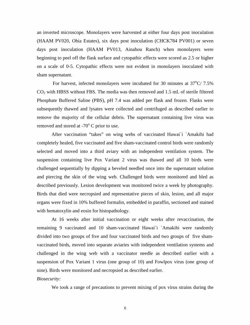
an inverted microscope. Monolayers were harvested at either four days post inoculation
(HAAM PV020, Ohia Estates), six days post inoculation (CHCK784 PV001) or seven
days post inoculation (HAAM PV013, Ainahou Ranch) when monolayers were
beginning to peel off the flask surface and cytopathic effects were scored as 2.5 or higher
on a scale of 0-5. Cytopathic effects were not evident in monolayers inoculated with
sham supernatant.
For harvest, infected monolayers were incubated for 30 minutes at 37oC/ 7.5%
CO2 with HBSS without FBS. The media was then removed and 1.5 mL of sterile filtered
Phosphate Buffered Saline (PBS), pH 7.4 was added per flask and frozen. Flasks were
subsequently thawed and lysates were collected and centrifuged as described earlier to
remove the majority of the cellular debris. The supernatant containing live virus was
removed and stored at -70o C prior to use.
After vaccination “takes” on wing webs of vaccinated Hawai`i `Amakihi had
completely healed, five vaccinated and five sham-vaccinated control birds were randomly
selected and moved into a third aviary with an independent ventilation system. The
suspension containing live Pox Variant 2 virus was thawed and all 10 birds were
challenged sequentially by dipping a beveled needled once into the supernatant solution
and piercing the skin of the wing web. Challenged birds were monitored and bled as
described previously. Lesion development was monitored twice a week by photography.
Birds that died were necropsied and representative pieces of skin, lesion, and all major
organs were fixed in 10% buffered formalin, embedded in paraffin, sectioned and stained
with hematoxylin and eosin for histopathology.
At 16 weeks after initial vaccination or eight weeks after revaccination, the
remaining 9 vaccinated and 10 sham-vaccinated Hawai`i `Amakihi were randomly
divided into two groups of five and four vaccinated birds and two groups of five sham-
vaccinated birds, moved into separate aviaries with independent ventilation systems and
challenged in the wing web with a vaccinator needle as described earlier with a
suspension of Pox Variant 1 virus (one group of 10) and Fowlpox virus (one group of
nine). Birds were monitored and necropsied as described earlier.
Biosecurity:
We took a range of precautions to prevent mixing of pox virus strains during the

7
experiment. Hawai`i `Amakihi infected with different pox virus isolates were housed in
separate rooms with independent ventilation systems to avoid potential cross
contamination from aerosols. All caging was washed with hot soapy water, sprayed with
a 10% solution of chlorine bleach, and rinsed with water prior to and after use. All food
dishes and Nekton® tubes were washed every day with hot soapy water in a commercial
dishwasher. Each room also had a separate set of brushes, buckets, and hoses to avoid
cross contamination. Personnel wore rubber boots, disposable Tyvek® pants, and a single
use laboratory coat, used a footbath containing 10% bleach when entering and leaving
individual rooms, and thoroughly washed their hands with soap before entering a new
room. Pants were discarded and laboratory coats were washed with bleach in a
commercial washer/dryer after use in each room. During vaccination, we also cared for
and measured control birds first every day to reduce the chance that virus might move
from contaminated to uncontaminated areas.
Statistical Analysis:
Weight and food consumption data were analyzed with a repeated measures
ANOVA using the statistical program Systat®, Version 11 (Systat Corp., Chicago, IL).
Survival analyses for birds infected with Variant 1 and Variant 2 were done using a
Kaplan Meier Survivorship Analysis with Systat®. Statistical tests were considered
significant when P < 0.05.
RESULTS
Analysis and Classification of Vaccine Virus:
Three of the four partial 4b core protein gene clones produced unambiguous
sequences that were 100% identical to each other. One representative sequence was used
in a neighbor-joining tree with other previously published sequences to establish
relationships (Figure 1). The attenuated Canarypox vaccine virus appears most similar to
the ATCC Canarypox sequence as well as field isolates of Avipoxvirus Variant 1 (Figure
1).
Vaccination:
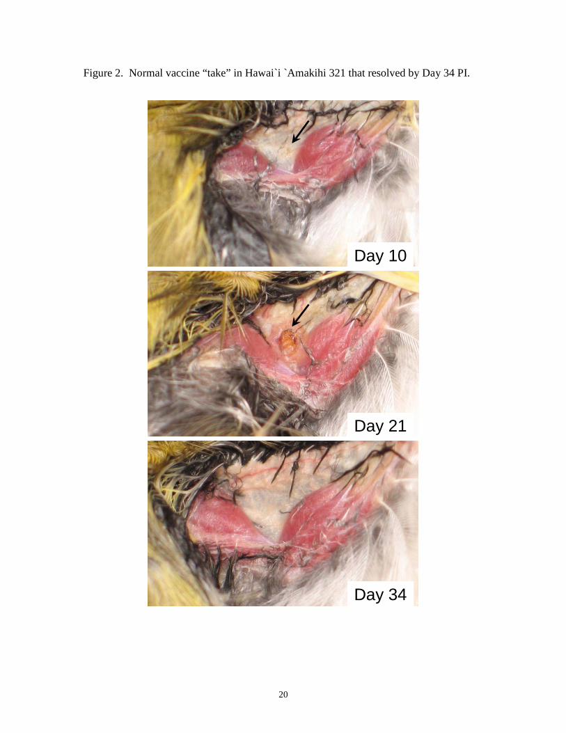
Approximately 7 days after vaccination with Poximmune C®, Hawai`i `Amakihi
developed small, 2-3 mm diameter areas of discoloration or swelling at the inoculation

8
site in the wing web (Figure 2). These swellings or vaccine “takes” increased in diameter
over the next two weeks for 6 birds (306, 316, 317, 318, 321, 78404) and then healed
within 3-6 weeks after inoculation, leaving no scar (Figure 2). By contrast, four Hawai`i
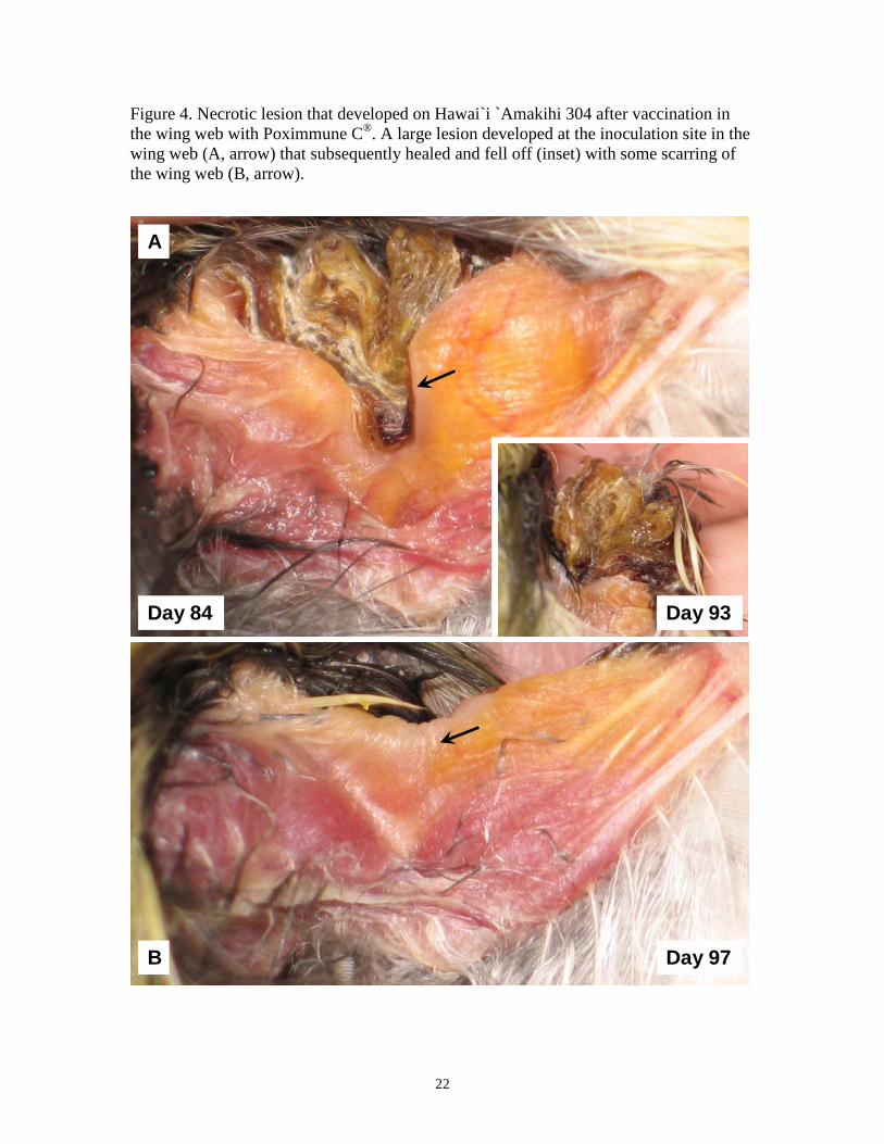
`Amakihi (302, 304, 305, 315) developed, extensive, necrotic wing lesions. Wing lesions
on three of these birds (302, 304, 305) took 7-8 weeks to heal (Figure 3). One bird (304)
developed a large scar in the wing membrane during the healing process (Figure 4), while
a second (305) developed a large necrotic lesion that eventually healed completely
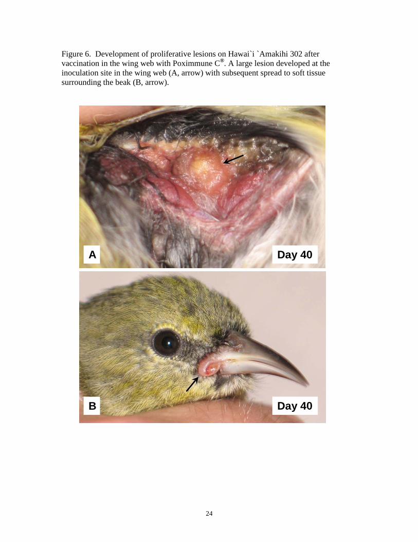
without scarring (Figure 5). Two birds (302, 315) developed proliferative lesions around
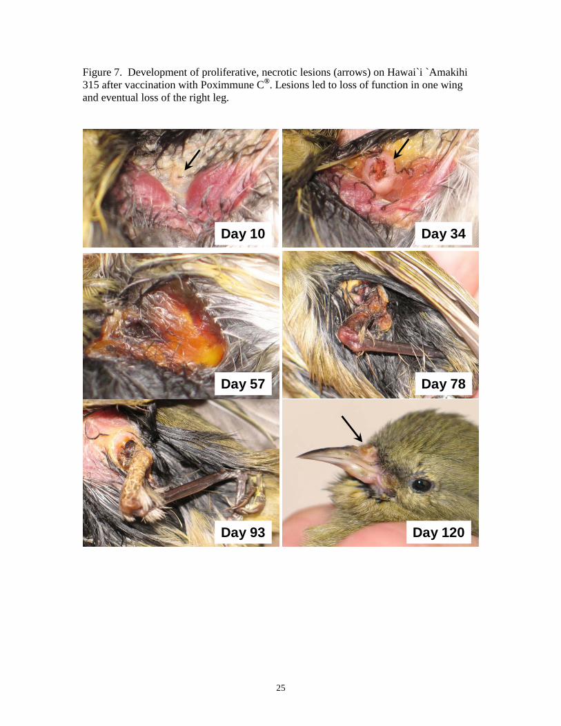
the beak (302, 315), eyelids (315), and also on the legs and feet (315) (Figures 6, 7).
Lesions on Hawai`i `Amakihi 315 were particularly bad, leading to loss of function in the
wing by six weeks after vaccination, loss of function in the right leg by 11 weeks after
vaccination, and complete loss of the right leg by 14 weeks after vaccination (Figure 7).
This bird subsequently died approximately 17 weeks after vaccination with persistent
active lesions on the beak and remaining leg.
Six Hawai`i `Amakihi (308, 312, 313, 324, 327, 328) did not develop vaccine
“takes” and were subsequently revaccinated eight weeks after the initial vaccination. Five
of these six birds developed “takes” that healed within 4-7 weeks after vaccination. One
individual, Hawai`i `Amakihi 313, did not develop a swelling that would indicate
successful vaccination, and was dropped from the experiment.
Among the 15 Hawai`i `Amakihi that developed clear vaccine takes, 11
developed wing lesions that resolved without evidence of scarring within 7 weeks after
vaccination. The remaining 4 birds, however, developed potentially life threatening
lesions that exhibited extensive necrosis, caused loss of function in a wing or leg, or
spread beyond the inoculation site to other parts of the body. None of the control birds
developed lesions following a sham-vaccination with distilled water.
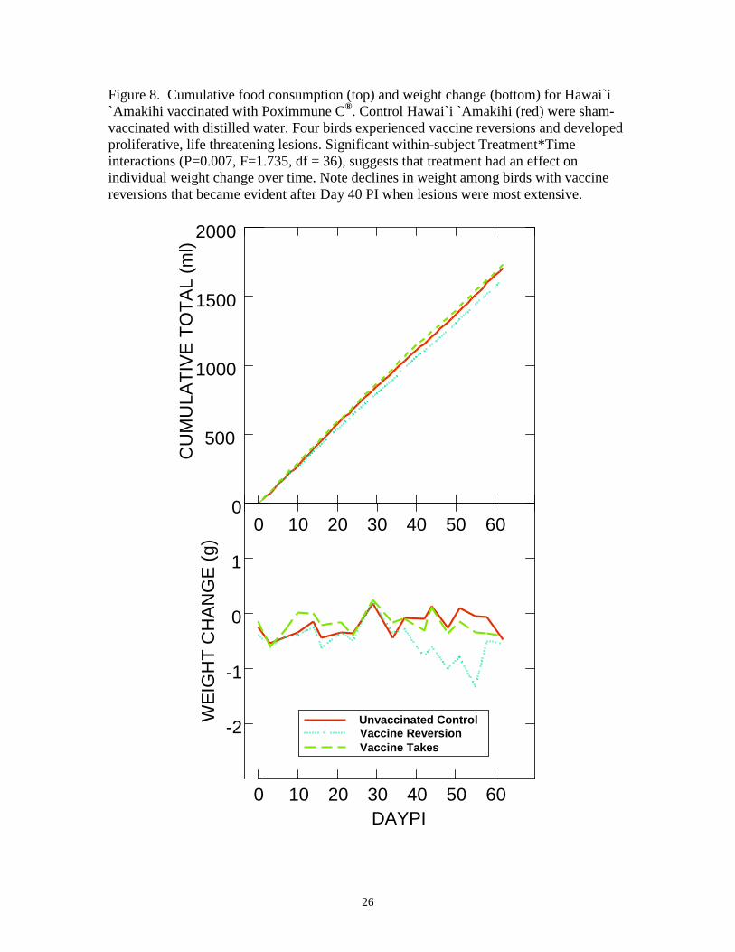
Among all three groups, cumulative food consumption among birds with large
vaccine-associated lesions was slightly lower than cumulative food consumption in
control birds and birds with small vaccine takes (Figure 8), but differences among
subjects were not significantly different (P= 0.211, F = 1.673, df = 2). There were,
however, significant within-subject Time (P<0.0001, F = 4565.685, df = 69) and
Treatment*Time interactions (P=0.004, F=1.367, df=138), suggesting that treatment had

9
an effect on individual food consumption over time.
Among the three experimental groups, birds with large vaccine-associated lesions
lost more weight over time than control birds or birds with small vaccine takes (Figure 8),
but differences among subjects were not significantly different (P=0.115, F=2.395, df=2).
There were, however, significant within-subject Time (P<0.0001, F = 1083.992, df = 18)
and Treatment*Time interactions (P=0.007, F=1.735, df=36), suggesting that treatment
had an effect on individual weight change over time.
Challenge with Pox Variant 2
After challenge with Pox Variant 2, five Hawai`i `Amakihi that had been
vaccinated once with Poximmune C® and five unvaccinated control Hawai`i `Amakihi
developed wing lesions as early as nine days after infection. Lesions were first evident as
reddish swellings at the inoculation site (Figure 9). Lesions on three of five vaccinated
birds (306, 318, 321) continued to grow, eventually leading to an extensive thickening
and inflammation of the wing web that spread throughout the dermis of inoculated birds
(Figure 9). Edematous swelling and wrinkling of the dermis spread, eventually involving
skin on the abdomen, thighs, head and neck, and all three of these birds died between 40-
50 days post-challenge. One bird (321) developed proliferative lesions that spread to the
foot and beak, eventually leading to loss of circulation in the right leg and loss of the
upper beak (Figure 9). The two remaining vaccinated birds (316, 317) developed small
swellings at the inoculation site by day 9 after challenge that resolved completely by day
21 after challenge.
All five unvaccinated control Hawai`i `Amakihi (307, 309, 319, 322, 331) died
between 24 – 30 days after challenge. Lesions were similar to those from the three
vaccinated birds, beginning as reddish swellings at the inoculation site by day nine that
rapidly led to extensive thickening and inflammation of the wing web and a generalized
edematous swelling and wrinkling of the epidermis that spread over the entire body
before death at three to four weeks after inoculation. While significantly fewer vaccinated
birds died after challenge with Pox Variant 2 virus (P = 0.002, X2 = 9.651, df = 1),
mortality was still 60% (3/5) (Figure 10).
In spite of the high mortality among both vaccinated and unvaccinated Hawai`i
`Amakihi, challenge with Pox Variant 2 had no significant effects on cumulative food

10
consumption (P = 0.950, F = 0.052, df = 2) or weight change (P = 0.958, F = 0.043, df =
2) during the first 23 days of the study when all birds could be included in the repeated
measures analysis (Figure 11). Within-subject effects for cumulative food consumption
were significant for Time (P < 0.0001, F = 3892.722, df = 33), indicating that daily food
consumption was consistent throughout the study, however, Treatment*Time interactions
were not significant (P =1.000, F = 0.476, df = 66), indicating that day to day variability
in cumulative food consumption was not affected by treatment status. There were
significant Time (P<0.0001, F=1385.366, df =7) and Treatment*Time (P<0.0001,
F=3.121, df =14) interactions for weight change, indicating that there were significant
day to day variations in weight change within subjects that were affected by treatment.
Most interesting was the increase in weight among unvaccinated Hawai`i `Amakihi
between Days 20-30 PI, immediately prior to death (Figure 11).
By the end of the experiment at Day 58, two vaccinated birds that were still alive
had lower totals for cumulative food consumption and slightly higher weights relative to
pre-infection values than uninfected control birds (Figure 11), but these differences were
not statistically significant (weight change, P=0.279, T = -1.144, df = 10; cumulative
food consumption, P= 0.135, T = 1.625, df = 10).
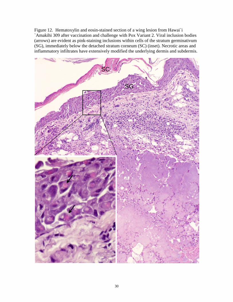
Gross and microscopic lesions were similar among vaccinated and unvaccinated
birds that died after challenge with Pox Variant 2. Wing lesions and proliferative lesions
that spread to other parts of the body had microscopic lesions typical of pox infections,
including proliferation and hypertrophy of epithelial cells, particularly cells of the stratum
germinativum (Figure 12). Viral inclusion or Bollinger Bodies were common in these
cells and the pink-staining inclusions were either amorphous or cuboidal in shape
(Figures 12, 13). Necrotic areas, extensive inflammatory infiltrates, and secondary
bacterial and fungal infections were prominent in the dermis and subdermis (Figures 12,
13). Epithelial hyperplasia and intracytoplasmaic Bollinger Bodies were evident in both
the stratum germinativum and subdermis of edematous and wrinkled skin from the
abdomen of vaccinated and unvaccinated birds (Figure 13). Both extracellular viral
inclusion bodies and mononuclear cells containing pink-staining inclusion bodies were
evident in the subdermis of some birds (Figure 14).
Challenge with Pox Variant 1

11
Five revaccinated Hawai`i `Amakihi and five unvaccinated Hawai`i `Amakihi
were challenged in the wing web with Pox Variant 1. Four of five revaccinated birds
(308, 324, 327, 328), i.e. birds that were revaccinated when they did not develop “takes”,
developed small lesions at the inoculation site that quickly resolved and disappeared as
early as 12 days after challenge (Figure 15). The fifth revaccinated bird (312) had a wing
lesion of moderate size after challenge that did not completely heal until 56 days after
exposure (Figure 16). The lesion had no effect on wing function and the bird was
otherwise healthy.
All five unvaccinated Hawai`i `Amakihi (303, 314, 323, 326, 78481) developed
necrotic wing lesions after challenge with Pox Variant 1 that were much larger and more
debilitating than lesions from the five vaccinated birds (Figure 17). Hawai`i `Amakihi
78481 developed an extensive thickening and inflammation of the wing web that spread
throughout the dermis of the body, leading a generalized edematous swelling and
wrinkling of the skin (Figure 18), but this was the only bird among the unvaccinated
Hawai`i `Amakihi challenged with Pox Variant 1 to exhibit this type of proliferative
lesion. Lesions in 3 other unvaccinated birds spread to the beak, feet, or both, where they
formed more typical tumor-like swellings. Two of the unvaccinated birds (323, 78481)
ultimately died (Figures 17, 18), but differences in survivorship between vaccinated and
unvaccinated groups were not statistically significant (P = 0.134, X2 = 2.242, df = 1)
(Figure 19). Gross and microscopic lesions, particularly the edematous, dermal lesions of
Hawai`i `Amakihi 78481, were similar to those in Hawai`i `Amakihi that were
challenged with Pox Variant 2 (Figures 12-14).
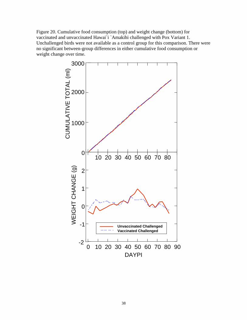
Challenge with Pox Variant 1 had no significant effects on food consumption (P =
0.881, F = 0.024, df = 1) or weight (P = 0.555, F = 0.380, df = 1) among vaccinated or
unvaccinated Hawai`i `Amakihi (Figure 20). Within-subject effects for cumulative food
consumption were significant for Time (P < 0.0001, F = 2968.578, df = 47), indicating
that daily food consumption was consistent throughout the study. However,
Treatment*Time interactions were not significant (P =0.997, F = 0.511, df = 47),
indicating that day to day variability in cumulative food consumption was not affected by
treatment status. Similarly, within subject effects for weight change were significant for
Time (P<0.0001, F = 687.821, df = 9), indicating that within-subject weight change

12
varied over time, but Treatment*Time interactions were not significant (P=0.877, F =
0.490, df = 9), indicating that day to day variability in weight change was not affected by
treatment status.
Challenge with Fowlpox
Four Hawai`i `Amakihi that were vaccinated once with Poximmune C® (78404,
302, 304, 305) were moved into individual cages in a separate room with independent
ventilation with five unvaccinated control Hawai`i `Amakihi and challenged with
Fowlpox virus as described earlier. Both vaccinated and unvaccinated control birds
developed small lesions at the inoculation site within 5 days after challenge that
completely healed by 12 days after challenge (Figure 21).
There were no differences between the two groups in lesion size or duration.
Challenge with Fowlpox had no significant effects on food consumption (P = 0.889, F =
0.021, df = 1) or weight change (P = 0.165, F = 2.401, df = 1) among vaccinated or
unvaccinated Hawai`i `Amakihi (Figure 22). Within-subject effects for cumulative food
consumption were significant for Time (P < 0.0001, F = 2880.587, df = 79), indicating
that daily food consumption was consistent throughout the study. However,
Treatment*Time interactions were not significant (P =1.000, F = 0.129, df = 79),
indicating that day to day variability in cumulative food consumption was not affected by
treatment status. Similarly, within subject effects for weight change were significant for
Time (P<0.0001, F = 258.435, df = 18), indicating that within-subject weight change
varied over time, but Treatment*Time interactions were not significant (P=0.983, F =
0.414, df = 18), indicating that day to day variability in weight change was not affected
by treatment status.
One bird (78404) developed neurological signs, including torticollis and loss of
balance on Day 69 PI and was found dead the following day. No gross lesions were
evident at necropsy and cause of death was unknown and presumably not related to the
experimental infection with pox virus. There were no significant differences in
survivorship between vaccinated and unvaccinated birds (P = 0.264, X2 = 1.250, df = 1)
(Figure 23).
DISCUSSION
While the impacts of introduced Avipoxvirus on native Hawaiian forest birds have

13
been widely documented (Warner 1968, VanderWerf 2001, van Riper et al. 2002),
relatively little information has been available about the identity and diversity of virus
strains in Hawaii or whether commercial pox vaccines are sufficiently immunogenic to be
protective (Tripathy et al. 2000 and Kim and Tripathy 2006a) provided the first evidence
that pox virus isolates from Hawaiian birds differ from Fowlpox, are non-pathogenic in
domestic chickens, and are closely related to Canarypox based on sequence of the virion
assembly protein gene. A more extensive survey of pox virus isolates from native and
non-native birds, seabirds, and domestic poultry from Hawaii based on sequence of the
virus 4b core protein was recently completed by Jarvi et al. (2008). Two clades (Variant 1
and Variant 2) from passerines and a third clade that formed a distinct basal cluster
containing known Fowlpox isolates were identified. The American Type Culture
Collection strain of Canarypox was most similar to viral isolates in clade 1 (Pox Variant
1). Sequence analysis of a 116 bp region from the vaccine strain we used (Poximmune
C®) also fell within this clade, suggesting that the attenuated vaccine might be
particularly protective against challenge with wild viruses that fell within clade 1.
Based on the close relationship between viral isolates that fell within clade 1 and
Canarypox (Kim and Tripathy. 2006a, Jarvi et al. 2008), we designed a series of
experiments to test both the safety and efficacy of Biomune Poximmune C® against three
known strains of Avipoxvirus that have been isolated from native and non-native forest
birds and domestic poultry from Hawaii. This experiment also provided the opportunity
to obtain information about relative susceptibility of a native honeycreeper to Fowlpox
and to obtain additional experimental data about relative pathogenicity of Pox Variants 1
and 2 (Jarvi et al. 2008).
For a vaccine to be useful for immunizing wild or captive birds, it should be
effective after a single dose against known viral variants that birds may encounter in the
field, produce long-lasting immunity, and have minimal side effects. During the first
phase of the study, we divided captive Hawai`i `Amakihi into two random groups and
vaccinated half with Poximmune C® according to the manufacturer’s instructions and
half with distilled water to access success of a single dose. Only 10 of 16 birds (63%)
developed “takes” after initial vaccination with Poximmune C®, indicating that they had
been successfully inoculated with the attenutated virus. The remaining six birds were
14
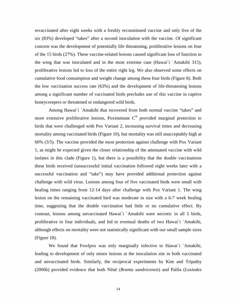
revaccinated after eight weeks with a freshly reconstituted vaccine and only five of the
six (83%) developed “takes” after a second inoculation with the vaccine. Of significant
concern was the development of potentially life threatening, proliferative lesions on four
of the 15 birds (27%). These vaccine-related lesions caused significant loss of function in
the wing that was inoculated and in the most extreme case (Hawai`i `Amakihi 315),
proliferative lesions led to loss of the entire right leg. We also observed some effects on
cumulative food consumption and weight change among these four birds (Figure 8). Both
the low vaccination success rate (63%) and the development of life-threatening lesions
among a significant number of vaccinated birds precludes use of this vaccine in captive
honeycreepers or threatened or endangered wild birds.
Among Hawai`i `Amakihi that recovered from both normal vaccine “takes” and
more extensive proliferative lesions, Poximmune C® provided marginal protection to
birds that were challenged with Pox Variant 2, increasing survival times and decreasing
mortality among vaccinated birds (Figure 10), but mortality was still unacceptably high at
60% (3/5). The vaccine provided the most protection against challenge with Pox Variant
1, as might be expected given the closer relationship of the attenuated vaccine with wild
isolates in this clade (Figure 1), but there is a possibility that the double vaccinations
these birds received (unsuccessful initial vaccination followed eight weeks later with a
successful vaccination and “take”) may have provided additional protection against
challenge with wild virus. Lesions among four of five vaccinated birds were small with
healing times ranging from 12-14 days after challenge with Pox Variant 1. The wing
lesion on the remaining vaccinated bird was moderate in size with a 6-7 week healing
time, suggesting that the double vaccination had little or no cumulative effect. By
contrast, lesions among unvaccinated Hawai`i `Amakihi were necrotic in all 5 birds,
proliferative in four individuals, and led to eventual deaths of two Hawai`i `Amakihi,
although effects on mortality were not statistically significant with our small sample sizes
(Figure 18).
We found that Fowlpox was only marginally infective to Hawai`i `Amakihi,
leading to development of only minor lesions at the inoculation site in both vaccinated
and unvaccinated birds. Similarly, the reciprocal experiments by Kim and Tripathy
(2006b) provided evidence that both Nēnē (Branta sandvicensis) and Palila (Loxiodes

15
balleuili) isolates of the virus have only minimal infectivity to domestic chickens, but
their precise relationship to Pox Variants 1 and 2 in our study are not clear since a
different gene region was sequenced. These data provide additional support to the idea
that domestic poultry and their associated Fowlpox infections may be of little threat to
native forest birds and that major pox epidemics that were described near the end of the
nineteenth century were associated with introduction of passerine Avipoxvirus from one
or more non-native perching birds (Tripathy et al. 2000, Jarvi et al. 2008).
Our data support limited experimental data from Jarvi et al. (2008) that Pox
Variants 1 and 2 differ in pathogenicity in Hawai`i `Amakihi. Mortality among
unvaccinated Hawai`i `Amakihi after challenge with Pox Variant 2 was 100%, with birds
rapidly succumbing to infection within 30 days after challenge from extensive spread of
the virus throughout epithelial tissue. Lesions in unvaccinated Hawai`i `Amakihi
challenged with Pox Variant 1, by contrast, were more typical tumor-like swellings on
the wing, beak, and feet, with only one bird exhibiting the disseminated, rapidly fatal,
epithelial lesions that we observed in birds challenged with Pox Variant 2. Differences in
survivorship between unvaccinated birds challenged with Pox Variant 1 and Pox Variant
2 were highly significant (P = 0.002, X2 = 9.496, df = 2) (Figures 10, 19). Effects on
cumulative food consumption and weight change over time were most evident for birds
challenged with Pox Variant 2, with an increase in weights within several days prior to
death that corresponded to the development of extensive, edematous swelling of
epithelial tissue in these birds. Similar epithelial lesions have not been reported in other
experimental studies of pox virus in passerine hosts, including the recent report by Jarvi
et al. (2008) that challenged Hawai`i `Amakihi by needle inoculation in the foot pad with
Pox Variants 1 and 2. There are no reports of comparable lesions in wild birds with pox
infections, and we have not seen similar lesions in native or non-native forest birds in
almost 20 years of field work in Hawai`i. Given how unusual the lesions are plus their
development in birds challenged with both pox variants, their pathogenesis may be
related to both the high host susceptibility of Hawai`i `Amakihi to the virus and route of
inoculation through the wing web. We found both extracellular viral inclusion bodies and
mononuclear cells with viral inclusion bodies in subdermal tissue of these lesions which
suggests that spread may have been through the lymphatic vessels in the subdermis or

16
movement of infected macrophages. While other studies have reported infection of
macrophages with viral inclusion bodies of avian pox (Giddens et al. 1971, Shivaprasad
et al. 2009), we did not find them in other tissues or organs and found no evidence of
disseminated infections in epithelial cells of other organs (Shivaprasad et al. 2009).
Based on our results, use of live attenuated vaccines for protecting native
honeycreepers from avian pox is risky and these vaccines should be carefully assessed
under controlled conditions to evaluate safety and efficacy prior to use in the field or in
captive threatened or endangered forest birds. Given the potential that attenuated vaccine
strains of the virus may revert in the wild and become more virulent, a preferable
approach may be to develop effective subunit vaccines that can stimulate immunity to
one or more wild variants of the virus without the risk that live viruses pose.
ACKNOWLEDGMENTS
We thank the U.S. Geological Survey Wildlife and Terrestrial Resources and Science
Support Programs for financial support, Bernard Rocha and Kawena Wise for providing
daily care for Hawai`i `Amakihi and collecting data on food consumption, Nick Shema
and the interns and technicians of the U.S. Geological Survey Palila Project for assistance
with capture of wild Hawai`i `Amakihi on Mauna Kea and Kevin Brinck for invaluable
advice about statistical analysis of the data. All animal challenges were conducted with
approval of the University of Hawai`i Institutional Animal Care and Use Committee,
Protocol 06-028.

17
LITERATURE CITED
Atkinson, C. T., K. L. Woods, R. J. Dusek, L. S. Sileo and W. M. Iko. 1995. Wildlife disease and conservation in Hawaii: pathogenicity of avian malaria (Plasmodium relictum) in experimentally infected Iiwi (Vestiaria coccinea). Parasitology 111:S59-S69.
Atkinson, C.T. and D.A. LaPointe. 2009. Introduced avian diseases, climate change, and
the future of Hawaiian honeycreepers. Journal of Avian Medicine and Surgery 23:53-63.
Docherty, D.E. and Slota, P.G. 1988. Use of Muscovy duck embryo fibroblasts for the
isolation of viruses from wild birds. Journal of Tissue Culture Methods 11:165-170.
Giddens, W. E. Jr., L. J. Swango, J. D. Henderson Jr., R. A. Lewis, D. S. Farner, A.
Carlos, and W. C. Dolowy. 1971. Canary pox in sparrows and canaries (Fringillidae) and in Weavers (Ploceidae): pathology and host specificity of the virus. Veterinary Pathology 8:260-280.
Jarvi, S.I., D. Triglia, A. Giannoulis, M. Farias, K. Bianchi, and C.T. Atkinson. 2008.
Diversity and virulence of Avipoxvirus in Hawaiian Forest Birds. Conservation Genetics 9:339-348.
Kim, T. and D.N. Tripathy. 2006a. Antigenic and genetic characterization of an avian
poxvirus isolated from an endangered Hawaiian Goose (Branta sandvicensis). Avian Diseases 50:15-21.
Kim, T. and D.N. Tripathy. 2006b. Evaluation of pathogenicity of avian poxvirus isolates
from endangered Hawaiian wild birds in chickens. Avian Diseases 50:288-291. Kumar, S., K Tamura, I. Jakobsen, and M. Nei. 2000. MEGA: Molecular Evolutionary
Genetics Analysis, Version 2.1. Arizona State University, Tempe, AZ. Shivaprasad, H. L., T. Kim, D. Tripathy, P. R. Woolcock, and F. Uzal. 2009. Unusual
pathology of canary poxvirus infection associated with high mortality in young and adult breeder canaries (Serinus canaria). Avian Pathology 38:311-316.
Smith, S.A., and G.J. Kotwal. 2002. Immune response to poxvirus infections in various
animals. Critical Reviews in Microbiology 28:149-185. Tripathy, D. N., W. M. Schnitzlein, P. J. Morris, D. L. Jannsen, J. K. Zuba, G. Massey,
and C. T. Atkinson. 2000. Characterization of poxviruses from forest birds in Hawai`i. J. Wildl. Dis. 36: 225-230.

18
U.S. Fish and Wildlife Service. 2006. Revised recovery plan for Hawaiian forest birds. U.S. Fish and Wildlife Service, Region 1, Portland, OR.
VanderWerf, E. A. 2001. Distribution and potential impacts of avian poxlike lesions in
‘Elepaio at Hakalau Forest National Wildlife Refuge. Pg. 247-253. in J. M. Scott, S. Conant, and C. van Riper III (editors). Evolution, ecology, conservation and management of Hawaiian birds: a vanishing avifauna. Studies in Avian Biology 22.
van Riper C. III. 1991. Parasite communities in wet and dry forest subpopulations of the
Hawaii common amakihi. Pp. 140-153 in J. E. Loye and M. Zuk (editors). Bird-Parasite Interactions, Ecology, Evolution and Behavior. Oxford University Press, New York.
van Riper C. III, S. G. van Riper, and W. R. Hansen. 2002. Epizootiology and effect of
avian pox on Hawaiian forest birds. Auk 119:929-942. van Riper C. III, and D. J. Forrester. 2007. Avian pox. Pp. 131-176 in N.J. Thomas, D. B.
Hunter, and C. T. Atkinson, (editors). Infectious Diseases of Wild Birds. Blackwell, Ames, IA.
Warner, R. E. 1968. The role of introduced diseases in the extinction of the endemic
Hawaiian avifauna. Condor 70:101-120.

19
Figure 1. A neighbor-joining tree (Kimura 2-parameter corrected distances) showing relationships among pox virus sequences amplified from native and non-native birds and Poximmune C® (Vaccine C06). Three clusters are present – one associated with Fowlpox from domestic poultry (bottom, FP), one that includes sequence from Canarypox (Variant 1, V1) and one that is closely related, but distinct from Canarypox (Variant 2, V2). All sequences in the Variant 2 cluster were isolated from birds in Hawai`i. Tree was generated using vaccine sequence plus sequences reported in Figure 1 from Jarvi et al. (2008).
AY30309CANARYAY30308 SPARROWAPAP14.1 MO 2003 (`Apapane)HAAM22.7 HI 1998 (Hawai`i `Amakihi)
ALAL21.1 HI 2003 (Hawaiian Crow)AY30310 CURLEWVACCINE C06HAAM18.1 HI 2004 (Hawai`i `Amakihi)APAP16.1 HI 2003 (`Apapane)AY318871 ATCC CANARYAPAP16.2 HI 2003 (`Apapane)
LAAL19.1 OA 2004 (Laysan Albatross)HAAM27.7 HI 2005 (Hawai`i `Amakihi)
ELEP1.1 HI 1900 (Hawai`i `Elepaio)HSFN24.1 HI 1998 (House Finch)APAP5.1 HI 2002 (`Apapane)HAAMB.07 HI 2002 (Hawai`i `Amakihi)HAAM15.4 HI 2003 (Hawai`i `Amakihi)IIWI2.12 HI 2002 (`I`iwi)PALI17.2 HI 2002 (Palila)HSFN4.3 MA 2002 (House Finch)AY30311 LOVE BIRD
AY530303 PIGEONAY530305 OSTRICH
AY530302 FOWLPOXCHKN23.1 HI 1993 (Domestic Chicken, Hawai`i)AY530304 TURKEYCHKN1.1 HI 2001 (Domestic Chicken, Hawai`i)CHKN23.2 HI 1993 (Domestic Chicken, Hawai`i)AJ005164 FOWLPOXM25781 FOWLPOX
99
98
76
65
99
100
79
0.05
V1
V2
FP
20
Figure 2. Normal vaccine “take” in Hawai`i `Amakihi 321 that resolved by Day 34 PI.
Day 10
Day 21
Day 34

21
Figure 3. Duration of vaccine lesions in 15 Hawai`i `Amakihi that were vaccinated in the wing web with Poximmune C®. Vaccine “takes” in four birds (315, 305, 304, 302) developed into potentially life-threatening lesions that involved most of the wing. Lesions spread to other areas of the body in two birds, leading to loss of a leg and eventual death in one individual (`Amakihi 315). Five Hawai`i` `Amakihi (306, 316, 317, 318, 321, 78404) developed vaccine “takes” that healed within 3-6 weeks after initial vaccination. Six Hawai`i `Amakihi (308, 312, 313, 324, 327, 328) did not develop vaccine “takes” and were subsequently revaccinated eight weeks after the initial vaccination. Five of these six birds developed “takes” that healed within 4-7 weeks after the second vaccination. `Amakihi 313 (not shown), did not develop a swelling that would indicate successful vaccination and was dropped from the experiment.
306316317
318321
78404302
304305
315308
312324
327328
0 20 40 60 80 100 120 140
Ban
d N
umbe
r
Duration of Lesion (Days)
22
Figure 4. Necrotic lesion that developed on Hawai`i `Amakihi 304 after vaccination in the wing web with Poximmune C®. A large lesion developed at the inoculation site in the wing web (A, arrow) that subsequently healed and fell off (inset) with some scarring of the wing web (B, arrow).
Day 93
Day 97
Day 84
A
B

23
Figure 5. Necrotic wing lesion (arrow) that developed on Hawai`i `Amakihi 305 after vaccination with Poximmune C®. The lesion eventually healed without leaving a scar.
Day 53
Day 105
24
Figure 6. Development of proliferative lesions on Hawai`i `Amakihi 302 after vaccination in the wing web with Poximmune C®. A large lesion developed at the inoculation site in the wing web (A, arrow) with subsequent spread to soft tissue surrounding the beak (B, arrow).
Day 40
Day 40
A
B
25
Figure 7. Development of proliferative, necrotic lesions (arrows) on Hawai`i `Amakihi 315 after vaccination with Poximmune C®. Lesions led to loss of function in one wing and eventual loss of the right leg.
Day 57
Day 10 Day 34
Day 78
Day 93 Day 120
26
Figure 8. Cumulative food consumption (top) and weight change (bottom) for Hawai`i `Amakihi vaccinated with Poximmune C®. Control Hawai`i `Amakihi (red) were sham-vaccinated with distilled water. Four birds experienced vaccine reversions and developed proliferative, life threatening lesions. Significant within-subject Treatment*Time interactions (P=0.007, F=1.735, df = 36), suggests that treatment had an effect on individual weight change over time. Note declines in weight among birds with vaccine reversions that became evident after Day 40 PI when lesions were most extensive.
DAYPI
0
500
1000
1500
2000
CU
MU
LATI
VE T
OTA
L (m
l)
10 20 30 40 50 60
-2
-1
0
1
WEI
GH
T C
HA
NG
E (g
)
Vaccine TakesVaccine ReversionUnvaccinated Control
10 20 30 40 50 600
0

27
Figure 9. Development of proliferative, necrotic lesions on Hawai`i `Amakihi 321 after challenge with Pox Variant 2. Lesions led to loss of circulation in the right leg (Day 50) and loss of the upper beak (Day 50).
Day 2 Day 9
Day 22 Day 31
Day 46 Day 46
Day 50 Day 50

28
Figure 10. Kaplan Meier Survival Plot of vaccinated and unvaccinated Hawai`i `Amakihi challenged with Pox Variant 2. A control group of unchallenged vaccinated Hawai`i `Amakihi was included for comparison. While mortality among vaccinated Hawai`i `Amakihi was significantly lower than unvaccinated birds (P = 0.002, X2 = 9.651, df = 1), mortality was still 60%.
0 10 20 30 40 50 60DAYPI
0.0
0.2
0.4
0.6
0.8
1.0S
UR
VIV
AL
FUN
CTI
ON
Unvaccinated, ChallengedVaccinated, Challenged
Control, Not Challenged

29
Figure 11. Cumulative food consumption (top) and weight change (bottom) among vaccinated and unvaccinated Hawai`i `Amakihi challenged with Pox Variant 2. Vaccinated control birds were not challenged with live virus. There were significant Time (P<0.0001, F=1385.366, df = 7) and Treatment*Time (P<0.0001, F=3.121, df = 14) interactions for weight change, indicating that there were significant day to day variations in weight change within subjects that were affected by treatment. Note increase in weight among unvaccinated Hawai`i `Amakihi between 20-30 Days PI, just prior to death.
0 10 20 30 40 500
500
1000
1500
2000
CU
MU
LATI
VE T
OTA
L (m
l)
0 10 20 30 40 50 60DAYPI
-2
-1
0
1
2
WEI
GH
T C
HA
NG
E (g
)
Vaccinated ChallengedUnvaccinated ChallengedVaccinated Not Challenged
30
Figure 12. Hematoxylin and eosin-stained section of a wing lesion from Hawai`i `Amakihi 309 after vaccination and challenge with Pox Variant 2. Viral inclusion bodies (arrows) are evident as pink-staining inclusions within cells of the stratum germinativum (SG), immediately below the detached stratum corneum (SC) (inset). Necrotic areas and inflammatory infiltrates have extensively modified the underlying dermis and subdermis.
SC
SG

31
Figure 13. Wrinkled, edematous abdominal skin (upper inset) and hematoxylin and eosin-stained section of abdominal skin from Hawai`i `Amakihi 307 after challenge with Pox Variant 2. Viral inclusion bodies are evident as pink-staining inclusions (arrows) within cells of the stratum germinativum (SG). Necrotic areas and inflammatory infiltrates have extensively modified the dermis and subdermis.
SG

32
Figure 14. Hematoxylin and eosin stained section of the subdermal region of Hawai`i `Amakihi 331. A. Macrophage with intracytoplasmic viral inclusion (arrow). B. Extracellular viral particles (arrows).
A
B

33
Figure 15. Development of small lesion in Hawai`i `Amakihi 328 after vaccination with Poximmune C® and challenge with Pox Variant 1. Four of 5 vaccinated birds developed small, self limiting lesions (arrow) that healed completely between 5 and 19 days after challenge.
Day 0
Day 19
Day 5

34
Figure 16. Development of a moderate lesion in Hawai`i `Amakihi 312 after vaccination with Poximmune C® and challenge with Pox Variant 1. The wing lesion (arrows) in this bird did not completely heal until day 62 after challenge.
Day 0 Day 5
Day 40 Day 62

35
Figure 17. Unvaccinated control Hawai`i `Amakihi 323 inoculated with Pox Variant 1. Note extensive swelling of the lesion (arrows) and eventual spread to the beak prior to death at day 68 post infection.
Day 0 Day 5
Day 40 Day 33
Day 62 Day 62

36
Figure 18. Unvaccinated control Hawai`i `Amakihi 78481 inoculated with Pox Variant 1. Note swelling of the lesion (arrows) that became hemorrhagic by day 19 post inoculation. At necropsy on day 36 post-inoculation, the skin over most of the body was edematous, and appeared wrinkled and blistered.
Day 0 Day 12
Day 19 Day 36

37
Figure 19. Kaplan Meier Survival Plot of vaccinated and unvaccinated Hawai`i `Amakihi challenged with Pox Variant 1. Mortality among unvaccinated Hawai`i `Amakihi was 40%, but this difference was not significantly different from vaccinated birds for this small sample size (P = 0.134, X2 = 2.242, df = 1).
0 10 20 30 40 50 60 70 80 90DAYPI
0.0
0.2
0.4
0.6
0.8
1.0SU
RVI
VAL
FUN
CTI
ON
Unvaccinated, ChallengedVaccinated, Challenged
38
Figure 20. Cumulative food consumption (top) and weight change (bottom) for vaccinated and unvaccinated Hawai`i `Amakihi challenged with Pox Variant 1. Unchallenged birds were not available as a control group for this comparison. There were no significant between-group differences in either cumulative food consumption or weight change over time.
10 20 30 40 50 60 70 800
1000
2000
3000C
UM
ULA
TIV
E T
OTA
L (m
l)
0 10 20 30 40 50 60 70 80 90DAYPI
-2
-1
0
1
2
WE
IGH
T C
HA
NG
E (g
)
Vaccinated ChallengedUnvaccinated Challenged

39
Figure 21. Small lesion (arrow) in the wing web of Hawai`i `Amakihi 302 after challenge with Fowlpox virus. Both vaccinated and unvaccinated Hawai`i `Amakihi developed similar small lesions that quickly resolved within 12 days after challenge.
Day 0
Day 5
Day 12

40
Figure 22. Weight change and cumulative food consumption for vaccinated and unvaccinated Hawai`i `Amakihi challenged with Pox Variant 1. Unchallenged birds were not available as a control group for this comparison. There were no significant between-group differences in either cumulative food consumption or weight change over time.
0 10 20 30 40 50 60 70 80 90DAYPI
-3
-2
-1
0
1
0
WEI
GH
T C
HA
NG
E (g
)
10 20 30 40 50 60 70 80
1000
2000
3000C
UM
ULA
TIVE
TO
TAL
(ml)
Vaccinated ChallengedUnvaccinated Challenged

41
Figure 23. Kaplan Meier Survival Plot of vaccinated and unvaccinated Hawai`i `Amakihi challenged with Fowlpox. One vaccinated bird died at Day 69 PI from causes that were not related to pox infection. There were no significant differences in survivorship between the two groups (P = 0.264, X2 = 1.250, df = 1).
0 10 20 30 40 50 60 70 80 90DAYPI
0.0
0.2
0.4
0.6
0.8
1.0
SU
RV
IVA
L FU
NC
TIO
N
Unvaccinated, ChallengedVaccinated, Challenged