efficient induction of functional ameloblasts from human
TRANSCRIPT
RESEARCH Open Access
Efficient induction of functionalameloblasts from human keratinocytestem cellsXuefeng Hu1,2, Jyh-Wei Lee3,4,5, Xi Zheng2, Junhua Zhang2, Xin Lin2, Yingnan Song2, Bingmei Wang2, Xiaoxiao Hu2,Hao-Hueng Chang6, Yiping Chen1,7, Chun-Pin Lin8* and Yanding Zhang1,2*
Abstract
Background: Although adult human tissue-derived epidermal stem cells are capable of differentiating intoenamel-secreting ameloblasts and forming teeth with regenerated enamel when recombined with mousedental mesenchyme that possesses odontogenic potential, the induction rate is relatively low. In addition,whether the regenerated enamel retains a running pattern of prism identical to and acquires mechanicalproperties comparable with human enamel indeed warrants further study.
Methods: Cultured human keratinocyte stem cells (hKSCs) were treated with fibroblast growth factor 8 (FGF8)and Sonic hedgehog (SHH) for 18 h or 36 h prior to being recombined with E13.5 mouse dental mesenchyme withimplantation of FGF8 and SHH-soaked agarose beads into reconstructed chimeric tooth germs. Recombinant toothgerms were subjected to kidney capsule culture in nude mice. Harvested samples at various time points wereprocessed for histological, immunohistochemical, TUNEL, and western blot analysis. Scanning electronic microscopyand a nanoindentation test were further employed to analyze the prism running pattern and mechanical properties ofthe regenerated enamel.
Results: Treatment of hKSCs with both FGF8 and SHH prior to tissue recombination greatly enhanced the rate oftooth-like structure formation to about 70%. FGF8 and SHH dramatically enhanced stemness of cultured hKSCs.Scanning electron microscopic analysis revealed the running pattern of intact prisms of regenerated enamel is similarto that of human enamel. The nanoindentation test indicated that, although much softer than human child and adultmouse enamel, mechanical properties of the regenerated enamel improved as the culture time was extended.
Conclusions: Application of FGF8 and SHH proteins in cultured hKSCs improves stemness but does not facilitateodontogenic fate of hKSCs, resulting in an enhanced efficiency of ameloblastic differentiation of hKSCs and toothformation in human–mouse chimeric tooth germs.
Keywords: Fibroblast growth factor 8, Sonic hedgehog, Keratinocyte stem cells, Ameloblast, Enamel
BackgroundVarious efforts to develop techniques for human tooth re-generative therapy and replacement have been attemptedfor decades [1, 2]. Currently, bioengineering of a wholetooth crown from embryonic tooth germ cells appears tobe the most successful approach for tooth regeneration in
several animal models including mouse, rat, pig, and dog[3–10]. Impressively, it was reported that implantation ofa bioengineered tooth germ, reconstructed from mouseembryonic dental epithelial and mesenchymal cells, into alost tooth socket in the alveolar bone of adult mice coulddevelop into a fully functional tooth [11–14], indicatingthe feasibility of future regenerative therapy in humans viaimplantation of bioengineered tooth germs. However, inpractice, it is impossible to use embryonic cells for suchclinical therapy. Thus, identification of adult cell sources,such as stem cells from adult tissue or induced pluripotent
* Correspondence: [email protected]; [email protected] Institute of Clinical Dentistry, School of Dentistry, National TaiwanUniversity and National Taiwan University Hospital, Taipei 10048, Taiwan1Southern Center for Biomedical Research, Fujian Normal University, Fuzhou350108, ChinaFull list of author information is available at the end of the article
© The Author(s). 2018 Open Access This article is distributed under the terms of the Creative Commons Attribution 4.0International License (http://creativecommons.org/licenses/by/4.0/), which permits unrestricted use, distribution, andreproduction in any medium, provided you give appropriate credit to the original author(s) and the source, provide a link tothe Creative Commons license, and indicate if changes were made. The Creative Commons Public Domain Dedication waiver(http://creativecommons.org/publicdomain/zero/1.0/) applies to the data made available in this article, unless otherwise stated.
Hu et al. Stem Cell Research & Therapy (2018) 9:126 https://doi.org/10.1186/s13287-018-0822-4

stem cells (iPSCs), for ex-vivo generation of implantabletooth germ is a prerequisite for the realization of humanbiotooth replacement therapy in the future.Stem cell-based tissue engineering has been proven a
prospective approach to repair or replace an injuredtissue or organ. Adult bone marrow stem cells (bonemarrow stromal cells) are the first adult cell source cap-able of participating in tooth formation when confrontedwith the mouse embryonic dental epithelium that pos-sesses odontogenic inducing capability [15]. At least fivetypes of mesenchymal stem cells from adult humanteeth have been isolated [16]. Among them, dental pulpstem cells (DPSCs), stem cells from exfoliated deciduousteeth (SHED), and stem cells from the apical papilla(SCAP) could generate dentin/pulp-like complexes inex-vivo culture [17–19]. Although these adult dentalstem cells do not possess either odontogenic inducingcapability or competence to support tooth formationwhen confronted with embryonic dental epithelia [20],they remain promising stem cell sources for regener-ation of tooth mesenchymal components. On the otherhand, the postnatal dental epithelium-derived stem cellsare more difficult to obtain due to ameloblastic apop-tosis during tooth eruption. It was reported that subcul-tured epithelial cell rests of Malassez can differentiateinto ameloblast-like cells and generate enamel-like tis-sues in combination with dental pulp cells at the crownformation stage [21]. We and others have reported pre-viously that nondental epithelia-derived human stemcells including human keratinocyte stem cells (hKSCs)[20, 22], gingival epithelial cells [23], and iPSCs [24],when recombined with either human or mouse embry-onic dental mesenchyme, could support tooth formationand differentiate into enamel-secreting ameloblasts.However, less than 30% and 10% of these recombinantexplants in subrenal culture formed teeth and producedenamel, respectively [22]. Such low efficiency of amelo-blastic differentiation prevents use of these human stemcells as realistic cell sources for tooth replacement ther-apy. In addition, whether hKSC-derived dental epitheliaexhibit an unusual life cycle and whether the regener-ated enamel acquires the unique physicochemicalcharacteristics remain elusive and warrant furtherexploration.Studies indicated that either FGF8 or SHH alone is
sufficient to promote limb regeneration in amphibian[25]. FGF8 or SHH is able to stimulate neurite out-growth and cavernous nerve regeneration in vitro, re-spectively [26, 27]. In the tooth, FGF8 promotes cellproliferation and inhibits apoptosis in diastemal toothepithelium, and revitalizes the tooth developmental pro-gram [28]. In this study, we developed an approach thatgreatly enhanced the ratio of ameloblastic differentiationof hKSCs and formation of tooth-like structures in tissue
recombinants. We further examined the developmentalprocess of differentiation of the hKSC-derived dentalepithelium and present evidence for rapid differentiationof human ameloblasts and production of regenerated en-amel with intact prisms the same as normal enamel.Meanwhile, we observed an increasing tendency formineralization effect with improved mechanical proper-ties in the regenerated enamel as cultivation extends.Our results provide a significant advance toward futureuse of human adult stem cells to generate implantabletooth organ ex vivo by tissue-engineering approaches.
MethodsCulture of hKSCs and application of recombinant proteinsCircumcised human foreskins from children 5–12 yearsold were collected immediately after surgery fromFuzhou Children's Hospital in Fujian Province. Primaryhuman keratinocytes were isolated and cultivated inKeratinocyte Serum-free Medium (KSFM; Gibco) ac-cording to the protocol described previously [22]. Kera-tinocyte stem cells were characterized by cell surfacemarkers as described previously [22]. Recombinant hu-man FGF8a (100 ng/ml; R&D Systems) and/or SHH(100 ng/ml; R&D Systems) proteins were applied to pas-sage 3 hKSCs cultured in KSFM at 90% confluence in10-cm culture dishes. These cells were continuously cul-tured for 18 h or 36 h prior to being used for subsequenttissue recombinant experiments or immunocytochemicalassay.
Tissue recombination and subrenal capsule cultureTissue recombination and mouse subrenal culture werecarried out as described previously [22]. Briefly, mandibularmolar tooth germs dissected from E13.5 mouse embryoswere incubated in 2.25% trypsin and 0.75% pancreatin inPBS on ice for 10 min and then dental epithelia were re-moved with fine forceps. Pieces of confluent hKSC sheetswere recombined with E13.5 mouse dental mesenchyme toreconstruct human–mouse chimeric tooth germs [22].Agarose beads (Bio-Rad) soaked with FGF8 (125 ng/μl;R&D Systems) and/or SHH (250 ng/μl; R&D Systems),respectively, were implanted into tissue recombinants asdescribed previously [22]. BSA beads were used as negativecontrol. Recombinant tooth germs were cultured in Tro-well type organ culture for 24 h prior to being subjected tosubrenal culture in immune-compromised adult male mice.Samples were harvested at various time points after subre-nal culture and processed for histological analysis andimmunohistochemical staining.
Histology, immunochemical staining, TUNEL assay, andwestern blot analysisMolar tooth germs dissected from surgically terminatedhuman fetuses of 12th-week, 16th-week, and 19th-week
Hu et al. Stem Cell Research & Therapy (2018) 9:126 Page 2 of 13

gestation were provided by Fujian Province Maternaland Child Health Hospital. Use of human embryonic tis-sues in this study was approved by the Ethics Committeeof Fujian Normal University, Fuzhou, China, and use ofanimals was approved by the Animal Use Committee ofFujian Normal University. Human fetus tooth germs andharvested recombinant samples treated with FGF8 andSHH protein prior to tissue recombination were fixed in4% paraformaldehyde (PFA) overnight at 4 °C on a rota-tor. Calcified tissues were further decalcified in 10% eth-ylenediaminetetraacetic acid (EDTA) for 1 week prior tobeing processed for dehydration and paraffin embed-ding. Sections were made at 10 μm, and were subjectedto hematoxylin/eosin staining or Azan dichromic stain-ing for histological analysis, and to immunohistochemi-cal staining by antigen recovery technique. Thefollowing antibodies were used: anti-human ameloblas-tin, anti-human K18, anti-human p63, anti-human K10,anti-human integrin-β1 (Santa Cruz Biotech, Inc.), anti-human amelogenin, anti-human Sp3 (Abcam), anti-human Sp6, and anti-human Msx2 (HPA). For negativecontrols, the primary antibodies were omitted. Immu-nostaining, immunofluorescence, and TUNEL assay(Roche) procedures followed the instructions of themanufacturers. For western blot analysis, culturedhKSCs were extracted with urea lysis buffer. Equalamounts of samples were electrophoresed on 12% SDSpolyacrylamide gels and transferred to NC membrane(Millipore). Immunoreactions were performed with thespecific primary antibodies as mentioned earlier, visual-ized with fluorescent secondary antibodies (LI-COR),and scanned on an Odyssey Clx Imager (LI-COR). Blotimages were quantified by densitometric analysis withImageJ software.
Scanning electronic microscopyThe surface morphologies of human tooth (adult andchild), mouse molar, and human–mouse chimeric toothcrown specimens that were treated with both FGF8 andSHH were investigated using a scanning electron micro-scope (S-3400 N; Hitachi, Japan) with an accelerationvoltage of 15 kV. Each specimen was cold mounted inresin, abraded with #1200 SiC paper, polished with 0.05μm alumina powder, etched with 25% EDTA for 60 s,washed in distilled water, and ultrasonically degreased inacetone. The conductive Pt thin film around 5 nm thickwas sputtered on each specimen before scanning elec-tronic microscopy (SEM) analysis.
Nanoindentation testThe nanoindentation hardness, H, and elastic modulus,E, of human (adult and child), mouse molar, and hu-man–mouse chimeric tooth crown specimens that weretreated with FGF8 and SHH were investigated by means
of a nanoindenter (TI-900, TriboIndenter; Hysitron,USA) with a Berkovich 142.3° diamond probe at differ-ent loads to achieve a fixed indentation depth of 70 nm.The loading rates were between 6 and 14 μN/s. Beforethe nanoindentation test, each specimen was coldmounted in resin, abraded with #1200 SiC paper,polished with 0.05 μm alumina powder, washed in dis-tilled water, and ultrasonically degreased in acetone. Tenindentation tests were performed on the enamel anddentin regions, respectively, for each specimen. Thehardness and elastic modulus of each indent were deter-mined on the basis of the Oliver and Pharr method [29].The elastic modulus, E, was expressed as follows:
1Er
¼ 1−ν2
Eþ 1−νi2
Ei;
where Er and ν are the reduced elastic modulus andPoisson’s ratio, respectively, for the specimen under test,and Ei (1140GPa) and νi (0.07) are the correspondingparameters of the diamond indenter. The Poisson ratio,ν, was 0.3 for each tooth sample [30]. The fused quartzstandard sample was used to calibrate the area functionof the nanoindenter [29].
ResultsEnhanced ameloblastic differentiation efficiency ofcultured hKSCs in the presence of FGF8 and SHHAlthough our previous studies manifested that hKSCs,when recombined with E13.5 mouse dental mesen-chyme, were induced to differentiate into enamel-secreting ameloblasts in the presence of FGF8-asorbedbeads, the efficiency was quite low with hKSCs differen-tiating to ameloblasts in 13 out of 41 formed teeth from146 tissue recombinants [22]. Enhancement of the ame-loblastic differentiation rate indeed warranted furtherexploration. SHH represents one of the pivotal cell-autonomous factors expressed in the developing humanand mouse dental epithelium, being required for thedevelopment of early tooth germ as well as involved inthe determination of ameloblastic cytodifferentiation andfunction [31]. This prompted us to investigate whetherapplication of FGF8/SHH-absorbed beads, instead ofFGF8-absorbed beads alone, in the human–mousechimeric tooth germ could increase the efficiency oftooth formation and ameloblastic differentiation. Indeed,our tissue recombination experiments showed that 12out of 25 recombinant samples developed into chimericteeth, with enamel deposition in eight of these cases(Table 1). The efficiency increased to around 50% fortooth formation and 65% for ameloblastic differentiationin formed tooth-like structures. These promising resultsagain encouraged us to test whether application of FGF8and SHH proteins in the cultured hKSCs prior to tissue
Hu et al. Stem Cell Research & Therapy (2018) 9:126 Page 3 of 13
recombination could further increase the ratio of toothformation and ameloblastic differentiation in the tissuerecombinants.We next treated the cultured hKSCs with either FGF8
(100 ng/ml) or SHH (100 ng/ml) proteins, or both ofthem, for 18 h or 36 h before proceeding to tissue re-combination and subrenal culture (Table 1). It is note-worthy that hKSCs treated with either FGF8 or SHHalone, or both of them, respectively, retain much morehealthy morphology with a smaller and typicalcobblestone-like cell shape by comparison with controlcells treated with PBS exhibiting a bigger and flattenedphenotype (data not shown). Histological examinationrevealed that in samples cultured for 18 h, although theratio of tooth formation remained around 50% (5 out of11 recombinant samples) in the presence of FGF8protein alone or in the presence of both FGF8 and SHH(9 out 17 recombinants), all teeth formed in both condi-tions exhibited a 100% ratio of ameloblastic differenti-ation (Table 1). We then extended the protein-treatedculture duration to 36 h, and the tooth forming rate in-creased to 69% (9/13) with enamel deposition in all casesof the tooth forming samples (Table 1). However, treat-ment of cultured hKSCs with FGF8/SHH for periodslonger than 36 h (48 and 60 h) resulted in decreasedtooth formation and reduced ameloblastic differentiationin tissue recombinants (data not shown). Our results,therefore, definitely demonstrated a dramaticallyenhanced efficiency of ameloblastic differentiation andtooth formation by application of FGF8 and SHH pro-tein in construction of human–mouse chimeric toothrecombinants.
FGF8 and SHH improves stemness but not ameloblasticfate of cultured hKSCsNext, we studied why addition of FGF8 and SHH pro-teins in the cultured hKSCs or in human–mouse tissuerecombinants could dramatically increase the efficiencyof tooth formation and ameloblastic differentiation. Thetranscription factors Msx2 [32, 33], Sp3 [34], and Sp6[35] have been demonstrated to be involved in amelo-blastic differentiation in mice. Mutations of each of these
genes in mice or humans disrupted ameloblastic differenti-ation and resulted in amelogenesis imperfecta. To unveilwhether these transcription factors were involved in ame-loblastic differentiation of hKSCs in the tissue recombinantin the presence or absence of FGF8 and SHH protein, wefirst carried out immunostaining to confirm whetherMSX2, SP3, and SP6 were expressed in human toothgerms. We did find that these proteins were all present inthe dental epithelium and differentiating ameloblasts in hu-man deciduous tooth germs (Additional file 1: Figure S1),exhibiting identical expression patterns to those in mice[32–36]. We then examined the expression of MSX2, SP3,and SP6 in the hKSC-derived dental epithelium inhuman–mouse chimeric tooth germs after 5-day subrenalculture. We found that MSX2 and SP3, but not SP6, pro-teins were presented in BSA control samples, which wereneither treated with FGF8 and SHH proteins in culturedhKSCs nor implanted with FGF8/SHH-soaked beads(Fig. 1a–c). In comparison, however, in addition to the ex-pression of MSX2 and SP3, SP6 was strongly activated inthe dental epithelium (Fig. 1d–f) of chimeric teeth withwell-differentiated preameloblasts (Fig. 1g–i) and enamel-secreting ameloblasts (Fig. 1j–l) in the FGF8/SHH-treatedsamples. Furthermore, unlike MSX2 and SP3 that exhib-ited more widely spread expression patterns, SP6 showedan exclusive expression pattern as in normal humantooth development in the inner enamel epithelium(Additional file 1: Figure S1), the dental epitheliumthat directly differentiates into functional ameloblasts,in the chimeric recombinants. Considering the pres-ence of MSX2 and SP3 proteins in the cultured KSCs(Fig. 2), these findings implied that the activation ofSP6 in the hKSC-derived epithelium by mouse embry-onic dental mesenchyme in the presence of FGF8 andSHH may be necessary for ameloblastic differentiationin the chimeric tooth.Since application of FGF8 and SHH proteins in cul-
tured hKSCs could dramatically increase the efficiencyof ameloblastic differentiation and tooth formation inthe human–mouse chimeric tooth germ, we investigatedwhether application of these proteins would commit cul-tured hKSCs to the odontogenic fate through induction
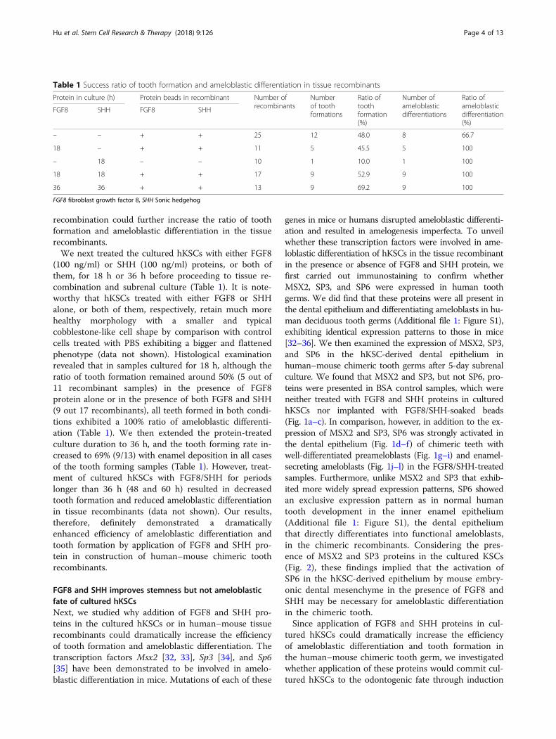
Table 1 Success ratio of tooth formation and ameloblastic differentiation in tissue recombinants
Protein in culture (h) Protein beads in recombinant Number ofrecombinants
Numberof toothformations
Ratio oftoothformation(%)
Number ofameloblasticdifferentiations
Ratio ofameloblasticdifferentiation(%)
FGF8 SHH FGF8 SHH
– – + + 25 12 48.0 8 66.7
18 – + + 11 5 45.5 5 100
– 18 – – 10 1 10.0 1 100
18 18 + + 17 9 52.9 9 100
36 36 + + 13 9 69.2 9 100
FGF8 fibroblast growth factor 8, SHH Sonic hedgehog
Hu et al. Stem Cell Research & Therapy (2018) 9:126 Page 4 of 13

Fig. 1 Immunohistochemical examination of MSX2, SP3, and SP6 expression in human–mouse chimeric teeth in presence or absence of FGF8and SHH. A–C Expression of MSX2, SP3, and SP6 in BSA-treated control samples after 5 days in subrenal culture. MSX2 (A) and SP3 (B), but notSP6 (C), are detected in recombinant implants. D–I Expression of MSX2, SP3, and SP6 in human–mouse chimeric tooth germs in presence ofFGF8 and SHH at various time points after subrenal culture. MSX2 and SP3 proteins detected in dental epithelium and mesenchyme, while SP6exclusively present in dental epithelium at 5 days (D–F) and 7 days (F–I) after grafting. All three proteins present in both ameloblasts and odonto-blasts at 12 days after grafting (J–l). Scale bar = 50 μm. BSA bovine serum albumin, FGF8 fibroblast growth factor 8, SHH Sonic hedgehog, 5d 5days, de dental epithelium, dm dental mesenchyme, am ameloblast, dp dental pulp
Fig. 2 Expression of MSX2, SP3, and SP6 in cultured hKSCs in presence and/or absence of FGF8 and/or SHH. hKSCs at passage 3 cultured with orwithout proteins for 36 h prior to immunofluorescent staining. No expression differences found among controls (a, e, i) and experimental groupstreated with FGF8 (b, f, j), SHH (c, g, k), or FGF8 + SHH (d, h, l). Scale bar = 100 μm. FGF8 fibroblast growth factor 8, SHH Sonic hedgehog
Hu et al. Stem Cell Research & Therapy (2018) 9:126 Page 5 of 13

of MSX2, SP3, and SP6 expression. We examined effectsof FGF8 and SHH proteins on the expression of thesethree transcription factors in cultured hKSCs using im-munofluorescence. Our results revealed that in controlcells MSX2 and SP3 proteins were detectable whereasSP6 was not detectable (Fig. 2a–i). However, expressionprofiles of SP6, as well as MSX2 and SP3, remained un-changed in cultured hKSCs that were treated either withFGF8 (Fig. 2b, f, j) or SHH (Fig. 2c, g, k) alone or as acombination (Fig. 2d, h, l) for 18 h or 36 h. These resultssuggested that enhanced efficiency of ameloblastic differ-entiation and tooth formation by application of FGF8and SHH in cultured hKSCs prior to tissue
recombination might not be associated with commit-ment of hKSCs to odontogenic fate by activation of tran-scription factors of odontogenic importance.Stemness is a critical element for differentiation capabil-
ity of stem cells. To illustrate possible roles of FGF8 andSHH for improving ameloblastic differentiation of culturedhKSCs, we further investigated expression patterns ofstemness markers of epidermal stem cells after growth fac-tor treatment, including K18, p63, integrin-β1, and K10[37]. Figure 3 shows immunocytochemical and westernblot analyses of the expression profile of cultured hKSCstemness markers. Our results indicated that, compared tothe controls that expressed a very low level of K18,
Fig. 3 Enhanced stemness of hKSCs in presence of FGF8 and/or SHH. A Immunofluorescence shows increased expression levels of K18 (a–d) andp63 (e–h), unaltered expression of integrin-β1 (i–l), and decreased K10 expression (m–p) in cultured hKSCs in the presence of FGF8 and/or SHH. B Western blot and densitometric quantification analyses further confirm results of immunofluorescence. Scale bar = 100 μm.FGF8 fibroblast growth factor 8, SHH Sonic hedgehog, IN integrin
Hu et al. Stem Cell Research & Therapy (2018) 9:126 Page 6 of 13

application of FGF8 or SHH protein alone or both almostdoubled the expression level of K18 in cultured hKSCs(Fig. 3Aa–d, B). In addition, nuclear p63, a key stemnessmarker of epidermal stem cells, was barely detectable inBSA-treated controls while its expression level was in-creased slightly in cultured hKSCs with either FGF8 orSHH treatment (Fig. 3Ae–g, B). On the contrary, p63 wasabundantly expressed in cultured hKSCs with treatment byboth FGF8 and SHH together (Fig. 3Ah, B). The expressionlevel of K10, a differentiating marker of epidermal stemcells, declined in these cultured cells, exhibiting a reversedpattern as compared to p63 and K18 after growth factortreatment (Fig. 3Am–p, B). Moreover, we observed no dif-ference in expression level of integrin-β1, another markerof epidermal stem cells, among the different experimentalgroups (Fig. 3Ai–l, B). These findings strongly suggestedthat application of FGF8 and SHH protein in culturedhKSCs improves stemness but does not facilitate odonto-genic fate of hKSCs, resulting in an enhanced efficiency ofameloblastic differentiation of hKSCs and tooth formationin human–mouse chimeric teeth. In addition, due to fastdecaying of active FGF8 and SHH in the cell culture,
analysis of hKSC cultured for 48 and 60 h revealed an ob-vious decrement of stemness (data not shown), furthersupporting the earlier idea.
The developmental process of differentiation of hKSC-derived dental epithelium into functional ameloblastsHuman deciduous teeth begin to develop during the 6thweek of gestation, but it is not until the 18th week thatthe dental epithelium starts to differentiate into enamel-secreting ameloblasts [38]. It takes about 400 days forhuman deciduous teeth to develop from initiation toeruption [39]. However, in our study, a completely differ-entiated human–mouse chimeric tooth crown was foundwithin only 4 weeks under the kidney capsule culture(Fig. 2h, i) [22]. Therefore, we sought to examine the de-velopmental process of this rapid differentiation of hKSC-derived dental epithelium to functional ameloblasts andregeneration of human enamel in the chimeric toothgerm. Histological staining indicated that hKSCs aggre-gated to a well-defined dental epithelial bud at day 5 insubrenal culture (Fig. 4A), corresponding to the bud stagein normal tooth development. This tooth bud underwent
Fig. 4 Histogenesis of hKSC-derived dental epithelium in human–mouse chimeric teeth. Sections through chimeric teeth retrieved from subrenalculture at different time points processed with hematoxylin and eosin (A–C) or Azan dichromic staining that stains dentin (d) blue and enamel(e) red (D–H). a hKSC-derived epithelial bud (de) formed after 5 days in subrenal culture. B–E Tooth buds underwent typical dental epithelialhistogenesis, forming the cap-like structure at day 6 (B), the bell-like structure at day 7 (C), elongated preameloblasts at day 8 (D), and well-differentiatedameloblasts (am) at day 9 (E), respectively, after subrenal culture. F hKSC-derived ameloblasts began to deposit enamel (e) on the surface of dentin (d)around day 12 after subrenal culture. G Thick layer of enamel secreted from hKSC-derived ameloblasts at around day 15 after subrenal culture. H Reducedthickness and compacted layer of enamel found in a graft cultured for 28 days. I A lateral (left) and top (right) view of human-mouse chimeric toothcrowns formed after 30 days in subrenal culture. J hKSC-derived ameloblasts expressed human SP6 but not GFP , and the odontoblasts and dental pulpcells of eGFP-mouse origin expressed GFP in human-mouse chimeric tooth crown after 8 days culture. stain Scale bar = 100µm (A-H, J), 500µm (I). 5d 5days, dm dental mesenchyme, dp dental pulp, GFP green fluorescent protein
Hu et al. Stem Cell Research & Therapy (2018) 9:126 Page 7 of 13

typical dental epithelial histogenesis within the tissue re-combinant, progressing to the cap stage at day 6 (Fig. 4B)and the bell stage at day 7 (Fig. 4C). The odontoblasts ofmouse origin produced dentin at day 8 (Fig. 4D, E) andthe hKSC-derived ameloblasts deposited enamel at day 12(Fig. 4F). Eventually, well-differentiated teeth were formedwithin 30 days (Fig. 4H). To further confirm the rapidameloblastic differentiation of hKSCs in chimeric toothgerms, we further performed immunohistochemistry andTUNEL assays to examine the expression profiles of ame-logenin and ameloblastin, two molecular markers for dif-ferentiating ameloblasts [40, 41], and the programmed celldeath at various time points [42]. Strikingly, we found thatexpression of amelogenin and ameloblastin was not de-tectable in the hKSC-derived dental epithelia in therecombinants at day 8 (Fig. 5Aa, b), but became strong atday 9 (Fig. 5Ac, d), further reduced to low levels at day 28(Fig. 5Ae, f), and quenched at day 30 when completelymineralized tooth crowns were formed (Fig. 4I andFig. 5Ag, h). In addition, TUNEL assay revealed that itwas not until 21 days that apparent apoptotic signalscould be detected (Fig. 5Ba–f ). Apoptotic signals reachedthe maximal level at day 28 (Fig. 5Bg–i) and were com-pletely quenched at day 30 (Fig. 5Bj–l), when ameloblastslost their healthy morphology with condensed nuclei inreduced enamel epithelia that underwent degeneration(Fig. 5Ag–h, Bi). These observations provide cogentevidence for the rapid differentiation of hKSC-derived am-eloblasts in human–mouse chimeric teeth.
In our previous report, we identified the human ori-gin of hKSC-derived dental epithelial component andthe mouse origin of the dental pulp with specificantibodies against human or mouse MHC antigen, re-spectively, in chimeric teeth to show no contamin-ation of the mouse dental epithelial tissue in therecombinant experiment [22]. In the present study,we further recombined hKSCs with mouse dentalmesenchyme genetically labeled with eGFP. Immuno-fluorescence studies indicated that no GFP-positivecells could be found in hKSC-derived ameloblasts thatwere marked with SP6 in chimeric teeth (Fig. 4J). Inaddition, we grafted E13.5 dental mesenchyme withremoval of dental epithelium after enzyme treatmentinto nude mice for subrenal culture for 4 weeks as afurther control. All 30 grafted samples either degener-ated or formed tiny pieces of bone-like tissues (datanot shown). These data provide more evidence to ruleout the possibility of mouse dental epithelium con-tamination in the recombinant experiment.
Microstructure and mechanical characteristics ofregenerated human enamelEnamel is the hardest calcified tissue of the body and isstructurally distinct from collagen-based calcified tissue.To illustrate whether the rapidly regenerated humanenamel is physically and functionally comparable to nor-mal enamel, we examined microstructure and physicalcharacteristics of regenerated human enamel on the
Fig. 5 Cytodifferentiation and apoptosis of hKSC-derived dental epithelium in human–mouse chimeric teeth. A Immunofluorescence shows expressionprofiles of amelogenin and ameloblastin in hKSC-derived ameloblasts at various time points in subrenal culture. Amelogenin and ameloblastin expressionnot detectable until day 9 (a–d), downregulated at day 28 (e, f), and completely silenced at day 30 (g, h), respectively, after subrenal culture. B TUNEL assayshows programmed cell death of hKSC-derived ameloblasts at various time points in subrenal culture. Obvious apoptosis signals not detected until day 21(a–f), reached maximal level at day 28 (g–h), and quenched at day 30 (j–l), respectively, after subrenal culture. Scale bar = 100 μm. 8d 8days, TUNEL terminal deoxynucleotidyl transferase dUTP nick end labeling, DAPI 4′,6-diamidino-2-phenylindole
Hu et al. Stem Cell Research & Therapy (2018) 9:126 Page 8 of 13

surface of human–mouse chimeric tooth utilizing scan-ning electronic microscopy (SEM) and nanoindentation,respectively. The microscopic characteristic structures ofhuman adult, human child, mouse molar, and human–mouse chimeric teeth after 20 and 60 days in subrenalculture are compared in Fig. 6. The enamel structures ofa human adult tooth on macro and micro scales aredepicted in Fig. 6A. The enamel prisms were clearlyobserved in the pattern of complex trajectory (enlargedin Fig. 6Ab, c). Similar results were also observed for theenamel structure of a human child tooth in Fig. 6B.Meanwhile, a more clear running pattern of the enamelprisms arranged in row with alternating orientation wasfound in the mouse molar (Fig. 6Ca–c). Upon compari-son with the microstructure of enamel prism of humanand mouse molar, the enamel prism of human–mousechimeric tooth cultured for 20 and 60 days was orderlyaligned as illustrated in Fig. 6D–F, manifesting that the
morphologies of enamel prisms of human–mousechimeric teeth cultured for 20 days (Fig. 6E) and 60 days(Fig. 6F) were almost identical to those of the humanmolar (Fig. 6A, B). It is well known that enamel is madeup of hydroxyl apatite crystals and then arranged inprisms. The appearance of prisms is determined by theorientation of the crystals. According to the study of en-amel structure in human molars, the enamel is dividedinto three layers by the running pattern of the enamelprisms [43, 44]. In this work, similar images of the run-ning pattern of the enamel prisms were observed for thehuman molar and the human–mouse chimeric teeth cul-tured for 20 or 60 days, indicating that the enamel prismmicrostructure of the human–mouse chimeric teeth islikely to grow completely after cultivation for more than20 days.As the hardest matrix of the body, enamel is brittle
and easily fractioned. It is supported by dentin, an
Fig. 6 Comparison of microstructure of enamel in human (adult and child), mouse, and human–mouse chimeric teeth by SEM. A SEM images ofmicrostructure of adult human tooth. B SEM images of child human tooth. C SEM images of adult mouse tooth. D Cross-section SEM images ofhuman–mouse chimeric tooth after 20 days in subrenal culture. E Vertical-section SEM images of human–mouse chimeric tooth grown after 20days in subrenal culture. F SEM images of human–mouse chimeric tooth after 40 days in subrenal culture. Frame regions in (a) and (b) enlargedin (b) and (c), respectively, in each panel
Hu et al. Stem Cell Research & Therapy (2018) 9:126 Page 9 of 13

underlying layer of a more resilient calcified matrix, tomaintain its integrity and hardness to withstand mech-anical force applied during tooth functioning. We there-fore tested mechanical characteristics of both enameland dentin in a whole tooth crown. The average elasticmodulus, E, and average hardness, H, of enamel anddentin regions for human (adult and child), mousemolar, and human–mouse chimeric tooth crown speci-mens are plotted in Fig. 7. The average E and H valuesfor the enamel of human–mouse chimeric teeth in-creased from 6.3 to 46.0 GPa and from 0.2 to 1.2 GPa,respectively, as the culture time increased from 20 to 60days. Notably, around a 6-fold to 7-fold increase wasfound for the E and H of enamel as the culture time in-creased, which indicates the mineralization effect in-creases with cultivation time. The average E and Hvalues for the regenerated enamel in human–mousechimeric teeth cultivated for 60 days were still 46% and70% less than those values of the mouse molar. As com-pared with the mechanical property of mouse molar en-amel in the literature [30], very similar results wereobtained for the E and H in this work. The highestvalues of E and H, 98.6 and 5.4 GPa, could be found forthe enamel of adult human teeth. On the other hand, forthe dentin of human–mouse chimeric teeth, the average Eand H values increased from 10.7 to 24.7 GPa and from0.3 to 0.8 GPa, respectively, as the culture time increased
from 20 to 60 days. A 2.7-fold increase was found for thehardness of dentin when the culture time increased from20 to 60 days, implying its increasing mineralization effect.The average E and H values for the dentin of human–mouse chimeric teeth cultivated for 60 days reachedaround 91.1% and 61.5%, respectively, by comparison withthose of the mouse molar; the findings were even higherthan those of adult human teeth. The mechanical propertyevaluation of enamel and dentin of tooth-like structuresby nanoindentation technique has been reported previ-ously [24]. As compared with those of the mouse andadult teeth, much lower values of hardness and elasticmodulus were found for the enamel and dentin of regen-erative teeth [24]. A similar tendency was also reflected inthis study. Meanwhile, the hardness of the regenerativeenamel might be increased by slowdown of tooth develop-ment process in culture since the mineralization effect issignificantly increased as the culture time increased.
DiscussionReciprocal heterotypic recombination of tissues of ectopicorigin has been long used as a routine approach to studyregulative interactions between tissue components in clas-sical experimental embryology. Mammalian tooth devel-opment is dependent upon inductive interactions betweenepithelium and adjacent mesenchyme [45]. Both epithelialand mesenchymal components in tooth germ are
Fig. 7 Comparisons of mechanical properties of enamel and dentin in humans (adult and child), mouse, and human–mouse chimeric tooth crownspecimens. a Statistical data on elastic modulus and hardness of enamel and dentin from nanoindentation tests. b Reduced elastic modulus andhardness of enamel and dentin for humans (adult and child), mouse, and human–mouse chimeric tooth crown specimens. E elastic modulus, WT wildtype, 60d 60 days
Hu et al. Stem Cell Research & Therapy (2018) 9:126 Page 10 of 13

indispensable for tooth development [46, 47]. Sequentialand reciprocal interactions between the stomadial epithe-lium and the cranial neural crest-derived mesenchymalcells regulate tooth morphogenesis and differentiation.Odontogenic potential represents an instructive inductioncapability of a tissue to induce gene expression in an adja-cent tissue and to initiate tooth formation, whereas odon-togenic competence indicates the capability of a tissue torespond to odontogenic inducing signals and to supporttooth formation. Tissue recombination experiments be-tween isolated mouse molar epithelial and mesenchymaltissues have demonstrated that, during early tooth devel-opment, odontogenic potential resides first in the dentalepithelium and then shifts to the mesenchyme [48, 49]. Atthe prebud stages of development (before and at E11.5),the presumptive dental epithelium possesses the potentialto induce tooth formation in nondental mesenchyme. Incontrast, at the early bud stage (E12.5) the odontogenicpotential has switched to the mesenchyme, and this odon-togenic mesenchyme is able to instruct nondental epithe-lium to form tooth-specific structures [48–50]. Ourprevious report demonstrated that such potential is alsoconserved in human embryonic dental mesenchymal tis-sues that are able to induce nondental epithelial tissues,such as human keratinocyte, and able to participate intooth formation [20]. In-vitro bioengineering of primordialtooth germs represents a promising approach for tooth re-placement therapy in the future [51]. Either in-vitro or ex-vivo generation of an implantable biotooth germ shouldfollow the principles of tooth development. Based on thisconcept, previous studies including ours have demon-strated that human epithelium-derived stem cells, includ-ing iPSC-derived epithelium-like tissue, could be inducedto participate in tooth formation when confronted withmouse dental mesenchyme with odontogenic potential.However, the efficiency of the induced ameloblastic differ-entiation of these cells was relatively low, and can be anobstacle for using adult stem cells as an epithelial cellsource to develop tooth replacement therapy. In thisstudy, in comparison with our previous report in whicharound 30% of tooth formation and 10% of ameloblasticdifferentiation were obtained [22], we achieved 70% and100% of tooth formation and ameloblastic differentiation,respectively, by application of two key growth factors,FGF8 and SHH, in cell culture and tissue recombination,demonstrating that in the presence of appropriate odonto-genic signals an efficient induction of enamel-secretingameloblasts from hKSCs could be achieved.FGF8 and SHH have been demonstrated to play pivotal
roles in mammalian tooth development. SHH acts as anautonomous signal to regulate dental epithelial cells toproliferate, grow, and differentiate into functional amelo-blasts [31, 52]. FGF8 is responsible for the determinationof tooth forming sites, induction of several tooth
developmental genes, and initiation of tooth development[53]. We did find that, in the present study, application ofthese two growth factors activated SP6 expression inaddition to that of MSX2 and SP3 expression in thehKSC-derived dental epithelium in tissue recombinants.The similar phenotypes in mice lacking the Msx2, Sp3, orSp6 gene, respectively, and their overlapping expressionpattern during tooth development raise the possibility thatthese transcription factors reside or interact closely withina signaling cascade to regulate amelogenesis [35, 36]. Ourstudy indicates that simultaneous activation of these threetranscription factors could likely be necessary for initiationof ameloblastic differentiation in hKSCs of the chimerictooth germ. However, our results also indicate that appli-cation of FGF8 and SHH in the cultured hKSCs did notalter the expression of MSX2, SP3, and SP6 but improvedstemness of hKSCs. These data strongly suggest that theenhanced efficiency of ameloblastic differentiation inhKSCs is associated with an improvement of culturedhKSC stemness but not their ameloblastic fate.In the developing human deciduous teeth, ameloblasts
begin to differentiate around the 15th week and start tosecrete and deposit enamel on the surface of dentinaround the 18th week of gestation [38]. It is not until 6months after birth that ameloblasts undergo apoptosiswhen the tooth erupts. Of interest, in our study, the dif-ferentiation and enamel deposit of hKSC-derived amelo-blasts in the chimeric teeth were completed within 4weeks under subrenal culture and this rapidly generatedenamel exhibited a microstructural pattern grossly iden-tical to normally developed enamel. Despite the mechan-ical property evaluation of enamel structures by ananoindentation technique revealing much lower valuesof hardness and elastic modulus for the regeneratedenamel than those of adult human and mouse teeth, anincreasing tendency for the mineralization effect withcultivation time was discovered in this study. Thisshould be of a significant impact for future clinical prac-tice, since implantable tooth primordia could grow rap-idly in the patient oral cavity in a relatively short periodof time and further mineralize and mature to attaineffective hardness and elastic modulus to withstandmechanical force applied during food chewing.
ConclusionWe developed a process for efficient induction of enamel-secreting ameloblasts and rapid generation of enamel fromhKSCs by treatment of hKSCs with both FGF8 and SHHproteins prior to recombination with mouse embryonicdental mesenchyme. FGF8 and SHH dramatically enhancedstemness of cultured hKSCs. Electron microscopic analysisand a nanoindentation test revealed the formation of intactprisms and an increasing tendency for the mineralizationeffect with cultivation time in the regenerated enamel. Our
Hu et al. Stem Cell Research & Therapy (2018) 9:126 Page 11 of 13
results provide an appealing idea for efficient induction ofadult stem cells into enamel-secreting ameloblasts.
Additional file
Additional file 1: Figure S1. Showing immunohistochemistry expressionpatterns of MSX2, SP3, and SP6 in developing human primary tooth germ.A MSX2, B SP3, and C SP6 protein distribution in cap and bell stages oftooth germs: (a, b) 12-week human primary incisor; (c, d) 16-week humanprimary incisor; (e, f) 19-week human primary incisor. Scale bar = 100μm (a, c, e), 50 μm (b, d, f). (TIFF 6518 kb)
AcknowledgementsThe authors thank Fuzhou Children's Hospital in Fujian Province forproviding circumcised human foreskins.
FundingThis study was supported by grants from the National Natural ScienceFoundation of China (81771034, 81271102, 81570036).
Availability of data and materialsData sharing not applicable to this article as no datasets were generated oranalyzed during the current study.
Authors’ contributionsXfH, C-PL, and YdZ were responsible for conception and experiment design.XfH, J-WL, XZ, JhZ, XL, and XxH were responsible for the preliminary datasearch, selection, and extraction. YnS, BmW, H-HC were responsible for dataanalysis. XfH, C-PL, and YdZ were responsible for assembly of data, data analysis,and manuscript writing. YpC was responsible for data interpretation andmanuscript revision. All authors read and approved the final manuscript.
Ethics approval and consent to participateAll mouse surgical procedures were approved by the Animal Care Committeeat Fujian Normal University. KSCs were harvested from circumcised humanforeskins from children 5–12 years old whose parents gave informed consentfor the study.
Consent for publicationNot applicable.
Competing interestsThe authors declare that they have no competing interests.
Publisher’s NoteSpringer Nature remains neutral with regard to jurisdictional claims inpublished maps and institutional affiliations.
Author details1Southern Center for Biomedical Research, Fujian Normal University, Fuzhou350108, China. 2Fujian Key Laboratory of Developmental and Neural Biology,College of Life Science, Fujian Normal University, Fuzhou 350108, China.3Department of Materials Engineering, Ming Chi University of Technology,New Taipei 24301, Taiwan. 4Center for Thin Film Technologies andApplications, Ming Chi University of Technology, New Taipei 24301, Taiwan.5College of Engineering, Chang Gung University, Taoyuan 33302, Taiwan.6School of Dentistry, National Taiwan University and National TaiwanUniversity Hospital, Taipei 10048, Taiwan. 7Department of Cell and MolecularBiology, Tulane University, New Orleans, LA 70118, USA. 8Graduate Instituteof Clinical Dentistry, School of Dentistry, National Taiwan University andNational Taiwan University Hospital, Taipei 10048, Taiwan.
Received: 3 February 2018 Revised: 26 February 2018Accepted: 1 March 2018
References1. Chai Y, Slavkin HC. Prospects for tooth regeneration in the 21st century: a
perspective. Microsc Res Tech. 2003;60(5):469–79.
2. Garcia-Godoy F, Murray PE. Status and potential commercial impact of stemcell-based treatments on dental and craniofacial regeneration. Stem CellsDev. 2006;15(6):881–7.
3. Duailibi MT, Duailibi SE, Young CS, Bartlett JD, Vacanti JP, Yelick PC.Bioengineered teeth from cultured rat tooth bud cells. J Dent Res. 2004;83(7):523–8.
4. Hu B, Nadiri A, Bopp-Kuchler S, Perrin-Schmitt F, Lesot H. Dental epithelialhistomorphogenesis in vitro. J Dent Res. 2005;84(6):521–5.
5. Honda MJ, Ohara T, Sumita Y, Ogaeri T, Kagami H, Ueda M. Preliminarystudy of tissue-engineered odontogenesis in the canine jaw. J OralMaxillofac Surg. 2006;64(2):283–9.
6. Kuo TF, Lin HC, Yang KC, Lin FH, Chen MH, Wu CC, et al. Bone marrowcombined with dental bud cells promotes tooth regeneration in miniaturepig model. Artif Organs. 2011;35(2):113–21.
7. Song YQ, Zhang ZY, Yu XY, Yan MQ, Zhang XY, Gu SP, et al. Application oflentivirus-mediated RNAi in studying gene function in mammalian toothdevelopment. Dev Dyn. 2006;235(5):1334–44.
8. Young CS, Terada S, Vacanti JP, Honda M, Bartlett JD, Yelick PC. Tissueengineering of complex tooth structures on biodegradable polymerscaffolds. J Dent Res. 2002;81(10):695–700.
9. Young CS, Kim SW, Qin C, Baba O, Butler WT, Taylor RR, et al.Developmental analysis and computer modelling of bioengineered teeth.Arch Oral Biol. 2005;50(2):259–65.
10. Young CS, Abukawa H, Asrican R, Ravens M, Troulis MJ, Kaban LB, et al. Tissue-engineered hybrid tooth and bone. Tissue Eng. 2005;11(9–10):1599–610.
11. Ikeda E, Morita R, Nakao K, Ishida K, Nakamura T, Takano-Yamamoto T, et al.Fully functional bioengineered tooth replacement as an organ replacementtherapy. Proc Natl Acad Sci U S A. 2009;106(32):13475–80.
12. Nait LA, Kuchler-Bopp S, Hu B, Haïkel Y, Lesot H. Vascularization ofengineered teeth. J Dent Res. 2008;87(12):1138.
13. Nakao K, Morita R, Saji Y, Ishida K, Tomita Y, Ogawa M, et al. Thedevelopment of a bioengineered organ germ method. Nat Methods. 2007;4(3):227–30.
14. Oshima M, Mizuno M, Imamura A, Ogawa M, Yasukawa M, Yamazaki H, et al.Functional tooth regeneration using a bioengineered tooth unit as a matureorgan replacement regenerative therapy. PLoS One. 2011;6(7):e21531.
15. Ohazama A, Modino SAC, Miletich I, Sharpe PT. Stem-cell-based tissueengineering of murine teeth. J Dent Res. 2004;83(7):518–22.
16. Huang GTJ, Gronthos S, Shi S. Mesenchymal stem cells derived from dentaltissues vs. those from other sources: their biology and role in regenerativemedicine. J Dent Res. 2009;88(9):792–806.
17. Gronthos S, Mankani M, Brahim J, Robey PG, Shi S. Postnatal human dentalpulp stem cells (DPSCs) in vitro and in vivo. Proc Natl Acad Sci U S A. 2000;97(25):13625–30.
18. Miura M, Gronthos S, Zhao MR, Lu B, Fisher LW, Robey PG, et al. SHED: stemcells from human exfoliated deciduous teeth. Proc Natl Acad Sci U S A.2003;100(10):5807–12.
19. Sonoyama W, Liu Y, Fang DAJ, Yamaza T, Seo BM, Zhang CM, et al.Mesenchymal stem cell-mediated functional tooth regeneration in swine.PLoS One. 2006;1(1):e79.
20. Hu X, Lin C, Shen B, Ruan N, Guan Z, Chen Y, et al. Conserved odontogenicpotential in embryonic dental tissues. J Dent Res. 2014;93(5):490–5.
21. Shinmura Y, Tsuchiya S, Hata KI, Honda MJ. Quiescent epithelial cell rests ofMalassez can differentiate into ameloblast-like cells. J Cell Physiol. 2008;217(3):728–38.
22. Wang B, Li L, Du S, Chao L, Xin L, Chen YP, et al. Induction of humankeratinocytes into enamel-secreting ameloblasts. Dev Biol. 2010;344:795.
23. Volponi AA, Kawasaki M, Sharpe PT. Adult human gingival epithelial cells asa source for whole-tooth bioengineering. J Dent Res. 2013;92(4):329–34.
24. Cai J, Zhang Y, Liu P, Chen S, Wu X, Sun Y, et al. Generation of tooth-likestructures from integration-free human urine induced pluripotent stemcells. Cell Regeneration. 2013;2(1):6.
25. Nacu E, Gromberg E, Oliveira CR, Drechsel D, Tanaka EM. FGF8 and SHHsubstitute for anterior-posterior tissue interactions to induce limbregeneration. Nature. 2016;533(7603):407.
26. Garcia-Hernandez S, Potashner SJ, Morest DK. Role of fibroblast growthfactor 8 in neurite outgrowth from spiral ganglion neurons in vitro. BrainRes. 2013;1529:39–45.
27. Angeloni NL, Bond CW, Tang Y, Harrington DA, Zhang SM, Stupp SI, et al.Regeneration of the cavernous nerve by Sonic hedgehog using alignedpeptide amphiphile nanofibers. Biomaterials. 2011;32(4):1091–101.
Hu et al. Stem Cell Research & Therapy (2018) 9:126 Page 12 of 13
28. Li L, Yuan G, Liu C, Zhang L, Zhang Y, Chen YP, et al. Exogenous fibroblastgrowth factor 8 rescues development of mouse diastemal vestigial tooth exvivo. Dev Dyn. 2011;240(6):1344–53.
29. Oliver WC, Pharr GMJ. An improved technique for determining hardnessand elastic modulus using load and displacement sensing indentation. JMater Res. 1992;7(6):1564–83.
30. Cheng ZJ, Wang Q, Wang XM, Cui FZ, Ge J, Chen D, et al. Enameldistribution, structure and mechanical alterations in col1-caPPR mice molar.Arch Oral Biol. 2011;56(10):1020–6.
31. Dassule HR, Lewis P, Bei M, Maas R, McMahon AP. Sonic hedgehogregulates growth and morphogenesis of the tooth. Development. 2000;127(22):4775–85.
32. Satokata I, Ma L, Ohshima H, Bei M, Woo I, Nishizawa K, et al. Msx2deficiency in mice causes pleiotropic defects in bone growth andectodermal organ formation. Nat Genet. 2000;24(4):391–5.
33. Bei M, Stowell S, Maas R. Msx2 controls ameloblast terminal differentiation.Dev Dyn. 2004;231(4):758–65.
34. Bouwman P, Gollner H, Elsasser HP, Eckhoff G, Karis A, Grosveld F, et al.Transcription factor Sp3 is essential for post-natal survival and late toothdevelopment. EMBO J. 2000;19(4):655–61.
35. Nakamura T, de Vega S, Fukumoto S, Jimenez L, Unda F, Yamada Y. Transcriptionfactor epiprofin is essential for tooth morphogenesis by regulating epithelial cellfate and tooth number. J Biol Chem. 2008;283(8):4825–33.
36. Ruspita I, Miyoshi K, Muto T, Abe K, Horiguchi T, Noma T. Sp6downregulation of follistatin gene expression in ameloblasts. J Med Investig.2008;55(1–2):87.
37. Alonso L, Fuchs E. Stem cells of the skin epithelium. Proc Natl Acad Sci U SA. 2003;100:11830–5.
38. Hu XF, Xu S, Lin CS, Zhang LS, Chen YP, Zhang YD. Precise chronology ofdifferentiation of developing human primary dentition. Histochem Cell Biol.2014;141(2):221–7.
39. Schoenwolf GC, Bleyl SB, Brauer PR, Francis -West PH. Larsen’s HumanEmbryology E-Book. London: Elsevier Health Sciences; 2014.
40. Cerný R, Slaby I, Hammarström L, Wurtz T. A novel gene expressed in ratameloblasts codes for proteins with cell binding domains. J Bone Miner Res.1996;11(7):883–91.
41. Krebsbach PH, Lee SK, Matsuki Y, Kozak CA, Yamada KM, Yamada Y. Full-length sequence, localization, and chromosomal mapping of ameloblastin.A novel tooth-specific gene. J Biol Chem. 1996;271(8):4431–5.
42. Abiko Y, Nishimura M, Arai J, Kuraguchi J, Saitoh M, Kaku T. Apoptosis in thereduced enamel epithelium just after tooth emergence in rats. MedElectron Microsc. 1996;29(2):84–9.
43. Lyngstadaas SP, Moinichen CB, Risnes S. Crown morphology, enameldistribution, and enamel structure in mouse molars. Anat Rec. 1998;250(3):268–80.
44. Yamamoto H, Chai J, Suzuki K, Yokota R, Chisaka H, Sakae T, et al. Studies onthe enamel structure of transplanted tooth germ. J Hard Tissue Biol. 2005;14(2):218–20.
45. Jussila M, Thesleff I. Signaling networks regulating tooth organogenesis andregeneration, and the specification of dental mesenchymal and epithelialcell lineages. Cold Spring Harb Perspect Biol. 2012;4(4):a008425.
46. Huggins C, McCarroll H, Dahlberg A. Transplantation of tooth germelements and the experimental heterotopic formation of dentin andenamel. J Exp Med. 1934;60(2):199–210.
47. Koch WE. In vitro differentiation of tooth rudiments of embryonic mice. I.Transfilter interaction of embryonic incisor tissues. J Exp Zool. 1967;165(2):155.
48. Mina M, Kollar EJ. The induction of odontogenesis in non-dentalmesenchyme combined with early murine mandibular arch epithelium.Arch Oral Biol. 1987;32(2):123.
49. Lumsden AG. Spatial organization of the epithelium and the role of neuralcrest cells in the initiation of the mammalian tooth germ. Development.1988;103(Suppl):155.
50. Kollar EJ, Baird GR. Tissue interactions in embryonic mouse tooth germs. II. Theinductive role of the dental papilla. J Embryol Exp Morphol. 1970;24(1):173.
51. Zhang Y, Chen Y. Bioengineering of a human whole tooth: progress andchallenge. Cell Regeneration. 2014;3(1):1–3.
52. Gritli-Linde A, Bei M, Maas R, Zhang XYM, Linde A, McMahon AP. Shhsignaling within the dental epithelium is necessary for cell proliferation,growth and polarization. Development. 2002;129(23):5323–37.
53. Zhang YD, Chen Z, Song YQ, Liu C, Chen YP. Making a tooth: growthfactors, transcription factors, and stem cells. Cell Res. 2005;15(5):301–16.
• We accept pre-submission inquiries
• Our selector tool helps you to find the most relevant journal
• We provide round the clock customer support
• Convenient online submission
• Thorough peer review
• Inclusion in PubMed and all major indexing services
• Maximum visibility for your research
Submit your manuscript atwww.biomedcentral.com/submit
Submit your next manuscript to BioMed Central and we will help you at every step:
Hu et al. Stem Cell Research & Therapy (2018) 9:126 Page 13 of 13