egfr inhibition by (-)-epigallocatechin-3-gallate and iif ... · fulvia farabegoli1, marzia...
TRANSCRIPT

Bioscience Reports (2017) 37 BSR20170168DOI: 10.1042/BSR20170168
Received: 27 February 2017Revised: 27 April 2017Accepted: 02 May 2017
Accepted Manuscript Online:02 May 2017Version of Record published:17 May 2017
Research Article
EGFR inhibition by (-)-epigallocatechin-3-gallate andIIF treatments reduces breast cancer cell invasionFulvia Farabegoli1 , Marzia Govoni2, Enzo Spisni3 and Alessio Papi31Department of Pharmacology and Biotechnology (FaBiT), University of Bologna, Bologna, Italy; 2Department of Specialistic, Diagnostic and Experimental Medicine (DIMES),University of Bologna, Bologna, Italy; 3Department of Biological, Geological, and Environmental Sciences, (BiGea), University of Bologna, Bologna, Italy
Correspondence: Fulvia Farabegoli ([email protected])
Epidermal growth factor receptor (EGFR) expression is an important marker in breast car-cinoma pathology and is considered a pivotal molecule for cancer cell proliferation, in-vasion and metastasis. We investigated the effects of epigallocatechin-3-gallate (EGCG),the most active green tea catechin, in combination with 6-OH-11-O-hydroxyphenanthrene(IIF), a synthetic retinoid X receptor-γ (RXRγ) agonist, on three breast carcinoma cell lines:MCF-7, MCF-7TAM and MDA-MB-231. EGFR and AKT activation and molecular mark-ers of cell motility and migration (CD44, extracellular matrix metalloproteinase (MMP) in-ducer (EMMPRIN), MMP-2, MMP-9 and tissue inhibitor of metalloproteinases (TIMPs)) werestudied after EGCG and IIF treatments. The EGCG + IIF treatment was the most active indown-regulating EGFR phosphorylation at Tyr1068 in all the investigated cell lines; p473AKTwas also down-regulated in MCF-TAM cells. EGCG + IIF was also the most active treatmentin reducing the expression of markers of invasion and migration in all the three cell lines:CD44, EMMPRIN, MMP-2 and -9 expression decreased, whereas TIMPs were up-regulated.Zymography and scratch assay also confirmed the reduced invasion tendency. We consid-ered that EGCG and IIF treatments could alter the molecular network based on EGFR, CD44and EMMPRIN expression interdependence and reduced the migration tendency in MCF-7,MCF-7TAM and MDA-MB-231 cells. These events only occurred in association with AKT in-activation in MCF-7TAM cells. In conclusion, the combination of EGCG and IIF significantlyattenuated the invasive behaviour of breast carcinoma cells.
IntroductionGreen tea, obtained from the leaves of Camellia sinensis, is a popular beverage traditionally consumedin the Asia-Pacific region, particularly in China and Japan. Numerous epidemiological in vivo and invitro studies suggest that the molecules present in green tea, chiefly catechins and the most abundantcatechin epigallocatechin-3-gallate (EGCG), have many biological activities, including antioxidant, freeradical scavenging and iron chelating properties [1-3]. Green tea catechin activity has also been widelyassociated with cancer prevention and treatment: epidemiological and laboratory studies have found teaconsumption inversely associated with the onset and development of certain cancer types [4-7]. EGCGwas found to behave as a multitarget molecule, with minimal or no side-effects, numerous sites of ac-tion and different mechanisms. Laboratory studies suggested that EGCG works mainly at cellular levelby down-regulating molecules involved in cell proliferation (p21, p27, p53, Cyclin D and E, CDK2, 4and 6) and apoptosis (Bax, Bcl-2 and p53). EGCG also targets molecules that regulate cell motility, inva-sion and metastasis, such as the HER family, vascular endothelial growth factor receptor (VEGFR), ma-trix metalloproteinases (MMPs), 67-kDa laminin receptor (67LR) and the phosphatidylinositol-3-kinase(PI3K)-Akt pathway [8-12]. Furthermore, EGCG is an interesting molecule for adjuvant combined ther-apies, as it can improve the efficacy of chemotherapeutics currently used in cancer therapy (tamoxifen,
c© 2017 The Author(s). This is an open access article published by Portland Press Limited on behalf of the Biochemical Society and distributed under the Creative Commons AttributionLicence 4.0 (CC BY).
1

Bioscience Reports (2017) 37 BSR20170168DOI: 10.1042/BSR20170168
doxorubicin and cisplatinum) by promoting synergistic cytotoxic effects [13-16]. The strategy of combining EGCGwith chemotherapeutics might become a potential vehicle to reduce drug-related toxicities in patients treated forcancer. Two of our previous studies showed that when EGCG was given in association with a synthetic retinoid Xreceptor-γ (RXRγ) agonist named 6-OH-11-O-hydroxyphenanthrene (IIF) to breast carcinoma [17], cholangiocar-cinoma and colorectal carcinoma cell lines, cytotoxicity increased [18]. RXR-selective compounds have been foundto have antitumour effects in mammary carcinoma, NSCLCs, myeloid leukemic cells, and prostate cancer cells with-out the dose-limiting toxicities associated with other retinoids. These compounds are currently under investigation astherapeutic agents in the treatment of cancer, particularly in tamoxifen-resistant breast cancer [19,20]. EGCG and IIFcombined treatments elicited apoptosis by inhibiting the AKT survival pathway and activating pro-apoptotic Foxo3a[17]. The PI3K-Akt pathway is a key cascade downstream of membrane-bound receptor tyrosine kinases, includ-ing the epidermal growth factor receptor (EGFR) family and can be regulated by phosphorylation, which activatesand represses a multitude of downstream molecules [21]. EGFR/AKT activation has been demonstrated to controlmolecules associated with cell proliferation, epithelial–mesenchymal transition (EMT), cell motility and invasion[22].
The present study better defined the molecular pathways modulated by the combined treatments of EGCG and IIFin three breast carcinoma cell lines: MCF-7, MCF-7TAM and MDA-MB-231. We investigated the effects of EGCG andIIF treatments on EGFR and AKT activation and several markers of cell motility and migration: CD44, extracellularMMP inducer (EMMPRIN), MMPs and tissue inhibitor of metalloproteinases (TIMPs), which are directly regulatedby EGFR and AKT and mark the tendency of neoplastic cells to invade and metastasize [23,24].
Materials and methodsCell linesMCF-7 and MDA-MB-231 were purchased from the American Type Culture Collection (Rockville, MD, U.S.A.) andgrown in E-MEM (MCF-7) or D-MEM (MDA-MB-231) supplemented with 10% FBS, 2 mM L-glutamine, 50 U/mlpenicillin, 50 μg/ml streptomycin and grown at 37◦C in a humidified atmosphere with 5% CO2. MCF-7 cells resistantto tamoxifen (MCF-7TAM) were selected by growing MCF-7 cells in MEM medium without Phenol Red, contain-ing 2 mM L-glutamine, 50 U/ml penicillin, 50 μg/ml streptomycin, 10 % FBS serum charcoal treated and 10−7 M4-OH-hydroxytamoxifen as previously described 17[17]. In order to avoid any risk of contamination, the cell lineswere assayed for oestrogen receptor α (ERα) expression by reverse transcription-PCR (RT-PCR) and immunoflures-cence every 3 months: MCF-7 expressed ERα to a great extent, whereas MDA-MB-231 was completely devoid ofERα expression. The MCF-7TAM cell line was grown in 10−7 M 4-OH-hydroxytamoxifen, which is cytotoxic forboth MCF-7 and MDA-MB-231 cells. Cell lines were routinely tested for mycoplasma infection by fluorescence mi-croscope inspection after DAPI staining.
ReagentsEGCG, 4-Hydroxytamoxifen, L-glutamine, penicillin-streptomycin, sulphorhodamine B, MEM medium with-out L-glutamine and Phenol Red, DAPI and 1,4-diazabicyclo[2.2.2]octane (DABCO), were all purchased fromSigma–Aldrich, MO, U.S.A. IIF (pat. WIPO W0 00/17143), was provided by Dr K. Ammar, Bologna, Italy. E-MEM ,D-MEM and FBS were purchased from the Lonza group, Basel, Switzerland. Formalin 40% was from Carlo Erba,Milano, Italy. Antibodies: anti-EGFR, anti-AKT and anti-p473AKT, were all from Thermo Scientific, Waltham,MA, U.S.A., anti-p1068EGFR from Novex, Life Technologies, Carlsbad, CA, U.S.A., anti-mouse-FITC conju-gated and anti-γ-tubulin (Sigma–Aldrich, MO, U.S.A.), anti-p308AKT (Rockland Immunochemicals, Pottstown,PA, U.S.A.), anti-CD44 (BD Bioscience, San Jose, CA, U.S.A.), anti-EMMPRIN (Zymed Lab, San Francisco,CA, U.S.A.), anti-MMP-2, MMP-9 and anti-TIMPs (all from Santa Cruz, Dallas, TX, U.S.A.), anti-rabbit andanti-mouse-peroxidase conjugated antibodies (GE Healthcare, Milano, Italy).
EGCG storage and treatmentsEGCG was dissolved in water with 3% ascorbic acid and stored at –20◦C in aliquots. According to Hong andco-workers [25], EGCG is soluble in water and it is more stable in mild acidic conditions than at neutral pH. Ascorbicacid minimizes the EGCG autoxidation that may occur after dilution in complete medium before treatments.
Western blot analysisThe cells were treated with EGCG and/or IIF for 24 h. After treatments, the cells were scraped and centrifugedat 300×g for 10 min. The pellets were suspended in lysis buffer (20 mM Tris/HCl, pH 7.5, 0.5 mM EDTA, 0.5%
2 c© 2017 The Author(s). This is an open access article published by Portland Press Limited on behalf of the Biochemical Society and distributed under the Creative Commons AttributionLicence 4.0 (CC BY).

Bioscience Reports (2017) 37 BSR20170168DOI: 10.1042/BSR20170168
Triton X-100, 5 mM Na3VO4) and sonicated on ice in the presence of protease inhibitors. Protein concentrationwas determined by the method of Lowry. Cell lysates (50 μg of protein per lane) were size-fractioned in 10%SDS-polyacrylamide before transfer to Hybond TM-C extra membranes (GE Healthcare, Milano, Italy) by stan-dard protocols. The membranes were blocked with 5% milk in transfer buffer saline (TBS) at RT for 2 h and laterthey were incubated with the antibodies overnight at 4◦C. The following antibodies, dissolved in TBS-5% milk wereused: anti-EGFR, anti-p1068Tyr EGFR, anti-AKT, anti-p473AKT and anti-p308AKT, anti-CD44, EMMPRIN, MMP-2,MMP-9 and TIMPs. The membranes were washed twice with TBS-5% milk and incubated for 1 h with the respec-tive peroxidase–conjugated antibodies. The primary antibodies were diluted 1:500 and the anti-rabbit or anti-mouseperoxidase–conjugated antibodies were diluted 1:1000. The proteins were detected by luminol. Bands were quanti-fied by using densitometric image analysis software (Imagemaster VDS, Pharmacia Biotechnology, Piscataway, NJ,U.S.A.). Protein loading was controlled by anti-actin (1:1000) or anti-γ-tubulin (1:1000) detection. Experiments wereperformed in triplicate, normalized against actin or γ-tubulin control and statistically evaluated.
ImmunofluorescenceCells were grown on coverslips. After treatments, the cells undergoing EGFR immunostaining were fixed in coldabsolute methanol for 15 min, air-dried and then washed twice in PBS. The cells undergoing CD44 immunostainingwere fixed in 1% formalin in PBS for 20 min and then in 70% ethanol, air-dried and washed twice in PBS. Thesamples were incubated in 10% BSA (Sigma–Aldrich, MO, U.S.A.) in PBS for 30 min at 37◦C and subsequently in theprimary antibody: mouse monoclonal antibody anti-EGFR (1:800 in 1% BSA in PBS) or mouse monoclonal antibodyanti-CD44 (1:600 in 1% BSA in PBS) overnight at 4◦C. Negative controls were also included: the primary antibodywas eliminated from the solution incubated overnight at 4◦C to assay the selective antigen localization. After washing,the samples were incubated in 1:800 (1% BSA in PBS) anti-mouse-FITC–conjugated secondary antibody for 1 h at37◦C, washed, air-dried and mounted in a solution of 1:500 DAPI in DABCO and analysed by a Nikon fluorescentmicroscope equipped with filters for FITC, TRITC and DAPI.
RNA extraction and reverse transcription-PCRTotal RNA was extracted from harvested cells by guanidinium-phenol-chloroform method as described by Chom-czynski and Sacchi [26] and quantified spectrophotometrically.
EGFR gene expression was evaluated by RT-PCR (Verso 1-Step RT-PCR Kit, Thermo Fisher Scientific, Monza,Italy) using β-actin as an internal reaction control.
EGFR; F: 5′-CTCACGCAGTTGGGCACTTT-3′, R: 5′-TCATGGGCAGCTCCTTCAGT-3′; β-actin; F:5′-ATCGTGCGTGACATTAAGGAGAAG-3′, R: 5′-AGGAAGGAAGGCTGGAAGAGTG-3′. Annealing tem-perature was 58◦C. PCR products were loaded on to a 1% agarose gel, run into an electrophoresis chamber, stainedby Ethidium Bromide and visualized with a UV-transilluminator. Bands were analysed by Kodak ElectrophoresisDetection and Analysis System (EDAS 290, Eastman Kodak Company, Rochester, NY, U.S.A.).
ZymographyCells were seeded and after 18 h, were placed in serum-free medium with EGCG and/or IIF for 24 h. MMP-2 andMMP-9 activity was determined by gelatin zymography as previously described [27]. MMP activities, indicated byclear bands of gelatin digestion on a blue background, were quantified by using densitometric image analysis software(Quantity One, Bio–Rad, Milano, Italy).
Monolayer scratch assayMCF-7, MCF-7TAM and MDA-MB-231 cells were grown until 80% confluence in 3.5-cm dishes and scratched usinga P1000 pipette tip to create a cross at the centre of the plate. Then, the cells were treated as already described. Photosof the scratches were taken at 0, 24, 48 and 72 h to monitor scratch closure. The distance migrated was calculated onthe basis of the reduction in the scratch width with respect to the zero hour time point using ImageJ software.
Statistical analysisAll experiments were performed in triplicate. Statistical significance was assessed by ANOVA multiple comparisontest with S.D., as appropriate, using PRISM 5.1 (GraphPad Software). The levels for accepted statistical significancewere P<0.05 and P<0.01.
c© 2017 The Author(s). This is an open access article published by Portland Press Limited on behalf of the Biochemical Society and distributed under the Creative Commons AttributionLicence 4.0 (CC BY).
3

Bioscience Reports (2017) 37 BSR20170168DOI: 10.1042/BSR20170168
ResultsEGCG and IIF treatments down-regulated EGFR expression andphosphorylation at Tyr1068
We demonstrated that EGCG treatment was cytotoxic to breast cell lines in vitro [17,27]. In two previous studies, onthe basis of dose-response curves, we found that combinations of suboptimal EGCG and IIF concentrations signifi-cantly increased cytotoxicity: additive or synergistic effects were detected in breast [17], colorectal and colangiocar-cinoma cell lines [18]. We found that 25 μg/ml EGCG and 15 or 30 μM IIF concentrations were significantly morecytotoxic than individual treatments in breast carcinoma cells. Apoptosis also increased significantly in the cotreatedbreast carcinoma samples with respect to individual treatments [17]. The present study investigated whether thecombination of 25 μg/ml EGCG and 15 or 30 μM IIF could be effective in down-regulating EGFR and markers ofmigration and invasion in three breast carcinoma cell lines differing in biomolecular characteristics.
EGFR expression was investigated by RT-PCR, Western blot and immunofluorescence. Firstly, we observed thatEGFR mRNA expression decreased sharply after IIF and IIF + EGCG treatments in the MCF-7 cell line, whereas itdid not change in MCF-7TAM cells or MDA-MB-231 (Figure 1A). Indeed, Western blot revealed that EGFR pro-tein dramatically decreased in MCF-7 and MCF-7TAM cells after all the treatments (Figure 1B). In addition to the170-kDa band, corresponding to the EGFR full-length form, MCF-7 cells also expressed EGFR low MW bands (re-sults not shown), possibly corresponding to a low molecular weight EGFR variant, named EGFR mLEEK [28]. EGFRimmunostaining was attenuated on the cytoplasmic membrane in MCF-7TAM and MDA-MB-231 cells after EGCGtreatment, whereas MCF-7 cells showed prevalent cytoplasmic localization (Supplementary Figure S1). Omission ofprimary antibody did not result in any detectable immunostaining (Supplementary Figure S2). EGFR activity wasinvestigated using a specific antibody for Tyr1068 (pTyr1068EGFR) located at the C-terminal tail that is important forsignal transduction. Tyr1068 is one of the major sites of EGFR autophosphorylation and is considered a marker ofEGFR activity [29]. We found that pTyr1068 EGFR was down-regulated in all the investigated cell lines after all thetreatments, and the combined treatment was the most effective (Figure 1B). Hence, the EGCG + IIF treatments wereable to down-regulate EGFR activity independent of the different EGFR abundance and regulation in the three dif-ferent cell lines.
EGCG and IIF treatments down-regulated AKT and AKT phosphorylationat 473 serine (p473Ser) and 308 threonine (p308Thr)EGFR can regulate AKT, one of the most important signalling pathways activated in human cancer. AKT activationoccurs by phosphorylation, chiefly at Ser473 and Thr308, which triggers cascades of further downstream phosphoryla-tion events that modulate several aspects of cancer cell behaviour. AKT protein was expressed in all the three cell linesinvestigated, as detected by Western blot (Figure 2). The effects of EGCG and IIF given individually and in combi-nation differed in the three cell lines. AKT expression increased in MCF-7 after EGCG and IIF individual treatmentsand was reduced by all treatments in MCF-7TAM and after IIF and EGCG + IIF in MDA-MB-231 cells (Figure 2A).p473SerAKT was down-regulated in MCF-7TAM after all the treatments, whereas p308ThrAKT expression decreasedafter EGCG + IIF treatment (Figure 2B). Interestingly, MCF-7 showed faint poorly detectable bands corresponding top473SerAKT and p308ThrAKT (Figure 2C). MDA-MB-231 showed decreased expression in p308ThrAKT after EGCGand EGCG + IIF treatments. In conclusion, MCF-7TAM only showed a significant EGFR and AKT inactivation afterEGCG and IIF treatments.
EGCG and IIF attenuated the expression of molecular markers of invasionand impaired MCF-7, MCF-7TAM and MDA-MB-231 cell migration in vitroWe investigated the expression of CD44, EMPRINN, MMP-2, MMP-9 and TIMPs after EGCG and IIF treatments andperformed a migration scratch test to investigate whether EGCG and IIF treatments could limit the acquisition of aninvasive phenotype and behaviour in MCF-7, MCF-7TAM and MDA-MB-231 cells. These molecules were consideredsuitable markers of invasion and are correlated with EGFR activation [30,31].
First, we analysed CD44 expression in MCF-7, MCF-7TAM and MDA-MB-231 cells by immunofluorescence andWestern blot. CD44 is a glycosylated protein highly expressed at the cell surface of breast, ovarian and prostate cancercells including cancer stem cell (CSC) [32]. CD44 was clearly expressed at the surface of all the cell lines investigated(Figure 3). EGCG treatment (individually and in combination with IIF) efficiently suppressed CD44 immunostainingin all the cell lines investigated but MDA-MB-231 cells: we observed that after EGCG + IIF treatment, some residual
4 c© 2017 The Author(s). This is an open access article published by Portland Press Limited on behalf of the Biochemical Society and distributed under the Creative Commons AttributionLicence 4.0 (CC BY).

Bioscience Reports (2017) 37 BSR20170168DOI: 10.1042/BSR20170168
Figure 1. EGFR receptor expression in MCF-7, MCF-7TAM and MDA-MB-231 cells
(A) RT-PCR for EGFR and β-actin (internal standard). RNA was isolated after 24 h treatments at the following concentrations: EGCG 25
μg/mL, IIF 15 μM in MCF-7 cells or 30 μM in MCF-7TAM and MDA-MB-231 cells and combined treatments EGCG + IIF at the aforementioned
concentrations. EGFR mRNA expression was expressed as percentage of treated on control samples (CTR). Each bar represents the mean
(+− S.D.) of three independent experiments. n.s., not significant. *P<0.05; **P<0.01. (B) Western blot for EGFR and p1068EGFR: proteins
were isolated after 24-h treatments at the following concentrations: EGCG 25 μg/mL, IIF 15 μM in MCF-7 cells or 30 μM in MCF-7TAM and
MDA-MB-231 cells and combined treatments of EGCG + IIF at the aforementioned concentrations. EGFR protein expression was expressed
as percentage of treated on control samples (CTR). γ-tubulin was used as an internal standard. Each bar represents the mean (+− S.D.) of
three independent experiments. n.s., not significant. *P<0.05; **P<0.01.
MDA-MB-231 cells showed intense staining, among a cell population showing a weak fluorescence signal. IIF indi-vidual treatment weakened the immunostaining in MCF-7 cells (Figure 3). Western blot showed that the EGCG +IIF treatment was the most effective in down-regulating CD44 protein expression (Figure 4).
Hyaluronan–CD44 interaction up-regulates EMMPRIN, a molecule belonging to the Ig superfamily present on theplasma membrane of normal and cancer cells and induces breast epithelial cell invasiveness by promoting EGFR sig-nalling and assembly with EGFR and CD44 in lipid raft like domains. We found that the inhibition of pTyr1068EGFRactivity was correlated with down-regulation of EMMPRIN in all the cell lines investigated, after both individual andcombined treatments (Figure 4). Furthermore, TIMPs, MMP-2 and MMP-9 expression was inversely correlated in allthe three cell lines (Figure 5). MMPs are enzymes that able to degrade ECM proteins and process numerous bioac-tive molecules: they are known to play a critical role in the invasive behaviour of breast cancer cells. In particular,
c© 2017 The Author(s). This is an open access article published by Portland Press Limited on behalf of the Biochemical Society and distributed under the Creative Commons AttributionLicence 4.0 (CC BY).
5

Bioscience Reports (2017) 37 BSR20170168DOI: 10.1042/BSR20170168
Figure 2. AKT, p473 AKT and p308 AKT expression in MCF-7, MCF-7TAM and MDA-MB-231 cells
(A) Western blot for AKT: proteins were isolated after 24-h treatments at the following concentrations: EGCG 25 μg/mL, IIF 15 μM in MCF-7
cells or 30 μM in MCF-7TAM and MDA-MB-231 cells and combined treatments of EGCG + IIF at the aforementioned concentrations. AKT
protein expression was expressed as percentage of treated over control samples (CTR). Each bar represents the mean (+− S.D.) of three
independent experiments. n.s. not significant. *P<0.05; **P<0.01. (B,C) Western blot for p473AKT and p308 AKT: proteins were isolated
after 24-h treatments at the following concentrations: EGCG 25 μg/mL, IIF μM in MCF-7 cells or 30 μM in MCF-7TAM and MDA-MB-231 cells
and combined treatments EGCG + IIF at the aforementioned concentrations. p473AKT and p308AKT protein expression were expressed
as percentage of treated over control samples (CTR). Each bar represents the mean (+− S.D.) of three independent experiments. n.s., not
significant. *P<0.05 ; **P<0.01.
MMP-2 and MMP-9 were found to be up-regulated in metastatic cancer cell lines [33]. TIMP1 and TIMP2 are en-dogenous regulators of MMP-9 and MMP-2 respectively: in fact, we detected an increase in TIMP expression afterthe treatments. Zymography demonstrated a major reduction in MMP-2 and -9 proteins in the medium after EGCG+ IIF treatments in all the three cell lines (Figure 6A, Supplementary Figure S3A), although the MMP-2 band wasvery weak in MDA-MB-231 cells. To study cell migration in vitro, we performed a scratch assay. Cell migration wasimpaired in all the cell lines investigated after all the treatments, but the most effective inhibitory effect was achievedafter combined treatments in a time-dependent manner (Figure 6B, Supplementary Figure S3B).
6 c© 2017 The Author(s). This is an open access article published by Portland Press Limited on behalf of the Biochemical Society and distributed under the Creative Commons AttributionLicence 4.0 (CC BY).

Bioscience Reports (2017) 37 BSR20170168DOI: 10.1042/BSR20170168
Figure 3. Immunofluorescence detection of CD44 in MCF-7, MCF-7TAM and MDA-MB-231 cells
Control and 24 h treated cells were fixed in 1% formalin in PBS and in 70% ethanol. The samples were incubated with the primary anti-CD44
antibody overnight at 4◦C. After washing, the cells were incubated with a secondary FITC–conjugated antibody. Nuclear DAPI staining in
blue. Original magnification ×60.
c© 2017 The Author(s). This is an open access article published by Portland Press Limited on behalf of the Biochemical Society and distributed under the Creative Commons AttributionLicence 4.0 (CC BY).
7

Bioscience Reports (2017) 37 BSR20170168DOI: 10.1042/BSR20170168
Figure 4. CD44 and EMMPRIN expression in MCF-7, MCF-7TAM and MDA-MB-231 cells
Western blot for CD44 and EMMPRIN: proteins were isolated after 24-h treatments at the following concentrations: EGCG 25 μg/mL, IIF
15 μM in MCF-7 cells or 30 μM in MCF-7TAM and MDA-MB-231 cells and combined treatments of EGCG + IIF at the aforementioned
concentrations. CD44 and EMMPRIN protein expression were expressed as percentage of treated over control samples (CTR). Each bar
represents the mean (+− S.D.) of three independent experiments. n.s., not significant. *P<0.05; **P<0.01.
DiscussionTargeted therapies are currently the focus of tailored cancer treatment: the discovery of multiple molecular targets incancer has increased the number of assayed molecules selectively directed to stop the malignant cell signalling path-ways. These molecules are less toxic than conventional chemotherapeutics and they could target the malignant cellsignal transduction machinery with minimal side-effects. The present study combined a widely investigated phyto-chemical present in green tea, EGCG, which has been demonstrated to have chemopreventive and therapeutic effects,and a synthetic derivative of all trans-retinoid acid (ATRA) named IIF, demonstrated to be a selective RXRγ inhibitor[34]. The EGCG + IIF combination was demonstrated to be cytotoxic to breast carcinoma [17], cholangiocarcinomaand colorectal carcinoma cells [18]. Since, we intended to investigate the molecular targets involved in the cytotoxiceffects, in the present study, we did not change EGCG and IIF concentrations. The EGCG + IIF combination wasmore effective than individual treatments and we could treat cells with a low EGCG concentration, comparable withthose measurable after consuming 1600 mg EGCG a day in capsules without side-effects [35]. Furthermore, recentimprovements in the field of polymeric carrier systems with controlled release (nanoparticles, liposomes and mi-celles) enhanced the stability and bioavailability of EGCG and might improve EGCG oral absorption [36]. These newapproaches might also overcome EGCG oxidation that occurs in vitro, although EGCG uptake in cell culture occursvery rapidly and make the molecule more stable than in the medium [37].
Apart from the expected selective effects on RXRγ receptors in the present study, EGCG and IIF were found todown-regulate pTyr1068EGFR. These findings are noteworthy as EGFR is one of the most frequently deregulatedsignalling pathways in human cancer and strictly correlated with invasion and metastasis. EGFR is an importantmarker in breast carcinoma pathology, a potential target of new therapies [38].
EGCG is thought to suppress EGFR activity by disturbing the “lipid raft” and therefore impairing EGFR dimer-ization and activation [39]. Tyr1068EGFR phosphorylation is considered critical for this function, since it has beenimplicated in receptor recycling after lipid raft alteration and regulation of CSC-related gene expression [40]. In thepresent study, Tyr1068 EGFR phosphorylation decreased in all the cell lines after all treatments, EGCG + IIF being themost effective of all.
EGCG and IIF treatments were also able to down-regulate molecular pathways downstream from EGFR, althoughwith remarkable differences in the three cell lines. EGFR and AKT interplay may be considered potentially responsible
8 c© 2017 The Author(s). This is an open access article published by Portland Press Limited on behalf of the Biochemical Society and distributed under the Creative Commons AttributionLicence 4.0 (CC BY).

Bioscience Reports (2017) 37 BSR20170168DOI: 10.1042/BSR20170168
Figure 5. TIMP and MMP expression in MCF-7, MCF-7TAM and MDA-MB-231 cells
(A,B) Western blot for TIMPs and MMPs: proteins were isolated after 24-h treatments at the following concentrations: EGCG 25 μg/mL,
IIF 15μM in MCF-7 cells or 30 μM in MCF-7TAM and MDA-MB-231 cells and combined treatments of EGCG + IIF at the aforementioned
concentrations. TIMP-1 and TIMP-2 (A) and MMP-2 and MMP-9 (B) protein expression were expressed as percentage of treated over control
samples (CTR). Each bar represents the mean (+− S.D.) of three independent experiments. n.s., not significant. *P<0.05; **P<0.01.
for CD44, EMPRIMM and MMPs down-regulation in MCF-7TAM cells. In fact, MCF-7TAM only showed a signif-icant EGFR and AKT inactivation after EGCG and IIF treatments. AKT promotes cancer cell invasion via increasedmotility and MMPs production [41]. Indeed, AKT down-regulation did not occur in MCF-7 and MDA-MB-231cells after EGCG and IIF treatments. In our hands, MCF-7 weakly expressed 170-kDa EGFR, as reported by Li andco-workers [42], whereas EGFR low MW bands were more abundant and visible. Furthermore, both p473SerAKTand p308ThrAKT bands were faint and poorly detectable. In MDA-MB-231 cells, p308ThrAKT phosphorylation wasonly down-regulated after individual treatments, although p1068TyrEGFR, MMP-2, CD44 and EMPRIMM were alldown-regulated. Therefore, we speculated that CD44, EMPRIMM and MMP-2 (and to a lesser extent, MMP-9)down-regulation were not dependent on AKT inactivation in MCF-7 and MDA-MB-231 cells, but they occurred
c© 2017 The Author(s). This is an open access article published by Portland Press Limited on behalf of the Biochemical Society and distributed under the Creative Commons AttributionLicence 4.0 (CC BY).
9

Bioscience Reports (2017) 37 BSR20170168DOI: 10.1042/BSR20170168
Figure 6. Zymography and scratch assay in MCF-7TAM cells
(A) Zymography on MCF-7TAM cells: MMP-2 and MMP-9 activity was measured after 24-h treatments at the following concentrations:
EGCG 25 μg/mL, IIF 15 μM in MCF-7 cells or 30 μM in MCF-7TAM and MDA-MB-231 cells and combined treatments of EGCG + IIF at
the aforementioned concentrations. The gelatin zymogram showed MMP-2 (62 kDa) and MMP-9 (83 kDa) in serum-free conditioned media.
CTR, control samples. (B) In vitro scratch assay on MCF-7TAM cells treated with EGCG 25 μg/mL + IIF 30 μM for 24 h and evaluated after
24, 48 and 72 h. Data are expressed as the number of cells per field. CTR, control samples. Each bar represents the mean (+− S.D.) of three
independent experiments. n.s., not significant. *P<0.05; **P<0.01. Original magnification ×20.
as a consequence of lipid raft disorganization that impaired EGFR dimerization and activation after treatments [39].Tyr1068EGFR phosphorylation is considered critical for this function since it has been implicated in receptor recyclingafter lipid raft alteration [40]. EGFR can activate cancer cell motility and invasion by regulating CD44 and EMMPRIN[31]. CD44 is a receptor for hyaluronic acid (HA), collagens and MMPs: adhesion with HA plays an important rolein cell migration and invadopodia formation [32]. EMMPRIN also induces breast epithelial cell invasiveness by pro-moting EGFR signalling and assembly with EGFR and multiprotein complexes including CD44 and MMPs in lipidraft domains [43]. In turn, EMMPRIN and CD44 can phosphorylate Tyr1068 EGFR, establishing a regulatory loop that
10 c© 2017 The Author(s). This is an open access article published by Portland Press Limited on behalf of the Biochemical Society and distributed under the Creative Commons AttributionLicence 4.0 (CC BY).
Bioscience Reports (2017) 37 BSR20170168DOI: 10.1042/BSR20170168
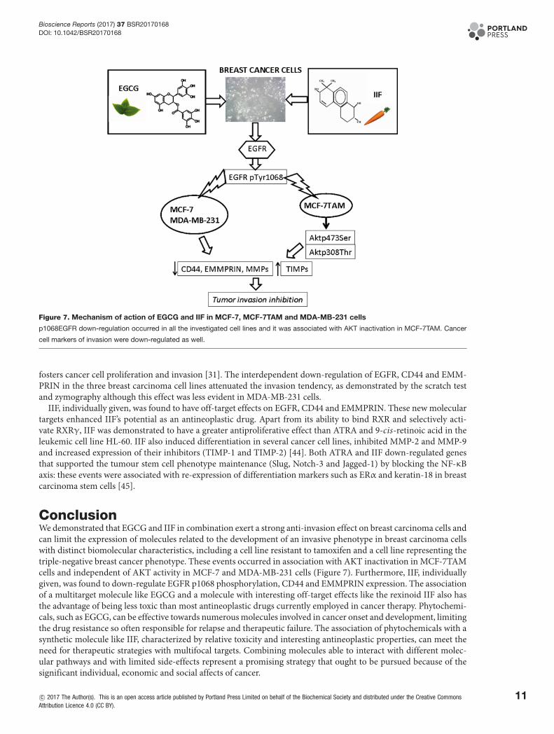
Figure 7. Mechanism of action of EGCG and IIF in MCF-7, MCF-7TAM and MDA-MB-231 cells
p1068EGFR down-regulation occurred in all the investigated cell lines and it was associated with AKT inactivation in MCF-7TAM. Cancer
cell markers of invasion were down-regulated as well.
fosters cancer cell proliferation and invasion [31]. The interdependent down-regulation of EGFR, CD44 and EMM-PRIN in the three breast carcinoma cell lines attenuated the invasion tendency, as demonstrated by the scratch testand zymography although this effect was less evident in MDA-MB-231 cells.
IIF, individually given, was found to have off-target effects on EGFR, CD44 and EMMPRIN. These new moleculartargets enhanced IIF’s potential as an antineoplastic drug. Apart from its ability to bind RXR and selectively acti-vate RXRγ, IIF was demonstrated to have a greater antiproliferative effect than ATRA and 9-cis-retinoic acid in theleukemic cell line HL-60. IIF also induced differentiation in several cancer cell lines, inhibited MMP-2 and MMP-9and increased expression of their inhibitors (TIMP-1 and TIMP-2) [44]. Both ATRA and IIF down-regulated genesthat supported the tumour stem cell phenotype maintenance (Slug, Notch-3 and Jagged-1) by blocking the NF-κBaxis: these events were associated with re-expression of differentiation markers such as ERα and keratin-18 in breastcarcinoma stem cells [45].
ConclusionWe demonstrated that EGCG and IIF in combination exert a strong anti-invasion effect on breast carcinoma cells andcan limit the expression of molecules related to the development of an invasive phenotype in breast carcinoma cellswith distinct biomolecular characteristics, including a cell line resistant to tamoxifen and a cell line representing thetriple-negative breast cancer phenotype. These events occurred in association with AKT inactivation in MCF-7TAMcells and independent of AKT activity in MCF-7 and MDA-MB-231 cells (Figure 7). Furthermore, IIF, individuallygiven, was found to down-regulate EGFR p1068 phosphorylation, CD44 and EMMPRIN expression. The associationof a multitarget molecule like EGCG and a molecule with interesting off-target effects like the rexinoid IIF also hasthe advantage of being less toxic than most antineoplastic drugs currently employed in cancer therapy. Phytochemi-cals, such as EGCG, can be effective towards numerous molecules involved in cancer onset and development, limitingthe drug resistance so often responsible for relapse and therapeutic failure. The association of phytochemicals with asynthetic molecule like IIF, characterized by relative toxicity and interesting antineoplastic properties, can meet theneed for therapeutic strategies with multifocal targets. Combining molecules able to interact with different molec-ular pathways and with limited side-effects represent a promising strategy that ought to be pursued because of thesignificant individual, economic and social affects of cancer.
c© 2017 The Author(s). This is an open access article published by Portland Press Limited on behalf of the Biochemical Society and distributed under the Creative CommonsAttribution Licence 4.0 (CC BY).
11

Bioscience Reports (2017) 37 BSR20170168DOI: 10.1042/BSR20170168
AcknowledgementsWe thank Dr E. Collins, editor of scientific publications and member of the European Medical Writers Association, who revisedEnglish language in the present manuscript.
FundingThis work was supported by the University of Bologna (RFO grant to F.F and to E.S.)].
Competing interestsThe authors declare that there are no competing interests associated with the manuscript.
Author contributionF.F. designed the study, performed the immunofluorescence and RT-PCR and prepared the manuscript. M.G. performed Westernblot. E.S. edited and reviewed the manuscript. A.P. performed the Western blot, zymography, acquired and analysed the datastatistically. All authors read and approved the final manuscript version that was submitted for peer review.
AbbreviationsATRA, all trans-retinoic acid; CSC, cancer stem cell; DABCO, diazabicyclo[2.2.2]octane; EGCG, epigallocatechin-3-gallate;EGFR, epidermal growth factor receptor; EMMPRIN, extracellular matrix metalloproteinase inducer; EMT,epithelial–mesenchymal transition; ERα, oestrogen receptor α; HA, hyaluronic acid; IIF, 6-OH-11-O-hydroxyphenanthrene; MMP,matrix metalloproteinase; PI3K, phosphatidylinositol-3-kinase; RT-PCR, reverse transcription-PCR; RXRγ, retinoid X receptor-γ;TIMP, tissue inhibitor of metalloproteinase.
References1 Forester, S.C. and Lambert, J.D. (2011) The role of antioxidant versus pro-oxidant effects of green tea polyphenols in cancer prevention. Mol. Nutr. Food
Res. 55, 844–8542 Mak, J.C. (2012) Potential role of green tea catechins in various disease therapies: progress and promise. Clin. Exp. Pharmacol. Physiol. 39, 265–2733 Yu, Y., Deng, Y., Lu, B.M., Liu, Y.X., Li, J. and Bao, J.K. (2014) Green tea catechins: a fresh flavor to anticancer therapy. Apoptosis 19, 1–184 Chowdhury, A., Sarkar, J., Chakraborti, T., Pramanik, P.K. and Chakraborti, S. (2016) Protective role of epigallocatechin-3-gallate in health and disease:
a perspective. Biomed. Pharmacother. 78, 50–595 Yang, C.S., Chen, J.X., Wang, H. and Lim, J. (2016) Lessons learned from cancer prevention studies with nutrients and non-nutritive dietary
constituents. Mol. Nutr. Food Res. 60, 1239–12506 Li, M.J., Yin, Y.C., Wang, J. and Jiang, Y.F. (2014) Green tea compounds in breast cancer prevention and treatment. World J. Clin. Oncol. 5, 520–5287 Butt, M.S., Ahmad, R.S., Sultan, M.T., Qayyum, M.M. and Naz, A. (2015) Green tea and anticancer perspectives: updates from last decade. Crit. Rev.
Food Sci. Nutr. 55, 792–8058 Rahmani, A.H., Al Shabrmi, F.M., Allemailem, K.S., Aly, S.M. and Khan, M.A. (2015) Implications of green tea and its constituents in the prevention of
cancer via the modulation of cell signalling pathway. Biomed. Res. Int. 2015, 925640, doi:10.1155/2015/9256409 Singh, B.N., Shankar, S. and Srivastava, R.K. (2011) Green tea catechin, epigallocatechin-3-gallate (EGCG): mechanisms, perspectives and clinical
applications. Biochem. Pharmacol. 82, 1807–182110 Amin, A.R., Karpowicz, P.A., Carey, T.E., Arbiser, J., Nahta, R., Chen, Z.G. et al. (2015) Evasion of anti-growth signaling: a key step in tumorigenesis and
potential target for treatment and prophylaxis by natural compounds. Semin. Cancer Biol. 35, S55–S7711 Tachibana, H. (2011) Green tea polyphenol sensing. Proc. Jpn. Acad. Ser. B Phys. Biol. Sci. 87, 66–8012 Shimizu, M., Adachi, S., Masuda, M., Kozawa, O. and Moriwaki, H. (2011) Cancer chemoprevention with green tea catechins by targeting receptor
tyrosine kinases. Mol. Nutr. Food Res. 55, 832–84313 Yiannakopoulou, E.C. (2014) Interaction of green tea catechins with breast cancer endocrine treatment: a systematic review. Pharmacology 94,
245–24814 Chen, H., Landen, C.N., Li, Y., Alvarez, R.D. and Tollefsbol, T.O. (2013) Enhancement of cisplatin-mediated apoptosis in ovarian cancer cells through
potentiating G2/M arrest and p21 upregulation by combinatorial epigallocatechin gallate and sulforaphane. J. Oncol. 2013, 87295715 Fujiki, H., Sueoka, E., Watanabe, T. and Suganuma, M. (2015) Synergistic enhancement of anticancer effects on numerous human cancer cell lines
treated with the combination of EGCG, other green tea catechins, and anticancer compounds. J. Cancer Res. Clin. Oncol. 141, 1511–152216 Lecumberri, E., Dupertuis, Y.M., Miralbell, R. and Pichard, C. (2013) Green tea polyphenol epigallocatechin-3-gallate (EGCG) as adjuvant in cancer
therapy. Clin. Nutr. 32, 894–90317 Farabegoli, F., Govoni, M., Ciavarella, C., Orlandi, M. and Papi, A. (2014) A RXR ligand 6-OH-11-O-hydroxyphenanthrene with antitumour properties
enhances (-)-epigallocatechin-3-gallate activity in three human breast carcinoma cell lines. Biomed. Res. Int. 2014, 85308618 Papi, A., Govoni, M., Ciavarella, C., Spisni, E., Orlandi, M. and Farabegoli, F. (2016) Epigallocatechin-3-gallate increases RXRγ-mediated pro-apoptotic
and anti-invasive effects in gastrointestinal cancer cell lines. Curr. Cancer Drug Targets 16, 373–38519 Altucci, L., Leibowitz, M.D., Ogilvie, K.M., de Lera, A.R. and Gronemeyer, H. (2007) RAR and RXR modulation in cancer and metabolic disease. Nat. Rev.
Drug Discov. 6, 793–810
12 c© 2017 The Author(s). This is an open access article published by Portland Press Limited on behalf of the Biochemical Society and distributed under the Creative CommonsAttribution Licence 4.0 (CC BY).

Bioscience Reports (2017) 37 BSR20170168DOI: 10.1042/BSR20170168
20 Uray, I.P. and Brown, P.H. (2011) Chemoprevention of hormone receptor-negative breast cancer: new approaches needed. Recent Results Cancer Res.188, 147–162
21 Wei, X. (2011) Mechanism of EGER-related cancer drug resistance. Anticancer Drugs 22, 963–97022 Elloul, S., Kedrin, D., Knoblauch, N.W., Beck, A.H. and Toker, A. (2014) The adherens junction protein afadin is an AKT substrate that regulates breast
cancer cell migration. Mol. Cancer Res. 12, 464–47623 Zuo, J.H., Zhu, W., Li, M.Y., Li, X.H., Yi, H., Zeng, G.Q. et al. (2011) Activation of EGFR promotes squamous carcinoma SCC10A cell migration and
invasion via inducing EMT-like phenotype change and MMP-9-mediated degradation of E-cadherin. J. Cell Biochem. 112, 2508–251724 Wang, S.J. and Bourguignon, L.Y. (2011) Role of hyaluronan-mediated CD44 signaling in head and neck squamous cell carcinoma progression and
chemoresistance. Am. J. Pathol. 178, 956–96325 Hong, J., Lu, H., Meng, X., Ryu, J.H., Hara, Y. and Yang, C.S. (2002) Stability, cellular uptake, biotransformation, and efflux of tea polyphenol
(-)-epigallocatechin-3-gallate in HT-29 human colon adenocarcinoma cells. Cancer Res. 62, 7241–724626 Chomczynski, P. and Sacchi, N. (1987) Single-step method of RNA isolation by acid guanidinium thiocyanate-phenol-chloroform extraction. Anal.
Biochem. 162, 156–15927 Farabegoli, F., Papi, A. and Orlandi, M. (2011) (-)-Epigallocatechin-3-gallate down-regulates EGFR, MMP-2, MMP-9 and EMMPRIN and inhibits the
invasion of MCF-7 tamoxifen-resistant cells. Biosci. Rep. 31, 99–10828 Piccione, E.C., Lieu, T.J., Gentile, C.F., Williams, T.R., Connolly, A.J., Godwin, A.K. et al. (2012) A novel epidermal growth factor receptor variant lacking
multiple domains directly activates transcription and is overexpressed in tumors. Oncogene 31, 2953–296729 Montermini, L., Meehan, B., Garnier, D., Lee, W.J., Lee, T.H., Guha, A. et al. (2015) Inhibition of oncogenic epidermal growth factor receptor kinase
triggers release of exosome-like extracellular vesicles and impacts their phosphoprotein and DNA content. J. Biol. Chem. 290, 24534–2454630 Appert-Collin, A., Hubert, P., Cremel, G. and Bennasroune, A. (2015) Role of ErbB receptors in cancer cell migration and invasion. Front. Pharmacol. 6,
28331 Grass, G.D., Tolliver, L.B., Bratoeva, M. and Toole, B.P. (2013) CD147, CD44, and the epidermal growth factor receptor (EGFR) signaling pathway
cooperate to regulate breast epithelial cell invasiveness. J. Biol. Chem. 288, 26089–2610432 Xu, H., Tian, Y., Yuan, X., Wu, H., Liu, Q., Pestell, R.G. et al. (2015) The role of CD44 in epithelial-mesenchymal transition and cancer development.
Onco. Targets Ther. 8, 3783–379233 Brown, G.T. and Murray, G.I. (2015) Current mechanistic insights into the roles of matrix metalloproteinases in tumour invasion and metastasis. J.
Pathol. 237, 273–28134 Papi, A., Tatenhorst, L., Terwel, D., Hermes, M., Kummer, M.P., Orlandi, M. et al. (2009) PPARgamma and RXRgamma ligands act synergistically as
potent antineoplastic agents in vitro and in vivo glioma models. J. Neurochem. 109, 1779–179035 Ullmann, U., Haller, J., Decourt, J.P., Girault, N., Girault, J., Richard-Caudron, A.S. et al. (2003) A single ascending dose study of epigallocatechin gallate
in healthy volunteers. J. Int. Med. Res. 31, 88–10136 Peter, B., Bosze, S. and Horvath, R (2017) Biophysical characteristics of proteins and living cells exposed to the green tea polyphenol
epigallocatechin-3-gallate (EGCg): review of recent advances from molecular mechanisms to nanomedicine and clinical trials. Eur. Biophys. J. 46, 1–24
37 Peter, B., Farkas, E., Forgacs, E., Saftics, A., Kovacs, B., Kurunczi, S. et al. (2017) Green tea polyphenol tailors cell adhesivity of RGD displayingsurfaces: multicomponent models monitored optically. Sci. Rep. 7, 42220
38 Cheng, W., Zhou, J., Tian, X. and Zhang, X. (2016) Development of the third generation EGFR tyrosine kinase inhibitors for anticancer therapy. Curr.Med. Chem. 23, 3343–3359
39 Masuda, M., Wakasaki, T., Toh, S., Shimizu, M. and Adachi, S. (2011) Chemoprevention of head and neck cancer by green tea extract: EGCG-the role ofEGFR signaling and “lipid raft”. J. Oncol. 2011, 540148
40 Sette, G., Salvati, V., Mottolese, M., Visca, P., Gallo, E., Fecchi, K. et al. (2015) Tyr1068-phosphorylated epidermal growth factor receptor (EGFR)predicts cancer stem cell targeting by erlotinib in preclinical models of wild-type EGFR lung cancer. Cell Death Dis. 6, e1850
41 Kim, S., Han, J., Kim, J.S., Kim, J.H., Choe, J.H., Yang, J.H et al. (2011) Silibinin suppresses EGFR ligand-induced CD44 expression through inhibition ofEGFR activity in breast cancer cells. Anticancer Res. 31, 3767–3773
42 Li, L., Qi, L., Liang, Z., Song, W., Liu, Y., Wang, Y. et al. (2015) Transforming growth factor-β1 induces EMT by the transactivation of epidermal growthfactor signaling through HA/CD44 in lung and breast cancer cells. Int. J. Mol. Med. 36, 113–122
43 Grass, G.D., Dai, L., Qin, Z., Parsons, C. and Toole, B.P. (2014) CD147: regulator of hyaluronan signaling in invasiveness and chemoresistance. Adv.Cancer Res. 123, 351–373
44 Papi, A. and Orlandi, M. (2016) Role of nuclear receptors in breast cancer stem cells. World J. Stem Cells 8, 62–7245 Papi, A., De Carolis, S., Bertoni, S., Storci, G., Sceberras, V., Santini, D. et al. (2014) PPARγ and RXR ligands disrupt the inflammatory cross-talk in the
hypoxic breast cancer stem cells niche. J. Cell Physiol. 229, 1595–1606
c© 2017 The Author(s). This is an open access article published by Portland Press Limited on behalf of the Biochemical Society and distributed under the Creative Commons AttributionLicence 4.0 (CC BY).
13
