electrochemical nucleic acid-based biosensors: · pdf fileelectrochemical nucleic acid-based...
TRANSCRIPT

1161
Pure Appl. Chem., Vol. 82, No. 5, pp. 1161–1187, 2010.doi:10.1351/PAC-REP-09-08-16© 2010 IUPAC, Publication date (Web): 20 April 2010
Electrochemical nucleic acid-based biosensors:Concepts, terms, and methodology (IUPACTechnical Report)*
Jan Labuda1,‡, Ana Maria Oliveira Brett2, Gennady Evtugyn3,Miroslav Fojta4,‡, Marco Mascini5, Mehmet Ozsoz6, Ilaria Palchetti5,Emil Paleček4, and Joseph Wang7
1Institute of Analytical Chemistry, Faculty of Chemical and Food Technology,Slovak University of Technology in Bratislava, Radlinského 9, 81237 Bratislava,Slovakia; 2Department of Chemistry, Faculty of Sciences and Technology,University of Coimbra, 3004-533 Coimbra, Portugal; 3Department of AnalyticalChemistry, A.M. Butlerov Institute of Chemistry, Kazan State University,Kremlevskaya 18, 420008 Kazan, Russia; 4Institute of Biophysics, Academy ofSciences, Královopolská 135, 612 65 Brno, Czech Republic; 5Department ofChemistry, University of Florence, Via della Lastruccia 3, 50019 Sesto Fiorentino,Italy; 6Faculty of Pharmacy, Ege University, Bornova-Izmir, Turkey 35100;7Department of Nanoengineering, University of San Diego, La Jolla, CA 92093,USA
Abstract: An electrochemical nucleic acid (NA)-based biosensor is a biosensor that integratesa nucleic acid as the biological recognition element and an electrode as the electrochemicalsignal transducer. The present report provides concepts, terms, and methodology related tobiorecognition elements, detection principles, type of interactions to be addressed, and con-struction and performance of electrochemical NA biosensors, including their critical evalua-tion, which should be valuable for a wide audience, from academic, biomedical, environ-mental, and food-testing, drug-developing, etc. laboratories to sensor producers.
Keywords: aptamers; biosensors; electrochemistry; DNA damage; IUPAC AnalyticalChemistry Division; nucleic acids; nucleic acid hybridization; nucleic acid interactions.
CONTENTS
1. INTRODUCTION2. BIORECOGNITION ELEMENTS
2.1 Nucleic acids used at biosensors 2.2 Nucleic acid aptamers
3. DETECTION PRINCIPLES3.1 Electroactivity of NA
3.1.1 Reduction and oxidation of nucleic acids 3.1.2 Tensammetric responses of nucleic acids
*Sponsoring body: IUPAC Analytical Chemistry Division: see more details on p. 1184.‡Corresponding authors: E-mail: [email protected] (J.L.), [email protected] (M.F.)

3.2 Effects of DNA structure3.2.1 Changes of DNA structure at charged electrode surface
3.3 Label-free techniques utilizing electrochemical and/or surface activity of nucleic acids3.3.1 Guanine oxidation at carbon electrodes3.3.2 Structure-sensitive responses of DNA at mercury-based electrodes
3.4 Noncovalent redox indicators3.5 Covalently bound labels
3.5.1 Electroactive groups 3.5.2 Enzymes3.5.3 Nanoobjects
3.6 Specific features of the detection techniques3.6.1 Signal-off vs. signal-on techniques3.6.2 Signal amplification
4. NUCLEIC ACID INTERACTIONS AND RELATED SENSORS4.1 DNA hybridization and sequence-specific DNA sensing
4.1.1 Detection techniques used in DNA hybridization sensors4.1.2 Primer extension-based sensing4.1.3 Detection of mutations and sequence polymorphisms
4.2 Other association interactions4.2.1 Nucleic acid interactions with low-molecular-mass compounds4.2.2 Nucleic acid interactions with proteins4.2.3 Aptamer–target interactions
4.3 DNA damage4.3.1 Label-free detection of strand breaks with mercury-based nucleic acid sensors4.3.2 Redox indicator-based sensing of DNA degradation with carbon-based nucleic acid
sensors4.3.3 Sensors based on guanine redox processes4.3.4 Electroactive products of DNA damage4.3.5 Layered assemblies for genotoxicity screening4.3.6 Molecular beacon-like sensor for nuclease and ligase activities
5. CONSTRUCTION OF BIOSENSORS5.1 Transducers5.2 Nucleic acid immobilization
5.2.1 Noncovalent binding5.2.2 Affinity binding5.2.3 Chemisorption5.2.4 Covalent binding
6. SPECIFIC PERFORMANCE CRITERIA7. CONCLUSIONS
LIST OF ABBREVIATIONSMEMBERSHIP OF SPONSORING BODIESREFERENCES
1. INTRODUCTION
The previously published IUPAC technical report “Electrochemical biosensors: Recommended defini-tions and classification” [1] did not deal extensively with nucleic acids (NAs) [2] as the biologicalrecognition element, but considered the work on other types of biosensors. Since that time, significantprogress in the development and application of electrochemical sensors based on deoxyribonucleic acid
J. LABUDA et al.
© 2010, IUPAC Pure Appl. Chem., Vol. 82, No. 5, pp. 1161–1187, 2010
1162

(DNA) and other NAs, including aptamers and peptide NAs, has been achieved. On the other hand, sofar there have been no efforts at essential classification in this dynamically developing field.
An electrochemical NA-based biosensor is a device that integrates an NA (natural and bio-mimetic forms of oligo- and polynucleotides) as the biological recognition element and an electrode asthe physicochemical transducer. In this regard, the previous IUPAC report on electrochemical bio -sensors [1] is fully acceptable for electrochemical NA-based biosensors, including the biosensor defi-nition and information obtained. NA-based biosensors belong to the family of chemical sensors, whichtransform (bio)chemical stimulus from an analyte in relation to NA into the analytically useful infor-mation (analyte type/concentration, NA chemical structure and/or its change, etc.). From the point ofview of electroanalytical chemistry, the electrochemical NA-based biosensor represents an NA-modi-fied electrode which is used mostly in voltammetric and chronopotentiometric detection modes [3].Electrochemical impedance spectroscopy (EIS) is also used as the investigation and detection technique[4]. The use of the concept of an electrode modified with the DNA layer has allowed a significant de-crease of the amount of DNA tested/determined.
Comment: Use of terms “sensor” and “sensing” (or assay) is sometimes confusing. Here, it isnecessary to distinguish strictly between NA biosensors and NA sensing. While inan electrochemical NA biosensor, the NA has to be in an intimate contact with theelectrode prior to and during the NA interaction with an analyte, the NA electro-chemical sensing has broader meaning. Product of an interaction of any NA with ananalyte (generated either in solution or at another surface) or NA itself can be de-tected or its concentration determined electrochemically, usually after accumulationonto the electrode surface. Most of the electrochemical NA biosensors mentioned inthis report employ detection principles which can be applied in alternative electro-chemical biosensing (micro) techniques as well. In some cases, the latter techniquescan be similar to or even more efficient than the biosensors. For example, DNA dam-age or association interactions can easily be monitored using simple ex situ (ad-sorptive transfer stripping) electrochemical analysis of DNA exposed to a damagingagent or interacting in solution (prior to adsorption at an electrode). A specific classof approaches that have complemented the classical concept of electrochemicalDNA sensors during the last decade employs magnetic beads as the surface on whichthe DNA biorecognition event (hybridization, interaction with protein, etc.) occurs.Then, target DNA, signaling probe, or other indicator molecules captured or gener-ated at the surface of the beads can be determined electrochemically. Such tech-niques are referred to as “double-surface” ones due to the two different surfaces in-volved (one—the magnetic beads—for the biomolecular interaction, and theother—the electrode—for detection) [5]. A more detailed description of the double-surface strategy is beyond the scope of this report.
According to [1], biosensors can be classified considering the biological specificity—conferringmechanism, mode of signal transduction, and analytes or reactions that they monitor. The classificationof the biosensors according to a third point of view (i.e., analytes or reactions) is also acceptable for theelectrochemical NA-based biosensors. However, the ratio of the utilization of the NA biosensors isshifted from the detection of analytes more to the reactions of NA when compared to the enzyme- andimmuno-sensors. In other words, the NA biosensors discussed in this report often deal with the inves-tigation of characteristic NA interactions rather than with the conventional determination of the con-centration of an analyte (a measurand).
Specificity of the biosensor response can also be accepted as a typical feature of the device, tak-ing advantage of the DNA strands bioaffinity properties. Typically, specificity/selectivity is induced byboth NA surface film and chemical properties of an analyte. The NA-based biosensors are specific ei-ther to the analyte (nucleotide bases sequence, protein) or to the NA itself (its damage).
© 2010, IUPAC Pure Appl. Chem., Vol. 82, No. 5, pp. 1161–1187, 2010
Electrochemical nucleic acid-based biosensors 1163
2. BIORECOGNITION ELEMENTS
2.1 Nucleic acids used at biosensors
Today, numerous types of natural and synthetic DNA and RNA molecules are available for electro-chemical biosensors, including chromosomal DNA as well as well-defined viral or plasmid NAs. Theplasmid and the viral DNA molecule can be cleaved into the fragments of various lengths by the actionof restriction endonucleases and/or amplified by polymerase chain reaction (PCR). End-labeled DNAs,polynucleotides with random or monotone sequences, and synthetically prepared oligonucleotides withprogrammable sequences (allowing modification of bases and/or backbones) are also commerciallyavailable. Oligonucleotides with RNA backbone are currently more expensive than oligodeoxyribo -nucleotides (ODNs).
Note: At present, mainly synthetic ODNs are used as probes in the DNA hybridization sen-sors. End-labels, such as thiols, disulfides, amines, or biotin, are incorporated to im-mobilize ODN to transducer surfaces. A long flexible spacer is usually added to pro-vide sufficient accessibility for surface attachment. Hydrocarbon linkers arefrequently used for this purpose. Selection of the probe nucleotide sequence dependsvery much on the target sequence. Certain specific applications require the rightchoice of probe length. For example, in discrimination of single-base mismatches,shorter probes are preferred because a single-base mismatch is more likely to disturbthe stability of a short DNA duplex and eventually prevent its formation.
Peptide nucleic acid (PNA) is a synthetic DNA mimic, which contains 2-aminoethylglycine link-ages instead of the negatively charged phosphodiesteric backbone of ODNs. The PNA probes are par-ticularly convenient for the detection of single-base mismatches (point mutations, SNPs) because thestability of DNA-PNA duplexes is strongly influenced by a single-base mismatch [6]. Other kinds ofsynthetic NA, such as locked nucleic acid (LNA), are also used.
Comment: In real DNA analyses, PCR-amplified genomic DNA segments are mostly used as tar-get DNAs. On the other hand, in a large number of papers, synthetic ODNs serve astarget DNAs. This may be acceptable if in principle new technology is being devel-oped. Otherwise, natural amplified or nonamplified target DNAs should be used tovalidate the new detection principles in analysis of real biological material. TargetDNAs can be labeled, which in the case of natural DNAs can be more difficult thanin the case of synthetic ODNs. Osmium tetraoxide complexes [5,7] are particularlysuitable for labeling of both natural and synthetic DNAs, RNAs, as well as for PNAend-labeling. Sometimes, it may be convenient to work with unlabeled target DNA.In such a case, either label-free detection is applied or labeled signaling (reporter)probes are used. Occasionally, the DNA probe may serve only as a capture probe (CP)and the signaling probe detects presence of the specific nucleotide sequence.Signaling probes are usually synthetic ODNs.
2.2 Nucleic acid aptamers
Nucleic acid aptamers are single-stranded (ss) oligonucleotides (mainly DNA or RNA) originating fromin vitro selection, which, starting from random sequence libraries, optimize the NAs for high affinitybinding to a given target [8–10]. The term “aptamer” derives from the Latin aptus, “to fit”, and em-phasizes the relationship between aptamer and its target. Aptamers, upon association with their target,fold into complex three-dimensional shapes in which the target becomes an intrinsic part of the NAstructure.
J. LABUDA et al.
© 2010, IUPAC Pure Appl. Chem., Vol. 82, No. 5, pp. 1161–1187, 2010
1164

Comments: The term “aptamer” should be clearly distinguished from other terms such as “ri-bozyme”, “DNAzyme”, and “aptazyme”, which are defined as follows. Ribozymesare catalytic RNAs; some ribozymes have been found in Nature and mediatephospho diester bond cleavage and peptide bond formation. In vitro selection has beenused to generate RNA enzymes with novel structures and catalytic functions (i.e.,Diels–Alder reactions, biphenyl isomerization, C–S bond by Michael reaction, etc.).DNAzymes are DNA-based catalysts that have not been found in Nature and are gen-erated only by in vitro selection. The ligand-binding and catalytic features of NA canbe combined to generate allosteric ribozymes or “aptazymes”. When ligands bind toan aptazyme, conformational changes in the ligand-binding domain are transduced toa change in the catalytic core and a concomitant modulation of catalytic activity. Theterm “aptamer” has been recently used also to denominate a new class of peptidicbioreceptors. To avoid misunderstanding in this report with the term, aptamers areonly considered NA-based aptamers.
Note: In vitro selection is an iterative method mainly known as Systematic Evolution ofLigands by EXponential enrichment (SELEX) developed by the Gold, Ellington, andSzostak laboratories in the early 1990s [11,12].
The properties of aptamers make them an attractive class of molecules that meet and exceed theproperties of antibodies for biosensor development. Both DNA and RNA aptamers bind their targetswith dissociation constants (Kd) which in the case of proteins are in the low picomolar to low nano molarrange, discriminating between related proteins that share common sets of structural domains. Affinitiesin the micromolar range can be observed in the case of aptamers—small molecule complexes. The char-acterization of affinity of aptamer binding is required for estimating the sensitivity and selectivity of theappropriate biosensors, also called “aptasensors”.
Aptamers with affinity for a large variety of molecules, including virtually any class of proteins(enzymes, membrane proteins, viral proteins, etc.), peptides, drugs, toxins, low-molar-mass ligands, andions have been isolated. The folding of NA around the target provides numerous discriminatory inter-molecular interactions. These interactions fall in the class of noncovalent bonding and are mainly stack-ing, shape complementarity, electrostatic, and hydrogen-bonding interactions. Multiple interactionscontribute to the same aptamer-target complexes. The molecular interactions govern the specific recog-nition of and discrimination between different target classes in aptamer complexes [8–10]. The follow-ing advantages of aptamers are mostly counted over other biorecognition elements: (i) chemical syn-thesis, which does not require biological raw materials (bioethics requirements); (ii) universal approachto selection procedure, which does not depend on a particular analyte (possibility to use toxins as wellas molecules that do not elicit a good immune response); (iii) cost-effective production; (iv) high affin-ity and molecular discrimination; (v) high thermal stability and opportunities for the further modifica-tion that provides the immobilization of aptamers onto solid support and mild alteration of specificityand selectivity of the binding.
3. DETECTION PRINCIPLES
3.1 Electroactivity of nucleic acids
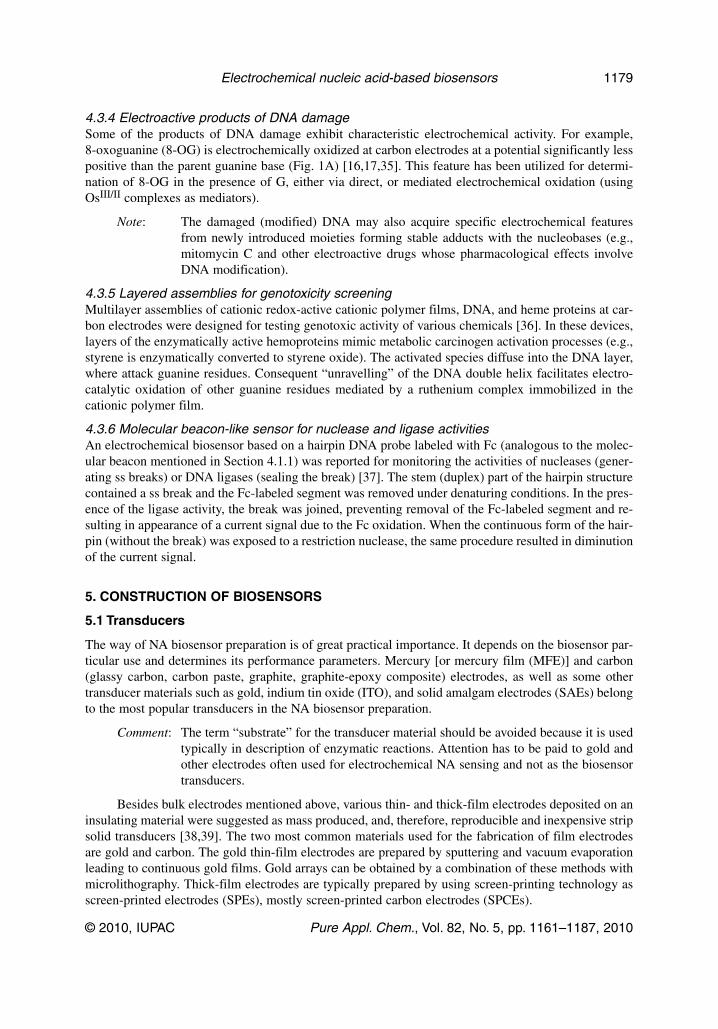
3.1.1 Reduction and oxidation of nucleic acidsThe electrochemical activity of NAs (both the native high-molecular ones as well as oligonucleotideswith rather short sequences) is conferred by the electroactivity of its components, nucleobases and sugarresidues [3]. At mercury-based electrodes, adenine (A) and cytosine (C) residues in ssNAs undergoreduction processes close to –1.4 V (against SCE) in neutral or weakly acidic medium (giving rise topeak CA, Fig. 1A). In cyclic voltammetric modes, chemically reversible reduction of guanine (G) in
© 2010, IUPAC Pure Appl. Chem., Vol. 82, No. 5, pp. 1161–1187, 2010
Electrochemical nucleic acid-based biosensors 1165

DNA and RNA leads to an anodic peak G (at about –0.3 V, Fig. 1A). Nucleobase residues in DNA andRNA are reduced in an adsorbed state. All NA bases have been reported to be electro chemically oxi-dized at carbon electrodes, but only adenine and (particularly) guanine oxidation peaks (Fig. 1A) havebeen widely utilized in NA biosensors. The oxidation of sugar residues in NA at copper electrodes hasalso been employed.
Comment: The electroactivity of NAs on the mercury electrodes has not been fully utilized be-cause of concerns regarding the toxicity of metal mercury. Recently, it has beenshown that with nontoxic solid dental amalgam electrodes, electrochemical responsessimilar to the hanging mercury electrode can be obtained [13,14]. There were alsofew reports about the guanine residue oxidation at gold electrodes [15].
J. LABUDA et al.
© 2010, IUPAC Pure Appl. Chem., Vol. 82, No. 5, pp. 1161–1187, 2010
1166
Fig. 1 (A) Scheme of intrinsic electrochemical responses of DNA. Solid curves represent voltammetric peaks dueto reduction or oxidation of nucleobase residues in the DNA at mercury/amalgam electrodes (CA: reduction ofcytosine and adenine, G: re-oxidation of an electrochemically generated reduction product of guanine) or carbonelectrodes (Gox, Aox, Tox, Cox: oxidation of standard nucleobases guanine, adenine, thymine, and cytosine; 8-OG,oxidation of a common product of oxidative DNA damage, 8-oxoguanine). Dotted or dashed curves showcapacitive (tensammetric) voltammetric responses of dsDNA lacking or containing free ends, respectively, atmercury-based electrodes (peak 1 is due to adsorption/desorption processes of the DNA sugar-phosphate backbone,while peak 3 is due to single-stranded DNA segments adsorbed via bases). (B) Scheme of redox potentials of someelectroactive species used as DNA labels/indicators (shown relative to the intrinsic DNA responses): Os,L-cat,catalytic signal of DNA labeled with osmium tetraoxide complexes (Os,L); echi, echinomycin; DM-Q, quinonemoiety of daunomycin; NP, nitrophenyl; AQ, anthraquinone; MB, methylene blue; DM-QH2, hydroquinone moietyof daunomycin. Bars in the upper part of the scheme show potential regions in which redox electrochemistry isyielded by “tunable” DNA labels such as Os,L (their redox potentials depend on the ligand L) or ferrocene (Fc; itsredox potential can be tuned by different substituents and/or electronic conjugation with another aromatic system).

3.1.2 Tensammetric responses of nucleic acidsThe polyanionic nature of the NAs causes them to undergo characteristic adsorption/desorption (reorientation) processes at the mercury-based electrodes upon applying (changing) negative potentialsdue to interplay between electrostatic repulsion and relatively strong adsorption via hydrophobic partsof the polynucleotide chains (particularly bases) [3,16]. In weakly alkaline media, these processes giverise to analytically useful tensammetric (capacitive) [2] current signals (Fig. 1A) that sensitively reflectchanges in the NA structure. DNA sugar-phosphate backbone undergoes adsorption/desorptionprocesses around –1.2 V (vs. SCE) yielding peak 1. Peak 2 (at –1.3 V) has been ascribed to distortedDNA double-helical segments with partially accessible edges of base pairs, which thus take part in theadsorption/desorption processes. ssDNA segments with freely accessible bases produce peak 3 close to–1.45 V (Fig. 1A).
3.2 Effects of DNA structure
Marked differences in the voltammetric and tensametric responses of native (double-stranded, ds)and denatured (ss) DNA have been observed at the mercury electrodes under certain conditions [3,16].Large current responses of ssDNA as compared to very small ones of dsDNA are explained by inaccessibility of the nucleobases in dsDNA for adsorption and the electroactive sites of cytosine andadenine for the reduction at the mercury electrode. Both faradaic and tensammetric [2] responses meas-ured at the mercury-based electrodes thus sensitively reflect subtle changes in DNA structure, resultingin uncovering the nucleobases. On the other hand, the primary oxidation sites of guanine and adenineare relatively well accessible in the dsDNA, making the oxidation responses at carbon electrodes lesssensitive to DNA structure changes.
3.2.1 Changes of DNA structure at charged electrode surface Using the faradaic and capacitive DNA responses, it was shown that at neutral and weakly alkaline pHvalues prolonged contact of dsDNA with the surface of the mercury electrode within a narrow poten-tial region around –1.2 V vs. SCE resulted in a relatively slow, irreversible opening of the DNA doublehelix at the surface [3,16]. No extensive duplex opening was found in covalently closed circular DNAsin agreement with an assumption that DNA unwinding starts from DNA strand ends.
Note: DNA unwinding was also observed at negatively charged gold and silver electrodesand at other surfaces.
3.3 Label-free techniques utilizing electrochemical and/or surface activity of nucleicacids
Nucleic acids are electrochemically active due to the presence of electrochemically oxidizable or re-ducible nucleobases, and they exhibit specific surface activity depending on which NA components takepart in adsorption at the electrode surface. Electrochemical analysis of the DNA can thus in principlebe conducted without introducing specific labels.
3.3.1 Guanine oxidation at carbon electrodesAlthough all common nucleobases have been reported to be electrochemically oxidizable at carbonelectrodes and adenine, cytosine, and guanine to give electrochemical responses at mercury-based elec-trodes, the vast majority of label-free DNA biosensors employ oxidation of the guanine moiety at car-bon or other solid electrodes as the source of analytical signals [3]. This choice is dictated by (a) rela-tively low redox potential of guanine, making it easily detectable by direct electrochemical oxidationwithout using additional reagents; (b) the fact that guanine is the most frequent target for many DNAdamaging species and its chemical modification is often accompanied by a loss of the guanine peak.
© 2010, IUPAC Pure Appl. Chem., Vol. 82, No. 5, pp. 1161–1187, 2010
Electrochemical nucleic acid-based biosensors 1167

Comment: Limitations of the guanine redox process-based label-free and reagent-less sensorsarise from (a) usually insufficient discrimination between the probe and target strandsin DNA hybridization assays due to almost even distribution of all four nucleobasesin most natural nucleotide sequences (see Section 4.1); (b) relatively low sensitivitytoward small local changes in DNA structure such as strand breakage (see Section3.2); and (c) the fact that only guanines in the close vicinity of the electrode surfacecan undergo direct electrooxidation. The latter obstacle can be overcome by applyingsoluble redox mediators such as rhodium or ruthenium complexes that can shuttleelectrons from guanine residues in distant parts of DNA chains to the electrode[17,18]
G(DNA) + Mz+ → G•+(DNA) + M(z–1)+
M(z–1)+ – e– → Mz+ (electrochemically)
3.3.2 Structure-sensitive responses of DNA at mercury-based electrodesReduction and tensammetric responses of NAs at the mercury and some amalgam electrodes arestrongly dependent on the hydrophobic nature of bases, on whether they are present in ss or ds regionsof the NAs, and/or whether the DNA adsorbed at the electrode surface can undergo a structural transi-tion connected with a change in the nucleobase accessibility [3,16]. Based on these principles, denatu-rational transitions, strand breaks (making the double helix susceptible to potential-induced surface de-naturation) or ss regions within dsDNA can be detected by AC voltammetry or impedance techniqueswith a considerable sensitivity (such as one strand break per 2 × 10⁵ nucleotides in circular duplexDNA).
Note: Thus, mercury-based electrodes modified with circular duplex DNA have been de-signed as label-free and reagent-less sensors for DNA damage involving (or convert-ible to) strand breaks (see Section 4.3.1).
3.4 Noncovalent redox indicators
Despite the analytical usefulness of the intrinsic NA electrochemical activity, a number of detectiontechniques have employed electroactive species serving as redox indicators of the events (such as hy-bridization, damage, complex formation with another substance) having undergone by the DNA at theelectrode surface.
Comment: Development of these detection techniques (which are still label-free in the sense ofno chemical modification of NA probes or targets) has been motivated by several lim-itations of the intrinsic DNA electroactivity-based approaches (see Section 3.3):
(a) only mercury or some amalgam electrodes possess a sufficiently negative po-tential window to observe reduction and tensammetric responses of unlabeledNA, and only carbon electrodes can be used for direct electrochemical detec-tion of the nucleobase oxidation. Other electrodes, including very popular goldones, exhibit insufficient hydrogen overvoltage on the negative side and elec-trooxidation of the electrode material at insufficiently positive potentials toallow detection of well-defined NA responses. On the other hand, typical redoxindicators are reduced/oxidized at less extreme potentials (Fig. 1B), thus ex-tending the choice of working electrodes.
(b) electrochemical reduction and oxidation of NA bases is irreversible and thuspreventing reusability of the sensors (recognition layers). This obstacle can beovercome by using redox indicators, producing their (often reversible) electro-chemical responses at “safe” potentials.
J. LABUDA et al.
© 2010, IUPAC Pure Appl. Chem., Vol. 82, No. 5, pp. 1161–1187, 2010
1168

Electrochemically (redox) active substances binding preferentially to either ssDNA or dsDNAhave been applied as indicators of DNA hybridization (to recognize an ss probe from the hybrid duplex)or DNA damage (to recognize intact dsDNA from degraded DNA that has lost its double-helical struc-ture) [3,16,17]. The noncovalent redox indicators encompass species interacting with DNA electrosta-tically, DNA intercalators, or groove binders (for more details on DNA association inter actions, seeSection 4.2). Some of the noncovalent indicators can act as redox mediators (electron shuttles) and/orelectrocatalysts, which may be either soluble and diffusionally free (see Section 3.1), or connected tothe dsDNA base pair stacks and take part in electron transfer mediated by the DNA double helix.
The electrostatic indicators respond to differences in negative charge density between ssDNAand dsDNA. Hybridization between a surface-confined probe with target DNA results in increase of thenegative charge density, while degradation of dsDNA covering the surface results in the opposite effect.Using peptide nucleic probe (bearing no negative charge) offers a better discrimination between the (un-charged) probe and the (negatively charged PNA�DNA) hybrid. Redox indicators such as the anioniccomplex hexacyanoferrate (III/II), [Fe(CN)6]3–/4–, (being repelled from the DNA-modified surface) orthe cationic complex [Ru(NH3)6]3+/2+ (being attracted to the negatively charged hybrids) have been em-ployed to monitor changes in the negative charge density using impedimetric or voltammetric tech-niques as well as scanning electrochemical microscopy [17,19,20].
Note: Some authors use the term “redox probe” for the [Fe(CN)6]3–/4– indication system. Itis recommended to avoid using the term “probe” in the context of DNA sensors dueto possible confusion with the “hybridization probe”.
Groove binders [such as bis-benzimide (fluorescent dye Hoechst 33258)] and intercalators(such as daunomycin, metal chelates with condensed aromatic nitrogenous heterocyclic ligands, pheno -xazines, etc.) recognize specific structural features of dsDNA and bind with a higher affinity to the du-plex NA, which results in selective accumulation of the indicators in dsDNA (after hybridization orprior to damage to the duplex DNA) layer at the electrode surface, thus increasing the peak currents ofthe indicators. To improve discrimination between ssDNA and dsDNA, bis-intercalators (e.g., echino-mycin) or threading intercalators [e.g., N,N-bis[3-(3-ferroceneacetylaminopropyl)methylamino-propyl]naphthalene-1,4,5,8-tetracarboxylic acid diimide (ferrocenyl naphthalene diimide)] have beenapplied, which form thermodynamically or kinetically more stable complexes with dsDNA than thesimple intercalators [16,17,21].
Substances associating preferentially with ssDNA have also been applied in electrochemicalsensors for the DNA hybridization. A typical example is a phenothiazine dye methylene blue (MB)which has been reported to associate with unpaired guanine residues. In dsDNA this interaction is ham-pered, which results in decrease of the current due to MB reduction [22].
3.5 Covalently bound labels
Introducing an electrochemically (directly or indirectly) detectable label (tracer) into the NA consider-ably improves specificity of the assay (typically a hybridization one) because the labeled NA can eas-ily be distinguished from the unlabeled one (e.g., labeled target DNA from unlabeled CP, labeled sig-naling probe from unlabeled target, or a labeled nucleotide introduced at specific position from othernucleotides in the NA molecule) owing to differences between redox potentials of the DNA componentsand those of the labels (Fig. 1B) [3,5,17]. This is usually difficult without DNA labeling in the major-ity of natural target DNAs due to more or less even distribution of all four nucleobases in both com-plementary strands. In addition, using different tags for different nucleotide sequences makes it possi-ble to analyze multiple targets in parallel (“multicolor” or “multipotential” NA labeling).
Comment: It is recommended to avoid using the term “marker” in the context of DNA labels dueto possible confusion with clinical diagnostic markers.
© 2010, IUPAC Pure Appl. Chem., Vol. 82, No. 5, pp. 1161–1187, 2010
Electrochemical nucleic acid-based biosensors 1169

Covalent labeling of NAs can in principle be conducted during chemical synthesis of oligo -nucleotides (often on commercial basis), by chemical modification of natural NAs or via enzymatic in-corporation of modified nucleotides (available in the form of deoxynucleotide triphosphates, dNTPs) byprimer extension or PCR (Table 1).
3.5.1 Electroactive groups Besides reasons mentioned in the previous paragraph, the purpose of using electroactive groups at-tached to NA is analogous as mentioned above for the noncovalent indicators, i.e., to get analyticallyuseful responses at relatively low overpotentials and to enable creation of reusable sensors based on la-bels undergoing reversible electrochemistry. Perhaps the most prominent electroactive DNA label hasbeen ferrocene (Fc) tethered to the ends of synthetic ODNs or internally incorporated using Fc-labeleddNTPs and DNA polymerases [23]. Similar applications have been found by other electroactive groups,such as daunomycin, anthraquinone, thionine, bipyridine chelates of Ru or Os, nitrophenyl oraminophenyl groups, etc. Osmium tetraoxide complexes with nitrogen ligands (OsVIII,L) [24,25] oranalogous osmate complexes (OsVI,L) [26] represent examples of electroactive tags attached to the nat-ural NAs (or synthetic ODNs composed of natural nucleotides). OsVIII,L bind preferentially to thyminebases in ssDNA and have been applied for oligoT tail-labeling of ODN probes, while OsVI,L react pri-marily with cis-diols and are, in principle, suitable for the labeling of 3'-terminal ribose in ribo -
J. LABUDA et al.
© 2010, IUPAC Pure Appl. Chem., Vol. 82, No. 5, pp. 1161–1187, 2010
1170
Table 1 Overview of detection techniques used in electrochemical DNA sensors.
Detection Examples Label-freea Reagent-lessa Typicalprinciple applications
in sensors
Intrinsic NA Guanine oxidation at Yes Yes DNA hybridization,electroactivity carbon-based electrodes (may be combined DNA damage
with redox mediators)
Tensammetric DNA Yes Yes DNA damageresponses at mercury-based electrodes
Redox indicators Electrostatic (anions or Yes No DNA hybridization,cations) DNA damage
host–guestinteractionsb
Groove binders, Yes No DNA hybridization,intercalators DNA damage,
host–guestinteractionsb
Covalently bound Organometallics (Fc), No Yes DNA hybridization,redox labels metal chelates, organic (may be employed as primer extension,(tracers) moieties, nanoparticles redox mediators using DNA-mediated
a soluble depolarizer) charge transfer,mismatch detection
Enzymes coupled Phosphatases, peroxidases No No DNA hybridization,to DNA PCR monitoring
aLabel-free techniques use no chemical modification of NA probes, targets, or other analytes interacting with NA. Reagent-lesstechniques use no additional chemical reagents (indicators, redox mediators, enzyme substrates) to generate an analyticalsignal.bThe interacting small-molecule partners may feature redox indicators to analyze properties of the surface-confined DNA (e.g.,DNA hybridization), or analytes to be determined via interaction with the DNA recognition layer (host–guest interactions).
nucleotides and RNAs. Many of the transition-metal-based electroactive tags are electrochemically“tunable” as their redox potentials can be influenced by choice or derivatization of the ligands [23,24].In addition, some redox labels coupled to nucleobases (such as Fc) respond to the nucleobase typeand/or to incorporation into DNA. Thus, the palette of NA redox markers offers many applications inDNA sensing which have been demonstrated and discussed in the literature.
3.5.2 EnzymesIn general, employment of the enzymes in biosensing is advantageous due to the inherent “biocatalytic”signal amplification (see Section 3.6.2) [5,17,27]. Enzymatic conversion of a substrate to a productwhich differs from the substrate by its electrochemical properties can serve for indirect electro chemicalsensing of a molecular interaction. Alkaline phosphatase (ALP) and peroxidases belong to the most fre-quently used enzymes in the NA biosensors. The ALP possesses broad substrate specificity, being ableto hydrolyze many phosphoesters (such as 1-naphthyl phosphate or p-aminophenyl phosphate whosedephosphorylated product can easily be determined at the carbon electrodes via irreversible or re-versible electrooxidation, respectively). Peroxidases and oxidases producing hydrogen peroxide haveusually been coupled to electrochemical (often amperometric) monitoring of H2O2 depletion or pro-duction. Typically, the enzymes are attached to NAs via biotin-avidin linkage, using enzyme-(strept)avidin conjugates and biotin-tagged NAs. The NA biotinylation can be attained via chemical ONsynthesis or via (terminal or internal) introduction of biotinylated nucleotides by enzymes.
3.5.3 NanoobjectsMetallic or semiconductor nanoparticles (nanocrystals, “quantum dots”) have found many applicationsin both optical and electrochemical DNA sensing as unique, electronically tunable tools [5,28].Nanoparticles or nanocrystals of gold, indium, zinc, cadmium, or lead chalcogenides and other materi-als have been used as labels covalently (often via thiol linkage) attached to DNA probes applied in am-plified (see Section 3.6.2) DNA sensing. By combination of various nanoparticles (such as ZnS, CdS,and PbS), electrochemical “multicolor” DNA coding has been attained [28]. The nanoparticle tags havebeen applied in the classical biosensor concept (NA recognition layer-modified electrode) as well as inthe magnetic bead-based approaches. The nanoparticle tracers were detected either in solid state aftermagnetic attraction of the beads bearing the hybridized DNA to an electrode surface (using “magneto-composite electrodes” or “magnetically switchable devices”), or by stripping voltammetric methodsafter dissolution of the nanoparticle material in a suitable solvent. Another popular type of nanoobjectsused as DNA tags are carbon nanotubes (CNTs), which may be loaded with multiple nanoparticles orenzyme molecules, thus offering a considerable signal enhancement (see Section 3.6.2).
3.6 Specific features of the detection techniques
3.6.1 Signal-off vs. signal-on techniquesElectrochemical DNA biosensors produce two types of responses. The first is based on appearance ofa signal (signal-on) resulting from a molecular interaction at the electrode surface. The signal-on tech-niques comprise, for example, the detection of strand breaks with mercury-based electrodes, hybridiza-tion sensors based on the guanine oxidation, covalently labeled NAs and noncovalent indicators asso-ciating preferentially with dsDNA, and sensor for host–guest interactions based on the electrochemicalactivity of the guest binders.
In the other group of techniques, diminution of a measured signal (signal-off) due to the inter-action of interest is observed. These techniques include most of the sensors for DNA damage based onthe guanine oxidation currents, hybridization sensors employing the indicators associating preferen-tially with ssDNA or based on anionic indicators, some types of electrochemical molecular beacons (seeSection 4.1) and all competitive assays.
© 2010, IUPAC Pure Appl. Chem., Vol. 82, No. 5, pp. 1161–1187, 2010
Electrochemical nucleic acid-based biosensors 1171

Comment: In general, the signal-on approaches can be expected to possess better analytical pa-rameters than the signal-off ones. The reason lies in strong background responses inthe signal-off techniques. When a decrease of an initially large signal is to be evalu-ated, change of the response (e.g., peak height) has to exceed standard deviation ofthe measurement, which limits sensitivity of the assay. For example, relatively largeportions of the guanine residues in DNA have to be damaged or CP hybridized to ob-serve reliable change (decrease) of the measured response. The signal-off techniquesusually work well in model systems but may be less effective in analysis of real sam-ples where, e.g., lower hybridization yields or relatively small portions of damagedguanines due to exposure to trace concentrations of genotoxic substances can be ex-pected. In addition, the signal diminution may be caused by nonspecific destructionof the DNA recognition layer, which may result in false-positives hardly recogniza-ble from specific responses of the sensor.
3.6.2 Signal amplification Amplification of analytical signals is an important feature of NA biosensing because it is often desir-able to detect a small amount of the analyte (specific nucleotide sequence, a point mutation, rare DNAlesions, etc.) in huge excesses of nonspecific NAs (other nucleotide sequences, intact DNA). Despiteaccumulation (enrichment) effects resulting from the biomolecular interactions themselves, as well asamplification of the genetic material to be analyzed by PCR, it is usually convenient to choose a (sig-nal-on) detection technique providing enhancement of a response resulting from a single interactionevent. This signal amplification can be attained by several ways [4,5,16,27,28], for example:
(a) employing multiple electrochemically active species in target DNA or signaling probes. Thesemay be intrinsic components of the NA (e.g., guanine residues) or introduced labels (e.g., multi-ple redox-active tags used in the tail-labeling techniques [24]);
(b) using labels undergoing multi-electron electrochemistry or electrocatalytic processes providinghigh electron yields (e.g., Os,L at mercury-based electrodes [3,14]);
(c) employing biocatalysis (one molecule of the enzyme used as an NA tag can catalyze conversionof many substrate molecules into a detectable product [5,21,27]);
(d) in the nanoparticle-based sensing strategies (by tethering one nanoparticle per RP molecule, alarge number of the trace atoms is collected per hybridization event; further signal enhancementcan be attained by precipitation of additional tracer amount [28]);
(e) multilevel signal amplification has been achieved by application of different kinds of particles(microbeads) or nanoobjects such as CNTs, each carrying many redox marker entities (simpleredox labels such as Fc, nanoparticles, or enzymes [28]); and
(f) in the mercury electrode-based sensors for DNA strand breaks, amplification of the signal isachieved through extensive surface denaturation of the DNA duplex around the lesion [16].
4. NUCLEIC ACID INTERACTIONS AND RELATED SENSORS
4.1 DNA hybridization and sequence-specific DNA sensing
DNA hybridization is based on the ability of ssDNA to form a DNA double helix (dsDNA) with itscounterpart exhibiting a complementary nucleotide sequence. In DNA hybridization sensors, a specifi-cally designed ssNA probe with a defined (known) nucleotide sequence is usually immobilized on a sur-face (in such a case, the NA probe is called capture probe, CP). The probe is used as a recognition el-ement to test nucleotide sequence of target DNA (tDNA) in the sample solution. If tDNA contains asequence complementary to the probe, hybrid dsDNA is formed [5,17,21]. This principle belongs topivotal principles of the methodic arsenal used in modern molecular biology. Similar considerations canbe applied to target RNA (tRNA). An NA biosensor is created by the immobilization of the probe onto
J. LABUDA et al.
© 2010, IUPAC Pure Appl. Chem., Vol. 82, No. 5, pp. 1161–1187, 2010
1172

a transducer surface in a manner allowing the probe to interact with a target analyte under optimum con-ditions (pH, temperature, and ionic strength). Formation and stability of the hybrid depends upon thedegree of complementarity (sequence matching) between the probe and target. By varying the pH, tem-perature, and the ionic strength conditions (hybridization stringency), the hybridization efficiency canbe controlled to allow hybridization of probe-target pairs that are complementary, either full or partial,allowing the detection of single- or multi-base mismatches (see Section 4.1.4).
4.1.1 Detection techniques used in DNA hybridization sensorsBasic principles of the electrochemical detection approaches applicable in the DNA biosensors areoverviewed in Section 3. Here, several examples of experimental arrangements typical for electro-chemical DNA hybridization sensing are mentioned.
Label-free and indicator-less detection of target DNA typically uses guanine residues in the tar-get DNA as the source of analytical signal. The guanine residues can be electrooxidized directly orusing the redox mediators to achieve the oxidation of guanines not being in close contact with the elec-trode (see Section 3.3.1).
Comment: These approaches are inherently suitable for analyzing nucleotide sequences exhibit-ing considerable bias of guanine amount in one of the complementary strands (in fact,excess of guanines strand serves as a marker of the tDNA hybridized with G-poorCP). To achieve reliable distinction between CP and complementary tDNA in anynucleo tide sequence, CPs in which guanine residues were replaced with hypo -xanthines have been introduced.
Noncovalent redox indicators featured by diverse redox-active electrostatically interactingspecies, groove binders and DNA intercalators have been employed to distinguish between the ssCP (in-dicating no hybridization having taken place) and hybrid duplex at the electrode surface (indicating suc-cessful hybridization). These indicators can respond simply to the change of DNA amount (negativecharge density) at the surface (electrostatic indicators) or can recognize DNA structure (groove bindersor intercalators selectively binding to duplex DNA).
Sandwich hybridization assay employing a covalently labeled reporter (signaling) probe (RP)involves two NA-NA recognition steps (CP-tDNA, tDNA-RP), thus in principle improving the selec-tivity [5,17,21]. The RPs are designed to hybridize with the tDNA at a site next to the sequence recog-nized by the CP to confer efficient electronic communication between the label and the electrode.
Comment: Positioning of the RP close to the electrode surface is less critical when enzyme la-bels (producing soluble, diffusion-free indicators) are used or, in general, in the “dou-ble-surface” bioassays [5] (see Section 4.1.2).
Electrochemical molecular beacons, employing hairpin-forming probes, have been introducedas an analogy of optical molecular beacons in which the on-off switching of fluorescence is achievedby a change of conformation of a probe-bearing fluorophore at one of its ends and quencher at the other[29]. Electrochemical variants involve an ODN immobilized at the electrode by one end, labeled witha reversible redox marker (usually Fc) at the other. Within hairpin (stem-loop) structure of the probe,the label is located close to the electrode surface and yields a characteristic electrochemical response.
Comment: In the presence of complementary tDNA, a rigid linear duplex DNA is formed andthe label is moved away from the electrode, resulting in elimination of the signal. Theimmobilized ODN need not necessarily form the stem-loop structure (which extendschoice of target sequences detectable by the electrochemical molecular beacons), asdifferences in the flexibilities of the labeled ss probe and the hybrid duplex are suffi-cient to switch on/off the measured signal.
© 2010, IUPAC Pure Appl. Chem., Vol. 82, No. 5, pp. 1161–1187, 2010
Electrochemical nucleic acid-based biosensors 1173
4.1.2 Primer extension-based sensorsThe basic principle of DNA hybridization, i.e., probe–target pairing, can be combined with primer ex-tension techniques [23,30]. An ODN probe with free 3'-hydroxy group hybridized to tDNA possessingss 5' overhang can serve as a primer for in vitro DNA synthesis in the presence of a DNA polymeraseand a mixture of deoxynucleotide triphosphates (dNTPs) on the target DNA template. When the dNTPmixture contains a labeled dNTP, the tag (or multiple tags) is introduced into the synthesized stretch,which can be utilized analytically. Because the newly synthesized DNA stretch is complementary to thetDNA overhang, this strategy not only allows one to indicate the probe (primer)-tDNA hybridization,but also to get information about the nucleotide sequence next to the probe–target hybrid (such as abun-dance of a particular nucleobase, detection of single nucleotide polymorphisms, etc.). The primer maybe represented by a surface-attached CP; thus, primer extension can be performed at the electrode sur-face.
4.1.3 Detection of mutations and sequence polymorphisms Detection of mutations (hereditable alterations in the genomic nucleotide sequence, such as substitu-tions of single base pairs, insertions or deletions of base pairs, or longer DNA stretches) is an impor-tant task due to its close connection with the genome function and pathogenesis of severe diseases.Electrochemical techniques used for the detection of single nucleotide polymorphisms (SNPs, pointmutations) include several approaches, some of which are analogous to those applied in connection withthe other detection techniques (such as fluorescence) [16,17,21]. One principle is based on different sta-bilities of duplexes that are fully complementary between the probe and tDNA (homoduplexes betweenwild-type probe and wild-type tDNA or mutant probe and mutant target) and those involving mis-matched nucleotides (heteroduplexes between wild-type probe and mutant target or vice versa).Discrimination of perfectly matched and mismatched duplexes can be achieved by performing DNA hy-bridization at stringent conditions achieved by elevated temperature, decreased ionic strength, or via ap-plying PNA probe instead of DNA. Under optimum conditions, the homoduplex gives positive hy-bridization response while the heteroduplex is not stable, thus giving a signal-off response to themutation in one of the hybridizing strands.
Another generally applicable technique relies on primer extension incorporation of a labelednucleo tide within the SNP site [23,27]. The target template is annealed with a primer complementaryto the target segment “upstream” (relative to DNA polymerase-catalyzed elongation of the primer,which always proceeds in the 5' → 3' direction) to the position of interest, and a labeled dNTP (e.g.,with biotin to attach an enzyme in the following step, or with a redox marker) is added to the reactionmixture. Under proper conditions, the labeled nucleotide is attached to the primer only when it is com-plementary to the base at first “free” position. Using different labels for different nucleotides, all fourpossible bases within the SNP site can be probed in a single reaction. These approaches have suc-cessfully been applied in both classical DNA biosensors and the alternative magnetic bead-based as-says [5].
Other electrochemical sensors designed for the SNP detection utilize electronic properties of theduplex DNA and perturbations in the DNA electronic properties in the presence of single-base mis-matches [31]. Disruption of the π-stacks within the DNA double helix due to presence of the mismatchhas been shown to prevent DNA-mediated charge transfer between electrode and an intercalator boundat the opposite (relative to the electrode surface) end of the double helix, which was efficient in the per-fectly matched (and perfectly base pair-stacked) homoduplex. Analogous principle was applied in sens-ing interactions of surface-attached DNA duplexes with proteins causing DNA bending and/or base flip-ping (for DNA–protein interactions, see Section 4.2.2).
Another important class of genomic mutations comprises expansion of the lengths of tri -nucleotide repeat sequences. Electrochemical determination of the length of guanine-containing tripletrepeats was achieved by the mediator-based guanine electrocatalytic oxidation technique (see Section3.3.1) combined with radioactive labeling. Other approaches applied for this purpose involve multiple
J. LABUDA et al.
© 2010, IUPAC Pure Appl. Chem., Vol. 82, No. 5, pp. 1161–1187, 2010
1174

hybridization of a labeled RP spanning several triplet units with the expanded triplet repeat [5,17]. Thenumber of RP molecules hybridized (or labels collected) per the tDNA strand is proportional to thelength of the repetitive sequence, which is—after proper normalization to the number of targetstrands—reflected by intensity of the measured signal.
4.2 Other association interactions
4.2.1 Nucleic acid interactions with low-molecular-mass compoundsThree main binding modes are recognized as noncovalent NA association host–guest interactions[16,17]:
(a) intercalation between the stacked base pairs of dsDNA,(b) binding at major or minor grooves of the DNA double helix, and(c) electrostatic interactions.
Note: The detection of NA (DNA) association with low-molecular-mass compounds likedrugs and chemicals represents an important aspect of studies in drug discovery andenvironmental processes. NA biosensors serve as effective screening tools for in vitrotests of NA interactions. Such tests are also of importance for the choice of the NAindicators. Due to the preconcentration effect within NA structure, specific (not se-lective) analytical detection/determination of a trace low-molecular-mass analyte orgroup of analytes could also be a result of such a study.
The intercalation represents an insertion of guest molecules between the stacked base pairs ofthe double-helix structure. It typically occurs at compounds of a planar structure with 3–4 aromaticrings. To accommodate an intercalating molecule, the dsDNA chain must lengthen and unwind slightly.Thus, intercalation can cause a lengthening of the DNA helix and perturbation of the phosphate back-bone. This can in turn lead to a long-range deformation of the DNA helix altering the structure and func-tionality of the molecule. The amount of intercalating molecules depends both on the NA primary se-quence and intercalator nature. For example, MB is intercalated primarily in guanine reach parts ofdsDNA to the average amount of one molecule per 3–4 base pairs (bps).
Comment: While some intercalators (e.g., doxorubicine, 1,10-phenanthroline complexes of tran-sient metals, or Fc naphthalene diimide) retain their electrochemical activity after theintercalation, some other, e.g., phenothiazines, do not show significant current signalsafter intercalation. The initial step of intercalation can result in secondary interactionswhich can be used for the detection, e.g., electron transfer from guanine residues(e.g., using the [Ru(bpy)2]2+ complex) or generation of oxygen reactive species ableto initiate oxidative cleavage of ribose cycles in the primary DNA sequence.
Major and minor groove binding molecules bind to the exterior of the grooves of dsDNA.Whereas the intercalating molecules tend to contain fused aromatic heterocycles, the minor groovebinders tend to be unfused aromatic heterocycles. Typical groove binding analyte (a drug) is a flat cres-cent-moon-shaped molecule that holds itself in the groove through hydrogen-bonding and van derWaals interactions.
Electrostatic interactions are formed between positively charged guest molecules and the nega-tively charged DNA sugar-phosphate backbone.
Comment: Depending on reaction conditions, these modes can be combined. For instance, thedsDNA interaction with positively charged metal complex compounds with aromaticligands is predominantly electrostatic at low ionic strength and predominantly inter-calative one at high ionic strength. A predominant character of the binding interactionof the components of electrically charged redox pairs (e.g., CoIII/CoII, FeIII/FeII, etc.)
© 2010, IUPAC Pure Appl. Chem., Vol. 82, No. 5, pp. 1161–1187, 2010
Electrochemical nucleic acid-based biosensors 1175

can be estimated from a net negative or positive formal potential shift when the firstone indicates the stabilization of the component in a higher oxidation state over thatin a lower oxidation state, i.e., the electrostatic interaction and the second one can beascribed to the intercalation [17,32]. As a result of the association interaction studies,specific parameters such as NA binding site, binding site size, etc. used to be ob-tained.
Note: Some compounds, particularly from the drug family (e.g., mitomycin C), form cova-lent bonds with NA bases to create adducts yielding specific electrochemical re-sponses [16]. Some of these compounds (e.g., cisplatin) are used either to add sub-stituents onto base residues or to form cross-links between different sections of DNAor between DNA and proteins.
For the detection of the association interactions, typically, the NA-modified electrode is ex-posed to the analyte solution, and, after allowing the interaction to take place on the surface, the elec-trochemical measurement is performed directly in the analyte solution or after the biosensor transferinto blank supporting electrolyte (a buffer solution). The electrochemical measurement itself is basedon the monitoring of the responses related either to an electrochemically active analyte or to the gua-nine and 8-oxoguanine—a product of the guanine oxidation promoted by internal electron transfer withthe participation of the analyte included in the complex with NA.
Comments: In both cases, changes in the responses measured prior to and after the contact of NAsensor with an analyte are considered. The direction and degree of the signal shift de-pend on the mechanism of interactions taking place onto the electrode surface. Thus,full intercalation of an analyte results in sufficient suppression of its signal. Partialinter calation or the coordination of the analyte molecule on the NA surface can leadto either decay of the analyte signal which is commonly much milder than that in theprevious case or even an increase in the analyte current, meaning preferable coordi-nation of the NA-analyte complex that promotes the electron transfer. In addition tothe above changes of the analyte response, simultaneous shifts of the guanine (8-ox-oguanine) voltammetric peak are often observed [16]. This can be taken as an inde-pendent evidence for the NA–analyte interaction and the role of NA altering the ana-lyte signal.
The distortion of the surface DNA layer can also be specified by appropriatechanges in the resistance of the charge transfer and capacity of the surface layer meas-ured by EIS. Impedimetric measurements also provide possibility to detect electro-chemically inactive analytes which do not exert remarkable changes in the guanineoxidation current [33].
Besides the net formal potential shift of a redox pair of the guest molecule, a competitive effectof another intercalator (electroactive one in the case of non-electroactive guest analyte under investiga-tion) and an effect of the medium ionic strength may indicate type of the interaction.
4.2.2 Nucleic acid interactions with proteinsThe NA biosensors can be applied in studies of NA–protein interactions in two ways. The first one issuitable for detecting catalytic activities of NA-processing enzymes such as nucleases, ligases, orpolymerases. Examples of the applications of the DNA sensors based on the NA enzymatic conversions(which are always preceded by physical interaction between the NA and the protein) are mentioned inSections 4.3.1 and 4.3.6. The other group concerns affinity interactions of proteins (which can butneed not be enzymes) with the NAs.
Affinity biosensors for DNA–protein interactions can in principle employ analogous detectiontechniques as mentioned above for DNA hybridization sensors. Proteins are electroactive owing to the
J. LABUDA et al.
© 2010, IUPAC Pure Appl. Chem., Vol. 82, No. 5, pp. 1161–1187, 2010
1176
presence of electrochemically active amino acid residues, allowing them to be detected electro -chemically without any labeling. For example, interaction of E. coli ss binding protein with DNA im-mobilized at single-walled CNTs modified screen-printed carbon electrodes was detected using currentresponses due to electrochemical oxidation of tyrosine and tryptophan residues in the protein [34].
In specific cases, binding of a protein to dsDNA can disturb base pair stacking via flipping-out anucleobase or via bending the duplex. These perturbations can affect the dsDNA-mediated charge trans-fer at a gold electrode (see also Section 4.1.3) [31], as reported for, e.g., a base-flipping enzyme MHhaI.On the other hand, duplex DNA was reported to conduct electrons between the electrode and [4Fe-4S]cluster in a DNA repair protein MutY, allowing detection of this protein binding to dsDNA anchored atthe electrode surface.
4.2.3 Aptamer–target interactionThe design for aptamer-based sensor (or aptasensor) largely relies on the inherently different recogni-tion modes of each aptamer-target complex. Generally, aptamers incorporate small molecules into theirNA structure, leaving little room for the interaction with a second molecule. Thus, small molecules aretypically detected by a single-region binding assay. By contrast, protein targets are structurally compli-cated, allowing the interplay of various discriminatory contacts. As a result, protein can be assayed viaboth single-region binding and dual-region binding assay (sandwich assay).
The majority of the detection principles described in Section 2 are applicable to electrochemicalaptasensors. Label-free modes and modes free of reagents are based upon the change in electrode sur-face behavior after the formation of the aptamer-target complex (generally monitored by EIS or FET)or upon the evaluation of the target properties (i.e., intrinsic electrochemical responses of the protein).Different label modes are possible. Redox-active compounds can be covalently tethered to an aptameror bound to an aptamer complementary sequence (which modulate the indicator signal upon the for-mation of aptamer-target complex), as well as present as indicators in the solution phase. Sandwichassay with a secondary aptamer (or an antibody) labeled with enzymes, metal nanoparticles, etc., aswell as methods based on the activity of the protein (in the case this protein is an enzyme) are other ex-amples.
4.3 DNA damage
The term “DNA damage” refers to any alteration in the chemical structure of the genetic material re-sulting from interactions with physical or chemical agents occurring in the environment, generated inthe organisms as by-products of metabolism or used as therapeutics [16]. The main types of DNA dam-age include interruptions of the sugar-phosphate backbone (strand breaks), release of bases due to hy-drolysis of N-glycosyl bonds (resulting in abasic sites) and a variety of nucleobase lesions (adducts) re-sulting from reactions of DNA with a broad range of oxidants, alkylating agents, etc. DNA damage mayaffect crucial cellular functions and can, when unrepaired, give rise to mutations.
Comment: Terms “(product of) DNA damage” (lesion, adduct…) and “mutation” should not beintermingled. Mutations refer to changes in DNA sequence—substitutions, dele-tions, or insertions of (one or more) base pairs; hence, mutated DNA sequences con-tain standard base pairs that are perfectly complementary (and from this point of vieware not damaged) but carry hereditably (irreversibly) altered genetic information. Onthe other hand, in damaged DNA the chemical nature of individual nucleotides ischanged, which can result in mutations via repeated replication of the damaged DNA,but the genetic information can still be preserved in the complementary strands, al-lowing proper DNA repair prior to the replication. In addition to changes of covalentbonds, the term “DNA damage” is sometimes extended to biological function-affect-ing alterations of DNA structure induced by noncovalent binders such as intercalators(see Section 4.2).
© 2010, IUPAC Pure Appl. Chem., Vol. 82, No. 5, pp. 1161–1187, 2010
Electrochemical nucleic acid-based biosensors 1177

Altered chemical, physico-chemical, and structural properties of damaged DNA are reflected inits behavior at the electrode, which has been utilized in numerous techniques designed for DNA dam-age detection. Electrochemical biosensors have been used not only to detect, but also to induce andcontrol DNA damage at the electrode surface via electrochemical generation of the damaging (usuallyradical) species [16,35].
4.3.1 Label-free detection of strand breaks with mercury-based nucleic acid sensorsElectrochemical behavior of DNA at the mercury-based electrodes is strongly influenced by its back-bone structure, allowing a perfect discrimination between DNA molecules containing or lacking freeends. Owing to the potential induced surface denaturation of DNA double helix (see Section 3.2.1),DNAs with free ends produce under certain conditions electrochemical responses specific for ssDNA(which are not produced by intact dsDNA). Denaturation of closed circular DNAs is prevented for topo-logical reasons. This variation in electrochemical behavior has been utilized for the sensitive detection(allowing us to recognize one lesion among ~105 intact nucleotides) of breakage to the DNA sugar-phosphate backbone.
Mercury and amalgam electrodes chemically modified with an adsorbed layer of super-coiledplasmid DNA have been used to monitor nicking of super-coiled plasmid DNA with enzymes (such asDNase I) as well as reactive radical species that destroy the deoxyribose moieties. The same principlecan be used for indirect detection of some types of nucleobase lesions after their conversion to strandbreaks by specific enzymes, as well as for monitoring of a reverse process, i.e., the repair of strandbreaks by action of the DNA ligases.
4.3.2 Redox indicator-based sensing of DNA degradation with carbon-based nucleic acidsensorsA redox indicator-based sensor was designed to detect DNA degradation by chemical systems produc-ing reactive oxygen species. The technique employs a metal complex like [Co(phen)3]3+ binding todsDNA at the electrode surface. Interaction of the indicator with intact dsDNA results in enhancementof its voltammetric signals. Degradation of DNA results in diminution of the indicator voltammetricpeak depending on the degree of DNA damage. The magnitude of peak current decrease represents theresponse to the DNA damage. This type of sensor was also applied in studies of anti-oxidative proper-ties of various natural substances preserving DNA from its damage [17].
4.3.3 Sensors based on guanine redox processesTechniques based on measurements of intrinsic responses due to the guanine residues belong to themost frequently applied techniques in the DNA biosensors due to (i) well-defined guanine responses atcarbon as well as mercury-based electrodes, and (ii) the fact that guanine is, among the DNA bases, themost frequent target for a broad range of genotoxic agents [16,35]. Due to chemically or photochemi-cally induced chemical changes in the guanine moiety, its electrochemical features may be altered andresponses corresponding to the parent base lost. Thus, decrease of the guanine peak height relative toits intensity yielded by intact DNA represents the response to damage to the nucleobase. Since naturalDNAs contain many guanine residues, partial decrease of the guanine peaks is usually observed, de-pending on the extent of the DNA damage. Decrease of the guanine redox peaks of DNA is obviouslyalso caused by a release of the base from the polynucleotide chains, an event often following modifi-cations within the guanine imidazole ring.
Comment: This mode of the DNA damage detection is sometimes even more sensitive to theconcentration/effect of DNA damaging species than other techniques used at theDNA electrochemical biosensors. Nevertheless, the guanine oxidation current wasalso reported first to increase and then to decrease, indicating more complex DNAdamage profile like helix opening and then degradation.
J. LABUDA et al.
© 2010, IUPAC Pure Appl. Chem., Vol. 82, No. 5, pp. 1161–1187, 2010
1178
4.3.4 Electroactive products of DNA damageSome of the products of DNA damage exhibit characteristic electrochemical activity. For example,8-oxoguanine (8-OG) is electrochemically oxidized at carbon electrodes at a potential significantly lesspositive than the parent guanine base (Fig. 1A) [16,17,35]. This feature has been utilized for determi-nation of 8-OG in the presence of G, either via direct, or mediated electrochemical oxidation (usingOsIII/II complexes as mediators).
Note: The damaged (modified) DNA may also acquire specific electrochemical featuresfrom newly introduced moieties forming stable adducts with the nucleobases (e.g.,mitomycin C and other electroactive drugs whose pharmacological effects involveDNA modification).
4.3.5 Layered assemblies for genotoxicity screeningMultilayer assemblies of cationic redox-active cationic polymer films, DNA, and heme proteins at car-bon electrodes were designed for testing genotoxic activity of various chemicals [36]. In these devices,layers of the enzymatically active hemoproteins mimic metabolic carcinogen activation processes (e.g.,styrene is enzymatically converted to styrene oxide). The activated species diffuse into the DNA layer,where attack guanine residues. Consequent “unravelling” of the DNA double helix facilitates electro-catalytic oxidation of other guanine residues mediated by a ruthenium complex immobilized in thecationic polymer film.
4.3.6 Molecular beacon-like sensor for nuclease and ligase activities An electrochemical biosensor based on a hairpin DNA probe labeled with Fc (analogous to the molec-ular beacon mentioned in Section 4.1.1) was reported for monitoring the activities of nucleases (gener-ating ss breaks) or DNA ligases (sealing the break) [37]. The stem (duplex) part of the hairpin structurecontained a ss break and the Fc-labeled segment was removed under denaturing conditions. In the pres-ence of the ligase activity, the break was joined, preventing removal of the Fc-labeled segment and re-sulting in appearance of a current signal due to the Fc oxidation. When the continuous form of the hair-pin (without the break) was exposed to a restriction nuclease, the same procedure resulted in diminutionof the current signal.
5. CONSTRUCTION OF BIOSENSORS
5.1 Transducers
The way of NA biosensor preparation is of great practical importance. It depends on the biosensor par-ticular use and determines its performance parameters. Mercury [or mercury film (MFE)] and carbon(glassy carbon, carbon paste, graphite, graphite-epoxy composite) electrodes, as well as some othertransducer materials such as gold, indium tin oxide (ITO), and solid amalgam electrodes (SAEs) belongto the most popular transducers in the NA biosensor preparation.
Comment: The term “substrate” for the transducer material should be avoided because it is usedtypically in description of enzymatic reactions. Attention has to be paid to gold andother electrodes often used for electrochemical NA sensing and not as the biosensortransducers.
Besides bulk electrodes mentioned above, various thin- and thick-film electrodes deposited on aninsulating material were suggested as mass produced, and, therefore, reproducible and inexpensive stripsolid transducers [38,39]. The two most common materials used for the fabrication of film electrodesare gold and carbon. The gold thin-film electrodes are prepared by sputtering and vacuum evaporationleading to continuous gold films. Gold arrays can be obtained by a combination of these methods withmicrolithography. Thick-film electrodes are typically prepared by using screen-printing technology asscreen-printed electrodes (SPEs), mostly screen-printed carbon electrodes (SPCEs).
© 2010, IUPAC Pure Appl. Chem., Vol. 82, No. 5, pp. 1161–1187, 2010
Electrochemical nucleic acid-based biosensors 1179
The choice of the electrode material is connected, on one hand, with the electrochemical processof interest. Mercury and carbon electrodes are of interest at the investigation of intrinsic NA responses.Mercury electrodes and some SAEs exhibiting high hydrogen overvoltage can operate at relatively highnegative potentials. The potential windows of most of the solid electrodes are shifted by approximately1 V to more positive values compared to the mercury-containing electrodes. The solid electrode are thustypically suitable for studies of the NA oxidation processes, while mercury electrodes (both liquid andsolid) are better suited for the NA reduction. On the second hand, the electrode material used is alsoclosely related to the choice of the NA immobilization technique.
Fully electrical biochip technology in this field is represented by DNA array sensors made in sil-icon technology [40]. At a low-density chip, the transducer is realized in several array positions (e.g.,of 0.5-mm diameter) with inter-digitated electrode (e.g., of 800-nm width separated by 400-nm-widegaps) [41]. Such microarrays can be freely designed for the particular use. For instance, alkanethiol-modified capture ODNs were attached to the gold surface and viral target DNA obtained by the PCRamplification was detected using hybridization event. Internal standards can be immobilized as well.Enzyme label can be introduced through the PCR primer and redox recycling of the enzymatic reactionproduct between the ultra microelectrodes is used to enhance the signal.
Label-free detection can be achieved by monitoring a change in conductance or resistance and ca-pacitance between neighboring electrodes, for instance, through the hybridization. Amino- or thiol-modified ssNA probes can be immobilized covalently on the gap between the electrodes (and not on theelectrodes) by using derivatized trimethoxysilane linkers. Difficulties of the frequency-dependent im-pedance method in low-frequency region can be overcome with transient techniques.
Nanotechnology-enabled sensors are already widely used in the field of biosensors includingNA-based sensors [42–44]. Gold nanoparticles [28] and carbon nanomaterials, particularly CNTs [45],have attracted attention due to their unique structural, electronic, mechanical, and chemical properties.The inherent electroactivity and effective electrode surface area of CNTs lead to a large enhancementof the current responses, compared to those obtained at conventional carbon electrodes. Moreover,CNTs can self-organize with DNA molecules. The mixed layer formed keeps stability of the surfacecoverage and can be used as a new electronic material, for example, to impart electrochemical proper-ties of some proteins. An ability of DNA assembled on nanotubes to interact with drug molecules canfacilitate the construction of new types of miniature DNA biosensors.
5.2 Nucleic acid immobilization
After the transducer choice, NA immobilization on the electrode surface is an initial step that playsa major role in the overall sensor performance. At this step, experimental conditions have to be opti-mized for each special application. For this purpose, a large spectrum of methods typically used atbiosensors can be utilized.
5.2.1 Noncovalent bindingThe NA adsorption on a transducer immersed into dilute NA solution (to create thin NA films), as wellas an evaporation of small volumes of the NA solution to dryness (to create thicker NA layers) are typ-ically used as ways of the NA physisorption. This binding is quite strong particularly at the mercuryand some carbon electrodes and may involve, depending on the NA structure and electrode surfacecharge, hydrophobic and/or electrostatic interactions of the NA bases and negatively charged sugar-phosphate backbone, respectively [3].
Comment: For the carbon paste electrode (CPE) and, sometimes, for SPCE, a pre-activation byanodic polarization at +1.7 V vs. Ag/AgCl for some time (several minutes) was sug-gested [17,46]. Such anodic pre-activation improves the stability of physisorbed DNAlayer even though the adsorption is performed in open-circuit mode after such elec-trode treatment. Adsorption can be performed at controlled potential. However, si-
J. LABUDA et al.
© 2010, IUPAC Pure Appl. Chem., Vol. 82, No. 5, pp. 1161–1187, 2010
1180

multaneous electroactivation of the transducer and immobilization of NA performedtypically in the NA solution could represent a risk of NA oxidative changes.
At the direct adsorption, an accessibility of the immobilized DNA by a guest molecule or anotherssDNA is generally limited due to the contact of the NA backbone with the electrode surface. This re-sults in poor detection efficiency of the hybridization.
Note: Surfactants such as cetyltrimethylammonium bromide adsorbed on the hydrophobicsurface of CPE were shown not only to improve electrochemical properties of thetransducer but also to be a material for the immobilization of dsDNA [47].
Experimental conditions have a strong effect on the adsorption of oligo- and poly-nucleotides onthe untreated glassy carbon electrode (GCE) [48]. Contrary to CPE, a negative effect of the potentio-static and air-oxidative pretreatment of the electrode surface was observed together with no effect of theaccumulation potential. Hence, there is a small contribution of the negatively charged phosphate back-bone to the NA adsorption at this electrode. The confined DNA layer is stable on air at room tempera-ture and the adsorption is strong enough to perform the measurement after the electrode transfer intoblank solution. The stability of the layer in solution depends on the quality of buffer and time. Duringthe ss- and dsDNA spontaneous adsorption on a highly oriented pyrolytic graphite electrode (HOPG),DNA condenses, forming complex network films with pores exposing the HOPG surface [49].
Note: Thin DNA films formed in pH 5.3 acetate buffer exhibit better coverage of the elec-trode surface with DNA molecules than the films formed in pH 7.0 phosphate buffersolutions. The application of a positive potential during the adsorption step enhancedthe robustness and stability of the DNA films with the formation of bigger networkholes and a more condensed and compact self-assembled DNA lattice [49].
An entrapment within polymeric films enables more stable immobilization and is of special in-terest for the genosensors and all-electronic microarrays. Cationic and conducting polymers are usedwhere the last ones have an advantage of electronic conductivity or electrochemical addressability.Various ways of immobilization of DNA and oligonucleotides anions were used, including poly -electrolyte interaction owing cationic groups of the polymeric film, physical entrapment, and others uti-lizing previously deposited polymer films, electropolymerization of a monomer-modified NA, copoly-merization of monomer and NA, etc. [50,51].
Comments: NA binding based on electrostatic physisorption exhibits advantages such as simplic-ity and mild conditions of immobilization together with accessibility of the immobi-lized DNA. On the other hand, adsorption of short ODNs is not stable and could bedone within the polymer host. Entrapment of NA in the bulk of a polymer can alsoresult in high loading. However, its conformational mobility could be restricted. Atthe modification of microelectrodes, the polymerization reaction should be initiatedelectrochemically, allowing the selective modification of individual electrode ele-ments in an array.
NAs were also entrapped within hybrids formed by the sol–gel techniques which combined thebiomolecule with inorganic materials [52].
Note: Simple and cheap noncovalent NA immobilization procedures are of interest particu-larly for disposable devices for routine and field use.
5.2.2 Affinity bindingExtremely strong avidin-biotin system is also often used to immobilize NA biotinylated at its 5' endby using avidin attached directly to carbon-based transducers [53]. These schemes have numerous vari-ations depending on the way of avidin attachment. For example, it can be physically adsorbed, attached
© 2010, IUPAC Pure Appl. Chem., Vol. 82, No. 5, pp. 1161–1187, 2010
Electrochemical nucleic acid-based biosensors 1181

via another biotin molecule, included in carbon paste, cross-linked with glutaraldehyde, incorporated inhydrogel, etc.
Comment: Avidin layer can be an inhibiting barrier for redox indicators. Utilization of avidin in-corporated into redox polymer could lead to prompt immobilization with higher hy-bridization efficiency.
5.2.3 ChemisorptionFormation of self-assembled monolayers (SAMs) of thiols and other sulfur-containing derivatives ofODNs and PNAs on gold (also silver, platinum, palladium, iron, mercury, and other) transducers arealso commonly used [54,55]. Typically, mixed monolayers of the 5'-thiol-derivatized NA and commer-cially available short-chain alkanethiols are formed where the adsorbed alkanethiol minimizes un-wanted nonspecific binding of nonderivatized NA. Thus, chemisorption enables relatively strong andsimple single-point attachment of the NA probe preserving its conformational mobility.
Comments: The abbreviation SAM for self-assembled monolayer is the same as used in the liter-ature for S-AdenosylMethionine (SAM).
The thiol-modification of NA is rather tedious, and the yield is quite low.Nonspecific binding of NA strands on gold surface may represent another problemfor the thiol-labeled NA which may be solved using mixed SAMs incorporating alka-nethiols [39]. Relatively little is still known about how the NA-transducer connectionaffects the film integrity. Regarding this, DNA molecules with thiol-terminated link-ers containing either ethane, hexane, or xylene spacers were used for the formation ofdensely packed monolayers on gold electrodes [56]. The dsDNA immobilization ongold can be controlled by electric potential. For instance, faster formation of a morecompact layer of the thiolated ssDNA was achieved under application of low positivepotential (+0.2 V vs. SCE) [57].
5.2.4 Covalent bindingProcedures involving the immobilization via one end of the NA molecule without damage to bases arerecommended in literature to preserve the original DNA recognition ability/specificity and thus hy-bridization efficiency, and to obtain benefits of long-lasting and reusable immobilization. The NA im-mobilization can be well controlled and molecules nonspecifically and weakly bound to the surface canbe removed.
The carbodiimide method has been widely reported. It is based on the fixation of chemicallymodified ODNs (e.g., amino-linked ODNs) onto activated electrode surface bearing oxidized (e.g., car-boxylate) groups [58].
Chemically grafted NAs (e.g., amino end-modified NA) can be covalently attached to the syn-thetic polymer films bearing reactive linkers such as biotin, complexation ligand, etc. Click chemistry-based DNA immobilization represents another promising strategy [59].
Comment: The reaction with amino groups of nucleobases is not always fully considered in thestudies.
6. SPECIFIC PERFORMANCE CRITERIA
As for chemical sensors and biosensors generally, at the NA-based biosensors there are also criteria foran evaluation of how the performance requirements are fulfilled for their particular application. Someof them characterize the NA biosensor and others the detection procedure used with the biosensor. Thecharacteristics of the sensor are also classified as either static or dynamic [44]. Static characteristics arethose that can be measured after all transient effects have stabilized to their final or steady state. Theyaddress the question of how much the sensor output changes in response to the input, etc. Dynamic
J. LABUDA et al.
© 2010, IUPAC Pure Appl. Chem., Vol. 82, No. 5, pp. 1161–1187, 2010
1182

characteristics describe the sensor’s transient properties, e.g., what rate is the output changing in re-sponse to the input, etc.
Many of the electrochemical NA biosensor performance criteria are influenced strongly by itsbuilding parts (type and amount of NA, type and pretreatment of the electrode), NA immobilization pro-cedure, NA biosensor pretreatment, electrochemical detection mode, and so on.
Specific criteria of the NA-based biosensor are represented by the response time, storage time,and lifetime stability under specific experimental conditions, its biocompatibility, portable size, etc.Reproducibility of the biosensor fabrication, particularly when mass-produced transducers (such asSPCE) are used, is an important attribute that characterizes strip-to-strip properties of the transducer it-self as well as the whole NA biosensor. The repeatability of the measurement with NA biosensor char-acterizes its ability to be used repeatedly within a given measurement/regeneration detection scheme.On the other hand, many NA biosensors are designed as single-use (disposable) devices. In some cases,irreversibility of the analyte-NA interaction (e.g., covalent NA damage) or of the electrode process giv-ing rise to the response (e.g., guanine oxidation) precludes reusability of the biosensor.
Specific criteria of an analytical procedure used with the NA biosensor are represented byspecificity/selectivity (i.e., recognition ability toward an analyte or group of analytes; see alsoSection 1), calibration curve parameters (limits of detection and determination, dynamic concentrationrange, and sensitivity) as well as trueness and accuracy. Some of them depend on accessibility of theimmobilized NA for the interaction with an analyte. For instance, multiple contact sites of ss probe withthe transducer may diminish the hybridization efficiency. The way of the sensor calibration is dictatedby its reusability (see above) and is performed either conventionally or using different single-usable de-vice(s)—one for detection and (an)other(s) for calibration.
Effects of experimental conditions such as pH, ionic strength, and temperature changes on theseparameters should be evaluated as well. Sample size and sample pretreatment also need to be consid-ered.
Comment: These data are often more or less absent in original studies or they are expressed indifferent ways to be compared. Commercial development of the DNA biosensorscould also be a reason for it. Nevertheless, the biosensor characteristics (as completeas possible) are of great importance for both their utilization and further development.
7. CONCLUSIONS
A unique feature of the NA-based sensors is that they detect specific interactions of the NA recognitionlayer with substances of interest. They represent a faster, cheaper, and simpler alternative to traditionassays such as gel electrophoresis or membrane blots. Modern electrochemical NA biosensors also offerremarkable sensitivity and compatibility with micro-fabrication technologies. Electrochemical DNAchips constitute a compact device with good cost performance. Hybridization genosensors and all-elec-tronic microarrays are of particular interest as they are expected to play a significant role in the futurediagnostic market. Here, principles and ways have been shown how direct electrical reading of NAinter actions is promising for development of simple and user-friendly NA sensing devices. Their criti-cal evaluation is presented together with a comparison to other procedures of electrochemical NAbiosensing.
LIST OF ABBREVIATIONS
A adenineAC alternating currentbp base pairC cytosine
© 2010, IUPAC Pure Appl. Chem., Vol. 82, No. 5, pp. 1161–1187, 2010
Electrochemical nucleic acid-based biosensors 1183

CNT carbon nanotubeCP capture probeCPE carbon paste electrodeDNA deoxyribonucleic acidDNase deoxyribonucleasedNTP deoxynucleotide triphosphateds double-strandeddsDNA double-stranded DNAEIS electrochemical impedance spectroscopyFc ferroceneFET field effect transistorG guanineHMDE hanging mercury drop electrodeHOPG highly oriented pyrolytic graphite electrodeITO indium tin oxideKd dissociation constantL ligandLNA locked nucleic acidMB methylene blueMFE mercury film electrodeNA nucleic acidODN oligodeoxyribonucleotide8-OG 8-oxoguaninePCR polymerase chain reactionPNA peptide nucleic acid RNA ribonucleic acidRP reporter probeSAE solid amalgam electrodeSCE saturated calomel electrode SELEX systematic evolution of ligands by exponential enrichmentSNP single-nucleotide polymorphism SPCE screen-printed carbon electrodeSPE screen-printed electrodess single-strandedssCP single-stranded capture probessDNA single-stranded DNAT thyminetDNA target DNA
MEMBERSHIP OF SPONSORING BODIES
Membership of the IUPAC Analytical Chemistry Division Committee for the period 2010–2011 is asfollows:
President: A. Fajgelj (Austria); Vice President: M. F. Camões (Portugal); Secretary: D. B.Hibbert (Australia); Titular Members: D. M. Bunk (USA); Z. Chai (China); T. A. Maryutina (Russia);Z. Mester (Canada); S. Motomizu (Japan); J. M. M. Pingarrón (Spain); H. M. M. Sirén (Finland);Associate Members: C. Balarew (Bulgaria); P. De Zorzi (Italy); P. DeBièvre (Belgium); H. Kim(Korea); M. C. F. Magalhães (Portugal); Y. Thomassen (Norway); National Representatives: S. K.Aggarwal (India); A. M. S. Alam (Bangladesh); R. Apak (Turkey); P. Bode (Netherlands); A. Felinger
J. LABUDA et al.
© 2010, IUPAC Pure Appl. Chem., Vol. 82, No. 5, pp. 1161–1187, 2010
1184
(Hungary); L. Y. Heng (Malaysia); M. Jarosz (Poland); M. Knochen (Uruguay); J. Labuda (Slovakia);T. C. Schmidt (Germany).
REFERENCES
1. D. R. Thevenot, K. Toth, R. A. Durst, G. S. Wilson. Pure Appl. Chem. 71, 2333 (1999).2. IUPAC. Compendium of Chemical Terminology, 2nd ed. (the “Gold Book”). Compiled by A. D.
McNaught and A. Wilkinson. Blackwell Scientific Publications, Oxford (1997). XML on-linecorrected version: http://goldbook.iupac.org (2006–) created by M. Nic, J. Jirat, B. Kosata; up-dates compiled by A. Jenkins.
3. E. Paleček, F. Jelen. In Electrochemistry of Nucleic Acids and Proteins. Towards ElectrochemicalSensors for Genomics and Proteomics, E. Paleček, F. Scheller, J. Wang, (Eds.), pp. 74–174,Elsevier, Amsterdam (2005).
4. E. Katz, I. Willner. In Technology and Performance, V. Mirsky (Ed.), pp. 67–106, Springler-Verlag, Berlin (2004).
5. E. Paleček, M. Fojta. Talanta 74, 276 (2007).6. J. Wang, E. Paleček, P. Nielsen, G. Rivas, X. Cai, H. Shiraishi, H. Dontha, D. Luo, P. A. M. Farias.
J. Am. Chem. Soc. 118, 7667 (1996).7. E. Paleček. In Methods in Enzymology, J. N. Abelson, M. I. Simon (Eds.), pp. 139–155,
Academic Press, New York (1992).8. T. Hermann, D. J. Patel. Science 287, 820 (2000).9. S. Klussman (Ed.). The Aptamer Handbook, Wiley-VCH, Weinheim (2006).
10. S. Tombelli, M. Minunni, M. Mascini. Biomol. Eng. 24, 191 (2007).11. A. D. Ellington, J. W. Szostak. Nature 346, 818 (1990).12. C. Tuerk, L. Gold. Science 249, 505 (1990).13. R. Fadrna, K. Kucharikova-Cahova, L. Havran, B. Yosypchuk, M. Fojta. Electroanalysis 17, 452
(2005).14. B. Yosypchuk, M. Fojta, L. Havran, M. Heyrovsky, E. Paleček. Electroanalysis 18, 186 (2006).15. E. E. Ferapontova, E. Dominguez. Electroanalysis 15, 629 (2003).16. M. Fojta. In Electrochemistry of Nucleic Acids and Proteins. Towards Electrochemical Sensors
for Genomics and Proteomics, E. Paleček, F. Scheller, J. Wang (Eds.), pp. 386–431, Elsevier,Amsterdam (2005).
17. J. Labuda, M. Fojta, F. Jelen, E. Paleček. In Encyclopedia of Sensors, C. A. Grimes, E. C. Dickey,M. V. Pishko (Eds.), pp. 201–228, American Scientific Publishers, Stevenson Ranch, CA (2006).
18. N. Popovich, H. Thorp. Interface 11, 30 (2002).19. J. Galandova, G. Ziyatdinova, J. Labuda. Anal. Sci. 24, 711 (2008).20. M. Steichen, Y. Decrem, E. Godfroid, C. Buess-Herman. Biosens. Bioelectron. 22, 2237 (2007).21. E. Paleček, M. Fojta. In Bioelectronics, I. Wilner, E. Katz (Eds.), pp. 127–192, Wiley-VCH,
Weinheim (2005).22. P. Kara, K. Kerman, D. Ozkan, B. Meric, A. Erdem, Z. Ozkan, M. Ozsoz. Electrochem. Commun.
4, 705 (2002).23. M. Hocek, M. Fojta. Org. Biomol. Chem. 6, 2233 (2008).24. M. Fojta, P. Kostecka, M. Trefulka, L. Havran, E. Paleček. Anal. Chem. 79, 1022 (2007).25. G. U. Flechsig, T. Reske. Anal. Chem. 79, 2125 (2007).26. M. Trefulka, V. Ostatna, L. Havran, M. Fojta, E. Paleček. Electroanalysis 19, 1281 (2007).27. E. Katz, B. Willner, I. Willner. In Electrochemistry of Nucleic Acids and Proteins. Towards
Electrochemical Sensors for Genomics and Proteomics, E. Paleček, F. Scheller, J. Wang (Eds.),pp. 195–246, Elsevier, Amsterdam (2005).
© 2010, IUPAC Pure Appl. Chem., Vol. 82, No. 5, pp. 1161–1187, 2010
Electrochemical nucleic acid-based biosensors 1185
28. J. Wang. In Electrochemistry of Nucleic Acids and Proteins. Towards Electrochemical Sensors forGenomics and Proteomics, E. Paleček, F. Scheller, J. Wang (Eds.), pp. 369–384, Elsevier,Amsterdam (2005).
29. C. Fan, K. W. Plaxco, A. J. Heeger. Proc. Natl. Acad. Sci. USA 100, 9134 (2003).30. I. Willner, E. Katz, B. Willner. Electroanalytical Methods for Biological Materials, A. Brajter-
Toth, J. Q. Chambers (Eds.), pp. 43–107, Marcel Dekker, New York (2002).31. A. A. Gorodetsky, M. C. Buzzeo, J. K. Barton. Bioconjugate Chem. 19, 2285 (2008).32. D. W. Pang, H. D. Abruna. Anal. Chem. 70, 3162 (1998).33. M. Gebala, L. Stoica, S. Neugebauer, W. Schuhmann. Electroanalysis 21, 325 (2009).34. E. Paleček, V. Ostatna. Electroanalysis 19, 2383 (2007).35. A. M. O. Brett, V. C. Diculescu, A. M. Chiorcea-Paquim, S. H. P. Serrano. In Electrochemical
Sensors Analysis, S. Alegret, A. Merkoci (Eds.), pp. 413–438, Elsevier, Amsterdam (2007).36. J. F. Rusling. In Electrochemistry of Nucleic Acids and Proteins. Towards Electrochemical
Sensors for Genomics and Proteomics, E. Paleček, F. Scheller, J. Wang (Eds.), pp. 433–449,Elsevier, Amsterdam (2005).
37. G. Zauner, Y. Wang, M. Lavesa-Curto, A. MacDonald, A. G. Mayes, R. P. Bowater, J. N. Butt.Analyst 130, 345 (2005).
38. M. B. Gonzalez-Garcia, M. T. Fernandez-Abedul, A. Costa-Garcia. In Electrochemical SensorsAnalysis, S. Alegret, A. Merkoci (Eds.), pp. 603–641, Elsevier, Amsterdam (2007).
39. M. J. Tarlov, A. B. Steel. In Biomolecular Films. Design, Function, and Applications, J. F.Rusling (Ed.), pp. 545–608, Marcel Dekker, New York (2003).
40. A. Sassolas, B. D. Leca-Bouvier, L. J. Blum. Chem. Rev. 108, 109 (2008).41. R. Hintsche, B. Elsholz, G. Piechotta, R. Woerl, C. G. J. Schabmueller, J. Albers, V. Dharuman,
E. Nebling, A. Hanisch, L. Blohm, F. Hofmann, B. Holzapfl, A. Frey, C. Paulus, M. Schienle,R. Thewes. In Electrochemistry of Nucleic Acids and Proteins. Towards Electrochemical Sensorsfor Genomics and Proteomics, E. Paleček, F. Scheller, J. Wang (Eds.), pp. 247–275, Elsevier,Amsterdam (2005).
42. A. M. O. Brett. In Electrochemistry at the Nanoscale, P. Schmuki, S. Virtanen (Eds.), pp.407–433, Springer, New York (2009).
43. A. Ferancova, J. Labuda. In Nanostructured Materials in Electrochemistry, A. Eftekhari, (Ed.),pp. 409–434, Wiley-VCH, Weinheim (2008).
44. K. Kalantar-zadeh, B. Fry. Nanotechnology: Enabled Sensors, Springer, New York (2008).45. K. Balasubramanian, M. Burghard. Anal. Bioanal. Chem. 385, 452 (2006).46. J. Wang, X. H. Cai, B. M. Tian, H. Shiraishi. Analyst 121, 965 (1996).47. C. G. Hu, S. S. Hu. Electrochim. Acta 49, 405 (2004).48. M. L. Pedano, G. A. Rivas. Biosens. Bioelectron. 18, 269 (2003).49. A. M. O. Brett, A. M. Chiorcea. Electrochem. Commun. 5, 178 (2003).50. J. Galandova, J. Labuda. Chem. Pap. 63, 1 (2009).51. P. Mailley, A. Roget, T. Livache. In Electrochemistry of Nucleic Acids and Proteins. Towards
Electrochemical Sensors for Genomics and Proteomics, E. Paleček, F. Scheller, J. Wang (Eds.),pp. 297–329, Elsevier, Amsterdam (2005).
52. S. Q. Liu, J. J. Xu, H. Y. Chen. Bioelectrochemistry 57, 149 (2002).53. H. Chen, C. K. Heng, P. D. Puiu, X. D. Zhou, A. C. Lee, T. M. Lim, S. N. Tan. Anal. Chim. Acta
554, 52 (2005).54. J. J. Gooding, F. Mearns, W. R. Yang, J. Q. Liu. Electroanalysis 15, 81 (2003).55. J. Watterson, P. A. E. Piunno, U. J. Krull. Anal. Chim. Acta 469, 115 (2002).56. B. J. Taft, M. O’Keefe, J. T. Fourkas, S. O. Kelley. Anal. Chim. Acta 496, 81 (2003).57. F. Y. Ma, R. B. Lennox. Langmuir 16, 6188 (2000).58. K. M. Millan, A. J. Spurmanis, S. R. Mikkelsen. Electroanalysis 4, 929 (1992).
J. LABUDA et al.
© 2010, IUPAC Pure Appl. Chem., Vol. 82, No. 5, pp. 1161–1187, 2010
1186

59. D. I. Rozkiewicz, J. Gierlich, G. A. Burley, K. Gutsmiedl, T. Carell, B. J. Ravoo, D. N. Reinhoudt.Chembiochem 8, 1997 (2007).
© 2010, IUPAC Pure Appl. Chem., Vol. 82, No. 5, pp. 1161–1187, 2010
Electrochemical nucleic acid-based biosensors 1187