electroconvulsive therapy in major depression
TRANSCRIPT

Electroconvulsive Therapy in Major Depression
A Clinical and Genetic Approach
A c t a U n i v e r s i t a t i s T a m p e r e n s i s 1102
ACADEMIC DISSERTATIONTo be presented, with the permission of
the Faculty of Medicine of the University of Tampere,
for public discussion in the small auditorium of Building K,
Medical School of the University of Tampere,
Teiskontie 35, Tampere, on October 7th, 2005, at 12 o’clock.
MARTTI HUUHKA
DistributionBookshop TAJUP.O. Box 61733014 University of TampereFinland
Cover design byJuha Siro
Printed dissertationActa Universitatis Tamperensis 1102ISBN 951-44-6400-1ISSN 1455-1616
Tampereen Yliopistopaino Oy – Juvenes PrintTampere 2005
Tel. +358 3 3551 6055Fax +358 3 3551 [email protected]/tajuhttp://granum.uta.fi
Electronic dissertationActa Electronica Universitatis Tamperensis 465ISBN 951-44-6401-XISSN 1456-954Xhttp://acta.uta.fi
ACADEMIC DISSERTATIONUniversity of Tampere, Medical SchoolTampere University Hospital, Department of PsychiatryFinland
Supervised byProfessor Esa LeinonenUniversity of Tampere
Reviewed byPertti Heikman, D.Med.Sc.University of HelsinkiDocent Jukka HintikkaUniversity of Kuopio

To Kaija
Contents LIST OF ORIGINAL PUBLICATIONS.............................................................................................7 ABBREVIATIONS .............................................................................................................................8 ABSTRACT.......................................................................................................................................10 TIIVISTELMÄ ..................................................................................................................................13 INTRODUCTION .............................................................................................................................17 1 REVIEW OF THE LITERATURE.................................................................................................19
1.1 Major depressive disorder........................................................................................................19 1.1.1 Epidemiology ....................................................................................................................20 1.1.2 Etiology .............................................................................................................................21
1.1.2.1. Monoamine hypothesis of depression .......................................................................22 1.1.2.2 Neurotrophic hypothesis of depression......................................................................24
1.1.3 Genetics of depression ......................................................................................................24 1.1.3.1 Candidate genes for MDD .........................................................................................25 1.1.3.2 Apolipoprotein E (APOE) ..........................................................................................25 1.1.3.3 Brain derived neurotrophic factor (BDNF) ...............................................................26 1.1.3.4 Serotonin transporter gene (5-HTT) ..........................................................................26 1.1.3.5 Other genes associated with MDD ............................................................................27
1.1.4 Depression in the elderly ..................................................................................................28 1.1.4.1 Vascular depression ...................................................................................................29 1.1.4.2 Suicidality...................................................................................................................30
1.1.5 Depression and physical illness ........................................................................................30 1.1.5.1 Depression and fibromyalgia.....................................................................................31
1.1.6 Treatment of MDD ............................................................................................................33 1.2 Electroconvulsive therapy (ECT).............................................................................................34
1.2.1 History of ECT ..................................................................................................................34 1.2.2 Mechanism of action of ECT.............................................................................................36
1.2.2.1 Neurotransmitters ......................................................................................................38 1.2.2.2 Neurohormones ..........................................................................................................39 1.2.2.3 Brain derived neurotrophic factor (BDNF) ...............................................................39 1.2.2.4 Cerebral blood flow (CBF) and glucose metabolism ................................................40
1.2.3 Indications for use of ECT ................................................................................................40 1.2.3.1 Major depressive episode...........................................................................................41 1.2.3.2 Mania .........................................................................................................................41 1.2.3.3 Schizophrenia.............................................................................................................41 1.2.3.4 Other indications .......................................................................................................42
1.2.4 ECT and pain ....................................................................................................................43 1.2.5 Efficacy of ECT in MDD ...................................................................................................44
1.2.5.1 Acute efficacy .............................................................................................................44 1.2.5.2 Long-term outcome ....................................................................................................44 1.2.5.3 Predictors of efficacy .................................................................................................45
1.2.6 Use of ECT in elderly patients ..........................................................................................49 1.2.6.1 Efficacy of ECT in elderly patients ............................................................................50
1.2.7 Adverse effects of ECT ......................................................................................................50 1.2.7.1 Mortality.....................................................................................................................50 1.2.7.2 Cognitive adverse effects ...........................................................................................51 1.2.7.3 Cardiovascular adverse effects ..................................................................................52 1.2.7.4 Other adverse effects..................................................................................................53
4

1.2.8 Continuation ECT .............................................................................................................54 1.3 Conclusions based on the literature .........................................................................................55
2 AIMS OF THE STUDY .................................................................................................................57 3 PATIENTS AND METHODS........................................................................................................59
3.1 Patients .....................................................................................................................................59 3.1.1 Patients in the APOE study (Study I) ................................................................................63 3.1.2 Elderly patients in the follow-up study (Study II) .............................................................63 3.1.3 Patients with concomitant MDD and fibromyalgia (Study III).........................................64 3.1.4 Patients with Holter monitoring (Study IV) ......................................................................64
3.2 Methods and designs of the studies .........................................................................................64 3.2.1 General assessment of patients .........................................................................................64 3.2.2 ECT procedure ..................................................................................................................67 3.2.3 Special features in different studies ..................................................................................68
3.2.3.1 Clinical assessment and APOE genotyping (Study I) ................................................68 3.2.3.2 Clinical assessment of elderly patients in follow-up study (Study II) ........................69 3.2.3.3 Clinical assessment in patients with concomitant MDD and fibromyalgia (Study III)................................................................................................................................................69 3.2.3.4 Holter monitoring of patients treated with ECT (Study IV).......................................70
3.2.4 Statistical methods ............................................................................................................71 4 RESULTS .......................................................................................................................................73
4.1 Apolipoprotein E polymorphism and response to ECT (I) ......................................................73 4.2 Outcome of elderly patients with MDD (II) ............................................................................74
4.2.1 Acute efficacy ....................................................................................................................74 4.2.2 Outcome in 12-month follow-up .......................................................................................74
4.3 Outcome in patients with concomitant MDD and fibromyalgia (III) ......................................75 4.3.1 Acute efficacy ....................................................................................................................75 4.3.2 Outcome in 3-month follow-up .........................................................................................76
4.4 Cardiac arrhythmias induced by ECT (IV) ..............................................................................76 5 DISCUSSION .................................................................................................................................79
5.1 Main findings ...........................................................................................................................79 5.2 Study populations.....................................................................................................................79 5.3 Study methods..........................................................................................................................80
5.3.1 ECT treatment ...................................................................................................................80 5.3.2 Rating scales used in the present studies ..........................................................................81 5.3.3 APOE genotyping..............................................................................................................83 5.3.4 Holter monitoring .............................................................................................................83
5.4 Apolipoprotein E polymorphism is not associated with response to ECT (I) .........................83 5.5 ECT is effective in acute treatment of elderly patients with MDD, but relapses are common (II) ..................................................................................................................................................85
5.5.1 Acute efficacy ....................................................................................................................85 5.5.2 Long-term outcome ...........................................................................................................86
5.6 Depression but not pain symptoms improved by ECT in patients with concomitant MDD and fibromyalgia (III) ...........................................................................................................................87 5.7 ECT increases cardiac arrhythmias in patients with pre-existing arrhythmias (IV) ................89 5.8 Limitations of the study ...........................................................................................................91
6 SUMMARY....................................................................................................................................93 7 CONCLUSIONS AND FUTURE IMPLICATIONS .....................................................................95 8 ACKNOWLEDGEMENTS............................................................................................................96 9 REFERENCES................................................................................................................................98
5

6
List of original publications
This series of studies is based on the following publications, referred to in the text by their
Roman numerals I-IV. Some additional data is also presented.
I. Huuhka M, Anttila S, Leinonen E, Huuhka K, Rontu R, Mattila KM, Huhtala H,
Lehtimäki T (2005): The apolipoprotein E polymorphism is not associated with response
to electroconvulsive therapy in major depressive disorder. J ECT 21:7-11.
II: Huuhka M, Korpisammal L, Haataja R, Leinonen E (2004): One-year outcome of elderly
inpatients with major depressive disorder treated with ECT and antidepressants. J ECT
20:179-185.
III. Huuhka M, Haanpää M, Leinonen E (2004): Electroconvulsive therapy in patients with
depression and fibromyalgia. Eur J Pain 8:371-376.
IV. Huuhka M, Seinelä L, Reinikainen P, Leinonen E (2003): Cardiac arrhythmias induced
by ECT in elderly psychiatric patients: Experience with 48-hour Holter monitoring. J
ECT 19:22-25.
7

Abbreviations ACTH Adrenocorticotropic hormoneADL Activities of Daily Living ADT Antidepressant drug treatment APA American Psychiatric Association APOE Apolipoprotein E BDI Beck Depression Inventory BDNF Brain derived neurotrophic factor BF Bifrontal BiTri Bigeminy/Trigeminy BL Bilateral CBF Cerebral blood flow CGI Clinical Global Impression CMR Cerebral metabolic rate for glucose CNS Central nervous system CORT Cortisol CRPS Complex regional pain syndrome DA Dopamine DSM-IV Diagnostic and Statistical Manual of Mental Disorders; fourth edition ECG Electrocardiogram ECS Electroconvulsive shocks ECT Electroconvulsive therapy EEG Electroencephalography FIQ The Fibromyalgia Impact Questionnaire FM Fibromyalgia GABA Gamma-aminobutyric acid HRV Heart rate variability 5-HT Serotonin 5-HTT Serotonin transporter 5-HTTLPR Serotonin transporter promoter gene region IADL Instrumental Activities of Daily Living LSS-A Life Satisfaction Scale A MADRS Montgomery and Åsberg Depression Rating Scale MDD Major depressive disorder MDE Major depressive episode MMSE Mini-Mental State Examination MRI Magnetic resonance imaging NE Norepinephrine NMS Neuroleptic malignant syndrome PET Positron emission tomography PRL Prolactin Q-Les-Q Quality of Life Enjoyment and Satisfaction Questionnaire RUL Right unilateral ST Seizure threshold SVES Supraventricular extrasystoles SVT Supraventricular tachycardia TPH Tryptophan hydroxylase TrkB Tyrosine kinase B TSH Thyroid-stimulating hormone
8

VES Ventricular extrasystoles VT Ventricular tachycardia
9

Abstract
Background: Electroconvulsive therapy (ECT) is the most effective treatment method in major
depression. It is also effective in some other serious mental diseases, such as mania and some
forms of schizophrenia, particularly with affective symptoms or catatonia. It has been reported
that ECT also has benefical effects on some pain symptoms. ECT is considered to be a safe and
effective treatment even in elderly patients and in somatically ill patients. It has been suggested
that the response to ECT of elderly patients in major depression is even better than in younger
patients. The most common side-effects of ECT are cognitive, including transient postictal
confusional state and longer acting anterograde and retrograde memory dysfunction. However,
the most serious complications of ECT are of a cardiovascular nature. Cardiovascular
complications most often occur in elderly patients and in patients with pre-existing
cardiovascular diseases. Despite the fact that ECT has been used over six decades its
fundamental mechanism is still obscure. It may be connected to neurochemical, neuroendocrine,
and neurophysiological effects. It has also been suggested that genetic factors may modulate the
treatment response.
Aims: The purposes of the present series of studies were to investigate the association between
the response of ECT and apolipoprotein E (APOE) gene polymorphism in major depressive
disorder (MDD) (Study I). To study the acute efficacy and long-term outcome of ECT and
antidepressant drug treatment (ADT) in elderly patients with MDD (Study II). To evaluate the
effects of ECT on depression, pain and other physical symptoms in patients with concomitant
MDD and fibromyalgia (FM) (Study III). To study if ECT induces cardiac arrhythmias in elderly
patients with major depressive episode (MDE) (Study IV).
10

Subjects and methods: All the patients (except 10 in Study III) were hospitalized because of
major depression. The study group in Study I consisted of 119 patients and 398 healthy blood
donors as controls. Genomic DNA was extracted from peripheral blood leukocytes. The DNA
samples were genotyped by employing the 5'exonuclease assay and fluorescent allele-specific
TaqMan probes. In Study II 30 patients were treated with ECT and 21 patients with
antidepressants. After discharge the patients were followed up for one year. The acute treatment
effects were measured by standard depression rating scales and the relapse rate was evaluated
during the follow-up. Study III consisted of 13 patients. The effects of ECT on depression, pain
and other physical symptoms of FM were evaluated with standard measurements. After ECT the
patients were followed up for three months. The follow-up assessments were carried out one
week, one month and three months after the last ECT session. The study group in Study IV
consisted of 31 patients. Automated Holter monitoring was performed for 48 hours, 24 hours
before ECT and 24 hours after ECT.
Results: APOE polymorphism was not associated with the treatment response to ECT in major
depression. Elderly patients with MDD had a good acute response to both ECT and ADT.
Relapses were frequent in both groups; many of these occurred during the first month after
discharge. ECT was effective in the treatment of the depressive symptoms of the patients with
concomitant MDD and FM, but had no effect on the pain symptoms of the patients. ECT caused
a significant increase in bigeminy/trigeminy and supraventricular tachycardia in elderly patients.
Pre-ECT arrhythmias predicted post-ECT arrhythmias. ECT caused a high incidence of ST
changes. Although arrhythmias were common, all the patients completed the ECT course.
Conclusions: APOE polymorphism may not be related to the response to ECT in MDD. In
accordance with previous reports the present results indicate that ECT is an effective treatment
11

method in elderly patients with MDD. However, relapses were common and occurred in many
patients soon after completion of treatment. This emphasizes the need for a careful follow-up and
in some cases even consideration of the continuation or maintenance ECT. ECT is also an
effective treatment in the depression of patients with concomitant MDD and FM, but it has no
effect on their pain symptoms. ECT is a safe treatment even in elderly patients with a somatic
illness, although it may increase some cardiac arrhythmias. These are usually clinically
inconsequential and do not prevent the course of ECT.
12

Tiivistelmä
Tausta: Psykiatrinen sähköhoito (electroconvulsive therapy, ECT) on tehokkain hoitomuoto
vaikeassa masennuksessa. Se on tehokas myös muissa vakavissa psykiatrisissa sairauksissa,
kuten maniassa ja skitsofrenian mielialaoireissa sekä katatoniassa. Sähköhoidolla on havaittu
olevan suotuisia vaikutuksia myös joidenkin kiputilojen hoidossa. Sitä pidetään turvallisena
hoitomuotona myös vanhuksilla ja somaattisesti sairailla potilailla. Sen on havaittu olevan jopa
tehokkaampi vanhusikäisillä kuin nuoremmilla depressiopotilailla. Yleisimmät sähköhoitoon
liittyvät haittavaikutukset ovat kognitiivisia, kuten ohimenevä toimenpiteen jälkeinen sekavuus
ja pidempikestoinen muistivaikeus. Vakavimmat sähköhoitoon liittyvät haittavaikutukset ovat
sydänperäisiä. Sydänperäisiä haittavaikutuksia ilmenee useimmiten vanhuksilla ja potilailla,
joilla on sydänsairauksia. Vaikka sähköhoitoa on käytetty jo yli kuusi vuosikymmentä, sen
vaikutusmekanismia ei vieläkään täysin tunneta. Vaikutusmekanismi saattaa liittyä sähköhoidon
aiheuttamiin neurokemiallisiin, neurohormonaalisiin ja neurofysiologisiin muutoksiin. On myös
esitetty, että geneettiset tekijät saattavat vaikuttaa hoitovasteeseen.
Tavoitteet: Näiden tutkimusten tarkoituksena oli selvittää apolipoproteiini E (APOE) geenin
polymorfismin yhteyttä sähköhoidon vasteeseen vaikean masennuksen (MDD) hoidossa
(tutkimus I). Tarkoituksena oli myös verrata sähköhoidon ja masennuslääkehoidon välitöntä ja
pitkäaikaista tehoa vaikeaa masennusta sairastavilla vanhuspotilailla (tutkimus II). Lisäksi
arvioitiin sähköhoidon vaikutusta masentuneiden fibromyalgiapotilaiden (FM) masennus- ja kipu
oireisiin (tutkimus III). Tarkoituksena oli myös selvittää, aiheuttaako sähköhoito sydänperäisiä
rytmihäiriöitä masennusta sairastavilla vanhuspotilailla (tutkimus IV).
Potilaat ja menetelmät: Kaikki potilaat (lukuun ottamatta 10 potilasta tutkimuksessa III) olivat
sairaalahoidossa vaikean masennuksen vuoksi. Tutkimuksessa I oli 119 potilasta ja verrokkeina
13

398 tervettä verenluovuttajaa. Genominen DNA eristettiin perifeerisen veren valkosoluista ja
DNA näytteet genotyypitettiin. Tutkimuksessa II 30 potilasta hoidettiin sähköhoidolla ja 21
potilasta masennuslääkkeillä. Sairaalasta kotiutumisen jälkeen potilaita seurattiin vuoden ajan.
Välitön hoitovaste arvioitiin vakiintuneilla masennusarviointiasteikoilla ja sairausjaksojen
uusiutuminen laskettiin seurannan aikana. Tutkimuksessa III 13 fibromyalgiaa sairastavaa
masennuspotilasta hoidettiin sähköhoidolla. Sen vaikutusta masenuusoireisiin, kipuun ja muihin
fibromyalgian fyysisiin oireisiin arvioitiin standardoiduilla arviointiasteikoilla. Potilaita
seurattiin hoitojakson jälkeen kolme kuukautta. Seurannan aikana arvioinnit tehtiin viikon,
kuukauden ja kolmen kuukauden kuluttua viimeisestä hoitokerrasta. Tutkimuksessa IV
tutkimusryhmä koostui 31 potilaasta. Automaattinen Holter-nauhoitus rekisteröitiin 48 tunnin
ajan, 24 tuntia ennen ja 24 tuntia toimenpiteen jälkeen.
Tulokset: APOE polymorfismi ei liittynyt sähköhoidon vasteeseen vaikean masennuksen
hoidossa. Vaikeasta masennuksesta kärsivät vanhukset saivat hyvän välittömän vasteen sekä
sähköhoidosta että lääkehoidosta. Masennusjaksojen uusiutuminen oli kuitenkin yleistä
molemmissa ryhmissä ja tapahtui usein pian sairaalasta kotiutumisen jälkeen. Sähköhoito oli
tehokas myös fibromyalgiapotilaiden masennusoireissa, mutta sillä ei ollut vaikutusta
kipuoireisiin. Sähköhoito lisäsi vanhuspotilaiden bigeminiaa, trigeminiaa ja supraventrikulaarista
takykardiaa. Rytmihäiriöt lisääntyivät erityisesti potilailla, joilla niitä esiintyi jo ennen hoitoa.
Sähköhoito aiheutti myös paljon ST-tason muutoksia. Vaikka rytmihäiriöt olivat yleisiä,
hoitosarjaa ei jouduttu kenelläkään niiden vuoksi keskeyttämään.
Johtopäätökset: APOE polymorfismi ei liity sähköhoidon vasteeseen vaikeassa masennuksessa.
Kuten on aiemmin todettu, myös tämän tutkimuksen perusteella sähköhoito on tehokas
hoitomuoto vaikeaa masennusta sairastavilla vanhuksilla. Masennusjakson uusiutuminen on
kuitenkin yleistä ja se tapahtuu usein pian hoitojakson jälkeen. Tämä puoltaa tarkkaa seurantaa
14

hoitojakson jälkeen ja joissakin tapauksissa tulisi harkita myös jatko- tai ylläpitosähköhoitoa.
Sähköhoito on myös tehokas hoitomuoto fibromyalgiapotilaiden masennusoireissa, mutta sillä ei
ole vaikutusta heidän kipuoireisiinsa. Sähköhoito on yleensä turvallinen hoitomuoto myös
vanhuspotilaille, joilla on somaattisia sairauksia, vaikkakin se voi lisätä sydämen rytmihäiriöitä.
Ne ovat kuitenkin yleensä kliinisesti merkityksettömiä, eivätkä ole este sähköhoidolle.
15

16

Introduction
Major depressive disorder (MDD) is a very common disorder (Kessler et al. 2003) causing both
individual suffering, and family and economic burden (Pincus and Pettit 2001, Katon et al.
2003). According to epidemiological studies most depressive persons in the general population
receive inadequate treatment (Kessler et al. 2003). Unrecognized, undertreated and treatment
resistant depression is a significant public health problem with profound effects on health care
costs (Greenberg et al. 2004, Russell et al. 2004).
Electroconvulsive therapy (ECT) is the most effective treatment for patients with severe
and treatment resistant depression [American Psychiatric Association (APA) 2001]. ECT is
generally used as a second line treatment. It is a treatment of choice in patients who have not
responded to antidepressant medication. The efficacy of ECT has also been well documented in
mania and some forms of schizophrenia (APA 2001). ECT has been used successfully in treating
the motor and psychiatric symptoms of Parkinson's disease (Rasmussen and Abrams 1991).
Some studies have been published on the effectiveness of ECT in chronic pain (Bloomstein et al.
1996, Rasmussen and Rummans 2000). In recent years many technical improvements in the
devices and the procedure for ECT have been introduced and the treatment is considered to be
safe even in medically ill patients and in elderly ones (Gormley et al. 1998, Manly et al. 2000).
The response of elderly patients to ECT has been reported to be as good as or even better than
that of middle-aged patients (Tew et al. 1999, Brodaty et al. 2000, O’Connor et al. 2001).
ECT has been used for nearly seven decades, however, its mechanism of action is still
largely unknown. Neurochemical, neuroendocrine, and neurophysiological effects may be
involved. It has also been suggested that the genetic factors may modulate the treatment
17

response. Fisman et al. (2001) reported that apolipoprotein E (APOE) genotype was associated
with the response to ECT in MDD.
In this dissertation the association between APOE gene polymorphism and response to
ECT in MDD was studied. Moreover, the acute treatment response and long-term outcome of
ECT was evaluated in elderly patients with MDD, likewise the efficacy of ECT in patients with
concomitant MDD and fibromyalgia (FM). Moreover, the cardiac arrhythmias induced by ECT
were studied.
18

1 Review of the literature
1.1 Major depressive disorder
Major Depressive Disorder (MDD) is characterized by one or more Major Depressive Episodes
(MDEs). The DSM-IV (American Psychiatric Association 1994) criteria for MDE are following:
A. Five (or more) of the following symptoms have been present during the same 2-week
period and represent a change from previous functioning; at least one of the symptoms is either
(1) depressed mood or (2) loss of interest or pleasure. Symptoms that are clearly due to a general
medical condition, or mood-incongruent delusions or hallucinations are not included.
1. Depressed mood most of the day, nearly every day, as indicated by either subjective report
(e.g., feels sad or empty) or observation made by others (e.g., appears tearful).
2. Markedly diminished interest or pleasure in all, or almost all, activities most of the day, nearly
every day (as indicated by either subjective account or observation made by others).
3. Significant weight loss when not dieting or weight gain (e.g., a change of more than 5% of
body weight in a month), or decrease or increase of appetite nearly every day.
4. Insomnia or hypersomnia nearly every day.
5. Psychomotor agitation or retardation nearly every day (observable by others, not merely
subjective feelings of restlessness or being slowed down).
6. Fatigue or loss of energy nearly every day.
7. Feelings of worthlessness or excessive or inappropriate guilt (which may be delusional) nearly
every day (not merely self-reproach or guilt about being sick).
8. Diminished ability to think or concentrate, or indecisiveness, nearly every day (either by
subjective account or as observed by others).
19

9. Recurrent thoughts of death (not just fear of dying), recurrent suicidal ideation without a
specific plan, or a suicide attempt or a specific plan for committing suicide.
B. The symptoms do not meet criteria for a mixed episode.
C. The symptoms cause clinically significant distress or impairment in social, occupational,
or other important areas of functioning.
D. The symptoms are not due to the direct physiological effects of a substance (e.g., a drug
of abuse, a medication) or a general medical condition (e.g., hypothyroidism).
E. The symptoms are not better accounted for by bereavement, i.e., after the loss of a loved
one, the symptoms persist for longer than 2 months or are characterized by marked functional
impairment, morbid preoccupation with worthlessness, suicidal ideation, psychotic symptoms, or
psychomotor retardation.
1.1.1 Epidemiology
MDD is a common psychiatric illness. About 16% of population have been reported to have an
MDE some time in their lives (Steffens et al. 2000, Kessler et al. 2003). The lifetime prevalence
of major depression varies widely in different studies and perhaps across countries. In a cross-
national study in 10 countries the prevalence rates of MDD varied between 1.5% and 19.0%
(Weissman et al. 1996). Steffens et al. (2000) reported that the estimated lifetime prevalence of
MDD in elderly population was 20.4% in women and 9.6% in men.
The 12-month prevalence rate of MDD has been reported to vary between 0.8% and 10.3%
(Kessler et al. 1993, Weissman et al. 1996, Kessler et al. 2003). In two large population studies
in Finland, the 12-month prevalence rate in the Finnish general population has been reported to
be 4.9% (Pirkola et al. 2005) and 9.3% (Lindeman et al. 2000). Pirkola et al. (2005) reported that
20

the 12-month prevalence of MDD among elderly people (over 65 years of age) in Finland was
2.7% in women and 1.1% in men. A prevalence rate around 2:1 between females and males has
been presented in several studies (Kessler et al. 1993, Weissman et al. 1996, Pirkola et al. 2005).
In a large population study by Steffens et al. (2000), the current prevalence of major
depression in elderly nondemented individuals was reported to be 4.4% in women and 2.7% in
men. In contrast to relatively low rates of major depression in the elderly in the community,
estimations of point prevalence in hospitalized medically ill elderly patients have varied between
11% and 21% (Koenig et al. 1988, Koenig et al. 1997). Bruce et al. (2002) reported that the
prevalence of major depression in elderly patients receiving home care was 13.5%. The
prevalence rate of major depression among elderly nursing home patients has been estimated to
vary between 8% and 13% (Rovner et al. 1991, Jongenelis et al. 2004).
Differences between countries in the rates of MDD suggest that there may be cultural
differences or different risk factors, which may affect the expression of this disorder (Weissman
et al. 1996). The differences in the prevalence rates may also be partly explained by the
evaluation methods (Narrow et al. 2002).
1.1.2 Etiology
Major depression is an etiologically complex and multifactorial disorder resulting from an
interaction of biological, environmental psychological and social factors (Kendler et al. 1999,
Manji et al. 2001, Kendler et al. 2002, Kendler et al. 2004, Young et al. 2004) and genetic
predisposition (Kendler et al. 1999, Sullivan et al. 2000, Lesch 2004, Hoefgen et al. 2005,
Kendler et al. 2005).
21

1.1.2.1. Monoamine hypothesis of depression
The major hypothesis about the neurobiological etiology of depression is based on
neurotransmitter depletion causing deficiency of monoamine neurotransmitters (Stahl 2001). The
monoamine neurotransmitters in the brain are catecholamines norepinephrine (NE) and
dopamine (DA) and indoliamine serotonin (5-HT). The diminished monoamine function is
associated with clinical depression. This hypothesis is supported by the mechanism of action of
antidepressant drugs (Delgado 2000). Antidepressants acutely increase the availability of
neurotransmitters at the synapse, either inhibiting their intraneuronal reuptake or metabolism, or
increasing their release by blocking the alpha2 auto- and heteroreceptors in the monoaminergic
neurons (Elhwuegi 2004).
On the other hand monoamine depletion does not exacerbate symptoms in unmedicated
depressed patients, nor does it cause depression in healthy volunteers with no depressive illness
(Delgado 2000, Berman et al. 2002). However, depletion of the serotonin precursor tryptophan
induced a transient return of depressive symptoms in some patients with remitted MDD
(Delgado et al. 1990, Neumeister et al. 2004).
Long-term tricyclic antidepressant drugs and electroconvulsive shocks enhance
neurotransmission across 5-HT synapses by sensitizing postsynaptic 5-HT neurons (Blier et al.
1990). It has been suggested that there may be an altered 5-HT1A autoreceptor function in
depression and that this may play a role in the mechanisms underlying treatment response to
selective serotonin reuptake inhibitors (SSRI) especially in late-life depression (Meltzer et al.
2004). A decrease in the 5-HT1A mRNA in the dorsolateral prefrontal cortex and hippocampus in
patients with MDD has been reported (Lopez-Figueroa et al. 2004). The hippocampal reduction
of 5-HT2A receptor binding has also been reported in MDD patients (Mintun et al. 2004).
22
There may be alteration in serotonin transporter (5-HTT) in patients with mood disorders.
Austin et al. (2002) found that depressed subjects who had committed suicide had a decrease in
serotonin transporter-immunoreactive axons in the prefrontal cortex. In addition, reductions were
found in the density of brain 5-HTT binding sites in depressed patients (Malison et al. 1998,
Ichimiya et al. 2002). However, Meyer et al. (2004) did not find any difference in the regional 5-
HTT binding potential between patients with MDE and healthy subjects except that severely
negativistic patients had significantly higher 5-HTT binding potential in some brain regions.
Laasonen-Balk et al. (2004) reported that recovery from depression was associated with
increased 5-HTT (as well as dopamine) binding in the midbrain.
It has been suggested that an acute increase in the amount of the norepinephrine (and other
monoamines) at the synapse during antidepressant treatment induces long-term adaptive changes
ending in the desensitization of the inhibitory auto- and heteroreceptors (Elhwuegi 2004). The
desensitization of these inhibitory receptors results in higher central monoaminergic activity that
coincides with the appearance of a therapeutic response (Elhwuegi 2004). The norepinephrine
transporter (NET) in the locus coeruleus (LC) in major depression may reflect a compensatory
downregulation of this transporter protein in response to an insufficient availability of its
substrate (norepinephrine) at the synapse (Klimek et al. 1997).
There are some studies suggesting the involvement of dopamine in unipolar depression.
However, the results are contradictory. McTavish et al. (2005) reported that tyrosine depletion
did not induce depressive symptoms in euthymic subjects with a past history of major
depression. McLean et al. (2004) suggested that dopaminergic factors are involved in disrupted
affect/reward-based processing characteristic of clinical depression. The mechanism of action of
antidepressants may also be linked to altered dopamine function in depression (Brunswick et al.
23

2003). Dremencov et al. (2004) suggested that the fast-onset of action of antidepressant
treatment also associated with the interaction of 5-HT and dopamine.
1.1.2.2 Neurotrophic hypothesis of depression
In recent years there has been growing evidence that the neurotrophic mechanisms are important
in the pathogenesis of depression as well as in the action of antidepressant medications (Gould et
al. 2003). Brain derived neurotrophic factor (BDNF) is a small dimeric protein and is a member
of the nerve growth factor family. It has been suggested that BDNF promotes neuronal survival,
differentiation and neuroprotection (Hashimoto et al. 2004). The action of BDNF is mediated by
its receptor, protein tyrosine kinase B (TrkB). It has been suggested that antidepressant drugs
increase TrkB and BDNF signalling in cerebral cortex and this induces formation and
stabilization of the synaptic connectivity (Saarelainen et al. 2003, Castren 2005). BDNF
signalling appears to be necessary for the clinical antidepressive effects (Saarelainen et al. 2003,
Castren 2004).
It has been reported that the serum level of BDNF is decreased in patients with MDD
compared with healthy controls (Karege et al. 2002). Chronic antidepressant medication has been
found to increase the serum BDNF levels in depressive patients (Chen et al. 2001, Aydemir et al.
2005). Neumeister et al. (2005) reported that tryptophan depletion increased the BDNF levels in
healthy volunteers, but not in the patients with remitted MDD. They suggested that this is related
to the complex interactions between serotonergic and neurotrophic systems.
1.1.3 Genetics of depression
The genetic influence of MDD has been shown in several adoption and twin studies (Wender et
al. 1986, Kendler et al.1992, Lyons et al. 1998, Bierut et al. 1999, Kendler et al. 2005).
24

According to a comprehensive review by Sullivan et al. (2000) the share of genetic contribution
is estimated to be about 31% to 42%. Compared with the general population, the first-degree
relatives of depressed individuals have a nearly three-fold increase in their risk of developing a
MDD (Lesch 2004).
1.1.3.1 Candidate genes for MDD
Some recently published linkage studies have suggested different candidate regions in different
chromosomes for susceptibility to MDD: 2q33-34 (Zubenko et al. 2002), 12q22-23.2 (Abkevich
et al. 2003) and 15q25.3-26.2 (Holmans et al. 2004). Gene association studies have reported a
number of candidate genes to be involved in psychopathology and treatment response in MDD.
However, the results are inconsistent and so far no susceptibility genes for MDD have been
established (Fanous and Kendler 2004).
1.1.3.2 Apolipoprotein E (APOE)
Apolipoprotein E (APOE) is located on chromosome 19q13.2. Human APOE exists in three
common isoforms, coded by different alleles: APOE ε2, ε3, and ε4. These result in different
genotypes ε2/ε2, ε2/ε3, ε2/ε4, ε3/ε3, ε3/ε4, and ε4/ε4 (Mahley and Rall 2000).
Most studies on the association between APOE ε4 and depression deal with geriatric
depression. The results are still contradictory. In some studies an increase in APOE ε4 allele
frequency in late-onset major depression has been found (Krishnan et al. 1996, Rigaud et al.
2001). However, most studies have failed to establish any such relationships (Zubenko et al.
1996, Schmand et al. 1998, Mauricio et al. 2000, Hickie et al. 2001, Steffens et al. 2003b,
Cervilla et al. 2004). It has also been suggested that patients with ε4 allele had an earlier onset of
depression (Lavretsky et al. 2000, Butters et al. 2003).
25

It has been suggested that APOE genotype may affect antidepressant treatment response
and response to electroconvulsive therapy. Murphy et al. (2003) reported that patients with ε4
allele had a rapid onset of mirtazapine action, whereas paroxetine-treated ε4 carriers showed a
slower onset of treatment response than non-carriers. Fisman et al. (2001) reported that ε4 allele
carrying patients were more likely to respond to ECT in late-onset depression.
1.1.3.3 Brain derived neurotrophic factor (BDNF)
The human BDNF gene is located on chromosome 11p13 (Maisonpierre et al. 1991). G196A
(val66met) polymorphism in the coding region of the BDNF gene is a functional polymorphism
(met allele decreases BDNF secretion) (Egan et al. 2003). Even though BDNF has been
connected with the pathophysiology of depression, two recent studies did not find any
association between BDNF G196A (val66met) polymorphism and MDD (Hong et al. 2003, Tsai
et al. 2003).
1.1.3.4 Serotonin transporter gene (5-HTT)
The 5-HTT gene is located on the long arm of chromosome 17. In a large population-based
sample of 549 adult twins Kendler et al. (2005) found that individuals with 2 short (S) alleles at
the 5-HTT locus were more sensitive to the depressogenic effects of stressful life events than
those with 1 or 2 long (L) alleles.
The 5'-flanking promoter region of the 5-HTT gene has a biallelic insertion/deletion (5-
HTTLPR). Hoefgen et al. (2005) reported that in a sample of 466 patients and 836 control
subjects the short allele of 5-HTTLPR was significantly more frequent in patients with MDD
than in control subjects.
26
Moreover, Caspi et al. (2003) reported that individuals with short allele of the 5-HTTLPR
(homo- or heterozygote) exhibited more depressive symptoms, diagnosable depression, and
suicidality in relation to stressful life events than individuals homozygous for the long allele. In a
recent study by Gillespie et al. (2005) this finding was not replicated. In that sample of 1206
male and female twins no interaction between the 5-HTTLPR genotype and stressful life events
predicting major depression was found. In one study an association between the homozygous
long allele genotype of 5-HTTLPR and the depressive response to tryptophan depletion has been
reported (Moreno et al. 2002). Steffens et al. (2002) found gender effects in 5-HTTLPR, with
23% of depressed men against only 5% of controls having two short alleles of 5-HTTLPR. The
authors suggest the possibility that this genetic locus may exert differential effects based on
gender, increasing the risk for depression in men.
1.1.3.5 Other genes associated with MDD
Research has been carried out on the association between several other genes and MDD. Sun et
al. (2004) found a significant difference in tryptophan hydroxylase (TPH1) gene T27224C
polymorphism C allele frequency between women with comorbid depression and anxiety and
healthy controls. They suggested that C allele confers a protective effect. An association between
tryptophan hydroxylase isoform (TPH2) gene and MDD has also been reported. Zill et al. (2004)
detected a significant association between a single-nucleotide polymorphism of the TPH2 gene
and MDD.
C825T polymorphism of the beta3 subunit of G protein (Gβ3) gene has been linked with
depression and response to antidepressant treatment. It has been suggested that T allele is more
frequent in MDD patients than in healthy controls (Zill et al. 2000, Serretti et al. 2003, Lee et al.
27

2004). Moreover, MDD patients bearing the T allele may show better response to antidepressant
treatment than those without the T allele (Zill et al. 2000, Lee et al. 2004).
There are also reports on the association between other genes and depression.
Polymorphisms on gamma-aminobutyric acid type A (GABA-A) receptor subunit genes α 1 and
α 6 have been linked with mood disorders in female patients (Yamada et al. 2003). Heilig et al.
(2004) found an association between depression and the T1128C and the T -399C
polymorphisms in the promoter region of neuropeptide Y (NPY) gene. cAMP-responsive
element-binding protein (CREB) is encoded by CREB1 gene. Zubenko et al. (2002) suggested
that this gene may be associated with MDD in women.
1.1.4 Depression in the elderly
Major depression often goes unrecognized and untreated especially in elderly people (Bruce et
al. 2002, Jongelis et al. 2004). The symptoms of depression in the elderly may differ from those
in younger patients and this may be the reason why depression often goes undiagnosed in this
age group (Glasser and Gravdal 1997). In addition to the usual depressive symptoms such as
psychomotor retardation, loss of weight, fatigue and feeling of guilt, elderly patients often
complain about symptoms such as neurocognitive impairment, somatic complaints and
hypochondriasis. Agitated behaviour and verbal aggressiveness may also be related with
depression (Fountoulakis et al. 2003). Neurocognitive impairment, ´pseudodementia´, refers to
the manifestation of dementia symptomatology, which in fact is due to depression and disappears
after antidepressant therapy (Plotkin et al. 1985, Koskinen 1991).
28

1.1.4.1 Vascular depression
It is suggested that late life depression can be divided into three subgroups with different
etiological pathways: (1) early-onset depression with longstanding psychobiological
vulnerability; (2) late-onset depression as a reaction to severe life stress; and (3) late-onset
depression with vascular risk factors (van den Berg et al. 2001). There is growing evidence that
cerebrovascular diseases are among the etiological factors in late-life depression (Alexopoulos
2005, Baldwin 2005). The term "vascular depression" describes depressive disorder in old age
associated with cerebrovascular disease. It is related to deep ischemic subcortical brain lesions,
particularly in frontal brain regions (O'Brien et al. 1998, Baldwin and O'Brien 2002, Camus et al.
2004, Kales et al. 2005). Such lesions, which typically occur in white matter, are seen as
hyperintensities in the Magnetic Resonance Imaging (MRI) scan. Accordingly, possible loss of
integrity in frontal and temporal white matter fiber tracts has been suggested in late life
depression (Nobuhara et al. 2004). It is hypothesized that lesions may be associated with
inflammatory processes (Baldwin 2005). Penninx et al. (2003) found that depressed patients had
higher plasma levels of interleukin (IL)-6 than nondepressed subjects. Genetic factors may also
be involved in vascular depression. Steffens et al. (2003c) reported an association between
subcortical gray matter lesions and the presence of APOE ε4 allele.
Typical depressive features in vascular depression include reduced depressive ideation,
greater psychomotor retardation, apathy and disturbance of executive function compared to
ordinary MDD (Alexopoulos et al. 1997, Rapp et al. 2005, Vataja et al. 2005). Vascular
depression is also associated with low depressive disorders in family history (Alexopoulos et al.
1997, Krishnan et al. 2004). Consequently, it has been suggested that recurrent early-onset MDD
and late-onset MDD in elderly people may represent distinct phenomenological entities (Rapp et
al. 2005).
29

Vascular depression is associated with poorer outcomes than nonvascular depression
(O'Brien et al. 1998, Alexopoulos et al. 2002, Taylor et al. 2003, Kales et al. 2005) and it has
been suggested that there is a bidirectional relationship between depression and vascular brain
diseases (Baldwin 2005, Kales et al. 2005).
1.1.4.2 Suicidality
Depressive symptoms are strongly associated with suicidal ideations in later life and seem to be
the most common risk factor for late-life suicide (Turvey et al. 2002, Pfaff and Almeida 2004). It
has been estimated that about 83-87% of elderly people who committed suicide were suffering
from a mood disorder and 65% of these from major depression (Conwell and Brent 1995, Wærn
et al. 2002). Depression and suicidal ideation are not always easily recognized in elderly patients.
Suominen et al. (2004) reported that in a sample of 81 elderly people who attempted suicide
depression was diagnosed in only 4% of the cases before the attempt, but in 57% of the cases
after the attempt, although the majority of the patients had an earlier health care contact. Pitkälä
et al. (2000) found that 70% of old people who had committed suicide had been in contact with
health care personal during the month before their death. However, depression and suicidal
thoughts of the patients were not recognized in these communications and only 8% had received
adequate antidepressive medication.
1.1.5 Depression and physical illness
Comorbid physical illnesses are common in patients with MDD (Proctor et al. 2003, Mueller et
al. 2004). Silverstone (1996) found that 5.1% of medically ill inpatients had a comorbid MDD.
Kisely and Goldberg (1996) reported that in general practice the prevalence of current
psychiatric morbidity was 25% and major depression was the most common diagnosis (17%). On
the other hand a significant percentage of patients with MDD suffer from concurrent general
30
medical conditions. Yates et al. (2004) found that 52.8% of outpatients with MDD suffered from
significant medical comorbidity.
Depression is associated with a variety of physical illnesses such as cardiovascular disease
(Hance et al. 1996, Lesperance et al. 1996, Musselman et al. 1998, Dam 2001, Ziegelstein 2001),
cancer (Spiegel and Giese-Davis 2003), endocrine disturbances (Anderson et al. 2001) and
Parkinson's disease (Nuti et al. 2004). Chronic painful physical conditions (joint/articular, limb,
or back pain, headaches, or gastrointestinal diseases) (Ohayon and Schatzberg 2003) and
especially fibromyalgia also have connections to depression (Goldenberg et al. 2004).
1.1.5.1 Depression and fibromyalgia
Fibromyalgia (FM) is a chronic pain syndrome. Its estimated prevalence is around 2% in the
general population, and up to 20% among rheumatology outpatients (White et al. 1999, White
and Harth 2001). FM is more common in women than in men (White et al. 1999). Besides
musculoskeletal pain, characteristic symptoms include fatigue and sleep disturbance. The
diagnosis of FM is based on a history of widespread pain and the presence of excessive
tenderness on applying pressure to 11 out of 18 specific muscle-tendon sites (Wolfe et al. 1990).
It has been suggested that patients with FM and control subjects generally detect sensory
stimulation at the same levels, but the level at which these stimuli become unpleasant or felt as
pain is lower in FM patients (Gibson et al. 1994, Kosek et al. 1996). The diagnosis and existence
of FM has been criticised, because it is based only on subjective symptoms without specific
pathophysiological characteristics (Croft 2003, Ehrlich 2003, Hadler 2003, van Houdenhove
2003). There are difficulties in distinguishing FM from other functional somatic syndromes such
as chronic fatigue syndrome, irritable bowel syndrome and from psychiatric disorders such as
depression and anxiety (Cathebras et al. 1998).
31

The etiology of FM is still poorly understood. FM has been associated with certain
infections (Buskila et al. 1997, Goldenberg 1999, Thomson and Barkhuizen 2003),
neuroendocrine system disturbance (Adler et al. 2002), biochemical and immunological
abnormalities (Richards and Cleare 2000, Panerai et al. 2002) and autonomic dysfunction (Cohen
et al. 2000, Rai et al. 2000). A recent functional MRI study supports the hypothesis that FM is
characterised by cortical and subcortical augmentation of pain processing (Gracely et al. 2002).
Psychological and psychosocial factors have also been suggested to influence the occurrence and
persistence of this disorder (Walker et al. 1997, Barsky and Borus 1999).
Several studies report that FM is comorbid with MDD (Hudson et al. 1985, Epstein et al.
1999, Okifuji et al. 2000). Hawley and Wolfe (1993) analysed more than 6000 consecutive
ambulatory patients with rheumatic disease and found that patients with FM were more
depressed than other patients. Okifuji et al. (2000) found that 30 out of 69 patients with FM had
concurrent depression and 18 of these met the diagnostic criteria for MDD. Rahinantti (1998)
studied the psychological factors related to FM in a sample of 61female FM patients and found
that 67% of them had depression. The lifetime prevalence of severe depression is reported to be
around 70% in FM patients compared to 13% in patients with arthritis (Hudson et al. 1985,
Epstein et al. 1999).
It has been hypothesised that FM is a disorder of affective spectrum, in which FM and
MDD are characterized by shared, familiarly mediated risk factors (Hudson et al. 2004, Raphael
et al. 2004). According to community-based family studies there is familial co-aggregation of
FM and major mood disorder (Arnold et al. 2004a, Hudson et al. 2004, Raphael et al. 2004).
Arnold et al. (2004a) reported that FM and reduced pressure pain thresholds aggregate in
32

families. They suggested that genetic factors may be involved in the etiology and pain sensitivity
of FM and that mood disorder and FM share some of these inherited factors.
The strongest evidence in pharmacological pain relief in FM is shown by amitriptyline and
other tricyclic antidepressants (Arnold et al. 2000, O' Malley et al. 2000, Goldenberg et al. 2004).
The pain relieving effect of tricyclic antidepressants is independent of their action on depressive
symptoms. The doses of tricyclic antidepressants used in pain alleviation in randomised
controlled studies were lower than those used in depression. Some positive therapeutic effects in
FM have been reported even with citalopram (Anderberg et al. 2000), fluoxetine (Arnold et al.
2002) and mirtazapine (Samborski et al. 2004). Samborski et al. (2004) reported that reduction of
FM symptoms with mirtazapine significantly correlated with the reduction of depressive
symptoms. In a recent study by Arnold et al. (2004b) duloxetine (serotonin and norepinephrine
reuptake inhibitor) was found to be effective for fibromyalgia symptoms in patients with or
without major depressive disorder. The mode of action of antidepressants in FM is unclear, but it
has been assumed to be related to potassium channel modulation and NMDA (N-methyl-D-
aspartate) receptor antagonism, and in addition to the modulation of monoamine
neurotransmitters (Lawson 2002). However, it has been suggested that the treatment of
fibromyalgia with pharmacotherapy as well as with other therapies is of little efficacy (Cathebras
et al. 1998) and of modest long-term prognosability (Henriksson 1994, Wolfe et al. 1997).
1.1.6 Treatment of MDD
The treatment of MDD includes pharmacotherapy, psychotherapy (cognitive, behavioural,
interpersonal, psychodynamic), combined pharmacotherapy and psychotherapy and ECT (APA
2000, Suomen Psykiatriyhdistys 2004). The choice of the acute phase treatment depends on
clinical and other factors such as the severity of symptoms of MDD and the preference of the
33
patient (APA 2000). Psychosocial treatment combined with antidepressant medication may be
considered as an initial treatment modality in patients with mild to severe MDD (APA 2000).
Combined psychological and antidepressant therapy has been associated with a higher
improvement rate than antidepressant treatment alone (Pampallona et al. 2004). In patients with
psychotic features combined antidepressant and antipsychotic medication should be used (APA
2000). ECT should be considered in moderate and severe MDD, especially in patients with
psychotic features, catatonic stupor or suicidality and in cases when a rapid response is required.
ECT should also be considered in patients who have not responded to antidepressant medication.
It is reported that 50-60% of medication resistant patients respond to ECT (Devanand et al. 1991,
Prudic et al. 1996).
1.2 Electroconvulsive therapy (ECT)
1.2.1 History of ECT
The origins of convulsive therapy go back to the observation that the psychotic symptoms of
patients with schizophrenia are sometimes alleviated after a spontaneous epileptic seizure. The
Hungarian neuropsychiatrist von Meduna hypothesized that induced seizure in patients reduced
their schizophrenic symptoms. Thus convulsive therapy has been clinically used since 1934.
Meduna induced seizures first with camphor and later with pentylenetetrazol (Meduna 1936).
The insulin coma was also introduced in 1933 by a Swiss psychiatrist Sakel (Fink 1984,
Kalinowski 1986).
ECT was a modification of chemically induced seizures. The first electrically induced
seizure was administered by the Italians Cerletti and Bini in 1938 to a patient with catanonic
schizophrenia (Cerletti and Bini 1938). The introduction of ECT was a remarkable turning-point
in the history of clinical psychiatry.
34
After its introduction, the use of ECT spread within a few years throughout the western
world (Fink 1984). Owing to its simplicity in utilization and its safety, it gradually replaced
pharmacoconvulsive therapies to become a first-line somatic therapy for schizophrenia and
affective disorders (Kolb and Vogel 1942, Gralnick 1946). Within a few years it became
apparent that ECT was even more effective in depression than in schizophrenia (Smith et al.
1943).
In the early 1940s a combination therapy with ECT and insulin or pentylenetetrazol was
commonly used (Sadler 1945, Kalpa 1947). ECT in combination with insulin coma treatment
was used in Finland as late as in the 1960s (Achte 1967). It was suggested that this combination
would increase the efficacy of the treatment (Sadler 1945).
The most important improvement in ECT treatment was the introduction of muscle
relaxation by succinyl choline and general anesthesia in the early 1950’s (Kalinowski 1986). As
late as the 1980s ECT was generally used in Finland without oxygenation, anesthetics, and
muscle-relaxants. Since the 1970s treatment without anesthesia has been considered
unacceptable (McCleave and Blakemore 1975).
The introduction of effective psychopharmacological agents for the treatment of
schizophrenia and affective disorders in the 1950s and 1960s caused a decrease in the use of
ECT (Weiner 1979). The negative public opinion associated with ECT contributed to a reduction
in the use of this treatment (Fink 1991). In recent decades, however, there has been a reawakened
interest in ECT (Fink 1993, Thomson et al. 1994). The small amount of research carried out in
Finland on the utility of ECT seems to suggest a relatively low rate of use (Strömgren 1991,
Isometsä et al. 1994, Suominen et al. 1998, Huuhka et al. 2000, Isometsä et al. 2000, Heikman
35

2002). In Pitkäniemi Hospital (District Mental Hospital / University Clinic of Psychiatry) 14% of
all inpatients were treated with ECT in both 1944 and 1964 and 2% in 1997 (Huuhka et al.
2000). The limited availability of ECT may be one of the reasons for its low rate of use. The
modern practice of ECT requires a high-level facility, such as special treatment unit with a
highly trained medical staff including a psychiatrist, an anesthetist and a treatment nurse. In the
last few decades this treatment method has received minimal education and training resources in
the psychiatric hospitals and medical shools of Finland.
1.2.2 Mechanism of action of ECT
The mechanism of action of ECT is not fully understood. A generalized epileptic seizure is
necessary but not sufficient for a therapeutic response. Traditionally, seizure duration of at least
20 seconds for the motor response and/or 25 seconds for the ictal electroencephalographic (EEG)
response are considered adequate (Beyer et al. 1998). However, slightly suprathreshold right
unilateral ECT (RUL ECT) seizure particularly, although adequate in duration may be
therapeutically insufficient (McCall et al. 2000, Sackeim et al. 2000). The clinical efficacy of
ECT is influenced by the electrical dose exceeding the seizure threshold (ST) (Sackeim et al.
2000). ST is higher in bilateral ECT (BL ECT) than RUL ECT (McCall et al. 1993), with sine
wave than with brief pulse stimulation (Weiner 1980a), with men than with women and with
elderly than with young persons (McCall et al. 1993, Boylan et al. 2000). Seizure duration is
related to patient characteristics and treatment factors. A brief seizure may occur with
insufficient or markedly suprathreshold stimulus doses (Sackeim et al. 1991). During the course
of ECT there is an increase in ST and decrease in seizure duration (Coffey et al. 1995, Kales et
al. 1997).
36

A higher electrical dosage (stimulus intensity) produces more intense ictal EEG expression
and greater postictal suppression (Luber et al. 2000, Nobler et al. 2000) and BL ECT produces
more intense ictal EEG expression and greater postictal suppression than RUL ECT (Perera et al.
2004). In a recent study by Perera et al. (2004) greater ictal power and coherence and postictal
suppression in EEG were found to correlate with a good outcome. It is assumed that inhibitory
processes during and immediately following seizures are involved in the mechanism of action of
ECT (Sackeim 1983, Perera et al. 2004). ECT produced a marked short-term increase in delta
and theta power activity in prefrontal cortex and this increase of slow-wave activity is linked to
the efficacy of ECT (Sackeim et al.1996, Heikman et al. 2001). Sackeim et al. (1996) reported
that interictally increased delta power in prefrontal regions was associated with the magnitude of
symptomatic improvement. Accordingly, Heikman et al. (2001) found in
magnetoencephalographic (MEG) recordings, that the increase of the theta activity in the left
frontal cortex correlated with the efficacy of the ECT treatment. In that study the change of the
ratio of left and right frontal theta activity to occipital theta activity had a positive correlation
with the therapeutic effect.
Electroconvulsive shocks (ECS) have been shown to regulate gene expression of distinct
neurotrophic signalling pathways particularly in the hippocampus of rats (Altar et al. 2004, Sun
et al. 2005). According to Altar et al. (2004) neurogenesis, neurite outgrowth, and neuronal
plasticity associated with BDNF, glutamate and cAMP-protein kinase A signalling pathways
may mediate the antidepressant effects of ECT in humans. ECT has been found to have various
acute effects on neurotansmitter, neuroendocrine and neurochemical systems. However, it has
been suggested that none of these acute biochemical changes has consistent associations with the
efficacy of ECT (Sackeim et al. 1995).
37

1.2.2.1 Neurotransmitters
Hofmann et al. (1996) reported that ECT increases the 5-hydroksyindoleacetic acid (5-HIAA)
serum level and they suggested that ECT improves serotonergic responsiveness and
neurotransmission. Markianos et al. (2002) studied the changes in the serotonergic and in
dopaminergic systems' responsivity before and after a therapeutic course of ECT. According to
them, the therapeutic effect of ECT in depression is not a result of considerable modifications in
the responsivity of these neurotransmitters although there may be a moderate increase in 5-HT1A
receptor responsivity. Repeated ECSs in the rat enhance 5-HT synaptic transmission by
increasing the sensitivity of postsynaptic 5-HT1A receptors (Chaput et al. 1991).
After a single ECT there is an acute increase in the blood levels of epinephrine and
norepinephrine (NE) (Weinger et al. 1991) correlating positively with the ECT dosage (Mann et
al. 1990). Werstiuk et al. (1996) found that ECT results in a reduction in platelet alpha2-
adrenoceptor numbers and increases leukocyte beta2-adrenoceptor densities in depressed
patients. Kelly and Cooper (1997) reported that, compared to baseline during a course of ECT,
there was a significant decrease in plasma NE in those patients with melancholic/psychotic
depression but an increase in those with a non-melancholic depressive illness. These authors
suggested that melancholic/psychotic depression involves disturbances in noradrenergic systems
and that this is not evident in non-melancholic depressions.
It has been suggested that the function of the inhibitory neurotransmitter gamma-
aminobutyric acid (GABA) is involved in mechanism of anticonvulsant and antidepressant
actions of ECT (Sackeim et al. 1983, Perera et al. 2004). Devanand et al. (1995) reported a
significant reduction in the free plasma GABA for up to 1 h after seizure termination. However,
Sanacora et al. (2003) reported a two-fold increase in occipital cortex GABA concentrations after
38

a course of ECT. Clinically successful ECT has been associated with increased vascular
perfusion and GABAergic neurotransmission in the right temporal and bilateral parietal cortices
(Mervaala et al. 2001).
Reduced cortical glutamate/glutamine levels in patients with MDD have been found to be
normalized after a successful ECT (Michael et al. 2003, Pfleiderer et al. 2003). Ende et al. (2000)
found in Proton Magnetic Resonance Spectroscopic Imaging of the hippocampal region that
compared with an age-matched control group, the choline-containing compounds signal in
patients with a MDE was significantly lower than normal before ECT and normalized during
ECT.
1.2.2.2 Neurohormones
After a single ECT there is an acute increase in the plasma thyroid-stimulating hormone (TSH)
(Esel et al. 2002), adrenocorticotropic hormone (ACTH) (Whalley et al. 1987, Kronfol et al.
1991), prolactin (PRL) (Lisanby et al. 1998), cortisol (CORT) (Kronfol et al. 1991) and
vasopressin (VP) (Weinger et al. 1991). According to Kronfol et al. (1991) there were significant
increases in post-ECT plasma ACTH, PRL and CORT levels. Compared to the first ECT,
repeated treatments were associated with a significant decrease in the magnitude of hormone
surge. These hormonal changes induced by ECT may reflect changes at the neurotransmitter
level. Esel et al. (2002) reported a significant increase in TSH levels 30 minutes after ECT
compared to the pre-ECT values and decrease in thyroxine values respectively.
1.2.2.3 Brain derived neurotrophic factor (BDNF)
It has been shown in animal experiments that repeated ECSs cause an increase in the BDNF
mRNA expression in the rat hippocampus and a corresponding increase in the proliferation and
39
survival of neurons, particularly serotonergic axon. (Zetterstrom et al. 1998, Vaidya et al. 1999,
Madhav et al. 2000, Malberg et al. 2000, Altar et al. 2004).
1.2.2.4 Cerebral blood flow (CBF) and glucose metabolism
There are contradictory results from studies concerning the effects of ECT on cerebral blood
flow (CBF) and the cerebral metabolic rate (CMR) for glucose. Bonne et al. (1996) reported an
increase in CBF in patients responding to ECT whereas CBF remained unchanged in patients not
responding to the treatment. Fukui et al. (2002) found an increase in the decreased thalamic CBF
in those pain patients obtaining pain relief from ECT. In contrast Nobler et al. (1994) reported
that, particularly in responders, ECT resulted in additional perfusion reductions of CBF and
CMR for glucose. In that study blood flow reductions in the anterior cortical regions were
strongly associated with a positive clinical response in both depression and mania. In a Positron
Emission Tomography (PET) study Henry et al. (2001) also found that a decrease in global CMR
for glucose correlated with the response to ECT. However, they also reported relative increases
in CMR for glucose in regions with known dopaminergic innervations (caudate and upper
brainstem). Nobler et al. (2001) found in PET a decreased regional cerebral glucose metabolism
after ECT especially in the frontal and parietal cortex, anterior and posterior cingulate gyrus, and
left temporal cortex. Similar evolution of frontal perfusion has been reported in a 12-month study
of elderly patients with MDD treated either with ECT or antidepressants (Navarro et al. 2004).
1.2.3 Indications for use of ECT
The main indications for clinical use of ECT are acute episodes of affective disorders and some
forms of schizophrenia (APA 2001).
40

1.2.3.1 Major depressive episode
Generally ECT is regarded as a second line treatment even in MDE if antidepressant treatment
has proved ineffective. ECT may be used as a first line treatment in cases in which a rapid
response is necessary because of psychiatric or medical condition. It may be used as a first line
treatment if the patient is severely suicidal, stuporous or has had a poor response to medication
or a good response to ECT in earlier MDEs (APA 2001, McCall 2005).
1.2.3.2 Mania
In mania ECT is generally used in patients with acute mania who have not responded to
pharmacological treatment (APA 2001). ECT has a rapid onset of action (Small et al. 1988) and
it is associated with remission or marked clinical improvement in 80% of manic patients
(Mukherjee et al. 1994). Volpe and Tavares (2004) reported that the use of ECT in mania
reduced the risk of readmission of the patients.
1.2.3.3 Schizophrenia
ECT is particularly appropriate in patients with an acute onset of symptoms, with a short episode
duration and with positive or affective symptoms or catatonia (Salzman 1980, Fink and Sackeim
1996, Chanpattana and Chakrabhand 2001, Suzuki et al. 2004). Reports exist that ECT in
combination with antipsychotic drugs is effective in some cases of treatment-resistant
schizophrenia (Sajatovic and Meltzer 1993, Tang and Ungvari 2003). In negative symptoms ECT
has a poor response (Sajatovic and Meltzer 1993, Chanpattana and Chakrabhand 2001).
In the 1950s the use of ECT for schizophrenia declined after the introduction of effective
antipsychotic agents. In USA about 11-20% of patients receiving ECT in 1970 and 1980 had
schizophrenia as a primary diagnosis (Thomson et al. 1994). In Scotland between the years 1997
41

and 1999 this rate was about 6% (Fergusson et al. 2004). In Hong Kong between the years 1997
and 2002 of patients receiving ECT 23% had schizophrenia as a primary diagnosis (Chung
2003). In some countries schizophrenia still constitutes the main indication for ECT. In Thailand
as many as 74% of patients treated with ECT in the years 2001-2002 (Chanpattana and Kramer
2004) and in Hungary 56% of patients in 2002 had schizophrenia as their primary diagnosis
(Gazdag et al. 2004).
1.2.3.4 Other indications
There are several reports of the efficacy of ECT in Parkinson’s disease (PD) especially with
comorbid depression (Rasmussen and Abrams 1991, Friedman and Gordon 1992, Aarsland et al.
1997, Wengel et al. 1998, Kennedy et al. 2003). ECT has been shown to improve both
depression and motor function and an improvement in motor function seems to be independent
of depression (Friedman and Gordon 1992, Aarsland et al. 1997, Kennedy et al. 2003). There is
also some evidence that continuation ECT may maintain the beneficial effects achieved during
the initial course of ECT (Aarsland et al. 1997, Wengel et al. 1998).
Neuroleptic malignant syndrome (NMS) is an unpredictable and rare, but potentially fatal
complication of antipsychotic medications. There are several reports that NMS has been
successfully treated with ECT (Davis et al. 1991, Scheftner and Shulman 1992, Nisijima and
Ishiguro 1999).
Because of the remarkable anticonvulsant effect of ECT it could be a useful treatment in
patients with intractable epilepsy or status epilepticus unresponsive to pharmacological treatment
(APA 2001). In practice, however, its use in this indication is limited.
42

1.2.4 ECT and pain
Beneficial effects of ECT in various pain states have been reported. Most of these studies are
case series or reports of severe and intractable pain syndromes of a single or a few patients
(Mandel 1975, Bloomstein et al. 1996, Hoshino et al. 1999). These include phantom limb pain
(Pisetsky 1946, Bornstein 1949, Gillis 1969, Rasmussen and Rummans 2000), reflex
sympathetic dystrophy (King and Nuss 1993), complex regional pain syndrome (CRPS) (Fukui
et al. 2002, McDaniel 2003), atypical facial pain (Hampf et al. 1992), postherpetic neuralgia
(Sameshima et al. 1999), and central post stroke pain (Doi et al. 1999). On the other hand, there
are reports that some patients did not benefit from ECT (Salmon et al. 1988, McCance et al.
1996).
There are several reports of the efficacy of ECT in chronic pain syndromes associated with
mood disorders (Mandel 1975, Bloomstein et al. 1996, Rasmussen and Rummans 2000,
McDaniel 2003). Bloomstein and others (1996) reported that 20 out of 21 patients with chronic
pain had experienced pain relief after ECT. Wasan et al. (2004) studied patients with
concomitant chronic pain and MDD. Patients were treated with ECT and antidepressants and the
controls were treated with antidepressant medications only. The authors suggested that ECT
seemed to have an analgesic action of its own, which was independent of the improvement of
MDD. It has been suggested that normalization of the balance of regional cerebral blood flow in
the thalamus may be related to the analgesic efficacy of ECT (Fukui et al. 2002).
Schreiber et al. (2003) researched the effect of bilateral ECT on the pressure pain threshold
and pressure pain tolerance. They evaluated deep bone-periosteal pain in 19 patients with MDD
and found that both the pain threshold and pain tolerance increased after alleviation of depression
by ECT. In contrast Gormsen et al. (2004) found no change in the pain threshold in MDD
43

patients treated with unilateral ECT even if the depression was improved. They compared 17
ECT treated MDD patients with 17 age and gender matched healthy controls (without ECT) in
pain detection and tolerance thresholds to pain.
1.2.5 Efficacy of ECT in MDD
ECT has been considered to be the most effective treatment in MDD. Some recently published
meta-analytic reviews of randomised and non-randomised controlled trials have indicated that
the efficacy of ECT is superior compared with simulated ECT or placebo or antidepressant drugs
(Kho et al. 2003, UK ECT Review Group 2003, Pagnin et al. 2004). Flint and Rifat (1998)
reported that elderly patients with psychotic depression had a significantly higher response rate
to ECT than to a combination of nortriptylene and perphenazine.
1.2.5.1 Acute efficacy
In patients to whom ECT has been given as a first line treatment as well as in those who have not
received adequate antidepressant drug treatment (ADT) during the present MDE, the response
rate is estimated to be 80-90% (APA 2001). In the clinical trials, however, the response rate has
been reported to vary between 65% and 90% (Flint and Rifat 1998, Stoudemire et al. 1998,
Sackeim et al. 2000, APA 2001, O’Connor et al. 2001, Petrides et al. 2001, Birkenhager et al.
2003, Husain et al. 2004).
1.2.5.2 Long-term outcome
There is a high rate of relapse after response to ECT. Birkenhager et al. (2004) reported the 6-
month relapse rate to be 28% and 12 months relapse rate to be 41%. Sackeim et al. (2000)
reported that 53% of patients relapsed within one year and 94% of the relapses occurred during
the first 6 months. Stoudemire et al. (1998) reported that 29% of elderly patients relapsed during
44

an 18-month follow-up period after ECT. Prudic et al. (2004) reported that 64.3% of the patients
who had remitted after ECT given in a community setting had relapsed within 24 weeks after
ECT.
After a successful acute response to ECT the long-term outcome depends on the post-ECT
continuation pharmacotherapy. In a study by McCall et al. (2002) where the one-month relapse
rate was 37%, the authors suggested that this might be connected to inadequate post-ECT
pharmacotherapy prophylaxis. In a study by Sackeim et al. (2001) MDD patients who remitted
after ECT were treated with combination of nortriptyline and lithium, nortriptyline alone or
placebo. The 6-month relapse rates of patients were 39%, 60%, and 84% respectively.
1.2.5.3 Predictors of efficacy
There is little evidence about biological and physiological factors predicting the response of
ECT. APOE polymorphism may affect treatment response. Fisman et al. (2001) reported that
patients carrying the ε4 allele were more likely to respond to ECT in late-onset depression. Old
age has been associated with better outcome (O’Connor et al. 2001). Of the physiological
markers, heart rate variability (HRV) has been suggested to predict the outcome. Nahshoni et al.
(2004) reported that elderly patients with MDD who respond to ECT, might show increased
vagal modulation. Low baseline HRV is associated with rapid relapse. Both high baseline HRV
and increasing HRV during ECT may predict a sustained outcome (Karpyak et al. 2004). The
clinical characteristics of patients and the technique of ECT may also predict the response of
ECT. These include severity and subtype of depression (Petrides et al. 2001, Birkenhager et al.
2003), duration of depressive episode (Prudic et al. 2004), medical and psychiatric comorbidity
(Heikman et al. 2002a, Feske et al. 2004), use of medications during ECT (Pettinati et al. 1990,
45

Klapheke 1993), medication resistance (Prudic et al. 1996), and electrode placement and
stimulus dose (UK ECT Review Group 2003).
There is evidence that the subtype of depression may play a role in the therapeutic response
to ECT. In most of the studies comparing psychotic and nonpsychotic depression a superior
response rate has been found in patients with psychotic depression (Mulsant et al. 1991, Black et
al. 1993, Petrides et al. 2001, Birkenhager et al. 2003, Kho et al. 2003). Birkenhager et al. (2003)
reported a response rate of 92% in patients with delusional depression compared to 55% in
patients with non-delusional depression. Petrides et al. (2001) reported an 87% overall remission
rate of 253 depressive patients given bilateral ECT; patients with psychotic depression had a
remission rate of 95% and those with nonpsychotic depression 83%. They also found that
psychotic patients achieve quicker antidepressant response to ECT compared with nonpsychotic
patients. It has been also suggested that psychomotor retardation predicts a good response
(Hickie et al. 1996). Longer episode duration has been associated with poorer outcome (Prudic et
al. 2004).
Black et al. (1993) reported that patients who responded to ECT were less likely to have
secondary depression. Comorbid somatic or psychiatric conditions have been shown to lower the
response to ECT (Sareen et al. 2000, DeBattista and Mueller 2001, Heikman et al. 2002a, Feske
et al. 2004, Prudic et al. 2004). In a study by Heikman et al. (2002a) it was found that the
response rate of patients with a pure moderate or severe depression was 63%, whereas only 8%
of patients with a variety of somatic or psychiatric comorbidities and milder depression
responded to ECT.
46

The benefits of using antidepressants in the course of ECT are poorly documented. There is
some evidence that antidepressants during ECT may improve the acute clinical outcome. Nelson
and Benjamin (1989) and Lauritzen et al. (1996) reported that tricyclic antidepressants improved
the clinical outcome whereas Mayur et al. (2000) found no advantage with continuation of
antidepressant during an ECT course in MDD. According to the APA ECT guideline, concurrent
use of antidepressant medication and ECT should be considered particularly for patients with
medication resistance (APA 2001). Starting the antidepressant during the course of ECT may
prevent relapse after ECT (Sackeim et al. 2001).
Combined use of antipsychotics and ECT may have synergistic effects in psychotic
depression (Klapheke 1993, APA 2001). However, anticonvulsant agents prescribed for mood
stabilization should be discontinued before ECT, because they increase the ST and interfere with
seizure expression (APA 2001). Using benzodiazepines during unilateral ECT, the maximum
therapeutic response may be compromised (Pettinati et al. 1990). Concurrent use of lithium and
ECT is suspected to increase neurotoxicity (Weiner et al. 1980b, Rudorfer et al. 1987). However,
in a retrospective case-control study by Jha et al. (1996) the use of lithium together with ECT
was not associated with a higher frequency of adverse effects. The APA Task Force Report
(APA 2001) warns of the potential toxic effects of the concurrent use of lithium and ECT.
However, for patients with severe and recurrent mood disorder complete discontinuation may not
be advisable.
It has been reported that the response to ECT is weaker in patients who have not responded
to previous antidepressant trials (Prudic et al. 1990, Devanand et al. 1991, Prudic et al. 1996,
Sackeim et al. 2000). Prudic et al. (1996) studied 100 patients who received ECT for primary,
unipolar, nonpsychotic subtype of MDE and found that only 63% of medication-resistant
47

patients responded, whereas the response rate of patients with inadequate pharmacotherapy
before ECT was 91%. However, in some recent studies no association between antidepressant
resistance and response to subsequent ECT was found (Pluijms et al. 2002, Husain et al. 2004,
van den Broek et al. 2004).
Bilateral ECT has been considered to be more effective than unilateral ECT and a high
dose ECT more effective than a low dose ECT (Abrams et al. 1983, UK ECT Review Group
2003). It is considered that an efficient stimulus dose in BL ECT is moderately suprathreshold,
i.e. between 50% and 150% above ST (APA 2001). The efficacy of RUL ECT administered just
above the ST is relatively poor when compared to BL ECT (Abrams et al. 1983, Sackeim et al.
1987, Tandon et al. 1988, McCall et al. 2000, Sackeim et al. 2000). Moreover, ST will rise
during the course of ECT (Coffey et al. 1995). Decreases in relative stimulus intensity over the
ECT course may affect the therapeutic potency. Ictal EEG indices may have considerable
potential for predicting such stimulus intensity changes and their effect on therapeutic outcome in
RUL ECT (Krystal et al. 1998).
RUL ECT has to be administered with a markedly suprathreshold stimulus dose to achieve
an antidepressant efficacy similar to BL ECT (McCall et al. 2000, Sackeim et al. 2000). In a
study by Sackeim et al. (2000) patients were given RUL ECT, with electrical dosages 50%,
150%, or 500% above the seizure threshold or BL ECT with an electrical dosage 150% above it.
The authors concluded that high-dosage RUL and BL ECT were equivalent in response rate
(65%) and approximately twice as effective as low-dosage (response rate 35%) or moderate-
dosage (response rate 30%) unilateral ECT. Tew et al. (2002) also found that in elderly patients
there were no significant differences in clinical response between RUL ECT 450% above ST and
BL ECT.
48

McCall et al. (2000) suggested that the dose-response relationship in RUL ECT is up to 8
or 12 times ST. However, an increase in the stimulus dose will result in a corresponding increase
in adverse effects. McCall et al. (2002) found no differences in the antidepressant efficacy or in
the memory adverse effects between RUL ECT at eight times and BL ECT at 1.5 times ST.
It has been suggested that bifrontal (BF) electrode placement induces fewer cognitive side
effects than does BL or RUL electrode placement (Lawson et al. 1990, Letemendia et al. 1993,
Bailine et al. 2000, Bakewell et al. 2004). However, considering efficacy, the results of these
studies are inconsistent. Bakewell et al. (2004) reported that BF ECT was not as effective as BL
ECT. In contrast, Letemendia et al. (1993) reported that the BF ECT had better efficacy than BL
or RUL ECT. Bailine et al. (2000) reported that BF ECT was as effective as BL ECT.
Heikman et al. (2002b) studied the treatment responses of a high dose (400% above the ST)
RUL ECT, a moderate dose (150% above the ST) RUL ECT and the low dose (just above the
ST) BF ECT. A high dose RUL ECT was associated with faster response than a low dose BF
ECT. Moreover, there was a tendency for a higher response rate with a high dose RUL ECT
when compared to either a moderate dose RUL ECT or BF ECT.
1.2.6 Use of ECT in elderly patients
ECT is proportionally more often used in elderly patients than in young or middle-aged ones
(Thomson et al. 1994, Olfson et al. 1998, Reid et al. 1998, Glen and Scott 1999, Prudic et al.
2001, Breakey and Dunn 2004). Old age seems to be one of the predisposing factors for
receiving ECT treatment for affective disorders. Thomson et al. (1994) reported that one third of
people receiving ECT in USA in 1986 were more than 65 years old. Reid et al. (1998) found that
48 % of patients treated with ECT in Texas 1993-1995 during a 19-month period were over 65
49
years of age. Olfson et al. (1998) reported that patients aged 65 years and older had an estimated
6.7 times greater likelihood of receiving ECT than younger adults (18-34 years). Glen and Scott
(1999) studied the use of ECT in Edinburgh in Britain between the years 1992 and 1997. The
rate of ECT use among the elderly population (65 years or older) was three times higher than that
in younger adults.
1.2.6.1 Efficacy of ECT in elderly patients
Several prospective studies have shown that ECT is highly effective in MDD in elderly patients
(Godber et al. 1987, Tew et al. 1999, Brodaty et al. 2000). The acute response of elderly patients
to ECT has been estimated to be as good as or even better than that of younger patients (Black et
al. 1993, Tew et al. 1999, Brodaty et al. 2001, O’Connor et al. 2001). A Consortium for Research
(C.O.R.E) in ECT reports the remission rates of 253 patients in three age-groups >65 years, 46-
64 years and <45 years to be 90%, 89.8% and 70% respectively (O’Connor et al. 2001). It has
been suggested that the better response to ECT in the elderly patients compared to younger
patients may be associated with a lower rate of comorbid Axis II pathology (APA 2001).
Psychotically depressed elderly patients treated with ECT have been reported to survive at least
as well as those treated with pharmacotherapy (Flint and Rifat 1998).
1.2.7 Adverse effects of ECT
1.2.7.1 Mortality
The rate of mortality in ECT has been estimated to be up to 1 death per 80,000 treatments (APA
2001). Shiwach et al. (2001) analysed mortality associated with ECT in all patients treated in
Texas between 1993 and 1998. Over this period, 8,148 patients received a total of 49,048 ECT
treatments. No deaths occurred during ECT over the five-year period. Thirty patients died within
14 days of ECT. Only one death, which occurred on the same day as the ECT, could be
50

specifically linked to the anesthesia associated with ECT. Nuttall et al. (2004) reported that there
were no ECT-related deaths among 2,279 patients given 17,394 treatments between the years
1988 and 2001 in Minnesota. Reid et al. (1998) reported that 2 patients died from possible
anesthesia complications in 15,240 treatments in Texas between September 1993 and April 1995.
1.2.7.2 Cognitive adverse effects
The most common adverse effects associated with ECT are transient memory loss and related
cognitive dysfunction. ECT induces acute postictal disorientation and anterograde and retrograde
amnesia (Calev et al. 1991a, Calev et al. 1991b, Sackeim et al. 2000). Anterograde amnesia
means difficulty in remembering learned materials after termination of a treatment course.
Retrograde amnesia means difficulty in retrieving materials learned before the ECT course
commenced. However, it has been shown that cognitive dysfunction induced by ECT is largely
reversible (Calev et al.1991b, Sackeim et al. 1993, McCall et al. 2002). Calev et al. (1991b)
reported that only one month after ECT both the anterograde and retrograde memory functions of
the patients were restored to the pre-ECT levels and after six months exceeded these levels. In
some cases retrograde amnesia may be long-lasting (Sobin et al. 1995, Lisanby et al. 2000). It
has been suggested that patients who have had cognitive impairment before ECT and patients
who have experienced prolonged disorientation in the acute postictal period after treatment may
be the most prone to persistent retrograde amnesia for autobiographical information (Sobin et al.
1995). However, most patients report subjective improvement in the memory after an ECT
course relative to baseline (Coleman et al. 1996, Sackeim et al. 2000). It has been suggested that
the subjective reports of cognitive functions are strongly influenced by the mood state and
patients with a poor response are more likely to report persistent memory deficit than patients
with a good response (Prudic et al. 2000).
51

Cognitive side effects correlate with the parameters of the treatment, e.g. duration of the
seizure, electrical stimulus intensity, electrode placement, number of treatments and electrical
waveform. Longer seizure duration and higher stimulus dosage above the seizure threshold
increase the risk of cognitive side-effects (Sackeim et al. 1987, Calev et al. 1991a, Sackeim et al.
1993, McCall et al. 2000). Brief-pulse ECT produces less memory impairment than sine-wave
ECT (Squire and Zouzounis 1986). BL ECT is associated with longer postictal disorientation and
greater anterograde and retrograde amnesia than RUL ECT (Sackeim et al. 1993, Lisanby et al.
2000, Sackeim et al. 2000). However, RUL ECT with markedly suprathreshold intensity has no
advantages in memory side-effects compared to BL (McCall et al. 2002). A twice weekly
schedule causes less severe cognitive impairment than a thrice weekly schedule (Shapira et al.
2000).
1.2.7.3 Cardiovascular adverse effects
The most serious complications of ECT are cardiovascular (Welch and Drop 1989, Zielinski et
al. 1993, Nuttall et al. 2004). The majority of cardiac events are transient arrhythmias and
myocardial ischemia (Zielinski et al. 1993, Nuttall et al. 2004), but even asystole may occur
(Burd and Kettl 1998, Robinson and Lighthall 2004). There is an increased risk of cardiac
complications in patients with pre-existing cardiac diseases and in the elderly. In a study by
Zielinski et al. (1993) 55% of the patients with cardiac disease had at least one complication such
as transitory arrhythmias and ST-segment changes in the electrocardiogram (ECG) during ECT
in contrast to 7.5% of those without a cardiovascular disease. In contrast, in a retrospective study
by Manly et al. (2000) cardiac complications were not reported in a sample of 39 patients aged
75 years or older despite the fact that 24 patients had a history of cardiac disease. ECT has even
been given to patients with cardiac pacemakers and patients with implantable cardioverter
defibrillators (Dolenc et al. 2004, Giltay et al. 2005). Dolenc et al. (2004) reported that only one
52

serious cardiac event occurred, a case of supraventricular tachycardia (SVT), in 26 patients with
cardiac pacemakers and 3 patients with implantable cardioverter defibrillators treated with ECT.
1.2.7.4 Other adverse effects
Prolonged seizure: The APA Task Force Report (2001) on ECT presents a cut-off of 180 seconds
for a prolonged EEG seizure. In a large retrospective study by Nuttall et al. (2004) the prolonged
seizure was reported as a complication in 0.31% of the 2,279 patients receiving ECT in the
United States. The usual practice in the United Kingdom is to define a prolonged seizure as one
lasting 120 seconds or more in EEG (Lock 1995, Benbow et al. 2003). According to this
definition Benbow et al. (2003) and Mayur et al. (1999) reported that prolonged seizure occurred
in between 16% and 19% of courses of ECT. A prolonged seizure is more likely to occur in
patients having medical conditions such as electrolyte imbalance or medications that lower ST or
prolong the seizure such as teophylline (APA 2001).
There are reports of cases with a nonconvulsive status epilepticus as a very rare
complication of ECT (Smith and Keepers 2000, Povlsen et al. 2003). This is important in
differential diagnosis in patients who develop prolonged confusion after ECT. Moreover,
spontaneous rare tardive seizures occurring within hours of treatments have been reported (APA
2001). In addition, prolonged apnea is a rare complication that occurs primarily in patients who
have slow metabolism of succinylcholine. Usually prolonged apnea resolves spontaneously
within 30-60 minutes (Kramer and Afrasiabi 1991, APA 2001).
Cerebrovascular complications of ECT are also rare. The APA Task Force on ECT (APA
2001) recommends that patients with elevated intracranial pressure should be considered on a
case-by-case risk-to-benefit ratio. There are case reports of successful use of ECT in patients
53

with idiopathic intracranial hypertension (Adam and Crowe 2003), with a brain tumor and
increased intracranial pressure (Patkar et al. 2000), with intracranial vascular masses (Salaris et
al. 2000) and with intracranial aneurysms (Bader et al. 1995).
Mania is a relatively uncommon complication of ECT. It develops in most cases in patients
with bipolar disorder (Devanand et al. 1988, APA 2001). There is no established strategy for the
management of manic symptoms emerging during the course of ECT. It could either be
discontinued or continued depending on clinical judgement (APA 2001).
1.2.8 Continuation ECT
Continuation ECT refers the continuation of the treatment after a successful ECT course. Usually
continuation treatment is started once a week and the period is gradually extended to a month
depending on clinical response. Continuation treatment exceeding 6 months is termed
maintenance ECT. Most of the patients referred to ECT are likely to be resistant to
antidepressant therapy. That is why the risk of relapse after successful ECT in these patients is
particularly high (Sackeim et al. 2001). Continuation and maintenance ECT have been shown to
be an effective treatment strategy to prevent relapses after successful ECT in patients with a high
relapse risk (Gagne et al. 2000, Andrade and Kurinji 2002). Gagne et al. (2000) reported that
after successful ECT the cumulative probability of surviving without relapse or recurrence at 2
years was 93% for patients treated with continuation ECT and 52% for patients treated with
antidepressants alone. At 5 years this declined to 73% for patients treated with continuation ECT
and to 18% for patients treated with antidepressant-alone. Continuation ECT should be
considered if pharmacotherapy alone has not been effective in the prevention of relapse, or if
pharmacotherapy cannot be safely administered, or if the patient prefers treatment with ECT
(APA 2001).
54

1.3 Conclusions based on the literature
The mechanism of the action of ECT is unknown. It may be connected to various
neurotransmitters, neurohormones, BDNF and cerebral blood flow and glucose metabolism. It
has also been suggested that genetic factors may be involved in the treatment response in MDD.
However, there is only one earlier study examining the influence of genetic polymorphism and
the treatment response of ECT. Fisman et al. (2001) reported that the APOE polymorphism was
associated with ECT response in MDD.
ECT is proportionally more often used in elderly patients than in young or middle-aged
ones. The acute response of elderly patients to ECT has been shown to be as good as or even
better than that of younger patients (Black et al. 1993, Tew et al. 1999, Brodaty et al. 2001,
O’Connor et al. 2001). The relapse rate in geriatric depression has been found to be high in
patients treated with both ECT and antidepressants (Stoudemire et al. 1998).
Fibromyalgia is a chronic pain syndrome with high comorbidity with depression. There are
several reports about the influence of ECT in chronic pain syndromes associated with mood
disorders (Mandel 1975, Bloomstein et al. 1996, Rasmussen and Rummans 2000, McDaniel
2003). However, no previous studies exist investigating the effect of ECT on the pain and other
symptoms in patients with FM.
ECT is usually considered to be a safe treatment even in elderly and somatically ill
patients. Serious complications are rare, but when they occur, they are generally cardiovascular
in nature (Welch and Drop 1989, Zielinski et al. 1993, Nuttall et al. 2004). The majority of
cardiac events are transient arrhythmias and ST-segment changes. The risk of cardiac
complications is increased in patients with pre-existing cardiac diseases and in the elderly. There
55

are only a few earlier studies using Holter monitoring to investigate the cardiac effects of ECT
and in most of these studies the recordings were taken only for a short time period during and/or
after the ECT (Dec et al. 1985, Zvara et al. 1997, Rasmussen et al. 2004).
56

2 Aims of the study
The overall aim of this study was to investigate the effect of genetic modulation on the outcome
of ECT in MDD patients and the outcome of this treatment in elderly patients as well as in
patients with concomitant somatic complaints.
The specific aims of this study were:
1. To study the relationships between the APOE polymorphism and response to ECT in
MDD (Study I).
2. To evaluate the acute efficacy and long-term outcome of ECT vs. antidepressants
treatment in elderly patients with MDD (Study II).
3. To evaluate the effects of ECT on depression, pain and other physical symptoms in
patients with concomitant MDD and fibromyalgia (Study III).
4. To evaluate the cardiac arrhythmias induced by ECT in patients with major depressive
episode (MDE) (Study IV).
57

58

3 Patients and methods
3.1 Patients
This prospective study was carried out between March 1997 and August 2003 at the Pitkäniemi
Hospital of the Department of Psychiatry, Tampere University Hospital. The selection of patients
for the various studies is shown in Figure 1.
59

60

All the study patients were referred for ECT because of treatment-refractory depression.
Treatment-resistance to antidepressants was defined as 2 or more unsuccessful trials of
antidepressant medication at an adequate dose for at least 4 weeks (6 weeks in the Study II)
(Russell et al. 2004). All patients had MDD except for five patients in Study lV, who had major
depressive episode (MDE) of bipolar disorder. These five patients with MDE were included in
Study IV because the aim of that study was only to evaluate ECT-induced cardiac arrhythmias.
Diagnoses based on semi-structured clinical interviews were made by an experienced
psychiatrist (MH). All patients (except those five in the Study IV) fulfilled the diagnostic criteria
for MDD according to DSM-IV with or without psychotic features. The sociodemographic and
clinical data of the patients are given in Table 1.
61

Table 1. Sociodemographic and clinical and ECT data of patients and controls (studies I-IV)
Study I Study II Study III Study IV Patients (n) 119 30 13 26 Gender, female, n (%) 65 (55) 22 (73) 9 (69) 18 (69) Age (mean±SD) 57.7±14.0 69.6±6.2 49±7.9 69.3±6.0 Patients with physical illness of 55 (46) 17 (57) 5 (38) * 21 (81) clinicacal importance, n (%) #
Psychotic depression, n (%) 51 (43) 10 (33) 0 (0) 3 (12) MADRS baseline (mean±SD) 32.5±8.2 31.6±8.5 26.2±5.0 MADRS after ECT (mean±SD) 11.3±8.8 8.1±6.0 13.2±10.5 BDI baseline (mean±SD) 29.4±10.3 BDI after ECT (mean±SD) 10.6±9.9 CGI change, n (%) ¤ 26 (87) 6 (46) MMSE baseline (mean±SD) 27.0±3.6 27.2±2.0 MMSE after ECT (mean±SD) 26.9±2.9 26.3±3.6 Controls (n) 398 21 Gender, female, n (%) 182 (46) 20 (95)
Age (mean±SD) 44.7±11.3 73.1±7.5 Patients with physical illness of 17 (81)
clinicacal importance, n (%)#
Psychotic depression, n (%) 3 (14) MADRS baseline (mean±SD) 28.5±5.4
MADRS after ECT (mean±SD) 13.4±10.6 BDI baseline (mean±SD) 28.9±6.6 BDI after ECT (mean±SD) 17.1±14.1 CGI change, n (%) ¤ 11 (52) MMSE baseline (mean±SD) 27.7±1.8 MMSE after ECT (mean±SD) 28.7±1.5 n; Number of patients (controls) # Other than exclusion criteria * Except fibromyalgia MADRS; Montgomery and Åsberg Depression Rating Scale (Montgomery and Åsberg 1979)) BDI; Beck Depression Inventory (Beck et al. 1961) CGI; Clinical Global Impression Change Scale (Guy 1976) ¤ much or very much improved MMSE; Mini-Mental State Examination (Folstein et al. 1975)
62

3.1.1 Patients in the APOE study (Study I)
The samples of analysis of APOE polymorphism were drawn from 119 patients (54 males and 65
females) with MDD, who were consecutively admitted for ECT (Table I). The sociodemographic
and clinical data of these patients are given in Table 1. Patients with neurological disorders,
dementia, schizophrenia, bipolar disorder and alcohol or other substance abuse were excluded
from the study. Three out of the 122 patients who met the inclusion criteria and were invited to
participate in the study declined. The controls were 398 (216 males and 182 females) healthy
blood donors (Table I).
3.1.2 Elderly patients in the follow-up study (Study II)
The study group consisted of 51 elderly (≥ 60 years) patients (9 males, 42 females) with MDD
(Table 1). Exclusion criteria were as follows: dementia, alcohol or other substance abuse, and
depression caused by organic factors.
According to clinical judgement, the patients were initially divided into 2 treatment groups,
the first one (n=30) receiving ECT and the second (n=21) antidepressant (ADT) drug treatment
(Table I). The criteria for choosing ECT were as follows: a rapid response was needed due to
severe psychotic or suicidal symptoms, a history of a poor response to antidepressants, and/or a
good previous response to ECT. Response to drug treatment was considered poor if at least 2
antidepressant trials (drugs of different classes, duration ≥6 weeks, the highest recommended
dose for the elderly) had failed. After discharge from hospital, the patients were followed up
until a new hospitalisation or until 1 year had passed. Antidepressants were chosen according to
clinical considerations such as history of previous response and side effects with ADTs, type of
depressive symptoms, possible concomitant physical illnesses, and somatic drug treatments
63

regarding interactions. The serum levels of the antidepressants used were measured when
available and when clinically needed.
3.1.3 Patients with concomitant MDD and fibromyalgia (Study III)
The study group consisted of 13 patients, four men and nine women, aged 36-61 years (Table 1).
The inclusion criteria were therapy refractory (at least two unsuccessful trials with
antidepressants of different types) MDD and the coexistence of fibromyalgia (FM). The
diagnosis of FM was made by a rheumatologist for all the patients and was based on the
diagnostic criteria for FM of the American College of Rheumatology (Wolfe et al. 1990).
Patients with epilepsy, anticonvulsive medication for any reason, substance abuse or any severe
medical or neurological diseases were excluded.
3.1.4 Patients with Holter monitoring (Study IV)
The study group consisted of 31 elderly patients referred for ECT because of treatment-
refractory depression. Bipolar depression was not an exclusion criterion in this study and five
patients with MDE of bipolar disorder were included. Of these 31 patients five were excluded
because of technical problems with the recordings. Thus, the final analyses were performed for
26 patients (Table 1).
3.2 Methods and designs of the studies
3.2.1 General assessment of patients
General pre- and post-ECT evaluation was performed for all the patients included in these
studies. The pre-ECT evaluation included the assessment of the patients’ psychiatric and medical
history and their psychiatric indication for ECT. It also included a medical examination focusing
particularly on neurological, cardiovascular and pulmonary systems according to the
64

recommendations of APA ECT reports (1990 and 2001). In Study IV a chest X-ray was taken of
all the patients. After the pre-ECT evaluation the patients were provided with oral and written
information about ECT. The pre- and post ECT evaluation of all patients included the
Montgomery and Åsberg Depression Rating Scale (MADRS) (Montgomery and Åsberg 1979),
the Beck Depression Inventory (BDI) (Beck et al. 1961), as well as the Clinical Global
Impression Change Scale (CGI) (Guy 1976) and the Mini-Mental State Examination Scale
(MMSE) (Folstein et al. 1975). The specific outcome measurements used in the different studies
are given in Table 2.
65
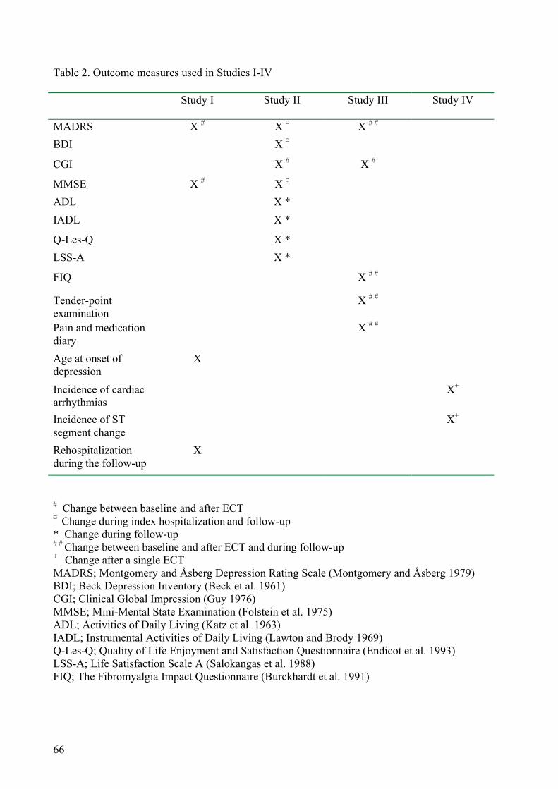
Table 2. Outcome measures used in Studies I-IV
Study I Study II
Study III Study IV
MADRS X # X ¤ X # # BDI X ¤
CGI X # X #
MMSE X # X ¤ ADL X * IADL X *
Q-Les-Q X * LSS-A X *
FIQ X # #
Tender-point examination
X # #
Pain and medication diary
X # #
Age at onset of depression
X
Incidence of cardiac arrhythmias
X+
Incidence of ST segment change
X+
Rehospitalization during the follow-up
X
# Change between baseline and after ECT ¤ Change during index hospitalization and follow-up
* Change during follow-up # # Change between baseline and after ECT and during follow-up + Change after a single ECT MADRS; Montgomery and Åsberg Depression Rating Scale (Montgomery and Åsberg 1979)BDI; Beck Depression Inventory (Beck et al. 1961) CGI; Clinical Global Impression (Guy 1976) MMSE; Mini-Mental State Examination (Folstein et al. 1975) ADL; Activities of Daily Living (Katz et al. 1963) IADL; Instrumental Activities of Daily Living (Lawton and Brody 1969) Q-Les-Q; Quality of Life Enjoyment and Satisfaction Questionnaire (Endicot et al. 1993) LSS-A; Life Satisfaction Scale A (Salokangas et al. 1988) FIQ; The Fibromyalgia Impact Questionnaire (Burckhardt et al. 1991)
66

3.2.2 ECT procedure
All the patients were treated at the ECT treatment unit in the Psychiatric Clinic of Tampere
University Hospital. The ECT-team consisted of an experienced staff including a treating
psychiatrist, an anesthesiologist, a treatment nurse and a recovery area nurse. The pre-ECT
evaluation according to the recommendations of the APA (1990, 2001) was carried out on all
patients by the treating psychiatrist (MH). The ECT was administered to all patients in these
studies by the same psychiatrist (MH), except for some isolated treatments (Maire Santala, Kaija
Huuhka).
ECT was administered 3 times a week with a brief-pulse, constant-current device
(Thymatron DGx, Somatics, Inc., Lake Bluff, IL). The initial stimulus dosage was adjusted with
the age method (Swartz and Abrams 1996) for all patients in Studies II and III. In Study I the
initial stimulus dosage was adjusted with the age method for 30 patients and with the stimulus
titration procedure (Sackeim et al. 1987, Sackeim et al. 1993) for 89 patients. In Study IV the age
method was used for 18 patients and the stimulus titration for 8 patients.
Anesthesia was induced with methohexital (except in 13 patients in Study IV with
propofol, because of the unavailability of methohexital at that time) and muscle relaxation with
succinylcholine. The initial dose was 1 mg/kg of methohexital (or 1.5 mg/kg of propofol) and 0.5
mg/kg of succinylcholine. If the seizure threshold was determined, patients were pretreated with
0.5 mg of atropine. At subsequent treatments the doses of anesthetic medications were adjusted
individually on clinical grounds.
The patients were ventilated with 100% oxygen until resumption of spontaneous
respiration. Physiological monitoring included pulse oximetry, blood pressure, ECG, 1-channel
67

electroencephalogram (EEG), and electromyography (EMG). The criteria for an adequate
generalized seizure duration was at least 20 seconds of motor response and 25 seconds EEG
seizure activity. During the treatment course of ECT, the dosage was adjusted upwards if needed
to maintain adequate seizure duration. In the Study III all the patients were treated with RUL
ECT and in Studies I, II and IV all the patients were treated with standard BL ECT.
The number of treatments in Studies I and II was based on the clinical judgement done of
the treating psychiatrist in the ECT unit together with the treating hospital ward. ECT was
continued until patients were free from symptoms or had received at least 8 treatments without
any further improvement being observed during the past 2 treatments. In Study I the number of
treatments ranged between 7 and 15, 9.4±1.8 (mean±SD) and in the Study II 5 and 12, 8.0±1.5.
In Study III the number of treatments was fixed at eight (with the exception of one patient with
seven treatments and another with three treatments because of excellent response). In Study IV
Holter monitoring was performed during the first ECT session.
3.2.3 Special features in different studies
3.2.3.1 Clinical assessment and APOE genotyping (Study I)
The clinical outcome measures are given in Table 2. The MADRS was used to assess the severity
of depression and the clinical change. Patients scoring less than 8 in MADRS after ECT were
considered to be responders and patients scoring more than 15 were considered to be non-
responders. Cognition was assessed in 78 patients using the MMSE. The patient’s age at first
onset of MDD was defined by using information of medical records and patient’s interview. The
age of 45 years was used as a cut-off between early and late onset depression.
68
Genomic DNA was extracted from peripheral blood leukocytes using a commercially
available kit (Qiagen Inc, Hilden, Germany) and DNA samples were then genotyped by
employing the 5’exonuclease assay (Livak 1999). For the APOE 112 genotyping, fluorogenic
allele-specific TaqMan probes and primers were used as described by Koch and coworkers
(2002). APOE 158 genotypes were, in turn, determined using primers and allele-specific
fluorogenic probes with conjugated minor groove binder group that were synthesized in
conjugation with Applied Biosystems (Foster City, CA, USA). Polymerase chain reaction (PCR)
mixture consisted of genomic DNA, 1 × Universal PCR Master Mix, 900 nM of each primer and
200 nM of each probe in a total reaction volume of 25 μl. Amplification was performed in 96-
well plates using the TaqMan Universal Thermal Cycling Protocol. After PCR, end-point
fluorescence intensity was measured by the ABI Prism 7000 Sequence Detection System
(Applied Biosystems, Foster City, CA, USA) and allelic discrimination performed. All
genotyping was performed blind to patient outcome. Negative and known control samples
previously typed by RFLP-PCR analysis were run in parallel with unknown DNA samples.
3.2.3.2 Clinical assessment of elderly patients in follow-up study (Study II)
The patients were assessed on admission, at discharge and after 12 months’ follow-up. The
change of MADRS in acute phase and rehospitalization in the follow-up were the primary
outcome measures. These and other measures used in this study are given in Table 2. The
functional capacity assessment and the quality of life evalutions were performed at disharge and
at month 12.
3.2.3.3 Clinical assessment in patients with concomitant MDD and fibromyalgia (Study III)
The baseline demographic and clinical data were assessed one week before the first ECT session
(Table 1). The patients were then followed up for three months after ECT. The follow-up
69

assessments were carried out one week, one month and three months after the last ECT session.
The MADRS and CGI were used to assess the severity of depression and the clinical change of
the patients (Table 2).
The physical symptoms of FM were evaluated by the Fibromyalgia Impact Questionnaire
(FIQ) (Table 2), a brief 10 item self-administered instrument (Burckhardt et al. 1991). The first
item of the FIQ focuses primarily on the patient’s ability to perform large muscle tasks and
contains 10 subitems. The next item asks the number of days in the past week that the patient felt
good. The next 2 items elicits the patient’s ability to work and the number of days they missed
work. The last 6 items measured by visual analog scales elicit pain, fatigue, morning tiredness,
stiffness, anxiety and depression.
Additionally, the tender-point examination using digital palpation with the force of 4 kg
(Wolfe et al. 1990) was performed. During the whole study the patients daily recorded their FM
pain and medication in the diary. The intensity of the pain was evaluated using a 6-point scale 0–
5 (0 = no pain, 5 = very severe pain). The mean values of the pain scores of the previous week
were calculated and used in the statistical analyses to represent the pain at the baseline and at the
follow-up visits.
3.2.3.4 Holter monitoring of patients treated with ECT (Study IV)
The outcome measure was the incidence of cardiac arrhythmias between pre and post treatment
(Table 2). Automated Holter monitoring (Del Mar model 461, Del Mar Avionics, Irvine, CA,
U.S.A.) was performed for 48 hours: for 24 hours before ECT, interrupted during the electrical
stimulation, and resumed immediately for 24 hours after the ECT. The incidence and number of
ECG changes were studied throughout this 48-hour period. The 24-hour post-ECT recording was
70

compared with that of the 24 hours pre-ECT, with each patient serving as his or her own control.
A separate comparison was performed between the records taken in the first hour after and the
last hour before ECT. Ventricular extrasystoles (VESs) were graded according to the
classification proposed by Lown and Wolf (1971). VESs occurring less often than 1/min were
regarded as isolated (even isolated VESs were counted), and others were regarded as frequent.
Ventricular tachycardia was defined as three or more consecutive ventricular ectopic beats at a
rate greater than 120/min, and supraventricular tachycardia (SVT) was defined as three or more
consecutive beats at a rate greater than 130/min. ST segment changes were analysed 60 msec
after the J-point. The Holter recordings were analysed blind to the pre-ECT and post-ECT
clinical status of the patients.
3.2.4 Statistical methods
The distributions of categorical variables between the different study groups were compared by
the Fisher exact test (I) or χ2-test (I, II). The difference in continuous clinical variables within
the patient groups before and after ECT was tested by paired t-test (I, II, III). Differences in
means for independent samples were tested using Student’s t test (II). One-way ANOVA was
used in determining differences in clinical variables between APOE genotype groups (I). Mean ±
SD were given (I, II, III). Pearson's correlation coefficient was used to find associations between
the MMSE score change and the number of ECT treatments and the age of the patients (I).
Comparisons of arrhythmias between pre-ECT and post-ECT periods were analysed using the
Wilcoxon matched-pairs signed ranks test (IV). This nonparametric test was used because the
variables were not normally distributed and the number of patients in the subgroups was small.
Medians (ranges) were given (IV).
71

A linear regression analysis was used to explain the correlation between the FIQ items,
tender point counts, self-assessed pain and MADRS (III) and the change of MADRS score by the
variables, which had statistically significant differences between the groups at baseline (II). The
Cox regression analysis was used to create a model to explain rehospitalization by age, length of
current period of depression, MADRS scores at discharge, somatic illnesses (5 variables) and the
explaining variables used in the linear regression analysis (II). These analyses were stratified by
“group” (II). The Kaplan-Meier method was used to test differences in rehospitalizations
between the groups (II) and the association between age at onset and APOE polymorphism (I).
The statistical analysis was carried out using SPSS/Win (Versions 10.0 (II), 11 (I) and
11.0.1 (III, IV), SPSS Inc, Chicago, IL). The limit for statistical significance was set equal to
0.05.
The study protocol was approved by the Ethics Committee of Tampere University Hospital
according to the principles of the Helsinki Declaration. All participants gave written informed
consent.
72

4 Results
4.1 Apolipoprotein E polymorphism and response to ECT (I)
In the whole group, the pre-ECT values of the MADRS score decreased from 32.5±8.2
(mean±SD) to 11.3±8.8 after ECT treatment (mean±SD, p<0.0001). Using the cut-off points < 8
for responders and > 15 for non-responders in the MADRS, 45 out of a total of 119 patients were
considered responders and 32 non-responders.
No association was found between APOE genotype and treatment response to ECT. APOE
alleles ε2, ε3, and ε4 were equally distributed between responders and non-responders (p=0.23,
p=0.64, and p=0.69 respectively). However, in non-psychotic patients ε2 allele tended to be more
frequent in responders (5/26) than in non-responders (0/18) (p=0.07).
APOE alleles ε2, ε3, and ε4 were equally distributed between patients and controls
(p=0.15, p=0.60, and p=0.60) respectively. However, in men ε3 allele was more frequent in
patients than in controls (p=0.03). In women, ε4 allele tended to be more frequent in patients vs.
controls (p=0.09).
An earlier onset of depression was observed in the patients with ε4 allele, but only in the
late onset depression group (p=0.0495). In the whole group there was no association between the
APOE genotypes and the change of the MMSE scores during ECT (p=0.16). In women,
however, there was a trend for APOE ε4 alleles to increase dose-dependently (groups having 0, 1
or 2 ε4 alleles respectively) the risk of lowered MMSE score (p=0.06). In addition, in women
APOE ε2 allele was associated with better or equal MMSE scores after the treatment course than
at baseline (p=0.04).
73

4.2 Outcome of elderly patients with MDD (II)
4.2.1 Acute efficacy
When elderly patients with MDD were treated even with ECT or with antidepressants the
MADRS total scores diminished during index hospitalization treatment within both groups: from
31.6±8.5 (mean±SD) to 8.1±6.0 in the ECT group (p<0.001), and from 28.5±5.4 to 13.4±10.6 in
the ADT group (p<0.001). There was no significant difference in MADRS scores between the
groups at admission. At discharge, however, this difference was significant (p=0.02).
Accordingly BDI scores decreased from 29.4±10.3 to 10.6±9.9 in the ECT group (p<0.001) and
from 28.9±6.6 to 17.1±14.1 in the ADT group (p=0.003).
At admission, according to CGI, the patients in the ECT group were more severely ill than
patients in the ADT group (p=0.002). After treatment 26 out of 30 patients in the ECT group and
11 out of 21 patients in the ADT group were much or very much improved according to the CGI
change scale (p=0.007).
MMSE scores diminished in the ECT group from 27.2±2.0 to 26.3±3.6 and increased in the
ADT group from 27.7±1.8 to 28.7±1.5; the difference between the groups at discharge was
significant (p=0.01).
4.2.2 Outcome in 12-month follow-up
Twenty-one out of a total 51 patients were rehospitalized. Four patients died (2 in each group)
during the 12-month follow-up. Five patients (3 in the ECT group and 2 in the ADT group)
declined to undergo the follow-up assessment. Thus, in total 21 patients were followed up for 12
months: 12 in the ECT group and 9 in the ADT group.
74

Thirteen out of 30 patients (43%) in the ECT group and 8 of 21 patients (38%) in the ADT
group were rehospitalized during the follow-up (p=0.71). Six out of 13 patients in the ECT group
and 1 out of 8 patients in the ADT group were rehospitalized during the first month after
discharge. The mean time before rehospitalization was 8.33 (95% CI: 6.68-9.98) months for the
ECT group and 9.14 (95% CI: 7.40-10.89) months for the ADT group (p=0.67). Hospitalization
during the 12 months before the index episode significantly predicted rehospitalization (Odds
ratio 4.00, 95% CI: 1.09-14.73, p=0.04).
There was a significant increase in the MMSE scores in the ECT group from discharge to
12-month assessment (p<0.001). In the ADT group there was no significant change in the
MMSE scores. No differences were found within the ECT or within the ADT group in functional
capacity and the quality of life assessments between discharge and 12-month follow-up (Study
II, Table 3). At discharge the IADL scores were better in the ECT group compared to the ADT
group (p=0.05). After 12-month follow-up there were no differences in any assessments of
functional capacity or the quality of life between the groups (Study II, Table 3).
4.3 Outcome in patients with concomitant MDD and fibromyalgia (III)
4.3.1 Acute efficacy
There was a significant improvement in MADRS and in those FIQ items, which may reflect
mood i.e., "feel good", "fatigue", "anxiety" and "depression" (Table 3). The MADRS total score
diminished from 27.17±5.01 (mean±SD) to 13.17±10.54 (p<0.001). According the CGI, six out
of 13 patients were "much or very much improved", four patients "minimally improved", while
there were three "no change" patients. There was no significant change in the number of tender-
point count and in the daily self-reported pain score (Table 3).
75

4.3.2 Outcome in 3-month follow-up
There was no change during the three-month follow-up in those parameters which had improved
after ECT: MADRS and FIQ items "feel good", "fatigue", "anxiety" and "depression" (Table 3).
4.4 Cardiac arrhythmias induced by ECT (IV)
The numbers of ventricular arrhythmias are presented in Table 4. There was a significant
increase in the incidence of bigeminy/trigeminy (p=0.02), but not in the other VES variables in
the 24-hour post-ECT recording when compared with the pre-ECT recording (p=0.22). There
were no differences in ventricular arrhythmias between 1 hour post-ECT and pre-ECT recordings
(p=0.18). Supraventricular tachycardias (SVTs) were more common in the post-ECT than in the
pre-ECT 24-hour Holter analysis (p=0.004) (Table 4). There was no significant difference in the
incidence of SVTs between the 1hour period after and before the ECT (p=0.16). In
76

supraventricular extrasystoles (SVES) no differences were found between the post-ECT and pre-
ECT recordings at either 24-hour or 1-hour intervals (Table 4).
Nine patients had an ST change of more than 1 mm, and five of them had an ST change of
more than 2 mm in the 1 hour post-ECT recording. Only two of them, however, reached their
maximal ST change during the first hour after treatment.
77

78

5 Discussion 5.1 Main findings
According to the present results APOE polymorphism was not associated with the treatment
response to ECT in major depression. Elderly patients with MDD had a good acute response to
ECT, however, relapses were frequent. ECT was also effective in the treatment of depressive
symptoms of the patients with concomitant MDD and FM, but had no effect on their pain
symptoms. In addition ECT increased the frequency of bigeminy/trigeminy and supraventricular
tachycardia and caused ST changes.
5.2 Study populations
The patients in the present studies included a representative sample of patients with a depression
severe enough to warrant ECT. More than 40% of the whole population had psychotic features
and as many even comorbid physical diseases. All the patients, except for 10 outpatients in Study
III, were hospitalized for severe acute MDD (and five for MDE of bipolar disorder in Study IV).
All of the patients who fulfilled the actual inclusion criteria and agreed to participate gave their
written consent. Only few patients declined. The excellent compliance of the patients may partly
be based on careful patient selection. In Study IV five patients were excluded because of
technical problems with the Holter recording.
All the patients in these studies were clinically carefully assessed. Most of them (except
those in Study III) were severely depressed and the reason for ECT in all cases was treatment
resistance to antidepressants.
79

5.3 Study methods
General evaluation of outcome for all the study population was performed according to the
recommendations of APA (1990, 2001).
5.3.1 ECT treatment
BL ECT was chosen because this method is considered to be the most effective strategy even
though it may induce cognitive disturbances more than would RUL ECT. However, it has been
shown that these cognitive dysfunctions are reversible (Calev et al. 1991b, Sackeim et al. 1993,
McCall et al. 2002) and that patients with poor response are more likely to report subjective
memory deficit after ECT than patients with a good response (Prudic et al. 2000). It has even
been suggested that there is no justification for favoring RUL ECT over BL ECT when treating
depressive patients (Fink et al. 2001). In addition, earlier rehospitalizations in patients treated
with RUL ECT compared to those with BL ECT have been reported (Little et al. 2004). In Study
III the RUL ECT was used because most of these patients were younger outpatients and less
depressed than the patients in the other studies. Prejudice against the cognitive side-effects of
ECT may have influenced the recruitment of patients in this study.
The initial stimulus dosage was adjusted with the age method (Swartz and Abrams 1996)
for all patients in Studies II and III. In Study I the initial stimulus dosage was adjusted with the
age method for 30 patients and with the stimulus titration procedure (Sackeim et al. 1987,
Sackeim et al. 1993) for 89 patients. In Study IV the age method was used in 18 patients and the
stimulus titration in 8 patients.
The dose titration method has been recommended because it is the most precise method for
the individual selection of ECT stimulus dose relative to the ST (Sackeim et al. 1987, Heikman
80

et al. 1999, APA 2001). The antidepressive effect of RUL ECT increases when the stimulus
dosage is increased in relation to the seizure threshold. However, the optimal dosage level may
still be unknown (Fink et al. 2001, Abrams 2002). However, the value of the dose-titration
method has also been criticized (Abrams 2002) and some practitioners still prefer the pre-
selected stimulus dose method (Petrides and Fink 1996, Swartz and Abrams 1996). The
Thymatron DGx ECT device user’s manual recommends a preselected dosing strategy based on
the age of the patient in which the dose (percentage of energy which is equal to the percentage of
maximum output charge) is adjusted according to the patient’s age in decades (Swartz and
Abrams 1996). However, it is a relatively rough method and some patients may thereby receive a
too low or a markedly suprathreshold stimulus dose. On the other hand, the dose-titration method
requires multiple stimulations, which may increase the risk of cardiovascular adverse events,
such as bradyarrhythmias and even asystole (Petrides and Fink 1996, Burd and Kettl 1998). In
the present series of studies most of the patients were elderly with various somatic diseases,
including cardiovascular disease, and therefore the age method was chosen for some studies (II,
III) and patients (I, IV).
5.3.2 Rating scales used in the present studies
The MADRS was chosen because it has been approved as a reliable and valid instrument to
evaluate the severity and the change of depressive symptoms (Davidson et al. 1986). The
sensitivity and specifity of the MADRS has been found to be good and it has been considered as
a suitable instrument even in the trials of management of older depressive patients (Mottram et
al. 2000). The BDI is a widely used self-administered scale in simple language and easy to score
(Yonkers and Samson 2000).
81

CGI is widely used in psychopharmacological clinical trials. It allows the clinician to rate
the severity of illness, the change over time and the efficacy of treatment. It is a rough but simple
and fast measure (Leon et al. 1993).
MMSE is a brief easily administered cognitive status examination. It is widely used in
clinical practice and in studies. MMSE has been shown to be sensitive in assessing the severity
of cognitive impairment and changes occurring over time. The sensitivity of this scale may still
not be high enough for detecting the minor changes in cognition (Tombaugh and McIntyre
1992).
Functional capacity (Study II) was assessed with the ADL and the IADL. The ADL is
intended to assess selfcare functions in chronically ill and aging populations. Because it attains
only the lower end of selfcare function, it may not be sensitive when functioning is normal or
nearly normal (Rubin et al. 1993). The IADL measures performance on everyday tasks
instrumental living in the community (Lehman et al. 2000).
The Q-Les-Q is a quality of life instrument that has been shown to be sensitive to change
following the treatment for depression. It has been approved as reliable and valid in its
psychometric properties (Ferrans and Powers 1992). The LSS-A is a 26-item scale developed to
assess life satisfaction in elderly Finnish people. It is easy to use and a reliable and valid scale for
measuring life satisfaction (Salokangas et al. 1988).
The Fibromyalgia Impact Questionnaire (FIQ) was designed to evaluate physical
functioning ability and was used to evaluate the severity and change of the FM symptoms (Study
82

III). It has been proven to be a reliable and valid instrument in both research and clinical
situations (Burckhardt et al. 1991).
5.3.3 APOE genotyping
The 5' nuclease assay has been successfully used to discriminate alleles that differ by a single
base substitution (Livak 1999). TaqMan assays allow for fast and sensitive genotyping and are
especially suitable for studies including large numbers of participants (Koch et al. 2002). A
standard method of analysis has been developed that enables automated genotype determination.
Applications of this assay have included typing a number of polymorphisms e.g. in human drug
metabolism genes (Livak 1999).
5.3.4 Holter monitoring
Most of the arrhythmias induced by ECT have been reported to occur during the ictal and briefly
post-ictal period (Zielinski et al. 1993, Zvara et al. 1997). However, even delayed arrhythmias
are possible. Holter monitoring enables a continuous long lasting recording of heart rhythm
during normal activity. It is a reliable method for determining cardiac arrhythmias and ST-
segment changes.
5.4 Apolipoprotein E polymorphism is not associated with response to ECT (I)
APOE has been found to be a major risk factor of Alzheimer’s disease (Strittmatter et al. 1993),
and may be associated with cerebrovascular diseases (Lavretsky et al. 2000). The polymorphism
of APOE has been previously connected to response of antidepressants (Murphy et al. 2003) and
ECT (Fisman et al. 2001).
83

In the present study no association was found between APOE genotype and the treatment
response to ECT according to MADRS. APOE alleles ε2, ε3, and ε4 were equally distributed
between responders and non-responders. Thus, the findings of Fisman et al. (2001) that the
patients with ε4 allele were more likely to respond to ECT in late-onset depression than patients
with other genotypes could not be replicated here. In the present study the definition of response
to ECT was stricter (responders < 8 and nonresponders > 15) than in the study by Fisman et al.
(2001) who used only one cut-off point (10) in the MADRS defining response or non-response.
Even when further analysing the data using the same cut-off point as Fisman, no significant
association was found in the present study group (data not shown).
Moreover, no difference was found in APOE allele distribution between the present
patients and controls. In men, however, ε3 allele was more frequent in patients than in controls.
In women, ε4 allele tended to be more frequent in patients vs. controls. The clinical significance
of these findings is obscure. In some studies the increased APOE ε4 allele frequency has been
associated with late-life depression (Krishnan et al. 1996, Zubenko et al. 1996, Rigaud et al.
2001).
In the present study earlier onset of depression was found in the patients with ε4 allele than
patients without this allele, but only in patients with late-onset depression. An earlier onset of
depression in ε4 carriers has also been reported by Lavretsky et al. (2000) and Butters et al.
(2003). The latter also found that age at onset of late-life depression was significantly reduced in
APOE ε4 carriers, which is in line with the present findings. In contrast, Fisman and others
(2001) found a trend towards later onset of depression in patients with ε4 allele. This finding
may be difficult to interpret, but the overall study group was small and the ε4 infrequent.
84

In the whole group there was no association between the APOE genotypes and the change
of the MMSE scores during ECT. However, in the present group of women, APOE ε2 could be
protective against cognitive impairment, whereas APOE ε4 may increase the risk of cognitive
impairment. These findings, however, are preliminary and should be replicated before further
conclusions are drawn.
5.5 ECT is effective in acute treatment of elderly patients with MDD, but relapses
are common (II)
5.5.1 Acute efficacy
Acute efficacy of ECT and antidepressive therapy in elderly patients with MDD was good. In the
present study the acute efficacy in the ECT group was clearly better than in the ADT group,
despite the fact that the patients in the ECT group may have been more severely ill than those in
the ADT group. The acute efficacy of ECT in late-life depression has been shown in several
previous studies (Mulsant et al. 1991, Black et al. 1993, Flint and Rifat 1998, O'Connor et al.
2001, Petrides et al. 2001) which are thus in line with the present findings.
In the ECT group there seemed to be a higher proportion (10/30) of patients with psychotic
depression than in the ADT group (3/21). The present results might be in line with the previous
ones, where psychotic depressions have responded to ECT even better than nonpsychotic
depression (Petrides et al. 2001, Birkenhager et al. 2003, Kho et al. 2003). Many practitioners
consider ECT to be a first-line treatment in psychotic major depression in the elderly
(Alexopoulos et al. 2004).
The MMSE scores decreased in the ECT group and rose in the ADT group. Thus, at
disharge the MMSE scores were higher in the ADT group than in the ECT group. This is in line
85
with the previous reports where the cognitive adverse effects induced by ECT have been
common (Sackeim et al. 1993, McCall et al. 2002).
5.5.2 Long-term outcome
In the present study the 1-year rehospitalization rate of the patients was relatively high in both
groups (around 40%), but there were no significant differences between the groups. According to
earlier follow-up studies the risk of relapse is associated with residual symptoms i.e. partial
remission (Pintor et al. 2003, Steffens et al. 2003a, Pintor et al. 2004). Pintor et al. (2003)
reported that 68% of patients with partial remission after ADT relapsed during 24-month follow-
up, whereas only 15% of patients with complete remission. In the present study groups, however,
the relapse rates were similar despite the fact that the patients in the ADT group had more
depressive symptoms than those in the ECT group at disharge (MADRS 13.4 vs. 8.1). In the
ADT group the patients continued to have the same medication which was originally effective in
acute treatment in contrast to patients in the ECT group where the continuation ECT was not
used.
A high relapse rate after successful ECT has been demonstrated in several clinical trials
and patients given ECT in a community setting (Sackeim et al. 2000, Sackeim et al. 2001, Prudic
et al. 2004). In a study by Sackeim et al. (2000) 53% patients with MDE responding to ECT
relapsed within one year, 94% of the relapses occurred during the first 6 months. Birkenhager et
al. (2004) reported a 6-month relapse rate of 28% and 12-month relapse rate of 41%. Prudic et al.
(2004) reported that 64.3% of the patients given ECT in a community setting had relapsed within
24-week after ECT. Stoudemire and others (1991) found that 28% of ECT treated patients and
25% of ADT treated patients were rehospitalized within 1 year. Psychopharmacological
treatment (and in some cases even continuation ECT) is essential to prevent early relapses after
86

ECT. Sackeim et al. (2001) found that 84% of patients had relapsed within six months when
treated with placebo after ECT. Grunhaus et al. (2001) reported that 28.5% of patients relapsed
within 3 months after a successful course of ECT despite subsequent pharmacotherapy with
fluoxetine. The findings in the present study replicate the previous reports showing a high
relapse rate in patients with MDD treated with ECT and/or antidepressant therapy. However, the
relapse rate may be still underestimated in this study because of the strict definition of relapse
judged by readmission.
There was a significant increase in the MMSE scores in the ECT group from disharge up to
12-month assessment in the present study. In 12 months the MMSE scores exceeded the baseline
scores. This finding is in accordance with earlier studies (Calev et al.1991b, Sackeim et al. 1993,
McCall et al. 2002). There were no significant changes between disharge and 12-month follow-
up in the depression rating scales, functional capacity assessment or quality of life assessment
within either the ECT or the ADT group. Moreover, there were no significant differences in
these clinical variables between the groups at the 12-month assessment.
5.6 Depression but not pain symptoms improved by ECT in patients with
concomitant MDD and fibromyalgia (III)
In patients who had concomitant MDD and FM there was a significant improvement after ECT
in depressive symptoms. However, no change was found in pain related FIQ items, self-reported
pain severity and tender-point count. The improvement in those parameters with a statistically
significant change after ECT (MADRS and FIQ items, "feel good", "fatigue", "anxiety" and
"depression") was maintained for the whole follow-up period of three months. These findings are
in line with previous suggestions that depression is independent of the pain symptoms of FM.
Okifuji et al. (2000) found that there were no differences in severity of pain, number of positive
87
tender points, and pain intensity of tender points in 39 depressive and 30 nondepressive FM
patients. Thus, the authors suggest that improvement of depression does not lead directly to
improvement of pain.
The prognosis of FM has been reported to be poor (Henriksson 1994, Wolfe et al. 1997). In
a 7-year follow-up by Wolfe et al. (1997) it was found that the symptoms of FM did not change
over time. The illness in the present patients also tented to be chronic. The mean duration of FM
had been 6.1 years. All the patients had several previous or present antidepressant medications
during their illness.
It has been shown that antidepressants are moderately effective in FM symptoms (O'Malley
et al. 2000, Goldenberg et al. 2004, Samborski et al. 2004). The best symptomatic benefits are
reported in fatigue, sleep and overall well-being as well as in pain severity (O'Malley et al.
2000). In the present patients fatigue and well-being also improved, but no change in pain was
noted. The present finding of no change in trigger-point tenderness but improvement in
depression is in line with some reports where antidepressants relieved depression but had no
effect on trigger points (Nicassio et al. 2000, Arnold et al. 2002).
The PET studies have shown decreased CBF in thalamus in patients with chronic
neuropathic pain (Di Piero et al. 1991, Iadarola et al. 1995). Di Piero et al. (1991) concluded that
chronic pain results in a decrease of synaptic activity in the thalamus. Fukui et al. (2002)
investigated the mechanism of the analgesic effect of ECT in patients with CRPS. They found in
a Single-Photon Emission Tomography that patients with CRPS had reduced ipsilateral regional
CBF in the thalamus and pain relief after ECT was associated with increase of regional CBF.
88

The authors suggested that the analgesic efficacy of ECT in chronic pain is related to the
normalization of the balance of regional cerebral blood flow in the thalamus.
In the abovementioned study by Fukui et al. (2002) BL ECT was used. It has been
speculated that BL ECT may be more effective than RUL ECT in chronic pain (King and Nuss
1993, McDaniel 2003). Electrode placement may thus partly explain the lack of efficacy of ECT
on pain symptoms in the present study. The patients were treated with RUL ECT to avoid
cognitive adverse effects. The number of treatments was 8 in 11 patients, 7 in one patient and 3
in one patient. Because the primary reason for ECT was depression and not the symptoms of FM,
ECT was continued until depression had been relieved or the patient had received at least 8
treatments without any further improvement being observed during the past 2 treatments. It is
unknown if an increase in the number of treatments would have improved the efficacy in pain
relief.
5.7 ECT increases cardiac arrhythmias in patients with pre-existing arrhythmias
(IV)
In this study, the influence of ECT on the incidence of arrhythmias and ST changes was
observed in elderly depressed inpatients using a 24-hour pre- and post-ECT Holter recording.
The major finding was an increased incidence of bigeminy/trigeminy and SVTs in the 24-hour
post-ECT recording when compared with the pre-ECT recording. Interestingly, this increase was
not found between 1-hour post-and pre-ECT recordings.
There are only a few previous studies using long-time Holter monitoring to investigate
cardiac arrhythmias induced by ECT (Troup et al. 1978, Zvara et al. 1997, Rumi et al. 2002).
Troup et al. (1978) performed a 24-hour Holter monitoring in young and middle-aged physically
89

healthy patients before, during and after ECT. They found no significant difference in the
frequency of premature ventricular or supraventricular contractions between the pre- and post-
ECT recordings. Rumi et al. (2002) performed a 24-hour Holter recording in 47 young patients
without cardiac disease. They found some isolated VES and SVES, but not other arrhythmias.
Zvara et al. (1997) performed Holter monitoring in 19 elderly patients for at least 2 hours after
two ECT sessions. They found a high incidence of ST-segment depression and premature
ventricular contractions during ECT and postictal period. In a recent study by Rasmussen et al.
(2004) no significant differences were found in ventricular or supraventricular arrhythmias in
medically healthy patients between post- and pre-ECT Holter recordings. However, their 3-hour
pre- ECT recordings were performed a day before ECT and 3-hour post-ECT recordings from
several hours up to 3 days after ECT. Obvious differences in the results compared to the present
ones may be due to different patient population and schedule.
Arrhythmias associated with ECT are relatively common findings, most of them being
premature atrial and ventricular contractions (Troup et al. 1978, Gerring and Shields 1982,
Mokriski et al. 1992, Zvara et al. 1997, McCully et al. 2003). In the present study it was also
found that pre-existing arrhythmias predict the development of post-ECT arrhythmias, which is
in line with several previous studies (Gerring and Shields 1982, Dec et al. 1985, Zielinski et al.
1993).
In contrast to the studies by Troup et al. (1978) and Rumi et al. (2002), an increase of some
ventricular and supraventricular arrhythmias was found after ECT in the present study. The
patients in the present study were old and physically ill, whereas the patients in the above
mentioned studies were young or middle-aged and physically healthy.
90

Consistent with earlier findings (Zielinski et al. 1993, Zvara et al. 1997, Rumi et al. 2002),
a high incidence (in nine out of 26 patients) of ST changes after ECT was found in the present
study. Only two of these patients with ST changes had a pre-existing cardiovascular disease (one
hypertension and one had a ischemic heart disease). The cardiac effects of ECT may be a direct
consequence of hypoxia during seizure, or a disturbance in central regulation (Mokriski et al.
1992, Robinson and Lighthall 2004). The clinical significance of ECT related cardiac side-
effects depends on their quality as well as on the age and possible concomitant diseases of the
patient.
5.8 Limitations of the study
Several limitations of the present studies should be noted. The main limitation in all the present
studies was relatively small sample size. In Study I the original number of patients was
moderate. However, in subanalyses the groups were small, which may increase the risk of false
negative findings (type II error). In Study II the initial number of patients in each treatment
group was reasonable enough, but during the follow-up there were some drop-outs. In Study III
it was more difficult than expected to recruit patients and the final number remained relatively
small. However, there were no drop-outs during the whole study period. In Study IV the Holter
recording failed for five patients who could therefore not be included in the analyses.
Another limitation of these studies was also that no formal structural clinical interview for
DSM-IV (SCID) was used and that the diagnostic interviews were conducted by only one
clinician. Moreover, the outcome evaluations were not blinded.
In addition, in Study II the patients were not randomly assigned to ECT and ADT groups,
which may have caused a selection bias. In the ECT group the patients were slightly more
91

severely depressed than in the ADT group. Thus the comparison between the groups may be
questionable. Another confounding factor is that the responders in the ECT group in follow-up
were switched to the antidepressant drug treatment, which had not previously helped them,
whereas in responders to pharmacotherapy the effective treatment was continued. Moreover, a
limitation in Study II was that the functional capacity assessments and the quality of life
evaluations were not performed at baseline and thus the effect of treatments in the index episode
could not been evaluated.
In Study III patients were given max. 8 RUL ECT. On the basis of the present study
procedure it is not possible to estimate whether BL ECT or a large number of treatments would
have been more efficacious for pain symptoms.
One limitation is also the variability in stimulus dosing strategy, age method versus dose
titration method, in Studies I and IV. Other limitations in Study IV were that the affects of
cardiovascular or other physical diseases, as well as medications could not be evaluated because
of small subgroups. Moreover, the limitation in this study was that anesthesia was induced either
with methohexital or with propofol, because of the unavailability of methohexital at that time.
Because the patients were not randomised concerning to the anesthetics, no comparison was
made.
92
6 Summary
The main findings of the study were:
Study I
APO-E polymorphism was not associated with the treatment response to ECT in major
depression.
Study II
Elderly patients with MDD had a good acute response to both ECT and ADT. Relapses,
however, were frequent in both groups and many of them occurred soon after discharge.
Study III
ECT is an effective treatment in depression of patients with MDD and concomitant FM.
However, it has no effect on the pain symptoms of these patients.
Study IV
ECT caused a significant increase in bigeminy/trigeminy and supraventricular tachycardia in
elderly patients. Pre-ECT arrhythmias predicted post-ECT arrhythmias. Moreover, ECT caused
ST changes in some patients. Although arrhythmias were common all the patients completed the
ECT course.
93

94

7 Conclusions and future implications
In accordance with earlier reports the present results indicate that ECT is an effective treatment
method in elderly patients with MDD. In spite of continuation pharmacotherapy relapses were
common and in many patients occurred soon after treatment. The high relapse rate emphasizes
the need for careful follow-up of elderly patients who have recovered from a depressive episode.
In some cases the continuation ECT should also be considered to prevent early relapses.
ECT is a safe treatment even in elderly patients with somatic illness, although it may
increase the risk of some cardiac arrhythmias. Arrhythmias caused by ECT are usually clinically
inconsequential and do not obstruct the course of ECT. However, especially in elderly patients,
the cardiovascular status should be carefully assessed before ECT.
There is only little data of the effectiveness of ECT in certain special groups such as pain
patients. Especially patients suffering from FM often also have depressive symptoms in addition
to pain and other somatic symptoms. According to the present study ECT is an effective
treatment of depression in these patients, but it has no effect on their pain symptoms. However,
on the basis of some reports, studies on the effectiveness of the ECT in pain patients should be
continued.
According to the present study the APOE polymorphism is not associated with the response
to ECT in MDD. In spite of this, studies on connections between genetics and treatment response
to ECT should be continued in order to find the possible genetic polymorphisms, which may
predict the response. Our group continues to study other gene polymorphisms which may be
associated with the outcome of ECT.
95

8 Acknowledgements
These studies were carried out in the Department of Psychiatry in Tampere University Hospital
during the years 1997-2003.
I owe my most heartfelt gratitude to my supervisor, Professor Esa Leinonen, MD, PhD. I
specially thank him for his never-ending patience and the time he devoted to advising and
helping me with this work. He inspired my interest in ECT and introduced me to the world of
scientific research. He always believed in me and in my work even in those days when I did not.
Without his encouragement and unfailing support throughout these years this dissertation would
never been completed.
I am very grateful to Pertti Heikman MD, PhD and Professor Jukka Hintikka, MD, PhD,
the official reviewers of this dissertation, for their constructive criticism and valuable advice
concerning the manuscript.
I would like to express my cordial thanks to Sami Anttila, MD, PhD, for his collaboration
and his contribution to this work. He also helped me a lot with the practical matters at the end of
this work. I am very grateful to Maija Haanpää, MD, PhD, for her collaboration, which has been
crucially important for this work. I also express my deepest gratitude to Lauri Seinelä MD, PhD,
and the late Pekka Reinikainen MD, for their expert collaboration. I also want to gratefully
acknowledge my other co-authors Professor Terho Lehtimäki MD, PhD, Kari M. Mattila PhD,
and Riikka Rontu PhD. I am fortunate to have these skilled co-workers who have shared their
knowledge with me.
96

I am grateful to Vesa Laukkanen MSc, Riina Haataja MSc and Heini Huhtala MSc, for
their help in the statistical analyses. I wish to thank Virginia Mattila MA for revising the English
language of the original articles and the text of the manuscripts of this dissertation.
Sincere thanks are due to Maire Santala MD, Anne Salonen RN, and Lea Korpisammal
RN, for their valuable help and collaboration. I am also thankful to my other colleagues and the
entire personnel at the Department of Psychiatry.
I also express my deepest appreciation to all the patients who participated in these studies.
Finally, I wish to express my most loving thanks to my dear wife, Kaija, for her love,
understanding and devoted support during these years. As my colleague and co-author she was
able to share the difficulties and joys in the field of research. To my children and relatives I want
to say thank you for reminding me that there is so much more to life than work and research.
This work was financially supported by the Medical Research Fund of Tampere University
Hospital.
Tampere, September 2005
Martti Huuhka
97

9 References
Aarsland D, Larsen JP, Waage O, Langeveld JH (1997): Maintenance electroconvulsive therapy for Parkinson's disease. Convuls Ther 13:274-277. Abrams R, Taylor MA, Faber R, Ts'o TO, Williams RA, Almy G (1983): Bilateral versus unilateral electroconvulsive therapy: efficacy in melancholia. Am J Psychiatry 140:463-465. Abrams R (2002): Stimulus titration and ECT dosing. J ECT 18:3-9. Abkevich V, Camp NJ, Hensel CH, Neff CD, Russell DL, Hughes DC, Plenk AM, Lowry MR, Richards RL, Carter C, Frech GC, Stone S, Rowe K, Chau CA, Cortado K, Hunt A, Luce K, O'Neil G, Poarch J, Potter J, Poulsen GH, Saxton H, Bernat-Sestak M, Thompson V, Gutin A, Skolnick MH, Shattuck D, Cannon-Albright L (2003): Predisposition locus for major depression
at chromosome 12q22-12q23.2. Am J Hum Genet 73:1271-1281. Achte KA (1967): On prognosis and rehabilitation in schizophrenic and paranoid psychoses. Acta Psychiatr Scand 43 (suppl 196):41-43. Adam LA, Crowe RR (2003): Use of ECT in idiopathic intracranial hypertension. J ECT 19:234-237. Adler GK, Manfredsdottir VF, Creskoff (2002): Neuroendocrine abnormalities in fibromyalgia. Curr Pain Headache Rep 6:289-298. Alexopoulos GS (2005): Depression in the elderly. Lancet 365:1961-1970. Alexopoulos GS, Meyers BS, Young RC, Campbell S, Silbersweig D, Charlson M (1997): 'Vascular depression' hypothesis. Arch Gen Psychiatry 54:915-922. Alexopoulos GS, Kiosses DN, Choi SJ, Murphy CF, Lim KO (2002): Frontal white matter microstructure and treatment response of late-life depression: a preliminary study. Am J Psychiatry 159:1929-1932. Alexopoulos GS, Streim J, Carpenter D, Docherty JP; Expert Consensus Panel for Using Antipsychotic Drugs in Older Patients (2004): Using antipsychotic agents in older patients. J Clin Psychiatry 65 (Suppl 2):5-99. Altar CA, Laeng P, Jurata LW, Brockman JA, Lemire A, Bullard J, Bukhman YV, Young TA, Charles V, Palfreyman MG (2004): Electroconvulsive seizures regulate gene expression of distinct neurotrophic signaling pathways. J Neurosci 24:2667-2677. American Psychiatric Association (1990): The Practice of Electroconvulsive Therapy: Recommendations for treatment, training and privileging: A Task Force Report of the American Psychiatric Association. American Psychiatric Press. Washington, DC. American Psychiatric Association (1994): Diagnostic and statistical manual of mental disorders, (4th edn.) American Psychiatric Press. Washington, DC.
98

American Psychiatric Association (2000): Practice guideline for the treatment of patients with major depressive disorder. In: Practice guideline for the treatment of psychiatric disorders. Compendium 2000, 1 Edition. pp. st 413-495. American Psychiatric Association. Washington, DC. American Psychiatric Association (2001): The practice of electroconvulsive therapy: Recommendations for treatment, training and privileging: A Task Force Report of the American Psychiatric Association. American Psychiatric Press. Washington, DC. Anderberg UM, Marteinsdottir I, von Knorring L (2000): Citalopram in patients with fibromyalgia a randomized, double-blind, placebo-controlled study. Eur J Pain 4:27-35. Anderson RJ, Freedland KE, Clouse RE, Lustman PJ (2001): The prevalence of comorbid depression in adults with diabetes: a meta-analysis. Diabetes Care 24:1069-1078. Andrade C, Kurinji S (2002): Continuation and maintenance ECT: a review of recent research. J ECT 18:149-158. Arnold LM, Keck Jr, PE, Welge JA (2000): Antidepressant treatment of fibromylagia. A meta-analysis and review. Psychosomatics 41:104-113. Arnold LM, Hess EV, Hudson JI, Welge JA, Berno SE, Keck PE (2002): A randomised, placebo-controlled, double-blind, flexible-dose study of fluoxetine in the treatment of women with fibromyalgia. Am J Med 112:191-197. Arnold LM, Hudson JI, Hess EV, Ware AE, Fritz DA, Auchenbach MB, Starck LO, Keck PE Jr (2004a): Family study of fibromyalgia. Arthritis Rheum 50:944-952. Arnold LM, Lu Y, Crofford LJ, Wohlreich M, Detke MJ, Iyengar S, Goldstein DJ (2004b): A double-blind, multicenter trial comparing duloxetine with placebo in the treatment of fibromyalgia patients with or without major depressive disorder. Arthritis Rheum 50:2974-2984. Austin MC, Whitehead RE, Edgar CL, Janosky JE, Lewis DA (2002): Localized decrease in serotonin transporter-immunoreactive axons in the prefrontal cortex of depressed subjects committing suicide. Neuroscience 114:807-815. Aydemir O, Deveci A, Taneli F (2005): The effect of chronic antidepressant treatment on serum brain-derived neurotrophic factor levels in depressed patients: a preliminary study. Prog Neuropsychopharmacol Biol Psychiatry 29:261-265. Bader GM, Silk KR, Dequardo JR, Tandon R (1995): Electroconvulsive therapy and intracranial aneurysm. Convuls Ther 11:139-143. Bailine SH, Rifkin A, Kayne E, Selzer JA, Vital-Herne J, Blieka M, Pollack S (2000): Comparison of bifrontal and bitemporal ECT for major depression. Am J Psychiatry 157:121-123. Bakewell CJ, Russo J, Tanner C, Avery DH, Neumaier JF (2004): Comparison of clinical efficacy and side effects for bitemporal and bifrontal electrode placement in electroconvulsive therapy. J ECT 20:145-153.
99

Baldwin RC (2005): Is vascular depression a distinct sub-type of depressive disorder? A review of causal evidence. Int J Geriatr Psychiatry 20:1-11. Baldwin RC, O'Brien J (2002): Vascular basis of late-onset depressive disorder. Br J Psychiatry 180:157-160. Barsky AJ, Borus JF (1999): Functional somatic syndromes. Ann Intern Med 130:910-921. Beck AT, Ward CH, Mendelson M, Mock J, Erbaugh J (1961): An inventory for measuring depression. Arch Gen Psychiatry 4:53-63. Benbow SM, Benbow J, Tomenson B (2003): Electroconvulsive therapy clinics in the United Kingdom should routinely monitor electroencephalographic seizures. J ECT 19:217-220. Berman RM, Sanacora G, Anand A, Roach LM, Fasula MK, Finkelstein CO, Wachen RM, Oren DA, Heninger GR, Charney DS (2002): Monoamine depletion in unmedicated depressed subjects. Biol Psychiatry 51:469-473. Beyer JL, Weiner RD, Glenn MD (1998): Electroconvulsive therapy, a programmed text, (2nd edn.) American psychiatric press, Inc., Washington, DC. Bierut LJ, Heath AC, Bucholz KK, Dinwiddie SH, Madden PAF, Statham DJ, Dunne MP, Martin NG (1999): Major depressive disorder in a community-based twin sample: are there different genetic and environmental contributions for men and women? Arch Gen Psychiatry 56:557-563. Birkenhager TK, Pluijms EM, Lucius SA (2003): ECT response in delusional versus non-delusional depressed inpatients. J Affect Disord 74:191-195. Birkenhager TK, Renes JW, Pluijms EM (2004): One-year follow-up after successful ECT: a naturalistic study in depressed inpatients. J Clin Psychiatry 65:87-91. Black DW, Winokur G, Nasrallah A (1993): A multivariate analysis of the experience of 423 depressed inpatients treated with electroconvulsive therapy. Convuls Ther 9:112-120. Blier P, de Montigny C, Chaput Y (1990): A role for the serotonin system in the mechanism of action of antidepressant treatments: preclinical evidence. J Clin Psychiatry. 51 (Suppl):14-20. Bloomstein JR, Rummans TA, Maruta T, Lin S, Pileggi TS (1996): The use of electroconvulsive therapy in pain patients. Psychosomatics 37:374-379. Bonne O, Krausz Y, Shapira B, Bocher M, Karger H, Gorfine M, Chisin R, Lerer B (1996): Increased cerebral blood flow in depressed patients responding to electroconvulsive therapy. J Nucl Med 37:1075-1080. Bornstein B (1949): Sur le phenomene du membre fantome. Encephale 38:32-46. [Article in French]
100

Boylan LS, Haskett RF, Mulsant BH, Greenberg RM, Prudic J, Spicknall K, Lisanby SH, Sackeim HA (2000): Determinants of seizure threshold in ECT: benzodiazepine use, anesthetic dosage, and other factors. J ECT 16:3-18. Breakey WR, Dunn GJ (2004): Racial disparity in the use of ECT for affective disorders. Am J Psychiatry 161:1635-1641. Brodaty H, Hickie I, Mason C, Prenter L (2000): A prospective follow-up study of ECT outcome in older depressed patients. J Affect Disord 60:101-111. Brodaty H, Berle D, Hickie I, Mason C (2001): "Side effects" of ECT are mainly depressive phenomena and are independent of age. J Affect Disord 66:237-245. Bruce ML, McAvay GJ, Raue PJ, Brown EL, Meyers BS, Keohane DJ, Jagoda DR, Weber C (2002): Major depression in elderly home health care patients. Am J Psychiatry 159:1367-1374. Brunswick DJ, Amsterdam JD, Mozley PD, Newberg A (2003): Greater availability of brain dopamine transporters in major depression shown by [99m Tc]TRODAT-1 SPECT imaging. Am J Psychiatry 160:1836-1841. Burckhardt CS, Clark, SH, Bennett RM (1991): The fibromyalgia impact questionnaire: development and validation. J Rheumatol 18:728-733. Burd J, Kettl P (1998): Incidence of asystole in electroconvulsive therapy in elderly patients. Am J Geriatr Psychiatry 6:203-211. Buskila D, Shnaider A, Neuman L, Zilberman D, Hilzenrat N, Sikuler E (1997): Fibromyalgia in hepatitis C virus infection. Another infectious disease relationship. Arch Intern Med 157:2497-2500. Butters MA, Sweet RA, Mulsant BH, Ilyas Kamboh M, Pollock BG, Begley AE, Reynolds CF 3rd, DeKosky ST (2003): APOE is associated with age-of-onset, but not cognitive functioning, in late-life depression. Int J Geriatr Psychiatry 18:1075-1081. Calev A, Cohen R, Tubi N, Nigal D, Shapira B, Kugelmass S, Lerer B (1991a): Disorientation and bilateral moderately suprathreshold titrated ECT. Convuls Ther 7:99-110. Calev A, Nigal D, Shapira B, Tubi N, Chazan S, Ben-Yehuda Y, Kugelmass S, Lerer B (1991b): Early and long-term effects of electroconvulsive therapy and depression on memory and other cognitive functions. J Nerv Ment Dis 179:526-533. Camus V, Kraehenbuhl H, Preisig M, Bula CJ, Waeber G (2004): Geriatric depression and vascular diseases: what are the links? J Affect Disord 81:1-16. Caspi A, Sugden K, Moffitt TE, Taylor A, Craig IW, Harrington H, McClay J, Mill J, Martin J, Braithwaite A, Poulton R (2003): Influence of life stress on depression: moderation by a polymorphism in the 5-HTT gene. Science 301:386-389. Castren E (2004): Neurotrophins as mediators of drug effects on mood, addiction, and neuroprotection. Mol Neurobiol 29:289-302.
101

Castren E (2005): Is mood chemistry? Nat Rev Neurosci 6:241-246. Cathebras P, Lauwers A, Rousset H (1998): Fibromyalgia. A critical review. Ann Med Interne (Paris).149:406-414. [Article in French] Cerletti U, Bini L (1938): L'Electroshock. Arch Gen Neuro Psichiat Psicoanal 19:266-268. Cervilla J, Prince M, Joels S, Russ C, Lovestone S (2004): Genes related to vascular disease (APOE, VLDL-R, DCP-1) and other vascular factors in late-life depression. Am J Geriatr Psychiatry 12:202-210. Chanpattana W, Chakrabhand ML (2001): Combined ECT and neuroleptic therapy in treatment-refractory schizophrenia: prediction of outcome. Psychiatry Res 105:107-115. Chanpattana W, Kramer BA (2004): Electroconvulsive therapy practice in Thailand. J ECT 20:94-98. Chaput Y, de Montigny C, Blier P (1991): Presynaptic and postsynaptic modifications of the serotonin system by long-term administration of antidepressant treatments. An in vivo electrophysiologic study in the rat. Neuropsychopharmacology 5:219-229. Chen B, Dowlatshahi D, MacQueen GM, Wang JF, Young LT (2001): Increased hippocampal BDNF immunoreactivity in subjects treated with antidepressant medication. Biol Psychiatry 50:260-265. Chung KF (2003): Electroconvulsive therapy in Hong Kong: rates of use, indications, and outcome. J ECT 19:98-102. Coffey CE, Lucke J, Weiner RD, Krystal AD, Aque M (1995): Seizure threshold in electroconvulsive therapy (ECT) II. The anticonvulsant effect of ECT. Biol Psychiatry 37:777-788. Cohen H, Neuman L, Shore M, Amir M, Cassuto Y, Buskila D (2000): Autonomic dysfunction in patients with fibromyalgia: application of power spectral analysis of heart rate variability. Semin Arthritis Rheum 29:217-222. Coleman EA, Sackeim HA, Prudic J, Devanand DP, McElhiney MC, Moody BJ (1996): Subjective memory complaints prior to and following electroconvulsive therapy. Biol Psychiatry 39:346-356. Conwell Y, Brent D (1995): Suicide and aging I: patterns of psychiatric diagnosis. Int Psychogeriat 7:149-164. Croft P (2003): Symptoms without pathology: should we try a little tenderness? [editorial]. Rheumatology 42:815-817. Dam H (2001): Depression in stroke patients 7 years following stroke. Acta Psychiatr Scand 103:287-293.
102

Davidson J, Turnbull CD, Strickland R, Miller R, Graves K (1986): The Montgomery-Asberg Depression Scale: reliability and validity. Acta Psychiatr Scand 73:544-548. Davis JM, Janicak PG, Sakkas P, Gilmore C, Wang Z (1991): Electroconvulsive therapy in the treatment of the neuroleptic malignant syndrome. Convuls Ther 7:111-120. Dec GW, Stern TA, Welch C (1985): The effects of electroconvulsive therapy on serial electrocardiograms and serum cardiac enzyme values. JAMA 253:2525-2529. Delgado PL (2000): Depression: the case for a monoamine deficiency. J Clin Psychiatry 61 (Suppl 6):7-11. Delgado PL; Charney DS; Price LH; Aghajanian GK; Landis H; Heninger GR (1990): Serotonin function and the mechanism of antidepressant action. Reversal of antidepressant-induced remission by rapid depletion of plasma tryptophan. Arch Gen Psychiatry 47:411-418. DeBattista C, Mueller K (2001): Is electroconvulsive therapy effective for the depressed patient with comorbid borderline personality disorder? J ECT 17:91-98. Devanand DP, Sackeim HA, Decina P, Prudic J (1988): The development of mania and organic euphoria during ECT. J Clin Psychiatry 49:69-71. Devanand DP, Sackeim HA, Prudic J (1991): Electroconvulsive therapy in the treatment-resistant patient. Psychiatr Clin North Am 14:905-923. Devanand DP, Shapira B, Petty F, Kramer G, Fitzsimons L, Lerer B, Sackeim HA (1995): Effects of electroconvulsive therapy on plasma GABA. Convuls Ther 11:3-13. Di Piero V, Jones AK, Iannotti F, Powell M, Perani D, Lenzi GL, Frackowiak RS (1991): Chronic pain: a PET study of the central effects of percutaneous high cervical cordotomy. Pain 46:9-12. Doi N, Nakamura M, Isse K, Nagao T, Mera H, Oikawa A, Takeyama S, Taira T (1999): Electroconvulsive therapy for central post-stroke pain. Abstracts, 9th World Congress on Pain. p. 436. Seattle: IASP Press. Dolenc TJ, Barnes RD, Hayes DL, Rasmussen KG (2004): Electroconvulsive therapy in patients with cardiac pacemakers and implantable cardioverter defibrillators. Pacing Clin Electrophysiol 27:1257-1263. Dremencov E, Gispan-Herman I, Rosenstein M, Mendelman A, Overstreet DH, Zohar J, Yadid G (2004): The serotonin-dopamine interaction is critical for fast-onset action of antidepressant treatment: in vivo studies in an animal model of depression. Prog Neuropsychopharmacol Biol Psychiatry 28:141-147. Egan MF, Kojima M, Callicott JH, Goldberg TE, Kolachana BS, Bertolino A, Zaitsev E, Gold B, Goldman D, Dean M, Lu B, Weinberger DR (2003): The BDNF val66met polymorphism affects activity-dependent secretion of BDNF and human memory and hippocampal function. Cell 112:257-269.
103

Ehrlich GE (2003): Pain is real; fibromyalgia isn’t [editorial]. J Rheumatol 30:1666-1667. Elhwuegi AS (2004): Central monoamines and their role in major depression. Prog Neuropsychopharmacol Biol Psychiatry 28:435-451. Ende G, Braus DF, Walter S, Weber-Fahr W, Henn FA (2000): The hippocampus in patients treated with electroconvulsive therapy: a proton magnetic resonance spectroscopic imaging study. Arch Gen Psychiatry 57:937-943. Endicott J, Nee J, Harrison W, Blumenthal R (1993): Quality of life enjoyment and satisfaction questionnaire: a new measure. Psychopharmacol Bull 29:321-326. Epstein SA, Kay G, Clauw D, Heaton R, Klein D, Krupp L, Kuck J, Leslie V, Masur D, Wagner M, Waid R, Zisook S (1999): Psychiatric disorders in patients with fibromyalgia. A multicenter investigation. Psychosomatics 40:57-63. Esel E, Turan T, Kula M, Reyhancan M, Gonul A, Basturk M, Sofuoglu S (2002): Effects of electroconvulsive therapy on hypothalamic-pituitary-thyroid axis activity in depressed patients. Prog Neuropsychopharmacol Biol Psychiatry 26:1171-1175. Fanous AH, Kendler KS (2004): The genetic relationship of personality to major depression and schizophrenia. Neurotox Res 6:43-50. Fergusson GM, Cullen LA, Freeman CPL, Hendry JD (2004): Electroconvulsive therapy in Scottish clinical practice: a national audit of demographics, standards, and outcome. J ECT 20:166-173. Ferrans CE, Powers MJ (1992): Psychometric assessment of the Quality of life index. Res Nurs Health 15:29-38. Feske U, Mulsant BH, Pilkonis PA, Soloff P, Dolata D, Sackeim HA, Haskett RF (2004): Clinical outcome of ECT in patients with major depression and comorbid borderline personality disorder. Am J Psychiatry 161:2073-2080. Fink M (1984): Meduna and the origins of convulsive therapy. Am J Psychiatry 141:1034-1041. Fink M (1991): Impact of antipsychiatry movement on the revival of electroconvulsive therapy in the United States. Psychiatr Clin North Am 14:793-801. Fink M (1993): History of electroconvulsive therapy in the United States in the last decades. Nervenarzt 64:689-695. [Article in German] Fink M, Sackeim HA (1996): Convulsive therapy in schizophrenia? Schizophr Bull 22:27-39. Fink M, Bailine S, Petrides G (2001): Electrode placement and electroconvulsive therapy: a search for the chimera. Arch Gen Psychiatry 58:607-609. Fisman M, Rabheru K, Hegele RA, Sharma V, Fisman D, Doering M, Appell J (2001): Apolipoprotein E polymorphism and response to electroconvulsive therapy J ECT 17: 11-14.
104

Flint AJ, Rifat SL (1998): The treatment of psychotic depression in later life: a comparison of pharmacotherapy and ECT. Int J Geriatr Psychiatry 13:23-28. Folstein MF, Folstein SE, McHugh PR (1975): A practical method for grading the cognitive state of patients for the clinician. J Psychiatric Res 12:189-198. Fountoulakis KN, O'Hara R, Iacovides A, Camilleri CP, Kaprinis G, Yesavage J (2003): Unipolar late-onset depression: A comprehensive review. Ann Gen Hosp Psychiatry 2:11. Friedman J, Gordon N (1992): Electroconvulsive therapy in Parkinson's disease: A report on five cases. Convuls Ther 8:204-210. Fukui S, Shigemori S, Nosaka S (2002): Changes in regional cerebral blood flow in the thalamus after electroconvulsive therapy for patients with complex regional pain syndrome type 1 (preliminary case series). Reg Anesth Pain Med 27:529-532. Gagne GG, Furman MJ, Carpenter LL, Price LH (2000): Efficacy of continuation ECT and antidepressant drugs compared with long-term antidepressants alone in depressed patients. Am J Psychiatry 157:1960-1965. Gazdag G, Kocsis N, Lipcsey A (2004): Rates of electroconvulsive therapy use in Hungary in 2002. J ECT 20:42-44. Gerring JP, Shields HM (1982): The identification and management of patients with a high risk for cardiac arrhythmias during modified ECT. J Clin Psychiatry 43:140-143. Gibson SJ, Littlejohn GO, Gorman MM, Helme RD, Granges G (1994): Altered heat pain thresholds and cerebral event-related potentials following painful CO2 laser stimulation in subjects with fibromyalgia syndrome. Pain 58:185-193. Gillespie NA, Whitfield JB, Williams B, Heath AC, Martin NG (2005): The relationship between stressful life events, the serotonin transporter (5-HTTLPR) genotype and major depression. Psychol Med 35:101-111. Gillis L (1969): The management of painful amputation stumps and phantom limbs. Bibl Psychiatr Neurol 139:159-163. Giltay EJ, Kho KH, Keijzer L, Leijenaar M, van Schaick HW, Blansjaar BA (2005): Electroconvulsive therapy (ECT) in a patient with a dual-chamber sensing, VDDR pacemaker. J ECT 21:35-38. Glasser M, Gravdal JA (1997): Assessment and treatment of geriatric depression in primary care settings. Arch Fam Med 6:433-438. Glen T, Scott AI (1999): Rates of electroconvulsive therapy use in Edinburgh (1992-1997). J Affect Disord 54:81-85. Godber C, Rosenvinge H, Wilkinson D, Smithies J (1987): Depression in old age: prognosis after ECT. Int J Geriatr Psychiatry 2:19-24.
105

Goldenberg DL (1999): Fibromyalgia a decade later: what have we learned. Arch Intern Med 159:777-785. Goldenberg DL, Burckhardt C, Crofford L (2004): Management of fibromyalgia syndrome. JAMA 292:2388-2395. Gormley N, Cullen C, Walters L, Philpot M, Lawlor B (1998): The safety and efficacy of electroconvulsive therapy in patients over age 75. Int J Geriatr Psychiatry 13:871-874. Gormsen L, Ribe AR, Raun P, Rosenberg R, Videbech P, Vestergaard P, Bach FW, Jensen TS (2004): Pain thresholds during and after treatment of severe depression with electroconvulsive therapy. Eur J Pain 8:487-493. Gould TD, Gray NE, Manji HK (2003): Cellular neurobiology of severe mood and anxiety disorders. In: Molecular neurobiology for the clinician. Ed. Charney DS. American Psychiatric Publishing Inc. Arlington. Gracely RH, Petzke F, Wolf JM, Clauw DJ (2002): Functional magnetic resonance imaging evidence of augmented pain processing in fibromyalgia. Arthritis Rheum 46:1333-1343. Gralnick A (1946): A three year survey of electroshock therapy. Am J Psychiatry 102:583-593. Greenberg P, Corey-Lisle PK, Birnbaum H, Marynchenko M, Claxton A (2004): Economic implications of treatment-resistant depression among employees. Pharmacoeconomics 22:367-373. Grunhaus L, Hirschman S, Dolberg OT, Schreiber S, Dannon PN (2001). Coadministration of melatonin and fluoxetine does not improve the 3-month outcome following ECT. J ECT 17:124-128. Guy W (1976): ECDEU Assessment manual for psychopharmacology. pp. 217-222. US Department of Health, Education and Welfare publication ADM 76-338. National Institute of Mental Health Rockville, MD. Hadler NM (2003): "Fibromyalgia" and the medication of misery [editorial]. J Rheumatol 30:1668-1670. Hampf G, Kuoppasalmi K, Henriksson M, Achte K (1992): Chronic facial pain together with severe depression is responsive to electroconvulsive therapy. A case report. Acta Odontol Scand 50:129-132. Hance M, Carney RM, Freedland KE, Skala J (1996): Depression in patients with coronary heart disease. A 12-month follow-up. Gen Hosp Psychiatry 18:61-65. Hashimoto K, Shimizu E, Iyo M (2004): Critical role of brain-derived neurotrophic factor in mood disorders. Brain Research Reviews 45:104-114. Hawley DJ, Wolfe F (1993): Depression is not more common in rheumatoid arthritis: a 10-year longitudinal study of 6,153 patients with rheumatic disease. J Rheumatol 20:2025-2031.
106

Heikman P (2002): Right unilateral and bifrontal electroconvulsive therapy in the treatment of depression with special reference to neurophysiological and clinical aspects. Academic dissertation. Yliopistopaino, Helsinki 2002. Heikman P, Tuunainen A, Kuoppasalmi K (1999): Value of the initial stimulus dose in right unilateral and bifrontal electroconvulsive therapy. Psychol Med 29:1417-1423. Heikman P, Salmelin R, Makela JP, Hari R, Katila H, Kuoppasalmi K (2001): Relation between frontal 3-7 Hz MEG activity and the efficacy of ECT in major depression. J ECT 17:136-140. Heikman P, Katila H, Sarna S, Wahlbeck K, Kuoppasalmi K (2002a): Differential response to right unilateral ECT in depressed patients: impact of comorbidity and severity of illness. BMC Psychiatry 2:2 [ISRCTN39974945]. Heikman P, Kalska H, Katila H, Sarna S, Tuunainen A, Kuoppasalmi K (2002b): Right unilateral and bifrontal electroconvulsive therapy in the treatment of depression: a preliminary study. J ECT 18:26-30. Heilig M, Zachrisson O, Thorsell A, Ehnvall A, Mottagui-Tabar S, Sjogren M, Asberg M, Ekman R, Wahlestedt C, Agren H (2004): Decreased cerebrospinal fluid neuropeptide Y (NPY) in patients with treatment refractory unipolar major depression: preliminary evidence for association with preproNPY gene polymorphism. J Psychiatr Res 38:113-121. Henriksson CM (1994): Longterm effects of fibromyalgia on everyday life. A study of 56 patients. Scand J Rheumatol 23:36-41. Henry ME, Schmidt ME, Matochik JA, Stoddard EP, Potter WZ (2001): The effects of ECT on brain glucose: a pilot FDG PET study. J ECT 17:33-40. Hickie I, Mason C, Parker G, Brodaty H (1996): Prediction of ECT response: validation of a refined sign-based (CORE) system for defining melancholia. Br J Psychiatry 169:68-74. Hickie I, Scott E, Naismith S, Ward PB, Turner K, Parker G, Mitchell P, Wilhelm K (2001): Late-onset depression: genetic, vascular and clinical contributions. Psychol Med 31:1403-1412. Hoefgen B, Schulze TG, Ohlraun S, von Widdern O, Hofels S, Gross M, Heidmann V, Kovalenko S, Eckermann A, Kolsch H, Metten M, Zobel A, Becker T, Nothen MM, Propping P, Heun R, Maier W, Rietschel M (2005): The power of sample size and homogenous sampling: association between the 5-HTTLPR serotonin transporter polymorphism and major depressive disorder. Biol Psychiatry 57:247-251. Hofmann P, Loimer N, Chaudhry HR, Pfersmann D, Schmid R, Wieselmann G (1996): 5-Hydroxy-indolacetic-acid (5-HIAA) serum levels in depressive patients and ECT. J Psychiatr Res 30:209-216. Holmans P, Zubenko GS, Crowe RR, DePaulo JR Jr, Scheftner WA, Weissman MM, Zubenko WN, Boutelle S, Murphy-Eberenz K, MacKinnon D, McInnis MG, Marta DH, Adams P, Knowles JA, Gladis M, Thomas J, Chellis J, Miller E, Levinson DF (2004): Genomewide significant linkage to recurrent, early-onset major depressive disorder on chromosome 15q. Am J Hum Genet 74:1154-1167.
107

Hong CJ, Huo SJ, Yen FC, Tung CL, Pan GM, Tsai SJ (2003): Association study of a brain-derived neurotrophic-factor genetic polymorphism and mood disorders, age of onset and suicidal behavior. Neuropsychobiology 48:186-189. Hoshino T, Sakamoto A, Suzuki N, Ogawa R, Kisi Y, Suzuki H (1999): Electroconvulsive therapy for the depressive patients associated with chronic pain. Masui 48:763-766. [Article in Japanese] Hudson JI, Hudson MS, Pliner LF, Goldenberg DL, Pope HG (1985): Fibromyalgia and major affective disorder: a controlled phenomenology and family history study. Am J Psychiatry 142:441-446. Hudson JI, Arnold LM, Keck PE Jr, Auchenbach MB, Pope HG Jr (2004): Family study of fibromyalgia and affective spectrum disorder. Biol Psychiatry 56:884-891. Husain MM, Rush AJ, Fink M, Knapp R, Petrides G, Rummans T, Biggs MM, O'Connor K, Rasmussen K, Litle M, Zhao W, Bernstein HJ, Smith G, Mueller M, McClintock SM, Bailine SH, Kellner CH (2004): Speed of response and remission in major depressive disorder with acute electroconvulsive therapy (ECT): a Consortium for Research in ECT (CORE) report. J Clin Psychiatry 65:485-491. Huuhka MJ, Korpisammal LA, Leinonen EVJ (2000): Historical perspective on electroconvulsive therapy in Pitkäniemi hospital: a comparison of practice in 1940s, 1960s and 1990s. Psychiatrica Fennica 31:55-64. Iadarola MJ, Max MB, Berman KF, Byas-Smith MG, Coghill RC, Gracely RH, Bennett GJ (1995): Unilateral decrease in thalamic activity observed with positron emission tomography in patients with chronic neuropathic pain. Pain 63:55-64. Ichimiya T, Suhara T, Sudo Y, Okubo Y, Nakayama K, Nankai M, Inoue M, Yasuno F, Takano A, Maeda J, Shibuya H (2002): Serotonin transporter binding in patients with mood disorders: a PET study with [11C](+)McN5652. Biol Psychiatry 51:715-722. Isometsä ET, Henriksson MM, Aro HM, Heikkinen ME, Kuoppasalmi KI, Lonnqvist JK (1994): Suicide in major depression. Am J Psychiatry 151:530-536. Isometsä ET, Katila H, Aro T (2000): Disability pension for major depression in Finland. Am J Psychiatry 157:1869-1872. Jha AK, Stein GS, Fenwick P (1996): Negative interaction between lithium and electroconvulsive therapy--a case-control study. Br J Psychiatry 168:241-243. Jongenelis K, Pot AM, Eisses AM, Beekman AT, Kluiter H, Ribbe MW (2004): Prevalence and risk indicators of depression in elderly nursing home patients: the AGED study. J Affect Disord 83:135-142. Kales H, Raz J, Tandon R, Maixner D, DeQuardo J, Miller A, Becks L (1997): Relationship of seizure duration to antidepressant efficacy in electroconvulsive therapy. Psychol Med 27:1373-1380.
108
Kales HC, Maixner DF, Mellow AM (2005): Cerebrovascular disease and late-life depression. Am J Geriatr Psychiatry 13:88-98. Kalinowski LB. History of convulsive therapy (1986): Ann Ny Acad Sci 462:1-4. Kalpa I (1947): Sokkikäsittelyn vaikutuksesta mielisairaalan toimintaan. [Effect of shocktherapy on mental hospital’s practice]. Duodecim 63:621-632. [Article in Finnish] Karege F, Perret G, Bondolfi G, Schwald M, Bertschy G, Aubry JM (2002): Decreased serum brain-derived neurotrophic factor levels in major depressed patients. Psychiatry Res 109:143-148. Karpyak VM, Rasmussen KG, Hammill SC, Mrazek DA (2004): Changes in heart rate variability in response to treatment with electroconvulsive therapy. J ECT 20:81-88. Katon WJ, Lin E, Russo J, Unutzer J (2003): Increased medical costs of a population-based sample of depressed elderly patients. Arch Gen Psychiatry 60:897-903. Katz S, Ford RW, Moskowitz RW, Jackson B, Jaffe MV (1963): Studies of illness in the aged: the index of ADL, a standardized measure of biological and psychological function. J Am Med Assoc 186:914-919. Kelly CB, Cooper SJ (1997): Plasma noradrenaline response to electroconvulsive therapy in depressive illness. Br J Psychiatry 171:182-186. Kendler KS, Neale MC, Kessler RC, Heath AC, Eaves LJ (1992): A population-based twin study of major depression in women: the impact of varying definitions of illness. Arch Gen Psychiatry 49:257-266. Kendler KS, Karkowski LM, Prescott CA (1999): Causal relationship between stressful life events and the onset of major depression. Am J Psychiatry 156:837-841. Kendler KS, Gardner CO, Prescott CA (2002): Toward a comprehensive developmental model for major depression in women. Am J Psychiatry 159:1133-1145. Kendler, KS, Kuhn, J, Prescott CA (2004): The interrelationship of neuroticism, sex, and stressful life events in the prediction of episodes of major depression. Am J Psychiatry 161:631-636. Kendler KS, Kuhn JW, Vittum J, Prescott CA, Riley B (2005): The interaction of stressful life events and a serotonin transporter polymorphism in the prediction of episodes of major depression: a replication. Arch Gen Psychiatry 62:529-535. Kennedy R, Mittal D, O'Jile J (2003): Electroconvulsive therapy in movement disorders: an update. J Neuropsychiatry Clin Neurosci 15:407-421. Kessler RC, McGonagle KA, Swartz M, Blazer DG, Nelson CB (1993): Sex and depression in the national comorbidity survey I: Lifetime prevalence, chronicity and recurrence. J Affect Disord 29:85-96.
109

Kessler RC, Berglund P, Demler O, Jin R, Koretz D, Merikangas KR, Rush AJ, Walters EE, Wang PS (2003): The epidemiology of major depressive disorder: results from the National Comorbidity Survey Replication (NCS-R). JAMA 289:3095-3105. Kho KH, van Vreeswijk MF, Simpson S, Zwinderman AH (2003): A meta-analysis of electroconvulsive therapy efficacy in depression. J ECT 19:139-147. King JH, Nuss S (1993): Reflex sympathetic dystrophy treated by electroconvulsive therapy: intractable pain, depression, and bilateral electrode ECT. Pain 55:393-396. Kisely SR, Goldberg DP (1996): Physical and psychiatric comorbidity in general practice. Br J Psychiatry 169:236-242. Klapheke MM (1993): Combining ECT and antipsychotic agents: benefits and risks. Convuls Ther 9:241-255. Klimek V, Stockmeier C, Overholser J, Meltzer HY, Kalka S, Dilley G, Ordway GA (1997): Reduced levels of norepinephrine transporters in the locus coeruleus in major depression. J Neurosci 17:8451-8458. Koch W, Ehrenhaft A, Griesser K, Pfeufer A, Muller J, Schomig A, Kastrati A (2002): TaqMan systems for genotyping of disease-related polymorphisms present in the gene encoding apolipoprotein E. Clin Chem Lab Med 40:1123-1131. Koenig HG, Meador KG, Cohen HJ, Blazer DG (1988): Depression in elderly hospitalized patients with medical illness. Arch Intern Med 148:1929-1936. Koenig HG, George LK, Peterson BL, Pieper CF (1997): Depression in medically ill hospitalized older adults: prevalence, characteristics, and course of symptoms according to six diagnostic schemes. Am J Psychiatry 154:1376-1383. Kolb L, Vogel VH (1942): The use of shock therapy in 305 mental hospitals. Am J Psychiatry 99:90-100. Kosek E, Ekholm J, Hansson P (1996): Sensory dysfunction in fibromyalgia patients with implications for pathogenic mechanisms. Pain 68:375-383. Koskinen T (1991): Pseudodementia vanhuuden depression ilmenemismuotona [Pseudodementia as manifestation of depression in elderly]. Kuopion yliopiston julkaisuja. Alkuperäistutkimukset 12/1991. Kuopion yliopiston painatuskeskus, Kuopio. Kramer BA, Afrasiabi A (1991): Atypical cholinesterase and prolonged apnea during electroconvulsive therapy. Convuls Ther 7:129-132. Krishnan KR, Tupler LA, Ritchie JC Jr., McDonald WM, Knight DL, Nemeroff CB, Carrol BJ (1996): Apolipoprotein E-epsilon 4 frequency in geriatric depression. Biol Psychiatry 40:69-71. Krishnan KR, Taylor WD, McQuoid DR, MacFall JR, Payne ME, Provenzale JM, Steffens DC (2004): Clinical characteristics of magnetic resonance imaging-defined subcortical ischemic depression. Biol Psychiatry 55:390-397.
110

Kronfol Z, Hamdan-Allen G, Goel K, Hill EM (1991): Effects of single and repeated electroconvulsive therapy sessions on plasma ACTH, prolactin, growth hormone and cortisol concentrations. Psychoneuroendocrinology 16:345-352. Krystal AD, Coffey CE, Weiner RD, Holsinger T (1998): Changes in seizure threshold over the course of electroconvulsive therapy affect therapeutic response and are detected by ictal EEG ratings. J Neuropsychiatry Clin Neurosci 10:178-186. Laasonen-Balk T, Viinamaki H, Kuikka JT, Husso-Saastamoinen M, Lehtonen J, Tiihonen J (2004): 123I-beta-CIT binding and recovery from depression. A six-month follow-up study. Eur Arch Psychiatry Clin Neurosci 254:152-155. Lauritzen L, Odgaard K, Clemmesen L, Lunde M, Ohrstrom J, Black C, Bech P (1996): Relapse prevention by means of paroxetine in ECT-treated patients with major depression: a comparison with imipramine and placebo in medium-term continuation therapy. Acta Psychiatr Scand 94:241-251. Lavretsky H, Lesser IM, Wohl M, Miller BL, Mehringer CM, Vinters HV (2000): Apolipoprotein-E and white-matter hyperintensities in late-life depression. Am J Geriatr Psychiatry 8:257-261. Lawton MP, Brody EM (1969): Assessment of older people: self-maintaining and instrumental activities of daily living. Gerontologist 9:179-186. Lawson K (2002): Tricyclic antidepressants and fibromyalgia: what is the mechanism of action? Expert Opin Investig Drugs 11:1437-1445. Lawson JS, Inglis J, Delva NJ, Rodenburg M, Waldron JJ, Letemendia FJ (1990): Electrode placement in ECT: cognitive effects. Psychol Med 20:335-344. Lee HJ, Cha JH, Ham BJ, Han CS, Kim YK, Lee SH, Ryu SH, Kang RH, Choi MJ, Lee MS (2004): Association between a G-protein beta 3 subunit gene polymorphism and the symptomatology and treatment responses of major depressive disorders. Pharmacogenomics J 4:29-33. Lehman AF, Azrin ST, Goldberg RW(2000): General health status, functioning, and disabilities measures. In: Handbook of psychiatric measures (1st edn) pp. 117-133. Eds. Rush AJ, Jr, Pincus HA, First MB, Blacker D, Endicott J, Keith SJ, Phillips KA, Ryan ND, Smith GR, Jr, Tsuang Mt, Widiger TA, Zarin DA. Washington DC: American psychiatric association. Leon AC, Shear MK, Klerman GL, Portera L, Rosenbaum JF, Goldenberg I (1993): A comparison of symptom determinants of patient and clinician global ratings in patients with panic disorder and depression. J Clin Psychopharmacol 13:327-331. Lesch KP (2004): Gene-environment interaction and the genetics of depression. J Psychiatry Neurosci 29:174-184. Lesperance F, Frasure-Smith N, Talajic M (1996): Major depression before and after myocardial infarction: its nature and consequences. Psychosom Med 58:99-110.
111

Letemendia FJ, Delva NJ, Rodenburg M, Lawson JS, Inglis J, Waldron JJ, Lywood DW (1993): Therapeutic advantage of bifrontal electrode placement in ECT. Psychol Med 23:349-360. Lindeman S, Hämäläinen J, Isometsä E, Kaprio J, Poikolainen K, Heikkinen M, Aro H (2000): The 12-month prevalence and risk factors for major depressive episode in Finland: representative sample of 5993 adults. Acta Psychiatr Scand 102:178-184. Lisanby SH, Devanand DP, Prudic J, Pierson D, Nobler MS, Fitzsimons L, Sackeim HA (1998): Prolactin response to electroconvulsive therapy: effects of electrode placement and stimulus dosage. Biol Psychiatry 43:146-155. Lisanby SH, Maddox JH, Prudic J, Devanand DP, Sackeim HA (2000): The effects of electroconvulsive therapy on memory of autobiographical and public events. Arch Gen Psychiatry 57:581-590. Little JD, Munday J, Atkins MR, Khalid A (2004): Does electrode placement predict time to rehospitalization? J ECT 20:213-218. Livak KJ (1999): Allelic discrimination using fluorogenic probes and the 5' nuclease assay. Genet Anal 14:143-149. Lock T (1995): Stimulus dosing. In: Freeman C (Ed.) The ECT Handbook. The second report of the Royal College of Psychiatrists Special Committee on ECT. pp. 72-87. Gaskell, London. Lopez-Figueroa AL, Norton CS, Lopez-Figueroa MO, Armellini-Dodel D, Burke S, Akil H, Lopez JF, Watson SJ (2004): Serotonin 5-HT1A, 5-HT1B, and 5-HT2A receptor mRNA expression in subjects with major depression, bipolar disorder, and schizophrenia. Biol Psychiatry 55:225-233. Lown B, Wolf M (1971): Approaches to sudden death from coronary heart disease. Circulation 44:130-142. Luber B, Nobler MS, Moeller JR, Katzman GP, Prudic J, Devanand DP, Dichter GS, Sackeim HA (2000): Quantitative EEG during seizures induced by electroconvulsive therapy: relations to treatment modality and clinical features. II. Topographic analyses. J ECT 16:229-243. Lyons MJ, Eisen SA, Goldberg J, True W, Lin N, Meyer JM, Toomey R, Raraone SV, Merla-Ramos M, Tsuang MT (1998): A registry-based twin study of depression in men. Arch Gen Psychiatry 55:468-472. Madhav TR, Pei Q, Grahame-Smith DG, Zetterstrom TS (2000): Repeated electroconvulsive shock promotes the sprouting of serotonergic axons in the lesioned rat hippocampus. Neuroscience 97:677-683. Mahley RW, Rall SC Jr (2000): Apolipoprotein E: far more than a lipid transport protein. Annu Rev Genomics Hum Genet 1:507-537.
112

Maisonpierre PC, Le Beau MM, Espinosa R 3rd, Ip NY, Belluscio L, de la Monte SM, Squinto S, Furth ME, Yancopoulos GD (1991): Human and rat brain-derived neurotrophic factor and neurotrophin-3: gene structures, distributions, and chromosomal localizations. Genomics 10:558-568. Malberg JE, Eisch AJ, Nestler EJ, Duman RS (2000): Chronic antidepressant treatment increases neurogenesis in adult rat hippocampus. J Neurosci 20:9104-9110. Malison RT, Price LH, Berman R, van Dyck CH, Pelton GH, Carpenter L, Sanacora G, Owens MJ, Nemeroff CB, Rajeevan N, Baldwin RM, Seibyl JP, Innis RB, Charney DS. Malison RT, Price LH, Berman R, van Dyck CH, Pelton GH, Carpenter L, Sanacora G, Owens MJ, Nemeroff CB, Rajeevan N, Baldwin RM, Seibyl JP, Innis RB, Charney DS (1998): Reduced brain serotonin transporter availability in major depression as measured by [123I]-2 beta-carbomethoxy-3 beta-(4-iodophenyl)tropane and single photon emission computed tomography. Biol Psychiatry 44:1090-1098. Mandel MR (1975): Electroconvulsive therapy for chronic pain associated with depression. Am J Psychiatry 132:632-636. Manji HK, Drevets WC, Charney DS (2001): The cellular neurobiology of depression. Nat Med 7:541-547. Manly DT, Oakley Jr. SP, Bloch RM (2000): Electroconvulsive therapy in old-old patients. Am J Geriatr Psychiatry 8:232-236. Mann JJ, Manevitz AZ, Chen JS, Johnson KS, Adelsheimer EF, Azima-Heller R, Massina A, Wilner PJ (1990): Acute effects of single and repeated electroconvulsive therapy on plasma catecholamines and blood pressure in major depressive disorder. Psychiatry Res 34:127-137. Markianos M, Hatzimanolis J, Lykouras L (2002): Serotonergic and dopaminergic neuroendocrine responses of male depressive patients before and after a therapeutic ECT course. Eur Arch Psychiatry Clin Neurosci 252:172-176. Mauricio M, O'Hara R, Yesavage JA, Friedman L, Kraemer HC, Van De Water M, Murphy GM Jr (2000): A longitudinal study of apolipoprotein-E genotype and depressive symptoms in community-dwelling older adults. Am J Geriatr Psychiatry 8:196-200. Mayur PM, Gangadhar BN, Janakiramaiah N, Subbakrishna DK (1999): Motor seizure monitoring during electroconvulsive therapy. Br J Psychiatry 174:270-272. Mayur PM, Gangadhar BN, Subbakrishna DK, Janakiramaiah N (2000): Discontinuation of antidepressant drugs during electroconvulsive therapy: a controlled study. J Affect Disord 58:37-41. McCall WV (2005): Concerns Over Antidepressant Medications and Suicide: What Does it Mean for ECT? [Editorial]. J ECT 21:1-2. McCall WV, Shelp FE, Weiner RD, Austin S, Norris J (1993): Convulsive threshold differences in right unilateral and bilateral ECT. Biol Psychiatry 34:606-611.
113

McCall WV, Reboussin DM, Weiner RD, Sackeim HA (2000): Titrated moderately suprathreshold vs fixed high-dose right unilateral electroconvulsive therapy: acute antidepressant and cognitive effects. Arch Gen Psychiatry 57:438-444. McCall WV, Dunn A, Rosenquist PB, Hughes D (2002): Markedly suprathreshold right unilateral ECT versus minimally suprathreshold bilateral ECT: antidepressant and memory effects. J ECT 18:126-129. McCance S, Hawton K, Brighous, Glynn C (1996): Does electroconvulsive therapy (ECT) have any role in the management of intractable thalamic pain? Pain 68:129-131. McCleave DJ, Blakemore WB. (1975): Anaesthesia for electroconvulsive therapy. Anaesth Intensive Care 3:250-256. McCully RB, Karon BL, Rummans TA, Black JL, Andreen KM, Oh JK, Seward JB, Tajik AJ (2003): Frequency of left ventricular dysfunction after electroconvulsive therapy. Am J Cardiol 91:1147-1150. McDaniel WW (2003): Electroconvulsive therapy in complex regional pain syndromes. J ECT 19:226-229. McLean A, Rubinsztein JS, Robbins TW, Sahakian BJ (2004): The effects of tyrosine depletion in normal healthy volunteers: implications for unipolar depression. Psychopharmacology (Berl) 171:286-297. McTavish SF, Mannie ZN, Harmer CJ, Cowen PJ (2005): Lack of effect of tyrosine depletion on mood in recovered depressed women. Neuropsychopharmacology 30:786-791. Meduna LJ (1936): New methods of medical treatment of schizophrenia. Arch Neurol Psychiatry 35:361-363. Meltzer CC, Price JC, Mathis CA, Butters MA, Ziolko SK, Moses-Kolko E, Mazumdar S, Mulsant BH, Houck PR, Lopresti BJ, Weissfeld LA, Reynolds CF (2004): Serotonin 1A receptor binding and treatment response in late-life depression. Neuropsychopharmacology 29:2258-2265. Mervaala E, Könönen M, Fohr J, Husso-Saastamoinen M, Valkonen-Korhonen M, Kuikka JT, Viinamäki H, Tammi AK, Tiihonen J, Partanen J, Lehtonen J (2001): SPECT and neuropsychological performance in severe depression treated with ECT. J Affect Disord 66:47-58. Meyer JH, Houle S, Sagrati S, Carella A, Hussey DF, Ginovart N, Goulding V, Kennedy J, Wilson AA (2004): Brain serotonin transporter binding potential measured with carbon 11-labeled DASB positron emission tomography: effects of major depressive episodes and severity of dysfunctional attitudes. Arch Gen Psychiatry 61:1271-1279. Michael N, Erfurth A, Ohrmann P, Arolt V, Heindel W, Pfleiderer B (2003): Metabolic changes within the left dorsolateral prefrontal cortex occurring with electroconvulsive therapy in patients with treatment resistant unipolar depression. Psychol Med 33:1277-1284.
114

Mintun MA, Sheline YI, Moerlein SM, Vlassenko AG, Huang Y, Snyder AZ (2004): Decreased hippocampal 5-HT2A receptor binding in major depressive disorder: in vivo measurement with [18F] altanserin positron emission tomography. Biol Psychiatry 55:217-224. Mokriski BK, Nagle SE, Papuchis GC, Cohen SM, Waxman GJ (1992): Electroconvulsive therapy induced cardiac arrhythmias during anesthesia with methohexital, thiamylal, or thiopental sodium. J Clin Anesth 4:208-221. Montgomery SA and Åsberg M (1979): A new depression scale designed to be sensitive to change. Br J Psychiatry 134:382-389. Moreno FA, Rowe DC, Kaiser B, Chase D, Michaels T, Gelernter J, Delgado PL (2002): Association between a serotonin transporter promoter region polymorphism and mood response during tryptophan depletion. Mol Psychiatry 7:213-216. Mottram P, Wilson K, Copeland J (2000): Validation of the Hamilton Depression Rating Scale and Montgommery and Asberg Rating Scales in terms of AGECAT depression cases. Int J Geriatr Psychiatry 15:1113-1119. Mukherjee S, Sackeim HA, Schnur DB (1994): Electroconvulsive therapy of acute manic episodes: a review of 50 years' experience. Am J Psychiatry 151:169-176. Mueller TI, Kohn R, Leventhal N, Leon AC, Solomon D, Coryell W, Endicott J, Alexopoulos GS, Keller MB (2004): Course of depression in elderly patients. Am J Geriatr Psychiatry 12:22-29. Mulsant BH, Rosen J, Thornton JE, Zubenko GS (1991): A prospective naturalistic study of electroconvulsive therapy in late-life depression. J Geriatr Psychiatry Neurol 4:3-13. Murphy GM Jr, Kremer C, Rodriques H, Schatzberg A and Mirtazapine versus Paroxetine study group (2003): The apolipoprotein E ε4 allele and antidepressant efficacy in cognitively intact elderly depressed patients. Biol Psychiatry 54:665-673. Musselman DL, Evans DL, Nemeroff CB (1998): The relationship of depression to cardiovascular disease: Epidemiology, biology, and treatment. Arch Gen Psychiatry 55:580-592. Narrow WE, Rae DS, Robins LN, Regier DA (2002): Revised Prevalence Estimates of Mental Disorders in the United States: Using a Clinical Significance Criterion to Reconcile 2 Surveys' Estimates. Arch Gen Psychiatry 59:115-123. Nahshoni E, Aizenberg D, Sigler M, Strasberg B, Zalsman G, Imbar S, Adler E, Weizman A (2004): Heart rate variability increases in elderly depressed patients who respond to electroconvulsive therapy. J Psychosom Res 56:89-94. Navarro V, Gasto C, Lomena F, Mateos JJ, Portella MJ, Massana G, Bernardo M, Marcos T (2004): Frontal cerebral perfusion after antidepressant drug treatment versus ECT in elderly patients with major depression: a 12-month follow-up control study. J Clin Psychiatry 65:656-661.
115

Nelson JP, Benjamin L (1989): Efficacy and safety of combined ECT and tricyclic antidepressant drugs in the treatment of depressed geriatric patients. Convuls Ther 5:321-329. Neumeister A, Nugent AC, Waldeck T, Geraci M, Schwarz M, Bonne O, Bain EE, Luckenbaugh DA, Herscovitch P, Charney DS, Drevets WC (2004): Neural and behavioral responses to tryptophan depletion in unmedicated patients with remitted major depressive disorder and controls. Arch Gen Psychiatry 61:765-773. Neumeister A, Yuan P, Young TA, Bonne O, Luckenbaugh DA, Charney DS, Manji H (2005): Effects of tryptophan depletion on serum levels of brain-derived neurotrophic factor in unmedicated patients with remitted depression and healthy subjects. Am J Psychiatry 162:805-807. Nicassio PM, Weissman MH, Schuman C, Young CW (2000): The role of generalized pain and pain behavior in tender point scores in fibromyalgia. J Rheumatol 27:1056-1062. Nisijima K, Ishiguro T (1999): Electroconvulsive therapy for the treatment of neuroleptic malignant syndrome with psychotic symptoms: a report of five cases. J ECT 15:158-163. Nobler MS, Sackeim HA, Prohovnik I, Moeller JR, Mukherjee S, Schnur DB, Prudic J, Devanand DP (1994): Regional cerebral blood flow in mood disorders, III. Treatment and clinical response. Arch Gen Psychiatry 51:884-897. Nobler MS, Luber B, Moeller JR, Katzman GP, Prudic J, Devanand DP, Dichter GS, Sackeim HA (2000): Quantitative EEG during seizures induced by electroconvulsive therapy: relations to treatment modality and clinical features. I. Global analyses. J ECT 16:211-228. Nobler MS, Oquendo MA, Kegeles LS, Malone KM, Campbell CC, Sackeim HA, Mann JJ (2001): Decreased regional brain metabolism after ect. Am J Psychiatry 158:305-308. Nobuhara K, Okugawa G, Minami T, Takase K, Yoshida T, Yagyu T, Tajika A, Sugimoto T, Tamagaki C, Ikeda K, Sawada S, Kinoshita T (2004): Effects of electroconvulsive therapy on frontal white matter in late-life depression: a diffusion tensor imaging study. Neuropsychobiology 50:48-53. Nuti A, Ceravolo R, Piccinni A, Dell'Agnello G, Bellini G, Gambaccini G, Rossi C, Logi C, Dell'Osso L, Bonuccelli U (2004): Psychiatric comorbidity in a population of Parkinson's disease patients. Eur J Neurol 11:315-320. Nuttall GA, Bowersox MR, Douglass SB, McDonald J, Rasmussen LJ, Decker PA, Oliver WC Jr., Rasmussen KG (2004): Morbidity and mortality in the use of electroconvulsive therapy. J ECT 20:237-241. O'Brien J, Ames D, Chiu E, Schweitzer I, Desmond P, Tress B (1998): Severe deep white matter lesions and outcome in elderly patients with major depressive disorder: follow up study. BMJ 317:982-984. O'Connor MK, Knapp R, Husain M, Rummans TA, Petrides G, Smith G, Mueller M, Snyder K, Bernstein H, Rush AJ, Fink M, Kellner C (2001): The influence of age on the response of major depression to electroconvulsive therapy: a C.O.R.E. Report. Am J Geriatr Psychiatry 9:382-390.
116

Ohayon MM, Schatzberg AF (2003): Using chronic pain to predict depressive morbity in the general population Arch Gen Psychiatry 60:39-47. Okifuji A, Turk DC, Sherman JJ (2000): Evaluation of the relationship between depression and fibromyalgia syndrome: why aren't all patients depressed? J Rheumatol 27:212-219. Olfson M, Marcus S, Sackeim HA, Thompson J, Pincus HA (1998): Use of ECT for the inpatient treatment of recurrent major depression. Am J Psychiatry 155:22-29. O'Malley PG, Balden E, Tomkins G, Santoro J, Kroenke K, Jackson JL (2000): Treatment of fibromyalgia with antidepressants: a meta-analysis. J Gen Intern Med 15:659-666. Pagnin D, de Queiroz V, Pini S, Cassano GB (2004): Efficacy of ECT in depression: a meta-analytic review. J ECT 20:13-20. Pampallona S, Bollini P, Tibaldi G, Kupelnick B, Munizza C (2004): Combined pharmacotherapy and psychological treatment for depression: a systematic review. Arch Gen Psychiatry 61:714-719. Patkar AA, Hill KP, Weinstein SP, Schwartz SL (2000): ECT in the presence of brain tumor and increased intracranial pressure: evaluation and reduction of risk. J ECT 16:189-197. Panerai AE, Vecchiet J, Panzeri P, Meroni P, Scarone S, Pizzigallo E, Giamberardino MA, Sacerdote P (2002): Peripheral blood mononuclear cell beta-endorphin concentration is decreased in chronic fatigue syndrome and fibromyalgia but not in depression: preliminary report. Clin J Pain 18:270-273. Penninx BW, Kritchevsky SB, Yaffe K, Newman AB, Simonsick EM, Rubin S, Ferrucci L, Harris T, Pahor M (2003): Inflammatory markers and depressed mood in older persons: results from the Health, Aging and Body Composition study. Biol Psychiatry 54:566-572. Perera TD, Luber B, Nobler MS, Prudic J, Anderson C, Sackeim HA (2004): Seizure expression during electroconvulsive therapy: relationships with clinical outcome and cognitive side effects. Neuropsychopharmacology 29:813-825. Petrides G, Fink M (1996): The "half-age" stimulation strategy for ECT dosing. Convuls Ther 12:138-146. Petrides G, Fink M, Husain MM, Knapp RG, Rush AJ, Mueller M, Rummans TA, O'Connor KM, Rasmussen KG Jr, Bernstein HJ, Biggs M, Bailine SH, Kellner CH (2001): ECT remission rates in psychotic versus nonpsychotic depressed patients: a report from CORE. J ECT 17:244-253. Pettinati HM, Stephens SM, Willis KM, Robin SE (1990): Evidence for less improvement in depression in patients taking benzodiazepines during unilateral ECT. Am J Psychiatry 147:1029-1035. Pfaff JJ, Almeida OP (2004): Identifying suicidal ideation among older adults in a general practice setting. J Affect Disord 83:73-77.
117

Pfleiderer B, Michael N, Erfurth A, Ohrmann P, Hohmann U, Wolgast M, Fiebich M, Arolt V, Heindel W (2003): Effective electroconvulsive therapy reverses glutamate/glutamine deficit in the left anterior cingulum of unipolar depressed patients. Psychiatry Res 122:185-192. Pincus HA, Pettit AR (2001): The societal costs of chronic major depression. J Clin Psychiatry 62 (Suppl 6):5-9. Pintor L, Gasto C, Navarro V, Torres X, Fananas L (2003): Relapse of major depression after complete and partial remission during a 2-year follow-up. J Affect Disord 73:237-244. Pintor L, Torres X, Navarro V, Matrai S, Gasto C (2004): Is the type of remission after a major depressive episode an important risk factor to relapses in a 4-year follow up? J Affect Disord 82:291-296. Pirkola SP, Isometsa E, Suvisaari J, Aro H, Joukamaa M, Poikolainen K, Koskinen S, Aromaa A, Lonnqvist JK (2005): DSM-IV mood-, anxiety- and alcohol use disorders and their comorbidity in the Finnish general population. Results from the Health 2000 Study. Soc Psychiatry Psychiatr Epidemiol 40:1-10. Pisetsky J (1946): The disappearance of painful phantom limbs after electric shock treatment. Am J Psychiatry 102:599-601. Pitkälä K, Isometsä ET, Henriksson MM, Lönnqvist JK (2000): Elderly suicide in Finland. Int Psychogeriatr 12:209-220. Plotkin DA, Mintz J, Jarvik LF (1985): Subjective memory complaints in geriatric depression. Am J Psychiatry 142:1103-1105. Pluijms EM, Birkenhager TK, Huijbrechts IP, Moleman PJ (2002): Influence of resistance to antidepressant pharmacotherapy on short-term response to electroconvulsive therapy. Affect Disord 69:93-99. Povlsen UJ, Wildschiodtz G, Hogenhaven H, Bolwig TG (2003): Nonconvulsive status epilepticus after electroconvulsive therapy. J ECT 19:164-169. Proctor EK, Morrow-Howell NL, Doré P, Wentz J, Rubin EH, Thompson S, Li H (2003): Comorbid medical conditions among depressed elderly patients discharged home after acute psychiatric care. Am J Geriatr Psychiatry 11:329-338. Prudic J, Sackeim HA, Devanand DP (1990): Medication resistance and clinical response to electroconvulsive therapy. Psychiatry Res 31:287-296. Prudic J, Haskett RF, Mulsant B, Malone KM, Pettinati HM, Stephens S, Greenberg R, Rifas SL, Sackeim HA (1996): Resistance to antidepressant medications and short-term clinical response to ECT. Am J Psychiatry 153:985-992. Prudic J, Peyser S, Sackeim HA (2000): Subjective memory complaints: a review of patient self-assessment of memory after electroconvulsive therapy J ECT 16:121-132.
118

Prudic J, Olfson M, Sackeim HA (2001): Electro-convulsive therapy practices in the community. Psychol Med 31:929-934. Prudic J, Olfson M, Marcus SC, Fuller RB, Sackeim HA (2004): Effectiveness of electroconvulsive therapy in community settings. Biol Psychiatry 55:301-312. Rahinantti P (1998): Psyykkiset tekijät fibromyalgiassa [Psychic factors in fibromyalgia]. Kuopion yliopiston julkaisuja D. Lääketiede 153. Kuopion yliopiston painatuskeskus, Kuopio. Rai SR, Broulillard D, Simson CS, Hopman WM, Abdollah H (2000): Dysautonomia among patients with fibromyalgia: a noninvasive assessment. J Rheumatol 27:2660-2665. Raphael KG, Janal MN, Nayak S, Schwartz JE, Gallagher RM (2004): Familial aggregation of depression in fibromyalgia: a community-based test of alternate hypotheses. Pain 110:449-460. Rapp MA, Dahlman K, Sano M, Grossman HT, Haroutunian V, Gorman JM (2005): Neuropsychological differences between late-onset and recurrent geriatric major depression. Am J Psychiatry 162:691-698. Rasmussen K, Abrams R (1991): Treatment of Parkinson's disease with electroconvulsive therapy. Psychiatr Clin North Am 14:925-933. Rasmussen KG, Rummans TA (2000): Electroconvulsive therapy for phantom limb pain. Pain 85:297-299. Rasmussen KG, Karpyak VM, Hammill S (2004): Lack of effect of ECT on Holter monitor recordings before and after treatment. J ECT 20:45-47. Reid WH, Keller S, Leatherman M, Mason M (1998): ECT in Texas: 19 months of mandatory reporting. J Clin Psychiatry 59:8-13. Richards S, Cleare A (2000): Fibromyalgia: biological correlates. Curr Opin Psychiat 13:623-628. Rigaud AS, Traykov L, Caputo L, Coste J, Latour F, Couderc R, Moulin F, Boller F, Forette F (2001): Association of the apolipoprotein E epsilon4 allele with late-onset depression. Neuroepidemiology 20:268-272. Robinson M, Lighthall G (2004): Asystole during successive electroconvulsive therapy sessions: a report of two cases. J Clin Anesth 16:210-213. Rovner BW, German PS, Brant LJ, Clark R, Burton L, Folstein MF (1991): Depression and mortality in nursing homes. JAMA 265:993-996. Rubin CD, Sizemore MT, Loftis PA, de Mola NL (1993): A randomized, controlled trial of outpatient geriatric evaluation and management in a large public hospital. J Am Geriatr Soc 41:1023-1028. Rudorfer MV, Linnoila M, Potter WZ (1987): Combined lithium and electroconvulsive therapy: pharmacokinetic and pharmacodynamic Interactions. Convuls Ther 3:40-45.
119

Rumi DO, Solimene MC, Takada JY, Grupi CJ, Giorgi DM, Rigonatti SP, Luz PL, Ramires JA (2002): Electrocardiographic and blood pressure alterations during electroconvulsive therapy in young adults. Arq Bras Cardiol 79:149-160. Russell JM, Hawkins K, Ozminkowski RJ, Orsini L, Crown WH, Kennedy S, Finkelstein S, Berndt E, Rush AJ (2004): The cost consequences of treatment-resistant depression. J Clin Psychiatry 65:341-347. Saarelainen T, Hendolin P, Lucas G, Koponen E, Sairanen M, MacDonald E, Agerman K, Haapasalo A, Nawa H, Aloyz R, Ernfors P, Castren E (2003): Activation of the TrkB neurotrophin receptor is induced by antidepressant drugs and is required for antidepressant-induced behavioral effects. J Neurosci 23:349-357. Sackeim HA, Decina P, Prohovnik I, Malitz S, Resor SR (1983): Anticonvulsant and antidepressant properties of electroconvulsive therapy: a proposed mechanism of action. Biol Psychiatry 18:1301-1310. Sackeim HA, Decina P, Kanzler M, Kerr B, Malitz S (1987): Effects of electrode placement on the efficacy of titrated, low-dose ECT. Am J Psychiatry 144:1449-1455. Sackeim HA, Devanand DP, Prudic J (1991): Stimulus intensity, seizure threshold, and seizure duration: impact on the efficacy and safety of electroconvulsive therapy. Psychiatr Clin North Am 14:803-843. Sackeim HA, Prudic J, Devanand DP, Kiersky JE, Fitzsimons L, Moody BJ, McElhiney MC, Coleman EA, Settembrino JM (1993): Effects of stimulus intensity and electrode placement on the efficacy and cognitive effects of electroconvulsive therapy. N Engl J Med 328:839-846. Sackeim HA, Devanand DP, Nobler MS (1995): Electroconvulsive therapy. In: Psychopharmacology. The Fourth Generation of Progress. Eds. Bloom F, Kupfer D. Raven. New York. Sackeim HA, Luber B, Katzman GP, Moeller JR, Prudic J, Devanand DP, Nobler MS (1996): The effects of electroconvulsive therapy on quantitative electroencephalograms. Relationship to clinical outcome. Arch Gen Psychiatry 53:814-824. Sackeim, HA, Prudic J, Devanand DP, Nobler MS, Lisanby SH, Peyser S, Fitzsimons, LR, Moody BJ, Clark JM (2000): A prospective, randomized, double-blind comparison of bilateral and right unilateral electroconvulsive therapy at different stimulus intensities. Arch Gen Psychiatry 57:425-434. Sackeim HA, Haskett RF, Mulsant BH, Thase ME, Mann JJ, Pettinati HM, Greenberg RM, Crowe RR, Cooper TB, Prudic J (2001): Continuation pharmacotherapy in the prevention of relapse following electroconvulsive therapy: a randomized controlled trial. JAMA 285:1299-1307. Sadler WS (1945): Modern psychiatry. ST. Louis: The C.V. Mosby Company.
120

Sajatovic M, Meltzer HY (1993): The effect of short-term electroconvulsive treatment plus neuroleptics in treatment-resistant schizophrenia and schizoaffective disorder. Convuls Ther 9:167-175. Salaris S, Szuba MP, Traber K (2000): ECT and intracranial vascular masses. J ECT 16:198-203. Salmon JB, Hanna MH, Williams M, Toone B, Wheeler M (1988): Thalamic pain – the effect of electroconvulsive therapy. Pain 33:67-71. Salokangas KR, Joukamaa M, Mattila V (1988): Measurement of life satisfaction. Developing a life satisfaction scale. Compr Gerontol [B] 2:262-267. Salzman C (1980): The use of ECT in the treatment of schizophrenia. Am J Psychiatry 137:1032-1041. Samborski W, Lezanska-Szpera M, Rybakowski JK (2004): Open trial of mirtazapine in patients with fibromyalgia. Pharmacopsychiatry 37:168-170. Sameshima T, Doi N, Mera H, Nakmura M, Isse K, Maeda T,Takeyama S (1999): Electroconvulsive therapy reduces pain in postherpetic neuralgia. Abstracts, 9th World Congress on Pain p. 191. Seattle: IASP Press. Sanacora G, Mason GF, Rothman DL, Hyder F, Ciarcia JJ, Ostroff RB, Berman RM, Krystal JH (2003): Increased cortical GABA concentrations in depressed patients receiving ECT. Am J Psychiatry 160:577-579. Sareen J, Enns MW, Guertin JE (2000): The impact of clinically diagnosed personality disorders on acute and one-year outcomes of electroconvulsive therapy. J ECT 16:43-51. Scheftner WA, Shulman RB (1992): Treatment choice in neuroleptic malignant syndrome. Convuls Ther 8:267-279. Schmand B, Hooijer C, Jonker C, Lindeboom J, Havekes LM (1998): Apolipoprotein E genotype is not related to late-life depression in a population-based sample. Soc Psychiatry Psychiatr Epidemiol 33:21-26. Schreiber S, Shmueli D, Grunhaus L, Dolberg OT, Feldinger E, Magora F, Shapira SC (2003): The influence of electroconvulsive therapy on pain threshold and pain tolerance in major depression patients before, during and after treatment. Eur J Pain 7:419-424. Serretti A, Lorenzi C, Cusin C, Zanardi R, Lattuada E, Rossini D, Lilli R, Pirovano A, Catalano M, Smeraldi E (2003): SSRIs antidepressant activity is influenced by G beta 3 variants. Eur Neuropsychopharmacol 13:117-122. Shapira B, Tubi N, Lerer B (2000): Balancing speed of response to ECT in major depression and adverse cognitive effects: role of treatment schedule. J ECT 16:97-109. Shiwach RS, Reid WH, Carmody TJ (2001): An analysis of reported deaths following electroconvulsive therapy in Texas, 1993-1998. Psychiatr Serv 52:1095-1097.
121

Silverstone PH (1996): Prevalence of psychiatric disorders in medical inpatients. J Nerv Ment Dis 184:43-51. Small JG, Klapper MH, Kellams JJ, Miller MJ, Milstein V, Sharpley PH, Small IF (1988): Electroconvulsive treatment compared with lithium in the management of manic states. Arch Gen Psychiatry 45:727-732. Smith K, Keepers G (2000): Nonconvulsive status epilepticus after ECT. Am J Psychiatry 157:1524. Smith LH, Hastings DW, Hughes J (1943): Immediate and follow up results of electroshock therapy. Am J Psychiatry 100:351-354. Sobin C, Sackeim HA, Prudic J, Devanand DP, Moody BJ, McElhiney MC (1995): Predictors of retrograde amnesia following ECT. Am J Psychiatry 152:995-1001. Spiegel D, Giese-Davis J (2003): Depression and cancer: mechanisms and disease progression. Biol Psychiatry 54:269-282. Squire LR, Zouzounis JA (1986): ECT and memory: brief pulse versus sine wave. Am J Psychiatry 143:596-601. Stalh SM (2001): Essential Psychopharmagology of Depression and Bipolar Disorder. Cambridge University Press, USA. Steffens DC, Skoog I, Norton MC, Hart AD, Tschanz JT, Plassman BL, Wyse BW, Welsh-Bohmer KA, Breitner JCS (2000): Prevalence of depression and its treatment in an elderly population. Arch Gen Psychiatry 57:601-607. Steffens DC, Svenson I, Marchuk DA, Levy RM, Hays JC, Flint EP, Krishnan KR, Siegler IC (2002): Allelic differences in the serotonin transporter-linked polymorphic region in geriatric depression. Am J Geriatr Psychiatry 10:185-191. Steffens DC, McQuoid DR, Krishnan KR (2003a): Partial response as a predictor of outcome in geriatric depression. Am J Geriatr Psychiatry 11:340-348. Steffens DC, Norton MC, Hart AD, Skoog I, Corcoran C, Breitner JC, Cache County Study Group (2003b): Apolipoprotein E genotype and major depression in a community of older adults. The Cache County Study. Psychol Med 33:541-547. Steffens DC, Trost WT, Payne ME, Hybels CF, MacFall JR (2003c): Apolipoprotein E genotype and subcortical vascular lesions in older depressed patients and control subjects. Biol Psychiatry 54:674-681. Stoudemire A, Hill CD, Morris R, Martino-Saltzman D, Markwalter H, Lewison B (1991): Cognitive outcome following tricyclic and electroconvulsive treatment of major depression in the elderly. Am J Psychiatry 148:1336-1340.
122

Stoudemire A, Hill CD, Marquardt M, Dalton S, Lewison BJ (1998): Recovery and relapse in geriatric depression after treatment with antidepressants and ECT in a medical-psychiatric population. Gen Hosp Psychiatry 20:170-174. Strittmatter WJ, Saunders AM, Schmechel D, Pericak-Vance M, Enghild J, Salvesen GS, Roses AD (1993): Apolipoprotein E: High-avidity binding to beta-amyloid and increased frequency of type 4 allele in late-onset familial Alzheimer disease. Proc Natl Acad Sci 90:1977-1981. Strömgren LS (1991): Electroconvulsive therapy in the Nordic countries, 1977-1987. Acta Psychiatr Scand 84:428-434. Sullivan P, Neale MC, Kendler, KS (2000): Genetic Epidemiology of Major Depression: Review and meta-analysis. Am J Psychiatry 157:1552-1562. Sun HS, Tsai HW, Ko HC, Chang FM, Yeh TL (2004): Association of tryptophan hydroxylase gene polymorphism with depression, anxiety and comorbid depression and anxiety in a population-based sample of postpartum Taiwanese women. Genes Brain Behav 3:328-336. Sun W, Park KW, Choe J, Rhyu IJ, Kim IH, Park SK, Choi B, Choi SH, Park SH, Kim H (2005): Identification of novel electroconvulsive shock-induced and activity-dependent genes in the rat brain. Biochem Biophys Res Commun 327:848-856. Suomen Psykiatriyhdistys [Finnish Psychiatric Association] (2004): Depression käypä hoito suositus [The National Finnish Current Care Guidelines for the Treatment of Depression]. Duodecim 120:744-764. Suominen KH, Isometsä ET, Henriksson MM, Ostamo AI, Lönnqvist JK (1998): Inadequate treatment for major depression both before and after attempted suicide. Am J Psychiatry 155:1778-1780. Suominen K, Isometsä E, Lönnqvist J (2004): Elderly suicide attempters with depression are often diagnosed only after the attempt. Int J Geriatr Psychiatry 19:35-40. Suzuki K, Awata S, Matsuoka H (2004): One-year outcome after response to ECT in middle-aged and elderly patients with intractable catatonic schizophrenia. J ECT 20:99-106. Swartz CM, Abrams R (1996): ECT Instruction Manual. 6th ed. Lake Bluff, Illinois: Somatics Inc. Tandon R, Grunhaus L, Haskett RF, Krugler T, Greden JF (1988): Relative efficacy of unilateral and bilateral electroconvulsive therapy in melancholia. Convuls Ther 4:153-159. Tang WK, Ungvari GS (2003): Efficacy of electroconvulsive therapy in treatment-resistant schizophrenia: a prospective open trial. Prog Neuropsychopharmacol Biol Psychiatry 27:373-379. Taylor WD, Steffens DC, MacFall JR, McQuoid DR, Payne ME, Provenzale JM, Krishnan KR (2003): White matter hyperintensity progression and late-life depression outcomes. Arch Gen Psychiatry 60:1090-1096.
123

Tew JD Jr, Mulsant BH, Haskett RF, Prudic J, Thase ME, Crowe RR, Dolata D, Begley AE, Reynolds CF 3rd, Sackeim HA (1999): Acute efficacy of ECT in the treatment of major depression in the old-old. Am J Psychiatry 156:1865-1870. Tew JD Jr, Mulsant BH, Haskett RF, Dolata D, Hixson L, Mann JJ (2002): A randomized comparison of high-charge right unilateral electroconvulsive therapy and bilateral electroconvulsive therapy in older depressed patients who failed to respond to 5 to 8 moderate-charge right unilateral treatments. J Clin Psychiatry 63:1102-1105. Thomson ME, Barkhuizen A (2003): Fibromyalgia, hepatitis C infection, and the cytocine connection. Curr Pain Headache Rep 7:342-347. Thomson JW, Weiner RD, Myers CP (1994): Use of ECT in the United States in 1975, 1980, and 1986. Am J Psychiatry 151:1657-1661. Tombaugh TN, McIntyre NJ (1992): The mini-mental state examination: a comprehensive review. J Am Geriatr Soc 40:922-935. Troup PJ, Small JG, Milstein V, Small IF, Zipes DP (1978): Effect of electroconvulsive therapy on cardiac rhythm, conduction and repolarization. Pacing Clin Electrophysiol 1:172-177. Tsai SJ, Cheng CY, Yu YW, Chen TJ, Hong CJ (2003): Association study of a brain-derived neurotrophic-factor genetic polymorphism and major depressive disorders, symptomatology, and antidepressant response. Am J Med Genet B Neuropsychiatr Genet 123:19-22. Turvey CL, Conwell Y, Jones MP, Phillips C, Simonsick E, Pearson JL, Wallace R (2002): Risk factors for late-life suicide. A prospective, community-based study. Am J Geriatr Psychiatry 10:398-406. UK ECT Review Group (2003): Efficacy and safety of electroconvulsive therapy in depressive disorders: a systematic review and meta-analysis. Lancet 361:799-808. Vaidya VA, Siuciak JA, Du F, Duman RS (1999): Hippocampal mossy fiber sprouting induced by chronic electroconvulsive seizures. Neuroscience 89:157-166. van den Berg MD, Oldehinkel AJ, Bouhuys AL, Brilman EI, Beekman ATF, Ormel J (2001): Depression in later life: three etiologically different subgroups. J Affect Disorder 65:19-26. van den Broek WW, de Lely A, Mulder PG, Birkenhager TK, Bruijn JA (2004): Effect of antidepressant medication resistance on short-term response to electroconvulsive therapy. J Clin Psychopharmacol 24:400-403. van Houdenhove B (2003): Fibromyalgia a challenge for modern medicine [editorial]. Clin Rheumatol 22:1-5. Vataja R, Pohjasvaara T, Mantyla R, Ylikoski R, Leskela M, Kalska H, Hietanen M, Aronen HJ, Salonen O, Kaste M, Leppavuori A, Erkinjuntti T (2005): Depression-executive dysfunction syndrome in stroke patients. Am J Geriatr Psychiatry 13:99-107.
124

Volpe FM, Tavares A (2004): Manic patients receiving ECT in a Brazilian sample. J Affect Disord 79:201-208. Walker EA, Keegan D, Gardner G, Sullivan M, Katon WJ, Bernstein D (1997): Psychosocial factors in fibromyalgia compared with rheumatoid arthritis: l. Psychiatric diagnosis and functional disability Psychosom Med 59:567-571. Wasan AD, Artin K, Clark MR (2004): A case-matching study of the analgesic properties of electroconvulsive therapy. Pain Med 5:50-58. Weiner RD (1979): The psychiatric use of electrically induced seizures. Am J Psychiatry 136:1507-1517. Weiner RD (1980a): ECT and seizure threshold: effects of stimulus wave form and electrode placement. Biol Psychiatry 15:225-241. Weiner RD, Whanger AD, Erwin CW, Wilson WP (1980b): Prolonged confusional state and EEG seizure activity following concurrent ECT and lithium use. Am J Psychiatry 137:1452-1453. Weinger MB, Partridge BL, Hauger R, Mirow A, Brown M (1991): Prevention of the cardiovascular and neuroendocrine response to electroconvulsive therapy: II. Effects of pretreatment regimens on catecholamines, ACTH, vasopressin, and cortisol. Anesth Analg 73:563-569. Welch CA, Drop LJ (1989): Cardiovascular effects of ECT. Convuls Ther 5:35-43. Wender PH, Kety SS, Rosenthal D, Schulsinger F, Ortmann J, Lunde I (1986): Psychiatric disorders in the biological and adoptive families of adopted individuals with affective disorders. Arch Gen Psychiatry 43:923-929. Wengel SP, Burke WJ, Pfeiffer RF, Roccaforte WH, Paige SR (1998): Maintenance electroconvulsive therapy for intractable Parkinson's disease. Am J Geriatr Psychiatry 6:263-269. Weissman MM, Bland RC, Canino GJ, Faravelli C, Greenwald S, Hwu HG, Joyce PR, Karam EG, Lee CK, Lellouch J, Lepine JP, Newman SC, Rubio-Stipec M, Wells JE, Wickramaratne PJ, Wittchen HU, Yeh EK (1996): Cross-national epidemiology of major depression and bipolar disorder. JAMA 276:293-299. Werstiuk ES, Coote M, Griffith L, Shannon H, Steiner M (1996): Effects of electroconvulsive therapy on peripheral adrenoceptors, plasma, noradrenaline, MHPG and cortisol in depressed patients. Br J Psychiatry 169:758-765. Whalley LJ, Eagles JM, Bowler GM, Bennie JG, Dick HR, McGuire RJ, Fink G (1987): Selective effects of ECT on hypothalamic-pituitary activity. Psychol Med 17:319-328. White KP, Harth M (2001): Classification, epidemiology and natural history of fibromyalgia. Curr Pain Headache Rep 4:320-329.
125

White KP, Speechley M, Harth M, Ostbye T (1999): The London fibromyalgia epidemiology study: the prevalence of fibromyalgia syndrome in London, Ontario. J Rheumatol 26:1570-1576. Wolfe F, Smythe HA, Yunus MB, Bennett RM, Bombardier C, Goldenberg DL, Tugwell P, Campbell SM, Abeles M, Clark P, Fam AG, Farber SJ, Fiechtner JJ, Franklin CM, Gatter RA, Hamaty D, Lessard J, Lichtbroun AS, Masi AT, McCain GA,Reunolds WJ, Romano TJ, Russell IJ, Sheon RP (1990): The American College of Rheumatology 1990 criteria for the classification of fibromyalgia. Report of the Multicenter Criteria Committee. Arthritis Rheum 33:160-172. Wolfe F, Anderson J, Harkness D, Bennett RM, Caro XJ, Goldenberg DL, Russell IJ, Yunus MB (1997): Health status and disease severity in fibromyalgia: results of a six-center longitudinal study. Arthritis Rheum 40:1571-1579. Wærn M, Runeson BS, Allebeck P, Beskow J, Rubenowitz E, Skoog I, Wilhelmsson K (2002): Mental disorder in elderly suicides: a case-control study. Am J Psychiatry 159:450-455. Yamada K, Watanabe A, Iwayama-Shigeno Y, Yoshikawa T (2003): Evidence of association between gamma-aminobutyric acid type A receptor genes located on 5q34 and female patients with mood disorders. Neurosci Lett 349:9-12. Yates WR, Mitchell J, Rush AJ, Trivedi MH, Wisniewski SR, Warden D, Hauger RB, Fava M, Gaynes BN, Husain MM, Bryan C (2004): Clinical features of depressed outpatients with and without co-occurring general medical conditions in STAR*D. Gen Hosp Psychiatry 26:421-429. Yonkers KA, Samson J (2000): Mood disorders measures. In Handbook of psychiatric measures (1st edn) pp. 515-548. Eds. Rush AJ, Jr, Pincus HA, First MB, Blacker D, Endicott J, Keith SJ, Phillips KA, Ryan ND, Smith GR, Jr, Tsuang Mt, Widiger TA, Zarin DA. Washington DC: American psychiatric association. Young EA, Abelson JL, Cameron OG (2004): Effect of comorbid anxiety disorders on the hypothalamic-pituitary-adrenal axis response to a social stressor in major depression. Biol Psychiatry 56:113-120. Zetterstrom TS, Pei Q, Grahame-Smith DG (1998): Repeated electroconvulsive shock extends the duration of enhanced gene expression for BDNF in rat brain compared with a single administration. Brain Res Mol Brain Res 57:106-110. Ziegelstein RC (2001): Depression in patients recovering from a myocardial infarction. JAMA 286:1621-1627. Zielinski RJ, Roose SP, Devanand DP, Woodring S, Sackeim HA (1993): Cardiovascular complications of ECT in depressed patients with cardiac disease. Am J Psychiatry 150:904-909. Zill P, Baghai TC, Zwanzger P, Schule C, Minov C, Riedel M, Neumeier K, Rupprecht R, Bondy B (2000): Evidence for an association between a G-protein beta3-gene variant with depression and response to antidepressant treatment. Neuroreport 11:1893-1897. Zill P, Baghai TC, Zwanzger P, Schule C, Eser D, Rupprecht R, Moller HJ, Bondy B, Ackenheil M (2004): SNP and haplotype analysis of a novel tryptophan hydroxylase isoform (TPH2) gene provide evidence for association with major depression. Mol Psychiatry 9:1030-1036.
126

Zubenko GS, Henderson R, Stiffler JS, Stabler S, Rosen J, Kaplan BB (1996): Association of the APOE epsilon 4 allele with clinical subtypes of late life depression. Biol Psychiatry 40:1008-1016. Zubenko GS, Hughes HB 3rd, Maher BS, Stiffler JS, Zubenko WN, Marazita ML (2002): Genetic linkage of region containing the CREB1 gene to depressive disorders in women from families with recurrent, early-onset, major depression. Am J Med Genet 114:980-987. Zvara DA, Brooker RF, McCall WV, Foreman AS, Hewitt C, Murphy BA, Royster RL (1997): The effect of esmolol on ST-segment depression and arrhythmias after electroconvulsive therapy. Convuls Ther 13:165-174.
127