electrodiagnostic testing (emg/ncv) · electromyography (emg) is the study and recording of...
TRANSCRIPT
Electrodiagnostic Testing (EMG/NCV) (CPG129) Page 1 of 63
Cigna Medical Coverage Policy- Therapy Services Electrodiagnostic Testing (EMG/NCV)
Effective Date: 9/15/2019 Next Review Date: 9/15/2020
INSTRUCTIONS FOR USE Cigna / ASH Medical Coverage Policies are intended to provide guidance in interpreting certain standard benefit plans administered by Cigna Companies. Please note, the terms of a customer’s particular benefit plan document may differ significantly from the standard benefit plans upon which these Cigna / ASH Medical Coverage Policies are based. In the event of a conflict, a customer’s benefit plan document always supersedes the information in the Cigna / ASH Medical Coverage Policy. In the absence of a controlling federal or state coverage mandate, benefits are ultimately determined by the terms of the applicable benefit plan document. Determinations in each specific instance may require consideration of:
1) the terms of the applicable benefit plan document in effect on the date of service 2) any applicable laws/regulations 3) any relevant collateral source materials including Cigna-ASH Medical Coverage Policies and 4) the specific facts of the particular situation
Cigna / ASH Medical Coverage Policies relate exclusively to the administration of health benefit plans. Cigna / ASH Medical Coverage Policies are not recommendations for treatment and should never be used as treatment guidelines. Some information in these Coverage Policies may not apply to all benefit plans administered by Cigna. Certain Cigna Companies and/or lines of business only provide utilization review services to clients and do not make benefit determinations. References to standard benefit plan language and benefit determinations do not apply to those clients. GUIDELINES Medically Necessary NERVE CONDUCTION/ELECTROMYOGRAPHY: PERFORMED TOGETHER
Nerve conduction velocity (NCV) testing AND needle electromyography testing (NEMG) are considered medically necessary when they are conducted and interpreted at the same time for ANY of the following indications:
• myopathy, including but not limited to ANY of the following: polymyositis dermatomyositis myotonic myopathy congenital myopathy
• disorder of brachial or lumbosacral plexus • plexopathy (e.g., idiopathic, trauma, infiltration)
Electrodiagnostic Testing (EMG/NCV) (CPG129) Page 2 of 63
• focal neuropathy, entrapment neuropathy, compressive lesion/syndrome, including but not limited to ANY of the following after failure of 4-6 weeks of conservative care (e.g. physical therapy, exercise, bracing):
carpal tunnel cubital tunnel syndrome tarsal tunnel syndrome peroneal nerve compression thoracic outlet syndrome
• diagnosis or confirmation of a generalized neuropathy, including but not limited to ANY of the following: metabolic and nutritional [diabetic, uremic, amyloidosis, hypothyroidism, immune, vitamin
B12 or thiamine deficiency]) toxic neuropathy (e.g., vincristine, amiodarone) hereditary polyneuropathy (e.g., Charcot-Marie Tooth disease) infectious neuropathy (e.g., HIV, Lyme disease, Leprosy) demyelinating neuropathy (e.g., Guillain-Barre syndrome) idiopathic peripheral neuropathy
• repetitive stimulation in the diagnosis of a neuromuscular junction disorder (e.g., myasthenia gravis, myasthenic syndrome, botulism)
• neurotrauma (e.g., traumatic nerve lesion) • symptom-based presentation suggesting nerve root, peripheral nerve, muscle, or neuromuscular
junction involvement, when pre-test evaluations are inconclusive and clinical assessment supports the need for the study, such as for ANY of the following:
muscle weakness muscle atrophy muscle fasciculation myokymia myotonia loss of dexterity spasticity hyperreflexia sensory deficits diplopia ptosis swallowing dysfunction dysarthria impaired bowel motility
• motor neuron disease (e.g., amyotrophic lateral sclerosis) • spine disorder and BOTH of the following:
imaging studies (CT scan, or MRI, myelogram) confirm nerve root impingement any one of the following:
o to differentiate radiculopathy from other neuropathies or non-neuropathic processes o to reconcile when pattern of pain, sensory impairment, or weakness does not match
imaging findings o to document degree of axonal nerve damage in an individual with weakness
NERVE CONDUCTION OR EMG: PERFORMED ALONE Nerve conduction velocity (NCV) testing performed alone is considered medically necessary for ANY of the above indications, in ANY of the following clinical presentations:
• current use of an anticoagulant • presence of lymphedema • carpal tunnel syndrome with BOTH of the following:
with high pre-test probability (eg., positive Tinel’s, thenar muscle atrophy or paresthesias in the radial three digits)
after failure of 4-6 weeks of conservative care (e.g., physical therapy, exercise, bracing)

Electrodiagnostic Testing (EMG/NCV) (CPG129) Page 3 of 63
NEMG testing is considered medically necessary when performed for determination of precise muscle location for an injection.
NEUROMUSCULAR JUNCTION TESTING
Neuromuscular junction testing is considered medically necessary for ANY of the following indications:
• myopathy • motor neuropathy (e.g., ALS) • botulinum toxicity • Myasthenia Gravis • Lambert Eaton myasthenic syndrome • the presence of ANY of the following:
diplopia dysphagia fatigue/weakness that progresses with repetitive activity
Somatosensory evoked potentials (SSEPs) are considered medically necessary when prior diagnostic testing has failed to confirm a diagnosis for ANY of the following:
• coma • myoclonus • multiple sclerosis and other demyelinating diseases (e.g., adrenoleukodystrophy,
adrenomyeloneuropathy, metachromatic leukodystrophy, and Pelizaeus-Merzbacher disease) • spinocerebellar degeneration • spinal cord trauma • subacute combined degeneration of the spinal cord (e.g., Lichtheim disease) • degenerative nontraumatic spinal cord lesions (e.g., cervical spondylotic myelopathy) • syringomyelia • hereditary spastic paraplegia
Not Medically Necessary
Neuromuscular junction testing for ANY other indication is considered not medically necessary.
Nerve conduction velocity testing when performed with NEMG testing for ANY other indication, including the following is considered not medically necessary:
• screening of the general population, in the absence of related symptoms • screening, monitoring of disease intensity or monitoring of treatment efficacy for polyneuropathy of
diabetes • screening, monitoring of disease intensity or monitoring of treatment efficacy for end stage renal disease
Experimental, Investigational, Unproven
The following electrodiagnostic tests are each considered experimental, investigational or unproven:
• nerve conduction velocity (NCV) testing performed without needle electromyography, other than when performed for follow-up testing, with current use of anticoagulants, the presence of lymphedema, or for carpal tunnel syndrome
• nerve conduction testing where the interpretation is delayed and not completed at the time of testing • nerve conduction velocity testing performed without the direct supervision of a trained electrodiagnostic
physician • automated noninvasive nerve conduction testing (e.g., NC-stat System, Brevio® nerve conduction
monitoring system)

Electrodiagnostic Testing (EMG/NCV) (CPG129) Page 4 of 63
• macro electromyography (EMG) • surface electromyography (e.g., surface EMG [SEMG], surface scanning EMG, high-density SEMG, HD-
sEMG) • paraspinal SEMG • needle electromyography study performed without a nerve conduction velocity study and/or late
response study for any indication, other than injection localization or intraoperative monitoring
SSEPs are considered experimental, investigational or unproven for ANY indication other than those listed above; including the evaluation of disorders of the lumbosacral roots, such as radiculopathies, thoracic root disorders, or cervical root disorders. DESCRIPTION This guideline addresses electrodiagnostic testing, including nerve conduction (NCV) studies, neuromuscular junction testing, electromyography (EMG) studies (including surface EMG).This guideline adopts many of the recommendations for the clinical necessity, contraindications, and proper performance of nerve conduction studies, needle electromyography, and somatosensory evoked potentials (SEPs) from the American Association of Neuromuscular & Electrodiagnostic Medicine (AANEM). GENERAL BACKGROUND Electrodiagnostic studies are frequently used to evaluate a subset of patients with suspected neuromuscular disorders and include needle electromyography and other nerve stimulation tests such as nerve conduction studies. Electrodiagnostic testing may provide an important means of diagnosing conditions attributable to nerve, muscle or neuromuscular junction weakness such as myopathies (muscle weakness), radiculopathies (nerve root disease), plexopathies (peripheral neuropathy), neuropathies (nerve disease), neuromuscular junction disorders, and nerve compression syndromes. In addition, electrodiagnostic testing may be indicated for symptom-based presentations, (e.g., pain in limb, muscle weakness) when appropriate pre-test evaluations are inconclusive and the clinical assessment unequivocally supports the need for the study (American Association of Neuromuscular and Electrodiagnostic Medicine [AANEM], 2010).
Electrodiagnostic Testing Nerve Conduction/Needle Electromyography: Nerve conduction studies (NCS), also referred to as nerve conduction velocity studies, are performed to diagnose disorders of the peripheral nervous system. Nerve conduction studies are used to measure action potentials resulting from peripheral nerve stimulation which are recordable over the nerve or from an innervated muscle. With this technique, responses are measured between two sites of stimulation, or between a stimulus and a recording site. Recording of the electrical response to stimulation of the nerve between these points along its route is conducted and compared to normal responses. The study measures speed (conduction velocity and/or latency), amplitude (size) and the shape of neurologic response for detecting demyelination and axon loss. Nerve conduction studies are of two general types: sensory and motor. Either surface or needle electrodes can be used to stimulate the nerve or record the response. Axonal damage or dysfunction generally results in loss of nerve or muscle potential response amplitude; whereas, demyelination leads to prolongation of conduction time and slowing of conduction velocity. Obtaining and interpreting NCS results requires extensive interaction between the performing qualified health care professional and patient, and is most effective when both obtaining raw data and interpretation are performed concurrently on a real-time basis. Results of the NCS reflect on the integrity and function of:
• The myelin sheath (Schwann cell derived insulation covering an axon), and • The axon (an extension of neuronal cell body) of a nerve.
Interruption of axon and dysfunction of myelin will both affect NCS results. It is often also valuable to test conduction status in proximal segments of peripheral nerves. The stimulation of nerves is similar across all NCSs; the characteristics of motor, sensory, and mixed NCSs are different and are discussed separately below. In each
Electrodiagnostic Testing (EMG/NCV) (CPG129) Page 5 of 63
case, an appropriate nerve is stimulated and recording is made either from the appropriate nerves or from muscle supplied by the motor nerve.
• Motor NCSs are performed by applying electrical stimulation at various points along the course of a motor nerve while recording the electrical response from an appropriate muscle. Response parameters include amplitude, latency, configuration, and motor conduction velocity.
• Sensory NCSs are performed by applying electrical stimulation near a nerve and recording the response from a distant site along the nerve. Response parameters include amplitude, latency, and configuration.
• Mixed NCSs are performed by applying electrical stimulation near a nerve containing both motor and sensory fibers (a mixed nerve) and recording from a different location along that nerve that also contains both motor and sensory nerve fibers. Response parameters include amplitude, latency, configuration, and motor conduction velocity."
Electromyography (EMG) is the study and recording of intrinsic electrical properties of skeletal muscles. This is carried out with a needle electrode. Generally, the needles are of two types: monopolar or concentric. EMG is undertaken together with NCS. Unlike NCS, however, EMG testing relies on both auditory and visual feedback to the electromyographer. This testing is also invasive in that it requires needle electrode insertion and adjustment at multiple sites, and at times anatomically critical sites. As in NCS during EMG studies the electromyographer depends on ongoing real-time interpretation based knowledge of clinical diagnosis being evaluated to decide whether to continue, modify, or conclude a test. This process requires knowledge of anatomy, physiology, and neuromuscular diseases. EMG results reflect not only on the integrity of the functioning connection between a nerve and its innervated muscle but also on the integrity of a muscle itself. The axon innervating a muscle is primarily responsible for the muscle’s volitional contraction, survival, and trophic functions. Thus, interruption of the axon will alter the EMG. A few prime examples of conditions in which EMG is potentially helpful are disc disease producing spinal nerve dysfunction, advanced nerve compression in peripheral lesions, Amyotrophic Lateral Sclerosis (ALS), polyneuropathy, etc. After an acute neurogenic lesion, EMG changes may not appear for several days to weeks in the innervated muscles. Primary muscle disease such as polymyositis will also alter a normal EMG pattern. Myotonic disorders may show a pattern of spontaneous repetitive discharges on needle exploration.
NCS are generally performed with needle electromyogram (NEMG), enabling the presence and extent of peripheral nerve pathology to be determined (Katirji, 2002; North American Spine Society [NASS], 2003; Aminoff, 2003; Asbury, 2004; AANEM] 2004). EMG studies measure the electrical activity of muscles. When performed together, they can be extremely helpful in detecting whether the pathology originates in the proximal or distal root ganglia and whether the neuromuscular dysfunction relates to peripheral nerve disease. Both EMGs and NCSs are required for a clinical diagnosis of peripheral nervous system disorders. EMG results reflect on the integrity of the functioning connection between a nerve and its innervated muscle and also on the integrity of a muscle itself. Performance of one does not eliminate the need for the other. Without awareness of the patterns of abnormality expected in different diseases and knowledge that the results of nerve conduction studies and electromyography may be similar in different diseases, diagnosis solely by EMG-NCS findings may be both inadequate and ultimately be detrimental to the patient. For example, EMG-NCS findings may overlap in the following pairs of disorders: inflammatory myopathies and ALS, ALS and multi-level radiculopathies, myotonia of channelopathies (periodic paralyses) and myotonic dystrophies, focal neuropathies as Carpal Tunnel Syndrome and proximal plexopathies. Other instances where knowledge of disease behavior is crucial are Chronic Inflammatory Demyelinating Neuropathy (CIDP) and Multifocal Motor Neuropathy. These entities display electrodiagnostic features that resemble generalized polyneuropathies. Neuromuscular transmission disorders require separation based on clinical presentation and electrical features. Without awareness of the disease spectrum, diagnosis solely by EMG-NCS findings may be either wrong or detrimental to the patient. Nerve conduction studies performed independent of needle electromyography (EMG) may only provide a portion of the information needed to diagnose muscle, nerve root, and most nerve disorders. When the nerve conduction study (NCS) is used on its own without integrating needle EMG findings or when an individual relies solely on a review of NCS data, the results can be misleading, and important diagnoses may be missed. For example, radiculopathies cannot be definitively diagnosed by NCS alone; EMG is performed to confirm the radiculopathy. According to the American Academy of Neurology (AAN), needle EMG (NEMG), in combination with nerve conduction studies, is the gold standard methodology for assessing the neurophysiologic
Electrodiagnostic Testing (EMG/NCV) (CPG129) Page 6 of 63
characteristics of neuromuscular diseases (Pullman, et al., 2000). In summary, axonal and muscle involvement are most sensitively detected by EMGs, and myelin and axonal involvement are best detected by NCSs.
EMG should always be performed by a physician or health care provider who is specially trained in electrodiagnostic medicine (neurologist, physiatrist, clinical neurophysiologist, board-certified physical therapist) with real-time interpretation (performed only by a physician), and is part of the complete electrodiagnostic examination (AANEM, 2004). EMG reports should include documentation of the muscle tested, the presence and type of spontaneous activity and the characteristics of the voluntary unit potentials.
NCS may be performed by a trained technologist under the direct supervision of a physician. Direct supervision implies that a physician is in close proximity to the patient undergoing testing, is immediately available to provide the trained technician with assistance and direction if necessary, and is responsible for determining the nerve conduction studies that are appropriate. In general, a physician assesses the results of the degree of myelination or axonal loss. H-reflex/F-wave Testing: Late response (H-reflex and F-wave testing) testing is a type of NCS usually performed on nerves more proximal to the spine. The H-reflex involves conduction from the periphery to and from the spinal cord. The H-reflex study involves the assessment of the gastrocnemius/soleus muscle complex in the calf, and is usually performed bilaterally due to the need to assess symmetrical results in determining abnormalities. The F-wave study is a late response similar to the H-reflex. F-wave studies are used to assess the proximal segments of the motor nerve function, and are performed in combination with the examination of motor nerves. Both studies are helpful in diagnosing conditions of radiculopathies, plexopathies, polyneuropathies, and proximal mononeuropathies (AANEM, 2004). Late response studies are additional studies complementary to NCV and are performed during the same patient evaluation.
Single Fiber EMG: Single fiber EMG uses a very highly selective electrode that can focus on a restricted number of muscle fibers. It is utilized to study neuromuscular jitter and muscle fiber density. Fiber density may be increased in neuromuscular disorders such as myasthenia gravis. Jitter is a measure of variation in neuromuscular transmission times and may be increased in some neuromuscular disorders (Sanders, Howard, 2008; Barboi and Barkhaus, 2004; Sanders, 2004). Single fiber EMG has many uses; however, it is most useful to confirm diagnosis for disorders of the neuromuscular junction in suspected myasthenia gravis when other tests are inconclusive or negative (Sanders, Howard, 2008; Gooch and Pullman, 2004).
Macro EMG: Macro EMG is less selective when compared to standard NEMG or single-fiber EMG and is primarily used in investigational settings. It is a method of analyzing the motor unit quantitatively. A surface electrode is used for reference, and motor unit action potentials (MUAP) are measured from a macro needle. Authors suggest that macro EMG evaluates a large recording area compared to other needle electrodes and is considered representative of the entire MUAP area (Barboi and Barkhous, 2004).
Surface EMG (SEMG): In contrast to NEMG, SEMG, also referred to as surface scanning EMG, is a non-invasive, computer-based technique that records the electrical impulses using electrodes placed on the surface of the skin overlying the nerve at rest (i.e., static) and during activity (i.e., dynamic). The procedure studies the topography of the motor unit action potential (MUAP) and is assessed by computer analysis of the frequency spectrum, amplitude or root mean square of the electrical action potential. The SEMG differs from the NEMG with respect to technical requirements and electrical properties. SEMG electrodes measure from a wide area of muscle, have a relatively narrow frequency band (range 20 to 500 Hz), have low-signal resolution, and are highly susceptible to movement artifact (Pullman, 2000). The proposed use for this type of EMG is to aid in the diagnosis of neuromuscular disorders and low back pain, and to aid in assessing the prognosis of disorders involving muscle lesions. The technology has also been used to monitor bruxism (i.e., grinding and clenching of teeth). The electrical activity of muscle may be recorded with surface EMG, although spontaneous electrical activity and voluntary motor units cannot be (Lange and Trojaborg, 2000). Although not widely used as a diagnostic tool, high-density SEMG (HD-sEMG) is a multichannel SEMG that records the input of multiple electrodes placed on one muscle and is being studied as a possible method of detecting single MU characteristics (Drost, et al. 2006). Nonetheless, the clinical utility of surface EMG testing outside of the investigative setting has not been proven in the peer-reviewed scientific literature.
Paraspinal EMG: Paraspinal EMG scanning, a type of SEMG, also referred to as paraspinal SEMG, has been investigated as a method of assessing the paraspinal muscles of patients which provide support to the spinal

Electrodiagnostic Testing (EMG/NCV) (CPG129) Page 7 of 63
column. Impairment of the paraspinal muscles may lead to abnormal motion and pain. The paraspinal SEMG is performed using a single electrode or an array of electrodes placed on the skin surface with recordings that are typically made at rest, in various positions, or after physical activity. The diagnostic utility of paraspinal EMG is not known, and its role in patient management has not been established. Somatosensory Evoked Potentials (SEPs) SEPs are an extension of the electrodiagnostic evaluation and can be used to test conduction in various sensory fibers of the peripheral and central nervous systems. SEPs may be used to assess the functional integrity of the central and peripheral sensory pathways. SEPs are noninvasive studies performed by repetitive submaximal stimulation of a sensory or mixed sensorimotor peripheral nerve and recording the averaged responses from electrodes placed over proximal portions of the nerve stimulated, plexus, spine, and scalp (AANEM, 2004). SSEPs are an extension of the electrodiagnostic evaluation and are used to evaluate nerves that cannot be studied by conventional nerve conduction studies, including electromyography. SEPs are typically elicited by stimulating mixed nerves (median, ulnar, tibial, and peroneal) to assess sensory pathways. Therefore, the application of standard SEPs to study radicular disease is necessarily limited to investigating the lumbar and cervical regions because of the limited number of sites to stimulate (AAN, 1997). The evoked potential response depends on the functional integrity of the nerve that is stimulated. An abnormal SSEP points to a problem in the nerve conduction mechanism that carries the impulse to the brain, however, the SSEP abnormality is not disease specific—an abnormal SSEP indicates impairments associated with certain disorders. An abnormal SSEP signifies an impaired pathway, helps to localize it, and provides a prognostic guide. The SSEP does not provide any indication about the nature of the underlying pathological processes. Although evoked potentials offer additional information regarding function that can be clinically useful, magnetic resonance imaging (MRI) is often the preferred test to determine structural abnormalities and provides more specific information regarding neurologic structures. SSEPs are altered by impairment of the somatosensory pathway which may occur as a result of both diffuse (e.g., diseases of myelin, hereditary system degenerations, coma) or local disorders (e.g., tumors, vascular lesions). SSEP abnormalities can be detected in a variety of different settings; therefore, the electrophysiologic findings should be interpreted in the clinical context in which they are obtained (e.g., assessing functional integrity, diagnostic purposes, determining the course of neurological disorders, determining pathological involvement). SSEPS are helpful in evaluating ill-defined complaints. A physician assesses the patient and determines a preliminary differential diagnosis; SSEP testing may then be performed by a trained technologist under the direct supervision of a trained electrodiagnostic physician. Direct supervision implies that a physician is in close proximity to the patient undergoing testing, is immediately available to provide the trained technician with assistance and direction if necessary, and is responsible for determining the SSEP studies that are appropriate. Evoked potentials are used to assist in diagnosing ill-defined neurological conditions and to categorize afferent pathways that may be responsible for the resulting symptoms experienced by the patient. Conditions for which SSEPS offer clinical utility include (American Association of Neuromuscular and Electrodiagnostic Medicine [AANEM], 2004):
• spinal cord trauma • subacute combined degeneration • non traumatic spinal cord lesions (e.g., cervical spondylosis) • multiple sclerosis • spinocerebellar degeneration • myoclonus • coma
In addition, SSEPs are also useful to evaluate spinal compression associated with syringomyelia and for assessment of hereditary spastic paraplegia (AANEM, 1999). SSEPs have been utilized to evaluate other peripheral nerve disorders such as acute inflammatory demyelinating polyradiculoneuropathy and focal neuropathies (e.g., entrapment neuropathies, carpal tunnel syndrome, lateral femoral cutaneous neuropathy, medial and lateral plantar neuropathy, saphenous neuropathy, intercostals neuropathy, trigeminal neuropathy, plexopathy) in addition to nerve root dysfunction (i.e., lumbosacral root [acute radiculopathies], thoracic root, cervical root). However, the diagnostic utility of SSEPs for these conditions remains controversial (AANEM, 1999). The AANEM reported that the available evidence is not convincing that SSEPs for these indications provide
Electrodiagnostic Testing (EMG/NCV) (CPG129) Page 8 of 63
information that cannot be obtained with conventional nerve conduction studies or needle electromyography. SSEPS are rarely used to assess peripheral neuropathy as standard nerve conduction velocity studies are the preferred test. There are no data to suggest a role for SSEPs in the evaluation of behavioral health disorders. The usefulness of evoked potential testing in psychiatry, including SSEPs, is still under investigation (Guse and Love, 2005). Recordings of SSEP can be normal even in patients with extreme sensory deficits due to the presence of multiple parallel, afferent somatosensory pathways. This procedure is often performed to investigate patients with multiple sclerosis (MS); various coma states, such as those from post-traumatic injury or post-anoxia; suspected brain death; and to indicate the extensiveness of lesion damage in spinal cord injuries. The return or presence of a cortically-generated response to stimulation of a nerve below the injured portion of the cord indicates an incomplete lesion and therefore may offer a better prognosis. SSEP testing is typically performed bilaterally. Depending on the clinical situation being investigated, several nerves in one extremity may have to be tested and compared with the opposite limb. The physician’s SSEP report should indicate which nerves were tested, latencies at various testing points and an evaluation of whether the results were normal or abnormal. Neuromuscular Junction Testing: The neuromuscular unit is made up of four components: the anterior horn cells of the spinal cord, the peripheral nerve, the neuromuscular junction, and the muscle being innervated. The level of disease determines the signs and symptoms an individual develops. Neuromuscular junction testing involves the stimulation of an individual motor nerve by means of repetitive electrical impulses with measurement of the resulting electrical activity of a muscle supplied by that nerve. Supramaximal electrical stimuli are delivered to the nerve. A surface electrode over, or percutaneous electrode placed in, a corresponding muscle records the evoked muscle action potentials using standard nerve conduction study techniques. The nerve is then stimulated electrically in a repetitive train at 2-3 Hz, or in special circumstances at higher rates up to 50 Hz. Testing may be performed in addition to NCS of the same nerves and/or EMG. In diseases of the neuromuscular junction, characteristic changes of a progressive decrease (decrement) in the compound action potential amplitude may be seen during the repetitive stimulation. Testing is indicated for suspected diseases of the neuromuscular junction (generally associated with progressive motor fatigability) which include myopathy, focal neuropathy, myasthenia gravis and Lambert Eaton myasthenic syndrome. Another condition that testing may be indicated for, botulism, is associated with a decrease in the amount of acetycholine released, and results in weakness (Juel, 2012; Shearer, Jagoda, 2009).
Automated Nerve Conduction Testing: Proponents of automated nerve conduction tests suggest that they can be used in a variety of clinical settings, including a physician’s office, without the need for specialized training or equipment, theoretically obtaining results within minutes. Portable, automated devices have been developed to provide nerve conduction studies at the point of care (e.g., primary care setting), particularly for carpal tunnel evaluation and evaluation of diabetic peripheral neuropathy, as an alternative to or as an adjunct to other conventional testing methods. Manufacturers state these devices have computational algorithms, provide delivery of stimulus, measure and analyze the patient’s response, and provide a detailed report of study results.
The NC-stat System and ADVANCETM NCS system (NEUROMetrix® Inc., Waltham, MA) are hand-held, noninvasive, automated nerve conduction testing systems that have been proposed as an alternative to conventional nerve conduction testing. The devices have been marketed for use in an office or clinic setting, to assess nerves of the upper and lower extremities assisting in the diagnosis of peripheral nerve disorders such as carpal tunnel syndrome, diabetic peripheral neuropathy, and sciatica. The manufacturer suggests that data can be analyzed and readily available within minutes and then transmitted to the physician via email, internet or as a faxed document. A computerized system interprets the data. The proposed benefits of these devices are ease of use and rapid results.
Another device proposed for automated testing of peripheral nerves is the Brevio nerve conduction monitoring system (Neurotron Medical, Inc., West Trenton, NJ). According to the manufacturer, the device calculates latency and amplitude for sensory, motor, and f-wave responses using a single noninvasive neuro-sensor for testing performed on the patient. Similar to the NC-stat device, when testing is performed, the results can be immediately sent to a printer in the office or through a Web service for an electronic report. Electrodiagnostic Testing General Principles Electrodiagnostic testing of nerve function is established as having diagnostic utility and is professionally recognized when such tests are ordered to clarify or confirm findings from history and physical examination including a neurological examination as described within this guideline. Current guidelines do not support the use
Electrodiagnostic Testing (EMG/NCV) (CPG129) Page 9 of 63
of these tests for initial or routine screening of patients in the absence of findings from physical examination or when the results of such tests are unlikely to influence treatment planning or patient management. In order to establish the necessity for special diagnostic testing, one needs to consider at least the following:
• Is there historical or chief complaint information that suggests a condition or lesion that can only be appropriately evaluated using special tests or was an appropriate physical examination performed that brought forth findings suggestive of a condition or lesion that can only be appropriately evaluated using special tests?
• For nerve function tests specifically, was a neurological examination of reflexes, sensory integrity, and motor function performed as part of the physical examination and were findings indicative of nerve insult (diminished reflexes, dermatome-specific sensory deficits, or nerve-root-specific muscle weakness)?
• Would the information or clarification anticipated from the results of the special tests influence treatment planning?
• If there is a strong indication for special testing because of suspicious findings on history or physical examination, would positive findings on special tests necessitate referral to a specialist where such testing might be repeated or duplicated; specifically, is the test most appropriately performed or ordered by the clinician evaluating the patient or by a specialist to whom the patient should be referred?
When patients present with neck or low back pain with associated extremity complaints of pain, numbness, or tingling it is hoped that a pattern match can be made between these complaints and objective physical examination demonstration of sensory loss, motor loss, or an associated deep tendon reflex decrease. Use of provocative maneuvers such as compression, distraction, or percussive maneuvers (e.g., Cervical Compression Test, Straight Leg Raise, Tinel’s sign) may further clarify the diagnosis. Other sources of the complaint should also be evaluated including referral from trigger points or facet irritation. Management should be based on the suspected cause. Consideration of electrodiagnostic testing may be warranted when:
• The diagnosis and treatment plan is not confirmed by the history and physical examination, • A preliminary diagnosis and trial of treatment are not resulting in improvement, • The patient’s condition does not respond to treatment or worsens, or • In order to make a proper diagnosis and treatment plan.
However, in most cases (i.e. for the conditions referenced above), it would be appropriate to initiate conservative care (e.g. 4-6 weeks), being sure to monitor for worsening or non-response to care, prior to utilizing invasive electrodiagnostic procedures (Souza, 2009). The electrodiagnostic evaluation is an extension of the neurologic portion of the physical examination. Both require a detailed knowledge of a patient and his/her disease. The electrodiagnostic consultation provides useful information in the evaluation of motor, sensory and autonomic neurons, nerve roots, brachial and lumbar plexi, peripheral nerves, neuromuscular junction, and muscles. Electrodiagnostic studies should enhance, but not replace, a careful history and physical examination. Training in the performance of electrodiagnostic procedures in isolation of knowledge about clinical diagnostic and management aspects of neuromuscular diseases, may not be adequate for proper performance of an electrodiagnostic evaluation and correct interpretation of electrodiagnostic test results. The broad diagnostic scope of NCS is recognizable by the foregoing description. There may be instances where questions about an indication, or need for a study, will arise. The clinical history and examination, carried out before the study, must always describe and document clearly and comprehensibly the need for the planned test. A "rule-out" diagnosis is typically not acceptable. Often, pain, paresthesia, or weakness in an extremity is the reason for an NCS or EMG. These common symptoms result not only from axonal and myelin dysfunction but also from systemic, non-neurological illnesses. EMG and NCV may help in making this distinction. Therefore, symptom-based diagnoses such as "pain in limb" weakness, disturbance in skin sensation or "paresthesia" are acceptable provided the clinical assessment unequivocally supports the need for a study. To cite but one example of many, an EMG or NCS is irrelevant as a first order diagnostic test for limb pain resulting from immediate antecedent trauma or acute bone injury. The intensity and extent of testing with EMG and NCS are matters of clinical judgment developed after the initial pre-test evaluation, and later modified during the testing procedure. Decisions to continue, modify or conclude a test also rely on a knowledge base of anatomy, physiology and neuromuscular diseases. There is a requirement for ongoing real-time clinical diagnostic evaluation, especially during EMG examination. Also, EMG examination is invasive. Needle placement in the exact muscle of interest is essential. It requires needle exploration near vital
Electrodiagnostic Testing (EMG/NCV) (CPG129) Page 10 of 63
structures as the pleura, femoral neurovascular bundle, peritoneum, intraspinal spaces, carotid artery, orbit and brachial plexus. Risk of infection from AIDS, Hepatitis B-E, Creutzfeldt-Jakob encephalopathy, and hemorrhage from anticoagulation can be managed by proper techniques. Needle EMG is relatively contraindicated in persons on anti-coagulant therapy with coumadin (Warfarin) or heparins that cannot be interrupted. Oh (2003) observed that patients with a variety of bleeding disorders may be referred for needle EMG. Oh (2003) recommended that the referring physician and the electromyographer examine each case individually, carefully weighing the potential risks and benefits. Cardiac pacemakers and implanted cardiac defibrillators (ICDs) are increasingly used in clinical practice, and no evidence exists indicating that performing routine electrodiagnostic studies on patients with these devices poses a safety hazard. However, there are theoretical concerns that electrical impulses of nerve conduction studies (NCSs) could be erroneously sensed by devices and result in unintended inhibition or triggering of output or reprogramming of the device (Schoeck, 2007). In general, the closer the stimulation site is to the pacemaker and pacing leads, the greater the chance for inducing a voltage of sufficient amplitude to inhibit the pacemaker. Despite such concerns, no immediate or delayed adverse effects have been reported with routine NCS (AANEM, 2014). In patients with external cardiac pacemakers, the conductive lead, inserted into the heart (usually transvenously) and connected to the external cardiac pacemaker, presents a serious potential hazard of electric injury to the heart (Al-Shekhlee et al., 2003). NCSs are not recommended in any patient with an external conductive lead terminating in or near the heart. The nature of recurrent and frequent electrical impulses that may occur with repetitive stimulation or eliciting somatosensory evoked potentials (SEP) pose a special circumstance. Nerve stimulation in the lower extremities or in distal upper extremities would be unlikely to have untoward effects upon pacemakers or ICDs. Repetitive stimulation for assessing integrity of the neuromuscular junction typically necessitates study of proximal and/or cranial nerve-innervated muscles, which may place the stimulating electrode closer to the cardiac device. Nonetheless, as there are no data to determine the safety of performing these procedures in patients with pacemakers or ICDs, proximal upper extremity and cranial nerve stimulation sites should be avoided for repetitive and SEP stimulation (AANEM, 2014). Needle EMG recording does not introduce electrical current into the body and, therefore, poses no risk of interference with implanted cardiac devices No known contraindications exist from performing needle EMG and NCSs on pregnant patients. In addition, no complications from these procedures have been reported in the literature. Evoked response testing, likewise, has not been reported to cause any problems when performed during pregnancy (AANEM, 2014).
The minimum standards recommended by the AANEM for electrodiagnostic testing (EDX) include the following: 1. EDX testing should be medically indicated. 2. Testing should be performed using EDX equipment that provides assessment of all parameters of the
recorded signals. Studies performed with devices designed only for “screening purposes” rather than diagnosis are not acceptable.
3. The number of tests performed should be the minimum needed to establish an accurate diagnosis. 4. NCSs should be either (a) performed directly by a physician or (b) performed by a trained individual
under the direct supervision of a physician. Direct supervision means that the physician is in close physical proximity to the EDX laboratory while testing is underway, is immediately available to provide the trained individual with assistance and direction, and is responsible for selecting the appropriate NCSs to be performed.
5. The needle EMG examination must be performed by a physician specially trained in EDX medicine, as these tests are simultaneously performed and interpreted. The EDX laboratory must have the ability to perform needle EMG. The needle EMG must include evaluation of both resting and voluntary activities. NCSs should not be performed without needle EMG except in unique circumstances. EMG and NCSs should be performed together in the same EDX evaluation when possible.
6. It is appropriate for only 1 attending physician to perform or supervise all of the components of the EDX testing (e.g., history taking, physical evaluation, supervision and/or performance of the EDX test, and interpretation) for a given patient and for all the testing to occur on the same date of service. The reporting of NCS and needle EMG study results should be integrated into a unifying diagnostic impression.
Electrodiagnostic Testing (EMG/NCV) (CPG129) Page 11 of 63
7. In contrast, dissociation of NCS and needle EMG results into separate reports is inappropriate unless specifically explained by the physician. Performance and/or interpretation of NCSs separately from that of the needle EMG component of the test should clearly be the exception (e.g. when testing an acute nerve injury) rather than an established practice pattern for a given practitioner.
In a position statement published by the AANEM regarding the performance and interpretation of electrodiagnostic studies (AANEM, 2006), the AANEM states, “The performance of or interpretation of NCS separately from the needle EMG component of the testing should clearly be the exception. Nerve conduction studies performed independent of needle EMG may only provide a portion of the information needed to diagnose muscle, nerve root, and most nerve disorders. When the NCS is used on its own without integrating needle EMG findings, or when an individual relies solely on a review of NCS data, the results can be misleading and important diagnoses may be missed. Moreover, individuals who interpret NCV data without patient interaction or who rely on studies that have delayed interpretation, who have interpretation made off-site, and who interpret results without complementary information obtained from EMG studies are not meeting the standards outlined in the AANEM policy recommendations.”
Except in limited clinical situations, performing nerve conduction studies (NCS) together with needle electromyography (NEMG) is required to diagnose peripheral nervous system disorders. According to the AANEM circumstances under which NCS and EMG should not be performed together include, but are not limited to, limited follow-up studies of neuromuscular structures that have undergone previous electrodiagnostic evaluation, the current use of anticoagulants, or the presence of lymphedema. In addition, the AANEM indicates that for suspected carpal tunnel syndrome, the extent of the needle EMG examination depends on the results of the NCSs and the differential diagnosis considered for the individual patient (AANEM, 2004). The AANEM (2010) does not support screening testing, monitoring disease intensity, or monitoring of treatment efficacy for polyneuropathy of diabetes or polyneuropathy of end stage renal disease (ESRD). NEMG is also not recommended for any of the following:
• testing of intrinsic foot muscles in the diagnosis of proximal lesions • definitive diagnostic conclusion from paraspinal EMG in regions bearing scars of previous surgeries,
such as previous laminectomy • pattern setting limited limb muscle examinations without paraspinal muscle testing for diagnosis
of radiculopathy • needle EMG testing performed shortly after trauma
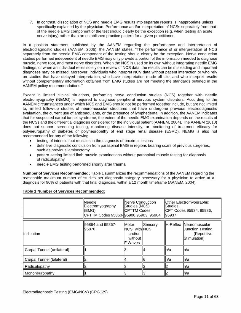
Number of Services Recommended; Table 1 summarizes the recommendations of the AANEM regarding the reasonable maximum number of studies per diagnostic category necessary for a physician to arrive at a diagnosis for 90% of patients with that final diagnosis, within a 12 month timeframe (AANEM, 2004).
Table 1 Number of Services Recommended: Needle Nerve Conduction Other Electromyographic Electromyography Studies (NCS) Studies (EMG) CPTTM Codes CPT Codes 95934, 95936, CPTTM Codes 95860- 95900,95903, 95904 95937
Indication
95864 and 95867- 95870
Motor NCS with
and/or without
F Waves
Sensory NCS
H-Reflex Neuromuscular Junction Testing
(Repetitive Stimulation)
Carpal Tunnel (unilateral) 1 3 4 n/a n/a
Carpal Tunnel (bilateral) 2 4 6 n/a n/a
Radiculopathy 2 3 2 2 n/a
Mononeuropathy 1 3 3 2 n/a
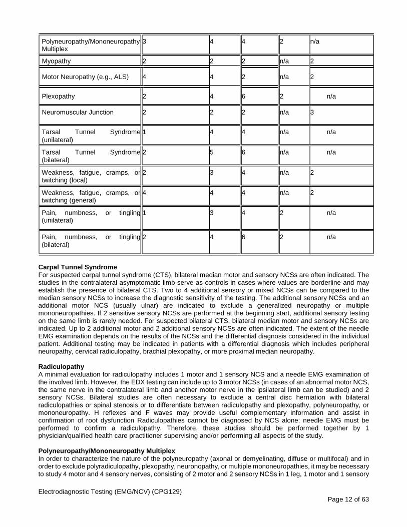
Electrodiagnostic Testing (EMG/NCV) (CPG129) Page 12 of 63
Polyneuropathy/Mononeuropathy Multiplex
3 4 4 2 n/a
Myopathy 2 2 2 n/a 2
Motor Neuropathy (e.g., ALS) 4 4 2 n/a 2
Plexopathy 2 4 6 2 n/a
Neuromuscular Junction 2 2 2 n/a 3
Tarsal Tunnel Syndrome (unilateral)
1 4 4 n/a n/a
Tarsal Tunnel Syndrome (bilateral)
2 5 6 n/a n/a
Weakness, fatigue, cramps, or twitching (local)
2 3 4 n/a 2
Weakness, fatigue, cramps, or twitching (general)
4 4 4 n/a 2
Pain, numbness, or tingling (unilateral)
1 3 4 2 n/a
Pain, numbness, or tingling (bilateral)
2 4 6 2 n/a
Carpal Tunnel Syndrome For suspected carpal tunnel syndrome (CTS), bilateral median motor and sensory NCSs are often indicated. The studies in the contralateral asymptomatic limb serve as controls in cases where values are borderline and may establish the presence of bilateral CTS. Two to 4 additional sensory or mixed NCSs can be compared to the median sensory NCSs to increase the diagnostic sensitivity of the testing. The additional sensory NCSs and an additional motor NCS (usually ulnar) are indicated to exclude a generalized neuropathy or multiple mononeuropathies. If 2 sensitive sensory NCSs are performed at the beginning start, additional sensory testing on the same limb is rarely needed. For suspected bilateral CTS, bilateral median motor and sensory NCSs are indicated. Up to 2 additional motor and 2 additional sensory NCSs are often indicated. The extent of the needle EMG examination depends on the results of the NCSs and the differential diagnosis considered in the individual patient. Additional testing may be indicated in patients with a differential diagnosis which includes peripheral neuropathy, cervical radiculopathy, brachial plexopathy, or more proximal median neuropathy. Radiculopathy A minimal evaluation for radiculopathy includes 1 motor and 1 sensory NCS and a needle EMG examination of the involved limb. However, the EDX testing can include up to 3 motor NCSs (in cases of an abnormal motor NCS, the same nerve in the contralateral limb and another motor nerve in the ipsilateral limb can be studied) and 2 sensory NCSs. Bilateral studies are often necessary to exclude a central disc herniation with bilateral radiculopathies or spinal stenosis or to differentiate between radiculopathy and plexopathy, polyneuropathy, or mononeuropathy. H reflexes and F waves may provide useful complementary information and assist in confirmation of root dysfunction Radiculopathies cannot be diagnosed by NCS alone; needle EMG must be performed to confirm a radiculopathy. Therefore, these studies should be performed together by 1 physician/qualified health care practitioner supervising and/or performing all aspects of the study. Polyneuropathy/Mononeuropathy Multiplex In order to characterize the nature of the polyneuropathy (axonal or demyelinating, diffuse or multifocal) and in order to exclude polyradiculopathy, plexopathy, neuronopathy, or multiple mononeuropathies, it may be necessary to study 4 motor and 4 sensory nerves, consisting of 2 motor and 2 sensory NCSs in 1 leg, 1 motor and 1 sensory

Electrodiagnostic Testing (EMG/NCV) (CPG129) Page 13 of 63
NCS in the opposite leg, and 1 motor and 1 sensory NCS in 1 arm. H-reflex studies and F-wave studies from 2 nerves may provide additional diagnostic information. At least 2 limbs should be studied by a needle EMG examination. Studies of related paraspinal muscles are indicated to exclude some conditions such as polyradiculopathy. Myopathy To diagnose a myopathy, a needle EMG examination of 2 limbs is indicated. To help exclude other disorders such as polyneuropathy or neuronopathy, 2 motor and 2 sensory NCSs are indicated. Two repetitive motor nerve stimulation studies may be performed to exclude a disorder of NM transmission. Motor Neuronopathy In order to establish the diagnosis of motor neuronopathy (for example, amyotrophic lateral sclerosis and to exclude other disorders in the differential diagnosis, such as multifocal motor neuropathy or polyneuropathy, up to 4 motor nerves and 2 sensory nerves may be studied. Needle EMG of up to 4 extremities (or 3 limbs and facial or tongue muscles) is often necessary to document widespread denervation and to exclude a myopathy. One repetitive motor nerve stimulation study may be indicated to exclude a disorder affecting NM transmission. Plexopathy To characterize a brachial plexopathy and to differentiate it from cervical radiculopathy and mononeuropathies, it is often necessary to study all major sensory and motor nerves that can be easily studied in both upper extremities (radial, median, ulnar, and medial and lateral antebrachial cutaneous sensory; radial, median, ulnar, and possibly axillary and musculocutaneous motor) and to perform a needle EMG examination in both upper extremities. To characterize the lumbosacral plexopathy and to differentiate it from lumbar radiculopathy and mononeuropathies, it is often necessary to study all major sensory and motor nerves that can be easily studied in both lower extremities (superficial peroneal and sural sensory; peroneal and posterior tibial motor) and to perform a needle EMG examination in both lower extremities. F-wave studies in the motor nerves and soleus H reflexes also provide useful information. Neuromuscular Junction To demonstrate and characterize abnormal NM transmission, repetitive nerve stimulation studies should be performed in up to 2 nerves and single fiber EMG (SFEMG) in up to 2 muscles. If any of these are abnormal, up to 2 motor and 2 sensory NCSs may be performed to exclude neuropathies that can be associated with abnormal NM transmission. At least 1 motor and 1 sensory NCS should be performed in a clinically involved limb, preferably in the distribution of a nerve studied with repetitive stimulation or SFEMG. At least 1 distal and 1 proximal muscle should be studied by a needle EMG examination to exclude a neuropathy or myopathy that can be associated with abnormal repetitive stimulation studies or SFEMG. At least 1 of the muscles should be clinically involved and both muscles should be in clinically involved limbs. Frequency of Electrodiagnostic Testing in a Given Patient Repeat EDX evaluation is therefore sometimes necessary and, when justifiable, should be reimbursed. Reasonable limits can be set concerning the frequency of repeat EDX testing per year in a given patient by a given EDX physician for a given diagnosis. The following numbers of tests per 12-month period per diagnosis per physician are acceptable:
1. Two tests for carpal tunnel-unilateral, carpal tunnel-bilateral, radiculopathy, mononeuropathy, polyneuropathy, myopathy, and NMJ disorders.
2. Three tests for motor neuronopathy and plexopathy. These limits should not apply if the patient requires evaluation by more than 1 EDX physician (i.e., a second opinion or an expert opinion at a tertiary care center) in a given year or if the patient requires evaluation for a second diagnosis in a given year.
Additional studies may be required or appropriate over and above these guidelines. In such situations, the reason for the repeat study should be included in the body of the report or in the patient's chart. Comparison with the previous test results should be documented. This additional documentation from the physician regarding the necessity for the additional repeat testing would be appropriate. Repeat EDX testing should not be necessary in a 12-month period in 80% of all cases. The Professional Practice Committee of the AANEM developed the following recommendations as part of the ABIM Choosing Wisely Initiative (AANEM, 2015):

Electrodiagnostic Testing (EMG/NCV) (CPG129) Page 14 of 63
• Don’t do a needle electromyography (EMG) test for isolated neck or back pain after a motor vehicle accident, as a needle EMG is unlikely to be helpful.
• Don’t do a four limb needle EMG/nerve conduction study (NCS) testing for neck and back pain after trauma.
• Don’t do nerve conduction studies without also doing a needle EMG for testing for radiculopathy, a pinched nerve in the neck or back.
Sensitivity and specificity reports for electrodiagnostic testing methods (in general) vary. A clearly established measure of comparison is lacking in the medical literature, making comparisons across studies difficult. Some studies have compared results with clinical examination findings, imaging studies such as magnetic resonance imaging, computed tomography, myelography, or the observation of nerve root compression during surgery. Interobserver differences, the variety of tests employed, the presence of symptoms that may influence patient outcomes (e.g., pain), the presence of abnormal imaging studies in asymptomatic patients, and the subjectivity of the surgeon’s interpretations may all lead to variances in sensitivity and specificity results. Despite these variances however, electrodiagnostic testing is commonly used to assist in diagnosing disorders involving the nerves, muscles and neuromuscular junction. Sensitivity and specificity data for automated/portable devices, used instead of or as an adjunct to standard nerve conduction testing, is insufficient to draw conclusions regarding predictive value. DOCUMENTATION GUIDELINES Documentation required justifying electrodiagnostic testing:
• Reason for the study, clinical history and examination findings are required • Numerical values are required – latency, amplitude and nerve conduction • Type of needle – monopolar or concentric • When documentation is required submit hard copy of waveforms and complete written report, including
test interpretation • Name, signature, professional designation of all individuals performing, interpreting or supervising the test
must be included Inadequate Documentation:
• Narrative reports alluding to ‘normal’ or ‘abnormal’ results without numerical data • Description of F-wave without reference to corresponding motor conduction data • Pattern-setting unilateral H-reflex measurements • Absence of clinical history, preferably written by the referral source, indicating the need for the test • Absence of documentation to support repeat testing on the same beneficiary or testing every beneficiary
referred for pain Nerve conduction studies must provide a number of response parameters in a real-time fashion to facilitate provider interpretation. Those parameters include amplitude, latency, configuration and conduction velocity, temperature of limb. Diagnostic studies that do not provide this information or those that provide delayed interpretation as substitutes for nerve conduction studies are not accepted. Raw measurement data obtained and transmitted trans-telephonically or over the Internet, therefore, does not qualify for the payment of the electrodiagnostic service codes included in this policy. Claims for nerve conduction testing accomplished with discriminatory devices that use fixed anatomic templates and computer-generated reports used as an adjunct to physical examination routinely on all patients are not accepted. The AANEM provides specific recommendations for reporting needle EMG and NCV results. According to the AANEM, the recommendation for documentation of nerve conduction and EMG testing should include (but are not limited to) a description of the patient’s clinical problem (demographics, reason for referral), the electrodiagnostic tests performed (techniques, distances, lab reference values, and temperature monitoring), all relevant data derived from these tests (nerves/muscles tested, numerical values for latencies and action potential), and the diagnostic interpretation of the data, including limitations. Complete NCV test measurements should also include amplitude measurements, normal reference values and criteria for abnormalities. The recommendations also include confirmation that limb temperature was monitored continuously during the NCS and repetitive

Electrodiagnostic Testing (EMG/NCV) (CPG129) Page 15 of 63
stimulation and that (a) the hand temperature was maintained between 32°C and 36°C and (b) the foot temperature was maintained between 30°C and 36°C. NCS abnormalities such as prolonged distal sensory or motor latencies could otherwise be due to coolness of the limb. For repetitive stimulation, if the limb is not warmed, the results may be assessed inaccurately as normal (AANEM, 2005; AANEM 2014). LITERATURE REVIEW
Automated Nerve Conduction Testing
Evidence evaluating the diagnostic utility of the Brevio and Virtual Medical Systems VT 3000 nerve conduction monitor systems (Automated Nerve Conduction Testing) is lacking. Evidence evaluating the diagnostic utility of the NC-stat System consists mainly of case series, case control studies and retrospective reviews. Some of these studies compare results obtained using automated devices with results obtained from standard diagnostic testing (NCV testing and EMG), other studies did not have a comparison to conventional testing. Most of the published clinical studies have evaluated use of the NC-stat device for assessment of median and ulnar nerves (Dale, et al., 2015; Megerian, et al., 2007; Kong, et al., 2006; Vinik, et al., 2004); other published studies evaluated use of the device for disorders such as lumbosacral radiculopathies (Fisher, et al., 2008) and sensorimotor polyneuropathy in diabetic patients (Perkins et al., 2008). In some of these studies a strong correlation has been demonstrated when comparing NC-stat with reference standards (Perkins, et al., 2006; Kong, et al., 2006). The diagnostic accuracy for other conditions, such as those involving the lower extremities, has not been sufficiently demonstrated in the literature. Data regarding diagnostic performance, sensitivity and specificity of the automated NCV testing devices compared to standard testing is inconsistent and does not lead to strong conclusions; the studies are not well-designed, involve small populations and the results cannot be generalized. In some studies authors have reported high sensitivity and specificity when examining NC-stat accuracy for carpal tunnel syndrome compared to controls (Dale, et al., 2015; Leffler, et al., 2000; Rotman, et al., 2004), other authors however have reported NC-stat is no more sensitive or specific than a traditionally performed distal motor latency for the diagnosis of carpal tunnel syndrome (Katz, 2006). In 2008 Armstrong and colleagues published the outcomes of a cohort study comparing the results obtained with the NC-stat device to traditional nerve conduction studies for carpal tunnel screening (n=33). All correlations were significant. The authors reported sensitivity, with respect to the traditional results, ranged from 93.8% to 100% and specificity ranged from 84.6% to 94.1%. Nonetheless, the authors did not address limitations such as lack of needle EMG testing and did not evaluate the clinical relevance to the results (Armstrong, et al., 2008). In a longitudinal study (n=134), Dale and colleagues (2015) compared automated nerve conduction using the NC Stat device to traditional electrodiagnostic studies for 62 subjects, who had prior evaluation for carpal tunnel syndrome in the parent study (n=780). The authors reported that NC Stat results agreed with traditional electrodiagnostic studies for detecting median nerve conduction abnormalities within a general population of workers. Ulnar nerve testing results were not as favorable however median nerve testing results had high sensitivity and specificity (86-100%) for median motor and sensory latency. The study is limited by small sample population of industrial workers; results cannot be generalized to the standard population. A technology assessment conducted by the Washington State Department of Labor and Industries (2006) concluded that the scientific evidence does not show NC-stat to be equivalent to conventional methods for nerve conduction testing. Authors generally agree that further studies are needed to determine the role automated testing has as a component of clinical care. Furthermore, some concerns remain among specialists regarding lack of standard EMG testing and incomplete assessment when using automated NCV testing devices. The AANEM recommends electrodiagnostic studies be performed by properly trained physicians and that interpretation of nerve conduction study data alone, absent face-to-face patient interaction and control over the process, provides substandard care (AANEM, 2006). The AANEM (2010) does not support the following:
• electrodiagnostic testing with automated, noninvasive nerve conduction testing devices • screening testing, monitoring disease intensity, or monitoring treatment efficacy for polyneuropathy
of diabetes or polyneuropathy of end stage renal disease (ESRD) Schmidt and colleagues (2011) reported on the use of an automated hand-held nerve conduction device compared to NCS or needle electrode examination (standard electrodiagnostic tests) in the evaluation of individuals with unilateral leg symptoms. A total of 50 participants with complaints of unilateral leg pain, numbness or weakness were included in the study and underwent history with physical exam and standard electrodiagnostic

Electrodiagnostic Testing (EMG/NCV) (CPG129) Page 16 of 63
testing. The participants were then tested using an automated hand-held nerve conduction device. A total of 22 participants had findings consistent with radiculopathy on standard electrodiagnostic test and 28 participants had a normal electrodiagnostic exam or evidence of another distinct neuromuscular diagnosis. During initial data analysis, a significant discrepancy was revealed between the results of standard electrodiagnostic tests and the automated test. For this reason, another 25 participants were recruited to serve as the control group. The control group participants had upper limb symptoms such as cervical radiculopathy, carpal tunnel syndrome or ulnar neuropathy. Of the 50 participants initially recruited, 28 were found to have normal standard electrodiagnostic tests. The automated tests corroborated the findings in 4 cases only. In the control group, all standard electrodiagnostic tests were normal, but the automated testing showed 18 of 25 participants had findings consistent with radiculopathy or polyneuropathy. Automated and standard testing correlated in 14 of 75 participants studied (11 of whom had normal exams with both testing methods). While this study has a small number of participants, the authors stated that "it is unlikely that larger study numbers would have increased specificity to acceptable levels of a clinically useful test, given the 95% confidence levels for the current data." In a position statement on the Proper Performance and Interpretation of Electrodiagnostic Studies and the Recommended Use of Electrodiagnostic Medicine from the American Association of Neuromuscular and Electrodiagnostic Medicine (AANEM, 2006 and 2014), although no specific reference to or recommendation for automated nerve conduction testing devices is made, it is noted that "Nerve conduction studies performed independent of needle EMG studies may only provide a portion of the information needed to diagnose muscle, nerve root, and most nerve disorders." And: Individuals without a medical education in neuromuscular disorders and without special training in EDX procedures typically are not qualified to interpret the waveforms generated by NCSs and needle EMGs or to correlate the findings with other clinical information to reach a diagnosis. It is also the recommendation of the American Association of Neuromuscular and Electrodiagnostic Medicine (AANEM) that electrodiagnostic testing/consultations are conducted by physicians who have a comprehensive knowledge of neurological and neuromusculoskeletal diseases, and in the application of neurophysiologic techniques for evaluation of those disorders. Although portable, automated, noninvasive testing of nerve conduction has been suggested as an easier method for providers to obtain rapid results, the AANEM recommends that electrodiagnostic studies of EMG and NCS be performed together, except in unique situations, in a study design determined and interpreted by a trained physician, so that healthcare decisions are based on complete diagnostic information (AANEM, 2014). Currently, there is insufficient evidence in the published data to demonstrate that automated nerve conduction testing devices are valid measures in the diagnosis of peripheral nerve disease. Since the clearance of the NC-stat, several other devices have also received FDA clearance listing the NC-stat as the predicate device. However to date there has been very limited published evidence to demonstrate the safety and efficacy of automated, noninvasive nerve conduction testing devices, as compared to conventional "gold standard" electrodiagnostic testing using EMG and NCS. Most of the published clinical studies have evaluated use of an automated device for assessment of the median and ulnar nerves only (Katz, 2006; Kong, 2006). Other Electrodiagnostic Testing Evidence in the peer reviewed scientific literature including textbook and professional society opinion supports clinical utility for electrodiagnostic testing, including neuromuscular junction testing, when used to assist in diagnosing disorders involving the nerves, muscles and neuromuscular junction. The AANEM has published guidance for the performance of nerve conduction studies and EMG. According to the AANEM a typical nerve conduction examination includes: development of a differential diagnosis based upon appropriate history and physical exam, the NCV study (recording and studying of electrical responses from peripheral nerves or muscles) and the completion of indicated needle EMG studies to evaluate the differential diagnosis and to complement the nerve conduction study. In addition, the AANEM supports that when performing nerve conduction studies the waveform must be reviewed on site and in real time, with reports prepared onsite by the examiner, consistent with current procedural terminology descriptions (AANEM, 2014). The AANEM defines the use of the term onsite as that where the history and physical, performance of NCV and EMG, analysis of electrodiagnostic data and determination of diagnosis occur in the same location, typically an electrodiagnostic laboratory. Similarly, real time is defined as that which allows for information from the physical and history to be integrated with the
Electrodiagnostic Testing (EMG/NCV) (CPG129) Page 17 of 63
performance of testing, allowing for the testing of both NCV and EMG to be tailored/modified to the individual circumstance as needed before leaving the lab. The use of nerve conduction studies including F-wave and H-reflex tests for the diagnosis of early stage polyneuropathies and proximal nerve lesions is confirmed in several reviews and studies (Maccabee et al., 2011; Kostera-Pruszczyk et al., 2004; Trujillo-Hernandez et al., 2005; Bal et al., 2006; Kocer et al., 2005; Mesrati and Vecchierini, 2004). The published scientific literature demonstrates somatosensory evoked potential (SEP) studies are useful when used to aid in the diagnosis of various neuromuscular disorders and have varying degrees of sensitivity and specificity. Nerve conduction studies are indicated for the following conditions: peripheral nerve entrapment (Omejec, 2014; Park, 2014; Calfee, 2012; Kwon, 2008); generalized neuropathies (Holiner,2013; Derr, 2009, Dyck, 2010, De Sousa, 2009); polyneuropathies (de Souza, 2015; Emeryk-Szajewska, 1998, Torvin Moller, 2009); plexopathy (Mullins, 2007); neuromuscular junction disorders (Meriggioli, 2005); myopathies including polymyositis, dermatomyositis, and congenital myopathies (Wang, 2010); motor neuron disease (Hammad, 2007); spine disorders and radiculopathy (Pawar, 2013; Alrawi, 2007; Haig, 2006); and guidance for botulinum toxin injection for spasmodic dysphonia or segmental dystonia, when it is difficult to isolate affected muscles (Molloy, 2002). Karami-Mohajeri et al (2014) presented a systematic review of the recent literature on the scientific support of EMG and NCV in diagnosing the exposure and toxicity of organophosphorus pesticides (OP). Specifically, this review focused on changes in EMG, NCV, occurrence of intermediate syndrome (IMS), and OP-induced delayed polyneuropathy (OPIDN) in human. All relevant bibliographic databases were searched for human studies using the key words "OP poisoning", "electromyography", "nerve conduction study," and "muscles disorders". Intermediate syndrome usually occurs after an acute cholinergic crisis, while OPIDN occurs after both acute and chronic exposures. Collection of these studies supported that IMS is a neuromuscular junction disorder and can be recorded upon the onset of respiratory failure. Due to heterogeneity of reports on outcomes of interest such as motor NCV and EMG amplitude in acute cases and inability to achieve precise estimation of effect in chronic cases meta-analysis was not helpful to this review. The OPIDN after both acute and low-level prolonged exposures develops peripheral neuropathy without preceding cholinergic toxicity and the progress of changes in EMG and NCV is parallel with the development of IMS and OPIDN. Persistent inhibition of acetylcholinesterase (AChE) is responsible for muscle weakness, but this is not the only factor involved in the incidence of this weakness in IMS or OPIDN suggestive of AChE assay not useful as an index of nerve and muscle impairment. The authors concluded that although several mechanisms for induction of this neurodegenerative disorder have been proposed, among them oxidative stress and resulting apoptosis can be emphasized. Nevertheless, they stated that there is little synchronized evidence on subclinical electrophysiological findings that limit these investigators to reach a strong conclusion on the diagnostic or prognostic use of EMG and NCV for acute and occupational exposures to OPs. Asad et al. (2009) compared the nerve conduction studies in clinically undetectable and detectable sensorimotor polyneuropathy in type 2 diabetics. Diagnosed diabetics (n = 60) were divided in two groups. Group 1 (n1 = 30) with clinically undetectable and group 2 (n2 = 30) with clinically detectable Diabetic Polyneuropathy. Detection of the sensorimotor neuropathy was done according to Diabetic Neuropathy Symptom Score and Diabetic Neuropathy Examination scores. The simplified nerve conduction studies protocol was followed in recording amplitudes, velocities and latencies of minimum two (Sural, Peroneal) and maximum six i.e. three sensory (Sural, Ulnar, Median) and three motor (Peroneal, Ulnar, Tibial) nerves. The comparisons were done between different parameters of nerve conduction studies with the neurological scores in undetectable and detectable groups using Pearson's chi square test. The amplitudes, velocities, latencies, outcome and grading of neuropathy in nerve conduction studies when compared with neurological detection scores showed a significant relation in each group regarding evaluation (p = 0.005, p = 0.004, p = 0.05, p = 0.00001, p = 0.003 respectively). Diabetic Neuropathy Symptom Score and Diabetic Neuropathy Examination Score together can help in prompt evaluation of the diabetic sensorimotor polyneuropathy though nerve conduction study is more powerful test and can help in diagnosing subclinical cases. Surface Electromyography (SEMG) There is a wide variety of Surface Electromyography (SEMG) hardware and software that is used depending upon the specific clinical purpose intended. However, all SEMG hardware and software have in common the following:
• Electrical signals are measured from skeletal muscles. • Sensing electrodes are placed on the skin overlying the muscle of interest.

Electrodiagnostic Testing (EMG/NCV) (CPG129) Page 18 of 63
• The electrical activity is measured when the muscle is active. • SEMG records a narrow frequency of electrical activity (20-500 Hz). • SEMG findings are based on computer analysis of either the frequency spectrum (spectral analysis),
amplitude of signal, or root mean square of electrical action potentials. The Evaluation of Specific Neuromuscular Pathologies The literature on the subject of SEMG use for neuromuscular disorders indicates that it is inferior in all parameters (sensitivity, specificity, spatial resolution, signal to noise ratio) to the invasive procedures such as needle electromyography (NEMG) or fine-wire electromyography (FWEMG) and thus cannot be used as a substitute for those procedures. Both systematic reviews of this subject explicitly reject SEMG for the diagnosis of neuromuscular disease. The gold standard for this type of evaluation is either NEMG or FWEMG. Because these procedures are both invasive and painful, there is an obvious desire to find equally useful, but less onerous diagnostic tests. There are, however, several inherent limitations to the use of SEMG for the analysis of neuromuscular pathology. SEMG records input from a much wider spatial field than do either of the invasive procedures. Muscles adjacent to those of interest can produce signals that appear to originate from the target muscles (which are located immediately beneath the sensing electrodes). Thus, the specificity of SEMG findings is always in doubt. SEMG is also very susceptible to movement artifact. Even with the most careful procedural safeguards, small (and even imperceptible) body movements may produce spurious signals. There is a much poorer signal to noise ratio with SEMG. This is particularly a problem when target muscles are located more than 10 mm below the skin surface. Finally, the electrical activity that is recorded by SEMG is only of skeletal muscle origins. It is not possible to capture any electrical activity along motor neuron axons, as it is with NEMG or FWEMG. The Evaluation of Movement and Gait Disturbances There are a variety of experimental applications such as studies of human movement, the study of nerve conduction velocities after electrical stimulation of peripheral nerves, etc., in which SEMG is considered standard. Because of its relative ease of use and non-invasive nature, SEMG is considered superior to NEMG and FWEMG for many of these applications. There are also thought to be advantages in using SEMG to evaluate/study movement disorders of CNS origins such as tremor, dystonia, dyskinesia, and myoclonus. While it is thought that SEMG can accurately measure these disorders, it is less clear what the clinical utility of these measurements might be. This is the only application for which the American Medical Association (AMA) Current Procedural Terminology (CPT) coding committee has developed a procedure code. The Evaluation of Functional Back Pain There are a number of studies that have investigated the possibility that SEMG may differentiate between those with and those without back pain by evaluating muscle fatigue through “spectral shift”. However, the findings are inconsistent and contradictory, the relationship between muscle fatigue and back pain is not established, and there may be unrelated factors affecting spectral shift. The clinical context in which chiropractors are most likely to use SEMG is for the evaluation of functional low back pain and neck pain. There are two proposed mechanisms by which SEMG is thought to relate to back pain. First is the presumed relationship between muscle fatigue and back pain. The theory posits that excessive muscle fatigue, due to deconditioning, may result in back pain. Further, it has been shown that when muscles fatigue they produce a different set of electrical frequencies as measured by SEMG. This phenomenon has been dubbed the “spectral shift.” Thus, it has been hypothesized that by using dynamic SEMG (recording muscle activity while exercising) it should be possible to differentiate those with back pain from those without back pain. There are a number of studies that have investigated this possibility and some have had success in doing so. However, this success is tempered by several caveats. First, these findings are inconsistent and somewhat contradictory. Second, the exact nature of the relationship between muscle fatigue and back pain is uncertain. In fact, the direction of the relationship is uncertain—does muscle fatigue cause back pain or does back pain cause muscle fatigue? Third, it is unclear what other factors might cause a spectral shift making the specificity of such findings doubtful. There is another mechanism by which it is proposed that SEMG can assist in the evaluation of back pain: the identification of hypertonic muscles. It is this mechanism that the leading chiropractic proponents of SEMG suggest is the most relevant to patient management. In effect, it is proposed that SEMG is a more objective and accurate tool than palpation in locating hypertonic muscles and thereby the identification of vertebral subluxations.

Electrodiagnostic Testing (EMG/NCV) (CPG129) Page 19 of 63
The literature relative to this mechanism is even more limited and of much poorer quality than is the literature on muscle fatigue and SEMG. It is also speculated that the finding of SEMG asymmetry is an indication of spinal dysfunction. There is no literature that finds a relationship between back pain and such asymmetry and at least one study that casts doubt on this hypothesis. SEMG is not reliable for assessing spinal dysfunction or subluxation. A recent analysis by Triano, et al. (2013) examined the techniques and procedures used by chiropractors to identify the appropriate site for the application of spinal manipulation. Consistent with previous reviews they found limited support for reliability of SEMG to identify cohorts of patients with abnormal neuromuscular control. However the review concluded that there was no support for the use of SEMG to localize treatment to a specific site. Another area of research for SEMG is its use as a prognostic tool. Studies have looked at flexion and extension movements to determine the prognosis of the patient relative to their low back pain recovery. Hu et al. (2014) evaluated the prognostic value of quantitative SEMG topographic analysis and attempted to verify the accuracy of the performance of proposed time-varying topographic parameters for identifying the patients who have better response toward the rehabilitation program. Thirty-eight patients with chronic nonspecific LBP and 43 healthy subjects were included in the study. These patients suffered from chronic nonspecific LBP without the history of back surgery and any medical conditions causing acute exacerbation of LBP during the clinical test were enlisted to perform the clinical test during the 12-week physiotherapy (PT) treatment. Low back pain patients were classified into two groups: "responding" and "nonresponding" based on the clinical assessment. The responding group referred to the LBP patients who began to recover after the PT treatment, whereas the nonresponding group referred to some LBP patients who did not recover or got worse after the treatment. The quantitative time-varying analysis of SEMG topography showed significant difference between the healthy and LBP groups. The discrepancies in quantitative dynamic SEMG topography of LBP group from normal group, were able to identify those LBP subjects who would respond to a conservative rehabilitation program focused on functional restoration of lumbar muscle. More research is needed to confirm results and evaluate its utility clinically. In assessing the appropriateness of SEMG for functional back pain, there are three levels of analysis to consider that remain pertinent:
1. Technical performance of the instrument. To what extent does the instrument accurately measure what it purports to measure (e.g., muscle fatigue, muscle spasm)? The above discussion regarding neuromuscular disorders identifies several inherent limitations in the technical performance of SEMG. All of those limitations (with the exception of the inability to measure axonal signals) are relevant to this issue as well. The lack of specificity, poor signal to noise ratio, and the problem of movement artifacts will all limit the accuracy and validity of SEMG for the evaluation of functional back pain.
2. Whether and how the instrument findings can be used in patient management. The use of SEMG
as a “subluxation detector” that can help identify specific levels of spinal dysfunction has not been substantiated and is entirely speculative.
If it has been determined that it is possible to identify hypo- or hypertonic muscles through the use of SEMG (keeping in mind the inherent technical limitations affecting specificity, accuracy, and validity), the question becomes how this information will be used in the management of the patient. To date, the only clinical correlation that has been established is that there may be differences between subjects with back pain and control subjects in their muscle fatigability as measured by SEMG. In other words, it may be possible to differentiate those with and without back pain using SEMG. But as one of the systematic reviews points out, the gold standard for the presence or absence of back pain is the clinical history, and it is far easier and more reliable to simply ask the person whether he or she has back pain. While potentially, it might be possible to use SEMG to identify malingerers, the procedure is currently far too unreliable to permit any such determination to be predicated on SEMG findings. In addition, several established malingering tests are available as taught within standard orthopedic examination courses in chiropractic, osteopathic, and medical schools.
3. Whether the use of an instrument results in better clinical outcomes. There is no evidence (and very
little theory) to indicate how specific SEMG findings should be used to manage individuals with back pain in order to produce better clinical outcomes. Ultimately what matters is whether or not the use of SEMG results in better clinical outcomes than does the management of back pain without the use of SEMG information. There have been no clinical trials that have addressed this question. In fact, there are no clinical trials of back pain that have used SEMG
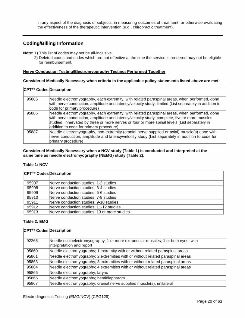
Electrodiagnostic Testing (EMG/NCV) (CPG129) Page 20 of 63
in any aspect of the diagnosis of subjects, in measuring outcomes of treatment, or otherwise evaluating the effectiveness of the therapeutic intervention (e.g., chiropractic treatment).
Coding/Billing Information Note: 1) This list of codes may not be all-inclusive. 2) Deleted codes and codes which are not effective at the time the service is rendered may not be eligible for reimbursement.
Nerve Conduction Testing/Electromyography Testing: Performed Together
Considered Medically Necessary when criteria in the applicable policy statements listed above are met:
CPT®* Codes Description
95885 Needle electromyography, each extremity, with related paraspinal areas, when performed, done with nerve conduction, amplitude and latency/velocity study; limited (List separately in addition to code for primary procedure)
95886 Needle electromyography, each extremity, with related paraspinal areas, when performed, done with nerve conduction, amplitude and latency/velocity study; complete, five or more muscles studied, innervated by three or more nerves or four or more spinal levels (List separately in addition to code for primary procedure)
95887 Needle electromyography, non-extremity (cranial nerve supplied or axial) muscle(s) done with nerve conduction, amplitude and latency/velocity study (List separately in addition to code for primary procedure)
Considered Medically Necessary when a NCV study (Table 1) is conducted and interpreted at the same time as needle electromyography (NEMG) study (Table 2):
Table 1: NCV
CPT®* Codes Description
95907 Nerve conduction studies; 1-2 studies 95908 Nerve conduction studies; 3-4 studies 95909 Nerve conduction studies; 5-6 studies 95910 Nerve conduction studies; 7-8 studies 95911 Nerve conduction studies; 9-10 studies 95912 Nerve conduction studies; 11-12 studies 95913 Nerve conduction studies; 13 or more studies
Table 2: EMG
CPT®* Codes Description
92265 Needle oculoelectromyography, 1 or more extraocular muscles, 1 or both eyes, with interpretation and report
95860 Needle electromyography; 1 extremity with or without related paraspinal areas 95861 Needle electromyography; 2 extremities with or without related paraspinal areas 95863 Needle electromyography; 3 extremities with or without related paraspinal areas 95864 Needle electromyography; 4 extremities with or without related paraspinal areas 95865 Needle electromyography; larynx 95866 Needle electromyography; hemidiaphragm 95867 Needle electromyography; cranial nerve supplied muscle(s), unilateral
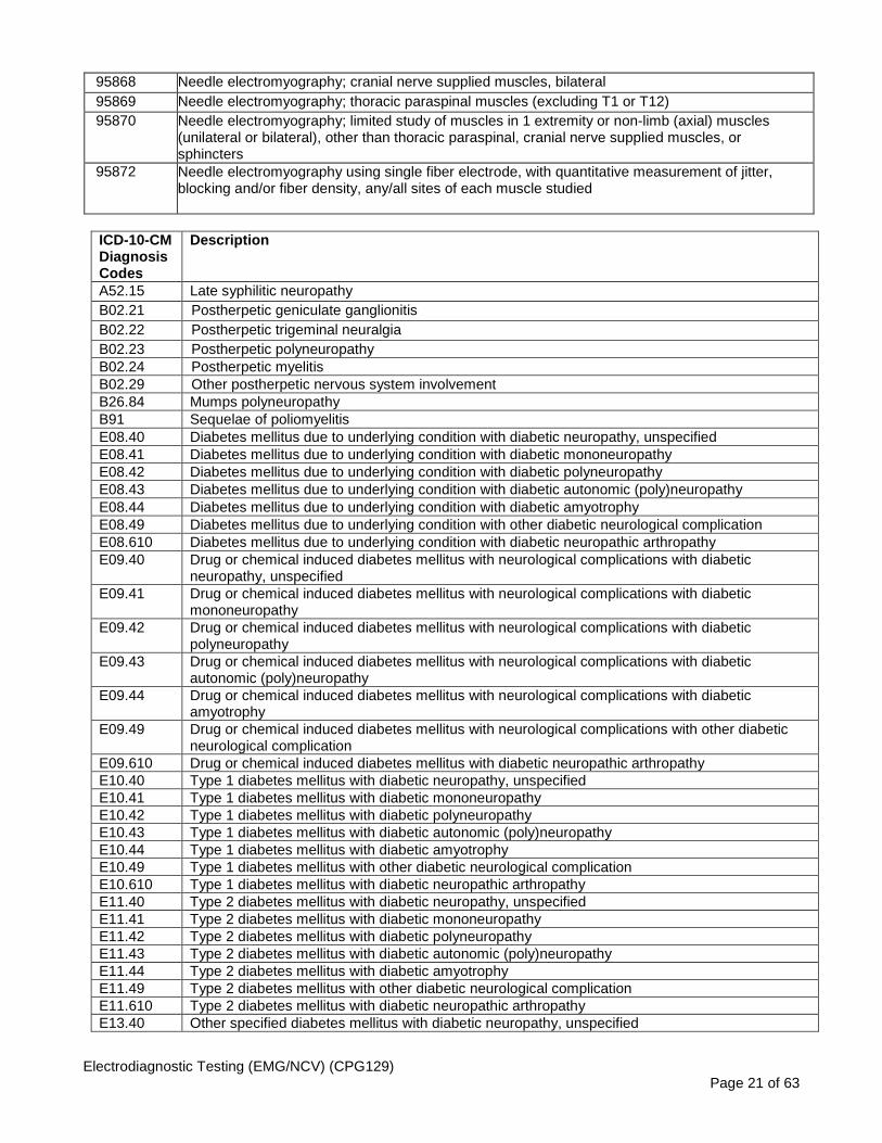
Electrodiagnostic Testing (EMG/NCV) (CPG129) Page 21 of 63
95868 Needle electromyography; cranial nerve supplied muscles, bilateral 95869 Needle electromyography; thoracic paraspinal muscles (excluding T1 or T12) 95870 Needle electromyography; limited study of muscles in 1 extremity or non-limb (axial) muscles
(unilateral or bilateral), other than thoracic paraspinal, cranial nerve supplied muscles, or sphincters
95872 Needle electromyography using single fiber electrode, with quantitative measurement of jitter, blocking and/or fiber density, any/all sites of each muscle studied
ICD-10-CM Diagnosis Codes
Description
A52.15 Late syphilitic neuropathy B02.21 Postherpetic geniculate ganglionitis
B02.22 Postherpetic trigeminal neuralgia
B02.23 Postherpetic polyneuropathy
B02.24 Postherpetic myelitis
B02.29 Other postherpetic nervous system involvement
B26.84 Mumps polyneuropathy B91 Sequelae of poliomyelitis E08.40 Diabetes mellitus due to underlying condition with diabetic neuropathy, unspecified E08.41 Diabetes mellitus due to underlying condition with diabetic mononeuropathy E08.42 Diabetes mellitus due to underlying condition with diabetic polyneuropathy E08.43 Diabetes mellitus due to underlying condition with diabetic autonomic (poly)neuropathy E08.44 Diabetes mellitus due to underlying condition with diabetic amyotrophy E08.49 Diabetes mellitus due to underlying condition with other diabetic neurological complication E08.610 Diabetes mellitus due to underlying condition with diabetic neuropathic arthropathy E09.40 Drug or chemical induced diabetes mellitus with neurological complications with diabetic
neuropathy, unspecified E09.41 Drug or chemical induced diabetes mellitus with neurological complications with diabetic
mononeuropathy E09.42 Drug or chemical induced diabetes mellitus with neurological complications with diabetic
polyneuropathy E09.43 Drug or chemical induced diabetes mellitus with neurological complications with diabetic
autonomic (poly)neuropathy E09.44 Drug or chemical induced diabetes mellitus with neurological complications with diabetic
amyotrophy E09.49 Drug or chemical induced diabetes mellitus with neurological complications with other diabetic
neurological complication E09.610 Drug or chemical induced diabetes mellitus with diabetic neuropathic arthropathy E10.40 Type 1 diabetes mellitus with diabetic neuropathy, unspecified E10.41 Type 1 diabetes mellitus with diabetic mononeuropathy E10.42 Type 1 diabetes mellitus with diabetic polyneuropathy E10.43 Type 1 diabetes mellitus with diabetic autonomic (poly)neuropathy E10.44 Type 1 diabetes mellitus with diabetic amyotrophy E10.49 Type 1 diabetes mellitus with other diabetic neurological complication E10.610 Type 1 diabetes mellitus with diabetic neuropathic arthropathy E11.40 Type 2 diabetes mellitus with diabetic neuropathy, unspecified E11.41 Type 2 diabetes mellitus with diabetic mononeuropathy E11.42 Type 2 diabetes mellitus with diabetic polyneuropathy E11.43 Type 2 diabetes mellitus with diabetic autonomic (poly)neuropathy E11.44 Type 2 diabetes mellitus with diabetic amyotrophy E11.49 Type 2 diabetes mellitus with other diabetic neurological complication E11.610 Type 2 diabetes mellitus with diabetic neuropathic arthropathy E13.40 Other specified diabetes mellitus with diabetic neuropathy, unspecified

Electrodiagnostic Testing (EMG/NCV) (CPG129) Page 22 of 63
E13.41 Other specified diabetes mellitus with diabetic mononeuropathy E13.42 Other specified diabetes mellitus with diabetic polyneuropathy E13.43 Other specified diabetes mellitus with diabetic autonomic (poly)neuropathy E13.44 Other specified diabetes mellitus with diabetic amyotrophy E13.49 Other specified diabetes mellitus with other diabetic neurological complication E13.610 Other specified diabetes mellitus with diabetic neuropathic arthropathy G04.1 Tropical spastic paraplegia G11.0 Congenital nonprogressive ataxia G11.2 Late-onset cerebellar ataxia G11.3 Cerebellar ataxia with defective DNA repair G11.4 Hereditary spastic paraplegia G11.8 Other hereditary ataxias G11.9 Hereditary ataxia, unspecified G12.0 Infantile spinal muscular atrophy, type I [Werdnig-Hoffman] G12.1 Other inherited spinal muscular atrophy G12.20 Motor neuron disease, unspecified G12.21 Amyotrophic lateral sclerosis G12.22 Progressive bulbar palsy G12.23 Primary lateral sclerosis G12.24 Familial motor neuron disease G12.25 Progressive spinal muscle atrophy G12.29 Other motor neuron disease G12.8 Other spinal muscular atrophies and related syndromes G12.9 Spinal muscular atrophy, unspecified G13.0 Paraneoplastic neuromyopathy and neuropathy G13.1 Other systemic atrophy primarily affecting central nervous system in neoplastic disease G14 Postpolio syndrome G23.0 Hallervorden-Spatz disease G23.1 Progressive supranuclear ophthalmoplegia [Steele-Richardson-Olszewski] G23.2 Striatonigral degeneration G23.8 Other specified degenerative diseases of basal ganglia G23.9 Degenerative disease of basal ganglia, unspecified G24.02 Drug induced acute dystonia G24.1 Genetic torsion dystonia G24.2 Idiopathic nonfamilial dystonia G24.3 Spasmodic torticollis G24.4 Idiopathic orofacial dystonia G24.5 Blepharospasm G24.8 Other dystonia G24.9 Dystonia, unspecified G25.3 Myoclonus G25.70 Drug induced movement disorder, unspecified G25.79 Other drug induced movement disorders G25.89 Other specified extrapyramidal and movement disorders G25.9 Extrapyramidal and movement disorder, unspecified G32.0 Subacute combined degeneration of spinal cord in diseases classified elsewhere G35 Multiple sclerosis G36.0 Neuromyelitis optica [Devic] G36.1 Acute and subacute hemorrhagic leukoencephalitis [Hurst] G36.8 Other specified acute disseminated demyelination G36.9 Acute disseminated demyelination, unspecified G37.0 Diffuse sclerosis of central nervous system G37.1 Central demyelination of corpus callosum G37.2 Central pontine myelinolysis G37.3 Acute transverse myelitis in demyelinating disease of central nervous system
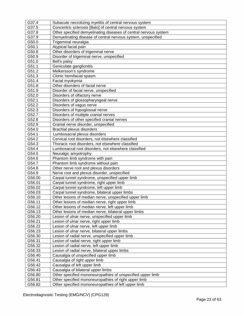
Electrodiagnostic Testing (EMG/NCV) (CPG129) Page 23 of 63
G37.4 Subacute necrotizing myelitis of central nervous system G37.5 Concentric sclerosis [Balo] of central nervous system G37.8 Other specified demyelinating diseases of central nervous system G37.9 Demyelinating disease of central nervous system, unspecified G50.0 Trigeminal neuralgia G50.1 Atypical facial pain G50.8 Other disorders of trigeminal nerve G50.9 Disorder of trigeminal nerve, unspecified G51.0 Bell’s palsy G51.1 Geniculate ganglionitis G51.2 Melkersson’s syndrome G51.3 Clonic hemifacial spasm G51.4 Facial myokymia G51.8 Other disorders of facial nerve G51.9 Disorder of facial nerve, unspecified G52.0 Disorders of olfactory nerve G52.1 Disorders of glossopharyngeal nerve G52.2 Disorders of vagus nerve G52.3 Disorders of hypoglossal nerve G52.7 Disorders of multiple cranial nerves G52.8 Disorders of other specified cranial nerves G52.9 Cranial nerve disorder, unspecified G54.0 Brachial plexus disorders G54.1 Lumbosacral plexus disorders
G54.2 Cervical root disorders, not elsewhere classified G54.3 Thoracic root disorders, not elsewhere classified G54.4 Lumbosacral root disorders, not elsewhere classified G54.5 Neuralgic amyotrophy G54.6 Phantom limb syndrome with pain G54.7 Phantom limb syndrome without pain G54.8 Other nerve root and plexus disorders G54.9 Nerve root and plexus disorder, unspecified G56.00 Carpal tunnel syndrome, unspecified upper limb G56.01 Carpal tunnel syndrome, right upper limb G56.02 Carpal tunnel syndrome, left upper limb G56.03 Carpal tunnel syndrome, bilateral upper limbs G56.10 Other lesions of median nerve, unspecified upper limb G56.11 Other lesions of median nerve, right upper limb G56.12 Other lesions of median nerve, left upper limb G56.13 Other lesions of median nerve, bilateral upper limbs G56.20 Lesion of ulnar nerve, unspecified upper limb G56.21 Lesion of ulnar nerve, right upper limb G56.22 Lesion of ulnar nerve, left upper limb G56.23 Lesion of ulnar nerve, bilateral upper limbs G56.30 Lesion of radial nerve, unspecified upper limb G56.31 Lesion of radial nerve, right upper limb G56.32 Lesion of radial nerve, left upper limb G56.33 Lesion of radial nerve, bilateral upper limbs G56.40 Causalgia of unspecified upper limb G56.41 Causalgia of right upper limb G56.42 Causalgia of left upper limb G56.43 Causalgia of bilateral upper limbs G56.80 Other specified mononeuropathies of unspecified upper limb G56.81 Other specified mononeuropathies of right upper limb G56.82 Other specified mononeuropathies of left upper limb

Electrodiagnostic Testing (EMG/NCV) (CPG129) Page 24 of 63
G56.83 Other specified mononeuropathies of bilateral upper limbs G56.90 Unspecified mononeuropathy of unspecified upper limb G56.91 Unspecified mononeuropathy of right upper limb G56.92 Unspecified mononeuropathy of left upper limb G56.93 Unspecified mononeuropathy of bilateral upper limbs G57.00 Lesion of sciatic nerve, unspecified lower limb G57.01 Lesion of sciatic nerve, right lower limb G57.02 Lesion of sciatic nerve, left lower limb G57.03 Lesion of sciatic nerve, bilateral lower limbs G57.10 Meralgia paresthetica, unspecified lower limb G57.11 Meralgia paresthetica, right lower limb G57.12 Meralgia paresthetica, left lower limb G57.13 Meralgia paresthetica, bilateral lower limbs G57.20 Lesion of femoral nerve, unspecified lower limb G57.21 Lesion of femoral nerve, right lower limb G57.22 Lesion of femoral nerve, left lower limb G57.23 Lesion of femoral nerve, bilateral lower limbs G57.30 Lesion of lateral popliteal nerve, unspecified lower limb G57.31 Lesion of lateral popliteal nerve, right lower limb G57.32 Lesion of lateral popliteal nerve, left lower limb G57.33 Lesion of lateral popliteal nerve, bilateral lower limbs G57.40 Lesion of medial popliteal nerve, unspecified lower limb G57.41 Lesion of medial popliteal nerve, right lower limb G57.42 Lesion of medial popliteal nerve, left lower limb G57.43 Lesion of medial popliteal nerve, bilateral lower limbs G57.50 Tarsal tunnel syndrome, unspecified lower limb G57.51 Tarsal tunnel syndrome, right lower limb G57.52 Tarsal tunnel syndrome, left lower limb G57.53 Tarsal tunnel syndrome, bilateral lower limbs G57.60 Lesion of plantar nerve, unspecified lower limb G57.61 Lesion of plantar nerve, right lower limb G57.62 Lesion of plantar nerve, left lower limb G57.63 Lesion of plantar nerve, bilateral lower limbs G57.70 Causalgia of unspecified lower limb G57.71 Causalgia of right lower limb G57.72 Causalgia of left lower limb G57.73 Causalgia of bilateral lower limbs G57.80 Other specified mononeuropathies of unspecified lower limb G57.81 Other specified mononeuropathies of right lower limb G57.82 Other specified mononeuropathies of left lower limb G57.83 Other specified mononeuropathies of bilateral lower limbs G57.90 Unspecified mononeuropathy of unspecified lower limb G57.91 Unspecified mononeuropathy of right lower limb
G57.92 Unspecified mononeuropathy of left lower limb G57.93 Unspecified mononeuropathy of bilateral lower limbs G58.7 Mononeuritis multiplex G58.8 Other specified mononeuropathies G58.9 Mononeuropathy, unspecified G60.0 Hereditary motor and sensory neuropathy G60.1 Refsum’s disease G60.2 Neuropathy in association with hereditary ataxia G60.3 Idiopathic progressive neuropathy G60.8 Other hereditary and idiopathic neuropathies G60.9 Hereditary and idiopathic neuropathy, unspecified G61.0 Guillain-Barre syndrome
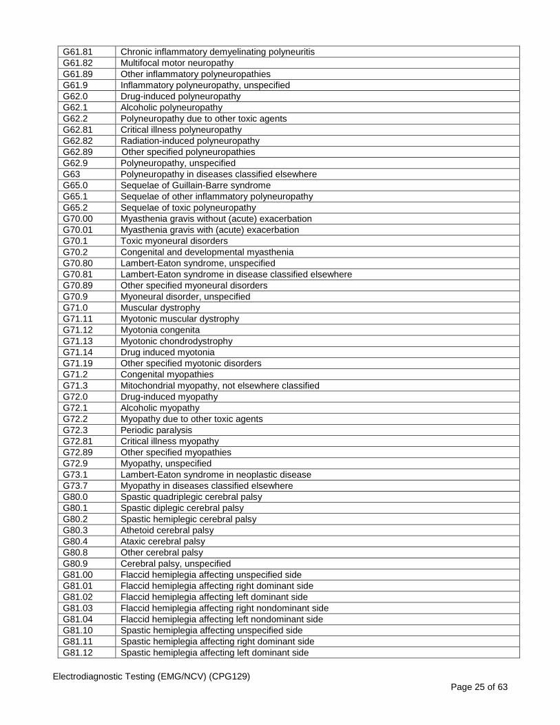
Electrodiagnostic Testing (EMG/NCV) (CPG129) Page 25 of 63
G61.81 Chronic inflammatory demyelinating polyneuritis G61.82 Multifocal motor neuropathy G61.89 Other inflammatory polyneuropathies G61.9 Inflammatory polyneuropathy, unspecified G62.0 Drug-induced polyneuropathy G62.1 Alcoholic polyneuropathy G62.2 Polyneuropathy due to other toxic agents G62.81 Critical illness polyneuropathy G62.82 Radiation-induced polyneuropathy G62.89 Other specified polyneuropathies
G62.9 Polyneuropathy, unspecified G63 Polyneuropathy in diseases classified elsewhere G65.0 Sequelae of Guillain-Barre syndrome G65.1 Sequelae of other inflammatory polyneuropathy G65.2 Sequelae of toxic polyneuropathy G70.00 Myasthenia gravis without (acute) exacerbation G70.01 Myasthenia gravis with (acute) exacerbation G70.1 Toxic myoneural disorders G70.2 Congenital and developmental myasthenia G70.80 Lambert-Eaton syndrome, unspecified G70.81 Lambert-Eaton syndrome in disease classified elsewhere G70.89 Other specified myoneural disorders G70.9 Myoneural disorder, unspecified G71.0 Muscular dystrophy G71.11 Myotonic muscular dystrophy G71.12 Myotonia congenita G71.13 Myotonic chondrodystrophy G71.14 Drug induced myotonia G71.19 Other specified myotonic disorders G71.2 Congenital myopathies G71.3 Mitochondrial myopathy, not elsewhere classified G72.0 Drug-induced myopathy G72.1 Alcoholic myopathy G72.2 Myopathy due to other toxic agents G72.3 Periodic paralysis G72.81 Critical illness myopathy G72.89 Other specified myopathies G72.9 Myopathy, unspecified G73.1 Lambert-Eaton syndrome in neoplastic disease G73.7 Myopathy in diseases classified elsewhere G80.0 Spastic quadriplegic cerebral palsy G80.1 Spastic diplegic cerebral palsy G80.2 Spastic hemiplegic cerebral palsy G80.3 Athetoid cerebral palsy G80.4 Ataxic cerebral palsy G80.8 Other cerebral palsy G80.9 Cerebral palsy, unspecified G81.00 Flaccid hemiplegia affecting unspecified side G81.01 Flaccid hemiplegia affecting right dominant side G81.02 Flaccid hemiplegia affecting left dominant side G81.03 Flaccid hemiplegia affecting right nondominant side G81.04 Flaccid hemiplegia affecting left nondominant side G81.10 Spastic hemiplegia affecting unspecified side G81.11 Spastic hemiplegia affecting right dominant side G81.12 Spastic hemiplegia affecting left dominant side
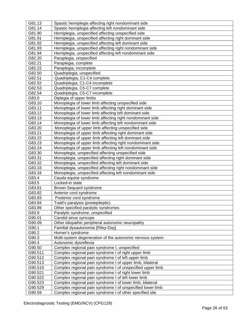
Electrodiagnostic Testing (EMG/NCV) (CPG129) Page 26 of 63
G81.13 Spastic hemiplegia affecting right nondominant side G81.14 Spastic hemiplegia affecting left nondominant side G81.90 Hemiplegia, unspecified affecting unspecified side G81.91 Hemiplegia, unspecified affecting right dominant side G81.92 Hemiplegia, unspecified affecting left dominant side G81.93 Hemiplegia, unspecified affecting right nondominant side G81.94 Hemiplegia, unspecified affecting left nondominant side G82.20 Paraplegia, unspecified G82.21 Paraplegia, complete G82.22 Paraplegia, incomplete G82.50 Quadriplegia, unspecified G82.51 Quadriplegia, C1-C4 complete
G82.52 Quadriplegia, C1-C4 incomplete G82.53 Quadriplegia, C5-C7 complete G82.54 Quadriplegia, C5-C7 incomplete G83.0 Diplegia of upper limbs G83.10 Monoplegia of lower limb affecting unspecified side G83.11 Monoplegia of lower limb affecting right dominant side G83.12 Monoplegia of lower limb affecting left dominant side G83.13 Monoplegia of lower limb affecting right nondominant side G83.14 Monoplegia of lower limb affecting left nondominant side G83.20
Monoplegia of upper limb affecting unspecified side
G83.21 Monoplegia of upper limb affecting right dominant side G83.22 Monoplegia of upper limb affecting left dominant side G83.23 Monoplegia of upper limb affecting right nondominant side G83.24 Monoplegia of upper limb affecting left nondominant side G83.30 Monoplegia, unspecified affecting unspecified side G83.31 Monoplegia, unspecified affecting right dominant side G83.32 Monoplegia, unspecified affecting left dominant side G83.33 Monoplegia, unspecified affecting right nondominant side G83.34 Monoplegia, unspecified affecting left nondominant side G83.4 Cauda equine syndrome G83.5 Locked-in state G83.81 Brown-Sequard syndrome G83.82 Anterior cord syndrome G83.83
Posterior cord syndrome
G83.84 Todd’s paralysis (postepileptic) G83.89 Other specified paralytic syndromes G83.9 Paralytic syndrome, unspecified G90.01 Carotid sinus syncope G90.09 Other idiopathic peripheral autonomic neuropathy G90.1 Familial dysautonomia [Riley-Day] G90.2 Horner’s syndrome G90.3 Multi-system degeneration of the autonomic nervous system G90.4 Autonomic dysreflexia G90.50 Complex regional pain syndrome I, unspecified G90.511 Complex regional pain syndrome I of right upper limb G90.512 Complex regional pain syndrome I of left upper limb G90.513 Complex regional pain syndrome I of upper limb, bilateral G90.519 Complex regional pain syndrome I of unspecified upper limb G90.521 Complex regional pain syndrome I of right lower limb G90.522 Complex regional pain syndrome I of left lower limb G90.523 Complex regional pain syndrome I of lower limb, bilateral G90.529 Complex regional pain syndrome I of unspecified lower limb G90.59 Complex regional pain syndrome I of other specified site
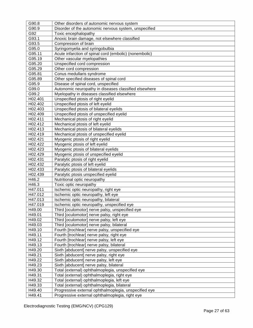
Electrodiagnostic Testing (EMG/NCV) (CPG129) Page 27 of 63
G90.8 Other disorders of autonomic nervous system G90.9 Disorder of the autonomic nervous system, unspecified G92 Toxic encephalopathy G93.1 Anoxic brain damage, not elsewhere classified G93.5 Compression of brain G95.0 Syringomyelia and syringobulbia G95.11 Acute infarction of spinal cord (embolic) (nonembolic) G95.19 Other vascular myelopathies G95.20 Unspecified cord compression G95.29 Other cord compression G95.81 Conus medullaris syndrome G95.89 Other specified diseases of spinal cord G95.9 Disease of spinal cord, unspecified G99.0 Autonomic neuropathy in diseases classified elsewhere G99.2 Myelopathy in diseases classified elsewhere H02.401 Unspecified ptosis of right eyelid H02.402 Unspecified ptosis of left eyelid H02.403 Unspecified ptosis of bilateral eyelids H02.409 Unspecified ptosis of unspecified eyelid H02.411 Mechanical ptosis of right eyelid H02.412 Mechanical ptosis of left eyelid H02.413 Mechanical ptosis of bilateral eyelids H02.419 Mechanical ptosis of unspecified eyelid H02.421 Myogenic ptosis of right eyelid H02.422 Myogenic ptosis of left eyelid H02.423 Myogenic ptosis of bilateral eyelids H02.429 Myogenic ptosis of unspecified eyelid H02.431 Paralytic ptosis of right eyelid H02.432 Paralytic ptosis of left eyelid H02.433 Paralytic ptosis of bilateral eyelids H02.439 Paralytic ptosis unspecified eyelid H46.2 Nutritional optic neuropathy H46.3 Toxic optic neuropathy H47.011 Ischemic optic neuropathy, right eye H47.012 Ischemic optic neuropathy, left eye H47.013 Ischemic optic neuropathy, bilateral H47.019 Ischemic optic neuropathy, unspecified eye H49.00 Third [oculomotor] nerve palsy, unspecified eye H49.01 Third [oculomotor] nerve palsy, right eye H49.02 Third [oculomotor] nerve palsy, left eye H49.03 Third [oculomotor] nerve palsy, bilateral H49.10 Fourth [trochlear] nerve palsy, unspecified eye H49.11 Fourth [trochlear] nerve palsy, right eye H49.12 Fourth [trochlear] nerve palsy, left eye H49.13 Fourth [trochlear] nerve palsy, bilateral H49.20 Sixth [abducent] nerve palsy, unspecified eye H49.21 Sixth [abducent] nerve palsy, right eye H49.22 Sixth [abducent] nerve palsy, left eye H49.23 Sixth [abducent] nerve palsy, bilateral H49.30 Total (external) ophthalmoplegia, unspecified eye H49.31 Total (external) ophthalmoplegia, right eye H49.32 Total (external) ophthalmoplegia, left eye H49.33 Total (external) ophthalmoplegia, bilateral H49.40 Progressive external ophthalmoplegia, unspecified eye H49.41 Progressive external ophthalmoplegia, right eye
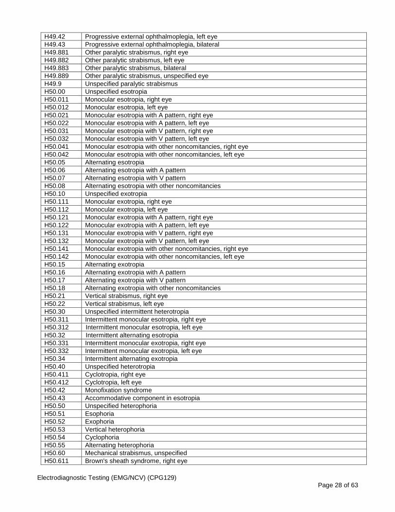
Electrodiagnostic Testing (EMG/NCV) (CPG129) Page 28 of 63
H49.42 Progressive external ophthalmoplegia, left eye H49.43 Progressive external ophthalmoplegia, bilateral H49.881 Other paralytic strabismus, right eye H49.882 Other paralytic strabismus, left eye H49.883 Other paralytic strabismus, bilateral H49.889 Other paralytic strabismus, unspecified eye H49.9 Unspecified paralytic strabismus H50.00 Unspecified esotropia H50.011 Monocular esotropia, right eye H50.012 Monocular esotropia, left eye H50.021 Monocular esotropia with A pattern, right eye H50.022 Monocular esotropia with A pattern, left eye H50.031 Monocular esotropia with V pattern, right eye H50.032 Monocular esotropia with V pattern, left eye H50.041 Monocular esotropia with other noncomitancies, right eye H50.042 Monocular esotropia with other noncomitancies, left eye H50.05 Alternating esotropia H50.06 Alternating esotropia with A pattern H50.07 Alternating esotropia with V pattern H50.08 Alternating esotropia with other noncomitancies H50.10 Unspecified exotropia H50.111 Monocular exotropia, right eye H50.112 Monocular exotropia, left eye H50.121 Monocular exotropia with A pattern, right eye H50.122 Monocular exotropia with A pattern, left eye H50.131 Monocular exotropia with V pattern, right eye H50.132 Monocular exotropia with V pattern, left eye H50.141 Monocular exotropia with other noncomitancies, right eye H50.142 Monocular exotropia with other noncomitancies, left eye H50.15 Alternating exotropia H50.16 Alternating exotropia with A pattern H50.17 Alternating exotropia with V pattern H50.18 Alternating exotropia with other noncomitancies H50.21 Vertical strabismus, right eye H50.22 Vertical strabismus, left eye H50.30 Unspecified intermittent heterotropia H50.311 Intermittent monocular esotropia, right eye H50.312 Intermittent monocular esotropia, left eye
H50.32 Intermittent alternating esotropia
H50.331 Intermittent monocular exotropia, right eye H50.332 Intermittent monocular exotropia, left eye H50.34 Intermittent alternating exotropia H50.40 Unspecified heterotropia H50.411 Cyclotropia, right eye H50.412 Cyclotropia, left eye H50.42 Monofixation syndrome H50.43 Accommodative component in esotropia H50.50 Unspecified heterophoria H50.51 Esophoria H50.52 Exophoria H50.53 Vertical heterophoria H50.54 Cyclophoria H50.55 Alternating heterophoria H50.60 Mechanical strabismus, unspecified H50.611 Brown's sheath syndrome, right eye

Electrodiagnostic Testing (EMG/NCV) (CPG129) Page 29 of 63
H50.612 Brown's sheath syndrome, left eye H50.69 Other mechanical strabismus H50.811 Duane's syndrome, right eye H50.812 Duane's syndrome, left eye H50.89 Other specified strabismus H51.0 Palsy (spasm) of conjugate gaze H51.11 Convergence insufficiency H51.12 Convergence excess H51.20 Internuclear ophthalmoplegia, unspecified eye H51.21 Internuclear ophthalmoplegia, right eye H51.22 Internuclear ophthalmoplegia, left eye H51.23 Internuclear ophthalmoplegia, bilateral H51.8 Other specified disorders of binocular movement H51.9 Unspecified disorder of binocular movement H53.2 Diplopia H71.00 Cholesteatoma of attic, unspecified ear H71.01 Cholesteatoma of attic, right ear H71.02 Cholesteatoma of attic, left ear H71.03 Cholesteatoma of attic, bilateral H71.10 Cholesteatoma of tympanum, unspecified ear H71.11 Cholesteatoma of tympanum, right ear H71.12 Cholesteatoma of tympanum, left ear H71.13 Cholesteatoma of tympanum, bilateral H71.20 Cholesteatoma of mastoid, unspecified ear H71.21 Cholesteatoma of mastoid, right ear H71.22 Cholesteatoma of mastoid, left ear H71.23 Cholesteatoma of mastoid, bilateral H71.30 Diffuse cholesteatosis, unspecified ear H71.31 Diffuse cholesteatosis, right ear H71.32 Diffuse cholesteatosis, left ear H71.33 Diffuse cholesteatosis, bilateral H71.90 Unspecified cholesteatoma, unspecified ear H71.91 Unspecified cholesteatoma, right ear H71.92 Unspecified cholesteatoma, left ear H71.93 Unspecified cholesteatoma, bilateral H72.00 Central perforation of tympanic membrane, unspecified ear H72.01 Central perforation of tympanic membrane, right ear H72.02 Central perforation of tympanic membrane, left ear H72.03 Central perforation of tympanic membrane, bilateral H72.10 Attic perforation of tympanic membrane, unspecified ear H72.11 Attic perforation of tympanic membrane, right ear H72.12 Attic perforation of tympanic membrane, left ear H72.13 Attic perforation of tympanic membrane, bilateral H72.2X1 Other marginal perforations of tympanic membrane, right ear H72.2X2 Other marginal perforations of tympanic membrane, left ear H72.2X3 Other marginal perforations of tympanic membrane, bilateral H72.2X9 Other marginal perforations of tympanic membrane, unspecified ear H72.811 Multiple perforations of tympanic membrane, right ear H72.812 Multiple perforations of tympanic membrane, left ear H72.813 Multiple perforations of tympanic membrane, bilateral H72.819 Multiple perforations of tympanic membrane, unspecified ear H72.821 Total perforations of tympanic membrane, right ear H72.822 Total perforations of tympanic membrane, left ear H72.823 Total perforations of tympanic membrane, bilateral ear H72.829 Total perforations of tympanic membrane, unspecified ear
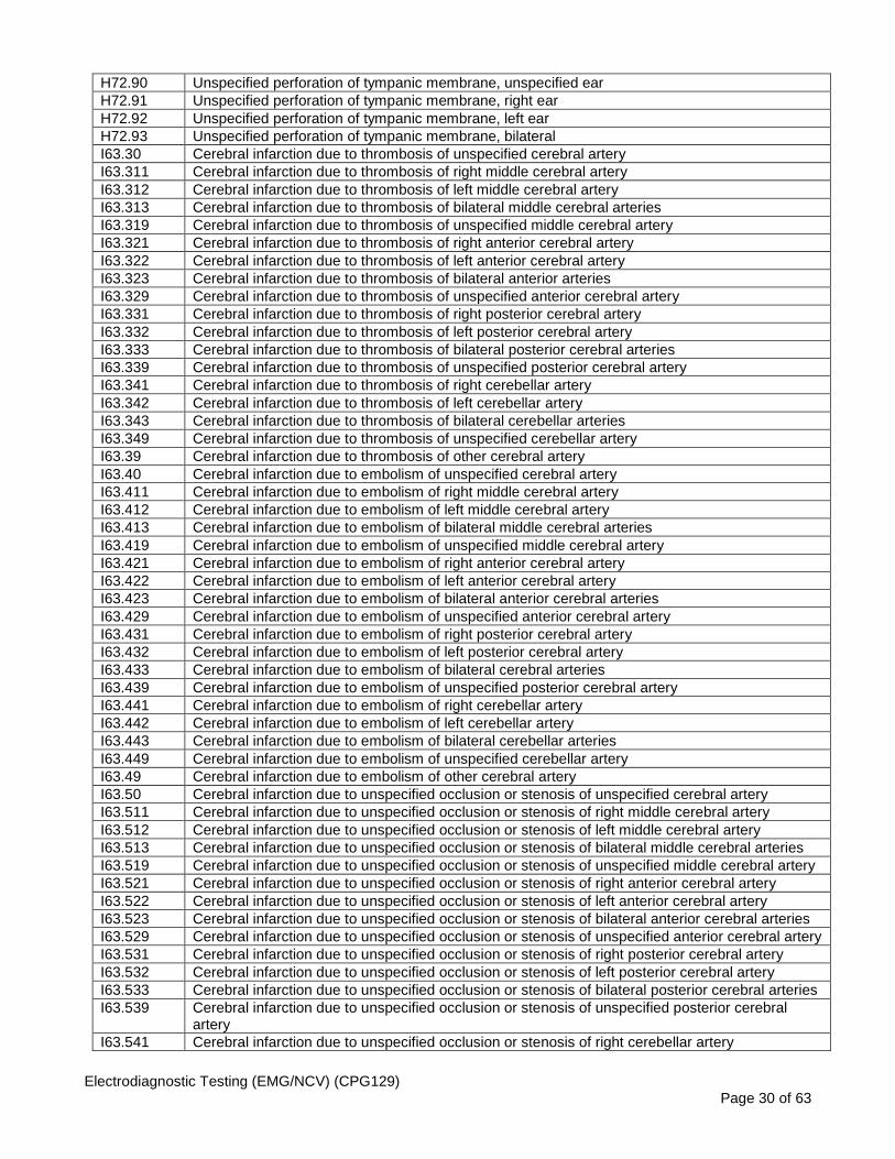
Electrodiagnostic Testing (EMG/NCV) (CPG129) Page 30 of 63
H72.90 Unspecified perforation of tympanic membrane, unspecified ear H72.91 Unspecified perforation of tympanic membrane, right ear H72.92 Unspecified perforation of tympanic membrane, left ear H72.93 Unspecified perforation of tympanic membrane, bilateral I63.30 Cerebral infarction due to thrombosis of unspecified cerebral artery I63.311 Cerebral infarction due to thrombosis of right middle cerebral artery I63.312 Cerebral infarction due to thrombosis of left middle cerebral artery I63.313 Cerebral infarction due to thrombosis of bilateral middle cerebral arteries I63.319 Cerebral infarction due to thrombosis of unspecified middle cerebral artery I63.321 Cerebral infarction due to thrombosis of right anterior cerebral artery I63.322 Cerebral infarction due to thrombosis of left anterior cerebral artery I63.323 Cerebral infarction due to thrombosis of bilateral anterior arteries I63.329 Cerebral infarction due to thrombosis of unspecified anterior cerebral artery I63.331 Cerebral infarction due to thrombosis of right posterior cerebral artery I63.332 Cerebral infarction due to thrombosis of left posterior cerebral artery I63.333 Cerebral infarction due to thrombosis of bilateral posterior cerebral arteries I63.339 Cerebral infarction due to thrombosis of unspecified posterior cerebral artery I63.341 Cerebral infarction due to thrombosis of right cerebellar artery I63.342 Cerebral infarction due to thrombosis of left cerebellar artery I63.343 Cerebral infarction due to thrombosis of bilateral cerebellar arteries I63.349 Cerebral infarction due to thrombosis of unspecified cerebellar artery I63.39 Cerebral infarction due to thrombosis of other cerebral artery I63.40 Cerebral infarction due to embolism of unspecified cerebral artery I63.411 Cerebral infarction due to embolism of right middle cerebral artery I63.412 Cerebral infarction due to embolism of left middle cerebral artery I63.413 Cerebral infarction due to embolism of bilateral middle cerebral arteries I63.419 Cerebral infarction due to embolism of unspecified middle cerebral artery I63.421 Cerebral infarction due to embolism of right anterior cerebral artery I63.422 Cerebral infarction due to embolism of left anterior cerebral artery I63.423 Cerebral infarction due to embolism of bilateral anterior cerebral arteries I63.429 Cerebral infarction due to embolism of unspecified anterior cerebral artery I63.431 Cerebral infarction due to embolism of right posterior cerebral artery I63.432 Cerebral infarction due to embolism of left posterior cerebral artery I63.433 Cerebral infarction due to embolism of bilateral cerebral arteries I63.439 Cerebral infarction due to embolism of unspecified posterior cerebral artery I63.441 Cerebral infarction due to embolism of right cerebellar artery I63.442 Cerebral infarction due to embolism of left cerebellar artery I63.443 Cerebral infarction due to embolism of bilateral cerebellar arteries I63.449 Cerebral infarction due to embolism of unspecified cerebellar artery I63.49 Cerebral infarction due to embolism of other cerebral artery I63.50 Cerebral infarction due to unspecified occlusion or stenosis of unspecified cerebral artery I63.511 Cerebral infarction due to unspecified occlusion or stenosis of right middle cerebral artery I63.512 Cerebral infarction due to unspecified occlusion or stenosis of left middle cerebral artery I63.513 Cerebral infarction due to unspecified occlusion or stenosis of bilateral middle cerebral arteries I63.519 Cerebral infarction due to unspecified occlusion or stenosis of unspecified middle cerebral artery I63.521 Cerebral infarction due to unspecified occlusion or stenosis of right anterior cerebral artery I63.522 Cerebral infarction due to unspecified occlusion or stenosis of left anterior cerebral artery I63.523 Cerebral infarction due to unspecified occlusion or stenosis of bilateral anterior cerebral arteries I63.529 Cerebral infarction due to unspecified occlusion or stenosis of unspecified anterior cerebral artery I63.531 Cerebral infarction due to unspecified occlusion or stenosis of right posterior cerebral artery I63.532 Cerebral infarction due to unspecified occlusion or stenosis of left posterior cerebral artery I63.533 Cerebral infarction due to unspecified occlusion or stenosis of bilateral posterior cerebral arteries I63.539 Cerebral infarction due to unspecified occlusion or stenosis of unspecified posterior cerebral
artery I63.541 Cerebral infarction due to unspecified occlusion or stenosis of right cerebellar artery
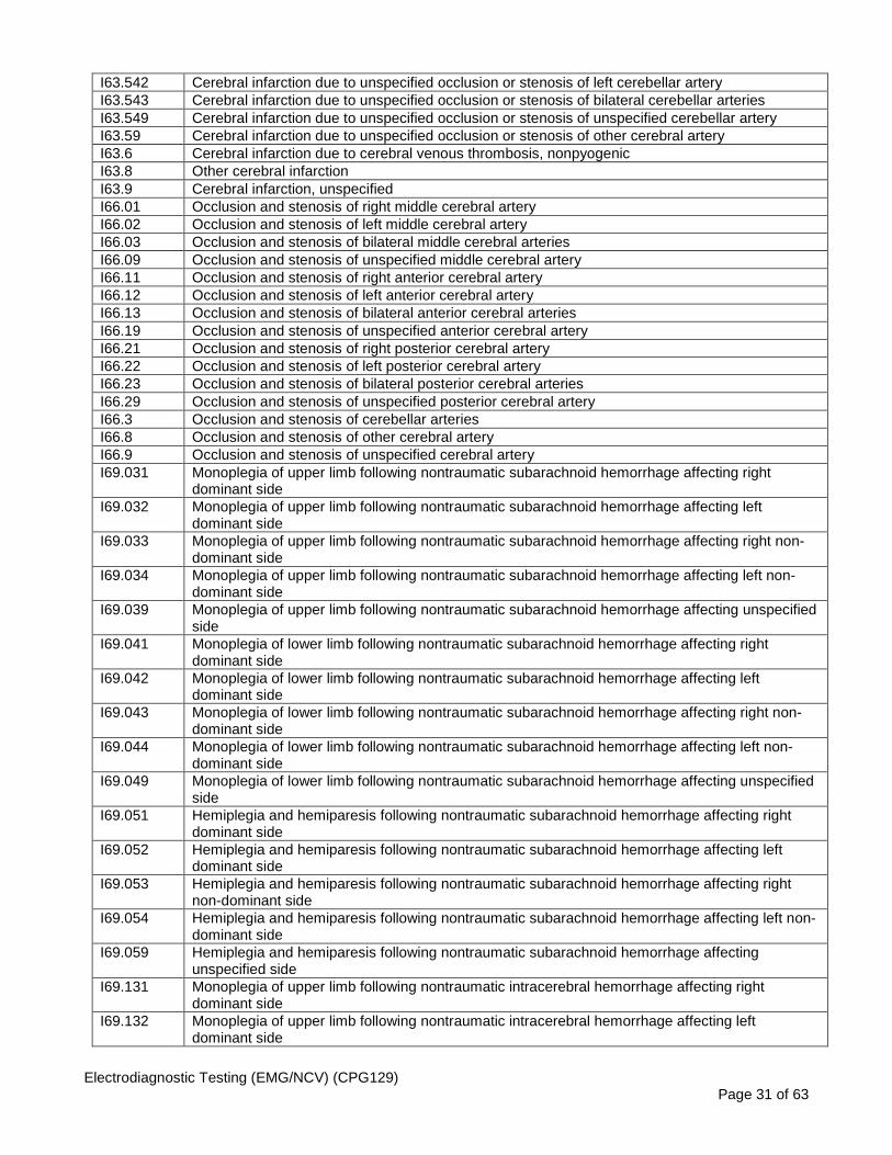
Electrodiagnostic Testing (EMG/NCV) (CPG129) Page 31 of 63
I63.542 Cerebral infarction due to unspecified occlusion or stenosis of left cerebellar artery I63.543 Cerebral infarction due to unspecified occlusion or stenosis of bilateral cerebellar arteries I63.549 Cerebral infarction due to unspecified occlusion or stenosis of unspecified cerebellar artery I63.59 Cerebral infarction due to unspecified occlusion or stenosis of other cerebral artery I63.6 Cerebral infarction due to cerebral venous thrombosis, nonpyogenic I63.8 Other cerebral infarction I63.9 Cerebral infarction, unspecified I66.01 Occlusion and stenosis of right middle cerebral artery I66.02 Occlusion and stenosis of left middle cerebral artery I66.03 Occlusion and stenosis of bilateral middle cerebral arteries I66.09 Occlusion and stenosis of unspecified middle cerebral artery I66.11 Occlusion and stenosis of right anterior cerebral artery I66.12 Occlusion and stenosis of left anterior cerebral artery I66.13 Occlusion and stenosis of bilateral anterior cerebral arteries I66.19 Occlusion and stenosis of unspecified anterior cerebral artery I66.21 Occlusion and stenosis of right posterior cerebral artery I66.22 Occlusion and stenosis of left posterior cerebral artery I66.23 Occlusion and stenosis of bilateral posterior cerebral arteries I66.29 Occlusion and stenosis of unspecified posterior cerebral artery I66.3 Occlusion and stenosis of cerebellar arteries I66.8 Occlusion and stenosis of other cerebral artery I66.9 Occlusion and stenosis of unspecified cerebral artery I69.031 Monoplegia of upper limb following nontraumatic subarachnoid hemorrhage affecting right
dominant side I69.032 Monoplegia of upper limb following nontraumatic subarachnoid hemorrhage affecting left
dominant side I69.033 Monoplegia of upper limb following nontraumatic subarachnoid hemorrhage affecting right non-
dominant side I69.034 Monoplegia of upper limb following nontraumatic subarachnoid hemorrhage affecting left non-
dominant side I69.039 Monoplegia of upper limb following nontraumatic subarachnoid hemorrhage affecting unspecified
side I69.041 Monoplegia of lower limb following nontraumatic subarachnoid hemorrhage affecting right
dominant side I69.042 Monoplegia of lower limb following nontraumatic subarachnoid hemorrhage affecting left
dominant side I69.043 Monoplegia of lower limb following nontraumatic subarachnoid hemorrhage affecting right non-
dominant side I69.044 Monoplegia of lower limb following nontraumatic subarachnoid hemorrhage affecting left non-
dominant side I69.049 Monoplegia of lower limb following nontraumatic subarachnoid hemorrhage affecting unspecified
side I69.051 Hemiplegia and hemiparesis following nontraumatic subarachnoid hemorrhage affecting right
dominant side I69.052 Hemiplegia and hemiparesis following nontraumatic subarachnoid hemorrhage affecting left
dominant side I69.053 Hemiplegia and hemiparesis following nontraumatic subarachnoid hemorrhage affecting right
non-dominant side I69.054 Hemiplegia and hemiparesis following nontraumatic subarachnoid hemorrhage affecting left non-
dominant side I69.059 Hemiplegia and hemiparesis following nontraumatic subarachnoid hemorrhage affecting
unspecified side I69.131 Monoplegia of upper limb following nontraumatic intracerebral hemorrhage affecting right
dominant side I69.132 Monoplegia of upper limb following nontraumatic intracerebral hemorrhage affecting left
dominant side
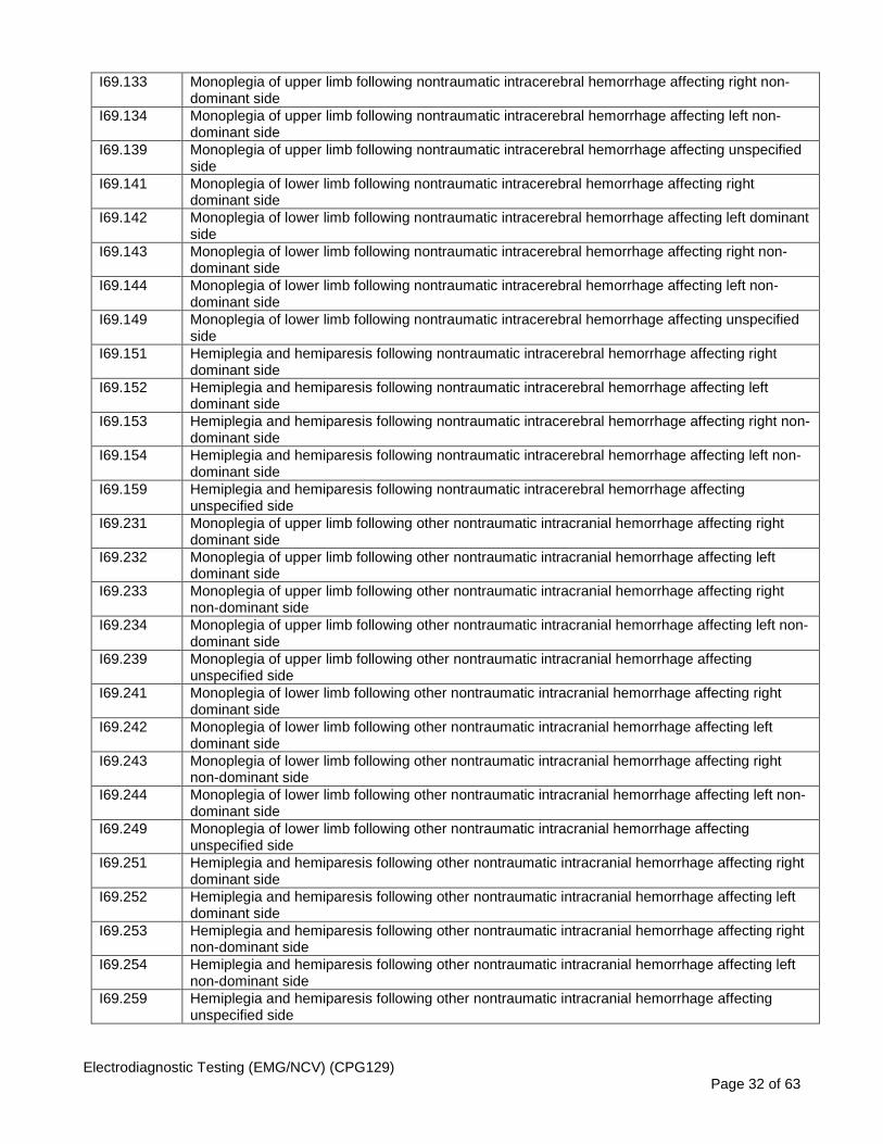
Electrodiagnostic Testing (EMG/NCV) (CPG129) Page 32 of 63
I69.133 Monoplegia of upper limb following nontraumatic intracerebral hemorrhage affecting right non-dominant side
I69.134 Monoplegia of upper limb following nontraumatic intracerebral hemorrhage affecting left non-dominant side
I69.139 Monoplegia of upper limb following nontraumatic intracerebral hemorrhage affecting unspecified side
I69.141 Monoplegia of lower limb following nontraumatic intracerebral hemorrhage affecting right dominant side
I69.142 Monoplegia of lower limb following nontraumatic intracerebral hemorrhage affecting left dominant side
I69.143 Monoplegia of lower limb following nontraumatic intracerebral hemorrhage affecting right non-dominant side
I69.144 Monoplegia of lower limb following nontraumatic intracerebral hemorrhage affecting left non-dominant side
I69.149 Monoplegia of lower limb following nontraumatic intracerebral hemorrhage affecting unspecified side
I69.151 Hemiplegia and hemiparesis following nontraumatic intracerebral hemorrhage affecting right dominant side
I69.152 Hemiplegia and hemiparesis following nontraumatic intracerebral hemorrhage affecting left dominant side
I69.153 Hemiplegia and hemiparesis following nontraumatic intracerebral hemorrhage affecting right non-dominant side
I69.154 Hemiplegia and hemiparesis following nontraumatic intracerebral hemorrhage affecting left non-dominant side
I69.159 Hemiplegia and hemiparesis following nontraumatic intracerebral hemorrhage affecting unspecified side
I69.231 Monoplegia of upper limb following other nontraumatic intracranial hemorrhage affecting right dominant side
I69.232 Monoplegia of upper limb following other nontraumatic intracranial hemorrhage affecting left dominant side
I69.233 Monoplegia of upper limb following other nontraumatic intracranial hemorrhage affecting right non-dominant side
I69.234 Monoplegia of upper limb following other nontraumatic intracranial hemorrhage affecting left non-dominant side
I69.239 Monoplegia of upper limb following other nontraumatic intracranial hemorrhage affecting unspecified side
I69.241 Monoplegia of lower limb following other nontraumatic intracranial hemorrhage affecting right dominant side
I69.242 Monoplegia of lower limb following other nontraumatic intracranial hemorrhage affecting left dominant side
I69.243 Monoplegia of lower limb following other nontraumatic intracranial hemorrhage affecting right non-dominant side
I69.244 Monoplegia of lower limb following other nontraumatic intracranial hemorrhage affecting left non-dominant side
I69.249 Monoplegia of lower limb following other nontraumatic intracranial hemorrhage affecting unspecified side
I69.251 Hemiplegia and hemiparesis following other nontraumatic intracranial hemorrhage affecting right dominant side
I69.252 Hemiplegia and hemiparesis following other nontraumatic intracranial hemorrhage affecting left dominant side
I69.253 Hemiplegia and hemiparesis following other nontraumatic intracranial hemorrhage affecting right non-dominant side
I69.254 Hemiplegia and hemiparesis following other nontraumatic intracranial hemorrhage affecting left non-dominant side
I69.259 Hemiplegia and hemiparesis following other nontraumatic intracranial hemorrhage affecting unspecified side

Electrodiagnostic Testing (EMG/NCV) (CPG129) Page 33 of 63
I69.331 Monoplegia of upper limb following cerebral infarction affecting right dominant side I69.332 Monoplegia of upper limb following cerebral infarction affecting left dominant side I69.333 Monoplegia of upper limb following cerebral infarction affecting right non-dominant side I69.334 Monoplegia of upper limb following cerebral infarction affecting left non-dominant side I69.339 Monoplegia of upper limb following cerebral infarction affecting unspecified side I69.341 Monoplegia of lower limb following cerebral infarction affecting right dominant side I69.342 Monoplegia of lower limb following cerebral infarction affecting left dominant side I69.343 Monoplegia of lower limb following cerebral infarction affecting right non-dominant side I69.344 Monoplegia of lower limb following cerebral infarction affecting left non-dominant side I69.349 Monoplegia of lower limb following cerebral infarction affecting unspecified side I69.351 Hemiplegia and hemiparesis following cerebral infarction affecting right dominant side I69.352 Hemiplegia and hemiparesis following cerebral infarction affecting left dominant side I69.353 Hemiplegia and hemiparesis following cerebral infarction affecting right non-dominant side I69.354 Hemiplegia and hemiparesis following cerebral infarction affecting left non-dominant side I69.359 Hemiplegia and hemiparesis following cerebral infarction affecting unspecified side I69.831 Monoplegia of upper limb following other cerebrovascular disease affecting right dominant side I69.832 Monoplegia of upper limb following other cerebrovascular disease affecting left dominant side I69.833 Monoplegia of upper limb following other cerebrovascular disease affecting right non-dominant
side I69.834 Monoplegia of upper limb following other cerebrovascular disease affecting left non-dominant
side I69.839 Monoplegia of upper limb following other cerebrovascular disease affecting unspecified side I69.841 Monoplegia of lower limb following other cerebrovascular disease affecting right dominant side I69.842 Monoplegia of lower limb following other cerebrovascular disease affecting left dominant side I69.843 Monoplegia of lower limb following other cerebrovascular disease affecting right non-dominant
side I69.844 Monoplegia of lower limb following other cerebrovascular disease affecting left non-dominant
side I69.849 Monoplegia of lower limb following other cerebrovascular disease affecting unspecified side I69.851 Hemiplegia and hemiparesis following other cerebrovascular disease affecting right dominant
side I69.852 Hemiplegia and hemiparesis following other cerebrovascular disease affecting left dominant side I69.853 Hemiplegia and hemiparesis following other cerebrovascular disease affecting right non-
dominant side I69.854 Hemiplegia and hemiparesis following other cerebrovascular disease affecting left non-dominant
side I69.859 Hemiplegia and hemiparesis following other cerebrovascular disease affecting unspecified side I69.931 Monoplegia of upper limb following unspecified cerebrovascular disease affecting right dominant
side I69.932 Monoplegia of upper limb following unspecified cerebrovascular disease affecting left dominant
side I69.933 Monoplegia of upper limb following unspecified cerebrovascular disease affecting right non-
dominant side I69.934 Monoplegia of upper limb following unspecified cerebrovascular disease affecting left non-
dominant side I69.939 Monoplegia of upper limb following unspecified cerebrovascular disease affecting unspecified
side I69.941 Monoplegia of lower limb following unspecified cerebrovascular disease affecting right dominant
side I69.942 Monoplegia of lower limb following unspecified cerebrovascular disease affecting left dominant
side I69.943 Monoplegia of lower limb following unspecified cerebrovascular disease affecting right non-
dominant side I69.944 Monoplegia of lower limb following unspecified cerebrovascular disease affecting left non-
dominant side
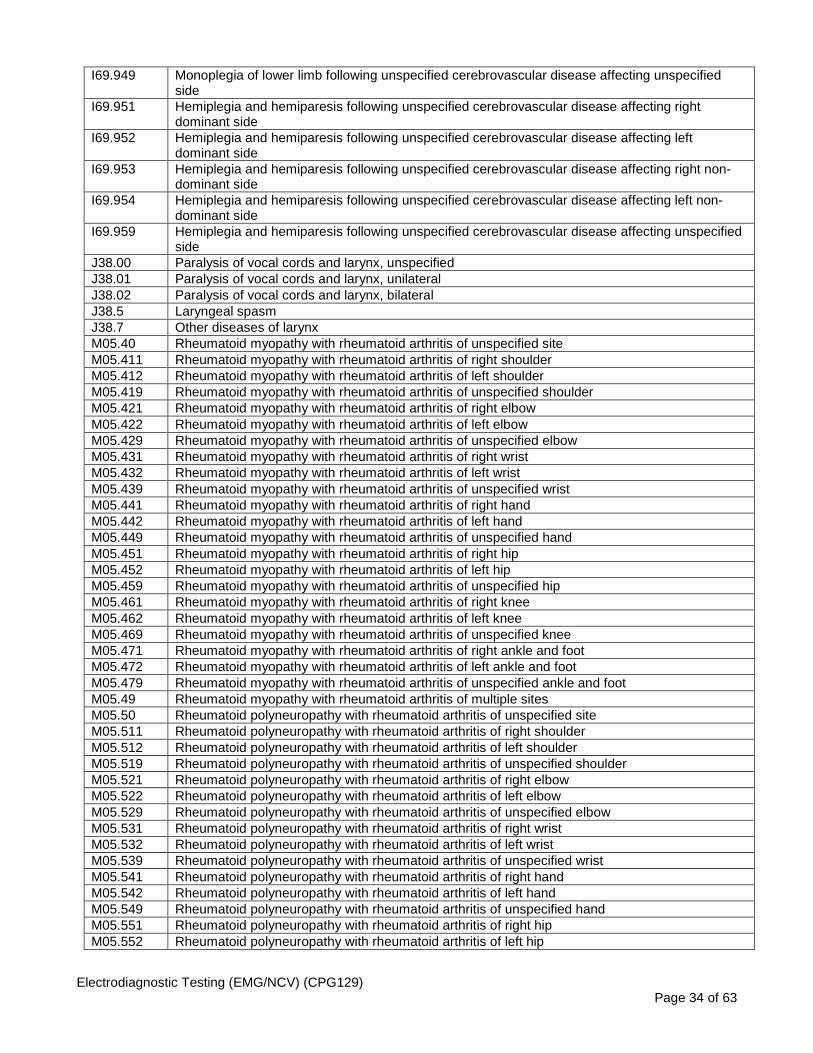
Electrodiagnostic Testing (EMG/NCV) (CPG129) Page 34 of 63
I69.949 Monoplegia of lower limb following unspecified cerebrovascular disease affecting unspecified side
I69.951 Hemiplegia and hemiparesis following unspecified cerebrovascular disease affecting right dominant side
I69.952 Hemiplegia and hemiparesis following unspecified cerebrovascular disease affecting left dominant side
I69.953 Hemiplegia and hemiparesis following unspecified cerebrovascular disease affecting right non-dominant side
I69.954 Hemiplegia and hemiparesis following unspecified cerebrovascular disease affecting left non-dominant side
I69.959 Hemiplegia and hemiparesis following unspecified cerebrovascular disease affecting unspecified side
J38.00 Paralysis of vocal cords and larynx, unspecified J38.01 Paralysis of vocal cords and larynx, unilateral J38.02 Paralysis of vocal cords and larynx, bilateral J38.5 Laryngeal spasm J38.7 Other diseases of larynx M05.40 Rheumatoid myopathy with rheumatoid arthritis of unspecified site M05.411 Rheumatoid myopathy with rheumatoid arthritis of right shoulder M05.412 Rheumatoid myopathy with rheumatoid arthritis of left shoulder M05.419 Rheumatoid myopathy with rheumatoid arthritis of unspecified shoulder M05.421 Rheumatoid myopathy with rheumatoid arthritis of right elbow M05.422 Rheumatoid myopathy with rheumatoid arthritis of left elbow M05.429 Rheumatoid myopathy with rheumatoid arthritis of unspecified elbow M05.431 Rheumatoid myopathy with rheumatoid arthritis of right wrist M05.432 Rheumatoid myopathy with rheumatoid arthritis of left wrist M05.439 Rheumatoid myopathy with rheumatoid arthritis of unspecified wrist M05.441 Rheumatoid myopathy with rheumatoid arthritis of right hand M05.442 Rheumatoid myopathy with rheumatoid arthritis of left hand M05.449 Rheumatoid myopathy with rheumatoid arthritis of unspecified hand M05.451 Rheumatoid myopathy with rheumatoid arthritis of right hip M05.452 Rheumatoid myopathy with rheumatoid arthritis of left hip M05.459 Rheumatoid myopathy with rheumatoid arthritis of unspecified hip M05.461 Rheumatoid myopathy with rheumatoid arthritis of right knee M05.462 Rheumatoid myopathy with rheumatoid arthritis of left knee M05.469 Rheumatoid myopathy with rheumatoid arthritis of unspecified knee M05.471 Rheumatoid myopathy with rheumatoid arthritis of right ankle and foot M05.472 Rheumatoid myopathy with rheumatoid arthritis of left ankle and foot M05.479 Rheumatoid myopathy with rheumatoid arthritis of unspecified ankle and foot M05.49 Rheumatoid myopathy with rheumatoid arthritis of multiple sites M05.50 Rheumatoid polyneuropathy with rheumatoid arthritis of unspecified site M05.511 Rheumatoid polyneuropathy with rheumatoid arthritis of right shoulder M05.512 Rheumatoid polyneuropathy with rheumatoid arthritis of left shoulder M05.519 Rheumatoid polyneuropathy with rheumatoid arthritis of unspecified shoulder M05.521 Rheumatoid polyneuropathy with rheumatoid arthritis of right elbow M05.522 Rheumatoid polyneuropathy with rheumatoid arthritis of left elbow M05.529 Rheumatoid polyneuropathy with rheumatoid arthritis of unspecified elbow M05.531 Rheumatoid polyneuropathy with rheumatoid arthritis of right wrist M05.532 Rheumatoid polyneuropathy with rheumatoid arthritis of left wrist M05.539 Rheumatoid polyneuropathy with rheumatoid arthritis of unspecified wrist M05.541 Rheumatoid polyneuropathy with rheumatoid arthritis of right hand M05.542 Rheumatoid polyneuropathy with rheumatoid arthritis of left hand M05.549 Rheumatoid polyneuropathy with rheumatoid arthritis of unspecified hand M05.551 Rheumatoid polyneuropathy with rheumatoid arthritis of right hip M05.552 Rheumatoid polyneuropathy with rheumatoid arthritis of left hip

Electrodiagnostic Testing (EMG/NCV) (CPG129) Page 35 of 63
M05.559 Rheumatoid polyneuropathy with rheumatoid arthritis of unspecified hip M05.561 Rheumatoid polyneuropathy with rheumatoid arthritis of right knee M05.562 Rheumatoid polyneuropathy with rheumatoid arthritis of left knee M05.569 Rheumatoid polyneuropathy with rheumatoid arthritis of unspecified knee M05.571 Rheumatoid polyneuropathy with rheumatoid arthritis of right ankle and foot M05.572 Rheumatoid polyneuropathy with rheumatoid arthritis of left ankle and foot M05.579 Rheumatoid polyneuropathy with rheumatoid arthritis of unspecified ankle and foot M05.59 Rheumatoid polyneuropathy with rheumatoid arthritis of multiple sites M21.331 Wrist drop, right wrist M21.332 Wrist drop, left wrist M21.339 Wrist drop, unspecified wrist M21.511 Acquired clawhand, right hand M21.512 Acquired clawhand, left hand M21.519 Acquired clawhand, unspecified hand M21.521 Acquired clubhand, right hand M21.522 Acquired clubhand, left hand M21.529 Acquired clubhand, unspecified hand M21.531 Acquired clawfoot, right foot M21.532 Acquired clawfoot, left foot M21.539 Acquired clawfoot, unspecified foot M21.541 Acquired clubfoot, right foot M21.542 Acquired clubfoot, left foot M21.549 Acquired clubfoot, unspecified foot M21.6X1 Other acquired deformities of right foot M21.6X2 Other acquired deformities of left foot M21.6X9 Other acquired deformities of unspecified foot M21.831 Other specified acquired deformities of right forearm M21.832 Other specified acquired deformities of left forearm M21.839 Other specified acquired deformities of unspecified forearm M25.50 Pain in unspecified joint M25.511 Pain in right shoulder M25.512 Pain in left shoulder M25.519 Pain in unspecified shoulder M25.521 Pain in right elbow M25.522 Pain in left elbow M25.529 Pain in unspecified elbow M25.531 Pain in right wrist M25.532 Pain in left wrist M25.539 Pain in unspecified wrist M25.541 Pain in joints of right hand M25.542 Pain in joints of left hand M25.549 Pain in joints of unspecified hand M25.551 Pain in right hip M25.552 Pain in left hip M25.559 Pain in unspecified hip M25.561 Pain in right knee M25.562 Pain in left knee M25.569 Pain in unspecified knee M25.571 Pain in right ankle and joints of right foot
M25.572 Pain in left ankle and joints of left foot M25.579 Pain in unspecified ankle and joints of unspecified foot M33.00 Juvenile dermatomyositis, organ involvement unspecified M33.01 Juvenile dermatomyositis with respiratory involvement M33.02 Juvenile dermatomyositis with myopathy M33.09 Juvenile dermatomyositis with other organ involvement

Electrodiagnostic Testing (EMG/NCV) (CPG129) Page 36 of 63
M33.10 Other dermatomyositis, organ involvement unspecified M33.11 Other dermatomyositis with respiratory involvement M33.12 Other dermatomyositis with myopathy M33.19 Other dermatomyositis with other organ involvement M33.20 Polymyositis, organ involvement unspecified M33.21 Polymyositis with respiratory involvement M33.22 Polymyositis with myopathy M33.29 Polymyositis with other organ involvement M33.90 Dermatopolymyositis, unspecified, organ involvement unspecified M33.91 Dermatopolymyositis, unspecified with respiratory involvement M33.92 Dermatopolymyositis, unspecified with myopathy M33.99 Dermatopolymyositis, unspecified with other organ involvement M34.83 Systemic sclerosis with polyneuropathy M35.3 Polymyalgia rheumatica M36.0 Dermato(poly)myositis in neoplastic disease M41.00 Infantile idiopathic scoliosis, site unspecified M41.02 Infantile idiopathic scoliosis, cervical region M41.03 Infantile idiopathic scoliosis, cervicothoracic region M41.04 Infantile idiopathic scoliosis, thoracic region M41.05 Infantile idiopathic scoliosis, thoracolumbar region M41.06 Infantile idiopathic scoliosis, lumbar region
M41.07
Infantile idiopathic scoliosis, lumbosacral region
M41.08 Infantile idiopathic scoliosis, sacral and sacrococcygeal region M41.112 Juvenile idiopathic scoliosis, cervical region M41.113 Juvenile idiopathic scoliosis, cervicothoracic region M41.114 Juvenile idiopathic scoliosis, thoracic region M41.115 Juvenile idiopathic scoliosis, thoracolumbar region M41.116 Juvenile idiopathic scoliosis, lumbar region M41.117 Juvenile idiopathic scoliosis, lumbosacral region M41.119 Juvenile idiopathic scoliosis, site unspecified M41.122 Adolescent idiopathic scoliosis, cervical region M41.123 Adolescent idiopathic scoliosis, cervicothoracic region M41.124 Adolescent idiopathic scoliosis, thoracic region M41.125 Adolescent idiopathic scoliosis, thoracolumbar region M41.126 Adolescent idiopathic scoliosis, lumbar region M41.127 Adolescent idiopathic scoliosis, lumbosacral region M41.129 Adolescent idiopathic scoliosis, site unspecified M41.20 Other idiopathic scoliosis, site unspecified M41.22 Other idiopathic scoliosis, cervical region M41.23 Other idiopathic scoliosis, cervicothoracic region M41.24 Other idiopathic scoliosis, thoracic region M41.25 Other idiopathic scoliosis, thoracolumbar region M41.26 Other idiopathic scoliosis, lumbar region M41.27 Other idiopathic scoliosis, lumbosacral region M43.00 Spondylolysis, site unspecified M43.01 Spondylolysis, occipito-atlanto-axial region M43.02 Spondylolysis, cervical region M43.03 Spondylolysis, cervicothoracic region M43.04 Spondylolysis, thoracic region M43.05 Spondylolysis, thoracolumbar region M43.06 Spondylolysis, lumbar region M43.07 Spondylolysis, lumbosacral region M43.08 Spondylolysis, sacral and sacrococcygeal region M43.09 Spondylolysis, multiple sites in spine M43.10 Spondylolisthesis, site unspecified
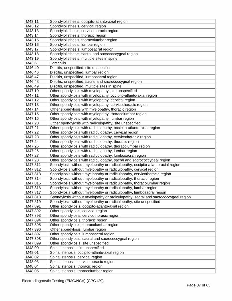
Electrodiagnostic Testing (EMG/NCV) (CPG129) Page 37 of 63
M43.11 Spondylolisthesis, occipito-atlanto-axial region M43.12 Spondylolisthesis, cervical region M43.13 Spondylolisthesis, cervicothoracic region M43.14 Spondylolisthesis, thoracic region M43.15 Spondylolisthesis, thoracolumbar region M43.16 Spondylolisthesis, lumbar region M43.17 Spondylolisthesis, lumbosacral region M43.18 Spondylolisthesis, sacral and sacrococcygeal region M43.19 Spondylolisthesis, multiple sites in spine M43.6 Torticollis M46.40 Discitis, unspecified, site unspecified M46.46 Discitis, unspecified, lumbar region M46.47 Discitis, unspecified, lumbosacral region M46.48 Discitis, unspecified, sacral and sacrococcygeal region M46.49 Discitis, unspecified, multiple sites in spine M47.10 Other spondylosis with myelopathy, site unspecified M47.11 Other spondylosis with myelopathy, occipito-atlanto-axial region M47.12 Other spondylosis with myelopathy, cervical region M47.13 Other spondylosis with myelopathy, cervicothoracic region M47.14 Other spondylosis with myelopathy, thoracic region
M47.15 Other spondylosis with myelopathy, thoracolumbar region M47.16 Other spondylosis with myelopathy, lumbar region M47.20
Other spondylosis with radiculopathy, site unspecified
M47.21 Other spondylosis with radiculopathy, occipito-atlanto-axial region M47.22 Other spondylosis with radiculopathy, cervical region M47.23 Other spondylosis with radiculopathy, cervicothoracic region M47.24 Other spondylosis with radiculopathy, thoracic region M47.25 Other spondylosis with radiculopathy, thoracolumbar region M47.26 Other spondylosis with radiculopathy, lumbar region M47.27 Other spondylosis with radiculopathy, lumbosacral region M47.28 Other spondylosis with radiculopathy, sacral and sacrococcygeal region M47.811 Spondylosis without myelopathy or radiculopathy, occipito-atlanto-axial region M47.812 Spondylosis without myelopathy or radiculopathy, cervical region M47.813 Spondylosis without myelopathy or radiculopathy, cervicothoracic region M47.814 Spondylosis without myelopathy or radiculopathy, thoracic region M47.815 Spondylosis without myelopathy or radiculopathy, thoracolumbar region M47.816 Spondylosis without myelopathy or radiculopathy, lumbar region M47.817 Spondylosis without myelopathy or radiculopathy, lumbosacral region M47.818
Spondylosis without myelopathy or radiculopathy, sacral and sacrococcygeal region
M47.819 Spondylosis without myelopathy or radiculopathy, site unspecified M47.891 Other spondylosis, occipito-atlanto-axial region M47.892 Other spondylosis, cervical region M47.893 Other spondylosis, cervicothoracic region M47.894 Other spondylosis, thoracic region M47.895 Other spondylosis, thoracolumbar region M47.896 Other spondylosis, lumbar region
M47.897 Other spondylosis, lumbosacral region M47.898 Other spondylosis, sacral and sacrococcygeal region M47.899 Other spondylosis, site unspecified M48.00 Spinal stenosis, site unspecified M48.01 Spinal stenosis, occipito-atlanto-axial region M48.02 Spinal stenosis, cervical region M48.03 Spinal stenosis, cervicothoracic region M48.04 Spinal stenosis, thoracic region M48.05 Spinal stenosis, thoracolumbar region

Electrodiagnostic Testing (EMG/NCV) (CPG129) Page 38 of 63
M48.061 Spinal stenosis, lumbar region without neurogenic claudication M48.062 Spinal stenosis, lumbar region with neurogenic claudication M48.07 Spinal stenosis, lumbosacral region M48.08 Spinal stenosis, sacral and sacrococcygeal region M48.30 Traumatic spondylopathy, site unspecified M48.31 Traumatic spondylopathy, occipito-atlanto-axial region M48.32 Traumatic spondylopathy, cervical region M48.33 Traumatic spondylopathy, cervicothoracic region M48.34 Traumatic spondylopathy, thoracic region M48.35 Traumatic spondylopathy, thoracolumbar region M48.36 Traumatic spondylopathy, lumbar region M48.37 Traumatic spondylopathy, lumbosacral region M48.38 Traumatic spondylopathy, sacral and sacrococcygeal region M50.00 Cervical disc disorder with myelopathy, unspecified cervical region M50.01 Cervical disc disorder with myelopathy, high cervical region M50.020 Cervical disc disorder with myelopathy, mid-cervical region, unspecified level M50.021 Cervical disc disorder at C4-C5 level with myelopathy M50.022 Cervical disc disorder at C5-C6 level with myelopathy M50.023 Cervical disc disorder at C6-C7 level with myelopathy M50.03 Cervical disc disorder with myelopathy, cervicothoracic region M50.10 Cervical disc disorder with radiculopathy, unspecified cervical region M50.11 Cervical disc disorder with radiculopathy, high cervical region M50.120 Mid-cervical disc disorder, unspecified M50.121 Cervical disc disorder at C4-C5 level with radiculopathy M50.122 Cervical disc disorder at C5-C6 level with radiculopathy M50.123 Cervical disc disorder at C6-C7 level with radiculopathy M50.13 Cervical disc disorder with radiculopathy, cervicothoracic region M50.20 Other cervical disc displacement, unspecified cervical region M50.21 Other cervical disc displacement, high cervical region M50.220 Other cervical disc displacement, mid-cervical region, unspecified level M50.221 Other cervical disc displacement at C4-C5 level M50.222 Other cervical disc displacement at C5-C6 level M50.223 Other cervical disc displacement at C6-C7 level M50.23 Other cervical disc displacement, cervicothoracic region M50.30 Other cervical disc degeneration, unspecified cervical region M50.31 Other cervical disc degeneration, high cervical region M50.320 Other cervical disc degeneration, mid-cervical region, unspecified level M50.321 Other cervical disc degeneration at C4-C5 level M50.322 Other cervical disc degeneration at C5-C6 level M50.323 Other cervical disc degeneration at C6-C7 level M50.33 Other cervical disc degeneration, cervicothoracic region M50.90 Cervical disc disorder, unspecified, unspecified cervical region M50.91 Cervical disc disorder, unspecified, high cervical region M50.920 Unspecified cervical disc disorder, mid-cervical region, unspecified level M50.921 Unspecified cervical disc disorder at C4-C5 level M50.922 Unspecified cervical disc disorder at C5-C6 level M50.923 Unspecified cervical disc disorder at C6-C7 level M50.93 Cervical disc disorder, unspecified, cervicothoracic region M51.04 Intervertebral disc disorders with myelopathy, thoracic region M51.05 Intervertebral disc disorders with myelopathy, thoracolumbar region M51.06 Intervertebral disc disorders with myelopathy, lumbar region M51.14 Intervertebral disc disorders with radiculopathy, thoracic region M51.15 Intervertebral disc disorders with radiculopathy, thoracolumbar region M51.16 Intervertebral disc disorders with radiculopathy, lumbar region M51.17 Intervertebral disc disorders with radiculopathy, lumbosacral region
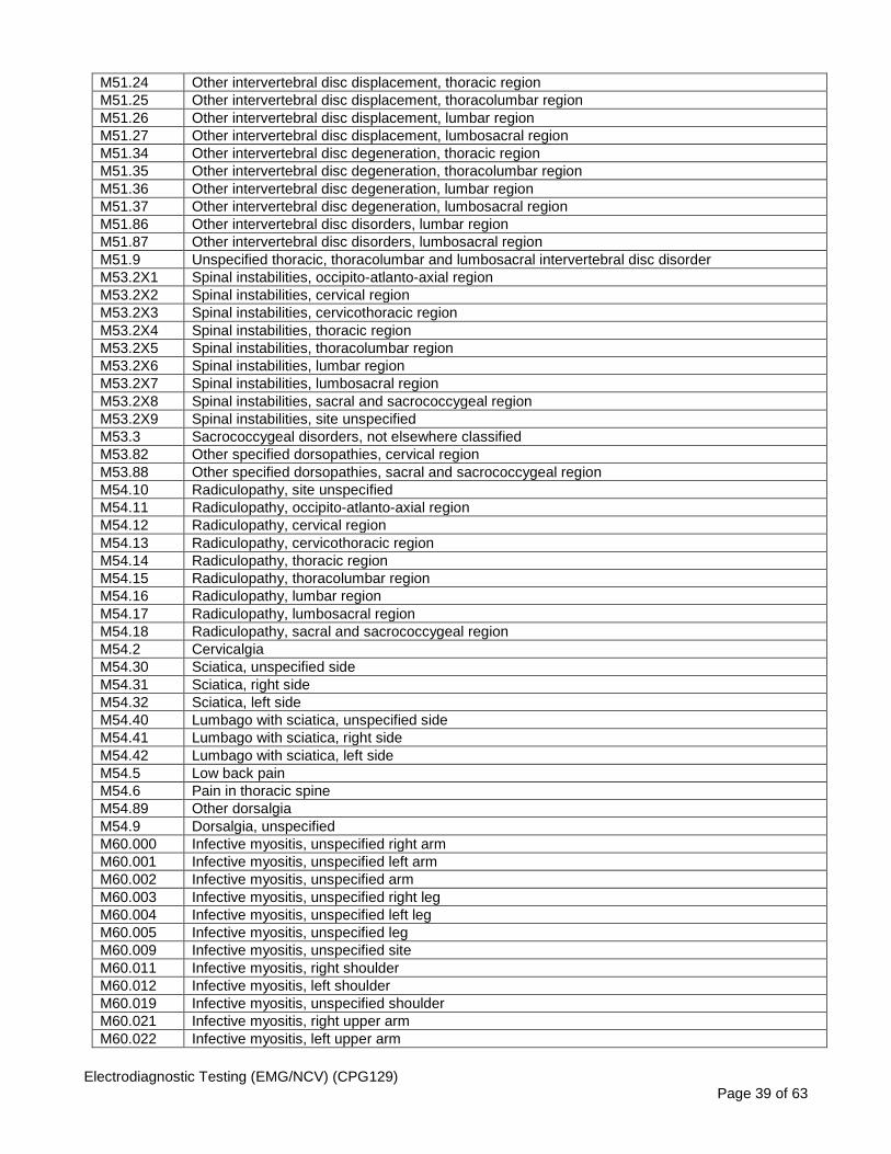
Electrodiagnostic Testing (EMG/NCV) (CPG129) Page 39 of 63
M51.24 Other intervertebral disc displacement, thoracic region M51.25 Other intervertebral disc displacement, thoracolumbar region M51.26 Other intervertebral disc displacement, lumbar region M51.27 Other intervertebral disc displacement, lumbosacral region M51.34 Other intervertebral disc degeneration, thoracic region M51.35 Other intervertebral disc degeneration, thoracolumbar region M51.36 Other intervertebral disc degeneration, lumbar region M51.37 Other intervertebral disc degeneration, lumbosacral region M51.86 Other intervertebral disc disorders, lumbar region M51.87 Other intervertebral disc disorders, lumbosacral region M51.9 Unspecified thoracic, thoracolumbar and lumbosacral intervertebral disc disorder M53.2X1 Spinal instabilities, occipito-atlanto-axial region M53.2X2 Spinal instabilities, cervical region M53.2X3 Spinal instabilities, cervicothoracic region M53.2X4 Spinal instabilities, thoracic region M53.2X5 Spinal instabilities, thoracolumbar region M53.2X6 Spinal instabilities, lumbar region M53.2X7 Spinal instabilities, lumbosacral region M53.2X8 Spinal instabilities, sacral and sacrococcygeal region M53.2X9 Spinal instabilities, site unspecified M53.3 Sacrococcygeal disorders, not elsewhere classified M53.82 Other specified dorsopathies, cervical region M53.88 Other specified dorsopathies, sacral and sacrococcygeal region M54.10 Radiculopathy, site unspecified M54.11 Radiculopathy, occipito-atlanto-axial region M54.12 Radiculopathy, cervical region M54.13 Radiculopathy, cervicothoracic region M54.14 Radiculopathy, thoracic region M54.15 Radiculopathy, thoracolumbar region M54.16 Radiculopathy, lumbar region M54.17 Radiculopathy, lumbosacral region M54.18 Radiculopathy, sacral and sacrococcygeal region M54.2 Cervicalgia M54.30 Sciatica, unspecified side M54.31 Sciatica, right side M54.32 Sciatica, left side M54.40 Lumbago with sciatica, unspecified side M54.41 Lumbago with sciatica, right side M54.42 Lumbago with sciatica, left side M54.5 Low back pain M54.6 Pain in thoracic spine M54.89 Other dorsalgia M54.9 Dorsalgia, unspecified M60.000 Infective myositis, unspecified right arm M60.001 Infective myositis, unspecified left arm M60.002 Infective myositis, unspecified arm M60.003 Infective myositis, unspecified right leg M60.004 Infective myositis, unspecified left leg M60.005 Infective myositis, unspecified leg M60.009 Infective myositis, unspecified site M60.011 Infective myositis, right shoulder M60.012 Infective myositis, left shoulder M60.019 Infective myositis, unspecified shoulder M60.021 Infective myositis, right upper arm M60.022 Infective myositis, left upper arm
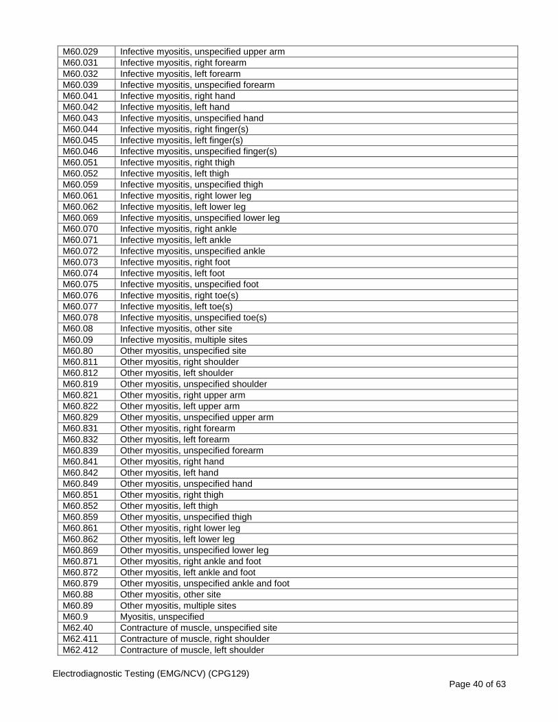
Electrodiagnostic Testing (EMG/NCV) (CPG129) Page 40 of 63
M60.029 Infective myositis, unspecified upper arm M60.031 Infective myositis, right forearm M60.032 Infective myositis, left forearm M60.039 Infective myositis, unspecified forearm M60.041 Infective myositis, right hand M60.042 Infective myositis, left hand M60.043 Infective myositis, unspecified hand M60.044 Infective myositis, right finger(s) M60.045 Infective myositis, left finger(s) M60.046 Infective myositis, unspecified finger(s) M60.051 Infective myositis, right thigh M60.052 Infective myositis, left thigh M60.059 Infective myositis, unspecified thigh M60.061 Infective myositis, right lower leg M60.062 Infective myositis, left lower leg M60.069 Infective myositis, unspecified lower leg M60.070 Infective myositis, right ankle M60.071 Infective myositis, left ankle M60.072 Infective myositis, unspecified ankle M60.073 Infective myositis, right foot M60.074 Infective myositis, left foot M60.075 Infective myositis, unspecified foot M60.076 Infective myositis, right toe(s) M60.077 Infective myositis, left toe(s) M60.078 Infective myositis, unspecified toe(s) M60.08 Infective myositis, other site M60.09 Infective myositis, multiple sites M60.80 Other myositis, unspecified site M60.811 Other myositis, right shoulder M60.812 Other myositis, left shoulder M60.819 Other myositis, unspecified shoulder M60.821 Other myositis, right upper arm M60.822 Other myositis, left upper arm M60.829 Other myositis, unspecified upper arm M60.831 Other myositis, right forearm M60.832 Other myositis, left forearm M60.839 Other myositis, unspecified forearm M60.841 Other myositis, right hand M60.842 Other myositis, left hand M60.849 Other myositis, unspecified hand M60.851 Other myositis, right thigh M60.852 Other myositis, left thigh M60.859 Other myositis, unspecified thigh M60.861 Other myositis, right lower leg M60.862 Other myositis, left lower leg M60.869 Other myositis, unspecified lower leg M60.871 Other myositis, right ankle and foot M60.872 Other myositis, left ankle and foot M60.879 Other myositis, unspecified ankle and foot M60.88 Other myositis, other site M60.89 Other myositis, multiple sites M60.9 Myositis, unspecified M62.40 Contracture of muscle, unspecified site M62.411 Contracture of muscle, right shoulder M62.412 Contracture of muscle, left shoulder

Electrodiagnostic Testing (EMG/NCV) (CPG129) Page 41 of 63
M62.419 Contracture of muscle, unspecified shoulder M62.421 Contracture of muscle, right upper arm M62.422 Contracture of muscle, left upper arm M62.429 Contracture of muscle, unspecified upper arm M62.431 Contracture of muscle, right forearm M62.432 Contracture of muscle, left forearm M62.439 Contracture of muscle, unspecified forearm M62.441 Contracture of muscle, right hand M62.442 Contracture of muscle, left hand M62.449 Contracture of muscle, unspecified hand M62.451 Contracture of muscle, right thigh M62.452 Contracture of muscle, left thigh M62.459 Contracture of muscle, unspecified thigh M62.461 Contracture of muscle, right lower leg M62.462 Contracture of muscle, left lower leg M62.469 Contracture of muscle, unspecified lower leg M62.471 Contracture of muscle, right ankle and foot M62.472 Contracture of muscle, left ankle and foot M62.479 Contracture of muscle, unspecified ankle and foot M62.48 Contracture of muscle, other site M62.49 Contracture of muscle, multiple sites M62.50 Muscle wasting and atrophy, not elsewhere classified, unspecified site M62.511 Muscle wasting and atrophy, not elsewhere classified, right shoulder M62.512 Muscle wasting and atrophy, not elsewhere classified, left shoulder M62.519 Muscle wasting and atrophy, not elsewhere classified, unspecified shoulder M62.521 Muscle wasting and atrophy, not elsewhere classified, right upper arm M62.522 Muscle wasting and atrophy, not elsewhere classified, left upper arm M62.529 Muscle wasting and atrophy, not elsewhere classified, unspecified upper arm M62.531 Muscle wasting and atrophy, not elsewhere classified, right forearm M62.532 Muscle wasting and atrophy, not elsewhere classified, left forearm M62.539 Muscle wasting and atrophy, not elsewhere classified, unspecified forearm M62.541 Muscle wasting and atrophy, not elsewhere classified, right hand M62.542 Muscle wasting and atrophy, not elsewhere classified, left hand M62.549 Muscle wasting and atrophy, not elsewhere classified, unspecified hand M62.551 Muscle wasting and atrophy, not elsewhere classified, right thigh M62.552 Muscle wasting and atrophy, not elsewhere classified, left thigh M62.559 Muscle wasting and atrophy, not elsewhere classified, unspecified thigh M62.561 Muscle wasting and atrophy, not elsewhere classified, right lower leg M62.562 Muscle wasting and atrophy, not elsewhere classified, left lower leg M62.569 Muscle wasting and atrophy, not elsewhere classified, unspecified lower leg M62.571 Muscle wasting and atrophy, not elsewhere classified, right ankle and foot M62.572 Muscle wasting and atrophy, not elsewhere classified, left ankle and foot M62.579 Muscle wasting and atrophy, not elsewhere classified, unspecified ankle and foot M62.58 Muscle wasting and atrophy, not elsewhere classified, other site M62.59 Muscle wasting and atrophy, not elsewhere classified, multiple sites M62.81 Muscle weakness (generalized) M62.831 Muscle spasm of calf M62.838 Other muscle spasm M62.9 Disorder of muscle, unspecified M72.9 Fibroblastic disorder, unspecified M79.0 Rheumatism, unspecified M79.1 Myalgia M79.2 Neuralgia and neuritis, unspecified M79.601 Pain in right arm M79.602 Pain in left arm

Electrodiagnostic Testing (EMG/NCV) (CPG129) Page 42 of 63
M79.603 Pain in arm, unspecified M79.604 Pain in right leg M79.605 Pain in left leg M79.606 Pain in leg, unspecified M79.609 Pain in unspecified limb M79.621 Pain in right upper arm M79.622 Pain in left upper arm M79.629 Pain in unspecified upper arm M79.631 Pain in right forearm M79.632 Pain in left forearm M79.639 Pain in unspecified forearm M79.641 Pain in right hand M79.642 Pain in left hand M79.643 Pain in unspecified hand M79.644 Pain in right finger(s) M79.645 Pain in left finger(s) M79.646 Pain in unspecified finger(s) M79.651 Pain in right thigh M79.652 Pain in left thigh M79.659 Pain in unspecified thigh M79.661 Pain in right lower leg M79.662 Pain in left lower leg M79.669 Pain in unspecified lower leg M79.671 Pain in right foot M79.672 Pain in left foot M79.673 Pain in unspecified foot M79.674 Pain in right toe(s) M79.675 Pain in left toe(s) M79.676 Pain in unspecified toe(s) M96.1 Postlaminectomy syndrome, not elsewhere classified N31.0 Uninhibited neuropathic bladder, not elsewhere classified N31.1 Reflex neuropathic bladder, not elsewhere classified N31.2 Flaccid neuropathic bladder, not elsewhere classified N31.8 Other neuromuscular dysfunction of bladder N31.9 Neuromuscular dysfunction of bladder, unspecified N32.81 Overactive bladder N39.3 Stress incontinence (female) (male) N39.41 Urge incontinence N39.42 Incontinence without sensory awareness N39.43 Post-void dribbling N39.44 Nocturnal enuresis N39.45 Continuous leakage N39.46 Mixed incontinence N39.490 Overflow incontinence N39.491 Coital incontinence N39.498 Other specified urinary incontinence O26.821 Pregnancy related peripheral neuritis, first trimester O26.822 Pregnancy related peripheral neuritis, second trimester O26.823 Pregnancy related peripheral neuritis, third trimester O26.829 Pregnancy related peripheral neuritis, unspecified trimester P11.3 Birth injury to facial nerve P11.4 Birth injury to other cranial nerves P11.5 Birth injury to spine and spinal cord P14.0 Erb’s paralysis due to birth injury P14.1 Klumpke’s paralysis due to birth injury
Electrodiagnostic Testing (EMG/NCV) (CPG129) Page 43 of 63
P14.3 Other brachial plexus birth injuries P14.8 Birth injuries to other parts of peripheral nervous system P14.9 Birth injury to peripheral nervous system, unspecified Q28.2 Arteriovenous malformation of cerebral vessels Q28.3 Other malformations of cerebral vessels Q76.2 Congenital spondylolisthesis R13.0 Aphagia R13.10 Dysphagia, unspecified R13.11 Dysphagia, oral phase R13.12 Dysphagia, oropharyngeal phase R13.13 Dysphagia, pharyngeal phase R13.14 Dysphagia, pharyngoesophageal phase R13.19 Other dysphagia R15.0 Incomplete defecation R15.1 Fecal smearing R15.2 Fecal urgency R15.9 Full incontinence of feces R20.0 Anesthesia of skin R20.1 Hypoesthesia of skin R20.2 Paresthesia of skin R20.3 Hyperesthesia R20.8 Other disturbances of skin sensation R20.9 Unspecified disturbances of skin sensation R25.2 Cramp and spasm R26.0 Ataxic gait R26.1 Paralytic gait R26.2 Difficulty in walking, not elsewhere classified R26.81 Unsteadiness on feet R26.89 Other abnormalities of gait and mobility R26.9 Unspecified abnormalities of gait and mobility R27.0 Ataxia, unspecified R27.8 Other lack of coordination R27.9 Unspecified lack of coordination R29.0 Tetany R29.1 Meningismus R29.2 Abnormal reflex R29.5 Transient paralysis R29.818 Other symptoms and signs involving the nervous system R29.891 Ocular torticollis R29.90 Unspecified symptoms and signs involving the nervous system R32 Unspecified urinary incontinence R33.0 Drug induced retention of urine R33.8 Other retention of urine R33.9 Retention of urine, unspecified R39.14 Feeling of incomplete bladder emptying R47.02 Dysphasia R47.1 Dysarthria and anarthria R47.89 Other speech disturbances R49.0 Dysphonia R49.8 Other voice and resonance disorders R49.9 Unspecified voice and resonance disorder S04.10XA-S04.10XS
Injury of oculomotor nerve, unspecified side
S04.11XA-S04.11XS
Injury of oculomotor nerve, right side

Electrodiagnostic Testing (EMG/NCV) (CPG129) Page 44 of 63
S04.12XA-S04.12XS
Injury of oculomotor nerve, left side
S04.20XA-S04.20XS
Injury of trochlear nerve, unspecified side
S04.21XA-S04.21XS
Injury of trochlear nerve, right side
S04.22XA-S04.22XS
Injury of trochlear nerve, left side
S04.30XA-S04.30XS
Injury of trigeminal nerve, unspecified side
S04.31XA-S04.31XS
Injury of trigeminal nerve, right side
S04.32XA-S04.32XS
Injury of trigeminal nerve, left side
S04.40XA-S04.40XS
Injury of abducent nerve, unspecified side
S04.41XA-S04.41XS
Injury of abducent nerve, right side
S04.42XA-S04.42XS
Injury of abducent nerve, left side
S04.50XA-S04.50XS
Injury of facial nerve, unspecified side
S04.51XA-S04.51XS
Injury of facial nerve, right side
S04.52XA-S04.52XS
Injury of facial nerve, left side
S04.60XA-S04.60XS
Injury of acoustic nerve, unspecified side
S04.61XA-S04.61XS
Injury of acoustic nerve, right side
S04.62XA- S04.62XS
Injury of acoustic nerve, left side
S04.70XA-S04.70XS
Injury of accessory nerve, unspecified side
S04.71XA-S04.71XS
Injury of accessory nerve, right side
S04.72XA-S04.72XS
Injury of accessory nerve, left side
S04.811A-S04.811S
Injury of olfactory [1st ] nerve, right side
S04.812A-S04.812S
Injury of olfactory [1st ] nerve, left side
S04.819A-S04.819S
Injury of olfactory [1st ] nerve, unspecified side
S04.891A-S04.891S
Injury of other cranial nerves, right side
S04.892A-S04.892S
Injury of other cranial nerves, left side
S04.899A-S04.899S
Injury of other cranial nerves, unspecified side
S04.9XXA-S04.9XXS
Injury of unspecified cranial nerve
S14.0XXA-S14.0XXS
Concussion and edema of cervical spinal cord
S14.101A-S14.9XXS
Other and unspecified injuries of cervical spinal cord

Electrodiagnostic Testing (EMG/NCV) (CPG129) Page 45 of 63
S24.0XXA-S24.0XXS
Concussion and edema of thoracic spinal cord
S24.101A-S24.9XXS
Other and unspecified injuries of thoracic spinal cord
S34.01XA-S34.01XS
Concussion and edema of lumbar spinal cord
S34.02XA-S34.02XS
Concussion and edema of sacral spinal cord
S34.101A-S34.9XXS
Other and unspecified injury of lumbar and sacral spinal cord
S44.00XA-S44.92XS
Injury of nerves at shoulder and upper arm level
S54.00XA-S54.92XS
Injury of ulnar nerve at forearm level
S64.00XA-S64.92XS
Injury of nerves at wrist and hand level
S74.00XA-S74.92XS
Injury of nerves at hip and thigh level
S84.00XA-S84.00XS
Injury of tibial nerve at lower leg level
S84.01XA-S84.01XS
Injury of tibial nerve at lower leg level, right leg
S84.02XA-S84.02SX
Injury of tibial nerve at lower leg level, left leg
S84.10XA-S84.10XS
Injury of peroneal nerve at lower leg level, unspecified leg
S84.11XA-S84.11XS
Injury of peroneal nerve at lower leg level, right leg
S84.12XA-S84.12XS
Injury of peroneal nerve at lower leg level, left leg
S84.20XA-S84.20XS
Injury of cutaneous sensory nerve at lower leg level, unspecified leg
S84.21XA-S84.21XS
Injury of cutaneous sensory nerve at lower leg level, right leg
S84.22XA-S84.22XS
Injury of cutaneous sensory nerve at lower leg level, left leg
S84.801A-S84.801S
Injury of other nerves at lower leg level, right leg
S84.802A-S84.802S
Injury of other nerves at lower leg level, left leg
S84.809A-S84.809S
Injury of other nerves at lower leg level, unspecified leg
S84.90XA-S84.90XS
Injury of unspecified nerve at lower leg level, unspecified leg
S84.91XA-S84.91XS
Injury of unspecified nerve at lower leg level, right leg
S84.92XA- S84.92XS
Injury of unspecified nerve at lower leg level, left leg
S94.00XA-S94.00XS
Injury of lateral plantar nerve, unspecified leg
S94.01XA-S94.01XS
Injury of lateral plantar nerve, right leg
S94.02XA-S94.02XS
Injury of lateral plantar nerve, left leg
S94.10XA-S94.10XS
Injury of medial plantar nerve, unspecified leg

Electrodiagnostic Testing (EMG/NCV) (CPG129) Page 46 of 63
S94.11XA-S94.11XS
Injury of medial plantar nerve, right leg
S94.12XA-S94.12XS
Injury of medial plantar nerve, left leg
S94.20XA-S94.20XS
Injury of deep peroneal nerve at ankle and foot level, unspecified leg
S94.21XA-S94.21XS
Injury of deep peroneal nerve at ankle and foot level, right leg
S94.22XA S94.22XS
Injury of deep peroneal nerve at ankle and foot level, left leg
S94.30XA-S94.30XS
Injury of cutaneous sensory nerve at ankle and foot level, unspecified leg
S94.31XA-S94.31XS
Injury of cutaneous sensory nerve at ankle and foot level, right leg
S94.32XA-S94.32XS
Injury of cutaneous sensory nerve at ankle and foot level, left leg
S94.8X1A-S94.8X1S
Injury of other nerves at ankle and foot level, right leg
S94.8X2A-S94.8X2S
Injury of other nerves at ankle and foot level, left leg
S94.8X9A-S94.8X9S
Injury of other nerves at ankle and foot level, unspecified leg
S94.90XA-S94.90XS
Injury of unspecified nerve at ankle and foot level, unspecified leg
S94.91XA-S94.91XS
Injury of unspecified nerve at ankle and foot level, right leg
S94.92XA-S94.92XS
Injury of unspecified nerve at ankle and foot level, left leg
Medical conditions supporting NCV testing without EMG
Considered Medically Necessary when criteria in the applicable policy statements listed above are met:
CPT®* Codes
Description
95907 Nerve conduction studies; 1-2 studies 95908 Nerve conduction studies; 3-4 studies 95909 Nerve conduction studies; 5-6 studies 95910 Nerve conduction studies; 7-8 studies 95911 Nerve conduction studies; 9-10 studies 95912 Nerve conduction studies; 11-12 studies 95913 Nerve conduction studies; 13 or more studies
ICD-10-CM Diagnosis Codes
Description
G56.00 Carpal tunnel syndrome, unspecified upper limb G56.01 Carpal tunnel syndrome, right upper limb G56.02 Carpal tunnel syndrome, left upper limb G56.03 Carpal tunnel syndrome, bilateral upper limbs I89.0 Lymphedema, not classified elsewhere I89.1 Lymphangitis I89.8 Other specified noninfective disorders of lymphatic vessels and lymph nodes I89.9 Noninfective disorder of lymphatic vessels and lymph nodes, unspecified I97.2 Postmastectomy lymphedema syndrome

Electrodiagnostic Testing (EMG/NCV) (CPG129) Page 47 of 63
Z79.01 Long term (current) use of anticoagulants
Considered Experimental/Investigational/Unproven:
ICD-10-CM Diagnosis Codes
Description
All other codes EMG Injection Localization: Performed Alone
Considered Medically Necessary for determination of precise muscle location for an injection:
CPT®* Codes
Description
95874 Needle electromyography for guidance in conjunction with chemodenervation (List separately in addition to code for primary procedure)
Neuromuscular Junction Testing
Considered Medically Necessary when criteria in the applicable policy statements listed above are met:
CPT®* Codes
Description
95937 Neuromuscular junction testing (repetitive stimulation, paired stimuli), each nerve, any 1 method
ICD-10-CM Diagnosis Codes
Description
A05.1 Botulism food poisoning A48.52 Wound botulism G12.21 Amyotrophic lateral sclerosis G12.22 Progressive bulbar palsy G12.23 Primary lateral sclerosis G12.24 Familial motor neuron disease G12.25 Progressive spinal muscle atrophy G12.29 Other motor neuron disease G12.8 Other spinal muscular atrophies and related syndromes G12.9 Spinal muscular atrophy, unspecified G61.0 Guillain-Barre syndrome G62.81 Critical illness polyneuropathy G70.01 Myasthenia gravis with (acute) exacerbation G70.1 Toxic myoneural disorders G70.80 Lambert-Eaton syndrome, unspecified G70.81 Lambert-Eaton syndrome in disease classified elsewhere G70.9 Myoneural disorder, unspecified G71.11 Myotonic muscular dystrophy G71.12 Myotonia congenita G71.13 Myotonic chondrodystrophy G71.19 Other specified myotonic disorders G71.3 Mitochondrial myopathy, not elsewhere classified G71.8 Other primary disorders of muscles G71.9 Primary disorder of muscle, unspecified G72.3 Periodic paralysis G72.49 Other inflammatory and immune myopathies, not elsewhere classified

Electrodiagnostic Testing (EMG/NCV) (CPG129) Page 48 of 63
G72.81 Critical illness myopathy G72.89 Other specified myopathies G72.9 Myopathy, unspecified G73.1 Lambert-Eaton syndrome in neoplastic disease H02.401 Unspecified ptosis of right eyelid H02.402 Unspecified ptosis of left eyelid H02.403 Unspecified ptosis of bilateral eyelids H02.409 Unspecified ptosis of unspecified eyelid H53.2 Diplopia M62.81 Muscle weakness (generalized) R13.0 Aphagia R13.10 Dysphagia, unspecified R13.11 Dysphagia, oral phase R13.12 Dysphagia, oropharyngeal phase R13.13 Dysphagia, pharyngeal phase R13.14 Dysphagia, pharyngoesophageal phase R13.19 Other dysphagia R47.02 Dysphasia R47.1 Dysarthria and anarthria R47.81 Slurred speech R47.89 Other speech disturbances R47.9 Unspecified speech disturbances
Considered Not Medically Necessary:
ICD-10-CM Diagnosis Codes
Description
All other codes Considered Medically Necessary when criteria in the applicable policy statements listed above are met:
CPT®* Codes
Description
95925 Short-latency somatosensory evoked potential study, stimulation of any/all peripheral nerves or skin sites, recording from the central nervous system; in upper limbs
95926 Short-latency somatosensory evoked potential study, stimulation of any/all peripheral nerves or skin sites, recording from the central nervous system; in lower limbs
95927 Short-latency somatosensory evoked potential study, stimulation of any/all peripheral nerves or skin sites, recording from the central nervous system; in the trunk or head
95938 Short-latency somatosensory evoked potential study, stimulation of any/all peripheral nerves or skin sites, recording from the central nervous system; in upper and lower limbs
ICD-10-CM Diagnosis Codes
Description
C72.0 Malignant neoplasm of spinal cord C72.1 Malignant neoplasm of cauda equina C79.31 Secondary malignant neoplasm of brain C79.49 Secondary malignant neoplasm of other parts of nervous system D33.4 Benign neoplasm of spinal cord D43.0 Neoplasm of uncertain behavior of brain, supratentorial D43.1 Neoplasm of uncertain behavior of brain, infratentorial D43.2 Neoplasm of uncertain behavior of brain, unspecified D43.4 Neoplasm of uncertain behavior of spinal cord

Electrodiagnostic Testing (EMG/NCV) (CPG129) Page 49 of 63
E03.5 Myxedema coma E71.50-E71.548
Peroxisomal disorders
E75.23 Krabbe disease E75.25 Metachromatic leukodystrophy E75.29 Other sphingolipidosis G04.1 Tropical spastic paraplegia G11.0-G11.9 Hereditary ataxia G25.3 Myoclonus G32.0 Subacute combined degeneration of spinal cord in diseases classified elsewhere G32.81 Cerebellar ataxia in diseases classified elsewhere G35 Multiple sclerosis G36.0-G36.9 Other acute disseminated demyelination G37.0-G37.9 Other demyelinating diseases of the central nervous system G82.20 Paraplegia, unspecified G82.21 Paraplegia, complete G82.22 Paraplegia, incomplete G95.0 Syringomyelia and syringobulbia G95.20 Unspecified cord compression G95.29 Other cord compression G95.9 Disease of spinal cord, unspecified M47.011-M47.019
Anterior spinal artery compression syndromes
M47.021-M47.029
Vertebral artery compression syndromes
M47.11 Other spondylosis with myelopathy, occipito-atlanto-axial region M47.12 Other spondylosis with myelopathy, cervical region M47.13 Other spondylosis with myelopathy, cervicothoracic region M47.14 Other spondylosis with myelopathy, thoracic region M47.15 Other spondylosis with myelopathy, thoracolumbar region M47.16 Other spondylosis with myelopathy, lumbar region M48.01 Spinal stenosis, occipito-atlanto-axial region M48.02 Spinal stenosis, cervical region M48.03 Spinal stenosis, cervicothoracic region M48.04 Spinal stenosis, thoracic region M48.05 Spinal stenosis, thoracolumbar region M48.061 Spinal stenosis, lumbar region without neurogenic claudication M48.062 Spinal stenosis, lumbar region with neurogenic claudication M50.00-M50.03
Cervical disc disorder with myelopathy
M99.20 Subluxation stenosis of neural canal of head region M99.21 Subluxation stenosis of neural canal of cervical region M99.22 Subluxation stenosis of neural canal of thoracic region M99.30 Osseous stenosis of neural canal of head region M99.31 Osseous stenosis of neural canal of cervical region M99.32 Osseous stenosis of neural canal of thoracic region M99.40 Connective tissue stenosis of neural canal of head region M99.41 Connective tissue stenosis of neural canal of cervical region M99.42 Connective tissue stenosis of neural canal of thoracic region
Electrodiagnostic Testing (EMG/NCV) (CPG129) Page 50 of 63
M99.50 Intervertebral disc stenosis of neural canal of head region M99.51 Intervertebral disc stenosis of neural canal of cervical region M99.52 Intervertebral disc stenosis of neural canal of thoracic region M99.60 Osseous and subluxation stenosis of intervertebral foramina of head region M99.61 Osseous and subluxation stenosis of intervertebral foramina of cervical region M99.62 Osseous and subluxation stenosis of intervertebral foramina of thoracic region M99.70 Connective tissue and disc stenosis of intervertebral foramina of head region M99.71 Connective tissue and disc stenosis of intervertebral foramina of cervical region M99.72 Connective tissue and disc stenosis of intervertebral foramina of thoracic region P11.5 Birth injury to spine and spinal cord Q06.0 Amyelia Q06.1 Hypoplasia and dysplasia of spinal cord Q06.3 Other congenital cauda equina malformations Q06.8 Other specified congenital malformations of spinal cord Q06.9 Congenital malformation of spinal cord, unspecified R40.20 Unspecified coma R40.2110-R40.2114
Coma scale, eyes open never
R40.2120-R40.2124
Coma scale, eyes open, to pain
R40.2130-R40.2134
Coma scale, eyes open, to sound
R40.2140-R40.2144
Coma scale, eyes open, spontaneous
R40.2210-R40.2214
Coma scale, best verbal response, none
R40.2220-R40.2224
Coma scale, best verbal response, incomprehensible words
R40.2230-R40.2234
Coma scale, best verbal response, inappropriate words
R40.2240-R40.2244
Coma scale, best verbal response, confused conversation
R40.2310-R40.2314
Coma scale, best motor response, none
R40.2320-R40.2324
Coma scale, best motor response, extension
R40.2330-R40.2334
Coma scale, best motor response, abnormal flexion
R40.2340-R40.2344
Coma scale, best motor response, flexion withdrawal
R40.2350-R40.2354
Coma scale, best motor response, localizes pain
R40.2360-R40.2364
Coma scale, best motor response, obeys commands
R40.2420- R40.2424
Glasgow coma scale score 9-12
R40.2430-R40.2434
Glasgow coma scale score 3-8
S14.0XXA-S14.0XXS
Concussion and edema of cervical spinal cord
S14.101A-S14.109S
Other and unspecified injury of cervical spinal cord
S14.111A-S14.119S
Complete lesion of cervical spinal cord

Electrodiagnostic Testing (EMG/NCV) (CPG129) Page 51 of 63
S14.121A-S14.129S
Central cord syndrome of cervical spinal cord
S14.131A-S14.139S
Anterior cord syndrome of cervical spinal cord
S14.141A-S14.149S
Brown-Sequard syndrome of cervical spinal cord
S14.151A-S14.159S
Other incomplete lesion of cervical spinal cord
S24.0XXA-S24.0XXS
Concussion and edema of thoracic spinal cord
S24.101A-S24.109S
Unspecified injury at level of thoracic spinal cord
S24.111A-S24.119S
Complete lesion at level of thoracic spinal cord
S24.131A-S24.139S
Anterior cord syndrome of thoracic spinal cord
S24.141A-S24.149S
Brown-Sequard syndrome of thoracic spinal cord
S24.151A-S24.159S
Other incomplete lesion of thoracic spinal cord
S34.01XA-S34.01XS
Concussion and edema of lumbar spinal cord
S34.02XA-S34.02XS
Concussion and edema of sacral spinal cord
S34.101A-S34.109S
Unspecified injury to lumbar spinal cord
S34.111A-S34.119S
Complete lesion of lumbar spinal cord
S34.121A-S34.129S
Incomplete lesion of lumbar spinal cord
S34.131A-S34.139S
Complete lesion of sacral spinal cord
S34.3XXA-S34.3XXS
Injury of cauda equina
Considered Experimental/Investigational/Unproven:
ICD-10-CM Diagnosis Codes
Description
All other codes Automated Hand-held Noninvasive Nerve Conduction Testing
Considered Experimental/Investigationa/Unproven when used to report automated or portable hand-held noninvasive nerve conduction testing/devices:
CPT®* Description
95905 Motor and/or sensory nerve conduction, using preconfigured electrode array(s), amplitude and latency/velocity study, each limb, includes F-wave study when performed, with interpretation and report
Macro EMG/Surface Electromyography/Paraspinal SEMG Considered Experimental/Investigational/Unproven:
Electrodiagnostic Testing (EMG/NCV) (CPG129) Page 52 of 63
HCPCS Codes
Description
S3900 Surface electromyography (EMG) *Current Procedural Terminology (CPT®) ©2018 American Medical Association: Chicago, IL. References
1. Ahern, D. K., Follick, M. J., Council, J. R., & Laser-Wolston, N. (1986). Reliability of lumbar paravertebral EMG assessment in chronic low back pain. Archives of Physical Medicine and Rehabilitation, 67(10), 762-765.
2. Ahern, D. K., Follick, M. J., Council, J. R., Laser-Wolston, N., & Litchman, H. (1988). Comparison of
lumbar paravertebral EMG patterns in chronic low back pain patients and non-patient controls. Pain, 34(2), 153-160.
3. Alemo S, Sayadipour A. Role of intraoperative neurophysiologic monitoring in lumbosacral spine fusion
and instrumentation: a retrospective study. World Neurosurg. 2010 Jan;73(1):72-6.
4. Alexiev, A. R. (1994). Some differences of the electromyographic erector spinae activity between normal subjects and low back pain patients during the generation of isometric trunk torque. Electromyography and Clinical Neurophysiology, 34(8), 495-499.
5. Alrawi MF, Khalil NM, Mitchell P, et al. The value of neurophysiological and imaging studies in predicting
outcome in the surgical treatment of cervical radiculopathy. Eur Spine J. 2007 Apr;16(4):495-500.
6. Al-Shekhlee A, Shapiro BE, Preston DC. Iatrogenic complications and risks of nerve conduction studies and needle electromyography. Muscle & nerve 2003;27:517-526.
7. Ambroz, C., Scott, A., Ambroz, A., & Talbott, E. O. (2000). Chronic low back pain assessment using
surface electromyography. Journal of Occupational and Environmental Medicine, 42(6), 660-669.
8. American Academy of Neurology. Assessment: intraoperative neurophysiology. Report of the Therapeutics and Technology Assessment Subcommittee of the American Academy of Neurology. Neurology. 1990 Nov;40(11):1644-6.
9. American Academy of Neurology (AAN). Principles of coding for intraoperative neurophysiologic
monitoring (IOM) and testing model medical policy. 2016. Retrieved on August 6, 2019 from https://www.aan.com/siteassets/home-page/tools-and-resources/practicing-neurologist--administrators/billing-and-coding/model-coverage-policies/16iommodelpolicy_tr.pdf
10. American Academy of Neurology (AAN). Position statement. Mobile electrodiagnostic laboratories
provide substandard patient care. 2015. Retrieved on August 6, 2019 from http://www.aanem.org/getmedia/21cc5d73-3d9f-4240-b780-5acd2f6eb15f/Position-Statement_Mobile-Electrodiagnostic-Laboratories.pdf
11. American Academy of Neurology (AAN). Assessment: Dermatomal Somatosensory Evoked Potentials.
Reports of the American Academy of Neurology’s Therapeutics and Technology Assessments Committee. Approved November 1995. Approved by the AAN Practice Committee December 1995. Approved by the AAN Executive Board January 1996. Copyright ©1997. Current guideline re-affirmed 10/17/2003. Published: Neurology 1997;49:1127-30.
12. American Association of Neuromuscular and Electrodiagnostic Medicine (AANEM). Model policy for
needle electromyography and nerve conduction velocity studies. 2016. Retrieved on August 6, 2019 from https://www.aanem.org/getmedia/65934187-d91e-4336-9f3c-50522449e565/Model-Policy.pdf

Electrodiagnostic Testing (EMG/NCV) (CPG129) Page 53 of 63
13. American Association of Neuromuscular and Electrodiagnostic Medicine (AANEM). Choosing Wisely.
Five Things Physicians and Patients Should Question. Released February 2015.
14. American Association of Neuromuscular and Electrodiagnostic Medicine (AANEM). 2011 Coding guide.
15. American Association of Neuromuscular and Electrodiagnostic Medicine (AANEM). Definition of real time onsite. Position Statement. July 2014. Retreived on August 6, 2019 from http://www.aanem.org/Advocacy/Position-Statements
16. American Association of Neuromuscular and Electrodiagnostic Medicine (AANEM). Needle EMG in
certain uncommon clinical contexts. Muscle Nerve. 2005;31:398-399.
17. American Association of Neuromuscular and Electrodiagnostic Medicine (AANEM). (2014). Risks in electrodiagnostic medicine. Retrieved on August 6, 2019 from http://www.aanem.org/getmedia/50f4dd83-835c-46cb-a832-930851440e9e/risksinEDX.pdf.aspx
18. American Association of Neuromuscular and Electrodiagnostic Medicine (AANEM). Practice parameter
for Repetitive Nerve Stimulation & Single Fiber EMG Evaluation of Adults w/Suspected Myasthenia Gravis. Reaffirmed October 2015. Retrieved on August 6, 2019 from http://www.aanem.org/getmedia/dc0ca43b-b108-41f3-894c-7f54c078a6f9/myastheniagravis_Reaffirmed.pdf
19. American Association of Neuromuscular and Electrodiagnostic Medicine (AANEM). Proper performance
and interpretation of electrodiagnostic studies. Muscle Nerve. 2006 Mar;33(3):436-9. Retrieved on August 6, 2019 from http://www.aanem.org/getmedia/6808cb2a-1d3e-4c09-9499-82bfae0621f7/Proper-Performance-of-EDX.pdf
20. American Association of Neuromuscular and Electrodiagnostic Medicine (AANEM). Position Statement.
Reporting the results of needle EMG and nerve conduction studies: and eduational report. May 2014. Retrieved on August 6, 2019 from: http://www.aanem.org/Advocacy/Position-Statements
21. American Association of Neuromuscular and Electrodiagnostic Medicine (AANEM). Somatosensory
evoked potentials. Clinical uses. Chapter 5. Muscle Nerve 22: Supplement 8: S111-S118, 1999a. Retrieved on August 6, 2019 from http://www.aanem.org/Practice/Guidelines-and-Performance-Measures
22. American Association of Neuromuscular and Electrodiagnostic Medicine (AANEM). Who is qualified to
practice electrodiagnostic medicine. Position statement. Updated and Re-approved: May 2012; November 2017 Accessed August 6, 2019. Available at URL address: http://www.aanem.org/Advocacy/Position-Statements
23. American Association of Neuromuscular and Electrodiagnostic Medicine (AANEM). Position Statement.
Technologists conducting nerve conduction studies and somatosensory evoked potential studies independently to be reviewed by a physician at a later time. Approved by the American Association of Electrodiagnostic Medicine May 1999b. Updated 2015 by the AANEM. Retrieved on August 6, 2019 from http://www.aanem.org/Advocacy/Position-Statements
24. American Association of Neuromuscular and Electrodiagnostic Medicine (AANEM). Recommended
policy for electrodiagnostic medicine. Endorsed by the American Academy of Neurology, The American Academy of Physical Medicine and Rehabilitation, and The American Association of Neuromuscular and Electrodiagnostic Medicine. Updated 2004. Copyright ©1995-2014; by the AANEM. Retrieved on August 6, 2019 from http://www.aanem.org/getmedia/884f5b94-a0be-447e-bdae-20d52aaf8299/Recommended-Policy-for-Electrodiagnostic-Medicine.pdf
25. American Association of Sensory Electrodiagnostic Medicine (AASEM). Sensory Pain Fiber
Electrodiagnostic (EDX) Medicine Guidelines. Accessed August 6, 2019. Available at URL address: http://www.aasem.org/pages/practice-guidelines
Electrodiagnostic Testing (EMG/NCV) (CPG129) Page 54 of 63
26. American Clinical Neurophysiology Society (ACNS). Standards for short latency somatosensory evoked
potentials. Feb 10, 2006. Retrieved on August 6, 2019 from https://www.acns.org/practice/guidelines
27. American Society of Electroneurodiagnostic Technologists (ASET). Simultaneous intraoperative monitoring. Professional Standards and Best Practices. Retrieved on August 6, 2019 from http://www.aset.org/i4a/pages/index.cfm?pageid=3288
28. American Society of Neurophysiological Monitoring (ASNM). Intraoperative monitoring using
somatosensory evoked potentials. A position statement by the American Society of Neurophysiological Monitoring. J Clin Monit Comput. 2005 Jun;241-58 (18).
29. American Speech-Language-Hearing Association. Neurophysiologic Intraoperative Monitoring. Position
Statement. Copyright © 2013 American Language-Speech-Hearing Association. Retrieved on August 6, 2019 from http://www.asha.org/policy/PS1992-00036/
30. Aminoff MJ, Eisen A. Somatosensory Evoked Potentials. In: Aminoff’s Electrodiagnosis in Clinical
Neurology. Ch 26. Sixth Edition © 2012, Elsevier Inc.
31. Aminoff, MJ, Weiskopf, RB. Electrophysiologic testing for the diagnosis of peripheral nerve injury. Anesthesiology. May 1, 2004;100(5):1298-303.
32. Armstrong TN, Dale AM, Al-Lozi MT, Franzblau A, Evanoff BA. Median and ulnar nerve conduction
studies at the wrist: criterion validity of the NC-stat automated device. J Occup Environ Med. 2008 Jul;50(7):758-64.
33. Asad A, Hameed MA, Khan UA, Butt MU, Ahmed N, Nadeem A. Comparison of nerve conduction studies
with diabetic neuropathy symptom score and diabetic neuropathy examination score in type-2 diabetics for detection of sensorimotor polyneuropathy. J Pak Med Assoc. 2009 Sep;59(9):594-8.
34. Asbury AK. Approach to the patient with peripheral neuropathy. In: Harrison’s Principles of Internal
Medicine. Part 15: Neurologic disorders. Section 3: Nerve and muscle disorders. Chapter 363. Electrodiagnosis. Copyright 2004 by The McGraw-Hill Companies, Inc.
35. Bal S, Celiker R, Palaoglu S, et al. F wave studies of neurogenic intermittent claudication in lumbar spinal
stenosis. Am J Phys Med Rehabil. 2006 Feb;85(2):135-40.
36. Barboi AC, Barkhaus PE. Electrodiagnostic testing in neuromuscular disorders. Neurol Clin. 2004 Aug;22:619-641.
37. Biedermann, H. J., Shanks, G. L., Forrest, W. J., & Inglis, J. (1991). Power spectrum analyses of
electromyographic activity. Discriminators in the differential assessment of patients with chronic low-back pain. Spine, 16(10), 1179-1184.
38. Bose B1, Wierzbowski LR, Sestokas AK. Neurophysiologic monitoring of spinal nerve root function
during instrumented posterior lumbar spine surgery. Spine (Phila Pa 1976). 2002 Jul 1;27(13):1444-50.
39. Braunwald: Harrison’s Principles of Internal Medicine. Electrophysiology studies of muscle and nerve: Electrophysiologic studies of the central and peripheral nervous systems. ©2001, The McGraw-Hill Companies, Inc. Chapter 357.
40. Calfee RP, Dale AM, Ryan D, et al. Performance of simplified scoring systems for hand diagrams in carpal tunnel syndrome screening. J Hand Surg Am. 2012 Jan;37(1):10-7.
41. Chang MH, Liu LH, Lee YC, et al. Comparison of sensitivity of transcarpal median motor conduction
velocity and conventional conduction techniques in electrodiagnosis of carpal tunnel syndrome. Clin Neurophysiol. 2006; 117(5):984-991.
Electrodiagnostic Testing (EMG/NCV) (CPG129) Page 55 of 63
42. Chawla J, Burneo JG, Barkley GL. Clinical Applications of Somatosensory Evoked Potentials. Medscape.
Updated March 2014, April 12, 2016. Retrieved on August 6, 2019 from http://emedicine.medscape.com/article/1139393-overview#showall
43. Chiappa K. Electrophysiologic monitoring during carotid endarterecteomies. In: Chiappa K, editor.
Evoked potentials in clinical medicine. Third edition. ©1997. Lippincott-Raven Publishers. Philadelphia –New York. Ch. 19.
44. Chiappa K, Cros D. Dermatomal somatosensory evoked potentials. In: Chiappa K, editor. Evoked
potentials in clinical medicine. Third edition. ©1997. Lippincott-Raven Publishers. Philadelphia –New York. Ch. 12.
45. Cho SC, Ferrante MA, Levin KH, et al. Utility of electrodiagnostic testing in evaluating patients with
lumbosacral radiculopathy: an evidence-based review. Muscle Nerve. 2010; 42(2):276-282.
46. Collins, G. A., Cohen, M. J., Naliboff, B. D., & Schandler, S. L. (1982). Comparative analysis of paraspinal and frontalis EMG, heart rate and skin conductance in chronic low back pain patients and normals to various postures and stress. Scandinavian Journal of Rehabilitative Medicine, 14(1), 39-46.
47. Cruccu G, Aminoff MJ, Curio G, Guerit JM, Kakigi R, Mauguiere F, Rossini PM, Treede RD, Garcia-
Larrea L. Recommendations for the clinical use of somatosensory-evoked potentials. Clin Neurophysiol. 2008 May 15.
48. Crum BA, Strommen JA. Peripheral nerve stimulation and monitoring during operative procedures.
Muscle Nerve. 2007 Feb;35(2):159-70.
49. Current Procedural Terminology (CPT®) © (Current Year) American Medical Association: Chicago, IL.
50. Dale AM, Agboola F, Yun A, Zeringue A, Al-Lozi MT, Evanoff B. Comparison of automated versus traditional nerve conduction study methods for median nerve testing in a general worker population. PM R. 2015 Mar;7(3):276-82.
51. Danneels, L. A., Cagnie, B. J., Cools, A. M., Vanderstraeten, G. G., Cambier, D. C., Witvrouw, E. E., et
al. (2001). Intra-operator and inter-operator reliability of surface electromyography in the clinical evaluation of back muscles. Manual Therapy, 6(3), 145-153.
52. DeGood, D. E., Stewart, W. R., Adams, L. E., & Dale, J. A. (1994). Paraspinal EMG and autonomic
reactivity of patients with back pain and controls to personally relevant stress. Perceptual and Motor Skills, 79(3 Pt 1), 1399-1409.
53. Derr JJ, Micklesen PJ, Robinson LR. Predicting recovery after fibular nerve injury: which
electrodiagnostic features are most useful? Am J Phys Med Rehabil. 2009 Jul;88(7):547-53.
54. De Sousa EA, Chin RL, Sander HW, et al. Demyelinating findings in typical and atypical chronic inflammatory demyelinating polyneuropathy: sensitivity and specificity. J Clin Neuromuscul Dis. 2009 Jun;10(4):163-9.
55. De Souza RJ, de Souza A, Nagvekar MD. Nerve conduction studies in diabetics presymptomatic and
symptomatic for diabetic polyneuropathy. J Diabetes Complications. 2015 May 21. pii: S1056-8727(15)00209-3.
56. Devlin VJ, Schwartz DM. Intraoperative neurophysiologic monitoring during spinal surgery. J Am Acad
Orthop Surg. 2007 Sep;15(9):549-60.
57. Drost G, Stegeman DF, van Engelen BG, Zwarts MJ. Clinical applications of high-density surface EMG: a systematic review. J Electromyogr Kinesiol. 2006 Dec;16(6):586-602.
Electrodiagnostic Testing (EMG/NCV) (CPG129) Page 56 of 63
58. Dyck PJ, Overland CJ, Low PA, et al. Signs and symptoms versus nerve conduction studies to diagnose diabetic sensorimotor polyneuropathy: Cl vs. NPhys trial. Muscle Nerve. 2010 Aug;42(2):157-64.
59. Edwards BM, Kileny PR. Intraoperative neurophysiologic monitoring: indications and techniques for
common procedures in otolaryngology-head and neck surgery. Otolaryngol Clin North Am. 2005 Aug;38(4):631-42, viii.
60. Eager M1, Shimer A, Jahangiri FR, Shen F, Arlet V. Intraoperative neurophysiological monitoring
(IONM): lessons learned from 32 case events in 2069 spine cases. Am J Electroneurodiagnostic Technol. 2011 Dec;51(4):247-63.
61. Eisen and Fischer. The F Wave. Recommendations for the Practice of Clinical Neurophysiology:
Guidelines of the International Federation of Clinical Physiology (EEG Suppl. 52) Editors: G. Deuschl and A. Eisen, 1999
62. Elkowitz SJ, Dubin NH, Richards BE, Wilgis EF. Clinical utility of portable versus traditional
electrodiagnostic testing for diagnosing, evaluating and treating carpal tunnel syndrome. Am J Orthop. 2005; 34(8):362-364.
63. Emerson RG, Adams DC, Nagle KJ. Monitoring of spinal cord function intraoperatively using motor and
somatosensory evoked potentials. In: Chiappa K, editor. Evoked potentials in clinical medicine. Third edition. ©1997. Lippincott-Raven Publishers. Philadelphia –New York. Ch 20.
64. Emeryk-Szajewska B, Badurska B, Kostera-Pruszczyk A. Electrophysiological findings in hereditary
motor and sensory neuropathy type I and II--a conduction velocity study. Electromyogr Clin Neurophysiol. 1998 Mar;38(2):95-101.
65. England JD, Franklin GM. Automated hand-held nerve conduction devices: raw data, raw interpretations.
Muscle Nerve. 2011; 43(1):6-8.
66. Erickson L, Costa V, McGregor M. Intraoperative neurophysiological monitoring during spinal surgery. Montreal: Technology Assessment Unit of the McGill University Health Centre (MUHC), 2005:39.
67. Fehlings MG, Brodke DS, Norvell DC, Dettori JR. The evidence for intraoperative neurophysiological
monitoring in spine surgery: Does it make a difference? Spine. 2010;35(9 Suppl):S37-S46.
68. Ferdjallah, M., & Wertsch, J. J. (1998). Anatomical and technical considerations in surface electromyography. Physical Medicine and Rehabilitation Clinics of North America, 9(4), 925-931.
69. Fisher, MA. H reflexes and f waves fundamentals, normal and abnormal patterns. Neurology Clinics. May
2002;20(2):339-60.
70. Fisher MA, Bajwa R, Somashekar KN. Acta Neurol Scand. 2008 Aug;118(2):99-105.
71. Geisser, M. E., Ranavaya, M., Haig, A. J., Roth, R. S., Zucker, R., Ambroz, C., et al. (2005). A meta-analytic review of surface electromyography among persons with low back pain and normal, healthy controls. Journal of Pain, 6(11), 711-726.
72. Gooch CL, Pullman S. Electromyography, nerve conduction studies, and magnetic stimulation. In:
Rowland PL, editor. Merritt’s Neurology. 11th edition. Lippincott Williams and Wilkins. Ch 15.
73. Gooch CL, Weimer LH. The electrodiagnosis of neuropathy: basic principles and common pitfalls. Neurol Clin. 2007 Feb;25(1):1-28.
74. Grassme, R., Arnold, D., Anders, C., van Dijk, J. P., Stegeman, D. F., Linss, W., et al. (2005). Improved
evaluation of back muscle SEMG characteristics by modelling. Pathophysiology, 12(4), 307-312.
Electrodiagnostic Testing (EMG/NCV) (CPG129) Page 57 of 63
75. Gunnarsson T, Krassioukov AV, Sarjeant R, Fehlings MG. Real-time continuous intraoperative electromyographic and somatosensory evoked potential recordings in spinal surgery: Correlation of clinical and electrophysiologic findings in a prospective, consecutive series of 213 cases. Spine. 2004;29(6):677-84.
76. Guse BH, Love MJ. Medical assessment and laboratory testing in psychiatry. In: Saydock BJ, Saydock
VA, editors. Kaplan and Saydock’s Comprehensive Textbook of Psychiatry. 8th ed, 2005. p. 919.
77. Haig AJ, Gelblum JB, Rechtien JJ, Gitter AJ. Technology review: The use of surface EMG in the diagnosis and treatment of nerve and muscle disorders. American Association of Neuromuscular and Electrodiagnostic Medicine (AANEM). 1999. Retrieved on July 10, 2018 from http://www.aanem.org/Practice/Technology-Reviews
78. Haig, A. J., Gelblum, J. B., Rechtien, J. J., & Gitter, A. J. (1996). Technology assessment: The use of
surface EMG in the diagnosis and treatment of nerve and muscle disorders. Muscle & Nerve, 19(3), 392-395.
79. Hammad M, Silva A, Glass J, et al. Clinical, electrophysiologic, and pathologic evidence for sensory
abnormalities in ALS. Neurology. 2007 Dec 11;69(24):2236-42.
80. Heinonen, P., Kautiainen, H., & Mikkelsson, M. (2005). Erector spinae SEMG activity during forward flexion and re-extension in ankylosing spondylitis patients. Pathophysiology, 12(4), 289-293.
81. Hilburn JW. General principles and use of electrodiagnostic studies in carpal and cubital tunnel
syndrome. With special attention to pitfalls and interpretation. Hand Clin. 1996; 12(2):205-221.
82. Hogrel, J. Y. (2005). Clinical applications of surface electromyography in neuromuscular disorders. Neurophysiologie Clinique, 35(2-3), 59-71.
83. Holiner I, Haslinger V, Lütschg J, et al. Validity of the neurological examination in diagnosing diabetic
peripheral neuropathy. Pediatr Neurol. 2013 Sep;49(3):171-7.
84. Holland NR. Intraoperative electromyography. J Clin Neurophysiol. 2002 Oct;19(5):444-53.
85. Hu Y, Kwok JW, Tse JY, Luk KD. Time-varying surface electromyography topography as a prognostic tool for chronic low back pain rehabilitation. Spine J. 2014 Jun 1;14(6):1049-56.
86. Jablecki CK, Andary MT, Floeter MK et al. Practice parameter: Electrodiagnostic studies in carpal tunnel
syndrome. Report of the American Association of Electrodiagnostic Medicine, American Academy of Neurology, and the American Academy of Physical Medicine and Rehabilitation. Neurology 2002;58(11):1589-92.
87. Jabre JF, Salzsieder BT, Gnemi KE. Criterion validity of the NC-stat automated nerve conduction
measurement instrument. Physiol Meas. 2007; 28(1):95-104.
88. Jalovaara, P., Niinimaki, T., & Vanharanta, H. (1995). Pocket-size, portable surface EMG device in the differentiation of low back pain patients. European Spine Journal, 4(4), 210-212.
89. Jameson LC, Janki DJ, Sloan TB. Electrophysiologic Monitoring in Neurosurgery. Anesthesiol Clin. 2007
Sep; 25(3): 605-30, x
90. Juel VC. Evaluation of neuromuscular junction disorders in the electromyography laboratory. Neurol Clin. 01 May 2012; 30(2):621.39.
91. Jurell, K. C. (1998). Surface EMG and fatigue. Physical Medicine and Rehabilitation Clinics of North
America, 9(4), 933-47, viii-ix.

Electrodiagnostic Testing (EMG/NCV) (CPG129) Page 58 of 63
92. Karami-Mohajeri S, Nikfar S, Abdollahi M. A systematic review on the nerve-muscle electrophysiology in human organophosphorus pesticide exposure. Hum Exp Toxicol. 2014;33(1):92-102.
93. Katifi HA, Sedgwick EM. Evaluation of the dermatomal somatosensory evoked potential in the diagnosis
of lumbo-sacral root compression. J Neurol Neurosurg Psychiatry. 1987 Sep;50(9):1204-10.
94. Katirji, B. The clinical electromyography examination: an overview. Neurology Clinics. May 2002;20(2):XI
95. Katz RT. NC-stat as a screening tool for carpal tunnel syndrome in industrial workers. J Occup Environ Med. 2006 Apr;48(4):414-8.
96. Katz JN, Simmons BP. Carpal tunnel syndrome. N Engl J Med. 2002; 346(23):1807-1812.
97. Kaufman MA. Differential diagnosis and pitfalls in electrodiagnostic studies and special tests for
diagnosing compressive neuropathies. Orthop Clin North Am. 1996; 27(2):245-252.
98. Kelleher MO, Tan G, Sarjeant R, Fehlings MG. Predictive value of intraoperative neurophysiological monitoring during cervical spine surgery: a prospective analysis of 1055 consecutive patients. J Neurosurg Spine. 2008 Mar;8(3):215-21.
99. Khan MH, Smith PN, Balzer JR, Crammond D, Welch WC, Gerszten P, Sclabassi RJ, Kang JD,
Donaldson WF. Intraoperative somatosensory evoked potential monitoring during cervical spine corpectomy surgery: experience with 508 cases. Spine. 2006 Feb 15;31(4):E105-13.
100. Kong X, Gozani SN, Hayes MT, Weinberg DH. NC-stat sensory nerve conduction studies in the median
and ulnar nerves of symptomatic patients. Clin Neurophysiol. 2006 Feb;117(2):405-13. Epub 2006 Jan
101. Kong X, Lesser EA, Megerian JT, Gozani SN. Repeatability of Nerve conduction Measurements using Automation. J Clin Monit Comput. 2006 Dec;20(6):405-10. Epub 2006 Sep 14.
102. Kostera-Pruszczyk A, Rowinska-Marcinska K, Owsiak S, et al. F-wave amplitude in peripheral nervous
system lesions. Neurol Neruochir Pol. 2004 Nov-Dec;38(6):465-70.
103. Kraft, GH. Dermatomal somatosensory-evoked potentials in the evaluation of lumbosacral spinal stenosis. Phys Med Rehabil Clin N Am. February 2003;14(1):71-5.
104. Krassioukov AV, Sarjeant R, Arkia H, Fehlings MG. Multimodality intraoperative monitoring during
complex lumbosacral procedures: indications, techniques, and long-term follow-up review of 61 consecutive cases. J Neurosurg Spine. 2004 Oct;1(3):243-53.
105. Krivickas, L. S. (1998). Electrodiagnosis in neuromuscular disease. Physical Medicine and
Rehabilitation Clinics of North America, 9(1), 83-114, vi.
106. Kwon BC, Jung KI, Baek GH. Comparison of sonography and electrodiagnostic testing in the diagnosis of carpal tunnel syndrome. J Hand Surg Am. 2008 Jan;33(1):65-71.
107. Lall RR, Lall RR, Hauptman JS, et al. Intraoperative neurophysiological monitoring in spine surgery:
indications, efficacy, and role of the preoperative checklist. Neurosurg Focus. 2012 Nov;33(5):E10.
108. Lange DJ, Trojaborg W. Electromyography and nerve conduction studies in neuromuscular disease. In: Merritt’s Neurology. Chapter 15. Copyright 2000 by Lippincott Williams & Wilkins.
109. Lariviere, C., Arsenault, A. B., Gravel, D., Gagnon, D., & Loisel, P. (2002). Evaluation of measurement
strategies to increase the reliability of EMG indices to assess back muscle fatigue and recovery. Journal of Electromyography and Kinesiology, 12(2), 91-102.

Electrodiagnostic Testing (EMG/NCV) (CPG129) Page 59 of 63
110. Lariviere, C., Gagnon, D., & Loisel, P. (2000). The comparison of trunk muscles EMG activation between subjects with and without chronic low back pain during flexion-extension and lateral bending tasks. Journal of Electromyography and Kinesiology, 10(2), 79-91.
111. Lehman, G. J., & McGill, S. M. (1999). The importance of normalization in the interpretation of surface
electromyography: A proof of principle. Journal of Manipulative and Physiological Therapeutics, 22(7), 444-446.
112. Leffler CT, Gozani SN, Cros D. Median neuropathy at the wrist: diagnostic utility of clinical findings and
an automated electrodiagnostic device. J Occup Environ Med. 2000 Apr;42(4):398-409.
113. Legatt AD. General Principles of Somatosensory Evoked Potentials: MedScape. Updated Dec 8, 2014. Retrieved on August 6, 2019 from http://emedicine.medscape.com/article/1139906-overview
114. Lehman RM. A review of neurophysiological testing. Neurosurg Focus. 2004 Apr 15;16(4):ECP1.
115. Lesser EA, Starr J, Kong X, Megerian JT, Gozani SN. Point-of-service nerve conduction studies: an
example of industry-driven disruptive innovation in health care. Perspect Biol Med. 2007 Winter;50(1):40-53.
116. Linden RD, Zappulla R, Shileds CB. Intraoperative evoked potential monitoring. In: Chiappa K, editor.
Evoked potentials in clinical medicine. Third edition. ©1997. Lippincott-Raven Publishers. Philadelphia – New York. Ch. 18.
117. Liu X, Aziz TZ, Bain PG. Intraoperative monitoring of motor symptoms using surface electromyography
during stereotactic surgery for movement disorders. J Clin Neurophysiol. 2005 Jun;22(3):183-91.
118. Local Coverage Determination (LCD): NERVE CONDUCTION Studies and Electromyography (L36526). Retrieved on August 6, 2019 from https://www.cms.gov/medicare-coverage-database/details/lcd-details.aspx?LCDId=36526&ver=21&CoverageSelection=Local&ArticleType=All&PolicyType=Final&s=All&KeyWord=nerve+conduction&KeyWordLookUp=Title&KeyWordSearchType=And&bc=gAAAACAAAAAA&
119. Lofland, K. R., Cassisi, J. E., Levin, J. B., Palumbo, N. L., & Blonsky, E. R. (2000). The incremental validity of lumbar surface EMG, behavioral observation, and a symptom checklist in the assessment of patients with chronic low-back pain. Applied Psychophysiology and Biofeedback, 25(2), 67-78.
120. Maccabee PJ, Eberle LP, Stein IA, et al. Upper leg conduction time distinguishes demyelinating
neuropathies. Muscle Nerve. 2011 Apr;43(4):518-30. doi: 10.1002/mus.21909.
121. Mahla ME, Black S, Cucchiara RF. Intraoperative monitoring of sensory evoked potentials. In: Miller RD, editor. Miller’s Anesthesia, 6th ed. Ch 38. Neurologic monitoring. Copyright © 2005. Churchill Livingstone.
122. Malhotra NR, Shaffrey CI. Intraoperative electrophysiological monitoring in spine surgery. Spine (Phila
Pa 1976). 2010 Dec 1;35(25):2167-79.
123. Marciniak C, Armon C, Wilson J, Miller R. Practice parameter: utility of electrodiagnostic techniques in evaluating patients with suspected peroneal neuropathy: an evidence-based review. Muscle Nerve. 2005; 31(4):520-527.
124. Meekins GD et al. Technology review: American Association Of Neuromuscular & Electrodiagnostic
Medicine Evidenced-Based Review: Use Of Surface Electromyography In The Diagnosis And Study Of Neuromuscular Disorders. 2008. Retrieved on August 6, 2019 from http://www.aanem.org/Practice/Technology-Reviews

Electrodiagnostic Testing (EMG/NCV) (CPG129) Page 60 of 63
125. Megerian JT, Kong X, Gozani SN. Utility of nerve conduction studies for carpal tunnel syndrome by family medicine, primary care, and internal medicine physicians. J Am Board Fam Med. 2007 Jan-Feb;20(1):60-4.
126. Mendell JR, Sahenk Z. Painful sensory neuropathy. Southern Reg Med CTR. September 19th 2003. N
Engl J Med. 2003; 348(13):1243-1255.
127. Meriggioli MN, Sanders DB. Advances in the diagnosis of neuromuscular junction disorders. American Journal of Physical Medicine and Rehabilitation 2005;84(8):627-38.
128. Mesrati F, Vecchierini MF. F-waves: neurophysiology and clinical value. Neurophysiol Clin. 2004
Dec;34(5):217-43.
129. Mohseni Bandpei MA, Rahmani N, Majdoleslam B, Abdollahi I, Ali SS, Ahmad A.Reliability of surface electromyography in the assessment of paraspinal muscle fatigue: an updated systematic review. J Manipulative Physiol Ther. 2014;37(7):510-21.
130. Molloy FM, Shill HA, Kaelin-Lang A, et aI. Accuracy of muscle localization without EMG: implications for
treatment of limb dystonia. Neurology. 2002 Mar 12;58(5):805-7.
131. Mondelli M, Aretini A, Arrigucci U, et al. Clinical findings and electrodiagnostic testing in 108 consecutive cases of lumbosacral radiculopathy due to herniated disc. Neurophysiol Clin. 2013; 43(4):205-215.
132. Mullins GM, O'Sullivan SS, Neligan A, et al. Non-traumatic brachial plexopathies, clinical, radiological
and neurophysiological findings from a tertiary centre. Clin Neurol Neurosurg. 2007 Oct;109(8):661-6.
133. NeuMed®, Inc. Brevio® Nerve Conduction Monitoring System. Retrieved on August 6, 2019 from http://www.neumedinc.com/products/brevio.htm
134. NeuroMetrix®. Product Information NC-Stat®/ DPN Check®. Retrieved on August 6, 2019 from
http://www.dpncheck.com/
135. Ney JP, van der Goes DN. Evidence-based guideline update: Intraoperative spinal monitoring with somatosensory and transcranial electrical motor evoked potentials. Report of the Therapeutics and Technology Assessment Subcommittee of the American Academy of Neurology and the American Clinical Neurophysiology Society. Neurology. 2012 Jul 17;79(3):292; author replies 292-4.
136. Ng, J. K., Kippers, V., Parnianpour, M., & Richardson, C. A. (2002). EMG activity normalization for trunk
muscles in subjects with and without back pain. Medicine and Science in Sports and Exercise, 34(7), 1082-1086.
137. Ng, J. K, Richardson, C. A., Parnianpour, M., & Kippers, V. (2002). EMG activity of trunk muscles and
torque output during isometric axial rotation exertion: A comparison between back pain patients and matched controls. Journal of Orthopaedic Research, 20(1), 112-121.
138. Ng, J. K., Richardson, C. A., Parnianpour, M., & Kippers, V. (2002). Fatigue-related changes in torque
output and electromyographic parameters of trunk muscles during isometric axial rotation exertion: An investigation in patients with back pain and in healthy subjects. Spine, 27(6), 637-646.
139. North American Spine Society. Electromyogram and nerve conduction study. North American Spine
Society. Retrieved on August 6, 2019 from https://www.spine.org/KnowYourBack/Treatments/AssessmentTools/ElectrodiagnosticTesting
140. Nuwer MR, Emerson RG, Galloway G, et al. Evidence-based guideline update: Intraoperative spinal
monitoring with somatosensory and transcranial electrical motor evoked potentials: Report of the Therapeutics and Technology Assessment Subcommittee of the American Academy of Neurology and the American Clinical Neurophysiology Society. Neurology 2012;78;585-589.
Electrodiagnostic Testing (EMG/NCV) (CPG129) Page 61 of 63
141. Oddsson, L. I., Giphart, J. E., Buijs, R. J., Roy, S. H., Taylor, H. P., & De Luca, C. J. (1997). Development of new protocols and analysis procedures for the assessment of LBP by surface EMG techniques. Journal of Rehabilitation Research and Development, 34(4), 415-426.
142. Oh SJ. Clinical Electromyography: Nerve Conduction Studies. 3d ed. Philidelphia, PA: Lippincott
Williams & Wilkins; 2003: 147-148.
143. Omejec G, Žgur T, Podnar S. Diagnostic accuracy of ultrasonographic and nerve conduction studies in ulnar neuropathy at the elbow. Clin Neurophysiol. 2014 Dec 8. pii: S1388-2457(14)00843-8.
144. Palmieri RM, Ingersoll CD, and Hoffman MA. The Hoffmann Reflex: Methodologic Considerations and
Applications for Use in Sports Medicine and Athletic Training Research. Journal of Athletic Training 2004;39(3):268–277
145. Park KM, Shin KJ, Park J, et al. The usefulness of terminal latency index of median nerve and f-wave
difference between median and ulnar nerves in assessing the severity of carpal tunnel syndrome. J Clin Neurophysiol. 2014 Apr;31(2):162-8.
146. Parker SL1, Amin AG, Farber SH, McGirt MJ, Sciubba DM, Wolinsky JP, Bydon A, Gokaslan ZL,
Witham TF. Ability of electromyographic monitoring to determine the presence of malpositioned pedicle screws in the lumbosacral spine: analysis of 2450 consecutively placed screws. J Neurosurg Spine. 2011 Aug;15(2):130-5.
147. Pawar S, Kashikar A, Shende V, et al. The study of diagnostic efficacy of nerve conduction study
parameters in cervical radiculopathy. J Clin Diagn Res. 2013 Dec;7(12):2680-2.
148. Peach, J. P., & McGill, S. M. (1998). Classification of low back pain with the use of spectral electromyogram parameters. Spine, 23(10), 1117-1123.
149. Pelosi L, Lamb J, Grevitt M, Mehdian SM, Webb JK, Blumhardt LD. Combined monitoring of motor and
somatosensory evoked potentials in orthopaedic spinal surgery. Clin Neurophysiol. 2002 Jul;113(7):1082-91.
150. Perkins BA, Grewal J, Ng E, Ngo M, Bril V. Validation of a novel point-of-care nerve conduction device
for the detection of diabetic sensorimotor polyneuropathy. Diabetes Care. 2006 Sep;29(9):2023-7.
151. Perkins BA, Orszag A, Grewal J, Ng E, Ngo M, Bril V. Multi-site testing with a point-of-care nerve conduction device can be used in an algorithm to diagnose diabetic sensorimotor polyneuropathy. Diabetes Care. 2008 Mar;31(3):522-4.
152. Pullman SL, Goodin DS, Marquinez AI, Tabbal S, Rubin M. Clinical utility of surface EMG. Report of the
Therapeutics and Technology Assessment Subcommittee of the American Academy of Neurology. American Academy of Neurology (AAN). Neurology, 2000;55:171-7.
153. Rau, G., Disselhorst-Klug, C., & Silny, J. (1997). Noninvasive approach to motor unit characterization:
Muscle structure, membrane dynamics and neuronal control. Journal of Biomechanics, 30(5), 441-446.
154. Raynor BL, Lenke LG, Bridwell KH, Taylor BA, Padberg AM. Correlation between low triggered electromyographic thresholds and lumbar pedicle screw malposition: analysis of 4857 screws. Spine (Phila Pa 1976). 2007 Nov 15;32(24):2673-8.
155. Resnick DK, Choudhri TF, Dailey AT, Groff MW, Khoo L, Matz PG, Mummaneni P, Watters WC 3rd,
Wang J, Walters BC, Hadley MN; American Association of Neurological Surgeons/Congress of Neurological Surgeons. Guidelines for the performance of fusion procedures for degenerative disease of the lumbar spine. Part 15: electrophysiological monitoring and lumbar fusion. J Neurosurg Spine. 2005 Jun;2(6):725-32.
Electrodiagnostic Testing (EMG/NCV) (CPG129) Page 62 of 63
156. Rotman MB, Enkvetchakul BV, Megerian JT, Gozani SN. Time course and predictors of median nerve conduction after carpal tunnel release. J Hand Surg [Am]. 2004 May;29(3):367-72.
157. Ruddy: Kelley’s Textbook of Rheumatology, 6th edition. Neurophysiology (electrodiagnostic studies).
©2001 W.B. Saunders Company. Page 470.
158. Sanders DB. Single-fiber EMG. eMedicine Specialties. Neurology. Electromyography and Nerve Conduction Studies. Updated January 2009. Retrieved on August 6, 2019 from http://www.emedicine.com/neuro/topic343.htm
159. Sanders DB, Howard JF. Disorders of neuromuscular transmission. In: Bradley: Neurology in Clinical
Practice, 5th edition. Ch 82. Copyright © 2008 Butterworth-Heinemann.
160. Schmidt K, Chinea NM, Sorenson EJ, et al. Accuracy of diagnoses delivered by an automated hand-held nerve conduction device in comparison to standard electrophysiological testing in patients with unilateral leg symptoms. Muscle Nerve. 2011; 43(1):9-13.
161. Schoeck AP, Mellion ML, Gilchrist JM, Christian FV. Safety of nerve conduction studies in patients with
implanted cardiac devices. Muscle & nerve 2007;35:521-524.
162. Seubert CN, Mahla ME. Neurologic monitoring. Ch 46. In: Miller’s Anesthesia. 7th ed. Copyright © 2009 Churchill Livingstone.
163. Sharan A1, Groff MW, Dailey AT, Ghogawala Z, Resnick DK, Watters WC 3rd, Mummaneni PV,
Choudhri TF, Eck JC, Wang JC, Dhall SS, Kaiser MG. Guideline update for the performance of fusion procedures for degenerative disease of the lumbar spine. Part 15: Electrophysiological monitoring and lumbar fusion. J Neurosurg Spine. 2014 Jul;21(1):102-5.
164. Snowden ML, Haselkorn JK, Kraft GH, Bronstein AD, Bigos SJ, Slimp JC, Stolov WC. Dermatomal
somatosensory evoked potentials in the diagnosis of lumbosacral spinal stenosis: comparison with imaging studies. Muscle Nerve. 1992 Sep;15(9):1036-44.
165. Souza, T. (2009). Differential Diagnosis and Management for the Chiropractor Protocols 1 and
Algorithms. 4th Ed. Boston: Jones and Bartlett Publishers.
166. Stalberg, E., & Falck, B. (1997). The role of electromyography in neurology. Electroencephalography and Clinical Neurophysiology, 103(6), 579-598.
167. Stegeman, D. F., Blok, J. H., Hermens, H. J., & Roeleveld, K. (2000). Surface EMG models: Properties
and applications. Journal of Electromyography and Kinesiology, 10(5), 313-326.
168. Storm SA, Kraft GH. The clinical use of dermatomal somatosensory evoked potentials in lumbosacral spinal stenosis. Phys Med Rehabil Clin N Am. 2004 Feb;15(1):107-15.
169. Sutter MA, Eggspuehler A, Grob D, Porchet F, Jeszenszky D, Dvorak J. Multimodal intraoperative
monitoring (MIOM) during 409 lumbosacral surgical procedures in 409 patients. Eur Spine J. 2007 Nov;16 Suppl 2:S221-8.
170. Tankisi H, Pugdahl K, Euglsang-Frederiksen A, et al. Pathophysiology inferred from electrodiagnostic
nerve tests and classification of polyneuropathies. Suggested guidelines. Clin Neurophysiol. 2005; 116(7):1571-1580.
171. Torvin Møller A, Winther Bach F, Feldt-Rasmussen U, Rasmussen A, Hasholt L, Lan H, Sommer C,
Kølvraa S, Ballegaard M, Staehelin Jensen T. Functional and structural nerve fiber findings in heterozygote patients with Fabry disease. Pain. 2009 Sep;145(1-2):237-45.

Electrodiagnostic Testing (EMG/NCV) (CPG129) Page 63 of 63
172. Triano JJ, Budgell B, Bagnulo A, Roffey B, Bergmann T, Cooperstein R, Gleberzon B, Good C, Perron J, Tepe R. Review of methods used by chiropractors to determine the site for applying manipulation. Chiropr Man Therap. 2013 Oct 21;21(1):36
173. Trujillo-Hernandez B, Huerta M, Trujillo X, et al. F-wave and H-reflex alterations in recently diagnosed
diabetic patients. J Clin Neurosci. 2005 Sep;12(7):763-6.
174. Tsai TM, Tsai CL, Lin TS, Lin CC, Jou IM. Value of dermatomal somatosensory evoked potentials in detecting acute nerve root injury: an experimental study with special emphasis on stimulus intensity. Spine. 2005 Sep;30(18):E540-6.
175. Urban MK. Anesthesia for Orthopedic Surgery. In: Miller: Miller’s Anesthesia, 7th edition. Ch 70.
Copyright © 2009 Churchill Livingstone.
176. U.S. Food and Drug Administration (FDA). CFR Title 21. Part 882. Neurological Devices. Updated April 2005. Retrieved on August 6, 2019 from http://www.accessdata.fda.gov/scripts/cdrh/cfdocs/cfCFR/CFRSearch.cfm?CFRPart=882
177. U.S. Food and Drug Administration. Brevio. 510(k) Summary K012069. June 29, 2001. Retrieved on
August 6, 2019 from http://www.accessdata.fda.gov/cdrh_docs/pdf/k012069.pdf
178. Wang Y, Cui LY, Chen L, et al. Nerve conduction studies in patients with dermatomyositis or polymyositis. Chin Med J (Engl). 2010 Mar;123(5):523-6.
179. Watson, P. J., Booker, C. K., Main, C. J., & Chen, A. C. (1997). Surface electromyography in the
identification of chronic low back pain patients: The development of the flexion relaxation ratio. Clinical Biomechanics (Bristol, Avon), 12(3), 165-171.
180. Yiannikas C. Short-latency somatosensory evoked potentials in peripheral nerve lesions, plexopathies,
and radiculopathies. In: Chiappa K, editor. Evoked potentials in clinical medicine. Third edition. Lippincott-Raven Publishers ©1997. Philadelphia –New York. Ch. 10.
181. Zwarts, M. J., & Stegeman, D. F. (2003). Multichannel surface EMG: basic aspects and clinical utility.
Muscle & Nerve, 28(1), 1-17.
182. Zwarts, M. J., Drost, G., & Stegeman, D. F. (2000). Recent progress in the diagnostic use of surface EMG for neurological diseases. Journal of Electromyography and Kinesiology, 10(5), 287-291.
“Cigna Companies” refers to operating subsidiaries of Cigna Corporation. All products and services are provided exclusively by or through such operating subsidiaries, including Cigna Health and Life Insurance Company, Connecticut General Life Insurance Company, Cigna Behavioral Health, Inc., Cigna Health Management, Inc., QualCare, Inc., and HMO or service company subsidiaries of Cigna Health Corporation. The Cigna name, logo, and other Cigna marks are owned by Cigna Intellectual Property, Inc. © 2019 Cigna.