electromagnetic fields in biological media part i
TRANSCRIPT

0
ELECTROMAGNETIC FIELDS in BIOLOGICAL MEDIA
PART I: DOSIMETRY-A PRIMER on BIOELECTROMAGNETICS
I •
U.S. DEPARTMENT OF HEALTH, EDUCATION, AND WELFARE Public Health Service
Food and Drug Administration
BRH PUBLICATIONS
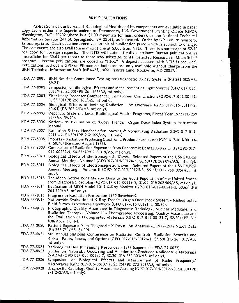
Publications of the Bureau of Radiological Health and its components are available in paper copy from either the Superintendent of Documents, U.S. Government Printing Office (GPO), Washington, D.C. 20402 (there is a $1.00 minimum for mail orders), or the National Technical Information Service (NTIS), Springfield, VA 22161, as indicated. Order by GPO or PB numbers, as appropriate. Each document receives an initial publication price which is subject to change. · The documents are also available in microfiche at $3.00 from NTIS. There is a surcharge of $2.50 per copy for foreign requests. The NTIS will automatically distribute Bureau publications as microfiche for $0.65 per report to those who subscribe to its "Selected Research in Microfiche" program. Bureau publications are coded as "HFX." A deposit account with NTIS is required. Publications without a GPO or PB number indicated are only available without charge from the BRH Technical Information Staff (HFX-25), 5600 Fishers Lane, Rockville, MD 20857.
FDA 77-8001
FDA 77-8002
FDA 77-8003
FDA 77-8004
FDA 77-8005
FDA 77-8006
FDA 77-8007
FDA 77-8008
FDA 77-8009
FDA 77-8010
FDA 77-8011
BRH Routine Compliance Testing for Diagnostic X-Ray Systems (PB 261 082/ AS, $9.25). Symposium on Biological Effects and Measurement of Light Sources (GPO 0l 7-015-00114-8, $3.30) (PB 261 167/AS, mf only). First Image Receptor Conference: Film/Screen Combinations (GPO 017-015-00115-6, $2. 50) (PB 261 166/ AS, mf only). Biological Effects of Ionizing Radiation: An Overview (GPO 0l 7-015-00117-2, $0.65) (PB 262 453/ AS, mf only). Report of State and Local Radiological Heal th Programs, Fiscal Year 197 5 (PB 259 947 / AS, $4.50). Nationwide Evaluation of X-Ray Trends: Organ Dose Index System-Instruction Manual. Radiation Safety Handbook for Ionizing & Nonionizing Radiation (GPO 017-015-00116-4, $1.70) (PB 262 109/ AS, mf only). Imports - Radiation-Producing Electronic Products (brochure) (GPO 0 17-015-00133-4, $0.70) (Revised August 1977). Comparison of Radiation Exposures from Panoramic Dental X-Ray Units (GPO 017-015-00122-9, $0.85) (PB 263 319/ AS, mf only). Biological Effects of Electromagnetic Waves - Selected Papers of the USNC/URSI Annual Meeting- Volume I (GPO 017-015-00124-5, $6.50) (PB 268 094/ AS, mf only). Biological Effects of Electromagnetic Waves - Selected Papers of the USNC/URSI Annual Meeting - Volume II (GPO 017-015-00125-3, $6.25) (PB 268 095/AS, mf only).
FDA 77-8013 The Mean Active Bone Marrow Dose to the Adult Population of the United States
FDA 77-8014
FDA 77-8015 FDA 77-8017
FDA 77-8018
FDA 77-8020
FDA 77-8021
FDA 77-8023 FDA 77-8025
FDA 77-8026
FDA 77-8028
from Diagnostic Radiology _(GPO 0 17-015-00119-9, $1.05) (PB 262 909/ AS, mf only). Evaluation of MOH Model 1015 X-Ray Monitor (GPO 017-012-00241-2, $0.65) (PB 263 725/ AS, mf only). Progress in Radiation Protection 197 5 (brochure). Nationwide Evaluation of X-Ray Trends: Organ Dose Index System - Radiographic Field Survey Procedures Handbook (GPO 017-015-00121-l, $0.80). Photographic Quality Assurance in Diagnostic Radiology, Nuclear Medicine, and Radiation Therapy. Volume II - Photographic Processing, Quality Assurance and the Evaluation of Photographic Materials (GPO 017-015-00123-7, $2.20) (PB 267 498/ AS, mf only). Patient Exposure from Diagnostic X Rays: An Analysis of 1972-1974 NEXT Data (PB 267 741/ AS, $4.00). 8th Annual National Conference on Radiation Control: Radiation Benefits and Risks: Facts, Issues, and Options (GPO 017-015-00126-1, $5.50) (PB 267 317/AS, mf only). Radiological Health Training Resources - 1977 (supersedes FDA 7 5-8027). Guides for Naturally Occurring and Accelerator-Produced Radioactive Materials (NARM) (GPO 017-015-00140-7, $2.20) (PB 272 303/AS, mf only). Symposium on Biological Effects and Measurement of Radio Frequency/ Microwaves (GPO 017-015-00137-7, $5.25) (PB 272 906/AS, mf only). Diagnostic Radiology Quality Assurance Catalog (GPO 017-015-00127-0, $4.00) (PB 271 248/ AS, mf only).

HEW Publication (FDA) 78 -8068
ELECTROMAGNETIC FIELDS in BIOLOGICAL MEDIA
PART I: DOSIMETRY-A PRIMER on BIOELECTROMAGNETICS
By
Stanley M. Neuder, Ph.D.
Division of Electronic Products
bureau of radiological health
~ WHO Collaborating Center for
Standardization of Protection
Against Nonionizing Radiation
July 1978
U.S. DEPARTMENT OF HEALTH, EDUCATION, AND WELFARE
Public Health Service Food and Drug Administration
Bureau of Radiological Health
Rockville, Maryland ~57

The opinions and statements contained in this report are those of the author and do not necessarily represent the views or stated policy of the World Health Organization (WHO).
ii
FOREWORD
The Bureau of Radiological Health conducts a national program to limit man's exposure to ionizing and nonionizing radiations. To this end, the Bureau (1) develops criteria and recommends standards for safe limits of radiation exposure, (2) develops methods and techniques for controlling radiation exposure, (3) plans and conducts research to determine health effects of radiation exposure, (4) provides technical assistance to agencies responsible for radiological health control programs, and (5) conducts an electronic product radiation control program to protect the public health and safety.
The Bureau publishes its findings in appropriate scientific journals and technical report and note series prepared by Bureau divisions and offices. Under a memorandum of agreement between the World Health Organization and the Department of Health, Education, and Welfare, three WHO Collaborating Centers have been established within the Bureau of Radiological Health, FDA:
WHO Collaborating Center for Standardization of Protection Against Nonionizing Radiations (Office of the Bureau Director)
WHO Collaborating Center for Training and General Tasks in Radiation Medicine (Division of Training and Medical Applications)
WHO Collaborating Center for Nuclear Medicine (Office of the Bureau Director)
As a WHO Collaborating Center, the Bureau makes available its technical reports and notes to participating WHO members.
Bureau publications provide an effective mechanism for disseminating results of intramural and contractor projects. The publications are distributed to State and local radiological health personnel, Bureau technical staff, Bureau advisory committee members, information services, industry, hospitals, laboratories, schools, the press, and other concerned individuals. These publications are for sale by the Government Printing Office and/or the National Technical Information Service.
Readers are encouraged to report errors or omissions to the Bureau. Your comments or requests for further information are also solicited.
ohn~.'!~ irector
Bureau of Radiological Health
iii
-··· - ..._..__. __ .,.. ____ . -~--~-· --·.
PREFACE
The present near ubiquity in the public domain of electronic devices emitting electromagnetic energy in the spectral region including the radiofrequency and microwave bands has stimulated an increasing level of concern for the impact of such devices on human health. Present human exposures range from fractional microwatts per centimeter squared to exceeding a milliwatt per centimeter squared in power density and fractional volts per meter to many hundred volts per meter in electric field strength at various frequencies within this interval. Exposures of humans to magnetic field strengths of several amps per meter in the radiofrequency spectrum during normal industrial operations have been reported.
The evaluation of the health impact of such exposures is hampered at present by the absence of adequate methodology for determining field distributions and the resulting power dissipation within the human body as a function of external field variables. In this spectral region, the science of dosimetry has not progressed to the state currently enjoyed for ionizing radiation.
In partial fulfillment of its mission in the implementation of The Radiation Control for Health and Safety Act of 1968, P.L. 90-602, the Division of Electronic Products has, in the last several years, conducted an active experimental and theoretical program within its Electromagnetics Branch to improve this situation. This report is one of several from that effort. Others have appeared in this FDA report series and in the open literature.
The present report discusses basic aspects of the interaction of radiofrequency and microwave fields with dielectric media and the determination of the resulting energy deposition.
Roger H. Schneider Director Di vision of Electronic Products
iv
CONTENTS
.. FOREWORD.
PREFACE.
ABSTRACT.
INTRODUCTION.
1. 2. 3. 4.
5. 6. 7.
8. 9.
10. 11. 12. 13. 14. 15. 16.
The RF and Microwave Spectrum. Electromagnetic Interaction with the Biological System Basic Field Equations. . • . . . . .... The Dielectric, Constant, Ke, and Relative Magnetic
Permeabiligy, Km, . . . . . . . • , • • The Loss Tangent, tan o • • • • •• • ••••••• Alternate Description -- The Complex Permittivity s* Relationship Between the Electrical Parameters, the Free Space
Wavelength \ 0 , and the Propagation Constant, K. Perfect and Lossy Dielectrics. The Internal Wavelength ............. . Not-So-Lossy Dielectrics ...•..•..•... Frequency and Temperature Dependence of Electrical Parameters. Power Absorption .. . Dosimetric Quantities ....... . Plane Waves. . . . . . . . . . . . . Normalization With Respect to the Incident Field Dosimetric Determinations.
APPENDIX I. Equivalent Expressions for the Poynting Vector
APPENDIX II. Conversion of Normalization of Incident Power Density.
REFERENCES ..•.... . . . . . . . . . . . . . . . . . . . . . .
V
Page
iii
iv
vi
1
2 2 2
4
5
6 7 9
11 11 12 13 14 14 16
17
18
20

ABSTRACT
Microwave and radiofrequency dosimetry - the determination of microwave and radiofrequency energy deposition in biological bodies - is discussed in terms of basic mathematical and physical considerations of electromagnetic interaction with biological systems. The electrical characteristics of the biological medium are mathematically related to the electromagnetic field properties. Variations of biological characteristics and internal field behavior as a function of exposure field frequency is discussed. Dosimetry aspects are stressed in terms of power absorption during field exposures.
vi

ELECTROMAGNETIC FIELDS IN BIOLOGICAL MEDIA
PART I: DOSIMETRY - A PRIMER ON BIOELECTROMAGNETICS
INTRODUCTION
The Bureau of Radiological Health is responsible for establishing radiation control programs to protect the public health and safety from electronic product radiation. In order to assist in establishing effective safety criteria, mathematical methods have been developed for predicting levels of absorption in biological systems exposed to nonionizing electromagnetic (EM) radiation.
When a biological system is exposed to radiofrequency or microwave radiation, electric and magnetic fields are induced within the system as well as scattered into the surrounding medium. The internally induced fields give rise to ionic currents and molecular excitations within the tissue and a subsequent heating of the tissue will insue. To understand biological effects in humans and animals exposed to nonionizing radiation, it is necessary to determine the induced field strength at internal points of the body. Knowing the electrical and physical characteristics of the irradiated body and the external exposure conditions,· it is then possible to calculate the absorbed power density (the distribution of absorbed power per unit volume) throughout the interior of the irradiated tissue. The absorbed power density will be the the source of a temperature rise and a subsequent steady-state temperature profile will be established which depends on thermal diffusion processes and cooling mechanisms of the biological system.
The magnitude of interior and exterior scattered fields depends on several factors: the frequency and extent of the incident field, the electrical characteristics of the various tissues, the shape, relative size, relative orientation and interior geometric structure of the system. For various combinations of these factors, extreme localized heating may occur and the internal energy distribution is generally not uniform.
Other factors being constant, higher absorption levels and lower penetration depths are found in biological media of relatively high dielectric constant such as brain, muscle, skin and tissue of high water content, whereas lower absorption levels and greater penetration depth are found in biological media of low dielectric constant such as bone and fat (1). Further, geometric structure, both size and shape, plays an important role in reflecting and refracting the incident field. Reflection and refraction are ultimately traced to the different electrical characteristics of adjacent interior regions and are responsible for the so-called "hot spots" or regions of concentrated power absorption within the biological system. The electrical conductivity strongly influences the internal fields but has a small effect on hot spot intensity (2). Since energy absorption depend on the geometric shape, size to wavelength ratio and relative orientation to the field, merely describing the exposure conditions will not adequately characterize the exposure hazard (3).
An exact mathematical determination of the internal fields is possible only for the simpler target geometries such as planes, spheres, and infinite cylinders exposed to well-defined incident radiation. Power absorption in complex geometries can only be calculated by approximation methods. Numerical

techniques and associated computer programs are presently being developed for treating complex arbitrary shapes, (4-6)(17-19).
This report reviews and unifies the physical and basic mathematical considerations of electromagnetic radiation in lossy dielectric media, and develops expressions for, and relationships between, the physical parameters pertinent to dosimetry. The behavior of these parameters in various biological tissues are discussed. Because of the vast subject matter only selected topics and typical behavior are discussed and the interested reader is referred to the cited references for greater detail. The list of references is however, not meant to be complete but rather a representative sampling of related research.
1. The RF and Microwave Spectrum
The RF and microwave spectrum extends in frequency from about 10~ Hz (RF) to 3 x 10 11 Hz, (microwave). The microwave range extends from 3 x 10 8 Hz to 3 x 10 11 Hz. In terms of wavelengths, the longest, at the RF end, are of the order of thousands of meters while the shortest, at the microwave end, are in the millimeter and submillimeter range.
The response of the biological system to the EM exposure varies widely across this range of frequencies, determined by the many factors already mentioned. The nature of this interaction will be discussed in the subsequent section. A large body of well-documented research exists on bioeffects in the megahertz (1 MHz= 10 6 Hz) to low gigahertz range (1 GHz= 10~ Hz), receiving impetus from the widespread industrial, medical and scientific applications at these frequencies. Outside this range, bioeffects have not been as extensively investigated and more work is necessary before any conclusions can be drawn.
2. Electromagnetic Interaction with the Biological System
The interaction of electromagnetic (EM) radiation with the biological system is characterized by classical electrodynamics rather than by quantum concepts so that the medium is considered to be a lossy dielectric described by the macroscopic electrical parameters, the permittivity€, the permeabilityµ, and the conductivity a, rather than by number densities and atomic properties. The radiation field is termed nonionizing because photon energies in the RF and microwave range do not produce atomic ionizations. This does not imply that microscopic processes do not occur. Indeed, molecular and ionic processes do exist in microwave fields and ultimately account for the frequency behavior of the electrical parameters and energy absorption by the biological system.
Characterizing the medium by €, µ, and a implies dissipative modes of interaction. Oscillations of free charge and molecular rotations are the mechanisms by which energy is removed from the field. This energy is dissipated in the medium, giving rise to what is called conduction losses and dielectric losses. The degree to which each loss shares in the total absorption in any given medium varies with frequency. Further discussion of physical processes has been deferred to section 8.
3, Basic Field Equations
Electromagnetic radiation is governed by Maxwell's equations, which in lossy dielectric media are of the form
2
• • aH
'ii X E = -µ at ( 1a)
• ( 1 b) • • aE 'ii X H aE + €-
at
• (1c) 'ii. E = 0
• ( 1 d) 'ii . H = 0
E and Hare the electric and magnetic field vectors, tis the time,µ, cr, E are the magnetic permeability, electrical conductivity, and electrical permittivity of the medium, respectively. Substituting the curl of equation (1a) into equation (1b) and using the vector identity V x V x E = V(V•E)-V2E, yields the time-dependent vector wave equation
The same expression applies time harmonic, of the form radiation, multiplied by 2TI. using equation (1c) yields the
(2)
to H. The radiation fields of interest here are Ee-iwt, where w is the frequency, f, of the
Taking the time derivatives in equation (2) and vector Helmholtz equation
(3)
where the square of the propagation constant, k, is given by
(4)
It is seen that the time harmonic electromagnetic fields are governed by the Helmholtz equation. Since k is a complex quantity, it has the form
k =a,+ i/3 (5)
where a and B, the real and imaginary parts of the propagation constant, are obtained by combining equations (4) and (5),
(l' = w fi [/1 +(~Y l½
+ lj
~ = w fi [ /1 + ( ~€ r _ 1] ½
(6)
(7)
(Units of k, a and Bare per-unit-length.) A particular solution of the timedependent vector wave equation is the description of a linearly-polarized plane wave propagating in the z-direction, with amplitude E0 along the x-direction, and Ho along they-direction.
E = 1 Eo ei(kz - wt)
(8)
• H = fHo ei(kz - wt)
3

Using equations (5) and (8) the electric field may be written as
• E = i'Eo e-/Jzei(o,z - wt) (9)
The term e-Bz indicates an exponential decay due to absorption of the field energy by the medium. It is specifically the imaginary part of the propagation constant that gives rise to this attenuation. Through B, the attenuation depends on the electrical properties of the medium. A plot of S vs. frequency using equation (7) indicates an increase in S with increasing frequency, at least in the range 100 MHz to 10 GHz. Of course, appropriate values of cr and€ must be chosen in constructing this graph since these quantities are frequency dependent. (This frequency dependence will be discussed in a section 8.) The increase simply indicates that the absorption increases, or penetration depth decreases, with increasing frequency. Further, plotting S for various biological tissues of high and low water content indicates that the absorption of the field energy is greater (penetration depth smaller) in the high water content media. It should be noted however, that besides the electrical properties, size and shape of the biological medium influences the distribution of absorbed power density too. Thus, the effective penetration may be different than the predicted exponential decay. This will be discussed further in reference (3).
The concept of wavelength, by definition, implies the existence of field fronts with constant phase. Thus, for example, plane wave fronts (see section 14 for plane wave description) propagating in the z direction are surfaces of constant phase described by (az-wt) = constant, as seen from equation (9). The wave fronts move with constant velocity, dz/dt. Differentiating the expression (az-wt) = constant with respect to time, yields
dz w dt a:
( 10)
where a is given by equation (6). Since w = 2Tif and by definition, dz/dt = Af, the wavelength associated with the electromagnetic field is
211' O'.
( 11 )
Through a, the wavelength depends on the electrical propertiesµ, E, and cr of the medium. It is thus apparent that the wavelength differs in different dielectric media. For example, the wavelength in high water-content tissue is about 1/7 the free space value. (Wavelength variation will be discussed in section 9.)
4. The Dielectric Constant, Ke, and Relative Magnetic Permeability, Km
The Dielectric constant (or relative permittivity), Ke, of a homogeneous dielectric medium is defined as
f K =
e fo ( 12)
where Ke is dimensionless and E:o = (36TI x 109)-1 F/m, the permittivity of free space. (Throughout this paper, unless otherwise specified, the subscript zero
4
refers to free space quantities.) The relative permeability, Km, of a homogeneous medium is defined as
K =--1!:_ m µo
where µ 0 = 4TI x 10-7 H/m. For biological tissue throughout the RF range, as well as for air, µ and µ 0 are essentially equal, so that Km= 1.
5, The Loss Tangent, tan o Equation (4) may be rewritten as
The quantity (a/we:) is termed the loss tangent, tan o.
tan cS a a
=-=-- ( 13)
The last expression is obtained from equation (12). The loss tangent is a direct indicator of the degree of energy absorption by the medium since it constitutes, by virtue of equation (4), the ratio of imaginary to real parts of the square of the complex propagation constant. Stated in other ways, it is the ratio of conduction currents to displacement currents, or the ratio of lossy components to lossless components, in the medium. (The concept of conduction and displacement current is further developed in the next section.) Thus, for a medium having zero loss tangent (a= 0, and S = 0), the wave passes through the medium unattenuated. This medium is referred to as a "perfect" dielectric. In biological tissue, values of the loss tangent at 2450 MHz generally range between 0.2 and 0.3.
6. Alternate Description -- The Complex Permittivity e:•
For a given frequency and geometry, two of the three parameters a, Ke and tan o must be specified in order to calculate the scattering and absorption in biological media exposed to electromagnetic radiation. An alternate description found in the literature is one in which the real and imaginary parts of a complex permittivity are specified. These quantities are developed as follows. Equation (1b) and E = E0 eiwt yields VxH = (cr+iwe:)E. Considering crE as a conduction current density and we:E as a displacement current density, the total current density Jt, is
• • lt = (a + iwE) E
This concept derives from Maxwell's equation being written as V x H = e:(aE;at). Substituting equation (12) into equation (14) yields
5
(14)
Jcond +

Now define a complex permittivity E* as
* (K . a ) (, . " E = € 0 e - t - = E E - zE ) WEO 0
( 15)
so that Jt : i(DE*E. permittivity are
The real and imaginary parts, E' and E", of the complex
" a (16) €
From equations (13) and (16), the loss tangent is
" tan o €
' ( 17)
€
In this description, the specification of any two of the three parameters E' , E11 and tan o is necessary in orde_r to calculate the induced fields. It should be noted that the quantities cr and Ke, or equivalently E' and E", are frequency dependent. Thus, tan o does not vary inversely with frequency, as may be erroneously inferred from equation (13). Similar remarks apply to the variation of E" in equation (16). Details of the frequency dependence of cr and Ke will be discussed in section 8.
7. Relationship Between the Electrical Parameters, the Free Space Wavelength Ao, and the Propagation Constant, k
From equations (6) and (11), the free space wavelength is obtained by setting cr = O, so that
27T
This, of course, reduces to \ 0 = elf, since the velocity of light, c = 1/✓µ 0E 0 and w = 2nf. Further, from equation (4) with E = KeEo andµ= Kmµo
Eliminating µ0 E0 between these two expressions and setting Km= 1 yields
Taking the square root and using equation (13), yields
k ~: JXe[l + itano] ½
( 18)
6
This relates the propagation constant, k, with the free space wavelength, Ao, the dielectric constant, Ke and the loss tangent, tan o. An alternate form for the propagation constant is obtained from substituting equations (16) and (17) into equation (18) to get
k = - € zE 2Tf j I + . II Ao
8. Perfect and Lossy Dielectrics
In the "perfect" or nonconducting dielectric medium, the propagation constant is real since the quantities a, £", tan o, and Bare all zero. As discussed earlier, there is no field attenuation in the medium when B = 0. In the case of the perfect dielectric, the propagation constant is, from equation ( 18),
( 19)
Moreover, if the perfect dielectric is air, then the propagation constant reduces to
k _ 2rr -~ ko
since Ke = 1. Similarly, using equations (6), (11) and (12) with Km= 1, the wavelength in a perfect dielectric is
(20)
If the perfect dielectric is air, then A= Ao, as expected. It is seen from the above that the wavelength is reduced in the perfect dielectric from its free space value, varying inversely as the square root of the dielectric constant, Ke·
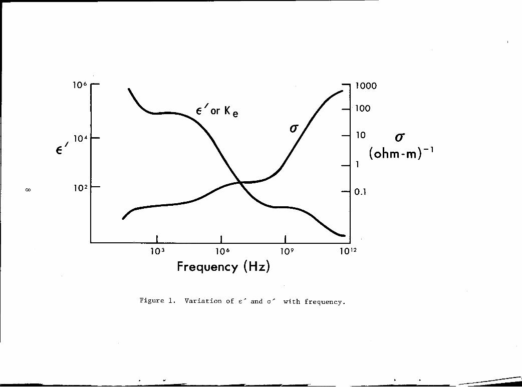
Since a perfect dielectric does not remove energy from the electromagnetic field the field propagates through the medium unattenuated. A lossy dielectric, by definition, has finite conductivity. Finite conductivity implies charge motion in the form of free ions (conduction loss), and molecular rotation (dielectric loss). Both processes result in energy transfer from the field to the medium with consequent heating of the medium. The energy transfer interaction attenuates the field. The degree of "lossiness" is a function of the frequency of the radiation. At low frequencies, below about 100 MHz, the response time of cell membrane allows the membrane to fully charge and discharge with the field oscillations, resulting in high capacitive reactance. Conduction currents are not sustained within the cells sa that the tissue is characterized by a high dielectric constant and low conductivity. As the field frequency increases well above membrane-response time, the membrane is no longer reactive and, in effect, allows free ion flow within and between cells. The tissue thus becomes electrically conducting. At still higher frequency (above about 3 GHz) molecular rotations of the water molecule occur. Thus, electrical conductivity of biological tissue increases and dielectric constant decreases with increasing frequency. Figure 1 shows the variations-of Ke and a as a function of frequency for typical biological tissue (7). Several biological sources contribute to these variations with frequency. These are the body fluids, free and bound water, ions, polymers and organic compounds. For detailed discussions see references (7 and 8).
7
10 6 1000
100
10 4 10 (J'
e' (ohm-m)- 1
1
00 10 2 0.1
10 3 10 6 10 9 1012
Frequency (Hz)
Figure 1. Variation of E~ and a~ with frequency.

Analytic expressions for the dependence of Ke and cr with frequency have been reported (9-11). These are
KL - KH Ke= KH +-------=-
1 + (w7) 2
where Tis the relaxation time of the medium. The subscripts Land H denote low frequency (de) and high frequency (optical) values. For biological tissue with high water content, T = 1/2Tif0 where f 0 is the rotational resonant frequency of the water molecule. Taking fo to be the characteristic frequency of water and also of biological tissue (11), these expressions become, after rearranging terms
a = 1 + (//20)2
Ke KH [KL (J/20)J = 1 + (//20)2 KH + J
with f in GHz. Values of Kand cr at low and high frequencies are reported in the literature (11). bther biological mechanisms contributing different expressions for T are discussed elsewhere (8). See figure 2 for variations of the electrical parameters in muscle tissue.
9. The Internal Wavelength
All biological media are lossy. Mathematically, this is indicated by a nonzero conductivity cr, a nonzero value of 8, a complex propagation constant k, and non-zero value of € 11
1 the imaginary part of E*. The internal wavelength in lossy media may be derived by substituting equations (11) and (12) into equation (6) and using the expression for Ao from section 7, to obtain
(21)
Or, from equation (13) the internal wavelength, in terms of the loss tangent is
' = }.; [½ + ½ Ji + ... ,,r It is seen from these equations that the wavelength in a lossy medium is
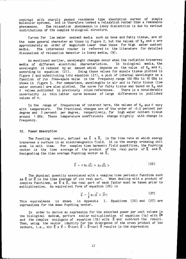
reduced from that in the perfect dielectric. For example, a free space wavelength of 10 cm (frequency = 3 GHz), will be reduced to about 1.5 cm in muscle tissue as shown in figure 3. Further, biological tissue having different electrical parameters will exhibit different internal wavelengths. From the above equations, the greater the dielectric constant and loss tangent of the biological tissue, the smaller the internal wavelength. For example, as shown in figure 3, a free space wavelength of 10 cm is reduced to 4.3 cm in fat compared with 1.5 cm in muscle. This is so because the dielectric constant and loss tangent values for muscle exceeds those for fat by factors of about 11 and 2, respectively.
9

100.0
.,, 10.0 Q) ::, .
~ .. Q)
'i E C .. C
Cl..
1.0
]08 109
Frequency (Hz)
tan o
1010
Figure 2. Parameter variations as a function of frequency for muscle tissue.
100
t E ~ .s:::. o, 10 C: Q)
Q) > 0
~ 0 C: ... Q) .... C:
- Field Frequency (GHz) 10 .1
10 100 1000
Free Space Wavelength (crn)-
Figure 3. Variation of internal wavelength in dielectric media.
10
--------------------------
10. Not-So-Lossy Dielectrics
As mentioned earlier, field attenuation is associated with non-zero 8 and the degree of attenuation is indicated by the relative magnitude of the loss tangent. At certain frequencies, the losses are relatively small in biological tissue. This is described mathematically as
·J
'. 2 ( a )2
tan /j = WE < < 1 (22)
When this condition prevails, the following approximations apply
(23)
(24)
(25)
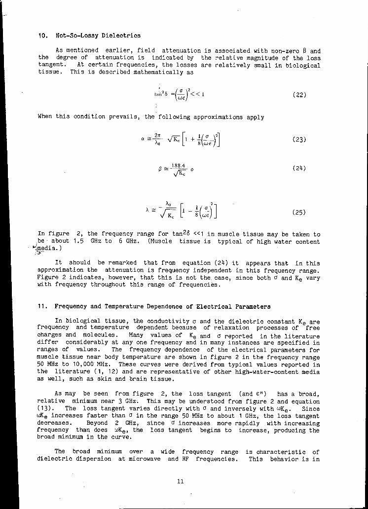
In figure _be· about
--;,media.) ;':J:"
2, the frequency range for tan2o <<1 in muscle tissue may be taken to 1.5 GHz to 6 GHz. (Muscle tissue is typical of high water content
It should be remarked that from equation (24) it appears that in this approximation the attenuation is frequency independent in this frequency range. Figure 2 indicates, however, that this is not the case, since both cr and Ke vary with frequency throughout this range of frequencies.
11. Frequency and Temperature Dependence of Electrical Parameters
In biological tissue, the conductivity cr and the dielectric constant Ke are frequency and temperature dependent because of relaxation processes of free charges and molecules. Many values of Ke and cr reported in the literature differ considerably at any one frequency and in many instances are specified in ranges of values. The frequency dependence of the electrical parameters for muscle tissue near body temperature are shown in figure 2 in the frequency range 50 MHz to 10,000 MHz. These curves were derived from typical values reported in the literature (1, 12) and are representative of other high-water-content media as well, such as skin and brain tissue.
As may be seen from figure 2, the loss tangent (and E: 11 ) has a broad, relative minimum near 3 GHz. This may be understood from figure 2 and equation (13). The loss tangent varies directly with cr and inversely with wKe. Since r.llKe increases faster than cr in the range 50 MHz to about 1 GHz, the loss tangent decreases. Beyond 2 GHz, since cr increases more rapidly with increasing frequency than does wKe, the loss tangent begins to increase, producing the broad minimum in the curve.
The broad minimum over a wide frequency range dielectric dispersion at microwave and RF frequencies.
11
is characteristic of This behavior is in
contrast with sharply peaked resonance type absorption curves of simple molecular systems, and is therefore termed a relaxation rather than a resonance phenomenon. The relaxation phenomenon in lossy dielectrics is due to various contributions of the complex biological structure.
Curves for low water content media such as bone and fatty tissue, are of the same general character as those in figure 2, but the values of Ke and a are approximately an order of magnitude lower than those for high water content media. The interested reader is referred to the literature for detailed discussions of relaxation behavior in lossy media, (9).
As mentioned earlier, wavelength changes occur when the radiation traverses media of different electrical characteristics. In biological media, the wavelength is reduced by an amount which depends on the value of Ke and a, according to equation (21). Using those values for muscle tissue plotted in figure 2 and substituting into equation (21), a plot of internal wavelength as a function of its free-space value in the frequency range 100 MHz to 10 GHz is shown in figure 3. For comparison, wavelengths in air and in fatty tissue (low water content) are also plotted. The curve for fatty tissue was based on Ke and a values published in previously cited references. There is a considerable uncertainty in this latter curve because of large differences in published values of a.
In the range of frequencies of interest here, the values of Ke and a vary with temperature. The fractional changes are of the order of -0.5 percent per degree and 2 percent per degree, respectively, for high water content tissue around 1 GHz. These temperature coefficients change slightly with change in frequency.
12. Power Absorption
The Poynting vector, defined as E x H, is the time rate at which energy traverses a surface in an electromagnetic field. It is the energy crossing unit area in unit time. For complex time harmonic field quantities, the Poynting vector is the time average of the product of the real parts of E and H. Designating the time average ·poynting vector as S,
• • • S = < Re (E) X Re (H) > (26)
The physical quantity associated with a complex time periodic function such as E or His the time average of its real part. When dealing with a product of complex functions, as Ex H, the real part of each factor must be taken prior to multiplication. An equivalent form of equation (26) is
• 1 • S = 2 Re (E X
This equivalence is shown in Appendix expressions for the mean Poynting vector.
• H*)
I. Equations (26) and
( 27)
(27) are
In order to derive an expression for the absorbed power per unit volume in the biological medium, perform scalar multiplication of equation (1a) with H* and the complex conjugate of equation (1b) with E and subtract the result. Then, using the vector identity for the divergence of the cross product of two vectors, i.e., div Ex H = H•curl E - E•curl H results in the expression
12
• • • ( • * • * a i ) "v • (E x tt*) = - E • J* _ it . ao + H • -at at (28)
where J is the conduction current, aE. Using the relationships J = aE, D = eE, B = µH, E = E0 e-j(ut and H :H0 e-jwt equation (28) becomes
• • * • • • "iJ • (E X H ) = -o IE 12 + jw(µIHl2 - EIEl2 )
(29)
Now, the mean Poynting vector is given by equation (27) and Poynting's theorem states that the divergence of the mean Poynting vector, V•(½ Re E x H*), measures the energy transformed per unit volume per second into heat. Applying this theorem to equation (29) yields
1 • • * 1 • 2
2 Re "i/ • E X H = - 2 a IE I
(The minus sign is to be interpreted as saying that ½alEl 2 is the rate at which energy is produced per unit volume rather than the rate at which energy is lost by the medium.) Thus, the power deposited per unit volume of biological medium by the electromagnetic field is
P = _l_olEl2 (30) 2
Another mathematical expression found in the literature may be derived from equations (16) and (30).
( 31)
Units of power density are W/m3 or mW/cm3, as discussed in the following section.
13. Dosimetric Quantities
The absorbed power density has units of J/sec-m3. An equivalent term found in the literature is "heating potential" (12). Other terminology found in the literature relates to "dose" ( 13). The "distributed dose rate" is the rate of energy absorption per unit mass with units of W/kg. This quantity is also called "absorbed dose rate." The conversion of W/m3 to W/kg is obtained by dividing the quantity in W/m3 by the mass density of the medium. The "integral dose rate," in watts, is the time rate of energy absorption by the biological system. The "integral dose" is the total absorbed energy, in joules. The "average dose" is the integral dose, in joules, per unit mass of the biological system. Mass densities of biological tissue are generally close to 1 gm/cm3. For example, the mass density of muscle, and fatty tissue are of the order of 1.07, and 0.94, gm/cm3, respectively (14). One other dosimetric term, the SAR, is used extensively. SAR is the specific absorption rate, in W/kg and is the same as the distributed dose rate or the absorbed dose rate, described above. Note that the integral dose rate, integral dose, and average dose are quantities relating to the whole body or regions of the body rather than to particular points in the biological system.
13
14. Plane Waves
The concepts and equations developed throughout this paper are of general applicability with respect to the externally incident field, i.e., tpe incident radiation need not be a plane wave in order that these equations be valid. For the purpose of subsequent discussion however, a description of the plane wave field will now be given.
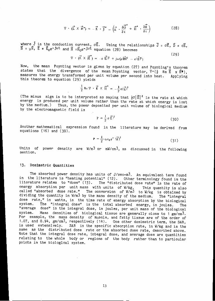
A plane wave is described mathematically by an oscillatory function of the space coordinates and time as for example, those functions given by equation (8). The field varies periodically in time with frequency f:w/2TI, time period T:1/f, and space period A. Relationships between these and other parameters describing the field have been derived earlier. A plane wave is characterized by surfaces of constant phase which at one instant·in time, t1, may be depicted as a series of stationary planar wave fronts containing the mutually perpendicular E and H vectors. As shown in figure 4a, the E and H vectors are in phase, with E always along the x-direction and H along they-direction. The two wave fronts shown are spaced A/2 apart. At a later time, t1 + T, the two fronts will have moved a distance A along the positive z-direction (not shown), with velocity given by equation (10). Consider a mathematical plane, transverse to the direction of propagation, fixed at a point along the z-axis. As depicted in figure 4b, at all points in this plane, the electric field oscillates in time with frequency f and amplitude E0 • All oscillations are in phase with each other. The magnetic field follows the same behavior. Equation (8) represents this linearly polarized, transverse electromagnetic (TEM) field where E, H, and the propagation direction are mutually perpendicular. In air, the TEM field has coupled transverse E and H fields that are in phase with a magnitude ratio 377 to 1. This ratio is called the intrinsic impedence of free space, denoted by Z0 • In a biological medium, E and Hare no longer in phase and their ratio is reduced by an ambunt which depends on the electrical parameters of the medium. Further, if the medium is inhomogeneous or if non-TEM conditions prevail, the ratio of E to H may vary from point to point. Thus, within a sufficiently close distance to a radiation source, as for example in the near field of a horn antenna, the ratio of E to H varies from point to point and the field is not TEM within a few wavelengths of the aperture.
Incident field strength or incident power density is a necessary descriptor in electromagnetic exposure studies and is often used for the normalization of numerical results. For TEM conditions the incident field may be specified either in terms of incident field strength or incident power density. For nonTEM incident fields, field strength rather than power density is the more meaningful description, although the concept of an 11 equivalent" power density, equal to E2/z0 , is sometimes used (See Appendix II).
15. Normalization With Respect To The Incident Field
It should be noted that absorbed power density has different units than does incident power density. The absorbed power density is the power absorbed per unit volume, in W/m3 whereas the incident power density is the power incident per unit area, in W/m2.
The externally incident electromagnetic radiation may be described in terms of field strength in V/m or in terms of energy crossing unit area per unit time~ in mW/cm2. Thus, if the reported internally absorbed power density is in W/mj it is usually understood to be normalized to either 1 V/m or to 1 mW/cm2 external field. To convert N-W/m3·per 1 V/m incident field to W/m3 per 1 mW/cm2
rms incident power density, N must be multiplied by 3770. (See Appendix II for conversion details.)
14

X
z
X 2
Figure 4a. Simultaneous space variation of the plane wave electromagnetic field.
X
z
X
z
·Figure 4b. Time variation of E and H at a fixed position in the plane wave field.
15
Other terms used for the incident field are "incident power density" and
"exposure rate," in mW/ cm2. These of course represent the incident energy
crossing unit area per unit time.
16. Dosimetric Determinations
Recently, theoretical and experimental techniques have been developed that
will allow the determination of the distribution of induced fields and power
deposition in a man or animal exposed to microwave radiation. Three
developments in particular will be mentioned. These are (a) thermography, (b)
nonperturbing miniature implantable probes, and (c) computer programs for
estimating induced field distributions.
Thermography is an experimental technique based on the use of an infrared
camera to record heating patterns in phantom models exposed to microwave
radiation. Phantom materials simulate various biological tissue by virtue of
their dielectric properties and capability of being molded into desirable shapes
(1,14).
Implantable electric field probes have recently been developed that do not
perturb nor interact with the electromagnetic field (15). These miniature
probes therefore may be used to measure the internal fields in biological
systems during irradiation. Non-perturbing temperature probes (16) allow
specific locations to be dosimetrically evaluated in terms of localized heating.
From the temperature rise per unit time, the SAR may be determined in bodies of
known thermal properties.
Primarily over the past 10 years, computer programs have been used to
calculate induced fields in regularly shaped lossy dielectric media such as
spheres and circular cylinders (2,12,13). These programs have been applied only
to idealized biological systems as for example, multilayered concentric spheres
representing the human head. Mathematical techniques that provide the
capability of calculating power deposition in biological media of arbitrary
shapes and complex internal structures are presently evolving (4-6, 17-19).
These techniques are based on a number of different numerical approximation
methods. The associated computer programs provide approximate solutions of
internal fields and absorbed power density at discrete points within the
biological structure.
16
APPENDIX I
Equivalent Expressions for the Poynting Vector
Expand< Re Ex Re H > where E = E0 e-jwt and H = H0e-jwt, as follows
Re E = Re E0 e-jwt
= Re (Er+ jEi)(cos wt+ j sin wt) = Er cos wt - Ei sin wt
where subscripts rand i mean real component and imaginary component, respectively. Similarly, expand Re Hand take the cross product with Re E, given above. This yields:
± + + + 2 + + l Re t; x Re H = Er x Hr cos 1.ut + Ei x Hi sin wt
- (Er x Hi + Ei x Hr) sin wt cos wt
so that
< Re E x Re H > = 1 /2 (Er x Hr + Ei x Hi)
since the time average over sin2 wt or cos 2 wt is 1/2 whereas the time average over sin wt cos wt is zero.
In a similar fashion expand Ex Has follows
E x H* = E0 x H0 = (Er + jEi) x (Hr - jHi)
= (Er x Hr + Ei x Hi) x j (Ei x Hr - Er x Hi)
so that
as above. Thus
S =<Re Ex Re H > = 1/2 Re (Ex H*)
17
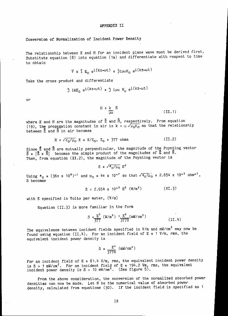
APPENDIX II
Conversion of Normalization of Incident Power Density
The relationship between E and H for an incident plane wave must be derived first.
Substitute equation (8) into equation (1a) and differentiate with respect to time
to obtain
V X 1 Eo ei(kz-wt) = JiµWHo ei(kz-wt)
Take the cross product and differentiate
J ikEo ei(kz-wt) = J iµw s0
ei(kz-wt)
or
H = k E µw ( II. 1)
where E and Hare the magnitudes of E and H, respectively. From equation
(19), the prop~gation constant in air is k = w /µ 0 E0 so that the relationship
between E and Hin air becomes
H = IE0 /µ 0 E = E/Z0 , Z0 = 377 ohms (II.2)
Since E and Hare mutually perpendicular, the magnitude of the Poynting vector
S = IE x HI becomes the simple product of the magnitudes of E and H. Then, from equation (II.2), the magnitude of the Poynting vector is
S = /t:ofµo E2
Using E0 = (36n x 10 9 )-1 and µ 0 = 4n x 10-7 so that /e0 /µ0 = 2.654 x 10- 3 ohm- 1 ,
S becomes
S = 2.654 x 10- 3 E2 (W/m 2) (II.3)
with E specified in Volts per meter, (V/m)
Equation (II.3) is more familiar in the form
2 2 S = E (W/m2 ) =_E_(mW/cm2 )
377 3770 (II.4)
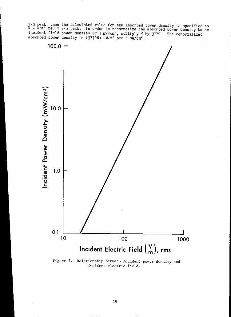
The equivalence between incident fields specified in V/m and mW/cm 2 may now be
found using equation (II.4). For an incident field of E = 1 Vim, rms, the
equivalent incident power density is
Ez z S = (mW/cm)
3770
For an incident field of E = 61.4 Vim, rms, the equivalent incident power density
is S = 1 mW/cm 2• For an incident field of E = 194.2 Vm, rms, the equivalent
incident power density is S = 10 mW/cm 2• (See figure 5).
From the above consideration, the conversion of the normalized absorbed power
densities can now be made. Let N be the numerical value of absorbed power
density, calculated from equations (30). If the incident field is specified as 1
18
V/m pea~, then the calculated value for the absorbed power density is specified as N - W/m per 1 V/m peak. In order to renormalize the absorbed power density to an incident field power density of 1 mW/cm 2
, multiply N by 3770. The renormalized absorbed power density is (3770N) -W/m 3 per 1 mW/cm2 •
100.0
-N
E ~ ~ E 10.0 ->--u,
C: Q)
0 L. Q)
~ 0 c... -C: Q) ~
u C:
1.0
0.1 10 100 1000
Incident Electric Field { ~), rms
Figure 5. Relationship between incident power density and incident electric field.
19
REFERENCES
1. Johnson, Systems.
C.C. and A.W. Guy. Nonionizing EM Wave Effects in Biological Proc IEEE 60, 692 (1972).
2. Neuder, S.M., R.B. Kellogg, and D. Hill. Microwave Absorption in a Spherical Multilayered Model of the Head. Effects of Electromagnetic Waves. HEW Publication (FDA) ( 1976).
Power Density In, Biological 77-8011, 199
3, Neuder, S.M. Electromagnetic Fields in Biological Media. Part IV. Microwave Scattering by Spherical Systems. FDA/BRH Rpt to be published.
4. Neuder, S.M. A Finite Element Technique for Calculating Induced Internal Fields and "Power Deposition in Biological Media of Complex Irregular Geometry. In, Symposium on Biological Effects and Measurement of Radio Frequency/Microwaves. HEW Publication (FDA) 77-8026 pp. 170-190, February 1977.
5. Neuder, S.M. and P.H.E. Meijer. Finite Element-Variational Calculus Approach to the Determination of Electromagnetic Fields in Irregular Geometry. In, Biological Effects of Electromagnetic Waves. HEW Publication (FDA) 77-8011, 193 (1976).
6. Neuder, S.M. and R.B. Kellogg. Numerical Calculation of Microwave Absorption in Arbitrary Geometry with Application to a Model of Man. URSI symposium on Electromagnetic Fields in Biological Systems, Ottawa, Canada, June 1978.
7, Grant, E.H. Microwaves - A Tool in Medical and Biological Research. In, Biological Effects and Health Hazards of Microwave Radiation. Proc. Int. Symp. Warsaw, Oct. 1973, pp. 309-316. Polish Medical Publishers, Poland, 1974.
8. Pressman, A.S. Electromagnetic Fields and Life. (1970).
Plenum Press, N.Y.
9. Von Hipple, A .R., Ed. Dielectric Materials and Applications. 1954.
MIT Press,
10. Debye, P. Polar Molecules. Dover N.Y. (1945).
11. Kritikos, H.N. and H.P. Schwan. Hot Spots Generated in Conducting Spheres by E.M. Waves. IEEE T-BME 19, 53 (1972).
12. Weill, C.C. Absorption of Multilayered Sphere UHF/Microwave Radiation. IEEE BME, 468 (1975).
Models Exposed to
13. Ho, H. and A.W. Guy. Development of Dosimetry for RF and Microwave Radiation. Health Physics 29, 317 (1975).
14. Guy, A.W., J.F. Lehmann, and J.B. Stonebridge. Therapeutic Applications of Electromagnetic Power. Proc. IEEE 62, 55 (1974).
15. Bassen, H., P. Herchenroeder, A. Cheung and S.M. Neuder. Evaluation of an Implantable Electric-Field Probe Within Finite Simulated Tissues. Radio Sciance 12, 15 (1977).
20

16.
17.
18.
Cetas, T.C. A birefringent Crystal Optical Thermometer for Measurements of EM Induced Heating. HEW Publication (FDA) 77-8011, 338 (1976).
Guru, B. and K. Chen. Experimental and Theoretical Studies of EM Fields Induced Inside Biological Bodies. IEEE T~MTT 24, 433 (1976).
Barber, P.W. Numerical Study of EM Power Deposition in Biological Tissue Bodies. HEW Publication (FDA) 77-8011, 119 (1976).
19. Hagmann, V.J., O.P. Gandhi and C.H. Durney. Numerical Calculation of EM URSI Symposium, 1977. Energy Deposition in a Realistic Model of Man.
21
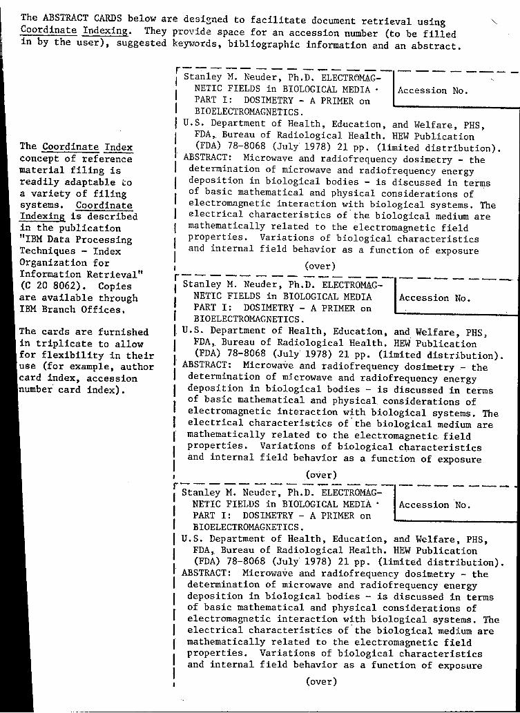
The ABSTRACT CARDS below are designed to facilitate document retrieval using ~-Coordinate Indexing. They provide space for an accession number (to be filled in by the user), suggested keywords, bibliographic information and an abstract.
The Coordinate Index concept of reference material filing is readily adaptable co a variety of filing systems. Coordinate Indexing is described in the publication "IBM Data Processing Techniques - Index Organization for Information Retrieval" (C 20 8062). Copies are available through IBM Branch Offices.
cards are furnished in triplicate to allow for flexibility in their use (for example, author card index, accession number card index).
r----- --- -...-- --- --------I Stanley M. Neuder, Ph.D. ELECTR~MAG- ,
I NETIC FIELDS in BIOLOGICAL MEDIA • Accession No. PART I: DOSIMETRY - A PRIMER on
I BIOELECTROMAGNETICS. I I I I
U.S. Department of Health, Education, and Welfare, PHS, FDA,. Bureau of Radiological Health. HEW Publication (FDA) 78-8068 (July 1978) 21 pp. (limited distribution).
ABSTRACT: Microwave and radiofrequency dosimetry - the determination of microwave and radiofrequency energy deposition in biological bodies - is discussed in terms of basic mathematical and physical considerations of · electromagnetic interaction with biological systems. The electrical characteristics of.the biological medium are mathematically related to the electromagnetic field properties. Variations of biological characteristics and internal field behavior as a function of exposure
I r I I I I , {over) r----- --- - ...... - --- -------· I Stanley M. Neuder, Ph.D. ELECTROMAG-
1 NETIC FIELDS in BIOLOGICAL MEDIA Accession No.
I PART I: DOSIMETRY - A PRIMER on BIOELECTROMAGNETICS.
I I I·
I
U.S. Department of Health, Education, and Welfare, PHS: FDA,. Bureau of Radiological Health. HEW Publication (FDA) 78-8068 (July' 1978) 21 pp. (limited distribution).
ABSTRACT: Microwave and radiofrequency dosimetry - the determination of microwave and radiofrequency energy deposition in biological bodies - is discussed in terms of basic mathematical and physical considerations of · electromagnetic interaction with biological systems. The electrical characteristics of.the biological medium are mathematically related to the electromagnetic. field properties. Variations of biological characteristics and internal field behavior as a function of exposure
I r I I I I , (over) r------ - -- - ...... - --- --- ----~ I Stanley M. Neuder, Ph.D. ELECTROMAG-
1 NETIC FIELDS in BIOLOGICAL MEDIA • Accession ·No. PART I: DOSIMETRY - A PRIMER on
I BIOELECTROMAGNETICS. I U.S. Department of Health, Education, and Welfare, PHS, I FDA,. Bureau of Radiological Health. HEW Publication
I. (FDA) 78-8068 (July 1978) 21 pp. (limited distribution).
ABSTRACT: Microwave and radiofrequency dosimetry - the I determination of microwave and radiofrequency energy I deposition in biological bodies - is discussed in terms of basic mathematical and physical considerations of f electromagnetic interaction with biological systems. The I electrical characteristics of.the biological medium are I mathematically related to the electromagnetic field
I properties. Variations of biological characteristics and internal field behavior as a function of exposure
I (over)

----~----- - - - - ,...... _______ ..,.... ____ _ f.ield frequency is discussed. Dosimetry aspects are
stressed in terms of power absorption during field
exposures.
KEYWORDS: Biological systems; Dosimetry; Electromag
netic interaction; Energy deposition; Exposure; Field
behavior; Microwave; Power absorption; Radiofrequency.
-·------- -----....... -~,.._...----.---1 field frequency is discussed. Dosimetry aspects are
stressed in terms of power absorption during field
exposures.
KEYWORDS: Biological systems; Dosimetry; Electromag
netic interaction; Energy deposition; Exposure; Field
behavior; Microwave; Power absorption; Radiofrequency.
I I I I I I I I I I I I I
_______________________ !
field frequency is discussed. Dosimetry aspects are 7 stressed in terms of power absorption during field I exposures.
KEYWORDS: Biological systems; Dosimetry; Electromag
netic interaction; Energy deposition; Exposure; Field
behavior; Microwave; Power absorption; Radiofrequency.
I I

FDA 77-8029 Course Manual for Machine Sources of X Rays (GS-461) (GPO 017-015-00131-8, $4.00) (PB 272 011/ AS, mf only) (supersedes FDA 73-8026).
FDA 77-8030 Course Manual for X-Ray Measurements (GS-462) (GPO017-015-00130-0, $3.50) (PB 272 012/ AS, mf only) (supersedes FDA 73-8027).
FDA 77-8031 Course Manual for X-Ray Applications (GS-463) (GPO 017-015-00132-6, $3.00) (PB 272 010/ AS, mf only) (supersedes FDA 73-8028).
FDA 77-8032 The Bureau of Radiological Health ••• A Look at FDA's Program to Protect the American Consumer from Radiation (GPO 017-015-00128-8, $1.20) (PB 272 869/ AS, mf only).
FDA 77-8033 BRH Publications Index (GPO 017-015-00129-6, $4.25) (PB 271 734/ AS, mf only). FDA 77-8034 Report of State and Local Radiological Health Programs, FY 1976 (PB 273 392/ AS,
$5.25). FDA 77-8035 The Developing Role of Short-Lived Radionuclides in Nuclear Medicine (GPO 017-
015-00139-3, $2.00) (PB 272 298/ AS, mf only). FDA 77-8036 Second Image Receptor Conference: Radiographic Film Processing (GPO 017-015-
00134-2, $3.00) (PB 273 287/AS, mf only). FDA 77-8037 Procedures for Field Testing Microwave Ovens (GPO 017-015-00138-5, $1.60)
(PB 272 839/ AS, mf only) (supersedes FDA 73-8016). FDA 77-8042 CSU-FDACollaborative Radiological Health Laboratory Annual Report 1976 (PB 273
560/ AS, $6.50). FDA 78-8015 Progress in Radiation Protection 1976 (brochure). FDA 78-8027 Directory of Personnel Responsible for Radiological Health Program (supersedes
FDA 77-8027). FDA 78-8038 Laser Compliance Measurements Handbook (supersedes FDA 76-8038-Prepublication
Copy) (PB 281 190/ AS, $9.00). FDA 78-8039 Exposure and Processing Guide for Dental Radiography (GPO 017-015-00146-6,
$1.20) (supersedes FDA 77-8039). FDA 78-8043 A Review of the Use of Ionizing Radiation for the Treatment of Benign Diseases
Volume I (GPO 017-015-00141-5, $2.10) (PB 274 032/ AS, mf only). Volume IIAppendixes A and B, FDA/BRH-78/2, NTIS only (PB 278 797 / AS, $14.00).
FDA 78-8044 Federal Recordkeeping Requirements for Television Receiver Dealers and Distributors (brochure) (GPO 017-015-00142-3, $0.50/3.75 per 100).
FDA 78-8047 Source Book of Educational Materials for Medical Radiographers (GPO 017-015-00143-1, $2.50) (PB 275 953/AS, mf only).
FDA 78-8048 Symposium on Biological Effects and Characterizations of Ultrasound Sources (GPO 017-015-00145-8, $4.25) (PB 277 945/AS, mf only).
FDA 78-8049 Measurements of the Performance Parameters of Gamma Cameras: Part I (GPO 017-015-00144-0, $2.20) (PB 277 482/ AS, mf only).
FDA 78-8050 A Practitioner's Guide to the Diagnostic X-Ray Equipment Standard (brochure) (GPO 017-015-00147-4, $0.80) (supersedes FDA 75-8005).
FDA 78-8053 An Instrument for Non-Invasive mAs Measurement (PB 280 548/ AS, $4.50). FDA 78-8054 9th Annual National Conference on Radiation Control - Meeting Today's Challenges
(GPO 017-015-00148-2, $5.75). FDA 78-8055 A Physical Basis of Electromagnetic Interactions With Biological Systems (PB AD-
A051218, $13.00). FDA 78-8056 Nationwide Evaluation of X-Ray Trends: Medical X-Ray Data (brochure). FDA 78-8057 Nationwide Evaluation of X-Ray Trends: Dental X-Ray Data (brochure). FDA 78-8058 We Want You to Know About Microwave Oven Radiation (pamphlet). FDA 78-8059 Wavelength Dependence of Ultraviolet-Enhanced Reactivation and Induction of
Mammalian Viruses (PB 281 534/ AS, $4.50). FDA 78-8060 Survey of Photocopier and Related Products (GPO 017-015-00149-1, $2.50). FDA 78-8062 Physical Mechanisms for Biological Effects of Ultrasound (PB 282 234/ AS, $5.25). FDA 78-8064 Computer Program for Organ Doses in Diagnostic Radiology. FDA 78-8065 Performance of X-Ray Measurement Instruments When Subjected to Environmental
Level RF Fields.

U.S. DEPARTMENT OF HEALTH, EDUCATION, AND WELFARE
Public Health Service Food and Drug Administration
Bureau al Radialagical Health Rackvi lie, Maryland 20857
OFFICIAL BUSINESS
Return this sheet to above address, if you
do NOT wish to receive this material 0 or if change of address is neededQ(indi•
cote change. including ZIP code).
f'OSTAGE AND FEES PAID
U.S. DEPARTMENT OF H.E.W. HEW 393
HEW Publication (FDA) 78 -8068
AN EQUAL OPPORTUNITY EMPLOYER
U.S.MAIL