electron microscope observations on the structure of the protein shell of turnip yellow mosaic virus
TRANSCRIPT

Neth. J. Plant Path. 70 (1964): 175-179
E L E C T R O N M I C R O S C O P E O B S E R V A T I O N S O N T H E S T R U C T U R E O F T H E P R O T E I N S H E L L O F T U R N I P
Y E L L O W M O S A I C V I R U S 1
HARI OM AGRAWAL Department of Virology, State Agricultural University, Wageningen
High resolution and high magnification electron micrographs of turnip yellow mosaic virus particles prepared by the negative staining procedure and the rotation technique are presented. Excellent agreement is found between these micrographs and a rhombic triacontahedron model with 32 subunits arranged in an icosahedral pattern with 5:3:2 symmetry. The protein shell of the virus is constructed of 32 morphological subunits. A discussion and interpretation of the electron microscope observations and the physico-chemical data is provided. All the evidence so far is strongly suggestive of 180 structural units representing possibly the total number of peptides in the virus protein shell Some of the artifacts of the rotation technique are also discussed.
INTRODUCTION
Electron microscope observations on turnip yellow mosaic virus (TYMV) employing the negative staining technique (BRENNER & HORNE, 1959) have been reported by HUXLEY & ZURAY (1960) and by NIXON & GIBBS (1960). On the basis of these observations and from the theoretical discussion of CRICK & WATSON (1956) on the structure of small viruses, it has been suggested that the TYMV protein shell has 32 subunits arranged in an icosahedral pattern with 5:3:2 symmetry. HUXLEY & ZUBAY (1960) suggested that these 32 subunits are possibly arranged on or near the vertices of either a pentakis dodecahedron or a rhombic triacontahedron. The virus particles (hydrated) were found to have a diameter of 275-282 A (MARKHAM, 1951). X-ray diffraction studies reported earlier by KLUG et al. (1957) suggested 60 approximately spherical protein subunits of about 60 A diameter, situated at the vertices of a snub dodecahedron.
Recently MARKHAM et aL (1963) suggested some methods for the enhance- ment of image detail in electron microscopy. Utilising the symmetry of the virus particle, they devised a rotation technique and obtained micrographs showing details which were normally distorted. AGRAWAL (1964) has used this technique in a study of the morphology of cowpea mosaic virus. The technique can be of help in some cases, provided the artifacts are taken into account very carefully. Employing the rotation technique in combination with the negative staining procedure, further observations in support of the 32 subunit structure for TYMV are presented here. The relationship of these 32 morphological subunits to the structural or the chemical subunits is discussed.
The purified TYMV used in the present studies was kindly supplied by the Department of Biochemistry, University of Leyden. The virus suspension was mixed with an equal quantity of 2 ~ phosphotungstic acid (PTA, dissolved in distilled water) adjusted to pH 7.0 and sprayed on to carbon-coated grids, according to the method of BRENNER & HORN~ (1959). The preparations were examined in a Siemens Elmiskop I electron microscope.
1 Accepted for publication 1 August, 1964.
175

RESULTS AND DISCUSSION
TYMV was reported by MARKHAM (1951) to consist of two components re- presenting two types of particles, viz. the top component constituting the empty particles or the virus protein shell (devoid of nucleic acid) and the bottom com- ponent consisting of the infective virus particles composed of the prbtein shell containing the nucleic acid. It appeared to us that the chances of the peripheral subunits being resolved for an empty particle were better than for the infective particle containing the nucleic acid, since in the negative staining procedure the PTA seems to penetrate the empty particle and fill the inside. An electron micrograph of such a particle at high magnification is shown in Fig. la. Ten subunits in the periphery can be counted. The same particle photographed by the rotation method, on rotating 5/5, shows the ten subunits very clearly (Fig. lb). Comparison is made with a model containing 32 subunits, represented by table tennis balls, arranged on the vertices of a rhombic triacontahedron (Fig 4a). The subunits in Fig. lb show an excellent agreement with the subunits in the periphery of the model in Fig. 4a. The pentagonal profile of the particle in Fig. lb corresponds very well with the appearance of the model at this (fivefold) symmetry axis. It may be pointed out that although this same particle appears to be hexagonal as if in twofold symmetry (Fig. la), this is probably due to a slight distortion of the particle.
A high magnification micrograph of a nucleic acid containing particle is shown in Fig. 2a. The same particle on rotating 6/6, 5/5, and 8/8 is shown in Figs. 2b, 3a, and 3b respectively. The micrographs in Fig. 2a and Fig. 3a show a good agreement with the model in fivefold symmetry (Fig. 4a). It may be point- ed out that the particle shows 10 subunits in the periphery only in fivefold symmetry when the subunits on one side completely conceal those on the other side. In all the other orientations the peripheral subunits are not seen so dis- tinctly. However, it should also be pointed out that the fivefold symmetry position is not a stable position for this type of particle, since the central subu- nit, surrounded by five others, is projected outside. But it is possible, that the particle is held in this position by the stain, by interfacial forces, or both. The particle shown in Fig. 2a, on rotating 6/6 shows an artificial differentiation as seen in Fig. 2b. The six structures seen in this photograph bear no resemblance to the unrotated micrograph in Fig. 2a, and appear to be only an artifact of the rotation technique. The micrograph in Fig. 2b resembles a micrograph pu- blished by MARKhaM et al. (1963) where they interpreted the six structures as representing six subunits. In the absence of the unrotated micrograph in their publication, a more exact comparison cannot be made. It is extremely important that the structures seen in the rotated micrographs must resemble the pattern in the unrotated micrograph before they can be regarded as true structures and not artifacts. In our opinion, the rotation technique can assist in enhancing only those structures which are already present in the unrotated micrograph (com- pare Fig. lb with la; 3a with 2a; and 5a with 5b). Any new "structures" seen in the rotated micrographs may represent nothing but artifacts.
A comparison of Figs. 3a and 3b, made after rotating 5/5 and 8/8 respectiv- ely, shows that the 5 and the 8 structures surrounding the centre are only artifacts. The apparent periodicity of these structures seems to be only due to an artificial differentiation on account of the rotation. In view of this observation,
176

b
FIG. 1. a. Electron micrograph of an empty particle of turnip yellow mosaic virus. b. The same particle as in (a) photographed by the rotation method, rotated 5/5. Ten subunits in the periphery of the particle can be counted. Magnification: • 2,700,000.

b
FIG. 2. a. Electron micrograph of a nucleic acid containing particle of turnip yellow mosaic virus. b. The same particle rotated 6/6, printed in reversed contrast, showing an artificial differentiation due to the rotation technique. Magnification: • 2,700,000.

\ .
b
Fro. 3. Electron micrographs of the same particle as in Fig. 2, printed in reversed contrast. a. Rotated 5/5; ten subunits can be counted at the periphery; the five white areas surrounding the centre are apparently due to the 5/5 rotation. b. The same particle as in (a) but rotated 8/8. The eight white areas surrounding the centre are again due to the 8/8 rotation and do not represent any virus structure. Magnification: • 2,700,000.

FIG. 4.
b
Photographs of a model of a rhombic triacontahedron constructed from table tennis balls each representing one of the 32 morphological subunits. The balls are arranged on the vertices of the triacontahedron according to 5:3:2 symmetry. a. The model viewed along a fivefold axis of symmetry. Ten distinct subunits can be counted round the periphery and an arrangement with one subunit surrounded by five others can be seen. b. The same model when viewed along a twofold axis of symmetry. The diamond- shaped pattern consisting of four subunits in the centre can be seen. The periphery does not show distinct subunits in this symmetry. The photographs have been reproduced on approximately the same scale as the virus electron micrographs for a better comparison.
b
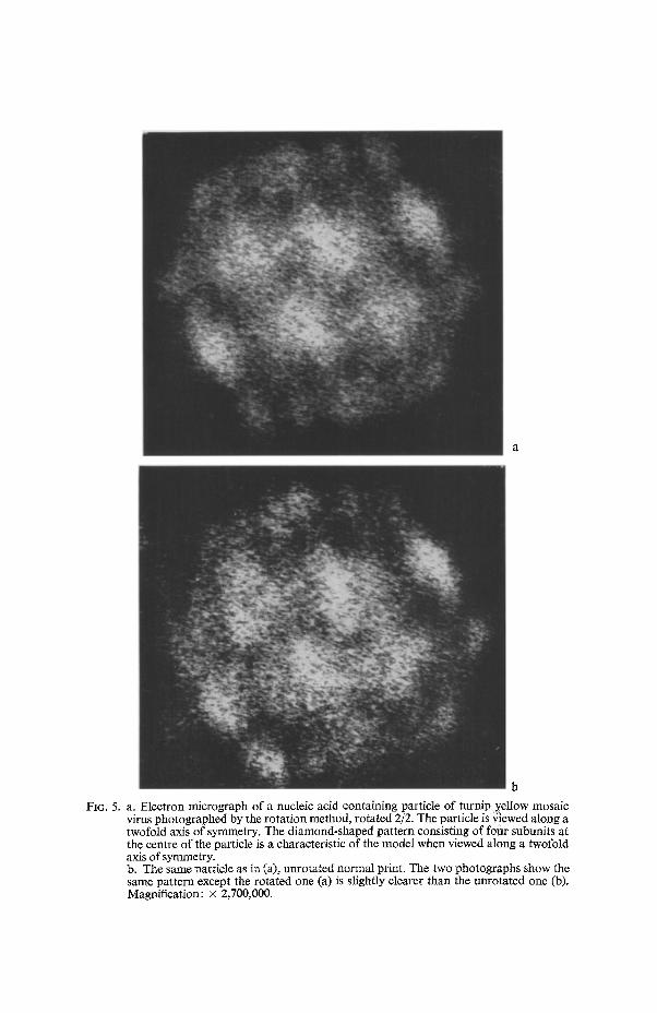
FIG. 5. a. Electron micrograph of a nucleic acid containing particle of turnip yellow mosaic virus photographed by the rotation method, rotated 2/2. The particle is ~!ewed along a twofold axis of symmetry. The diamond-shaped pattern consisting of fotlr subunits at the centre of the particle is a characteristic of the model when viewed along a twofold axis of symmetry. b. The same particle as in (a), unrotated normal print, The two photographs show the same pattern except the rotated one (a) is slightly clearer than the unrotated one (b). Magnification: • 2,700,000.

it is difficult to say if the five structures surrounding the centre, in the micro- graph published by MARKHAM et al. (1963), are really true structures represent- ing 5 peptides as suggested by them.
Fig. 5a shows a nucleic acid containing particle photographed after rotation 2/2, representing the particle as it would appear when viewed along a twofold axis of symmetry. The diamond- or the rhombus-pattern in the centre due to the four subunits on the vertices of a central rhombus can be seen quite clearly. Some of the other subunits corresponding with the model (Fig. 4b) can also be seen in this micrograph. It may be pointed out that this diamond-pattern in the centre, representing the particle in twofold symmetry, is encountered very often, and is apparently the most likely orientation for a stable equilibrium in this polyhedron. It is also interesting to note that the particle in twofold symmetry exhibits a hexagonal profile (Fig. 5a, and 5b), routinely observed in the virus preparations. The other most encountered orientation, where the particle also shows a hexagonal profile, as reported by HUXLEY & ZUBAY (1960), NIXON & GIBBS (1960), and MARKHAM et al. (1963), consists of a hexagonal ring of six subunits about a central subunit. This pattern represents the particle in three- fold symmetry, and also provides a stable position for the proposed model.
In a model of the rhombic triacontahedron type discussed here, the subunits are present in two locations depending on the arrangement of their neighbours; twelve are surrounded by five adjacent subunits and the remaining twenty by six. Since these locations are not equivalent, and since there is no indication that more than one type of peptide chain constitutes the protein shell, it seems possible that these morphological subunits might differ in respect of the number of peptide chains in each. On the basis of their micrographs MARKHAM et al. (1963) suggested that all the morphological subunits of the virus have five sub- structures in each which correspond to the five peptide chains, thus giving a to- tal of 160 structural units. The micrographs presented here do not support this figure of 160, neither does this number fit into CRICK and WATSON'S hypothesis. KLUG et al. 's (1957) X-ray diffraction studies established for the virus particle as a whole a 2: 3 point-group symmetry, suggesting that the particle was built up of 12 or a multiple of 12 subunits. Thus the number of 160 structural subunits is not supported by X-ray data.
MARKrIA~ (1951) calculated a molecular weight of 5.0 x 10⁶ for the bottom component and 3.0x 10⁶ for the top component. MARKHAM & REICHMANN (unpublished, quoted in MARKIJAM et aL, 1963) found a molecular weight of 5.13 x 10⁶ for the virus. SYMONS et aI., (1963) gave a molecular weight of ap- proximately 2.0 x 10 a for the peptide chain. MARKHAM et al. 's (1963) suggestion of 160 peptides representing the structural units is based on these figures. These values, however, cannot be regarded as final since the values reported by other workers show quite some disagreement. HARRIS & HINDLEY (1961) reported a molecular weight of approximately 2.1 x I04 for the peptide chain. HOR~ et al. (1963) reported a molecular weight of 5.8 x 10⁶ for a TYMV preparation free from the top component. KUI'KE & BEAMS (1963) reported a particle weight of 6.0 x 10⁶, the highest value reported so far. GOOSCHALK (1964) calculated his results on the basis of a molecular weight of 5.9 x 10⁶, a mean of the values reported by HORN et aL, and KUPKE 8r BEAMS. HASELKORN (1962) reported a molecular weight of 2.3 • 106 for the RNA from TYMV. This would give a molecular weight of 3.6 • 106 for the protein. This figure supports the idea that
177

there are 180 peptides or subunits of molecular weight of approximately 2.0 • 104, constituting the protein shell. The reasons for the discrepancies in molecular weight reported by different workers are not discussed here.
A "dynamic" model of 180 structural (chemical) units (corresponding with the 32 morphological units), which can assemble only in one way to form a stable shell, arranged with icosahedral symmetry, was made by CASI'AR & KLIJG (1962). The bonding pattern chosen in this model leads to a clustering into 20 hexamers and 12 pentamers. These 180 structural units may well correspond to the total number of peptides.
BOCKSTAHLER & KAESBERG (1962) calculated the molecular weight of the protein portion of bromegrass mosaic virus (BMV) as 3.6 • 106. Since the BMV subunit was found to have a molecular weight of about 20,000, they suggested that the number of protein subunits was about 180.
MAYER (1964) in a study of type 1 poliovirus (Mahoney strain) by means of negative staining technique found an excellent agreement between poliovirus micrographs and the 32 morphological subunit model described above for TYMV. It was, however, not possible to decide the number of structural or chemical subunits present in this case, although MAYER suggested a possibility of 120 or 180 subunits.
As illustrated here, TYMV particles can be precisely represented by a true rhombic triacontahedron. This contrasts with MARKHAM et al.'s (1963) conclu- sion which was based on rotation photographs, and accepted by HORI,~E & WILDY (1963). Thus it appears that the virus protein shell does indeed have true 5:3:2 symmetry, as suggested by KLUG et aL (1957).
Since the definite structure of RNA in the virus protein shell is not known at present and since it is possible that it has some influence on the appearance of the structural subunits as seen by means of the electron miroscope and the negative staining procedure, a final conclusion as to their number on the basis of these techniques cannot be made yet. The agreement of the physico-chemical data, electron microscope observations, CRICK and WATSON'S hypothesis, and the proposed "dynamic model", strongly suggests that the TYMV protein shell has 180 structural units.
ACKNOWLEDGEMENTS
The author thanks Prof. Dr. J. P. H. VAN DER WANT, Wageningen, and Prof. Dr. L. Bosch and his associates, Leyden, for the helpful discussions; Messrs S. HENSTRA and A. MASTENRROEK of the Electron Microscopy Section of the Service Institute for Technical Physics in Agriculture, Wageningen, for the electron microscopy, and the International Agricultural Centre for part of the financial support. Thanks are also due to Dr. R. MARKHAM, Cambridge, for kindly going through the manuscript and for his valuable comments.
REFERENCES
AGRAWAL, H. OM, - 1964. Identification of cowpea mosaic virus isolates. Meded. Landb- Hogesch., Wageningen 64-5: 1-53.
BOCKSTAHLER, L. E. & P. KAESBERG, -- 1962. The molecular weight and other biophysical properties of bromegrass mosaic virus. Biophys. J. 2: 1-9.
BRENNER, S., & R. W. HORNE, -- 1959, A negative staining method for high resolution electron microscopy of viruses. Biochim, biophys. Acta 34:103-110.
178

CASPAR, D. L. D. & A. KLUG, -- 1962. Physical principles in the construction of regular viruses. Cold Spr. Harb. Sym. quant. Biol. 27: 1-24.
Cmc~, F. H. C. & J. D. WATSON, - 1956. Structure of small viruses. Nature, Lond. 177: 473- 475.
GODSCHALK, W., - 1964. Interaction of turnip yellow mosaic virus with mercurials. Disserta- tion, University of Leyden.
HARRIS, J. I. & J. HINDLEY, -- 1961. The protein subunit of turnip yellow mosaic virus. J. mol. Biol. 3 : 117-120.
HASELKORN, R., - 1962. Studies on infectious RN A from turnip yellow mosaic virus. J. tool. Biol. 4: 357-367.
HORNE, R. W., & P. WILDY, -- 1963. Virus structure revealed by negative staining. Adv. Virus Res. 10: 101-170.
HORN, P., L. HIRTH, & P. CANALS, -- 1963. D6termination de la masse mol6culaire du virus de la mosaique jaune du navet. Ann. Inst. Pasteur 105: 99-102.
HUXLEY, H. E. & G. ZUBAY, - 1960. The structure of the protein shell of turnip yellow mosaic virus. J. mol. Biol. 2: 189-196.
KLUG, A., J. T. FINCH, & R. E. FRANKLIN, -- 1957. Structure of turnip yellow mosaic virus: X-ray diffraction studies. Biochim. biophys. Acta 25: 242-252.
KUPKE, D. W., & J. W. BEAMS, - 1963. The molecular weights of four additional viruses. In: Research and Technology News Coltmm. Chem. Engng News 41 (6): 39.
MARKHAM, R., - 1951. Physicochemical studies of the turnip yellow mosaic virus. Disc. Fara- day Soc. 11 : 221-227.
MARKHAM, R., S. FREY, & G. J. HILLS, -- 1963. Methods for the enhancement of image detail and accentuation of structure in electron microscopy. Virology 20:88-102.
MAYOR, H. D . , - 1964. Picornavirus symmetry. Virology 22: 156-160. NIxoN, H. L. & A. J. GIBBS, - 1960. Electron microscope observations on the structure of
turnip yellow mosaic virus. J. tool. Biol. 2:197-200. SYMONS, R. H., M. W. REES, MARGARET N. SHORT & R. MARKHAM, -- 1963. Relationships
between the ribonucleic acid and protein of some plant viruses. J. tool. Biol. 6: 1-15.
179