electron microscopy in diagnosis of infectious diseases€¦ · electron microscopy in diagnosis of...
TRANSCRIPT

Electron Microscopy in Diagnosis of Infectious Diseases
Sara E. Miller, Ph. D.
Department of Pathology Duke University Medical Center
Durham, NC 27710 [email protected]
(919) 684-9141
Duke Medicine Society for Ultrastructural Pathology
January 29, 2013
A
Outline Part 1
A. Virology 1. Advantages of using EM in diagnostic virology 2. Limitations of using EM in diagnostic virology 3. Workarounds 4. Virus structure
a. In negative stains b. In thin sections
Outline Part 1
B. Other organisms 1. Bacteriology 2. Mycology 3. Photology (algae) 4. Protozoology
Questions/Discussion
Outline Part 2
C. Virus look-alikes 1. Examples of confusing things in fluids 2. Examples of cell organelles that resemble
viruses in tissues
Outline Part 2
D. Real cases 1. Examples of organisms diagnosed from patients 2. Quiz for fun

Outline Part 1
A. Virology 1. Advantages of using EM in diagnostic virology 2. Limitations of using EM in diagnostic virology 3. Workarounds 4. Virus structure
a. In negative stains b. In thin sections

A. Virology
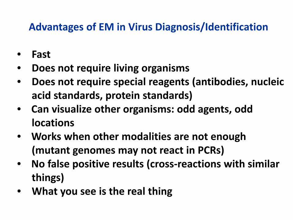
Advantages of EM in Virus Diagnosis/Identification • Fast • Does not require living organisms • Does not require special reagents (antibodies, nucleic acid standards, protein standards) • Can visualize other organisms: odd agents, odd locations • Works when other modalities are not enough (mutant genomes may not react in PCRs) • No false positive results (cross-reactions with similar things) • What you see is the real thing
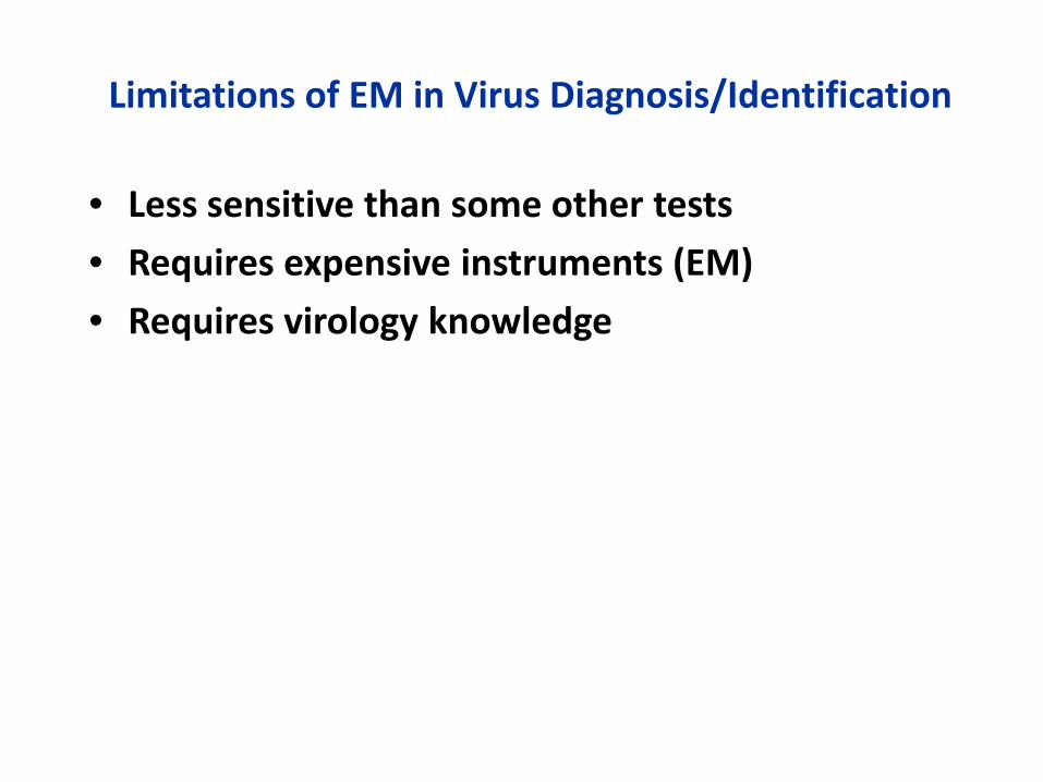
Limitations of EM in Virus Diagnosis/Identification
• Less sensitive than some other tests
• Requires expensive instruments (EM)
• Requires virology knowledge

Ways To Get Around Limitations • Ultracentrifugation • Antibody concentration • Confocal microscopy of wet tissue slabs • Multiple tissue locations • Semi-thin sections of epoxy-embedded tissues • Where to look in tissues: inflammation, nucleated cells, necrosis edge, unusual ultrastructure for the tissue type), syncytia
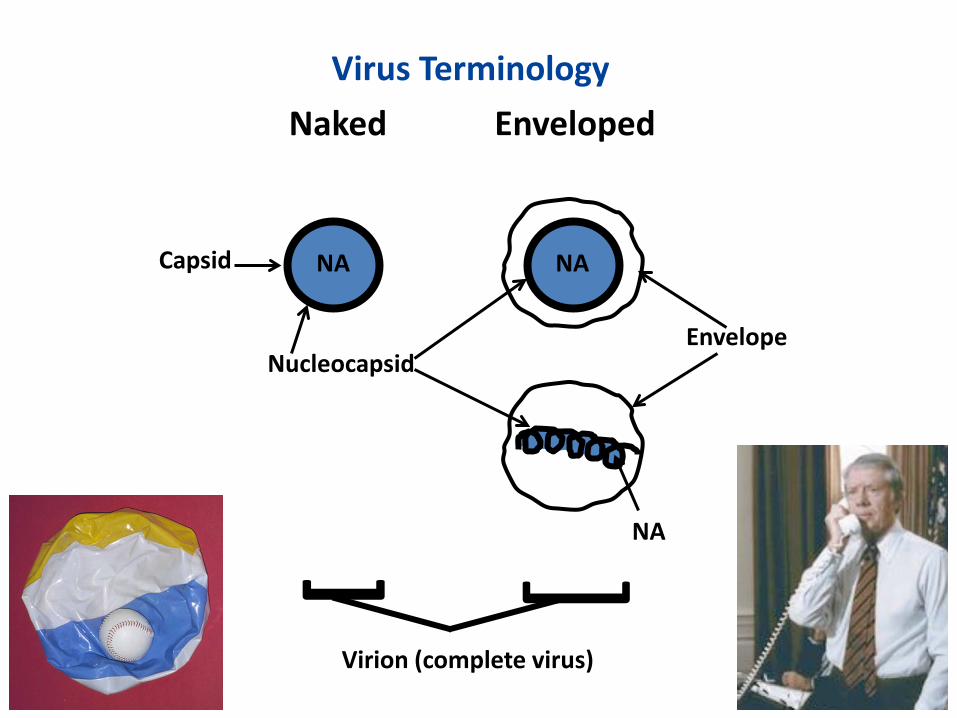
Virus Terminology
Naked Enveloped
Nucleocapsid
Capsid
Envelope
NA NA
NA
Virion (complete virus)
] ]
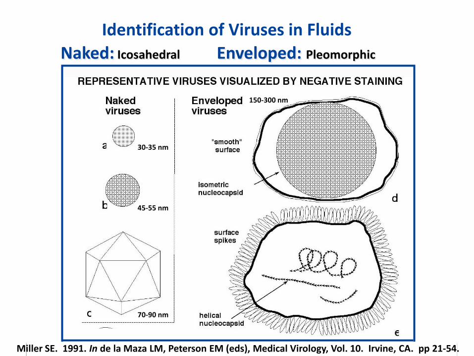
Identification of Viruses in Fluids
70-90 nm
Naked: Icosahedral
Enveloped: Pleomorphic
30-35 nm
45-55 nm
70-90 nm
150-300 nm
Miller SE. 1991. In de la Maza LM, Peterson EM (eds), Medical Virology, Vol. 10. Irvine, CA. pp 21-54.

80 nm x 300 nm 80 nm x 1400 nm
200 nm x 400 nm
Enveloped: Not Pleomorphic
Identification of Viruses in Fluids, Con’t.
Miller SE. 1991. In de la Maza LM, Peterson EM (eds), Medical Virology, Vol. 10. Irvine, CA. pp 21-54.

Polyomavirus from Urine Enteric Viruses
Influenzavirus
G
100 nm
Negative Staining of Naked and Enveloped Viruses
Rotavirus
Coronavirus
Calicivirus
Adenovirus
H
Astrovirus
Picornavirus
100 nm
100 nm
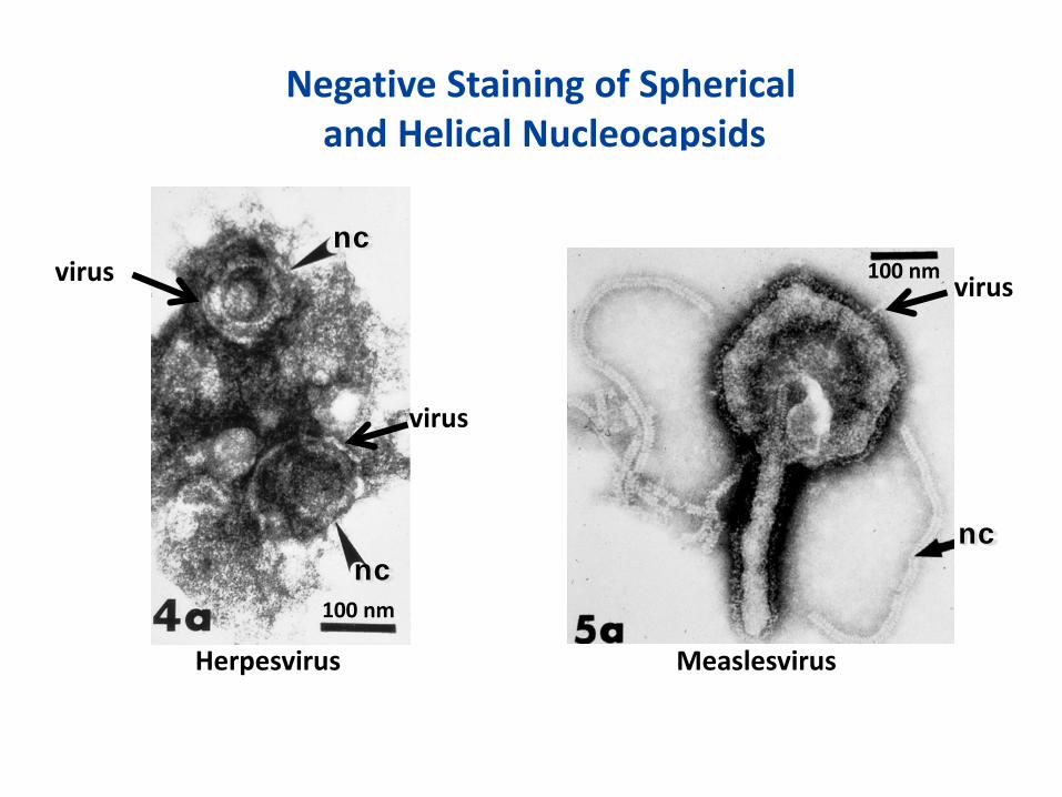
Negative Staining of Spherical and Helical Nucleocapsids
virus
100 nm
Herpesvirus
nc
nc
virus
virus 100 nm
Measlesvirus
nc
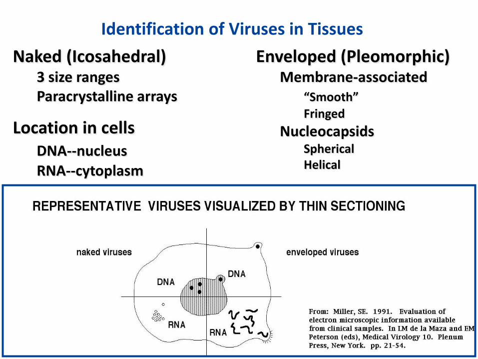
Naked (Icosahedral) 3 size ranges Paracrystalline arrays
Enveloped (Pleomorphic) Membrane-associated “Smooth” Fringed Nucleocapsids Spherical Helical
Identification of Viruses in Tissues
Location in cells DNA--nucleus RNA--cytoplasm

DNA Viruses: Usually produced in the nucleus
RNA Viruses: Usually produced in the cytoplasm
1.O µm
Reovirus µm
Adenovirus
N
N

Nuclear Membrane Budding
1 µm
100 nm
Plasma Membrane Budding
HTLV
nc
Herpesvirus
Vesicular Budding
Budding
N
N 100 nm
100 nm
N virus
Hantavirus
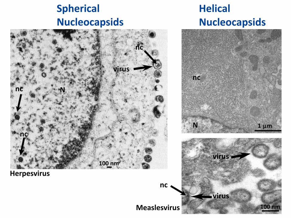
Helical Nucleocapsids
Spherical Nucleocapsids
N
100 nm
100 nm
µm 1
Measlesvirus
Herpesvirus
N nc
nc
nc nc
nc virus
virus
virus
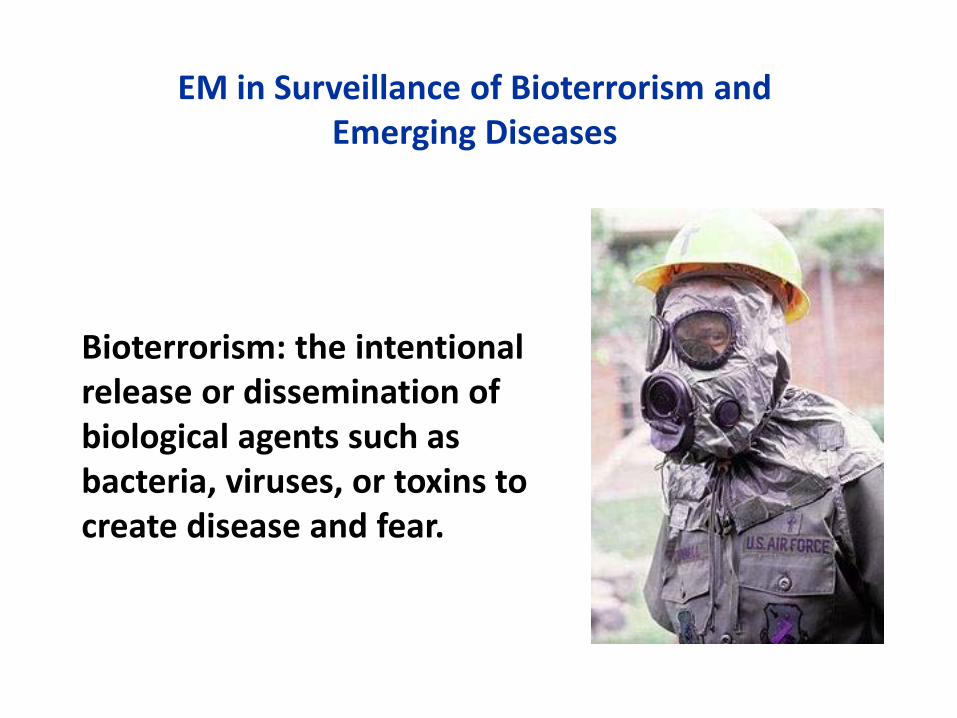
Bioterrorism: the intentional release or dissemination of biological agents such as bacteria, viruses, or toxins to create disease and fear.
EM in Surveillance of Bioterrorism and Emerging Diseases
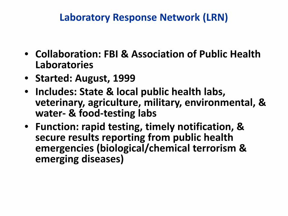
Laboratory Response Network (LRN)
• Collaboration: FBI & Association of Public Health
Laboratories • Started: August, 1999 • Includes: State & local public health labs,
veterinary, agriculture, military, environmental, & water- & food-testing labs
• Function: rapid testing, timely notification, & secure results reporting from public health emergencies (biological/chemical terrorism & emerging diseases)
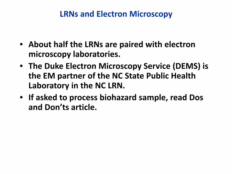
LRNs and Electron Microscopy
• About half the LRNs are paired with electron
microscopy laboratories. • The Duke Electron Microscopy Service (DEMS) is
the EM partner of the NC State Public Health Laboratory in the NC LRN.
• If asked to process biohazard sample, read Dos and Don’ts article.
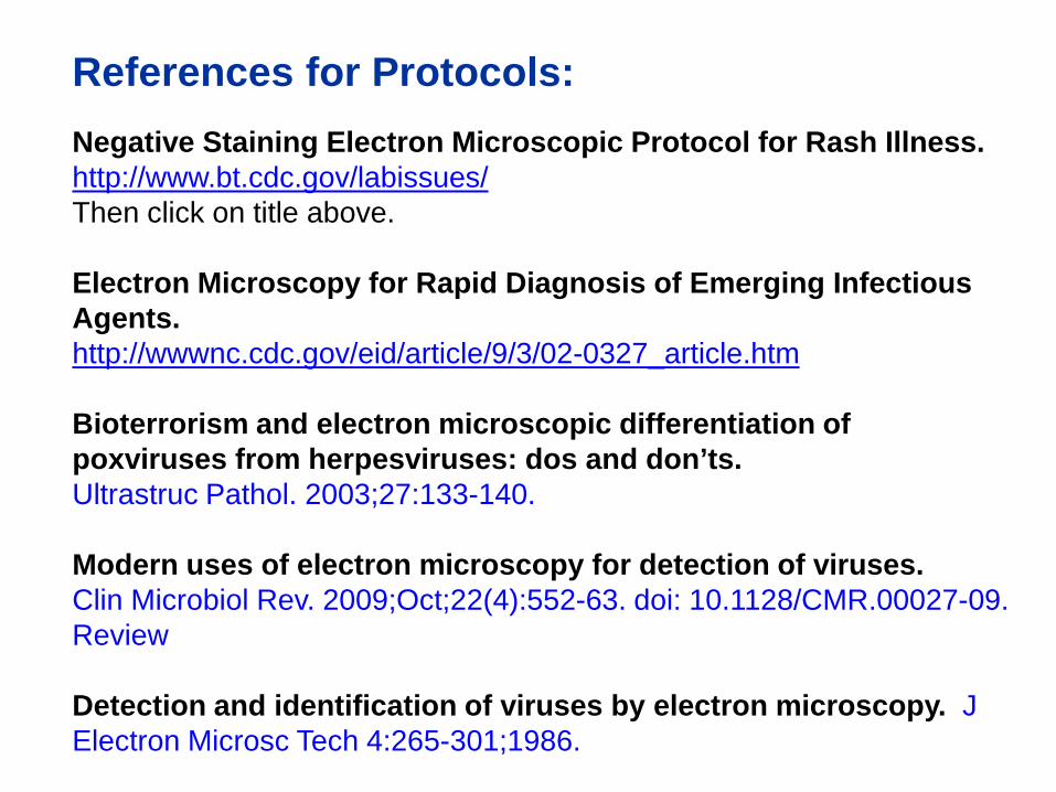
References for Protocols: Negative Staining Electron Microscopic Protocol for Rash Illness. http://www.bt.cdc.gov/labissues/ Then click on title above. Electron Microscopy for Rapid Diagnosis of Emerging Infectious Agents. http://wwwnc.cdc.gov/eid/article/9/3/02-0327_article.htm Bioterrorism and electron microscopic differentiation of poxviruses from herpesviruses: dos and don’ts. Ultrastruc Pathol. 2003;27:133-140. Modern uses of electron microscopy for detection of viruses. Clin Microbiol Rev. 2009;Oct;22(4):552-63. doi: 10.1128/CMR.00027-09. Review Detection and identification of viruses by electron microscopy. J Electron Microsc Tech 4:265-301;1986.

Smallpox virus • Easily disseminated • Easily weaponized • High mortality rate, survivors badly scarred • Previously mass-produced • Believed to have been distributed after USSR collapse Hemorrhagic fever viruses • Filoviruses (Marburg, Ebola) • Arenaviruses (Lassa, Machupo)
Class A Viral Agents of Bioterrorism

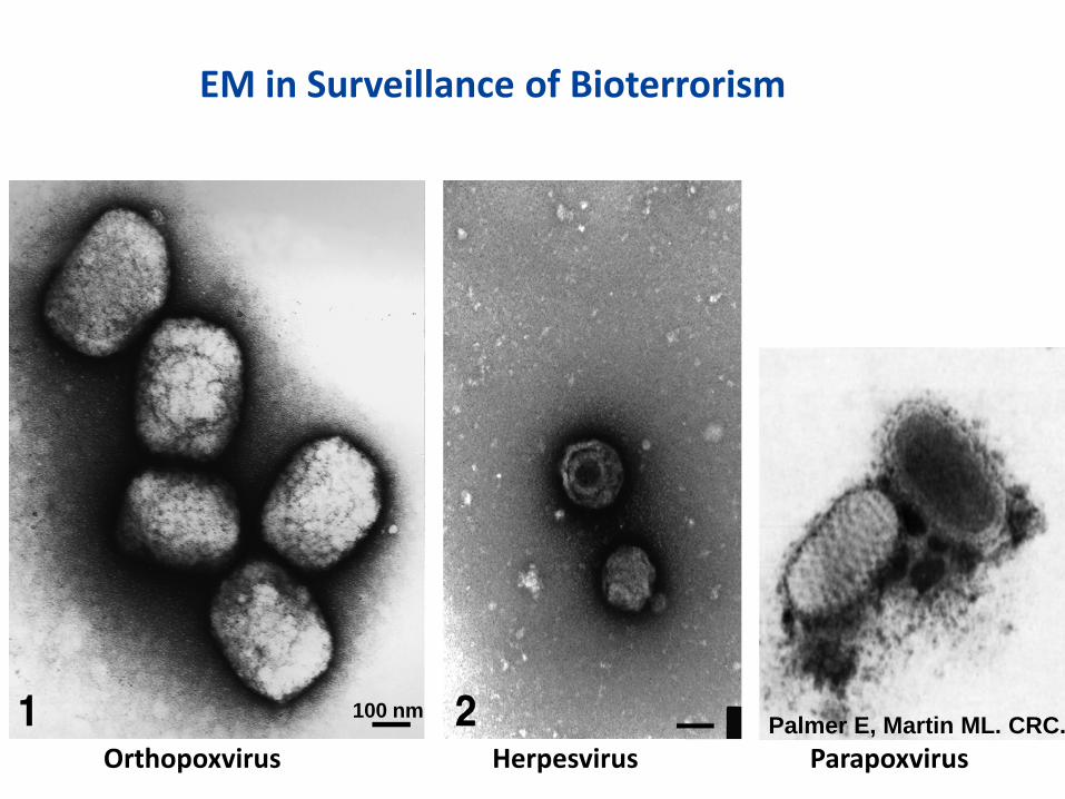
Because of ID speed, EM is on the front line in bioterrorism surveillance (e.g., smallpox).
Poxvirus lesions are most likely to be confused with varicella-zoster virus (VZV) (a herpesvirus) lesions.
Palmer E, Martin ML. CRC.
EM in Surveillance of Bioterrorism
Orthopoxvirus Parapoxvirus Herpesvirus
100 nm
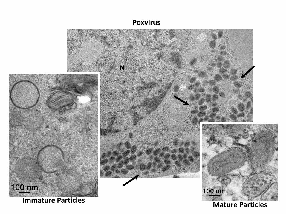
Poxvirus
N
Immature Particles Mature Particles
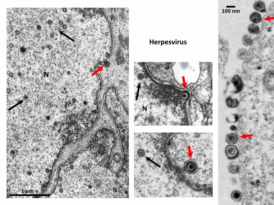
Herpesvirus
100 nm
1 μm
N
N

EM in Surveillance of Emerging Diseases
Viral emerging diseases identified first by EM • Parvovirus B-19 • Monkeypox • SARS coronavirus • Metapneumovirus • Morbilivirus • Nipah virus

Costs (11/02 – 7/03): 774 Deaths, 9.6% fatality (China, Hong Kong, Taiwan, Canada) $11 billion (Asia alone)
EM was crucial in identifying the coronavirus in the SARS outbreak.

Courtesy of Cynthia Goldsmith, CDC, Atlanta.
Thin Section of SARS Coronavirus
100 nm

EM was crucial in identifying the monkeypox virus outbreak

Thin Section of Monkeypox Virus Infected Cell
Negative Stain
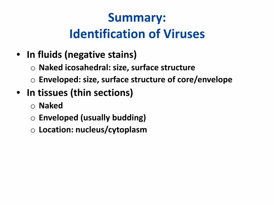
Summary: Identification of Viruses
• In fluids (negative stains) o Naked icosahedral: size, surface structure o Enveloped: size, surface structure of core/envelope
• In tissues (thin sections) o Naked o Enveloped (usually budding) o Location: nucleus/cytoplasm