electron microscopy of the centrifuged sea urchin egg ... · pdf fileelectron microscopy of...
TRANSCRIPT

Electron Microscopy of the Centrifuged Sea Urchin Egg, with a Note on the Structure of the Ground Cytoplasm*
BY PAUL R. GROSS, PI~.D., D E L B E R T E. P H I L P O T T , and SYLVAN NASS
(From the Department of Biology, New York University, and the Marine Biological Laboratory, Woods Hole, Massachusetts)
PLATES 58 TO 63
(Received for publication, July 25, 1959)
ABSTRACT
Centrifuged, unfertilized eggs of the sea urchin, Arbada punctulata, have been studied with the electron microscope. Subcellular particles were stratified by centrifuging living cells, known to be normally fertilizable, for five minutes at 3,000 g. The layered subcellular particles, including cortical granules, 16 m~ RNP par- ticles, pigment, yolk, mitochondria, and oil droplets, possess characteristic ultra- structural features by which they may be identified in situ. The clear zone contains 16 m~ particles, most of them freely dispersed, scattered mitoehondria, and a few composite structures made up of annulate lamellae in parallel layers or in associ- ation with dense, spherical aggregates of the RNP particles. Free 16 m~ particles are found, in addition, throughout the cell, in the interstices between the stratified larger particles. They show a tendency to form ramifying aggregates resulting from certain types of injury to the cell. A few vesicular structures, found mainly in the clear zone, have attached RNP particles, and appear to be related to the ER of tissue cells. Other vesicles, bounded by smooth membranes, are found through- out the cell. These are extremely variable in size, number, and distribution; their total number appears to depend upon conditions of fixation. It is suggested that limited formation of such structures is a normal property of the ground cytoplasm in this cell, but that fixed cells with very large numbers of smooth surfaced vesicles have produced the latter as a response to chemical injury. A model of the ground cytoplasm is proposed whose aim is to reconcile the rheological behavior of the living cell with the ultrastructural features observed.
The egg of the sea urchin, Arbacia punctulata, is a cell for which relatively large amounts of bio- physical and biochemical information have been accumulated. A compilation of experimental papers on this cell occupies much of the recent book by Harvey (10). In connection with a series of studies on the molecular interactions underlying physical changes in the cytoplasm, we have made an elec- tron microscopic survey of the centrifuged, un- fertilized Arbacia egg. Some of our observations may serve to supplement or modify already pub-
* Aided by grant A-2302 from the National Institute of Arthritis and Metabolic Diseases, United States Public Health Service, and by grants from the National Science Foundation, the Marine Biological Laboratory, and the Graduate Research Fund of New York University.
lished studies. Afzelius (1-3) has contributed a fundamental series of observations on the gametes of several European sea urchins, while Pasteels et al. (17) have recently reported observations on centrifuged eggs of Paracentrotus lividus. Merriam (15) has studied the "annulate lamellae" in oocytes of the sand dollar, Echinarachnius parma. Exami- nations of centrifugally stratified A rbacia eggs have been made by McCulloch (14) and by Lansing, Hillier, and Rosenthal (13). Gross et al. (9) have
studied dividing Arbacia eggs. The present report describes the ultrastructural
features of each of the layers formed in the cell by centrifugal stratification, with special emphasis upon the morphology of the "clear zone." The lat- ter represents the ground substance of the cell, and a detailed examination of its fine structure will
135
J. BIOPH¥SIc. AND BIOCIIE~. CYTOL., 1965, Vol. 7, No. I

136 CENTRIFUGED SEA URCHIN EGG
doubtless help in the interpretation of those organi- zational changes which are manifested as sol-gel transformations. Tha t these changes are functions of the .ground substance independent of the larger cytoplasmic inclusions has been clear since the early studies of E. B. Harvey on parthenogenetic merogones (@ 10).
Materials and Methods
Specimens of Arbacia punctulata, obtained near Woods Hole, were induced to shed gametes by electrical stimulation or by injection of 0.5 ml. of 0.53 M KC1. Only egg batches which proved capable of better than 90 per cent fertilization and first cleavage were used for the preparations. The unfertilized eggs were centri- fuged for 5 minutes at 3,000 g. It was found that with larger forces, stratification was more rapid, but often less complete and accompanied by a considerable incidence of cell injury. The cells were examined microscopically before fixation, and when populations appeared both well stratified and otherwise normal, fixation was begun. The eggs were quickly concentrated by a second, light centrifugation, and then resuspended in an isotonic, veronal-buffered 1 per cent OsO4 solution. Fixation was continued for 30 minutes in the cold, with one change of solution at 15 minutes to remove the cells from contact with material extracted from the pigment granules. Treatment of the cells was thereafter conventional, and embedding was done in a 4:1 mixture of n-butyl and methyl methacrylates, with benzoyl peroxide as catalyst. Polymerization took place in an oven regulated at 45°C., and the specimens were sectioned on the thermal advance microtome described by Philpott (18). Glass knives were used for survey work and a diamond knife for sectioning of material to be examined at high magnification. Thick sections cut just prior to normal thin sectioning from each block were examined under the light microscope for evidence of compression, and only those of normal (i.e. circular) appearance were chosen for examination with the RCA model EMU-2C electron microscope. 250 micron condenser and 65 micron objective apertures were used. The substrates were either formvar or carbon films.
RESULTS
The stratification pattern of the inclusions in the Arbaeia egg is well known. From the centrifugal to the centripetal pole, the order is as follows: pig- ment granules (actually vacuoles), yolk particles, mitochondria, the "clear zone," the nucleus, and a cap of oil droplets. A single layer of cortical gran- ules can be seen in the cortex of the unfertilized egg; these particles are not dislodged except at very high shear stresses, and this has often been cited as evidence for the relatively high rigidity ("vis-
cosity") of the cell cortex. The cortical granules, as was shown by Afzelius (1), differ morphologic- ally (after OsO4 fixation) from the pigment vac- uoles, and hence an occasional stray found in the endoplasm of the cell can be identified.
The electron microscope reveals the following structural features of the several layers, proceeding from the "heavy" to the "l ight" pole of the egg.
Pigment Granules.--The substance contained in the pigment granules is extracted during fixation and subsequent dehydration; this leaves a layer of empty, membrane-bounded vesicles at the centrif- ugal pole. Fig. 1 is a photograph of this region, with a row of cortical granules marking the "bot- tom" of the egg. The complex internal structure of these particles contrasts with the empty interior of the pigment granules. The latter have a mean diameter of 1.4 v in these preparations, a value which agrees surprisingly well with dimensions ob- tained from light microscope measurements in situ and in homogenates.
In the pigment layer are found a few yolk platelets, very few mitochondria, and a consider- able number of dense 16 m~ particles, lodged in the interstices between the pigment vacuoles.
Yolk Particles.--A section through the yolk layer is shown in Fig. 2. Intact platelets, when isolated, are yellow to brown ellipsoidal structures. The fixed particles show, under the electron micro- scope, a thin, peripheral membrane, about 5 m~ across the dense line in profile, and are from one to 1.7 g long. Two cortical granules, with their char- acteristic internal structure, are shown in the figure. This is an unusual finding in a non-periph- eral region of the egg, for the cortical granules are restricted almost exclusively to the outer 2 # of this cell. The yolk layer contains a very few mito- chondria, fewer pigment vacuoles, and the same large number of interstitial 16 mv particles as found in the pigment layer. Also present are varying numbers of vesicles possessing membranes of differing thickness and electron density. The diameters of the vesicles also vary greatly, i.e., from 35 m/~ to about 0.3 g (in profile, of course). Their numbers appear to change with the quality of the fixation.
Some evidence for the existence of two classes of yolk particle might be adduced from the observa- tion of large, rather granular structures together with others which are less dense and quite uniform in internal texture. Both types can be distinguished in Fig. 2.
The mitochondria are concentrated in a layer

P. R. GROSS, D. E. PHILPOTT, AND S. NASS 137
centripetal to the yolk. The mitochondria are not very sharply stratified at the low centrifugal forces used in these preparations, but this does not invali- date our conclusion that mitochondria move to positions both above and below the clear zone, since such a distribution has been observed by us and others (see below) at higher forces and since the higher forces would merely increase the rate of stratification without changing its direction. Fig. 3 shows a section through the "upper," or centripetal region of the mitochondrial layer. The mitochondria possess the complex inter- nal structure now recognized as characteristic for these organelles. Also found in this layer are a few yolk platelets, many vesicles, and a profusion of the dense, 16 m# particles.
Although random sections through rod-like particles should produce a majority of profiles shaped as ellipses, our failure to find any truly rod-like profiles among a very large number of sec- tions examined suggests that in this cell the parti- cles are ordinarily ellipsoids and not rods. "Waisted" mitochondria are seen occasionally, suggesting that we have sectioned an elongate structure through the arms of a U-shaped bend, or perhaps that the particles have been arrested in the process of fission. A few circular profiles of mito- chondria, easily recognizable as such, have been found with diameters as small as 0.15 t~. The longest profiles have been somewhat smaller than 1 p. Most are ellipses with major axes of 0.3 to 0.5 #.
The eristae seem to lie well within, and usually (but not always) detached from the outer mito- chondrial membrane; frequently they run parallel to one another but take no definite direction with respect to the particle axes. The total thickness of the outer membrane is about 16 m#; that of the cristae somewhat greater; the osmiophilic layers of the membranes are each about 6 m# thick.
The very loose packing of the mitochondria in these specimens (compared with the good stratifi- cation of the pigment and yolk) suggests that their density is not greatly different from that of the "clear zone" considered as a continuum. Not only do the mitochondria begin to diffuse out of their strata very quickly after centrifugation, but, in addition, these particles are found "above" as well as "below" the clear layer.
The clear zone of the centrifugally stratified egg has no sharp centrifugal boundary when relatively low centrifugal forces are employed; the number of mitochondria decreases quite gradually as one ap- proaches the top of the mitochondrial layer and this
merges, quite imperceptibly at the submicroscopic level, with the clear zone. With certain important exceptions, the latter is free of large inclusion bodies; it is, however, crowded with very large numbers of free 16 m# particles, which may be regarded as the RNP particles of Palade (3, 16, 17). The rare large bodies found in the clear zone are: (a) a very few scattered mitochondria; (b) annu- late lamellar systems, sometimes arranged as are the "heavy bodies" of Afzelius (3) and also very few in number; and (c) a few representatives of all particles types, found most often in the immedi- ately subcortical endoplasm of the clear zone, sug- gesting a graded increase in rigidity outward from the most peripheral four or five micra. These excep- tions should not obscure the fact that the clear zone is essentially free of microscopically visible inclu- sion bodies.
Fig. 4 is a section through the clear zone. The dominant feature is the profusion of RNP particles, mostly free but sometimes linked in chains. Many of the chains are circular (spherical) and enclose spaces or vesicles of low electron density. Possibly some of the interparticle linkages seen in Fig. 4 result from aggregation during fixation.
Fig. 5, also in the clear endoplasm, shows one of the annulate lamellar systems, containing here eight parallel double layers. These are surrounded by free RNP particles, although some of the latter adhere to the lamellae in such a way as to mimic the cisternal structure of the endoplasmic reticu- lure in mammalian tissue cells. The periodic undulations in the lamellar profiles, however, reveal that the membranes are related to the nuclear membrane, which has a similar structure.
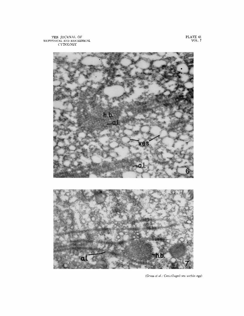
When sectioned tangentially, the membranes of these annulate lamellae show the regular pattern of annular densities observed by Afzelius (3), Rebhun (20), and Merriam (15). Fig. 6 shows such a section. The membranes are occasionally found in association with dense clusters of the 16 m# par- ticles; such complexes were called "heavy bodies" by Afzelius (3), but they do not fall to the centrif- ugal pole in the Arbacia egg; instead, they remain in the lower part of the clear zone. In Fig. 6 one of these bodies has been sectioned tangentially; while in Fig. 7, the section cuts the membranes in such a way as to reveal their undulating profiles. Fig. 7 shows the spherical aggregate of dense 16 m# particles.
I t is of interest that the particles contained within these aggregates appear identical with the cytoplasmic RNP particles found elsewhere, sug-
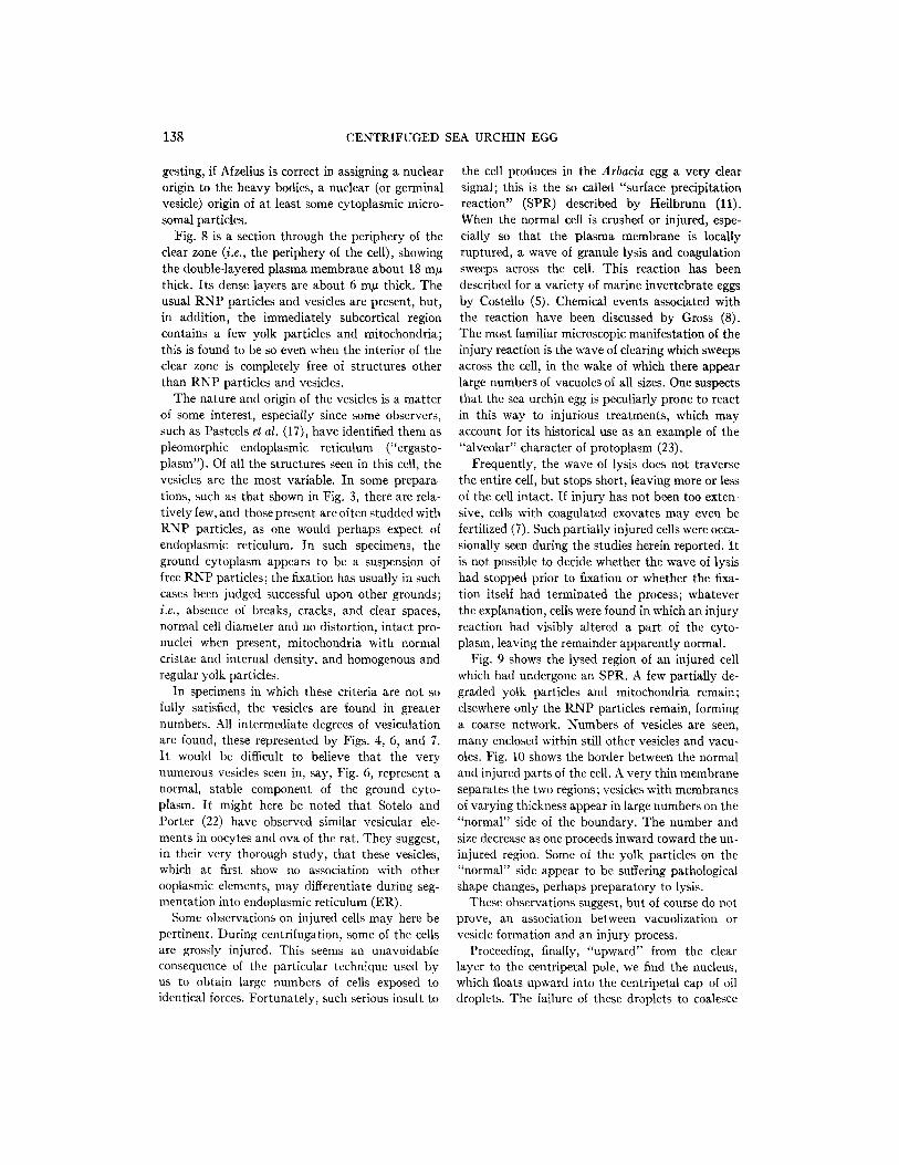
138 CENTRIFUGED SEA URCHIN EGG
gesting, if Afzelius is correct in assigning a nuclear origin to the heavy bodies, a nuclear (or germinal vesicle) origin of at least some cytoplasmic micro- somal particles.
Fig. 8 is a section through the periphery of the clear zone (i.e., the periphery of the cell), showing the double-layered plasma membrane about 18 m# thick. Its dense layers are about 6 m/z thick. The usual RNP particles and vesicles are present, but, in addition, the immediately subcortical region contains a few yolk particles and mitochondria; this is found to be so even when the interior of the clear zone is completely free of structures other than RNP particles and vesicles.
The nature and origin of the vesicles is a matter of some interest, especially since some observers, such as Pasteels et al. (17), have identified them as pleomorphic endoplasmic reticulum ("ergasto- plasm"). Of all the structures seen in this cell, the vesicles are the most variable. In some prepara- tions, such as that shown in Fig. 3, there are rela- tively few, and those present are often studded with RNP particles, as one would perhaps expect of endoplasmic reticulum. In such specimens, the ground cytoplasm appears to be a suspension of free RNP particles; the fixation has usually in such cases been judged successful upon other grounds; i.e., absence of breaks, cracks, and clear spaces, normal cell diameter and no distortion, intact pro- nuclei when present, mitochondria with normal cristae and internal density, and homogenous and regular yolk particles.
In specimens in which these criteria are not so fully satisfied, the vesicles are found in greater numbers. All intermediate degrees of vesiculation are found, these represented by Figs. 4, 6, and 7. It would be difficult to believe that the very numerous vesicles seen in, say, Fig. 6, represent a normal, stable component of the ground cyto- plasm. It might here be noted that Sotelo and Porter (22) have observed similar vesicular ele- ments in oocytes and ova of the rat. They suggest, in their very thorough study, that these vesicles, which at first show no association with other ooplasmic elements, may differentiate during seg- mentation into endoplasmic reticulum (ER).
Some observations on injured cells may here be pertinent. During centrifugation, some of the cells are grossly injured. This seems an unavoidable consequence of the particular technique used by us to obtain large numbers of cells exposed to identical forces. Fortunately, such serious insult to
the cell produces in the Arbacia egg a very clear signal; this is the so called "surface precipitation reaction" (SPR) described by Heilbrunn (11). When the normal cell is crushed or injured, espe- cially so that the plasma membrane is locally ruptured, a wave of granule lysis and coagulation sweeps across the cell. This reaction has been described for a variety of marine invertebrate eggs by Costello (5). Chemical events associated with the reaction have been discussed by Gross (8). The most familiar microscopic manifestation of the injury reaction is the wave of clearing which sweeps across the cell, in the wake of which there appear large numbers of vacuoles of all sizes. One suspects that the sea urchin egg is peculiarly prone to react in this way to injurious treatments, which may account for its historical use as an example of the "alveolar" character of protoplasm (23).
Frequently, the wave of lysis does not traverse the entire cell, but stops short, leaving more or less of the cell intact. If injury has not been too exten- sive, cells with coagulated exovates may even be fertilized (7). Such partially injured cells were occa- sionally seen during the studies herein reported. I t is not possible to decide whether the wave o[ lysis had stopped prior to fixation or whether the fixa- tion itself had terminated the process; whatever the explanation, cells were found in which an injury reaction had visibly altered a part of the cyto- plasm, leaving the remainder apparently normal.
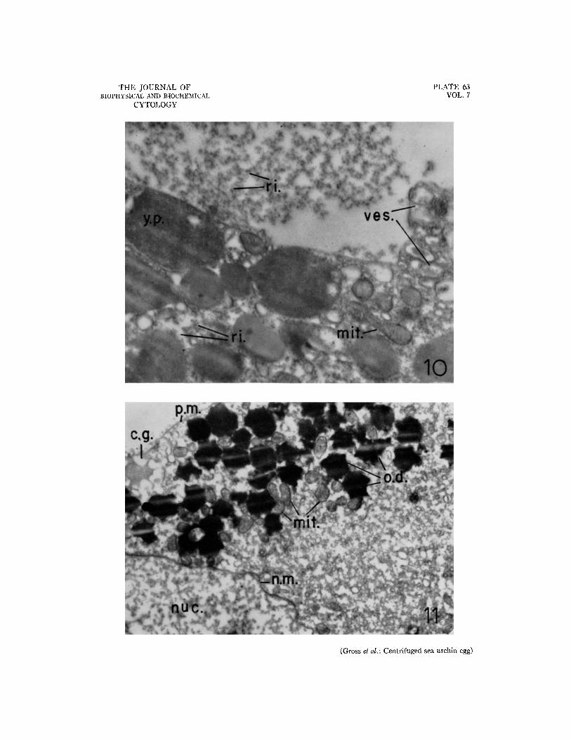
Fig. 9 shows the lysed region of an injured cell which had undergone an SPR. A few partially de- graded yolk particles and mitochondria remain; elsewhere only the RNP particles remain, forming a coarse network. Numbers of vesicles are seen, many enclosed within still other vesicles and vacu- oles. Fig. 10 shows the border between the normal and injured parts of the cell. A very thin membrane separates the two regions; vesicles with membranes of varying thickness appear in large numbers on the "normal" side of the boundary. The number and size decrease as one proceeds inward toward the un- injured region. Some of the yolk particles on the "normal" side appear to be suffering pathological shape changes, perhaps preparatory to lysis.
These observations suggest, but of course do not prove, an association between vacuolization or vesicle formation and an injury process.
Proceeding, finally, "upward" from the clear layer to the centripetal pole, we find the nucleus, which floats upward into the centripetal cap of oil droplets. The failure of these droplets to coalesce

P. R. GROSS, D. E. PHILPOTT, AND S. NASS 139
probably means that they possess a non-oily surface, as has been widely assumed; e.g., by Frey- Wyssling (6).
The nuclear membrane is double and shows the same structure in profile as do the annulate lamel- lae (this is not easily seen in low power micrographs such as Fig. 11, which is included for another purpose). At the resolution available to us, no fibrous structures could be discerned within the nucleus; instead, its internal structure is that of a coarse aggregate of particles having diameters of from 10 to 50 m#. The texture of this aggregate, however, differentiates it sharply from the sur- rounding cytoplasm, where the abundant dispersed RNP particles and the more or less abundant vesi- cles produce an altogether different appearance.
In every cell examined, the centripetal oil drop- lets were associated with clusters of mitochondria. This has been reported by Lansing et al. (13) and by others. Association of mitochondria with oil droplets is also quite commonly observed in the non-stratified unsegmented egg (7). The presence of mitochondria among the centripetal oil droplets would suggest that the clear quarters of the eggs (10) might also contain them, since such merogones are derived from the clear zone and the centripetal oil cap. The small centrifugal forces and possibly incomplete stratification of mitochondria in the present experiments do not, as already mentioned, weaken the suspicion, since increased force fields should serve merely to accentuate the separation. Thus, the presence of mitochondria in larvae de- veloped from clear quarters need not necessarily signify their de novo origin. Certainly this should be examined, preferably by an electron microscopic study of the merogones.
DISCUSSION
The observations reported herein confirm, in general, those made by Afzelius (1-3) on the European sea urchins. The stable subcellular parti- cles, i.e., cortical granules, pigment vacuoles, yolk particles, mitochondria, RNP particles, and oil droplets, have characteristic ultrastructural fea- tures. The establishment of these regularities of structure should make it possible to identify the particles in homogenates and in fractions; distor- tions effected by cell breakage and subsequent treatments should be recognizable. The subcellular particles do, in fact, appear to retain their ultra- structural identity when released into a suitable
homogenization medium (7), and this will be the subject of a separate communication.
Of more than descriptive interest, however, are several aspects of the fine structure of the clear zone. The absence or extreme reduction of prolif- erated, cisternal endoplasmic reticulum is in ac- cordance with the suggestions of Palade (16) con- cerning its reduction in embryonic cells generally and with emphasis of Porter (19) upon the extreme pleomorphism of this system. It would indeed ap- pear unlikely that a stable cisternal system, ramifying throughout the cytoplasm, would be found in cells which must divide repeatedly, for in such cells drastic rearrangements must occur in the ground cytoplasm at the macromolecular and small particulate levels.
I t is perhaps not an original speculation that the ER in its cisternal-trabecular form serves as a con- ductile or sequestration system, in those cells in which there must occur either transport of ions normally foreign to the endoplasm or synthesis of large quantities of enzymes, whose site of action is external to the endoplasm and which would, in soluble form, prove dangerous to cell proteins. Such teleology can, of course, be justified only by the lack of direct experimental evidence.
We consider now the nature of the numerous vesicles found in the clear zone and elsewhere in the fixed and sectioned egg. The ease with which mem- branes can be converted into vesicles both in vivo (19) and in vitro (16) must be taken into account in the interpretation of these structures. They might indeed be considered an aberrant form of the membraneous ER. Such a conclusion (e.g., 17) seems, however, to be hasty. Our own tendency is to regard the regularly observed, particle-studded vesicles as pleomorphic ER, but to interpret with extreme caution those with smooth membranes of widely varying thickness.
Two pieces of evidence, derived from the obser- vations presented in this paper, would appear to cast doubt upon a proposal that all of the ground substance vesicles seen represent a stable cyto- plasmic component related to the ER. The first is the extreme variability in size, number, and distri- bution of these vesicles in different preparations of the same material. The second resides in the failure of the vesicles to stratify in any regular manner. Thus, if they were all stable structures to be found in viva, one might expect that they would stratify as a layer with position depending upon their rela- tive density. Alternatively, if they existed in vivo as

140 CENTRIFUGED SEA URCHIN EGG
a population of spheres of widely varying diameter (and identical density), they might be distributed after sedimentation in a gradient, with the largest at one pole and the smallest at the other. Only in the unlikely case of exactly equal density of vesi- cles and the remainder of the hyaloplasm would one expect no redistribution in a high gravitational force field.
Yet precisely this is found: the vesicles are distri- buted throughout the otherwise stratified cell, with no regularity in position or in size; even the uni- formity is not predictable, for occasional cells are observed, say, with many vesicles in some layers and few in others.
These considerations would appear to mitigate against a large normal population of vesicles in the cell, at least against one as large as that seen in electron micrographs such as Fig. 6. I t is likely, on the other hand, that some cytoplasmic vesicles are normally present; perhaps those so often seen after OsO4 fixation represent an extreme in a vesiculation process normal to the cytoplasm of the living cell. Such a suggestion finds support when one examines injured cells, such as are seen in Figs. 9 and I0.
indirect evidence pertinent to the problem exists in the literature of cellular physiology. As was shown long ago by Heilbrunn (reviewed in 12), the ground cytoplasm of the sea urchin egg behaves as a Newtonian fluid of relatively low viscosity. "Viscosity" values, calculated from centrifugation and Brownian movement experiments, are from 2 to 5 centipoises for the "granule-free" cytoplasm.
Such values should perhaps not be uncritically accepted as implying that the ground substance is in fact a Newtonian liquid. The "incompressible Newtonian fluid" is a mathematical abstraction (see, e.g., Alfrey, 4), and even pure liquids show deviations from linearity between stress and flow in certain ranges.
What does, however, emerge from Heilbrunn's work is that in some cells, at least, there are re- sponses to shearing stress which are essentially Newtonian over wide ranges of applied stress; thus, for example, the Stokes "viscosity" measured by centrifugation of Cumingia eggs remains about 3.5 centipoises (12) over a 16-fold increment in shear stress (311 to 4,968 g). Such behavior is surprising in view of the not insignificant quantities of dis- solved protein in the "hyaline protoplasm," not to mention the large numbers of suspended RNP par- ticles we now known it to contain. One must con- clude from this that very few covalent junctions or entanglements occur among the dissolved
macromolecules. Furthermore, the freely suspended RNP particles must be fairly independent in the resting cell, for were they not, one should obtain ramifying aggregates and marked structural viscos~ ity with high yield values.
If vesicles were normally present in the ground cytoplasm in numbers as large as in Figs. 6 and 10, it might be expected to behave theologically as an emulsion; at least such behavior would be expected if the vesicles had any stability, which their as- sumed permanence would imply. Such emulsions would hardly be expected to flow normally under stress (i.e., to allow stratification of inclusion bodies at low shear stresses). More likely, the vesicles would pack closely, resist displacement at low stress, and respond ultimately by some sort of elastic deformation. But the ground cytoplasm is demonstrably either Newtonian or plastic; hence the emulsion model does not adequately represent the behavior of the system. We conclude, therefore, that large numbers of smooth surfaced vesicles are not a normal condition of the ground cytoplasm in these cells.
Employing the morphologic data here described with the more or less known mechanical properties of the cytoplasm, we would propose the following model for the ground substance, recognizing its inherent oversimplifications. The ground cyto- plasm is a liquid containing a significant concentra- tion of dissolved macromolecular material. The macromoleeules should be mainly corpuscular, al- though they can and do interact. Fibrous and highly cross-linked macromolecules are restricted mainly to the particulate and membranous compo- nents of the cell. Suspended in the liquid are large numbers of 16 m/~ RNP particles and some vesicles related to the ER. Some of the soluble macromole- cules are capable of forming sheets, vesicles, and other condensed systems, perhaps by interaction through phospholipid components, but normally such condensed systems do not contribute signifi- cantly to the physical properties of the ground substance in the sea urchin egg.
Such a system would show relatively low viscos- ity and quasi-Newtonian behavior over a certain region of applied stress. The suspended larger particulates would easily sediment through it. Non- Newtonian behavior might be expected at very low shear rates, in which the weak interactions among the dissolved macromolecules would be im- portant; published (21) and unpublished experi- ments (7) indicate that the weak interactions may be primarily through intermolecular hydrogen
P. R. GROSS, D. E. PHILPOTT, AND S. NASS 141
bonding. In such a system (and the reader is re- mained that conditions in a highly differentiated tissue cell would be different), the formation of ramifying aggregates of dissolved macromolecules or of the suspended RNP particles could convert the cytoplasm from a "sot" to a "gel," or at least, to a fluid with pronounced structural viscosity and the capacity for the formation of regions of high local order.
BIBLIOGRAPHY
1. Afzelius, B. A., Exp. Cell Research, 1956, 10, 257. 2. Afzelius, B. A., Exp. Cell Research, 1956, 11, 67. 3. Afzelius, B. A., Z. Zellforseh. u. mikr. Anat., 1957,
45, 660. 4. Alfrey, T., Jr., Mechanical Behavior of High
Polyrriers, New York, Interscience Publishers, 1948.
5. Costello, D. P., Protoplasma, 1932, 17, 239. 6. Frey-Wyssling, A., Submicroscopic Morphology of
Protoplasm, Amsterdam, Elsevier Publishing Co., 1953.
7. Gross, P. R., unpublished experiments. 8. Gross, P. R., Biol. Bull., 1954, 107, 364. 9. Gross, P. R., Philpott, D. E., and Nass, S., J.
Ultrastruct. Research, 1958, 2, 55. 10. Harvey, E. B., The American Arbacia and Other
Sea Urchins, Princeton, Princeton University Press, 1956.
11. Heilbrunn, L. V., Protoplasma, 1930, 11, 558. 12. Heilbrunn, L. V., The Viscosity of Protoplasm,
Protoplasmatologia. II, C-l, Vienna, Springer Verlag, 1958.
13. Lansing, A. I., Hillier, J'., and Rosenthal, T. B., Biol. Bull., 1952, 103, 294.
14. McCulloch, D., J. Exp. Zool., 1952, 119, 47. 15. Merriam, R. W., J. Biophysic. and Biochem.
Cytol., 1959, 5, 117. 16. Palade, G. E., in Microsomal Particles and Protein
Synthesis, (R. B. Roberts, editor), New York, Pergamon Press, 1958.
17. Pasteels, J. J., Castiaux, P., and Vandermeerssche, G., J. Biophysic. and Biochem. Cytol., 1958, 4, 575.
18. Philpott, D. E., Exp. Med. and Surg., 1955, 13, 189. 19. Porter, K. R., Fed. Proc., 1955, 14, 673. 20. Rebhun, L. I., J. Biophysic. and Biochem. Cylol.,
1956, 2, 93. 2l. Runnstr6m, J., and Kriszat, G., Exp. Celt Research,
1950, 1, 284. 22. Sotelo, J. R., and Porter, K. R., J. Biophysic. and
Biochem. Cytol., 1959, 5, 327. 23. Wilson, E. B., The Cell in Development and
Heredity, 3rd edition, New York, The Macmillan Company, 1925.

142 CENTRIFUGED SEA URCHIN EGG
a.l., annulate lamellae c.g., cortical granules h.b., heavy bodies mit., mitochondria n.m., nuclear membrane nuc., nucleus o.d., oil droplets
EXPLANATION OF PLATES
A bbreviatiom
p.g., pigment granules (vacuoles) p.m., plasma membrane ri., "ribosomes," RNP particles yes., vesicles X, artefact y.p., yolk particles
PLATE 58
FIG. I. Electron micrograph of the centrifugal pore of a centrifuged egg. A few yolk particles appear among the packed pigment vacuoles. A layer of cortical granules is seen at the periphery of the cell. RNP particles are interspersed among the larger bodies. X 25,700.
FJ6. 2. Electron micrograph of a section through the yolk layer of a centrifuged egg. The periodic variations in density are due to irregularities in the glass knife used to cut this section. Two cortical granules are seen in the cell interior, an unusual location. Interspersed among the closely packed yolk platelets are numerous RNP par- ticles and vesicles of varying size. X 24,100.

THE JOURNAL OF BIOPHYSICAL AND BIOCHEMICAL
CYTOLOGY
PLATE 58 VOL. 7
(Gross et d.: Centrifuged sea urchin egg)

P~ATE 59
F~G. 3. Electron micrograph of the mitochondrlal zone in a centrifuged egg. This section is from a preparation which was judged, by means of criteria discussed in the text, to have been well fixed. Loosely packed mitochondria are shown with their characteristic double limiting membranes and internal cristae. A few yolk platelets are found in this layer. Very large numbers of RNP particles and a few vesicles are present. The section represents the mar- ginal zone between the mitochondrial and clear layers. X 42,000.

THE JOURNAL OF BIOPHYSICAL AND BIOCHEMICAL
CYTOLOGY
PLATE 59 VOL. 7
(Gross et al.: Centrifuged sea urchin egg)

PLATE 60
FIG. 4. Electron micrograph of a section through the clear zone of a centrifuged egg. These sections contain k N P particles almost exclusively. A very few scattered mitochondria are found, as are annulate lamellae and associated structures. Vesicles, with and without attached RNP particles, are present. The free RNP particles show some aggregation in this specimen. X 39,500.
F1G. 5. Electron micrograph of the clear zone in a centrifuged egg, showing profiles of eight parallel layers of annulate lamellae. X 39,500.

THE JOURNAL OF BIOPHYSICAL AND BIOCHEMICAL
CYTOLOGY
PLATE 60 VOL. 7
(Gross et al.: Centrifuged sea urchin egg)

PLATE 61
Fro. 6. Electron micrograph of a section through the clear zone of a centrifuged egg, showing a heavy body and annulate lamellae, the latter sectioned tangentially. R N P particles and a very large number of vesicular structures are present. This section is from a preparation in which fixation was judged less successful than that in Fig. 3. X 34,700.
FIG. 7. Electron micrograph of a section through the clear zone of a centrifuged egg. This figure shows a heavy body with its spherical aggregate of 16 m~ dense particles surrounded by profiles of annulate lamellae. X 42,000.
T H E J O U R N A L OF BIOPHYSICAL AND BIOCHEMICAL
CYTOLOGY
P L A T E 61 VOL. 7
(Gross et al. : Centrifuged sea urchin egg)

PLATE 62
FIO. 8. Electron micrograph of a section through the peripheral clear zone of a centrifuged egg. At the top of the photograph is the plasma membrane, and immediately below it, the cell cortex containing cortical granules and RNP particles. Some subcellular particles remain in the subcortical endoplasm. X 42,000.
FIO. 9. Section through the injured region of an egg which had undergone a surface precipitation reaction (see text). Clustered and aggregated RNP particles are seen along with disintegrating remnants of yolk particles and mitochondria. The degenerating regions show an abundance of vesicular structures. X 16,600.

THE JOURNAL OF BIOPHYSICAL AND BIOCHEMICAL
CYTOLOGY
PLATE 62 VOL. 7
(Gross et al. : Centrifuged sea urchin egg)

PLATE 63
FIO. 10. Electron micrograph of a section through an injured cell with an SPR. In the "normal" region (below) are found mitochondria and yolk platelets, with some of the latter at the lysis border beginning to show pathological shape changes. The RNP particles are uniformly dispersed among the larger particles. These particles are grossly aggregated in the injured region. The "normal" region immediately adjacent to the border of lysis shows a very high degree of vesiculation. X 33,200.
FIo. 11. Electron micrograph of a section through the centripetal pole of a centrifuged egg; centripetal di- rection upward. Numerous strongly osmiophilic oil droplets form a centripetal cap. Trapped among these are many mitochondria. The nucleus lies immediately below the oil layer. Adjacent thereto is a portion of the clear zone. X 16,600.
THE JOURNAL OF BIOPHYSICAL AND BIOCHEMICAL
CYTOLOGY
PLATE 63 VOL. 7
(Gross et a/.: Centrifuged sea urchin egg)