electron optics and electron microscopy: a personal ... · field in an electron lens, immensely...
TRANSCRIPT

electron optics
11MicroscopyandAnalysis | 25th Anniversary Issue September 2012
Electron optics and electron microscopy: a personal retrospective Peter Hawkes, CEMES-CNRS, Toulouse, France
IntroductionMany years ago, I wrote an article entitled “Where are we going and how far have we come?” Here, I have been invited to ask almost the same questions: “How far had we got when Microscopy & Analysis was launched by Jean Gordon in 1987, and how far have we come in the succeeding 25 years?” Certainly we have come a very long way but the progress made in the preceding half-century was breathtaking. I shall therefore allow myself a few columns of pre-history before responding to these questions.
The first 50 yearsIn the first decade, the 1930s, electron optics and electron microscopy moved very fast, though to Ernst Ruska and his friends struggling to obtain encouragement and funding for the electron mi-croscope, progress doubtless seemed frustratingly slow. As well as Knoll and Ruska’s microscope with magnetic lenses, a model with electrostatic lenses was being developed at AEG; and still in Berlin, Manfred von Ardenne was assembling the first scanning microscope, strictly speaking a scanning transmission electron microscope (STEM), since the image was recorded down-stream from the specimen, which inspired André Léauté to build a similar instrument at the Ecole Polytechnique in Paris. All the basic theory was developed in this decade, including Otto Scher-zer’s unwelcome discovery that electron lenses are extremely poor and that none of the simple rem-edies used for glass lenses is applicable. In 1939, an excellent textbook by L. M. Myers appeared in English [1], complementing the German works; I mention it here because, for Myers, the AEG electrostatic work was primordial – after his early publications, Ruska’s efforts had little impact until Siemens began delivering their commercial model just before the outbreak of war.
Despite the wartime conditions, electron microscopes were constructed in the early 1940s: home-made models by Gaston Dupouy in Toulouse, Pierre Grivet in Paris and Jan Le Poole in The Netherlands; prototypes in Japan; a replacement for the confiscated Siemens in Italy; and the commercial models of RCA in the USA. Among the theoretical papers, we should not forget the introduction by Walter Glaser of the bell-shaped model of the axial magnetic field in an electron lens, immensely valuable in pre-computer times. The second half of the decade saw two major events: the publication by Otto Scherzer of several ways of correcting the huge spherical aberration of electron lenses, which limited electron microscope resolution to Figure 1
An early home-made electron microscope, Toulouse 1942.
about a hundred wavelengths at best and, again to circumvent spherical aberration, the invention of holography by Dennis Gabor. In the absence of coherent sources of electrons and of the laser, yet to be invented, it is not surprising that the first attempts to perform holography were flops. T. E. Allibone at AEI wrote: “We spent a great amount of time investigating this idea, solving very many problems in sequence, such as keeping the speci-men free from contamination for half an hour and free from vibration to the order of 1 Å and holding the voltage constant to 0.1 V in 100,000 V for half an hour, but the best holograms we could produce failed to give us a reconstructed image as good as the image we could then achieve by direct microscopy and we were obliged to drop the work [2].” In 1949, an often-overlooked paper by Ehren-berg and Siday predicted on semi-classical (and easily understood) grounds what today we call the Aharonov-Bohm effect. (Attempts to rename the effect to honour its discoverers, notably by Stur-rock [3], continue, though doomed to failure.)
For electron opticians, the high points of the 1950s were the publication of Glaser’s encyclo-paedic Grundlagen der Elektronenoptik and its much improved abridgement in the Handbuch der Physik. For microscopists in the physical sciences, the formal study of image formation of crystal specimens was advancing, to culminate in ‘Hirsch et al.’ in 1965 [4]. For the biologists,
the most important good news was probably the arrival of the ultramicrotome. Meanwhile, Scherzer’s list of potential aberration correctors began to be explored. Seeliger was a pioneer in the use of lenses without rotational symmetry, and the realisation by Archard that the inconvenient cylindrical (two-dimensional) lenses proposed by Scherzer could advantageously be replaced by quadrupoles was a small but vital step. Work on all these types of corrector continued intermit-tently until success was finally achieved in the 1990s. Important landmarks were the demonstra-tion that the chromatic aberration of combined electrostatic and magnetic quadrupoles can change sign [5], confirmed experimentally by David Hardy [6], the description of the proper-ties of the ‘Russian quadruplet’ [7] and the proof of principle experiments of Hans Deltrap [8]; the work of Meyer on parasitic aberrations of quadru-pole systems helped to explain why these were so difficult to put into practice [9]. In Paris, analyti-cal electron microscopy was in gestation with the construct-ion of a microanalyser by Raymond Castaing; a scanning version was built soon after by Peter Duncumb in Cambridge. An important publication was the study by Tretner of the lower limits of the spherical and chromatic aberration coeff-icients in real electron lenses [10].
The 1960s were an exciting decade for electron optics and microscopy. In the hope of seeing living material with electrons, Gaston Dupouy built the first high-voltage microscope in Toulouse; the first images were published in 1960 and HVEM projects were soon launched by AEI in the UK, GESPA in France, RCA in the USA and Hitachi and JEOL in Japan, though it was no longer expected that any organism could survive the discomfort of life in the column of a microscope. These huge instruments, many of which required a special building, provoked opti-cians to ask how the heavy magnetic lenses could be made less unwieldy. Superconducting lenses, Isolde Dietrich’s ‘shielding lens’ in particular, the laminated lenses of Robert Murillo in which the yoke consists of layers to prevent the flux from building up, and the family of pancake, snorkel, boiling water and other peculiar lenses that Tom Mulvey would take out of his pocket and brandish before his audience at congresses, all generated a large literature. In 1965, the first of the papers by Karl-Joseph Hanszen and colleagues in which Pierre Duffieux’s contrast-transfer theory was introduced into electron optics appeared and Thon’s ‘rings’ soon showed that it was essentially

electron optics
12 25th Anniversary Issue September 2012 | MicroscopyandAnalysis
Figure 3Light-optical diffraction patterns of members of a focal series of micrographs of an amorphous object. After F. Thon, Z. Natur-forsch. 21a, 1968, 476-478.
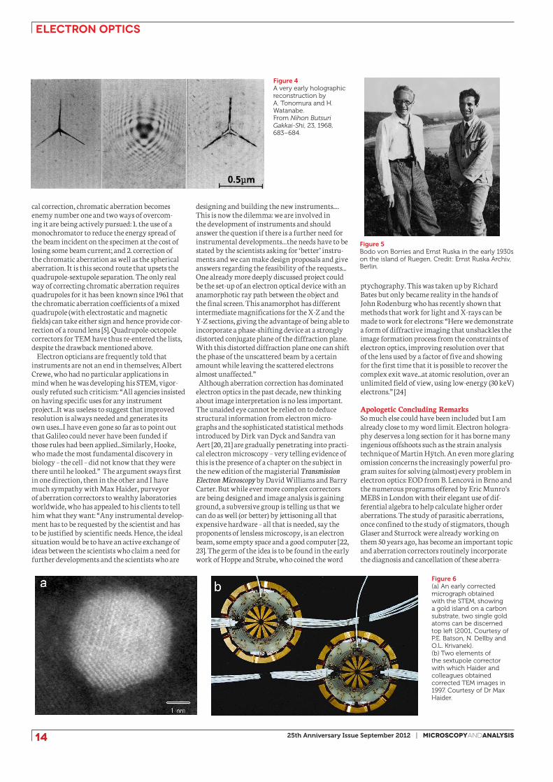
correct. The first three-dimensional reconstruc-tion by David de Rosier and Aaron Klug appeared in Nature in 1968. Two related but unconnected major instrumental developments came to frui-tion: prototype scanning electron microscopes had been built in the Cambridge University Engineering Department under Charles Oatley’s direction since 1950 [11-13] and in 1965, the first commercial version, the Stereoscan, was launched by the Cambridge Instrument Company (origi-nally founded in 1881 by Charles Darwin’s son Horace to manufacture specialized scientific equipment). In the Argonne National Laboratory and later in the University of Chicago, Albert Crewe was developing the scanning transmission electron microscope [14]. And at last, electron holography became a reality: in Japan, Akira Tonomura and Hiroshi Watanabe succeeded in recording and reconstructing an in-line hologram while in Tübingen, Gottfried Möllenstedt and Herbert Wahl used an electron biprism to gener-ate and reconstruct an off-axis hologram. And before leaving the 1960s, we must just mention the introduction of ptychography by Walter Hoppe and G. Strube in Munich; we shall return to this below.
Computers had been making their appearance in laboratories in the 1960s and the 1970s saw the arrival of smaller models such as the PDP, with memories of the order of 8-24 kilobytes. It was no coincidence that in the early 1970s, Ralph Gerchberg and Owen Saxton conceived and tested an iterative algorithm to solve the phase problem that arises from the fact that in many areas, infor-mation is coded in the phase of a complex signal whereas only the intensity can be recorded. This is exactly the situation in high-resolution electron microscopy, where the phase of the wave func-tion carries information about the specimen but only the intensity (current density) is recorded. The Gerchberg-Saxton algorithm has given rise to a gigantic literature and is used in one form or another in many disciplines. As the decade progressed, commercial models of Crewe’s STEM appeared; the transmission microscope manufac-turers AEI and Siemens both launched models and Siemens in practice built some six instru-ments but it was Vacuum Generators (VG) that captured the market [15]. The arrival of the com-puter also transformed practical electron optics. It was now possible to calculate the axial fields or potentials in electron lenses, the trajectory equa-tions could be solved numerically and aberration coefficients calculated. Eric Munro, who later created the company MEBS to sell electron optical software, was a pioneer in this area [16]. In 1979 and 1980, Vernon Beck, Crewe and David Kopf
discussed the use of sextupoles for aberration correction and a more realistic arrangement was proposed soon after by Harald Rose.
International, regional and thematic congresses on electron microscopy had been held ever since 1949, when the first international meeting was held in Delft. In 1980, however, the first of a series of meetings designed specifically for charged-particle optics was held in Giessen, organized by Hermann Wollnik, Karl Brown and myself; a second meeting was held in Albuquerque in 1986, since when they have been held every four years (the next will be held in Brno in 2014). This brings us to the brink of our ‘official’ starting date, 1987 but before turning to the quarter-century of M&A, we must celebrate 1986, when the Nobel Prize Committee at last (and just in time) awarded the prize to Ernst Ruska (1906-1988). The most vivid description of the occasion was given by Judith Reiffel, Elmar Zeitler’s secretary in Berlin (where Zeitler was now director of Ruska’s institute and editor of Ultramicroscopy): “Just after your call the telephone rang again – and didn’t stop ringing until five minutes ago. Because Ruska got the Nobel prize, sharing it with Binnig and Rohrer. Isn’t that a gas? Ruska is on holiday, and the BZ (Berliner Zeitung, yellow journal) sent a helicopter and found him taking a walk and schlepped him back to the hotel, where they interviewed him...jesus it’s a good thing we’ve got a festschrift in press.” This gave great pleasure to the electron microscopy community with the exception of some descendants of the founders of the subject who were not happy with the wording of the Nobel prize declaration: “Several scientists, among them Hans Busch, Max Knoll and Bodo von Borries, contributed to the development of the instrument, but Ernst Ruska deserves to be placed foremost.” An objective assessment of the rights and wrongs of the situation was attempted by a Chinese historian of science [17] but the subject is far from closed.
25 Years of Microscopy and AnalysisWith the 1990s, the world of electron optics was rejuvenated. Outside the small world of aberration specialists, there was no feeling that
aberration correction would ever work but the specialists were much more hopeful. A very prom-ising corrector design based on sextupoles had been found by Harald Rose and Max Haider was convinced that this could be made into a practical corrector. In 1991, the Volkswagen Foundation courageously agreed to provide funds. A corrector was built and the first tests showed that it did indeed provide correction. After a means of con-trolling the numerous parasitic aberrations had been devised, a resolution of about 140 picometres was achieved in 1997 – after initial resistance, Nature published these results a year later [18]. In the same years, Ondrej Krivanek was attempting to use quadrupoles and octopoles to correct the spherical aberration of the probe-forming lens of a STEM, research funded by the Paul Instrument Fund, and the successful outcome of this project was announced at the 1997 EMAG meeting in Cambridge at which the centenary of the electron was celebrated [19]. Meanwhile in the early 1990s, a quadrupole-octopole corrector had been tested by Zach and Haider on a low-voltage SEM, where it was simply required to provide some reduction of probe-size and this it succeeded in doing. The aberration-corrected era had begun; quadru-poles and octopoles for probe-forming lenses, sextupoles for TEM objectives. At first, this seemed a natural division of labour. Quadrupole systems have linear paraxial properties: the paths of rays in the two symmetry planes (X-Z and Y-Z where Z is the optic axis) are very different and quadrupoles have numerous off-axis aberrations as well as their own ‘spherical’ aberrations. Since a probe-forming system mainly requires correction of the axial aberrations, the complexity of the quadrupole optics is not too serious. Sextupoles, on the other hand, have no linear focusing effect; their primary effect is second-order, after which they exhibit third-order aberrations, one of which provides the correction. They are thus suit-able for TEM, where the whole of the extended field illuminated must be corrected.
But this neat sharing out of the correctors be-tween Krivanek’s company Nion (quadrupoles for STEM) and Haider’s CEOS (sextupoles for TEM) is becoming untenable. With the success of spheri-
Figure 2(a) Schematic and (b) photo of Seeliger’s corrector. (c) Shadow patterns demonstrating that Deltrap’s corrector worked. Fig. 2 a,b courtesy of H. Rose and Elsevier.
electron optics
14 25th Anniversary Issue September 2012 | MicroscopyandAnalysis
Figure 4A very early holographic reconstruction by A. Tonomura and H. Watanabe.From Nihon Butsuri Gakkai-Shi, 23, 1968, 683–684.
Figure 5Bodo von Borries and Ernst Ruska in the early 1930s on the island of Ruegen. Credit: Ernst Ruska Archiv, Berlin.
cal correction, chromatic aberration becomes enemy number one and two ways of overcom-ing it are being actively pursued: 1. the use of a monochromator to reduce the energy spread of the beam incident on the specimen at the cost of losing some beam current; and 2. correction of the chromatic aberration as well as the spherical aberration. It is this second route that upsets the quadrupole-sextupole separation. The only real way of correcting chromatic aberration requires quadrupoles for it has been known since 1961 that the chromatic aberration coefficients of a mixed quadrupole (with electrostatic and magnetic fields) can take either sign and hence provide cor-rection of a round lens [5]. Quadrupole-octopole correctors for TEM have thus re-entered the lists, despite the drawback mentioned above.
Electron opticians are frequently told that instruments are not an end in themselves; Albert Crewe, who had no particular applications in mind when he was developing his STEM, vigor-ously refuted such criticism: “All agencies insisted on having specific uses for any instrument project...It was useless to suggest that improved resolution is always needed and generates its own uses...I have even gone so far as to point out that Galileo could never have been funded if those rules had been applied...Similarly, Hooke, who made the most fundamental discovery in biology – the cell – did not know that they were there until he looked.” The argument sways first in one direction, then in the other and I have much sympathy with Max Haider, purveyor of aberration correctors to wealthy laboratories worldwide, who has appealed to his clients to tell him what they want: “Any instrumental develop-ment has to be requested by the scientist and has to be justified by scientific needs. Hence, the ideal situation would be to have an active exchange of ideas between the scientists who claim a need for further developments and the scientists who are
Figure 6(a) An early corrected micrograph obtained with the STEM, showing a gold island on a carbon substrate, two single gold atoms can be discerned top left (2001, Courtesy of P.E. Batson, N. Dellby and O.L. Krivanek). (b) Two elements of the sextupole corrector with which Haider and colleagues obtained corrected TEM images in 1997. Courtesy of Dr Max Haider.
designing and building the new instruments.…This is now the dilemma: we are involved in the development of instruments and should answer the question if there is a further need for instrumental developments…the needs have to be stated by the scientists asking for ‘better’ instru-ments and we can make design proposals and give answers regarding the feasibility of the requests...One already more deeply discussed project could be the set-up of an electron optical device with an anamorphotic ray path between the object and the final screen. This anamorphot has different intermediate magnifications for the X-Z and the Y-Z sections, giving the advantage of being able to incorporate a phase-shifting device at a strongly distorted conjugate plane of the diffraction plane. With this distorted diffraction plane one can shift the phase of the unscattered beam by a certain amount while leaving the scattered electrons almost unaffected.”
Although aberration correction has dominated electron optics in the past decade, new thinking about image interpretation is no less important. The unaided eye cannot be relied on to deduce structural information from electron micro-graphs and the sophisticated statistical methods introduced by Dirk van Dyck and Sandra van Aert [20, 21] are gradually penetrating into practi-cal electron microscopy – very telling evidence of this is the presence of a chapter on the subject in the new edition of the magisterial Transmission Electron Microscopy by David Williams and Barry Carter. But while ever more complex correctors are being designed and image analysis is gaining ground, a subversive group is telling us that we can do as well (or better) by jettisoning all that expensive hardware – all that is needed, say the proponents of lensless microscopy, is an electron beam, some empty space and a good computer [22, 23]. The germ of the idea is to be found in the early work of Hoppe and Strube, who coined the word
ptychography. This was taken up by Richard Bates but only became reality in the hands of John Rodenburg who has recently shown that methods that work for light and X-rays can be made to work for electrons: “Here we demonstrate a form of diffractive imaging that unshackles the image formation process from the constraints of electron optics, improving resolution over that of the lens used by a factor of five and showing for the first time that it is possible to recover the complex exit wave...at atomic resolution, over an unlimited field of view, using low-energy (30 keV) electrons.” [24]
Apologetic Concluding RemarksSo much else could have been included but I am already close to my word limit. Electron hologra-phy deserves a long section for it has borne many ingenious offshoots such as the strain analysis technique of Martin Hÿtch. An even more glaring omission concerns the increasingly powerful pro-gram suites for solving (almost) every problem in electron optics: EOD from B. Lencová in Brno and the numerous programs offered by Eric Munro’s MEBS in London with their elegant use of dif-ferential algebra to help calculate higher order aberrations. The study of parasitic aberrations, once confined to the study of stigmators, though Glaser and Sturrock were already working on them 50 years ago, has become an important topic and aberration correctors routinely incorporate the diagnosis and cancellation of these aberra-
electron optics
16 25th Anniversary Issue September 2012 | MicroscopyandAnalysis
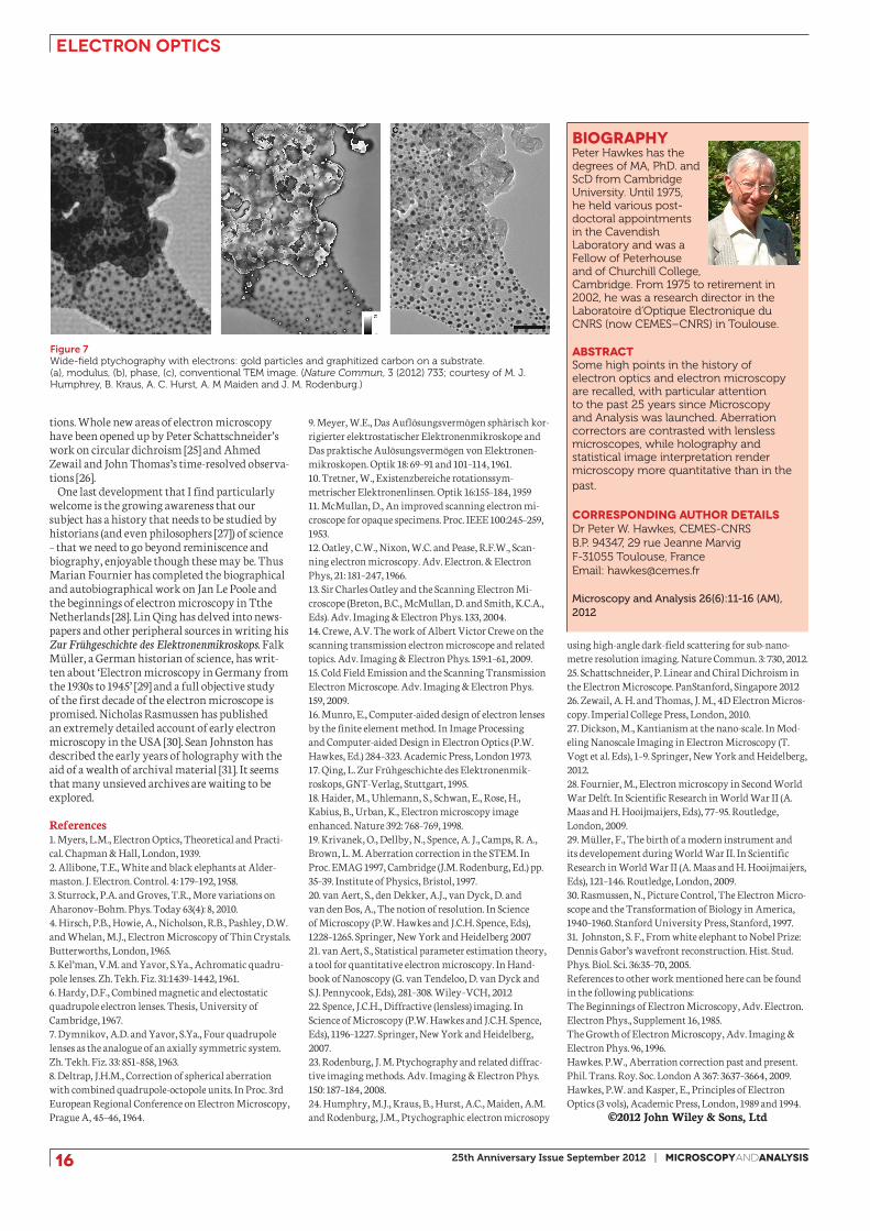
biography Peter Hawkes has the degrees of MA, PhD. and ScD from Cambridge University. Until 1975, he held various post-doctoral appointments in the Cavendish Laboratory and was a Fellow of Peterhouse and of Churchill College, Cambridge. From 1975 to retirement in 2002, he was a research director in the Laboratoire d’Optique Electronique du CNRS (now CEMES–CNRS) in Toulouse.
abstractSome high points in the history of electron optics and electron microscopy are recalled, with particular attention to the past 25 years since Microscopy and Analysis was launched. Aberration correctors are contrasted with lensless microscopes, while holography and statistical image interpretation render microscopy more quantitative than in the past.
CORRESPONDING AUTHOR DETAILSDr Peter W. Hawkes, CEMES-CNRSB.P. 94347, 29 rue Jeanne MarvigF-31055 Toulouse, FranceEmail: [email protected]
Microscopy and Analysis 26(6):11-16 (AM), 2012
Figure 7Wide-field ptychography with electrons: gold particles and graphitized carbon on a substrate.(a), modulus, (b), phase, (c), conventional TEM image. (Nature Commun, 3 (2012) 733; courtesy of M. J. Humphrey, B. Kraus, A. C. Hurst, A. M Maiden and J. M. Rodenburg.)
tions. Whole new areas of electron microscopy have been opened up by Peter Schattschneider’s work on circular dichroism [25] and Ahmed Zewail and John Thomas’s time-resolved observa-tions [26].
One last development that I find particularly welcome is the growing awareness that our subject has a history that needs to be studied by historians (and even philosophers [27]) of science – that we need to go beyond reminiscence and biography, enjoyable though these may be. Thus Marian Fournier has completed the biographical and autobiographical work on Jan Le Poole and the beginnings of electron microscopy in Tthe Netherlands [28]. Lin Qing has delved into news-papers and other peripheral sources in writing his Zur Frühgeschichte des Elektronenmikroskops. Falk Müller, a German historian of science, has writ-ten about ‘Electron microscopy in Germany from the 1930s to 1945’ [29] and a full objective study of the first decade of the electron microscope is promised. Nicholas Rasmussen has published an extremely detailed account of early electron microscopy in the USA [30]. Sean Johnston has described the early years of holography with the aid of a wealth of archival material [31]. It seems that many unsieved archives are waiting to be explored. References1. Myers, L.M., Electron Optics, Theoretical and Practi-cal. Chapman & Hall, London, 1939.2. Allibone, T.E., White and black elephants at Alder-maston. J. Electron. Control. 4: 179–192, 1958.3. Sturrock, P.A. and Groves, T.R., More variations on Aharonov–Bohm. Phys. Today 63(4): 8, 2010.4. Hirsch, P.B., Howie, A., Nicholson, R.B., Pashley, D.W. and Whelan, M.J., Electron Microscopy of Thin Crystals. Butterworths, London, 1965.5. Kel’man, V.M. and Yavor, S.Ya., Achromatic quadru-pole lenses. Zh. Tekh. Fiz. 31:1439–1442, 1961.6. Hardy, D.F., Combined magnetic and electostatic quadrupole electron lenses. Thesis, University of Cambridge, 1967.7. Dymnikov, A.D. and Yavor, S.Ya., Four quadrupole lenses as the analogue of an axially symmetric system. Zh. Tekh. Fiz. 33: 851–858, 1963.8. Deltrap, J.H.M., Correction of spherical aberration with combined quadrupole-octopole units. In Proc. 3rd European Regional Conference on Electron Microscopy, Prague A, 45–46, 1964.
9. Meyer, W.E., Das Auflösungsvermögen sphärisch kor-rigierter elektrostatischer Elektronenmikroskope and Das praktische Aulösungsvermögen von Elektronen-mikroskopen. Optik 18: 69–91 and 101–114, 1961.10. Tretner, W., Existenzbereiche rotationssym-metrischer Elektronenlinsen. Optik 16:155–184, 195911. McMullan, D., An improved scanning electron mi-croscope for opaque specimens. Proc. IEEE 100:245–259, 1953.12. Oatley, C.W., Nixon, W.C. and Pease, R.F.W., Scan-ning electron microscopy. Adv. Electron. & Electron Phys, 21: 181–247, 1966.13. Sir Charles Oatley and the Scanning Electron Mi-croscope (Breton, B.C., McMullan, D. and Smith, K.C.A., Eds). Adv. Imaging & Electron Phys. 133, 2004.14. Crewe, A.V. The work of Albert Victor Crewe on the scanning transmission electron microscope and related topics. Adv. Imaging & Electron Phys. 159:1–61, 2009.15. Cold Field Emission and the Scanning Transmission Electron Microscope. Adv. Imaging & Electron Phys. 159, 2009.16. Munro, E., Computer-aided design of electron lenses by the finite element method. In Image Processing and Computer-aided Design in Electron Optics (P.W. Hawkes, Ed.) 284–323. Academic Press, London 1973.17. Qing, L. Zur Frühgeschichte des Elektronenmik-roskops, GNT-Verlag, Stuttgart, 1995.18. Haider, M., Uhlemann, S., Schwan, E., Rose, H., Kabius, B., Urban, K., Electron microscopy image enhanced. Nature 392: 768–769, 1998.19. Krivanek, O., Dellby, N., Spence, A. J., Camps, R. A., Brown, L. M. Aberration correction in the STEM. In Proc. EMAG 1997, Cambridge (J.M. Rodenburg, Ed.) pp. 35–39. Institute of Physics, Bristol, 1997.20. van Aert, S., den Dekker, A.J., van Dyck, D. and van den Bos, A., The notion of resolution. In Science of Microscopy (P.W. Hawkes and J.C.H. Spence, Eds), 1228–1265. Springer, New York and Heidelberg 200721. van Aert, S., Statistical parameter estimation theory, a tool for quantitative electron microscopy. In Hand-book of Nanoscopy (G. van Tendeloo, D. van Dyck and S.J. Pennycook, Eds), 281–308. Wiley–VCH, 201222. Spence, J.C.H., Diffractive (lensless) imaging. In Science of Microscopy (P.W. Hawkes and J.C.H. Spence, Eds), 1196–1227. Springer, New York and Heidelberg, 2007.23. Rodenburg, J. M. Ptychography and related diffrac-tive imaging methods. Adv. Imaging & Electron Phys. 150: 187–184, 2008.24. Humphry, M.J., Kraus, B., Hurst, A.C., Maiden, A.M. and Rodenburg, J.M., Ptychographic electron microsopy
using high-angle dark-field scattering for sub-nano-metre resolution imaging. Nature Commun. 3: 730, 2012.25. Schattschneider, P. Linear and Chiral Dichroism in the Electron Microscope. PanStanford, Singapore 201226. Zewail, A. H. and Thomas, J. M., 4D Electron Micros-copy. Imperial College Press, London, 2010.27. Dickson, M., Kantianism at the nano-scale. In Mod-eling Nanoscale Imaging in Electron Microscopy (T. Vogt et al. Eds), 1–9. Springer, New York and Heidelberg, 2012.28. Fournier, M., Electron microscopy in Second World War Delft. In Scientific Research in World War II (A. Maas and H. Hooijmaijers, Eds), 77–95. Routledge, London, 2009.29. Müller, F., The birth of a modern instrument and its developement during World War II. In Scientific Research in World War II (A. Maas and H. Hooijmaijers, Eds), 121–146. Routledge, London, 2009.30. Rasmussen, N., Picture Control, The Electron Micro-scope and the Transformation of Biology in America, 1940–1960. Stanford University Press, Stanford, 1997.31. Johnston, S. F., From white elephant to Nobel Prize: Dennis Gabor’s wavefront reconstruction. Hist. Stud. Phys. Biol. Sci. 36:35–70, 2005.References to other work mentioned here can be found in the following publications:The Beginnings of Electron Microscopy, Adv. Electron. Electron Phys., Supplement 16, 1985.The Growth of Electron Microscopy, Adv. Imaging & Electron Phys. 96, 1996.Hawkes. P.W., Aberration correction past and present. Phil. Trans. Roy. Soc. London A 367: 3637–3664, 2009.Hawkes, P.W. and Kasper, E., Principles of Electron Optics (3 vols), Academic Press, London, 1989 and 1994.
©2012 John Wiley & Sons, Ltd