electronic supplementary information final of potential sirna molecules by dicer enzyme to generate...
TRANSCRIPT
Enzymatic size control of RNA Particles with
complementary rolling circle transcription (cRCT) for
efficient siRNA production
Electronic Supplementary Information
Daehoon Han, Yongkuk Park, Hyangsu Nam and Jong Bum Lee*
Department of Chemical Engineering, University of Seoul, Seoul, 130-743, South Korea
*Corresponding Author
Email: [email protected], Tel: +82 26490 2372, Fax: +82 26490 2364
Electronic Supplementary Material (ESI) for ChemComm.This journal is © The Royal Society of Chemistry 2014
Experimental Section
Materials and Reagents
DNA oligonucleotides were commercially synthesized from Integrated DNA Technologies
(IDT). T4 DNA ligase (cat. No. M1804) was purchased from Promega. T7 RNA polymerase
(cat. No. M0251L), 10X RNA Polymerase Reaction buffer (cat. No. B9012S) and
Ribonucleotide Solution Mix (cat. No. N0466S) were purchased from New England BioLabs.
SYBR Green II RNA gel stain (cat. No. S7564) was purchased from Invitrogen. Dulbecco's
Modified Eagle's Medium (DMEM, cat. No. LM001-10) and Fetal bovine serum (FBS, cat.
No. S 101-01) were purchased from Welgene. Opti-MEMI (cat. No. 31985-070), Hoechst-
33342 (Solution 15, cat. No. 910-3015) and transfection agent (TransIT-X2) were purchased
from Gibco, Chemometec and Mirus, respectively.
DNA sequences for generating RNA Particles Strand DNA Sequences
Single stranded DNA
(Length: 92 nt)
5’-Phospate-ATA GTG AGT CGT ATT AAC TTA CGC TGA
GTA CTT CGA TTC AAG TCC AGT CCA TAA TAC TGC
GTG TGC GTT GGT ACG TTA ATA CGA CTA TCC CT-3’
Complementary
single stranded DNA
(Length: 92 nt)
5’- Phospate -ATA GTG AGT CGT ATT AAC GTA CCA
ACG CAC ACG CAG TAT TAT GGA CTG GAC TTG AAT
CGA AGT ACT CAG CGT AAG TTT AGA GGC ATA TCC
CT-3’
Promoter DNA
(Length: 22 nt) 5’-TAA TAC GAC TCA CTA TAG GGA T-3’
Methods
Circularization of single stranded DNA
1 µM of circular DNA was synthesized with phosphorylated single stranded DNA (ssDNA)
and Promoter DNA. 1 µM of ssDNA was mixed with equimolar concentration of Promoter
DNA in nuclease-free water. To hybridize these two DNA strands, the solution was heated
for 2 min at 95 °C and gradually cooled to 25 °C for 1 hour using a PCR thermal cycler
(T100™ Thermal Cycler, Bio-Rad). To ligate the nick in the circularized DNA, 0.06 U µl-1 of
T4 ligase (1-3 U µl-1) and ligase buffer (50 mM Tris-HCl, 10 mM MgCl2, 10 mM DTT and 1
mM ATP) were added into the solution and incubated for overnight at room temperature. 1
µM of synthesized circular DNA was concentrated to 15 µM with centrifugal filter (YM-3,
Millipore).
Self-assembly of RNA Particles using cRCT
Circular DNA (final concentration of 2.5 µM) and Complementary circular DNA (final
concentration of 2.5 µM) were mixed with 0.625 mM of Ribonucleotide Solution Mix (25
mM of each NTP), 2X Reaction buffer (400 mM Tris-HCl, 20 mM spermidine, 60 mM
MgCl2 and 100 mM dithiothreitol) and T7 RNA polymerase (50 units µl-1) to prepare the
reaction solution. In this study, 1, 10 or 40 units µl-1 of T7 RNA polymerase was used to
generate the different size of RNA particles (RPs). The reaction solution was incubated at
37 °C for 20 hours for generating RPs.
Characterization of the morphologies of RNA Particles
The morphologies of RPs were confirmed by SEM (SNE model, SEC) and AFM (NX-10,
Park systems Corp.). AFM was used with non-contact silicon cantilever (OMCL-AC160TS,
Olympus) and XEI software. To prepare the sample, the reaction solution was centrifuged for
4 min at 2,400 g and replaced with fresh nuclease-free water. To disperse connected RPs into
individual RNA particle, the solution was pipetted and sonicated (less than 3 min) repeatedly.
2 µl of RPs sample was dropped on silicon wafer (for SEM) or mica substrate (for AFM).
After dropping the sample, the sample was dried at room temperature for 12 hours. The
morphology of dried sample was observed by SEM or AFM.
Size and surface charge (zeta potential) of RNA Particles
The size and surface charge of RPs were analyzed by Zetasizer (Zeetasizer Nano ZS90,
Malvern). To prepare the sample solution, generated RPs solution was diluted tenfold with
nuclease-free water. All samples were measured at 25 °C.
Young’s modulus of RNA Particles
To analyze the Young’s modulus of RPs, AFM (NX-10, Park systems Corp.) was used with
non-contact silicon cantilever (OMCL-AC160TS, Olympus) and XEI software. RPs were
prepared on the mica substrate.

Generation of potential siRNA molecules by Dicer enzyme
To generate the potential siRNA molecules, the RPs were incubated for 24 hours with 1 unit
of recombinant Dicer (Genlantis) and reaction solution (1 mM ATP, 2.5 mM MgCl2, 40 %
Dicer Reaction Buffer) in nuclease-free water (final volume of 10 µl). After incubation, 2 µl
of Dicer Stop Solution (Genlantis) was added to terminate enzyme reaction. To confirm the
generation of potential siRNA molecules, gel electrophoresis was used with 3 % agarose gel.
In vitro transfection experiment
HeLa cells were maintained in growth media comprised of essential Dulbecco's Modified
Eagle's Medium (DMEM) supplemented with 10 % fetal bovine serum (FBS), 1 % penicillin-
streptomycin and 1 % antibiotic-antimycotic. One day prior to transfection, cells were seeded
in 24-well plates and 8-well chamber slides at the seeding density of 50,000 cells per well. To
form RPs/TransIT complex, 4 µl of fluorescently labelled RPs was added to 70 µl of Opti-
MEMI with 6 µl of transfection agent (TransIT-X2), and the solution was incubated at 37 °C
for 15 minutes. To deliver the RPs into the cell, 40 µl of RPs/TransIT complex was added to
each well of cells and incubated at 37 °C for 6 hours. After incubation, cells were trypsinized
and stained with Hoechst-33342 (final concentration of 10 µg ml-1) at 37 °C for 15 minutes.
The cellular transfection was analyzed by Nucleocounter (NC-3000, Chemometec) with 24-
well plates and visualized with confocal microscope (Leica TCS SPE, Leica) with 8-well
chamber slides.
Supplementary Figures
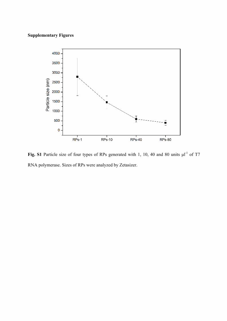
Fig. S1 Particle size of four types of RPs generated with 1, 10, 40 and 80 units µl-1 of T7
RNA polymerase. Sizes of RPs were analyzed by Zetasizer.
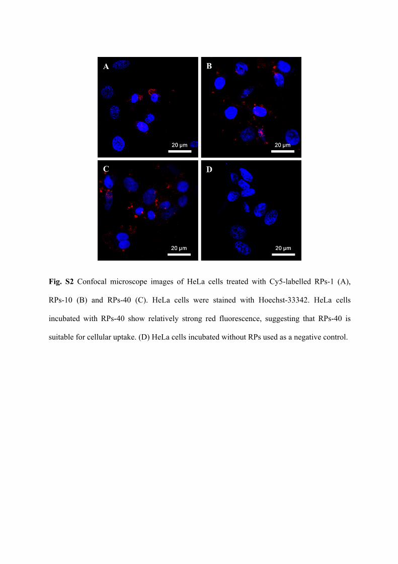
Fig. S2 Confocal microscope images of HeLa cells treated with Cy5-labelled RPs-1 (A),
RPs-10 (B) and RPs-40 (C). HeLa cells were stained with Hoechst-33342. HeLa cells
incubated with RPs-40 show relatively strong red fluorescence, suggesting that RPs-40 is
suitable for cellular uptake. (D) HeLa cells incubated without RPs used as a negative control.
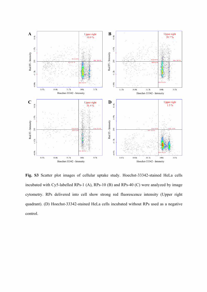
Fig. S3 Scatter plot images of cellular uptake study. Hoechst-33342-stained HeLa cells
incubated with Cy5-labelled RPs-1 (A), RPs-10 (B) and RPs-40 (C) were analyzed by image
cytometry. RPs delivered into cell show strong red fluorescence intensity (Upper right
quadrant). (D) Hoechst-33342-stained HeLa cells incubated without RPs used as a negative
control.