electronystagmography - fac.ksu.edu.sa · spontaneous nystagmus (eliminating suppression ) ......
TRANSCRIPT

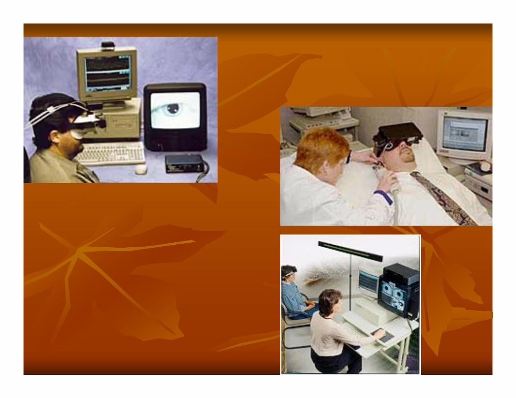
ELECTRONYSTAGMOGRAPHYELECTRONYSTAGMOGRAPHY


ELECTRONYSTAGMOGRAPHYELECTRONYSTAGMOGRAPHY
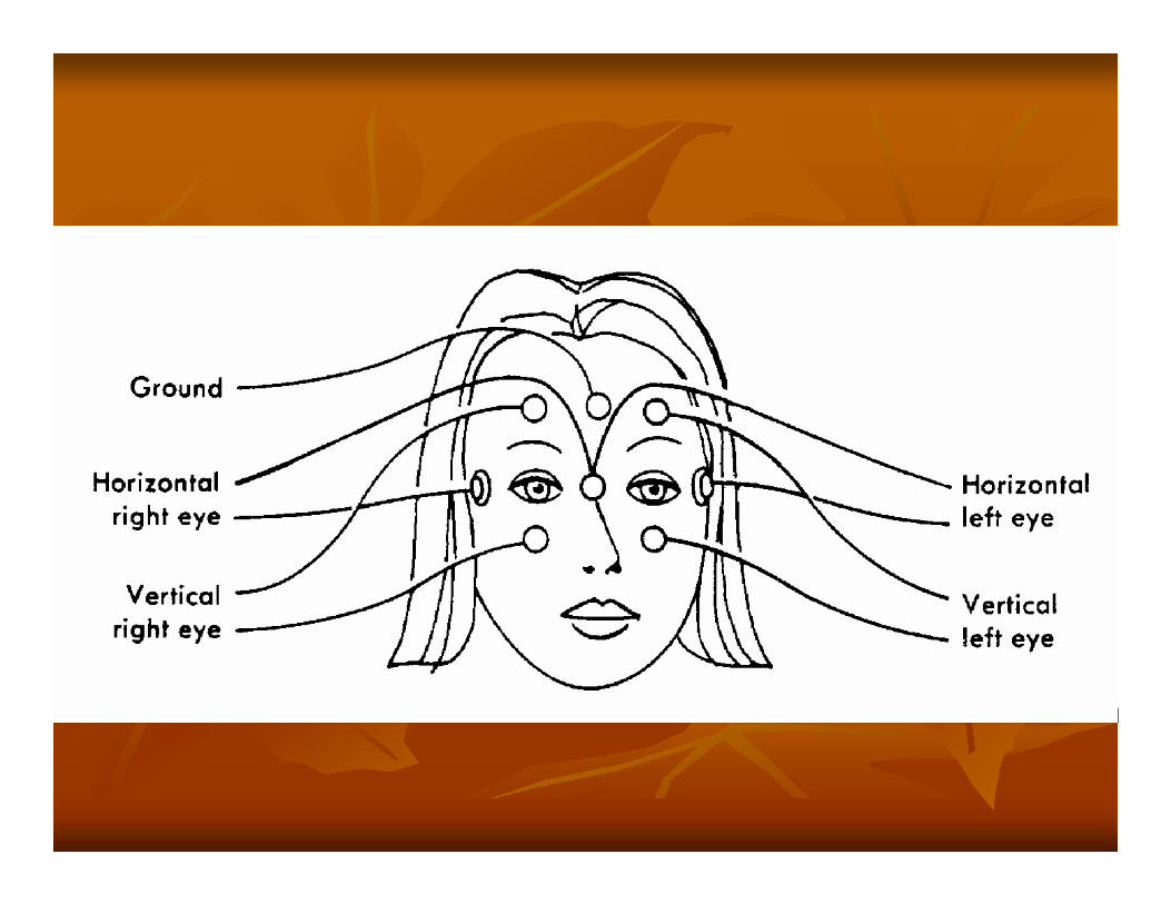
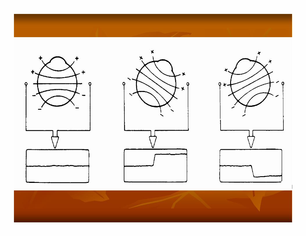
ENG measurements based on the presence ofENG measurements based on the presence ofthe the corneoretinalcorneoretinal potentialpotential
Cornea has a positive poleCornea has a positive pole
Retina has a negative poleRetina has a negative pole


ELECTRONYSTAGMOGRAPHYELECTRONYSTAGMOGRAPHY
Essentially ENG consists of 3 parts Essentially ENG consists of 3 parts oculomotoroculomotor evaluationevaluation
CalibrationCalibrationGazeGazeFixationFixationSaccadeSaccadeTracking (Pursuit)Tracking (Pursuit)OptokineticOptokinetic
positioning/positional testingpositioning/positional testingcaloric stimulation. caloric stimulation.

HistoryHistory
Stop all medications 24Stop all medications 24--72 h prior to testing72 h prior to testing72 hours Alcohol (agonist or antagonist )72 hours Alcohol (agonist or antagonist )Any medications taken should be clearly noted Any medications taken should be clearly noted on the test results on the test results limit food intake prior to examination limit food intake prior to examination arrange for transportation after the arrange for transportation after the examinationexamination

Examination Examination
Large perforations Large perforations increase air stimulation above expectation increase air stimulation above expectation cooling effect for warm (evaporation). cooling effect for warm (evaporation).
cerumencerumen must be removed must be removed Middle ear fluid affects stimulationMiddle ear fluid affects stimulation

Saccades (calibration) Saccades (calibration)
Dots on the wall or ceiling Dots on the wall or ceiling center and 10center and 10°°, 20, 20°°, and 30, and 30°° off center off center patient to look back and forth between the dotspatient to look back and forth between the dotshead fixed head fixed


GazeGaze
spontaneous spontaneous nystagmusnystagmusnystagmusnystagmus in the absence of stimulation in the absence of stimulation
presence or absence of spontaneous presence or absence of spontaneous nystagmusnystagmuspresence, absence, or exacerbation of presence, absence, or exacerbation of nystagmusnystagmuswith addition of offwith addition of off--center gaze center gaze fixation suppression of spontaneous fixation suppression of spontaneous nystagmusnystagmus

Gaze TestGaze TestGaze Test
NystagmusNystagmus present with eyes open and present with eyes open and enhanced by eye closure enhanced by eye closure -- lesion is peripherallesion is peripheralNystagmusNystagmus is enhanced with ocular fixation is enhanced with ocular fixation and reduced by eye closure and reduced by eye closure -- lesion is centrallesion is central
Administration Administration
For gaze testingFor gaze testingthe patient is instructed to look straight ahead and then to the patient is instructed to look straight ahead and then to fixate on a target 30fixate on a target 30°° to the right, left, up, and down. to the right, left, up, and down. Fixation is maintained for approximately 30 seconds in Fixation is maintained for approximately 30 seconds in center gaze and 10 seconds in eccentric gaze. center gaze and 10 seconds in eccentric gaze.
Spontaneous Spontaneous nystagmusnystagmus (eliminating suppression )(eliminating suppression )eyes open in a dark room eyes open in a dark room eyes closed. eyes closed. mental tasks (mental tasks (egeg, answering questions, counting by twos). , answering questions, counting by twos).

GazeGazeGaze
Normal gaze position Normal gaze position -- patient is able to patient is able to maintain position with eyes open and closedmaintain position with eyes open and closed

Spontaneous Spontaneous nystagmusnystagmusEither central or peripheral pathology. Either central or peripheral pathology. with eyes open is always diagnostically significant. with eyes open is always diagnostically significant. Peripheral indicators Peripheral indicators
Horizontal or horizontal rotary Horizontal or horizontal rotary Suppressed by visual fixation Suppressed by visual fixation NondirectionNondirection changing changing Exacerbated by gazing in the direction of the fast phase*Exacerbated by gazing in the direction of the fast phase*
Central indicators Central indicators Vertical Vertical Not suppressed by fixation Not suppressed by fixation Direction changing Direction changing

Alexander's lawAlexander's law
NystagmusNystagmus increases when the patient gazes in increases when the patient gazes in the direction of the fast phase. the direction of the fast phase. NystagmusNystagmus decreases or disappears when the decreases or disappears when the gaze in the direction of slow phase. gaze in the direction of slow phase. This pattern is often seen in peripheral This pattern is often seen in peripheral vestibular disorders and occasionally in central vestibular disorders and occasionally in central disorders. disorders.

Unilateral gazeUnilateral gaze--paretic paretic nystagmusnystagmus
NystagmusNystagmus only occurs with eccentric gaze in only occurs with eccentric gaze in one direction. one direction. Elicited Elicited nystagmusnystagmus beats in the direction of the beats in the direction of the gaze. gaze. consistent with CNS pathology consistent with CNS pathology
Bilateral gazeBilateral gaze--paretic paretic nystagmusnystagmus
right gazeright gaze rightright--beating beating nystagmusnystagmusleft gaze left gaze leftleft--beating beating nystagmusnystagmussuggests CNS pathology suggests CNS pathology

BrunsBruns nystagmusnystagmus
Combination of Combination of Unilateral gazeUnilateral gaze--paretic paretic nystagmusnystagmusVestibular Vestibular nystagmusnystagmus
Asymmetrical Asymmetrical nystagmusnystagmus in both directions of in both directions of a gazea gazeassociated with extraassociated with extra--axial mass lesions on the axial mass lesions on the side of the gazeside of the gaze--paretic paretic nystagmusnystagmus

EwaldEwald lawlaw
Eyes always move in the plane of the canal Eyes always move in the plane of the canal being stimulated and in the direction of being stimulated and in the direction of endolymphendolymph flowflowAmpulopetalAmpulopetal in HSCC causes greater response in HSCC causes greater response than than ampulofugalampulofugalAmpulofugalAmpulofugal in vertical in vertical SCCsSCCs cause greater cause greater response than response than ampulopetalampulopetal
Resting (firing) level is 1.0 Hz can not be <0 but can be high as 10 Hz

FixationFixation
Congenital Congenital nystagmusnystagmusGazeGaze--Evoked Evoked NystagmusNystagmusRebound Rebound nystagmusnystagmusSquare-wave jerks
Congenital Congenital nystagmusnystagmus
Spiky appearance Spiky appearance increases with lateral gaze. increases with lateral gaze. decrease in velocity or completely disappear decrease in velocity or completely disappear with eyes closed with eyes closed

Congenital Gaze FindingsCongenital Gaze FindingsCongenital Gaze Findings

GazeGaze--Evoked Evoked NystagmusNystagmus
Drift of the eye which is only present for certain Drift of the eye which is only present for certain directions of gaze directions of gaze EOG recordings, any persistent EOG recordings, any persistent nystagmusnystagmus for ocular for ocular displacements < 30 degrees is abnormal displacements < 30 degrees is abnormal Causes of GazeCauses of Gaze--evoked evoked nystagmusnystagmus
Medication Medication Brainstem or Brainstem or cerebellarcerebellar disorder disorder Normal variant Normal variant Ocular muscle fatigue Ocular muscle fatigue Congenital Congenital nystagmusnystagmus
Rebound Rebound nystagmusnystagmus
Burst of Burst of nystagmusnystagmusbegins when the eyes are returned to center begins when the eyes are returned to center gaze. gaze. lasting 5 seconds lasting 5 seconds brainstem or brainstem or cerebellarcerebellar lesions lesions

Square-wave jerks
the most common abnormality with eyes the most common abnormality with eyes closed. closed. healthy patients healthy patients increasing frequency with increasing age. increasing frequency with increasing age. abnormal if abnormal if
In young patientsIn young patientsmore frequently than 1 per second more frequently than 1 per second eyes open. eyes open.
suggestive of a suggestive of a cerebellarcerebellar disorder. disorder.

Fixation suppressionFixation suppression
For peripheral lesions, For peripheral lesions, nystagmusnystagmus that is that is evident with eyes closed or in the dark should evident with eyes closed or in the dark should be suppressed by visual fixation. be suppressed by visual fixation. If not CNS pathology is possible. If not CNS pathology is possible.
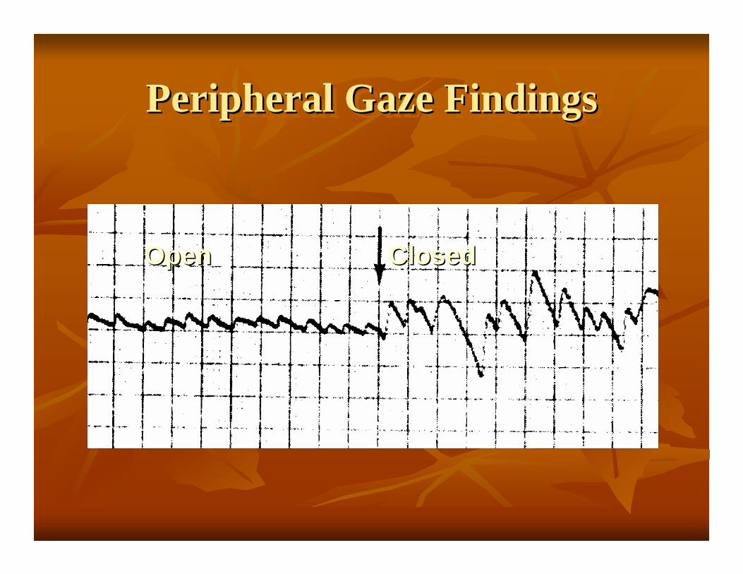
Peripheral Gaze FindingsPeripheral Gaze FindingsPeripheral Gaze Findings
ClosedClosedOpenOpen

Gaze Findings With CNS LesionGaze Findings With CNS LesionGaze Findings With CNS Lesion
NystagmusNystagmus may be horizontal, vertical, may be horizontal, vertical, rotatoryrotatoryMay May demonstatedemonstate variation in amplitudevariation in amplitudeIf cause by a stable pathology, it declines If cause by a stable pathology, it declines slowly in timeslowly in timeEnhanced by ocular fixationEnhanced by ocular fixationIf horizontal, most often bilateral If horizontal, most often bilateral (bidirectional)(bidirectional)

CNS Gaze FindingsCNS Gaze FindingsCNS Gaze Findings
RR
LL
SaccadesSaccades InterpretationInterpretation
Accuracy Accuracy Latency Latency Velocity Velocity

Accuracy Accuracy Normal or basal ganglia pathologyNormal or basal ganglia pathology
HypometricHypometric –– undershootsundershoots
CNS pathologyCNS pathologyOcular flutter Ocular flutter -- spiky overshootspiky overshoot
Cerebellum Cerebellum Hypermetric overshoot then a correction. Hypermetric overshoot then a correction. MultistepMultistep saccades undershoots then multiple saccadessaccades undershoots then multiple saccadesPostsaccadicPostsaccadic drift (Glissade) eye drifting after saccade. drift (Glissade) eye drifting after saccade.
PICA PICA PulsionPulsion :pulling to left or right after vertical saccades. :pulling to left or right after vertical saccades.

LatencyLatency
Short latency Short latency artifact artifact patient anticipating the position of the target. patient anticipating the position of the target. suggestive of CNS pathology. suggestive of CNS pathology.
Asymmetrical latencies Asymmetrical latencies occipital occipital parietal cortex. parietal cortex.
Velocity Velocity
Saccadic slowing Saccadic slowing drug effects. drug effects. CNS degenerative conditions, basal ganglia pathology, and CNS degenerative conditions, basal ganglia pathology, and cerebellarcerebellardisorders. disorders. ocular disorders, including ocular disorders, including oculomotoroculomotor weakness, weakness,
Abnormally fast saccades Abnormally fast saccades artifact and may be due to technical difficulties. artifact and may be due to technical difficulties. CNS CNS ocular pathology ocular pathology
Asymmetrical velocity Asymmetrical velocity -- between the eyes or between directions. between the eyes or between directions. ocular nerve ocular nerve muscle pathology (muscle pathology (ieie, lesions or palsies). , lesions or palsies). CNS pathology may also be indicated. A lesion in the MLFCNS pathology may also be indicated. A lesion in the MLF

Saccadic AbnormalitiesOvershootOvershoot

Saccadic AbnormalitiesSaccadic AbnormalitiesSaccadic AbnormalitiesSaccadic SlowingSaccadic Slowing

Smooth pursuit trackingSmooth pursuit tracking
follow a sinusoidal moving target with eyes follow a sinusoidal moving target with eyes only. only. Tracking targets within the visual fieldTracking targets within the visual fieldinterpreting with care in geriatric and pediatricinterpreting with care in geriatric and pediatricaffected by attention and patient cooperation. affected by attention and patient cooperation.
Interpretation Interpretation results should resemble a smooth sinusoid. results should resemble a smooth sinusoid. Breakup of movement Breakup of movement CNS pathology. CNS pathology.
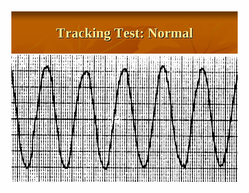
Tracking Test: NormalTracking Test: NormalTracking Test: Normal

Tracking Test: AbnormalTracking Test: AbnormalTracking Test: Abnormal
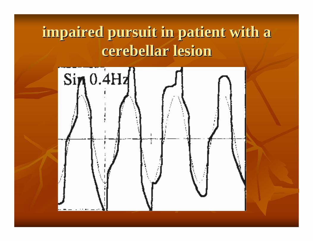
impaired pursuit in patient with a impaired pursuit in patient with a cerebellarcerebellar lesion lesion

OptokineticOptokinetic
tracks multiple stimuli. tracks multiple stimuli. stripes on a rotating drum stripes on a rotating drum stream of lighted dots across a light bar stream of lighted dots across a light bar full field array of moving stars or trees. full field array of moving stars or trees.
moved at 300, 400, or 600 per second moved at 300, 400, or 600 per second asymmetrical responses asymmetrical responses CNS pathologyCNS pathology


Opokinetic Test:NormalOpokineticOpokinetic Test:NormalTest:Normalsymmetrysymmetry
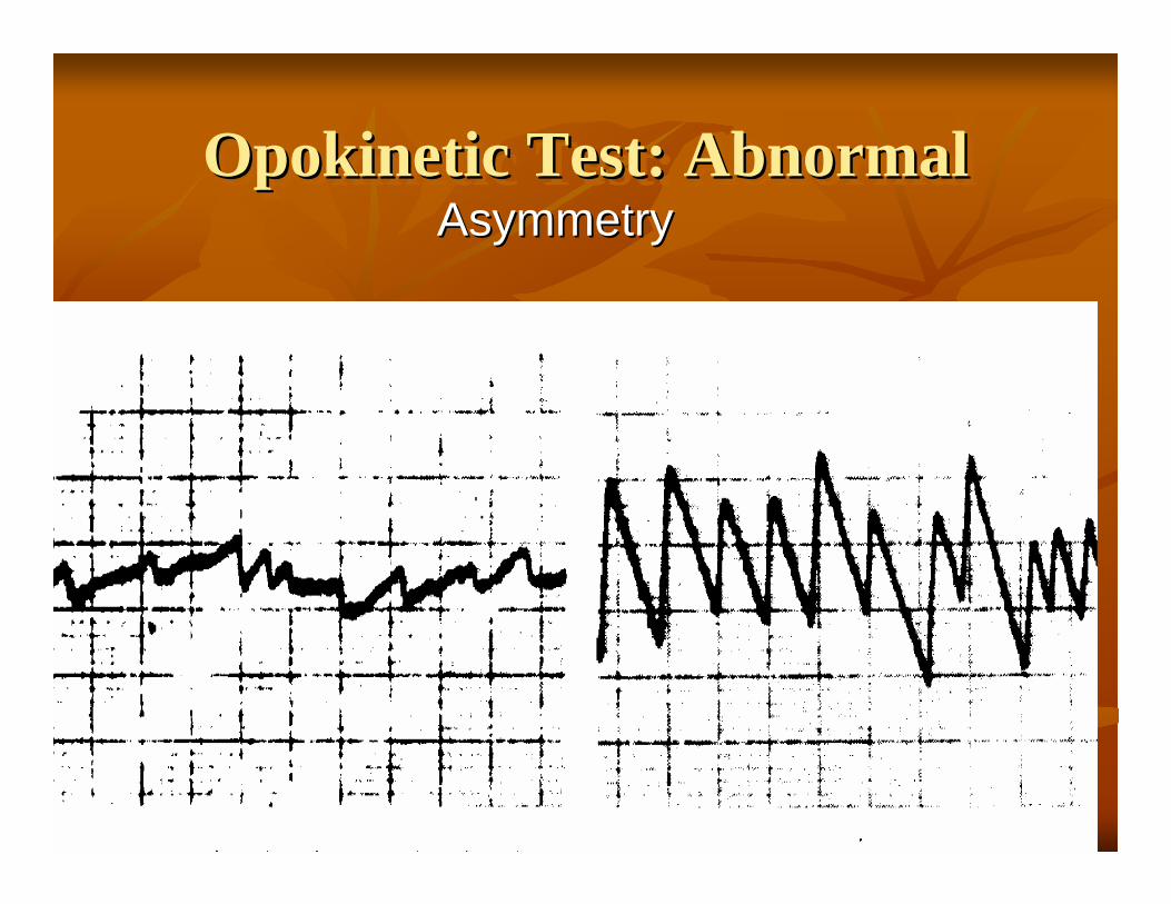
Opokinetic Test: AbnormalOpokineticOpokinetic Test: AbnormalTest: AbnormalAsymmetryAsymmetry
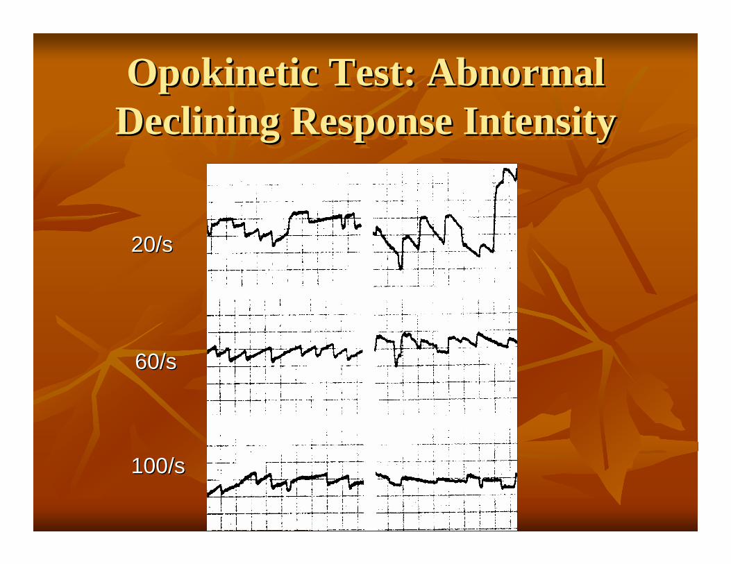
Opokinetic Test: AbnormalDeclining Response IntensityOpokineticOpokinetic Test: AbnormalTest: Abnormal
Declining Response IntensityDeclining Response Intensity
20/s20/s
60/s60/s
100/s100/s

Positioning Positioning DixDix--HallpikeHallpike maneuvermaneuver
should be completed prior to any other should be completed prior to any other positional testing. positional testing. Delayed onset Delayed onset -- observe patient for at least 20 observe patient for at least 20 seconds seconds Transient burst of Transient burst of nystagmusnystagmus -- Lasts about 10Lasts about 10--15 seconds 15 seconds Subjective report of vertigo Subjective report of vertigo Fatigability Fatigability
Positional tests Positional tests minimum of 20minimum of 20--30 seconds30 secondsMental tasking infrared goggles or with the patient's eyes Mental tasking infrared goggles or with the patient's eyes closed with electrodes closed with electrodes
Head hanging Head hanging Supine Supine Supine, head right Supine, head right Supine, head left Supine, head left Lateral right Lateral right Lateral left Lateral left
considered abnormal considered abnormal exceed 60 per second exceed 60 per second change direction in any 1 positionchange direction in any 1 positionpersist in at least 3 different positionspersist in at least 3 different positionsintermittent in all positionsintermittent in all positions

Positional testsPositional tests
Peripheral indicators include the following: Peripheral indicators include the following: DirectionDirection--fixed fixed geotropic direction changing in different positions, geotropic direction changing in different positions, horizontal SCC variant of BPPVhorizontal SCC variant of BPPVLatency of onset Latency of onset Fatigable Fatigable
Central indicators include the following: Central indicators include the following: ageotropicageotropic direction changing in different positions, direction changing in different positions, Direction changing in a single position, Direction changing in a single position, Immediate onset Immediate onset Not fatigableNot fatigable

Positional Test: AbnormalPeripheral
Positional Test: AbnormalPositional Test: AbnormalPeripheralPeripheral
RLRL
LLLL
EE
DirectionFixedDirectionFixed
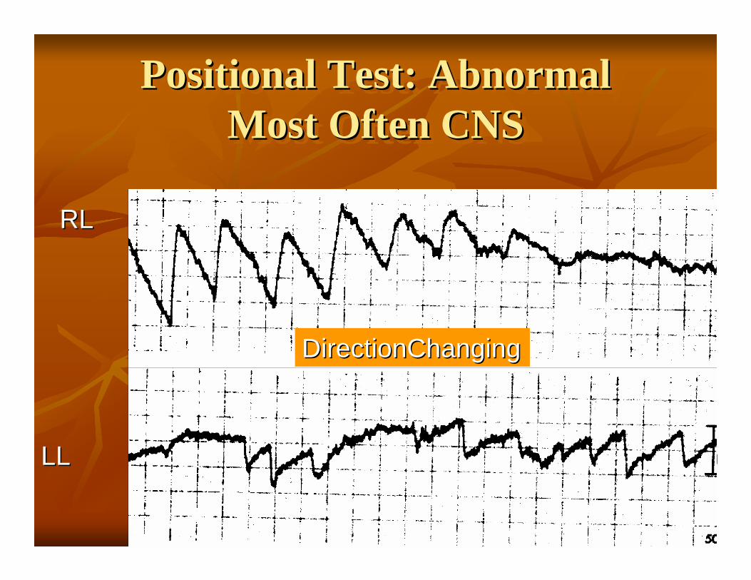
Positional Test: AbnormalMost Often CNS
Positional Test: AbnormalPositional Test: AbnormalMost Often CNSMost Often CNS
DirectionChangingDirectionChanging
RLRL
LLLL

Caloric stimulation Caloric stimulation
The most informative ENG subtestThe most informative ENG subtestwater, air, and closedwater, air, and closed--loop cuff loop cuff Water Water caloricscalorics provide a strong stimulus provide a strong stimulus air, and closedair, and closed--loop cuff used with PET or loop cuff used with PET or perforation of TM perforation of TM cool = 30 C warm = 44 Ccool = 30 C warm = 44 CResponse pattern follows the form of COWSResponse pattern follows the form of COWS

Caloric test disadvantageCaloric test disadvantage
Low frequency(0.003 Hz)* = PTA @125HzLow frequency(0.003 Hz)* = PTA @125HzIndirect ( depend on heat transferring capacity Indirect ( depend on heat transferring capacity of EE+ME)of EE+ME)Lateral SCC Lateral SCC LOCLOC
*0.01-10.0 Hz Vestibular system

Caloric stimulationCaloric stimulationhead at an angle of 30head at an angle of 30°°LSCC in the vertical planeLSCC in the vertical planespontaneous spontaneous nystagmusnystagmus is evaluated 1is evaluated 1stst
Bilateral weakness Bilateral weakness Average responses of <60/sAverage responses of <60/sbilateral peripheral or centralbilateral peripheral or centraldrug effects should be excludeddrug effects should be excluded
Fixation after each testFixation after each testR/O CNS No reduce R/O CNS No reduce nystagmusnystagmusFast recovery . Fast recovery .
no responseno response Ice water for residualIce water for residual

Caloric Test: NormalCaloric Test: NormalCaloric Test: NormalFixation SuppressionFixation Suppression
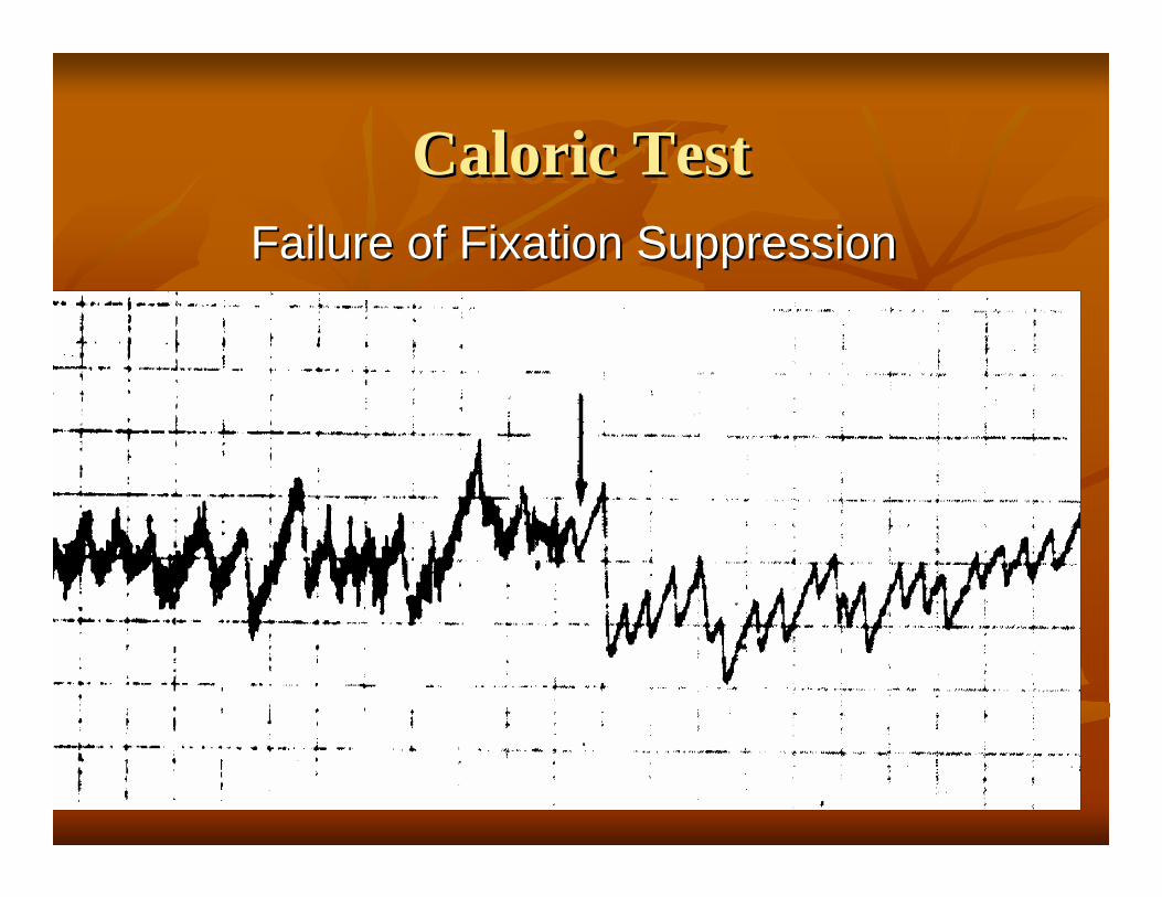
Caloric TestCaloric TestCaloric TestFailure of Fixation SuppressionFailure of Fixation Suppression
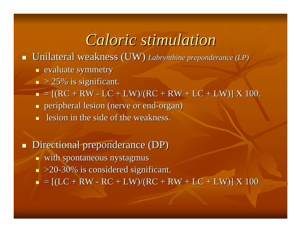
Caloric stimulationCaloric stimulationUnilateral weakness (UW) Unilateral weakness (UW) LabrynthineLabrynthine preponderance (LP) preponderance (LP)
evaluate symmetryevaluate symmetry> 25% is significant. > 25% is significant. = [(RC + RW = [(RC + RW -- LC + LW)/(RC + RW + LC + LW)] X 100.LC + LW)/(RC + RW + LC + LW)] X 100.peripheral lesion (nerve or endperipheral lesion (nerve or end--organ) organ) lesion in the side of the weakness. lesion in the side of the weakness.
Directional preponderance (DP) Directional preponderance (DP) with spontaneous with spontaneous nystagmusnystagmus>20>20--30% is considered significant. 30% is considered significant. = [(LC + RW = [(LC + RW -- RC + LW)/(RC + RW + LC + LW)] X 100 RC + LW)/(RC + RW + LC + LW)] X 100

Which direction? Which direction?