elena vecino and sansar c. sharma - cdn.intechweb.orgcdn.intechweb.org/pdfs/23828.pdf · elena...
TRANSCRIPT

15
Glaucoma Animal Models
Elena Vecino1 and Sansar C. Sharma2 1Departament of Cell Biology and Histology, Faculty of Medicine, Science and Technology
University of the Basque Country UPV/EHU, Leioa, Vizcaya, 2Departament of Ophthalmology, NY Medical College, Valhalla,
1Spain 2USA
1. Introduction
1.1 Glaucoma is a progressive neuropathy
Glaucoma is an optic neuropathy that is considered to be the second leading cause of blindness worldwide. This disease is characterized by selective death of retinal ganglion cells (RGC) and a progressive loss of vision. Elevation of intraocular pressure (IOP) is a critical risk factor for glaucoma progression, and its lowering has become a major focus of intervention. However, many patients continue to lose vision despite IOP management. Additionally, some patients develop what is known as normal tension glaucoma, which is not associated with increased IOP. Taken together, the continued deterioration of some patients’ vision despite IOP management as well as the incidence of normal tension glaucoma illustrate that several pressure-independent mechanisms are responsible for the development and progression of glaucomatous neuropathy (Pinar-Sueiro & Vecino, 2010). Glaucoma is difficult to study in humans. The damage present at the time of diagnosis precludes the study of disease development from onset. Additionally, obtaining retinas at equivalent pathologic states is rare, confounding comparisons and limiting conclusions. For these reasons, the development of animal models has been necessary for the study of the pathophysiology of glaucoma. Animal studies have articulated the mechanisms of the formation and evacuation of aqueous humour as well as the maintenance of intra-ocular pressure, thereby informing glaucoma etiology and therapeutic development. In 1901, Lauber determined that blood from the anterior ciliary veins of dogs contained fewer erythrocytes per unit volume than did blood from their paws. As early as 1903, Leber noted a significant histological connection between the Schlemm´s canal and the episcleral veins. Numerous investigators then demonstrated that various dyes and tracer substances injected into the anterior chamber later appeared in the anterior ciliary veins. It was not until 1942, when Ascher first described the appearance of aqueous veins and their connections to the episcleral venus plexus, and a connection between Schlemm’s canal and eipiscleral veins was demonstrated under normal conditions in vivo. Ascher compressed the episcleral veins using a fine glass rod, thereby inhibiting aqueous flow into them. This flow resumed when the rod was removed. He described two types of stratification: vessels with a superior layer of aqueous
www.intechopen.com

Glaucoma - Basic and Clinical Concepts
320
flow and an inferior layer of blood flow and vessels with a center of aqueous flow surrounded by layers of blood above and below. The detection of aqueous veins confirmed a continuous evacuation of aqueous from the eye and indirectly confirmed local formation of aqueous for homeostasis (Ascher, 1942). Through a diversity of drainage angles and functional structures across species, comparative animal studies have broadened the understanding of glaucoma. Due to their contribution to the understanding of hypertension and spontaneous or induced glaucoma, animal models have also facilitated the development of therapeutic strategies, which could not have been developed otherwise.
2. Natural-occurring glaucoma models
A variety of natural-occurring glaucoma models have been described in different animal species. Kolker et al. (1963) described a group of albino New Zealand rabbits that exhibited spontaneous alterations in trabecular mesh development. These rabbits presented a reduction in the number of lamelles, increased inter-cellular spaces between lamelles, vacuolation of the endothelial cells, and fragmentation of the basal cell. The description of these alterations gave ground to the hypothesis that a reduction in the structural support of the trabeculae could be the cause of elevated IOP. In addition, high fibrin levels detected in the aqueous humor suggested that the obstruction of aqueous evacuation associated with elevated IOP could be a result of fibrin accumulation. However, the rabbit is an inadequate animal model for studying alterations in the retina or its vascularization in glaucoma due to the absence of a lamina cribosa, the partial myelinization of the optic axons within the retina, and the existence of a prominent vasculous sac. Later observations by veterinary ophthalmologists led to the development of a dog model of closed angle glaucoma in Beagles, Cockers, and Basset hounds. Cockers develop glaucoma at an early age, whereas Beagles and Bassets begin to develop the disease between 6 and 12 months of age (Gelatt et al., 1977). Beagles expressing autosomal recessive phenotype present a pre-glaucoma stage characterized by increased IOP and an open angle. The angle begins to close within 2-3 years as the glaucoma develops. Chronic pressures of 30-40mmHg with transient peaks of up to 60 - 80 mmHg can induce excavation of the optic nerve head. In these animals, glaucoma can be treated pharmacologically with drugs utilized in humans including pilocarpine, epinephrine, acetazolamide, and dichlorphenamide. In 1974, Gaasterland et al. developed the first laser model of glaucoma in non-human primates, predating the description of spontaneous glaucoma in dogs. In 1993, a group of Macque monkeys in quay Santiago was characterized by a maternal inheritance pattern associated with a 40% prevalence of increased IOP. Affected animals exhibited a loss of retinal ganglion cells, excavation of the optic nerve, and electrophysiological evidence of damages in the retina peripheral field (Dawson et al., 1993). Similar to humans, the disadvantage of a naturally occurring glaucoma model in non-human primates is the difficulty in controlling the onset of the disease, thus, obtaining a homogenous experimental group to observe cellular and molecular mechanisms or test possible treatments.
3. Induced glaucoma models
To create the proper conditions for controlled experiments, induced glaucoma models have been developed over the decades. These models have provided the ability to examine both the onset and the pathological progression in a controlled, reproducible manner.
www.intechopen.com

Glaucoma Animal Models
321
3.1 Non-human primates
The earliest models of induced glaucoma were developed in non-human primates. The idea to induce elevated IOP via intraocular injections of the proteolytic enzyme alpha chymotrypsin was initially developed following cataract surgery in humans (Kalvin et al., 1966). Alpha chymotrypsin, however, produced highly variable IOP responses depending on the doses and region of the eye into which it was applied. These initial experiments suggested that the substrate underlying IOP elevation in this primate model primarily resided in the posterior chamber, and that the drug has direct, dose-dependent degenerative effects on the neural retina and vasculature. Observed atrophy of the ciliary body associated with the corneal lesions and dislocation of the lens in the presence of alpha chymotrypsin suggested that the most likely cause of elevated IOP in this primate model is blockage of the anterior chamber drainage channels by drug-induced lysates of the ciliary body (Lessell & Kuwabara, 1969). In an attempt to overcome the difficulties associated with the alpha chymotrypsin model and to develop a primate model more analagous to human primary open glaucoma, Gaasterland & Kupfer (1974) developed laser induced scar formation of the trabecular meshwork (TM) using a gonio lens and a slit lamp equipped with an Argon laser. This model became the gold standard for laser-induced glaucoma in non-human primates. Quigley and Hohman (1983) investigated different combinations of laser treatment duration, power, and number of application spots. Recently, the use of laser to generate pressure-induced experimental glaucoma in non-human primates was implemented with a high-power diode laser (Wang et al., 1998). The use of the laser technique requires access to skilled personnel with highly specialized ophthalmic equipment. Following a trabeculoplasty with Argon laser in humans and non-human primates, there is a temporary increase in IOP, which seems to be caused by the formation of fibrin meshes obstructing the spaces of the trabecula. In non-human primates, there is an additional fixed midriasis probably due to the damage suffered by the ciliary nerves. In some non-human primate cases, large fluctuations in IOP mandate repeated laser sessions to sustain high pressure, causing severe inflammation in the ocular globe and trabecular alterations that preclude pharmacological studies. In spite of these difficulties, non-human primates have been broadly used for improving clinical indicators of initial optic nerve damage in glaucoma. To circumvent the negative effects of the laser techniques, other methods were explored to elevate IOP. Quigley and Addicks (1980) injected autologous fixed red blood cells or ghost blood cells into the anterior chamber of the monkeys, but this method did not allow the visualization of the ocular fundus. The injection of latex microspheres into the anterior chamber of the rhesus monkey eye introduced a new, inexpensive technique. The microspheres do not induce ocular inflammation and do not compromise visibility of the optic disc necessary for clinical assessment of disease onset and progression (Weber & Zelenak, 2001). Our group obtained reliable results by modifying this method by injecting hydroxyproopylmethylcellulose added to the microspheres in rats and in pigs (Urcola et al., 2006; Ruiz-Ederra et al., 2005b). Non-human primate models of induced glaucoma are useful for understanding human glaucoma. These studies can evaluate RGC density from standard clinical perimetry. Examination of the retina, trabecular meshwork, lamina cribosa, and optic nerve head has been well reviewed, as has the improvement of non-invasive assessments of glaucoma onset and progression through in vivo measurements of neural structure and function (Weber & Viswanathan, 2008).
www.intechopen.com

Glaucoma - Basic and Clinical Concepts
322
3.2 Pigs and minipigs
Though non-human primates make excellent animal models for studying human disease, ethical and economical factors negatively affect their availability. The pig model is more accessible than non-human primates in addition to being phylogenetically close to humans. The pig eye/retina shares many similarities with that of the human (Ruiz-Ederra et al., 2005a; Ruiz-Ederra et al., 2005a). The retina is more similar to the human retina than that of other larger mammals such as the dog, goat, cow, or ox (Prince et al., 1960). In minipigs, the central venous ring is formed by various vessels and occupies the center of the optic disc, making visualization of the lamina cribosa more difficult than in humans (Galdos et al., 2011a). The pig has recently been used to genetically reproduce a retinitis pigmentosa condition similar to one found in human (Li et al., 1998). The diagnostic tools, such as optical coherence tomography, corneal topography imaging or multi-focal electroretinography can be applied to the pig, supporting its use as an excellent model for diseases of the eye (Vecino et al., 2011). Image analysis of the porcine retina has been characterized well (Lalonde et al., 2006) and is useful in developing human transplantation experiments (Klassen et al., 2008).
Fig. 1. Fundoscopy (left) and angiography (right) of a minipig eye. Note the lamina cribosa at the end of the optic nerve in the fundoscopic picture. In the angiography the veins appear dark and emerge from the optic nerve
The pig retina has been extensively studied. We have characterized three classes of RGCs (small, medium, and large) based on soma size (Garcia et al., 2002), and, in a detailed study of pig RGC topography, revealed that the distribution of the three classes is very similar in the porcine and human retina. This similarity may help elucidate the mechanisms implicated in the selective death of large RGCs generally accepted to be differentially vulnerable in human and experimental glaucoma (Glovinsky et al., 1991; Ruiz-Ederra et al., 2005a). We have found that porcine Müller cells in vitro secrete neuroprotective factors that facilitate the survival and axonal growth of large RGCs (Garcia et al., 2002). We have further described expression patterns of neurotrophins and their receptors in different RGC types in vivo and in vitro, suggesting that the expression of most of the molecules studied was preserved after either dissociation or regeneration in vitro (Garcia et al., 2003; Vecino, 2008b; Vecino, 2008a). We have described the similarity of the two classes of astrocytes in the adult human and pig retina in terms of their expression of the high affinity neuronal growth factor receptor TrkA
www.intechopen.com

Glaucoma Animal Models
323
(Ruiz-Ederra et al., 2003). The comparative description of the three neurofilament subunits were considered when describing the similarities between RGCs from human and pig (Ruiz-Ederra et al., 2004). Considering the similarities between the porcine and human retinas, we used episcleral vein cauterization described by Sharma’s group (Shareef et al., 1995) to induce elevation of IOP in the pig and minipig. Similarly, we used the injection of latex fluorospheres into the anterior chamber (Ruiz-Ederra et al., 2005b) to induce glaucoma in pigs and minipigs (Fig. 2). The use of minipigs facilitated the experiments because these animals are easy to handle and grow slowly (www.minipig.com). Glaucoma was identified by the presence of elevated IOP, altered eye fundus morphology, and RGC loss (Ruiz-Ederra et al., 2005a; Galdos et al., 2011a; Galdos et al., 2011b). Animals were kept for 21 weeks with up to a 1.4-fold increase of IOP in the operated eye versus the control fellow eye. Several factors that implicated in glaucoma etiology and development were analyzed in the pig and minipig models.
Fig. 2. Cryostat section of the minipig eye angle after Fluorospheres injection. Note the
location of the latex fluorospheres in the trabecular meshwork and the aqueous humor
evacuation channels. Upper left: light microscope picture. Upper right: fluorescent
microscopic picture. Bottom left: higher magnification of the trabecular meshwork with
fluorospheres. bottom right: scanning electronic microscopic picture of the injected 15
micrometres latex fluorospheres
www.intechopen.com

Glaucoma - Basic and Clinical Concepts
324
Angiographic and fundoscopic changes
The minipig model of chronic, open-angle glaucoma induced by episcleral venous cauterization presented no angiopathic changes after elevation of the IOP. Therefore, neither surgery with episcleral vein cauterization nor elevated IOP induced changes in the chorio-retinal circulation. However, displacement of the vessels in the optic disc reflected significant differences in the neuroretinal ring that were also observed in humans with primary open angle glaucoma (POAG). In minipigs with experimental glaucoma, some arterioles were nasally displaced and incurved medially once outside of the optic disc. In minipigs, the central venous ring is formed by various vessels and occupies the center of the optic disc, making visualization of the lamina cribosa more difficult than in humans (see Fig. 1, Galdos et al., 2011a).
Molecular and ultrastructural changes of the trabecular meshworks
Increased resistance to aqueous humour outflow is generally accepted as a predominant risk factor for increased IOP in glaucomatous eyes. To date, neither the associated structures nor the pathophysiology of this increased resistance is well understood. In the pig model of glaucoma, we have reported the expression of the endothelial leukocyte adhesion molecule 1 (ELAM-1), which was identified as the first molecular marker for glaucomatous trabecular meshwork cells in humans (Wang et al., 2001). ELAM-1 was expressed by trabecular meshwork cells in eyes with various types of glaucoma, but not in non-glaucomatous controls. ELAM-1, also known as E-Selectin, is a 115 kDa cytokine endothelial cell surface glycoprotein that mediates the adhesion of neutrophils, monocytes, eosinophils, NK cells, and a subset of T cells to activated endothelium (Bevilacqua et al., 1989). Based on our previous findings in the porcine trabecular meshwork, we propose that induced glaucoma begins when cauterization of three of the four episcleral veins produces a decrease in outflow of aqueous humor, leading to altered drainage in the front of the eye and subsequent elevation of IOP. The elevation of IOP induces ELAM-1 expression at the anterior chamber as a response to cellular stress (Suarez & Vecino, 2006). The finding that ELAM-1 is upregulated in the outflow pathway in pig and human experimental glaucoma will undoubtedly promote further studies about its treatment. The induction of glaucoma by increasing the post-trabecular resistance to aqueous outflow allows the observation of ultrastructural changes in the trabecular meshwork secondary to increased IOP without chemically or mechanically changing the TM cells. The Gotِtingen minipig has a system with multiple vessels of the angular aqueous plexus contrasting with the unique Schelm’s Canal of primates. In experimental eyes, ultrastructural differences were observed in the sub-endothelium of the inner wall of the juxta canalicular vessels, corresponding to the cribriform region in humans. Secondary ultrastructural changes in the optic disc in animals with induced elevation of IOP were noted. In the animals that presented statistically significant chronic increase of the IOP and were then subject to ultrastructural analysis, changes were observed in vessels at the subendothelial region. The differences detected included: greater amounts of fibrillar material and elastic-like (EL) fibers, a smaller number of empty spaces (ES) and larger cisternae of the endoplasmic reticulum (ER) filled with electron-lucent material. Both in control and glaucomatous eyes, pores were observed in both the deepest and the most superficial vessels, although, pores were only present in the inner wall of the latter. Similarly, giant vacuoles were found in both the deepest the most superficial vessels, and could be seen in both the inner and outer wall of the vessels of the angular aqueous
www.intechopen.com

Glaucoma Animal Models
325
Fig. 3. Electron Microscopic pictures of different channals of the minipig trabeculum. Note transport of vesicles (arrows) and elastic like fibers (EL) in the upper picture. In the bottom picture the large vesicles (VG) are characteristic of the aqueous transport in the endothelial cells as well as the colagenous (col) fibres (v)vesicles, ev(external vessel)
www.intechopen.com
Glaucoma - Basic and Clinical Concepts
326
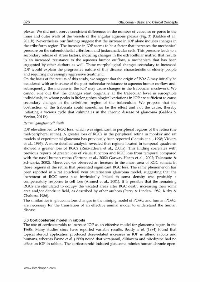
plexus. We did not observe consistent differences in the number of vacuoles or pores in the
inner and outer walls of the vessels of the angular aqueous plexus (Fig. 3) (Galdos et al.,
2011b). Nevertheless, our findings suggest that the increase in IOP alone induces changes in
the cribriform region. The increase in IOP seems to be a factor that increases the mechanical
pressure on the subendothelial cribriform and juxtacanalicular cells. This pressure leads to a
secondary release of stress factors, inducing changes in the extracellular matrix, that results
in an increased resistance to the aqueous humor outflow, a mechanism that has been
suggested by other authors as well. These morphological changes secondary to increased
IOP would explain the progressive nature of this disease, characteristic of elderly people
and requiring increasingly aggressive treatment.
On the basis of the results of this study, we suggest that the origin of POAG may initially be
associated with an increase of the post-trabecular resistance to aqueous humor outflow and,
subsequently, the increase in the IOP may cause changes in the trabecular meshwork. We
cannot rule out that the changes start originally at the trabecular level in susceptible
individuals, in whom peaks in lifelong physiological variations in IOP are sufficient to cause
secondary changes in the cribriform region of the trabeculum. We propose that the
obstruction of the trabecula could sometimes be the effect and not the cause, thereby
initiating a vicious cycle that culminates in the chronic disease of glaucoma (Galdos &
Vecino, 2011b).
Retinal ganglion cell death
IOP elevation led to RGC loss, which was significant in peripheral regions of the retina (the
mid-peripheral retina). A greater loss of RGCs in the peripheral retina in monkey and rat
models of experimental glaucoma has previously been reported (Laquis et al., 1998; Vickers
et al., 1995). A more detailed analysis revealed that regions located in temporal quadrants
showed a greater loss of RGCs (Ruiz-Ederra et al., 2005a). This finding correlates with
previous reports of greater loss of visual function and RGC loss from temporal compared
with the nasal human retina (Fortune et al., 2002; Garway-Heath et al., 2002; Takamoto &
Schwartz, 2002). Moreover, we observed an increase in the mean area of RGC somata in
those regions of the retina that presented significant RGC loss. The same phenomenon has
been reported in a rat episcleral vein cauterisation glaucoma model, suggesting that the
increment of RGC soma size intrinsically linked to soma density was probably a
compensatory response to cell loss (Ahmed et al., 2001). It is possible that the remaining
RGCs are stimulated to occupy the vacated areas after RGC death, increasing their soma
area and/or dendritic field, as described by other authors (Perry & Linden, 1982; Kirby &
Chalupa, 1986).
The similarities in glaucomatous changes in the minipig model of POAG and human POAG
are necessary for the translation of an effective animal model to understand the human
disease.
3.3 Corticosteroid model in rabbits
The use of corticosteroids to increase IOP as an effective model for glaucoma began in the
1960s. Many studies since have reported variable results. Beatty et al. (1984) found that
topical steroid application produced dose-related increases in IOP in albino rabbits and
humans, whereas Payne et al. (1990) noted that verapamil, diltiazem and nifedipine had no
effect on IOP in rabbits. The corticosteroid-induced glaucoma mimics human chronic open-
www.intechopen.com

Glaucoma Animal Models
327
angle glaucoma. In contrast to most of the induced experimental models for glaucoma,
corticosteroid glaucoma is also observed in ophthalmological practice after topical,
periocular or systemic administration of corticosteroids, strengthening the parallel between
the animal and human disease. It is important to remember that steroids can cause
undesired secondary effects including cataracts and the accumulation of cellular debris at
the trabecular meshwork. The increase in IOP is variable between species. Rabbits have been
a common animal used by the pharmaceutical companies to test drugs; however, IOP
measurements are difficult to standardize because the rabbit eye dries variably depending
on the stress of the animal. In addition, the rabbit retina is partially myelinated by
oligodendrocytes not present in human or other mammalian retina. Taken together, these
reasons make the steroids treatment, as well as the rabbit, a sub-optimal model for studying
glaucoma.
3.4 Rats and mice
Much of the progress in the study of glaucoma has been driven by the development of rat models. These animals offer advantages in economics and husbandry in addition to presenting fewer ethical restrictions. In 1995, the group of Dr. Sharma developed the cauterization of episcleral veins in rats as a model for obtaining chronically high intraocular pressures (Shareef et al., 1995). This model protects the trabecula structure, and it does not affect the ciliary nerve as the laser model does. In addition, elevated IOP can be maintained for up to 6 months (25% of a rat’s lifetime). This animal model has allowed pharmacological trials of pressure-reducing and neuron-protecting drugs of different combinations and concentrations before subsequent studies in larger animals and clinical trials in humans. In this context, rats are making an enormous contribution to anti-glaucoma research. Also in 1995, the group of Dr. Sharma described, for the first time, the death of RGCs following a pattern or program known as apoptosis (Garcia-Valenzuela et al., 1995). This pattern involves cleavage enzymes known as caspases, which can conversely confer neuroprotection if inhibited at a specific time of the damage-inducing process. The main advantage of the rat as an animal model is that it can be generated in high numbers, so that many pharmacological tests can be performed simultaneously, but the small size of the rat eye limits its use in some areas of ophthalmology. The use of the episcleral vein cauterization model of glaucoma has been the most commonly used method to induce glaucoma since its inception (Shareef et al., 1995). The method is less invasive than laser photocoagulation and induces no complications in the anterior chamber. Its efficacy and accessibility led to an explosion of research, and the majority of the molecular and functional studies in the experimental glaucoma field have used this method. Other rat models for induced glaucoma have emerged. A few years after the episcleral vein
cauterization was published, a hypertonic saline solution injection to the episcleral veins
was developed as a variant (Morrison et al., 1997). The objective of this method was also to
increase the IOP by reducing the drainage of the aqueous humour. One disadvantage of this
method is that sequential hypertonic saline injections are needed to maintain chronic,
elevated IOP (Ruduzinski & Saragovi H.U., 2005).
Laser energy has been employed in rats as a tool to perform burns directed at the trabecular
meshwork alone (Ueda et al., 1998) and episcleral veins (Levkovitch-Verbin et al., 2002).
To compare the effects of IOP elevation on ganglion cell size and death, we used three
experimental glaucoma models in rats: (i) injections of latex microspheres into the anterior
www.intechopen.com

Glaucoma - Basic and Clinical Concepts
328
chamber of the eye (ii) injections of microspheres and hydroxypropylmethylcellulose into
the anterior chamber, and (iii) cauterization of three episcleral veins. A significant increase
in IOP was found following each of the three methods (Urcola et al., 2006). Thirteen to 30
weeks later, RGCs were retrogradely labelled with fluorogold. Cell death was evident in the
glaucomatous eyes when compared with controls, but no statistically significant effect was
observed on the extent of cell death. The present results indicate that in animal groups
subjected to the injection of microspheres alone and microsphres together with
hydroxypropylmethylcellulose, nine and six injections, respectively, are necessary to
achieve sustained, elevated IOP. The number of microsphere injections necessary to induce
sustained, elevated IOP in rat is similar to that which has been reported for the monkey
(Weber & Zelenak, 2001). Regarding episcleral vein cauterization, we observed IOP
elevation more constant for at least 24 weeks as compared with the other two experimental
glaucoma methods tested (Urcola et al., 2006). Similar results were observed when the three
methods were compared in pigs (Ruiz-Ederra et al., 2005b).
Recently, it has been reported the induction of elevation of IOP in rats by using injection of magnetic microspheres to induce the same effect of the latex micropspheres but in this case the microspheres could be directed by an handheld magnet (Samsel et at., 2011). Due to the small size of the mouse eye, injection of the latex microspheres (Fluo-Spheres, Molecular Probes) into the anterior chamber of mice has become a popular method to induce increase IOP. Authors that now use the spheres to increase IOP in rats and mice (Sappington et al., 2010) use the term “microbeads occlusion model” to refer to the same model that Urcola et al. published in 2006 in rats and Ruiz-Ederra et al., 2005b published in pigs. To determine the effects of elevated IOP following episcleral vein cauterization, a detailed analysis of 15 different molecular markers was conducted for the different retinal cell types at different time points. The changes observed in the distribution of the immunoreactivity in the hypertensive rat retina were more severe in the inner retina than in the outer retina, especially in the AII amacrine cells. To our knowledge, we described for the first time that the rod bipolar cells pathway was also damaged in the hypertensive eye, as evidenced by changes in anti-PKC-α antibody. Changes in bipolar cells are not surprising when one considers that the RGCs lose their presynaptic connections. It is possible that the changes noticed may represent a plasticity mechanism in neuronal circuitry (Hernandez et al., 2009).
3.5 Transgenic mice
The appearance of the DBA/2J mouse, which develops a progressive increase in IOP leading
to the death of retinal ganglion cells (John et al., 1998) resulted in a large amount of studies
to establish the existence of homologies related to glaucoma in humans. The increase in IOP
in these animals appears at 8 months of age, and the pressure remains chronically high until
death. The limiting factors of these studies include the small globe and the absence of lamina
cribosa. This animal model of spontaneous, chronic, high IOP is suitable for studying the
causes of this pathology. However, with the exception of the DBA/2J mice, animals with
glaucoma are difficult to obtain, especially at similar stages of pathology. More recently,
other transgenic mouse models have emerged, including one with a targeted mutation in
the gene for the alpha-1 subunit of collagen type 1, which demonstrates a gradual elevation
of IOP and progressive optic nerve axon loss (Mabuchi et al., 2004). Another mouse,
www.intechopen.com

Glaucoma Animal Models
329
deficient in the glutamate transporters Glast or Eaac1, demonstrates retinal ganglion cell
death and optic nerve degeneration without elevated IOP (Harada et al., 2007). The
transgenic mouse expressing a mutant form of human myocilin protein has also been
characterized (Senatorov et al., 2006; Zhou et al., 2008). Finally, mutations affecting a serine
protease (PRSS56) cause a mouse phenotype resembling closed-angle glaucoma (Nair et al.,
2011).
Just as progress in glaucoma diagnosis has been linked to developments in technology,
progress in glaucoma treatment has been linked to the development of effective animal
models. By using these animal models, we hope to continue the processes of glaucoma
prevention as well as treatment.
Mechanism Procedure Species Reference
Ghost red blood Monkey (Quigley & Addicks, 1980)
Human (in vitro) (Benson et al., 1983) Viscolastic
Rabbit (Torngren et al., 2000)
Monkey (Weber & Zelenak, 2001)
Pig/Minipig (Ruiz-Ederra et al., 2005 a,b) Microspheres (beans)
Rat (Urcola et al., 2006)
Pig/Minipig (Ruiz-Ederra et al., 2005 a,b)
Rat (Urcola et al., 2006)
Pre-trabecular
Microspheres + viscolastic
Mouse (Sappington et al., 2010)
Steroids Rabbit (Bonomi et al., 1978)
Monkey (Pederson & Gaasterland, 1984) Trabecular Laser photocoagulation
Rat (Ueda et al., 1998)
Rat (Shareef et al., 1995) Episcleral veins cauterization
Pig/Minipig (Ruiz-Ederra et al., 2005 a,b) Post-trabecular
Saline injection Rat (Morrison et al., 1995)
DBA mouse (John et al., 1998) Genetic
Myocilin mouse (Senatorov et al., 2006)
Table 1.
4. Acknowledgments
Basque Government Grant Grupos Consolidados (IT43710), Red Patología Ocular RETICS
(RD07/0062/2004), ONCE (Organization for Spanish blind persons).
www.intechopen.com

Glaucoma - Basic and Clinical Concepts
330
5. References
Ahmed, F. A., Chaudhary, P., & Sharma, S. C. (2001). Effects of increased intraocular
pressure on rat retinal ganglion cells. Int. J. Dev. Neurosci., 19, 209-218.
Ascher, K. W. (1942). The aqueous veins. Physiological importance of the visible elimination
of intraouclar fluid. 25[American Journal of Ophthalmology], 1174-1209.
Beatty, J. F., Krupin, T., Nichols, P. F., & BECKER, B. (1984). Elevation of intraocular
pressure by calcium channel blockers. Arch. Ophthalmol., 102, 1072-1076.
Benson, F. G., Patterson, M. M., & Einstein D.L. (1983). Obstruction of aqueous outflow by
sodium hyaluronate in enucleated human eyes. Am. J. Ophthalmol., 95, 668-672.
Bevilacqua, M. P., Stengelin, S., Gimbrone, M. A., Jr., & Seed, B. (1989). Endothelial
leukocyte adhesion molecule 1: an inducible receptor for neutrophils related to
complement regulatory proteins and lectins. Science, 243, 1160-1165.
Bonomi, L., Perfetti, S., Noya, E., Bellucci, R., & Tomazzoli, L. (1978). Experimental
corticosteroid ocular hypertension in the rabbit. Albrecht. Von. Graefes Arch. Klin.
Exp. Ophthalmol., 209, 73-82.
Dawson, W. W., Brooks, D. E., Hope, G. M., Samuelson, D. A., Sherwood, M. B., Engel, H.
M. et al. (1993). Primary open angle glaucomas in the rhesus monkey.
Br.J.Ophthalmol., 77, 302-310.
Fortune, B., Cull, G., Wang, L., Van Buskirk, E. M., & Cioffi, G. A. (2002). Factors affecting
the use of multifocal electroretinography to monitor function in a primate model of
glaucoma. Doc. Ophthalmol., 105, 151-178.
Gaasterland, D. & Kupfer, C. (1974). Experimental glaucoma in the rhesus monkey. Invest
Ophthalmol., 13, 455-457.
Galdos, M., Bayon, A., Mico, C., & Vecino, E. (2010). Fundoscopic and ultrastructural
evaluation in experimental glaucoma model in minipigs. ARVO e-abstract 6392.
Galdos, M., Bayon, A., Rodriguez, F. D., Mico, C., Sharma, S. C., & Vecino, E. (2011a).
Morphology of retinal vessels in the optic disc in a Gtِtingen minipig experimental
glaucoma model.Veterinary Journal, In press.
Galdos, M. & Vecino, E. (2011b). Ultraestructural changes in the travecular meshwork and
IOP elevation. Which come first, the chicken or the egg? Archivos de la Sociedad
Espaoٌla de Oftalmologia, In press.
Garcia, M., Forster, V., Hicks, D., & Vecino, E. (2002). Effects of muller glia on cell survival
and neuritogenesis in adult porcine retina in vitro. Investigative Ophthalmology and
Visual Science, 43, 3735-3743.
Garcia, M., Forster, V., Hicks, D., & Vecino, E. (2003). In vivo expression of neurotrophins
and neurotrophin receptors is conserved in adult porcine retina in vitro.
Investigative Ophthalmology and Visual Science, 44, 4532-4541.
Garcia-Valenzuela, E., Shareef, S., Walsh, J., & Sharma, S. C. (1995). Programmed cell
death of retinal ganglion cells during experimental glaucoma. Exp.Eye Res., 61, 33-
44.
Garway-Heath, D. F., Holder, G. E., Fitzke, F. W., & Hitchings, R. A. (2002). Relationship
between electrophysiological, psychophysical, and anatomical measurements in
glaucoma. Invest Ophthalmol.Vis.Sci., 43, 2213-2220.
www.intechopen.com

Glaucoma Animal Models
331
Gelatt, K. N., Peiffer, R. L., Jr., Gwin, R. M., Gum, G. G., & Williams, L. W. (1977). Clinical
manifestations of inherited glaucoma in the beagle. Invest Ophthalmol.Vis.Sci., 16,
1135-1142.
Glovinsky, Y., Quigley, H. A., & Dunkelberger, G. R. (1991). Retinal ganglion cell loss is size
dependent in experimental glaucoma. Invest Ophthalmol.Vis.Sci., 32, 484-491.
Harada, T., Harada, C., Nakamura, K., Quah, H. M., Okumura, A., Namekata, K. et al.
(2007). The potential role of glutamate transporters in the pathogenesis of normal
tension glaucoma. J.Clin.Invest, 117, 1763-1770.
Hernandez, M., Rodriguez, F. D., Sharma, S. C., & Vecino, E. (2009). Immunohistochemical
changes in rat retinas at various time periods of elevated intraocular pressure.
Molecular Vision, 15, 2696-2709.
John, S. W., Smith, R. S., Savinova, O. V., Hawes, N. L., Chang, B., Turnbull, D. et al. (1998).
Essential iris atrophy, pigment dispersion, and glaucoma in DBA/2J mice. Invest
Ophthalmol.Vis.Sci., 39, 951-962.
Kalvin, N. H., Hamasaki, D. I., & Gass, J. D. (1966). Experimental glaucoma in monkeys. I.
Relationship between intraocular pressure and cupping of the optic disc and
cavernous atrophy of the optic nerve. Arch.Ophthalmol., 76, 82-93.
Kirby, M. A. & Chalupa, L. M. (1986). Retinal crowding alters the morphology of alpha
ganglion cells. J.Comp Neurol., 251, 532-541.
Klassen, H., Warfvinge, K., Schwartz, P. H., Kiilgaard, J. F., Shamie, N., Jiang, C. et al. (2008).
Isolation of progenitor cells from GFP-transgenic pigs and transplantation to the
retina of allorecipients. Cloning Stem Cells, 10, 391-402.
Kolker, A. E., Moses, R. A., Constant, M. A., & Becker, B. (1963). The Development of
Glaucoma in Rabbits. Invest Ophthalmol., 2, 316-321.
Lalonde, M. R., Chauhan, B. C., & Tremblay, F. (2006). Retinal ganglion cell activity from the
multifocal electroretinogram in pig: optic nerve section, anaesthesia and intravitreal
tetrodotoxin. J.Physiol, 570, 325-338.
Laquis, S., Chaudhary, P., & Sharma, S. C. (1998). The patterns of retinal ganglion cell death
in hypertensive eyes. Brain Res., 784, 100-104.
Lessell, S. & Kuwabara, T. (1969). Experimental alpha-chymotrypsin glaucoma.
Arch.Ophthalmol., 81, 853-864.
Levkovitch-Verbin, H., Martin, K. R., Quigley, H. A., Baumrind, L. A., Pease, M. E., &
Valenta, D. (2002). Measurement of amino acid levels in the vitreous humor of rats
after chronic intraocular pressure elevation or optic nerve transection. J.Glaucoma.,
11, 396-405.
Li, Z. Y., Wong, F., Chang, J. H., Possin, D. E., Hao, Y., Petters, R. M. et al. (1998). Rhodopsin
transgenic pigs as a model for human retinitis pigmentosa. Invest
Ophthalmol.Vis.Sci., 39, 808-819.
Mabuchi, F., Lindsey, J. D., Aihara, M., Mackey, M. R., & Weinreb, R. N. (2004). Optic nerve
damage in mice with a targeted type I collagen mutation. Invest Ophthalmol.Vis.Sci.,
45, 1841-1845.
McMenamin, P. G. & Steptoe, R. J. (1991). Normal anatomy of the aqueous humour outflow
system in the domestic pig eye. J.Anat., 178, 65-77.
www.intechopen.com
Glaucoma - Basic and Clinical Concepts
332
Morrison, J. C., Fraunfelder, F. W., Milne, S. T., & Moore, C. G. (1995). Limbal
microvasculature of the rat eye. Invest Ophthalmol.Vis. Sci., 36, 751-756.
Morrison, J. C., Moore, C. G., Deppmeier, L. M., Gold, B. G., Meshul, C. K., & Johnson, E. C.
(1997). A rat model of chronic pressure-induced optic nerve damage. Exp.Eye Res.,
64, 85-96.
Nair, K. S., Hmani-Aifa, M., Ali, Z., Kearney, A. L., Salem, S. B., Macalinao, D. G. et al.
(2011). Alteration of the serine protease PRSS56 causes angle-closure glaucoma in
mice and posterior microphthalmia in humans and mice. Nat.Genet. 43(6), 579-
584.
Payne, L. J., Slagle, T. M., Cheeks, L. T., & Green, K. (1990). Effect of calcium channel
blockers on intraocular pressure. Ophthalmic Res., 22, 337-341.
Pederson, J. E. & Gaasterland, D. E. (1984). Laser-induced primate glaucoma. I. Progression
of cupping. Arch.Ophthalmol., 102, 1689-1692.
Perry, V. H. & Linden, R. (1982). Evidence for dendritic competition in the developing
retina. Nature, 297, 683-685.
Pinar-Sueiro, S. & Vecino, E. (2010). Potential neuroprotective strategis for glaucoma.
J.Inherit.Metab Dis., 16, 1-38.
Prince, J. H., Diesem, C. C., Eglitis, I., & Ruskell, G. L. (1960). The pig. In C.C.Thomas
(Ed.), Anatomy and Histology of the Eye and Orbit in Domestic Animals (pp. 210-230).
Illinois.
Quigley, H. A. & Addicks, E. M. (1980). Scanning electron microscopy of
trabeculectomy specimens from eyes with open-angle glaucoma. Am. J.
Ophthalmol., 90, 854-857.
Quigley, H. A. & Hohman, R. M. (1983). Laser energy levels for trabecular meshwork
damage in the primate eye. Invest Ophthalmol. Vis. Sci., 24, 1305-1307.
Ruduzinski, M. & Saragovi H.U. (2005). Glaucoma: validated and facile in vivo experimental
models of a chronic neurodegenerative disease for drug development. Current
Med.Chem, 5, 43-49.
Ruiz-Ederra, J., Garcia, M., Hernandez, M., Urcola, H., Araiz and Vecino E. (2005a). The
pig eye as a novel model of glaucoma. Exp Eye Res, 81, 561-569.
Ruiz-Ederra, J., Garcia, M., Hicks, D., & Vecino, E. (2004). Comparative study of the three
neurofilament subunits within pig and human retinal ganglion cells. Mol Vis., 10,
83-92.
Ruiz-Ederra, J., Garcia, M., Martin, F., Urcola, H., Hernandez, M., Araiz, J. et al. (2005b).
Comparison of three methods of inducing chronic elevation of intraocular pressure
in the pig (experimental glaucoma. Arch Soc.Esp Oftalmol., 80, 571-579.
Ruiz-Ederra, J., Hitchcock, P. F., & Vecino, E. (2003). Two classes of astrocytes in the adult
human and pig retina in terms of their expression of high affinity NGF receptor
(TrkA). Neurosci Lett., 337, 127-130.
Samsel, P.A., Kisiswa L, Erichsen; J.T., Cross, S.D., & Morgan J. (2011) A novel method for
the induction of experimental glaucoma using magnetic microspheres. Invest
Ophthalmol.Vis.Sci., 52, 1671-1675.
Sappington, R. M., Carlson, B. J., Crish, S. D., & Calkins, D. J. (2010). The microbead
occlusion model: a paradigm for induced ocular hypertension in rats and mice.
Invest Ophthalmol.Vis.Sci., 51, 207-216.
www.intechopen.com

Glaucoma Animal Models
333
Senatorov, V., Malyukova, I., Fariss, R., Wawrousek, E. F., Swaminathan, S., Sharan, S. K. et
al. (2006). Expression of mutated mouse myocilin induces open-angle glaucoma in
transgenic mice. J.Neurosci., 26, 11903-11914.
Shareef, S. R., Garcia-Valenzuela, E., Salierno, A., Walsh, J., & Sharma, S. C. (1995). Chronic
ocular hypertension following episcleral venous occlusion in rats. Exp.Eye Res., 61,
379-382.
Suarez, T. & Vecino, E. (2006). Expression of endothelial leukocyte adhesion molecule 1 in
the aqueous outflow pathway of porcine eyes with induced glaucoma. Mol Vis., 12,
1467-1472.
Takamoto, T. & Schwartz, B. (2002). Differences by quadrant of retinal nerve fiber layer
thickness in healthy eyes. J.Glaucoma., 11, 359-364.
Torngren, L., Lundgren, B., & Madsen, K. (2000). Intraocular pressure development in the
rabbit eye after aqueous exchange with ophthalmic viscosurgical devices. J.Cataract
Refract.Surg., 26, 1247-1252.
Ueda, J., Sawaguchi, S., Hanyu, T., Yaoeda, K., Fukuchi, T., Abe, H. et al. (1998).
Experimental glaucoma model in the rat induced by laser trabecular
photocoagulation after an intracameral injection of India ink. Jpn. J. Ophthalmol., 42,
337-344.
Urcola, J. H., Hernandez, M., & Vecino, E. (2006). Three experimental glaucoma models in
rats: comparison of the effects of intraocular pressure elevation on retinal ganglion
cell size and death. Exp Eye Res, 83, 429-437.
Vecino, E. (2008a). Neurotrophins:responsible for death and survival. In A. Mangas, R.
Coveäs & Geffard M. Brain Molecules: from vitamins to Molecules for Axon Guidance
(pp. 181-193).
Vecino, E. (2008b). Gene therapy against retinosis pigmentary. Arch Soc. Esp Oftalmol., 83,
213-214.
Vecino, E., Bayon, A., Mico, C., & Palao C. (2011). Retrobulbar Optic Nerve Section in Pig:
Optical coherence tomography an Multifocal Electroretinogram study. ARVO
e-abstract 4683 .
Vickers, J. C., Schumer, R. A., Podos, S. M., Wang, R. F., Riederer, B. M., & Morrison,
J. H. (1995). Differential vulnerability of neurochemically identified
subpopulations of retinal neurons in a monkey model of glaucoma. Brain Res.,
680, 23-35.
Wang, N., Chintala, S. K., Fini, M. E., & Schuman, J. S. (2001). Activation of a tissue-specific
stress response in the aqueous outflow pathway of the eye defines the glaucoma
disease phenotype. Nat Med, 7, 304-309.
Wang, R. F., Schumer, R. A., Serle, J. B., & Podos, S. M. (1998). A comparison of argon laser
and diode laser photocoagulation of the trabecular meshwork to produce the
glaucoma monkey model. J.Glaucoma., 7, 45-49.
Weber, A. J. & Viswanathan, S. (2008). The Primate Model of Experimental Glaucoma. In
J.Tombran-Tink, C. J. Barnstable, & M. B. Shields (Eds.), Mechanisms of the
Glaucomas (pp. 551-577). Humana Press.
Weber, A. J. & Zelenak, D. (2001). Experimental glaucoma in the primate induced by latex
microspheres. J. Neurosci. Methods, 111, 39-48.
www.intechopen.com

Glaucoma - Basic and Clinical Concepts
334
Zhou, Y., Grinchuk, O., & Tomarev, S. I. (2008). Transgenic mice expressing the Tyr437His
mutant of human myocilin protein develop glaucoma. Invest Ophthalmol. Vis. Sci.,
49, 1932-1939.
www.intechopen.com
Glaucoma - Basic and Clinical ConceptsEdited by Dr Shimon Rumelt
ISBN 978-953-307-591-4Hard cover, 590 pagesPublisher InTechPublished online 11, November, 2011Published in print edition November, 2011
InTech EuropeUniversity Campus STeP Ri Slavka Krautzeka 83/A 51000 Rijeka, Croatia Phone: +385 (51) 770 447 Fax: +385 (51) 686 166www.intechopen.com
InTech ChinaUnit 405, Office Block, Hotel Equatorial Shanghai No.65, Yan An Road (West), Shanghai, 200040, China
Phone: +86-21-62489820 Fax: +86-21-62489821
This book addresses the basic and clinical science of glaucomas, a group of diseases that affect the opticnerve and visual fields and is usually accompanied by increased intraocular pressure. The book incorporatesthe latest development as well as future perspectives in glaucoma, since it has expedited publication. It isaimed for specialists in glaucoma, researchers, general ophthalmologists and trainees to increase knowledgeand encourage further progress in understanding and managing these complicated diseases.
How to referenceIn order to correctly reference this scholarly work, feel free to copy and paste the following:
Elena Vecino and Sansar C. Sharma (2011). Glaucoma Animal Models, Glaucoma - Basic and ClinicalConcepts, Dr Shimon Rumelt (Ed.), ISBN: 978-953-307-591-4, InTech, Available from:http://www.intechopen.com/books/glaucoma-basic-and-clinical-concepts/glaucoma-animal-models