elin pettersen sørgjerd - core · en autoimmun sykdom der kroppens immunforsvar angriper og...
TRANSCRIPT
Markers of autoimmunity in Latent Autoimmune Diabetes in Adults (LADA) and non-diabetic adults: Impact in phenotype and genetic predisposition
Results from the Nord-Trøndelag health study
Thesis for the degree of Philosophiae Doctor
Levanger, April 2013
Norwegian University of Science and TechnologyFaculty of MedicineDepartment of Cancer Research and Molecular Medicine
Elin Pettersen Sørgjerd

NTNUNorwegian University of Science and Technology
Thesis for the degree of Philosophiae Doctor
Faculty of MedicineDepartment of Cancer Research and Molecular Medicine
© Elin Pettersen Sørgjerd
ISBN 978-82-471-4326-1 (printed ver.)ISBN 978-82-471-4327-8 (electronic ver.)ISSN 1503-8181
Doctoral theses at NTNU, 2013:113
Printed by NTNU-trykk
Markører for autimmunitet hos pasienter med LADA og en ikke-diabetisk voksen befolkning: påvirkning av fenotype og genetisk predisposisjon.-Resultater fra Helseundersøkelsen i Nord-Trøndelag.
Diabetes er ikke en enhetlig sykdom, men finnes i flere former. Diabetes er hovedsakelig klassifiert i to hovedgrupper; type 1 og type 2 diabetes. Type 1 diabetes er en autoimmun sykdom der kroppens immunforsvar angriper og ødelegger beta-cellene som produserer insulin. Pasienter med type 2 diabetes har fremdeles bevart en delinsulin produksjon, men en viss grad reduksjon sammen med dårlig insulin virkning på cellene i kroppen fører til økende blodsukker og diabetes. I 1986 kom den første rapporten om en gruppe pasienter som avvek fra den klassiske type 2 diabetes diagnosen. De hadde tegn til autoimmunitet i form av påvisbart antistoff (hovedsakelig antiGAD) mot de insulinproduserende beta-cellene i samme grad som type 1 diabetikere, men pasientene hadde likevel fremdeles god beta-celle funksjon. Pasientene kunne i starten kost- og tablett-behandles som type 2 diabetikere før de senere ble insulin avhengige, men ofte på et tidligere tidspunkt en type 2 diabetikere. Denne pasientgruppen ble etter hvert kalt «Latent Autoimmune Diabetes in Adult» (LADA). Ilikhet med type 2 diabetes er LADA pasientene voksne og ofte overvektige ogkarakterisert ved såkaldt metabolsk syndrom. Likevel har LADA pasientene høy risiko for progresjon mot insulin-avhengighet. Dette tyder på at LADA kan være en mellomting mellom type 1 og type 2 diabetes.
LADA har en minst like høy prevalens blant befolkingen som type 1 diabetes.Men sykdomsbildet til LADA er i mye mindre grad forklart enn hos type 1 og type 2 diabetes. Målet med denne studien var å kartlegge både den genetiske og fenotypiskebakgrunnen til LADA. Vi ville også se på hvordan tilstedeværelsen av antiGADpositivitet påvirket en generell voksen og ikke-diabetisk befolkning. Studien ble basert på data fra den andre (1995-1997) og tredje (2006-2008) helseundersøkelsen i Nord-Trøndelag.
Artikkel 1: Målet var å kartlegge de genetiske risikofaktorene som påvirkerutviklingen av LADA. Dette ble gjort ved å se på allerede kjente risiko gener for både type 1 og type 2 diabetes og deres kobling til LADA pasientene som hadde deltatt iHUNT2. I tillegg ble grad av autoimmunitet hos LADA pasientene bestemt ut fra antiGAD titer fra serumprøver. Det ble funnet genetiske likheter med både type 1 og type 2 diabetes hos LADA pasientene. Type 1 diabetes genene var assosiert med LADA pasienter som hadde høy antiGAD titer, mens type 2 diabetes genene var assosiert med LADA pasienter som hadde lav antiGAD titer. Samlet indikerer dataene at LADA pasienter med høy autoimmunitet er genetisk mer type 1 diabetes lik, mens LADA pasienter med lav autoimmunitet er genetisk mer type 2 diabetes lik.
Artikkel 2: Målet var å studere den autoimmune prossesen hos LADA pasientene både før og etter de hadde fått sin diagnose. Dette ble gjort ved å måle ulike antistoffer som man visste var relatert til autoimmunitet hos type 1 diabetes pasienter (antiGAD, antiIA-2 og antiZnT8) i LADA pasienter som hadde deltatt i både HUNT2 og HUNT3. Blant disse LADA pasientene hadde over 50 % ikke lenger de målte antistoffene i blodet (antistoff negativ) etter 10 års perioden. LADA pasientene som ble antistoff negative var mer type 2 diabetes like - de var bl.a. tykkere og hadde høyerealder når de fikk sin sykdom enn de som beholdt sin positivitet. Men, de antistoff negative LADA pasienten hadde betydelig lavere C-peptidverdier (et mål på egen
1

insulinprosduksjon) enn type 2 diabetikere. Dette tyder på at selv en kort periode med antistoff positivitet er av klinisk betydning ved at slike LADA pasienter får dårligerebeta-celle funksjon. Det ble også funnet at mange av dem som utviklet LADA i tiden etter HUNT2 hadde påvisbart antistoff (antiGAD) i blodet allerede ved HUNT2 – altså før de fikk sykdommen. En del LADA pasienter har derfor en lang periode med ”prediabetes” i form av en pågående autoimmun prosess. LADA pasienter med tidlig positivitet for antistoff var mer type 1 diabetes lik sammenlignet med de som var antistoff negative ved HUNT2. Disse funnene viser at antistoff mønsteret hos LADA pasientene er assosiert med både sykdomsutvikling og fenotype.Artikkel 3: Tilstedeværelsen og kliniske implikasjoner av antiGAD positivitet i ikke-diabetiske populasjoner er dårlig belyst. Disse aspektene ble undersøkt prospektivt i et utplukk av voksne ikke-diabetikere (n=4496) som hadde deltatt i både HUNT2 og HUNT3. AntiGAD positivitet ble funnet i 1,7 % av denne gruppen. Positivitet var ikke assosiert med kjønn, første grad familiehistorie med diabetes (FHD), røyking, glukose eller BMI. Men HLA-DQA1/DQB1, en risiko-haplotype for autoimmun diabetes ble forbundet med antiGAD positivitet. Det samme ble positivitet for antiTPO, et antistoff funnet ved hypotyreose med autoimmun årsak. Ca. 50 % av pasientene som var antiGAD positive ved HUNT2 var senere antiGAD negative (HUNT3). AntiGADpositivitet i vedvarende ikke-diabetiske individer er delvis konsistent, er ikke forbundet med kliniske parametre relatert til diabetes, men forbundet med HLA risiko ogautoimmunitet i skjoldbruskkjertelen.
Kandidat: Elin Pettersen SørgjerdInstitutt: Instutt for kreftforskning og molekylærmedisinVeileder: Valdemar GrillBiveiledere: Frank Skorpen og Kirsti KvaløyFinansieringskilder: Stipendiat fra Samarbeidsorganet mellom Helse Midt-Norge og NTNU. Driftsmidler fra tidligere Kontaktutvalget et samarbeid mellom St.Olav Hospital og NTNU
Ovennevnte avhandling er funnet verdig til å forsvares offentlig for graden PhD i Molekylærmedisin
Disputas finner sted i Auditoriet BS31,Bevegelsessenteret.Mandag 29. April 2013, kl. 12:15.
2
Table of contents Acknowledgments........................................................................................................... 5
List of publications ......................................................................................................... 7
Abbreviations.................................................................................................................. 9
Summary ....................................................................................................................... 11
1 Introduction 1.1 World-wide scope of diabetes and classification ................................................. 15 1.2 Type 1 diabetes..................................................................................................... 16
In general ................................................................................................................ 16 The epidemiology and etiology .............................................................................. 16 Genetics .................................................................................................................. 17 Molecular pathogenesis .......................................................................................... 18
1.2 Type 2 diabetes..................................................................................................... 19 In general ................................................................................................................ 19 The epidemiology and etiology .............................................................................. 19 Genetics .................................................................................................................. 19 Pathogenesis and treatment .................................................................................... 20
1.3 Latent autoimmune diabetes in adult.................................................................... 21 In general ................................................................................................................ 21 The epidemiology and etiology .............................................................................. 21 Genetics of LADA.................................................................................................. 22 Pathophysiology of LADA..................................................................................... 23
1.5 Antibodies in autoimmune diabetes ..................................................................... 23 In general ................................................................................................................ 23 AntiGAD ................................................................................................................ 24 AntiIA-2 ................................................................................................................. 25 AntiZnT8 ................................................................................................................ 25
1.6 AntiGAD in the general non-diabetic population................................................. 25 2 Aims ............................................................................................................................ 27 3 Methods ...................................................................................................................... 29
3.1 Study population................................................................................................... 29 The HUNT Study ................................................................................................... 29 Data collection........................................................................................................ 30 Classification of diabetes........................................................................................ 31 Classification of diabetic cases with missing data on insulin treatment (paper I).. 31 Final study population ............................................................................................ 32
3.2 Biochemical analysis ............................................................................................ 36 C-peptide measurements......................................................................................... 36 Antibody measurements ......................................................................................... 36
3.3 Genetic analysis.................................................................................................... 38
3
DNA extraction ...................................................................................................... 38 Selection of SNPs ................................................................................................... 39 Single SNP genotyping analysis............................................................................. 39 HLA-haplotyping ................................................................................................... 43
3.4 Statistical methods................................................................................................ 44 Paper I..................................................................................................................... 44 Paper II ................................................................................................................... 44 Paper III .................................................................................................................. 45
4 Summary of results 4.1 Paper I................................................................................................................... 47 4.2 Paper II ................................................................................................................. 49
Cross-sectional ....................................................................................................... 49 Prospectively .......................................................................................................... 49
4.3 Paper III ................................................................................................................ 51
5 Discussion 5.1 Methodological considerations............................................................................. 53
The HUNT study .................................................................................................... 53 Antibody measurements (information bias) ........................................................... 54 Storage time of serum samples............................................................................... 55 Classification criteria of LADA ............................................................................. 56 Candidate gene studies ........................................................................................... 57
5.2 Genetic and autoimmune markers associated with risk of developing LADA .... 57 5.3 The significance of autoimmunity in a general adult non-diabetic population .... 58
6 Conclusions ................................................................................................................ 61
7 Future perspectives ................................................................................................... 63
8 References................................................................................................................... 65
Appendix I: Q1 HUNT2
Appendix II: Q1 HUNT3
Appendix III: Diabetes questionnaire HUNT2
Appendix IV: Diabetes questionnaire HUNT3
4

Acknowledgments
The present PhD thesis was carried out during the years 2007-2012 at the Department of
Cancer Research and Molecular Medicine, Faculty of Medicine, Norwegian University
of Science and Technology.
First of all I am deeply grateful for having the opportunity for having professor
Valdemar Grill as my main supervisor. Thank you, Valdemar, for introducing me to de
field of science and for shearing your never-ending knowledge in the etiology and
physiology of diabetes. You have always been there for discussion and assistance.
I also want to thanks my two co-supervisors professor Frank Skorpen and Dr. Kristi
Kvaløy. Thank you both for introducing me to the fields of human genetics. Special
thanks to you Kirsti for our many good discussions were I think we both always learned
something new.
In addition I want to thanks all my colleagues at the Faculty of Medicine, specially the
HUNT gang, who has created a fantastic working environment.
A thanks goes also to Dr. Peter A Torjesen and Kari Julien at Oslo University Hospital,
Aker Hospital for always answering my questions about the antibody measurements.
Torill Flatvad and Anne Heidi Skogholt at NTNU for their appreciated assistance with
the SNPlex analysis and Oddrun Storrø, Marit Aarhaug and Anne Kristin Lysakerrud at
St Olavs Hospital, for their technical assistance with the HLA haplotyping.
I want to acknowledge all the participants in the Nord-Trøndelag Health Study, and the
people who started and accomplished the HUNT study. Thanks to Kristian Midthjell
who made the diabetes part in the three HUNT surveys possible and for me to complete
this work.
5
Finally, a special thanks to my husband and soul mate Arve and to the rest of my family
and friends, for endlessly love and support.
I could never have done this work without you all. Thanks.
Our project was supported by the Liaison Committee between the Central Norway
Regional Health Authority and NTNU and the Liaison Committee between St.Olav’s
Hospital Trust and the Faculty of Medicine, NTNU.
6
List of publications
This thesis is based on the following three papers
Paper IElin Pettersen, Frank Skorpen, Kirsti Kvaløy, Kristian Midthjell, Valdemar Grill. Genetic heterogeneity in latent autoimmune diabetes (LADA) is linked to a varying degree of autoimmune activity. Results from the Nord-Trøndelag Health Study.Diabetes 2010;59(1):302-310
Paper IIElin Pettersen Sørgjerd, Frank Skorpen, Kirsti Kvaløy, Kristian Midthjell, Valdemar Grill. Time dynamics of auto-antibodies are coupled to phenotypes and add to the heterogeneity of autoimmune diabetes in adults: the HUNT Study, Norway.Diabetologia 2012;55(5):1310-1318
Paper IIIElin Pettersen Sørgjerd, Frank Skorpen, Kirsti Kvaløy, Valdemar Grill. Presence of antiGAD; its clinical influence in a non-diabetic population of adults. Results from the HUNT study. Manuscript
7

8

Abbreviations
ADA American Diabetes Association
ai antibody index
antiGAD Antibody for Glutamic Acid Decarboxylase
antiIA-2 Antibody for tyrosine phosphatase-like protein Insulinoma Antigen-2
antiTPO Antibody for Thyroid Peroxide
antiZnT8 Antibody for Zinc Transporter 8
APC Antigen-Presenting Cell
ASO Allele Specific Oligonucleotides
BMI Body Mass Index
CI Confidence Intervals
cpm counts per minute
CV Coefficient of Variation
DASP Diabetes Autoantibody Standardization Program
ELISA Enzyme-Linked Immunosorbent Assays
FHD First-degree family History of Diabetes
GABA Gamma-Aminobutyric Acid
GAD Glutamic Acid Decarboxylase
GAD67 67kDalton Glutamic Acid Decarboxylase
GWAS Genome Wide Association Studies
HLA Human Leukocyte Antigen
HUNT The Nord-Trøndelag Health Study
IA-2 tyrosine phosphatase-like protein Insulinoma Antigen-2
ICA Islet cell Cytoplasmic Antibody
IDF the International Diabetes Federation
LADA Latent Autoimmune Diabetes in Adult
LSO Locus Specific Oligonucleotide
LYP Lymphoid protein tyrosine Phosphatase
99

MAF Minor Allele Frequency
MHC Major Histocompatibility Complex
OR Odds Ratio
PCR Polymerase Chain Reaction
PE R-Phycoerytrin-bound
Q1 Questionnaire 1
Q2 Questionnaire 2
RIA Radiobinding Assays
SNP Single Nucleotide Polymorphisms
SSO Sequence-Specific Oligonucleotide
TCR T-Cell Receptor
T1D Type 1 diabetes
WHO World Health Organization
ZnT8 Zinc Transporter 8
1010
SummaryDiabetes is mainly classified: type 1 and type 2 diabetes. Type 1 diabetes is an
autoimmune disease in which the body's immune system attacks and destroys the beta
cells that produce insulin. Patients with type 2 diabetes have somewhat reduced insulin
production which coupled to poor insulin efficiency leads to increased levels of blood
glucose.
In 1986 a group of patients who deviated from the classical type 2 diabetes diagnosis
was reported. These patients showed signs of autoimmunity in form of detectable
antibodies (mainly antiGAD) against the insulin-producing beta cells, antibodies which
are commonly found in type 1 diabetes. The patients had still considerable good beta-
cell function and were initially diet and/or orally treated like type 2 diabetes. However,
as a group they developed insulin dependency faster than type 2 diabetes. This patient
group was later called Latent Autoimmune Diabetes in Adult (LADA). As with type 2
diabetes the LADA patients are older at diagnosis and often overweight. Nevertheless,
the LADA patients display a high risk for progression to insulin dependency. This
suggests that the etiology of LADA is a mix of type 1 and type 2 diabetes.
The prevalence of LADA is similar to that of type 1 diabetes; however the etiology and
phenotype of LADA is less characterized than type 1 and type 2 diabetes. The aim of
this study was therefore primarily to investigate the genetic and phenotypic background
of LADA. We also looked at the presence and clinical implications of antiGAD
positivity in a general adult non-diabetic population. The study was based on data from
the second (HUNT2: 1995-1997) and third (HUNT3:2006-2008) Nord-Trøndelag health
surveys.
Paper I: The aim was to identify genetic risk factors that could affect the development
of LADA. This was done by looking at known risk genes for both type 1 and type 2
diabetes and their link to LADA. Genetic similarities were found with both type 1 and
type 2 diabetes. Further, the type 1 diabetes genes were associated with LADA with
higher degree of autoimmunity (high titres of antiGAD), while type 2 diabetes genes
1111

were associated with LADA with lower autoimmunity. Overall, the data suggest that
LADA patients with high autoimmunity are genetically more similar to type 1 diabetes,
and LADA patients with low autoimmunity are genetically more similar to type 2
diabetes.
Paper II: The aim was to study the autoimmune process in LADA patients, both before
and after diagnosis of diabetes. We followed the LADA patients who had participated in
both HUNT2 and HUNT3 by measuring antibodies that are known to be related to
autoimmunity in patients with type 1 diabetes (anti-GAD, anti-IA-2 and anti-ZnT8).
Over 50% of the LADA patients, who had participated in both HUNT2 and HUNT3,
were antibody negative after the 10-year period between HUNT2 and HUNT3. LADA
patients who were antibody negative were more type 2 diabetes like; i.e. they were more
obese and older when they developed diabetes, than those who kept their positivity.
However, the antibody negative LADA patients had significantly lower C-peptide
values than patients with type 2 diabetes. This suggests that even a short period of
antibody positivity is of clinical importance. Samples analysed for antiGAD also
showed that many of the LADA patients who developed LADA after HUNT2 had
detectable antibody in the blood at HUNT2, i.e. before the onset of the disease. Thus,
for some LADA patients there is a long period of pre-diabetes in the form of an on-
going autoimmune process. LADA patients with positivity for antibodies at HUNT2
were more type 1 diabetes like compared with those who were antibody negative. These
findings show that the antibody patterns in LADA patients affect the LADA patients'
disease progression and phenotype.
Paper III: The presence and clinical implications of antiGAD positivity in non-diabetic
populations are poorly elucidated. We examined these aspects prospectively in a cohort
of adult non-diabetic patients (n = 4496) who had participated in both HUNT2 and
HUNT3. AntiGAD positivity was found in 1.7% of the group. Positivity was not
associated with gender, first-degree family history of diabetes (FHD), smoking, glucose
or BMI. However, the HLA-DQA1/DQB1 haplotype, a known risk haplotype for type 1
diabetes was associated with antiGAD positivity. Association was also found with
positivity for antiTPO, an antibody found in hypothyroidism. Approximately 50% of the
1212

patients who were positive by antiGAD at HUNT2 had turned antiGAD negative at
HUNT3. We conclude that antiGAD positivity in persistently non-diabetic individuals
is partly consistent, is not associated with clinical parameters related to diabetes, but is
associated with high risk HLA haplotypes and autoimmunity in the thyroid gland.
1313

1414

1 Introduction
1.1 World-wide scope of diabetes and classification
Diabetes mellitus is a chronic metabolic disorder characterized by increased plasma
glucose occurring when insulin is not acting as it should (insulin resistance) and/or the
insulin production from pancreas is poor (insulin deficiency). According to the
International Diabetes Federation (IDF), there are today estimated more than 350
million people worldwide with diabetes and the incidence every year is still rising1
(http://www.idf.org/diabetesatlas). Diabetes has become a serious global health
problem. Despite the effort of many researchers across the world, the etiology of the
disease is still not fully elucidated. However, investigations support the fact that
diabetes is a heterogeneous disease.
The World Health Organization (WHO) has since 1965 given advice on definitions,
diagnosis and classifications of diabetes based on published epidemiological studies
regarding etiology and pathogenesis of diabetes. Before 1999 the major forms of
diabetes were classified by type of treatment: e.g. insulin dependence or non-insulin
dependence at diagnosis. In the late 1990ies, an international expert committee,
sponsored by the American Diabetes Association (ADA), and WHO recommended a
change in the classification system from a treatment-based one to one more based on
etiology2,3. Diabetes primarily caused by beta-cell destruction and prone to ketoacidosis
should then be classified as type 1 diabetes. Type 1 diabetes includes two subgroups; A)
the major subgroup, autoimmune diabetes, with beta-cell destruction due to an
autoimmune process and B) a minor subgroup, idiopathic diabetes, where beta-cell
destruction is evident but (as of today) no evidence of autoimmunity has been found.
Diabetic patients who do not have signs of autoimmunity, who are insulin resistant and
who have to some degree insulin deficiency are classified as type 2 diabetes. Other
types of diabetes including gestational diabetes, genetic syndromes and monogenic
disorders including maturity-onset diabetes of the young are not the main focus of this
study.
1515

1.2 Type 1 diabetes
In general
Type 1 diabetes manifests itself in all age groups and accounts for about 10% of all
diabetic cases. The disease is mainly caused by an immune-mediated destruction of the
insulin producing beta cells in the pancreas. Diabetes becomes overt when the beta cells
are no longer able to meet the body’s requirement of insulin4. This leads to reliance on
insulin treatment.
The epidemiology and etiology
The disease is found in all ethnic groups; however, it is more prevalent in European
populations, especially in the Northern countries, with Finland showing the highest
incidence rate. Most studies show a worldwide rapid increase in incidence of childhood
type 1 diabetes5-7. The epidemiological studies are mainly performed in children and
therefore little is known about the trends in incidence rates in adults.
Type 1 diabetes is a multifactorial disease where both gene predispositions and
environmental factors interact. There is a high familial aggregation of type 1 diabetes,
with up to 15-fold higher risk of developing the disease in siblings compared to the
general population8. As outlined below, a predisposing genetic background is indeed a
strong factor. However, 90% of the type 1 diabetic patients do not have a first degree
relative with type 1 diabetes. This indicates also a strong influence by environmental
factors4. Potential environmental factors include diet (e.g. breast vs. bottle feeding and
D-vitamin intake), environmental toxins (e.g. nitrosamines) and viral infections both
intrauterine and in childhood (e.g. enteroviruses and congenital rubella), however the
evidence for the importance of such factors is conflicting9-14. The “hygiene hypothesis”
proposes that improved hygiene and living conditions in the 20th century have decreased
the frequency of childhood infections. This situation may then modulate the immune
system and increase the risk for type 1 diabetes and also other autoimmune diseases15.
1616
Genetics
The strongest susceptibility genes for type 1 diabetes are found in the human leukocyte
antigen (HLA) class II genes which account for almost 50% of the genetic risk16. These
genes are located in the Major Histocompatibility Complex (MHC), on the short arm of
chromosome 6. The HLA haplotypes DQA1*03:01-DQB1*03:02 (DQ8) and
DQA1*05:01-DQB1*02:01 (DQ2) are the two high risk haplotypes known to be
associated most strongly with type 1 diabetes17-20. About 90% of type 1 diabetic
children have at least one or both of these high risk haplotypes. On the other hand
DQA1*01:02-DQB1*06:02 is a strongly protective HLA haplotype with a frequency of
about 20% in the general population and <1% in individuals with type 1 diabetes17. This
protection is not absolute since some patients with type 1 diabetes are found to harbor
the protective DQB1*06:02 allele21. The mechanism behind why DQ2 and DQ8 are
important risk factors for type 1 diabetes remains to be fully elucidated. A leading
hypothesis relates to the three-dimensional configuration of different haplotypes for the
groove that harbors a presenting antigen (see further below).
Before the advent of genome wide association studies (GWAS) only a few non-HLA
loci had been found to be associated with type 1 diabetes, such as the insulin gene
(INS)22, protein tyrosine phosphatase, non-receptor type 22 (PTPN22)23,24, cytotoxic T-
lymphocyte-associated protein-4 (CTLA4)25 and interleukin-2 receptor-alpha
(IL2RA)26,27. In the last five-six years the numbers of susceptibility genes associated
with type 1 diabetes have increased due to GWAS. More than 40 genetic markers with
an underlying risk of developing type 1 diabetes have been identified28-32. Many of
these genes are suggested to influence immune function or beta-cell function and their
discovery may be important for the identification of different disease pathways33. The
impact of these genes on the development of type 1 diabetes is however limited
compared to certain of the HLA haplotypes.
The risk allele of the INS gene (class I allele) associates with decreased insulin levels in
both the pancreas and in the thymus. Lower expression of insulin in the thymus is
suggested to affect the specialized antigen presenting cells in the thymus and in the
1717

elimination of autoreactive T-cells something that could influence the development of
autoimmunity33,34. The PTPN22 gene codes for the lymphoid protein tyrosine
phosphatase (LYP) that, together with Csk kinase, suppresses T-cell activation24. The
risk allele of PTPN22 (arginine to tryptophan) is found to disrupt the interaction
between LYP and Csk, resulting in weakened suppression of autoreactive T-cells.
Molecular pathogenesis
The severe reduction or abolishment of insulin production in type 1 diabetes is thought
to occur due to an irreversible T-cell mediated autoimmune destruction of the insulin-
producing pancreatic beta-cells12. One of the hypotheses to explain this T-cell response
includes the so-called trimolecular complex35. This complex consists of the T-cell
receptor (TCR), an antigenic peptide, and a HLA molecule on antigen-presenting cells
(APCs). The APCs present the peptide, which is bound to the HLA molecules on the
surface of an APC, to the TCR. The TCR is then able to recognize it and with varying
affinity bind to the peptide. The TCR is crucial for T-cell selection in the thymus. If the
TCR recognition of a certain self-peptide is modest (due to weak binding) thymus may
fail to “kill” autoreactive T-cells which can then react with self-antigens in the
periphery and trigger an immune response that may end in tissue destruction.
T-cell activation is regarded as the major cause of autoimmunity in type 1 diabetes.
However, there are also signs of humoral autoimmunity in form of antibodies against
islet proteins36-39. Well-documented antibodies that are of clinical interest are glutamic
acid decarboxylase (antiGAD), tyrosine phosphatase-like protein insulinoma antigen-2
(antiIA-2), insulin (antiIA), zinc transporter 8 (antiZnT8) and islet cell cytoplasmic
antibody (ICA). These antibodies are described in more detail below.
1818

1.2 Type 2 diabetes
In general
Type 2 diabetes is the most common type of diabetes and comprises more than 80% of
the diabetic population world-wide. Patients with type 2 diabetes are characterized by
being insulin resistant and/or having inadequate insulin secretion with the disease
typically developing in adulthood and old age. Type 2 diabetes is usually, but not
always, accompanied by obesity.
The epidemiology and etiology
The incidence of type 2 diabetes has been rising in all age groups even in children,
although the risk of type 2 diabetes increases with age. The increase may be due to more
people getting obese. Regions with the highest prevalence of diabetes in adults are the
Middle East and North Africa followed by North America and the Caribbean (data from
2011)1.
Type 2 diabetes, like type 1 diabetes, is a heterogeneous disorder; however both the
predisposing genes and environmental factors involved are different from the ones
implicated in type 1 diabetes. Behavioral risk factors like overweight, smoking, diet and
lack of physical activity are strongly associated with type 2 diabetes, with overweight as
the most important one40-42. It has been estimated that approximately 80% of all new
type 2 diabetes cases are due to overweight43 and both physical activity and a healthy
diet significantly reduce the risk of type 2 diabetes41,44. Also low birth weight, which is
an indicator of fetal malnutrition, is a risk factor for developing type 2 diabetes later in
life45-47.
Genetics
The heritability of type 2 diabetes is as high as in type 1 diabetes40. However, the risk
genes so far documented for type 2 diabetes only explain a small part of the risk
deduced from family history. This is in contrast to the situation in type 1 diabetes. A
1919
“simplified” conclusion would be that environmental factors are well documented in
type 2 diabetes, genetic factors less so, whereas the opposite is true for type 1 diabetes.
The genes coding for calpain 10 (CAPN10), transcription factor-7-like 2 (TCF7L2), the
pancreatic beta cell KATP channel subunit Kir6.2 (KCNJ11), peroxisome proliferator-
activated receptor gamma (PPARG) and wolframin (WFS1) were the first genes to be
associated with type 2 diabetes through linkage and candidate gene studies48-52. Many
more risk loci have later been identified through GWAS and meta-analysis53-55. The
majority of the genes found are considered to be important for reduced insulin secretion
through reduced beta-cell mass and beta cell dysfunction. This pertains to the TCF7L2,
HHEX, KCNJ11, WFS1, HNF1B, SLC30A8, CDKAL1, IGF2BP2, CDKN2A, CDKN2B,
THADA, TSPAN8 and KCNQ1 genes56-58. Only a few genes, such as
ADAMTS9 and FTO, affect insulin sensitivity57,58. The FTO gene is also strongly
associated with obesity59,60. The clinical pay-off of genetic studies in type 2 diabetes has
however been minor. Hence, the associations found have modest effect sizes and the
associated genes have limited predictive ability, and only 5-10% of the genetic
susceptibility is currently explained57,61.
Pathogenesis and treatment
Hyperglycemia, which leads to development of type 2 diabetes, occurs because of a
combinations of A) insulin resistance in different tissues in the body most importantly
skeletal muscles, adipose tissue and liver and B) beta-cell defects and/or reduced beta-
cell mass leading to impaired insulin secretion. During pre-diabetes the beta-cell is still
able to compensate for the insulin resistance and produces enough insulin to maintain
normal glucose levels. At onset of type 2 diabetes one finds disparity between insulin
and glucose levels. Thus insulin levels are “normal” despite high glucose levels which
should have resulted in elevated insulin levels. This indicates that the insulin secretion
is no longer able to compensate for the insulin resistance62. It is still unclear whether a
reduction in beta-cell mass or cellular signal secretion defects is the most important
factor behind insufficient insulin secretion.
2020

Obesity, especially with abdominal fat distribution, (a feature which is more strongly
associated with type 2 diabetes than body mass index, BMI), lowers insulin
sensitivity63. The degree to which the beta cells are able to compensate for the insulin
resistance determines whether type 2 diabetes develops or not.
At diagnosis patients with type 2 diabetes are still able to produce much insulin and the
disease can usually be treated with diet and oral antidiabetic drugs. As the disease
progresses many patients gradually lose their ability to produce insulin and will
therefore eventually benefit from insulin treatment.
1.3 Latent autoimmune diabetes in adult
In general
Adult patients with signs of autoimmunity may masquerade as type 2 diabetes64,65.
These patients are termed slow-onset type 1 diabetes or more commonly latent
autoimmune diabetes in adult (LADA). There is still an ongoing discussion whether
LADA is a subgroup of type 1 diabetes, a mixture of type 1 and type 2 diabetes or an
entity of its own. However, by WHO definitions, LADA is classified as type 1 diabetes
or autoimmune diabetes3.
The epidemiology and etiology
The frequency of LADA among diabetic patients varies between 4-10% in different
populations indicating that the prevalence of LADA is as high as type 1 diabetes66,67.
There are few reports on the incidence of LADA. Available information indicates about
10 per 100,000 people per year68,69.
Clinical features of LADA share similarities with both type 1 and type 2 diabetes.
LADA patients, like type 1 diabetes, have (by definition) autoantibodies indicating an
autoimmune disease, but are more likely to be positive for only one antibody than being
multiple antibody positive70. Similar to type 2 diabetic patients, LADA patients develop
2121

diabetes as adults, and are often, but not always, overweight. Compared to type 1
diabetic patient LADA patients have higher C-peptide levels and do not need insulin
treatment at diagnosis66,71,72. LADA patients are, however, more prone to progress
earlier to insulin dependence than patients with type 2 diabetes.
Age, antibody positivity and initiation time of insulin treatment are common criteria
used to classify LADA. However, definition of these criteria varies between studies.
Some studies use no age limit73,74, others use cut-offs like age >3068,72 years or >35
years75. LADA patients should be antibody positive for at least one antibody, however
which antibody is not defined. AntiGAD is the most commonly used and also shown to
be the most prevalent one in LADA70. LADA patients should be non-insulin dependent
at diagnosis, but for how long after diagnosis is unclear. Some studies use three
months74, others six months68 and some use up to 12 months73.
Genetics of LADA
The genetic risk factors of LADA have not been elucidated to the same extent as for
type 1 and type 2 diabetes. Some evidence suggest that the genetic risk of LADA is a
mixture of type 1 and type 2 diabetes associated genes as described below.
Like in type 1 diabetes, the high risk HLA-DQB1*03:02 and DQB1*02:01 alleles are
associated with a higher risk of developing LADA66,74,76. However, compared to type 1
diabetes, the frequency of the high risk HLA alleles DQB1*03:02/*02:01 is reported to
be lower and the highly protective allele DQB1*06:02 is higher in LADA66. Studies
have also reported that increased frequency of HLA-DQB1*02:01 may be the most
prevalent risk HLA allele in LADA67,74. Regarding susceptibility genes in addition to
HLA the INS gene, as well as the PTPN22 and the CTLA4 genes, are reported to be
associated with higher risk in LADA76-78.
When this study started in the fall 2007, there had been few studies looking at the
association between susceptibility genes for type 2 diabetes and LADA. A few studies
reported that the TCF7L2 gene, highly associated with type 2 diabetes, was also
2222

associated with LADA76,79-81. There were no reports on genes associated with LADA
only.
Pathophysiology of LADA
Available results on LADA indicate an interaction of both autoimmunity (shown by
presence of antibodies) and insulin resistance. On one hand studies have shown that
LADA patients require insulin after a much shorter time subsequent to diagnosis
compared to type 2 diabetic patients73,82. Hence, an autoimmune attack against the beta-
cells in LADA patients is bound to have an impact over time, although at a slower rate
than in type 1 diabetic patients. In line with an impact of autoimmunity on insulin
producing cells, the levels of antibodies like antiGAD correlate with a need of insulin
treatment among LADA patients73. On the other hand, many patients with LADA are
obese, with obesity being a marker of insulin resistance, and studies have shown insulin
resistance in LADA patients to the same degree as in type 2 diabetic patients71,83.
1.5 Antibodies in autoimmune diabetes
In general
The first autoantibody found to be associated with type 1 diabetes was the ICA84,85.
Later several other autoantibodies have been defined. These includes antibodies against
GAD36, insulin39, IA-238 and most recently ZnT837.
ICA is detected by indirect immunofluorescence on cryocut sections of human pancreas.
This antibody is difficult to measure since the method is labor-intensive and requires
human pancreas. AntiGAD, antiIA-2, antiIA and antiZnT8 are usually analyzed by
immunoprecipitation (radiobinding) assays (RIA) with 3H- or 35S-methionine as labeled
reagent. However, also enzyme-linked immunosorbent assays (ELISA) are used.
Several international workshops, in particular the Diabetes Autoantibody
Standardization Program (DASP) which was established in 2000, have resulted in
standardization of the antibody assays. This has over the years led to improvement of
23
both the sensitivity and specificity of the assays86-88. Presently, measurements of
autoantibody markers are the most reliable diagnostic tool to identify and predict type 1
diabetes88.
There is still no evidence that these antibodies have an active role in development of
type 1 diabetes. Rather, they appear to reflect an ongoing autoimmune process. Thus
development of autoimmune diabetes is strongly associated with the presence of
autoantibody markers. About 90-95% of newly diagnosed type 1 diabetic patients are
positive for at least one antibody89,90. It is also well known that antibodies can be
present months and up to several years before clinical diagnosis of type 1 diabetes,
indicating a long pre-diabetic phase with autoimmune activity91-93. Further, the
appearance of multiple antibodies is highly predictable of the development of type 1
diabetes94,95.
AntiGAD
Glutamic acid decarboxylase (GAD) is an enzyme which catalyzes the conversion of
glutamic acid to the inhibitory neurotransmitter gamma-aminobutyric acid (GABA).
GAD was by Baekkeskov et al in 199036 discovered to be the 64kDa beta-cell antigen
which earlier was found to be a target of antibodies in type 1 diabetes. GAD is found to
be highly expressed in the nervous system, but is also found in other tissues such as
pancreas96. The function of GAD in the pancreas is not clear, however the presence of
both GAD and GABA and of GABA receptors on the islet beta cells suggests that
GABA is involved in paracrine signaling within these cells97. Other isoforms of GAD
like the 67kDa GAD (GAD67) have been discovered98, but have added little to the
detection of type 1 diabetes compared to antibodies against the 64kDa GAD99.
AntiGAD is present in up to 80% of new-onset type 1 diabetic patients100. AntiGAD
does not seem to be influenced by age to the same extent as antiIA and ICA101,102.
24
AntiIA-2
The tyrosine phosphatase-like protein insulinoma antigen-2 (IA-2) is an enzymatically
inactive member of the tyrosine phosphatase family. It is a transmembrane glycoprotein
located in islet secretory granules and may be involved in insulin secretion103. AntiIA-2
was identified when the 64kDa immunoprecipitate was trypsin treated and revealed
three different fragments of 37kDa, 40kDa and 50kDa. The 50kDa fragment was
identified by GAD antibodies; however the 37kDa and 40kDa fragments seemed to be
derived from a different autoantigen which was found to be IA-238.
AntiIA-2 is found in about 80% of newly diagnosed type 1 diabetic patients. This
antibody is found to have higher prevalence in younger age groups compared to
adults104.
AntiZnT8
In 2004 Chimienti et al. identified and cloned a beta-cell specific zinc transporter 8
(ZnT8) which is a product of the gene SLC30A105. The zinc transporter was found to be
localized together with insulin in the insulin secretory granules. Since zinc is an
important part of insulin storage and secretion, ZnT8 is believed to play an important
role for maintaining zinc in the beta-cells, something which is necessary for insulin
maturation and storage106. Antibodies against ZnT8 are the fourth major and the most
recently identified antibody marker in autoimmune diabetes37. AntiZnT8 has been
found in about 60-80% of newly diagnosed type 1 diabetic cases90,107,108 and 10-20% of
LADA patients70,109,110. Some studies have also shown that AntiZnT8 is present in about
1-7% of patients originally diagnosed with type 2 diabetes and presumably antibody
negative70,111.
1.6 AntiGAD in the general non-diabetic population
Previous studies in adults and school children have shown that antibodies, in particular
antiGAD, are present in a minority of non-diabetic subjects who do not have close
relatives with autoimmune diabetes94,112-115. The frequency (which varies between 1-4%
25

between studies) and the clinical importance of antiGAD in non-diabetic individuals is
however still unclear and debated. It is still not known if the presence of anti-GAD in
adult non-diabetic individuals reflects an extremely slow progress of beta-cell
destruction or whether it is attributable to other factors, such as aging. It has been
argued that positivity under these conditions does not predict the development of
diabetes116,117, that it is unspecific (particularly if it is weak), and should be regarded as
falsely positive118. However, it is also reported that the high risk type 1 diabetes HLA
haplotypes are associated with antiGAD positivity and high antiGAD levels in adult
non-diabetic individuals from the general population112,119. This may indicate that a
genetic predisposition can induce antibody positivity, but that other factors drive or are
at least necessary for development of autoimmune mediated diabetes.
26
2 Aims
The specific aims of this study were to investigate;
1) A: the association of type 1 and type 2 diabetes candidate genes in LADA
patients compared to non-diabetic controls and B: the variability of the genetic
background in LADA patients in relation to a marker of autoimmunity
(antiGAD titre) and to a phenotypic risk factor for type 2 diabetes (BMI) (Paper
I).
2) A: prospectively the pre-diabetic appearance of antiGAD, antiIA-2 and
antiZnT8, B: the persistence of these antibodies in LADA patients after a 10-13
years follow-up and C: cross-sectionally the presence of the same antibodies in
LADA and adult-onset type 1 diabetes in relation to diabetes onset and other
phenotypic characteristics (Paper II)
3) The prevalence, persistence and the potential clinical impact of antiGAD
positivity in a persistently non-diabetic population of adults (Paper III).
.
27

28
3 Methods
3.1 Study population
The HUNT Study
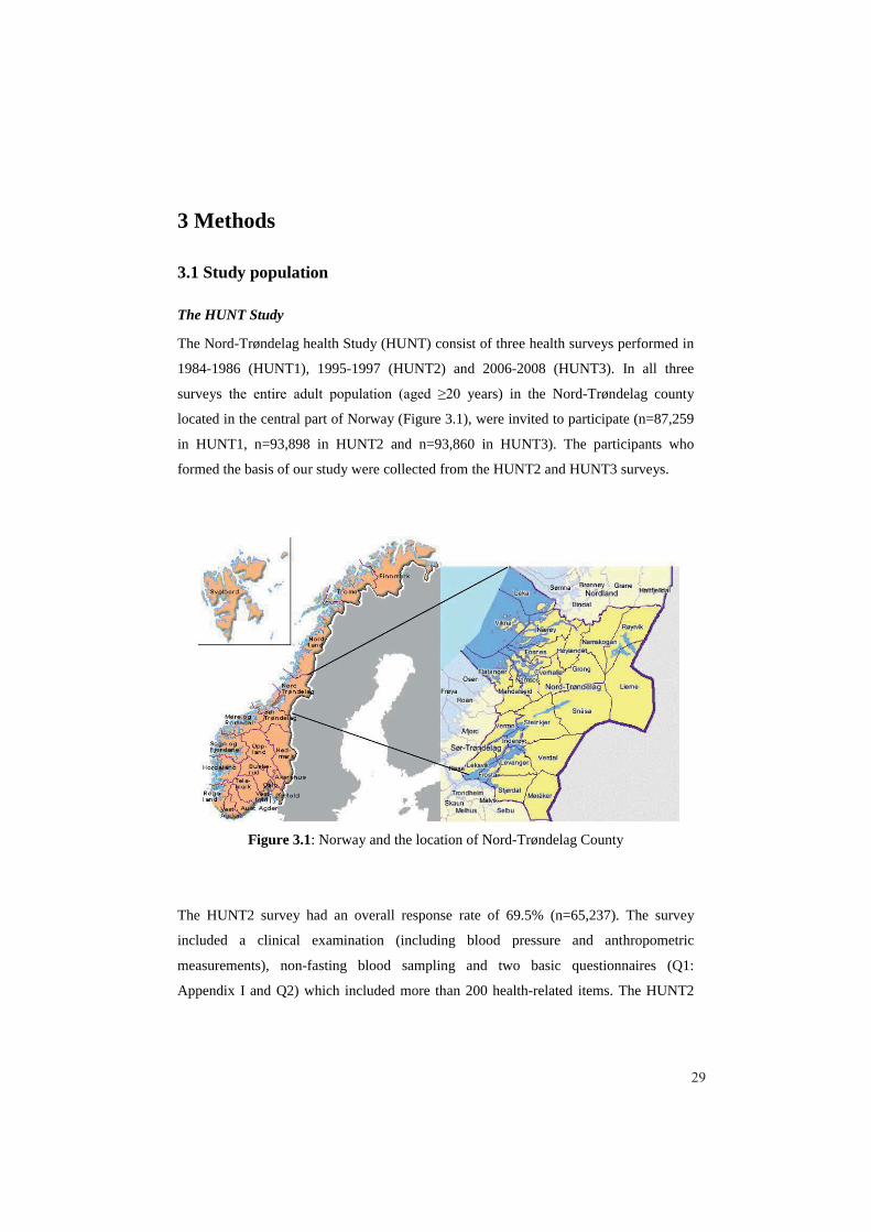
The Nord-Trøndelag health Study (HUNT) consist of three health surveys performed in
1984-1986 (HUNT1), 1995-1997 (HUNT2) and 2006-2008 (HUNT3). In all three
surveys years) in the Nord-Trøndelag county
located in the central part of Norway (Figure 3.1), were invited to participate (n=87,259
in HUNT1, n=93,898 in HUNT2 and n=93,860 in HUNT3). The participants who
formed the basis of our study were collected from the HUNT2 and HUNT3 surveys.
Figure 3.1: Norway and the location of Nord-Trøndelag County
The HUNT2 survey had an overall response rate of 69.5% (n=65,237). The survey
included a clinical examination (including blood pressure and anthropometric
measurements), non-fasting blood sampling and two basic questionnaires (Q1:
Appendix I and Q2) which included more than 200 health-related items. The HUNT2
29
survey has been described in detail elsewhere120. The HUNT3 survey had an overall
response rate of 54.1% (n=50,807). Fifty-seven percent of the participants in HUNT2
also participated in HUNT3 (n=37,059). The HUNT3 survey had a similar design as
HUNT2 and thus included clinical examination, blood-sampling and two basic
questionnaires (Q1: Appendix II and Q2) as described in detail121. Biological samples
collected from HUNT2 and HUNT3 were stored at HUNT Biobank (Levanger,
Norway) prior to analysis, serum being stored at minus 70oC and DNA at minus 20oC.
Data collection
Papers I, II and III
Individuals with diabetes were identified from a self-reported answer of “Yes” to the
question “Do you have or have you had diabetes?” in the Q1 questionnaire from
HUNT2 and HUNT3. In HUNT2, 1,972 individuals and in HUNT3 2,189 answered
affirmative. At both HUNT2 and HUNT3 participants declaring diabetes were invited to
a diabetes-oriented follow-up investigation. They completed a more detailed
questionnaire concerning diabetes (HUNT2: Appendix III and HUNT3: Appendix IV)
and underwent an interview by a nurse to ensure year of diagnosis and details on type
and start of treatment. They furthermore provided a fasting blood sample for
measurements of blood glucose, serum C-peptide, and antiGAD. In HUNT3 antiIA-2
was also measured. A total of 1,630 and 1,824 participants filled out the diabetes-
oriented questionnaire and a total of 1,455 and 1,168 participants rendered a fasting
blood sample at the follow-up respectively in HUNT2 and HUNT3.
Subsequent to the surveys we additionally analyzed antiGAD in participants who had
declared diabetes but had not provided a blood sample at the follow-up, but had serum
available from the baseline blood sampling (n=432 in HUNT2 and n=984 in HUNT3).
Analysis of antiGAD in the HUNT2 samples was performed in the spring of 2008
(average 12 years after sampling) and the HUNT3 samples were analyzed in late 2009
(average 3 years after sampling). This gave us the opportunity to classify all cases who
had answered affirmative to the question on diabetes.
30

Participants who answered “no” to the question of having diabetes served as controls.
They were frequency-matched by sex and by 10 year of age category to the diabetic
patients.
Paper III
Equal numbers of men and women who had stated that they did not have diabetes both
at the HUNT2 and HUNT3 surveys were randomly selected from different age groups:
500 individuals from the age group 20-29 years, 500 from the age group 30-34 years
etc. up to the last age group of 65 years and above. Altogether a total of 4,500 non-
diabetic individuals were sampled to represent the general adult population.
Classification of diabetes
Diabetic cases were classified as having type 1 diabetes if they had started insulin
treatment within 12 months of diagnosis and were 1) antibody positive, or 2) antibody
negative but in addition had fasting C-peptide levels <150 pmol/l.
Cases were classified as having LADA if they were antibody positive and had not been
treated with insulin within 12 months of diagnosis. No age limit was set for LADA.
Cases were classified as having type 2 diabetes if they were antibody negative and had
not been treated with insulin within 12 months of diagnosis.
Classification of diabetic cases with missing data on insulin treatment (paper I)
For the identified diabetic cases in HUNT2 who did not attend the follow-up
investigation we lacked data on insulin treatment. These non-attendees were therefore
classified by less stringent criteria; i.e. as type 1 diabetes if antiGAD positive and age at
diagnosis 35 years old, LADA if antiGAD positive and age at diagnosis >35 years old
and as type 2 diabetes if antiGAD negative and age of diagnosis >35 years old.
31
Attended follow-up n=1,455
Diabetesn=1,972
Did not attend the follow-up
n=517
LADAn=126
Type 2 diabetesn=1090
Type 1 diabetesn=120
Non-diabeticControlsn=1503
HUNT2 Populationn=65,237
Analysed for antiGAD n=432
LADAn=18
Type 2 diabetesn=255
Type 1 diabetesn=16
262 excluded due tomissing DNA, other
types of diabetes or non-classifiable
85 excluded due tomissing serum
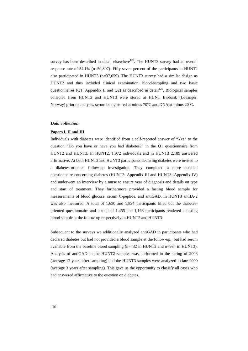
Figure 3.2: Study population in paper I.
Final study population
Paper I
This was a case-control study nested within the HUNT2 cohort (Figure 3.2). All
diabetic cases identified at baseline who had DNA available (n=1,642) and 1,503 age
and gender matched healthy non-diabetic controls were included in the study. The
32

diabetic cases classified by the criteria that included insulin treatment, comprised of 120
type 1 diabetic patients, 126 LADA patients and 1,090 type 2 diabetic patients. Sixteen
type 1 diabetic, 18 LADA and 255 type 2 diabetic patients were classified by the less
stringent criteria. Cases that we were not able to classify (n=17) were excluded.
Paper II
This was both a prospective and a cross-sectional study. Serum samples were collected
from the diabetic subjects classified as type 1 diabetes and LADA from both the
HUNT2 and HUNT3 surveys. Diabetic cases were analyzed for antiIA-2 (if not done
already in HUNT3) and for antiZnT8. Also serum samples from HUNT2 were used to
analyze antiGAD, antiIA-2 and antiZnT8 in incident cases in HUNT3 (i.e. in those not
having a diagnosis of diabetes in HUNT2). All of these antibody measurements were
done in late 2009.
For LADA and type 1 diabetic cases we included for analysis those with complete data
on all three antibody assays. These cases comprised 120 type 1 diabetic and 120 LADA
cases from HUNT2 and 147 type 1 diabetic and 85 LADA cases from HUNT3 (Figure
3.3). Type 2 diabetic cases who had participated in both HUNT2 and HUNT3 surveys
(n=302) were also included for comparison.
Prospective data were obtained (i.e. from cases that had participated in both HUNT2
and HUNT3; providing 10-13 years of follow-up) on 44 LADA, 59 type 1 diabetic and
302 type 2 diabetic cases from HUNT2 who were followed to HUNT. In addition we
analyzed data from 31 LADA and 24 type 1 incident cases of diabetes from HUNT3
who also participated and were non-diabetic in HUNT2.
33

T1DN=120
LADAN=120
HUNT2 (1995-1997)
Not attended HUNT3
N=76Not attended HUNT3
N=61
Diabetes free HUNT2
N=31
Not attended HUNT2
N=10
Diabetes free HUNT2
N=24
Not attended HUNT2
N=64
HUNT3 (2006-2008)
T1DN=147
LADAN=85
Participated both surveys
T1DN=59
LADAN=44
Incidents HUNT3 Incidents HUNT3
Figure 3.3: Study population in paper II. T1D = type 1 diabetes
Paper III
This was a prospective study. Serum samples from HUNT2 to be used for antiGAD
measurements were available from 4,496 of the 4,500 selected individuals (Figure 3.4).
All individuals who were antiGAD positive in HUNT2 were analysed for positivity in
HUNT3. For these individuals antiGAD was measured in the fall of 2011.
Additionally, 55 incident diabetic cases (Figure 3.4) who developed autoimmune
diabetes between HUNT2 and HUNT3 (i.e. reported not having diabetes in HUNT2 but
reported having diabetes in HUNT3, n=24 type 1 diabetes and n=31 LADA) were
included for analysis. Thirty-three of these cases were antiGAD positive already at
HUNT2, i.e. several years before diagnosis (n=13 type 1 diabetes and n=21 LADA).
Data from these patients who over time developed diabetes, were compared with data
from the antiGAD positive persistently non-diabetic population.
34

Persistently non-diabetic individuals who were antiGAD positive at HUNT2 as well as
a control group of antiGAD negative non-diabetic individuals were typed for HLA-
DQA1 and HLA- DQB1. A control group was age and gender matched to the antiGAD
positive, non-diabetic group. Two controls were selected per antiGAD positive
individual. The same HLA genotypes were also analysed in individuals who developed
autoimmune diabetes during the interval between HUNT2 and HUNT3.
N=4530 Participants HUNT2
Non-diabetic HUNT2N=4496
Pre-diabetic antiGAD pos
HUNT2N=34
N=76AntiGAD pos
HUNT2
N=4420AntiGAD neg
HUNT2
N=35Anti-GAD pos
HUNT3
N=34Autoimmune diabetes in
HUNT3
Persistently non-diabetic HUNT3
N=41Anti-GAD neg
HUNT3
Figure 3.4: Study population paper III
35
3.2 Biochemical analysis
C-peptide measurements
The most common way to assess insulin secretion is by measurements of C-peptide. C-
peptide is a cleavage product from pro-insulin and is released together with insulin. C-
peptide is therefore a measure of insulin release.
Serum levels of C-peptide were analysed at the Hormone Laboratory of Aker Hormone
laboratory, Oslo University hospital (Oslo, Norway) by radioimmunoassay (Diagnostic
system Laboratories, USA).
Antibody measurements
All antibody measurements were carried out at the Aker Hormone laboratory, Oslo
University hospital (Oslo, Norway).
AntiGAD
AntiGAD was measured by immuno-precipitation using translation labeled 3H-GAD65
as labeled reagent (Novo Nordisk, Denmark). Separation of bound antiGAD and free
labeled antigen was done by protein A coupled to Sepharose (procedure developed at
the Hormone laboratory). Antibody levels were expressed as an antibody index (ai)
relative to a standard serum given by the formula [(counts per minute (cpm) in the
patients sample – cpm from negative reference sample) / (cpm of a positive reference
sample – cpm from negative reference sample)]. The lower limit of detection was
0.01ai, whereas no upper limit was defined. Intra-assay variation coefficient (CV) was
14% in the lowest (0.11ai), 8% in the middle (0.22ai) and 17% in the highest (2.0ai)
range of measurements. Total assay CV was 19% in the lower (0.21ai) and 23% in the
higher (0.66ai) measurement range.
In paper I and II an antibody index of 0.08ai or greater was considered positive. This
cut-off level of positivity was the one used by the Hormone laboratory based on
participation in DASP. Cut-off was set to achieve the highest possible specificity with
36

an acceptable corresponding sensitivity. Based on participation in DASP this
corresponds to a 68% workshop-sensitivity and 100% workshop-specificity.
In paper III subjects above the 98.5th percentile of the antiGAD levels in the total
cohort were considered to be antiGAD “positive”. This corresponded to a value >0.05ai.
Based on Aker Hormone Laboratory’s participation in DASP this corresponded to an
82% workshop-sensitivity and a 99% workshop-specificity.
AntiIA-2
Antibody to IA-2 was measured by immuno-precipitation using translation labeled 3H-
IA-2ic as a labeled reagent. Separation of bound antiIA-2 and free labeled antigen was
done by protein A coupled to Sepharose, using a procedure developed at the Hormone
laboratory. Antibody levels were expressed as an index value relative to a standard
serum. A value of 0.11ai or greater was considered positive (method range: 0.01-
3.00ai). The level of cut-off was based on the same considerations as for antiGAD. As
calculated from DASP 2003 this assay has 70% workshop-sensitivity and 99%
workshop-specificity. Intra-assay CV was 17% in the lowest (0.10 ai), 10% in the
middle (0.48 ai) and 7% in the highest (1.96 ai) range of measurements. Total assay CV
was 22% in the lower (0.14 ai) and 11% in the higher (3.60 ai) range of measurements.
AntiZnT8
AntiZnT8 was measured by immuno-precipitation using a translation labeled 3H-ZnT8
C-terminal Arg325 variant fused to C-terminal Trp325 variant as a labeled reagent
(based on a plasmid pJH5.2 SP6, a Dimer human ZnT8 C-terminal Arg325 variant
fused to human ZnT8 C-terminal Trp variant from Dr. Hutton, University of Colorado,
Denver, CO, USA). Separation of bound antiZnT8 and freely labeled antigen was
achieved by protein A plus protein C coupled to Sepharose using a procedure developed
at the Hormone laboratory. Antibody levels were expressed as an index value relative to
a standard serum. A value greater of 0.08ai was considered positive (method range:
>0.01 ai). The level of cut-off was based on the same considerations as for antiGAD. As
calculated from DASP 2010 the antiZnT8 assay has 46% workshop-sensitivity and
100% workshop-specificity. Intra-assay CV was 7% in the lowest (0.18 ai) and 6% in
37

the highest (0.88 ai) range measurements. Total assay CV was 20% in the lower (0.18
ai) and 16% in the higher (0.85 ai) range of measurements.
3.3 Genetic analysis
DNA extraction
DNA samples were mainly collected from the HUNT2 survey. DNA from HUNT2 was
extracted from peripheral blood leukocytes from EDTA whole blood or blood clots
using the Gentra Purgene blood kit (QIAGEN Science, Maryland, USA). EDTA blood
samples were kept frozen at -70oC, whereas clots were stored at -20oC. The blood
samples were removed from the freezer and thawed in a 37oC water bath immediately
before DNA extraction and transferred to 50 ml tubes (Sarstedt). The clots were
liquidized using an OMNI TH homogenizer with disposable OMNI Tip generator
probes, using one cycle of 20 sec.
DNA from liquidized clots (5-10ml) and EDTA blood (1-5ml) were isolated on an
Autopure LS instrument according to protocols designed by Gentra, or manually, using
the same reagents and protocols. In brief, RBC Lysis Solution and Cell Lysis Solution
were added to lysate the red and white blood cells. Protein Precipitation Solution and
Proteinase K (only for blood clots) were added to precipitate the proteins in the solution.
Then the free DNA was precipitated in 100% isopropanol added Gentra Glycogen
Solution (only for EDTA blood) and finally the DNA pellet was washed in 70%
ethanol. The DNA was rehydrated in DNA Hydration Solution (Tris-EDTA-buffer).
DNA from low volumes (EDTA blood <400 l) was isolated on a GenoVision
BioRobot GenoM-48 (QIAGEN Science, Maryland, USA) according to protocols
designed by GenoVision.
In paper III a few DNA samples were collected from the HUNT3 survey when DNA
was not available from HUNT2. DNA from HUNT3 was extracted from buffy-coat
which was fractionated from 10 ml EDTA whole blood at sampling. The buffy-coat was
38

stored at -70oC at HUNT Biobank prior to DNA extraction. The DNA extraction
protocol was in general the same as that used for the HUNT2 samples.
Selection of SNPs
The selected single nucleotide polymorphisms (SNPs) were based on publicly available
results (mainly retrieved from searches on the PubMed database) from studies focusing
on genetic association analysis in type 1 (Table 3.1) and type 2 diabetes (Table 3.2).
Single SNP genotyping analysis
The genotyping technologies used for SNP analysis in this study were Taq-Man
Discrimination analysis and SNPlex assay (both from Applied Biosystems, Foster City,
CA, USA).
TaqMan Discrimination analysis
SNPs genotyped by applying TaqMan SNP allelic discrimination using ABI 7900HT
Fast Real-Time PCR System (Applied Bioasystems, Foster City, CA, USA) are
indicated in the table 3.1 and 3.2.
The TaqMan allelic discrimination assay is an endpoint analysis in which the presence
of two primer and probe pairs in each reaction allows you to differentiate between two
possible variations in a single SNP. Each probe is color labeled with its own reporter at
the 5'-end. The reporter is a specific fluorescent (typically VIC and FAM) which helps
to distinguish between the two alleles. In addition a non-fluorescent quencher which
suppresses the fluorescence signal of the reporter is bound at the 3'-end of the probe.
During the amplification the probes hybridize specifically to each complementary target
sequence (wild-type and mutant) between the primer sites. The DNA polymerase
enzyme then cleaves the reporter from the probe and quencher, resulting in increased
fluorescence signal from the reporter. The polymerase can only cleave probes
hybridized to the target sequence. The fluorescence signal generated during the
amplification is therefore an indicator of the alleles present in the sample. After
amplification an endpoint reading of the fluorescence signal is done using the Sequence
39
Detection System (SDS) software. This software plots the signals from each sample in a
scatterplot where each signal indicates which alleles are present in the sample.
SNPlex analysis
The SNPs genotyped by applying SNPlexTM genotyping system (Applied Biosystems,
Foster City, CA, USA) are indicated in tables 3.1 and 3.2.
The SNPlex assay is a multiplex assay which at the time of the study allowed us to
analyse up to 48 SNPs simultaneously. The assay is a migration specific assay designed
to discriminate alleles by the application of three SNP specific probes. Two of the
probes are allele specific oligonucleotides (ASO) designed to discriminate the two
alleles at each SNP. The third probe is a locus specific oligonucleotide (LSO). All
probes have a universal PCR priming site; however, the ASO probes have a unique
ZipCode identifier that hybridizes to the added complementary ZipChute probe. The
ZipChute probe allows for the discrimination between the SNPs in the assay by
providing a unique migration pattern for each SNP. Fluorescent signals from each SNP
are interpreted by using Applied Biosystem GeneMapper Software (Applied
Biosystems, Foster City, CA, USA).
Genotyping performance:
Cases and controls were equally distributed with four or more negative controls per
384-plate. Criteria to pass the assay were 1) call rates >90%, 2) minor allele frequency
(MAF) >1% in the genotyped population and 3) agreement with Hardy-Weinberg
equilibrium in the whole population (if p-value <0.001 the assay did not pass). The SNP
assays that did not pass quality control were excluded from further analysis.
40

Tab
le 3
.1:S
elec
ted
SNPs
ass
ocia
ted
with
hig
her r
isk
of ty
pe 1
dia
bete
s
Chr
omo-
som
eSN
PG
ene
Full
gene
nam
eA
llels
*M
AF†
Ref
eren
ceA
naly
se
met
hod
1p13
rs24
7660
1rs
2488
457
PTP
N22
Prot
ein
tyro
sine
pho
spha
tase
, non
-rec
epto
r typ
e 22
A/G
C/G
0.12
0.25
23,2
4Ta
qMan
TaqM
an
2q33
.2rs
2317
75rs
3087
243
CTL
A4
Cyt
otox
ic T
-lym
phoc
yte-
asso
ciat
ed p
rote
in-4
G/A
A/G
0.43
0.42
25,3
0,12
2Ta
qMan
TaqM
an2q
24rs
1990
760
IFIH
1In
terfe
ron-
indu
ced
with
hel
icas
e C
dom
ain
1C
/T0.
3625
,29
SNP
lex
5p13
.2rs
1445
898
CA
PS
LC
alcy
phos
ine-
like
T/C
0.42
25Ta
qMan
6p21
rs22
9633
6IT
PR
3In
osito
l1,4
,5-tr
isph
osph
ate
rece
ptor
, typ
e 3
C/G
0.32
123
TaqM
an
10p1
5.1
rs31
1847
0rs
7067
78rs
9663
421
IL2R
AIn
terle
ukin
-2 re
cept
or-a
lpha
C/T
T/C
T/C
0.40
0.45
0.22
26,1
24Ta
qMan
TaqM
anSN
Ple
x
11p1
5.5
rs68
9rs
3842
753
INS
Insu
linA/
TA/
C0.
260.
2722
,122
TaqM
an
16p1
3.13
rs29
0369
2KI
AA0
350
C-ty
pe le
ctin
dom
ain
fam
ily 1
6, m
embe
r AA/
G0.
3331
SNP
lex
*Min
or a
llele
is li
sted
firs
t†M
AF=
Min
or a
llele
freq
uenc
y
41

Tab
le 3
.2:S
elec
ted
SNPs
ass
ocia
ted
with
risk
of t
ype
2 di
abet
es
Chr
omo-
som
eSN
PG
ene
Full
gene
nam
eA
llels
*M
AF†
Ref
eren
ceA
naly
sem
etho
d1p
12rs
1092
3931
NO
TCH
2Pr
ospe
ro-r
elat
ed h
omeo
box-
1T/
G0.
1255
TaqM
an1p
12rs
2641
348
ADA
M30
ADA
M m
etal
lope
ptid
ase
dom
ain
30G
/A0.
1355
TaqM
an1q
32.1
rs13
4238
7AD
IPO
R1
Adip
onec
tin re
cept
or 1
A/G
0.46
122,
123
SNP
lex
2p16
.1rs
1049
0072
BCL1
1AB-
cell
cell/
lym
phom
a 11
AC
/T0.
2555
TaqM
an2p
21rs
7578
597
THAD
ATh
yroi
d ad
enom
a as
soci
ated
C/T
0.08
55Ta
qMan
3q25
.2rs
1801
282
PPAR
GPe
roxi
som
epr
olife
rato
r-ac
tivat
ed re
cept
or-g
amm
aG
/C0.
1451
,54
TaqM
an
3p27
.2rs
4402
960
rs76
3367
5IG
F2B
P2
Insu
lin-li
ke g
row
th fa
ctor
2 m
RN
A-bi
ndin
g pr
otei
n-2
T/G
G/T
0.30
0.30
54,1
24Ta
qMan
TaqM
an3p
25rs
1703
6101
SYN
2/P
PAR
GPe
roxi
som
e pr
olife
rato
r-ac
tivat
ed re
cept
or g
amm
aA/
G0.
0755
TaqM
an3p
14.1
rs46
0710
3AD
AM
TS9
ADA
M m
etal
lope
ptid
ase
with
thro
mbo
spon
din
type
1 m
otif,
9T/
G0.
2455
TaqM
an4p
16.1
rs10
0101
31W
FS1
Wol
fram
syn
drom
e 1
(wol
fram
in)
A/G
0.41
52Ta
qMan
6p22
.3rs
7754
840
CKA
L1C
DK5
regu
lato
ry s
ub-u
nit-a
ssoc
iate
d pr
otei
n-1-
like
1C
/G0.
3154
,124
TaqM
an6p
12rs
9472
138
VEG
FAVa
scul
ar e
ndot
helia
l gro
wth
fact
or A
T/C
0.32
55Ta
qMan
6p22
-q23
rs10
4449
8EN
PP1
Ecto
nucl
eotid
e py
roph
osph
atas
e/ph
osph
odie
ster
ase
1C
/A0.
1612
5,12
6SN
Ple
x7p
15.2
rs86
4745
JAZF
1Ju
xtap
osed
with
ano
ther
zin
c fin
ger g
ene-
1C
/T0.
4755
TaqM
an8q
24.1
1rs
1326
6634
SLC
30A
8S
olut
e ca
rrie
r fam
ily 3
0 (z
inc
trans
porte
r), m
embe
r 8T/
C0.
2953
,54
SNP
lex
9p21
.3rs
1081
1661
CD
KN2A
/BC
yclin
-dep
ende
nt k
inas
e in
hibi
tor 2
A/B
C/T
0.15
54,1
24Ta
qMan
10p1
3rs
1277
9790
CD
C12
3/C
AMK1
DC
ell d
ivis
ion
cycl
e 12
3 H
omol
ogue
/cal
cium
/cal
mod
ulin
-de
pend
ent p
rote
in k
inas
e ID
G/A
0.17
55Ta
qMan
10q2
5.2
rs79
0314
6TC
F7L2
Tran
scrip
tion
fact
or-7
like
2T/
C0.
2949
,53,
54Ta
qMan
10q2
3.33
rs11
1187
5H
HE
XH
omeo
box
hem
atop
oiet
ical
ly e
xpre
ssed
A/G
0.43
53,5
4SN
Ple
x11
p12
rs93
0003
9In
trage
nic
regi
onA/
C0.
1354
TaqM
an11
p15.
1rs
5219
KC
NJ1
1AT
P-se
nsiti
ve in
war
d re
ctifi
er p
otas
sium
chan
nel
T/C
0.43
54,1
24Ta
qMan
12q2
1.1
rs79
6158
1TS
PAN
8/LG
R5
Tetra
span
in 8
/leuc
ine-
rich
repe
at-c
onta
inin
g G
pro
tein
-co
uple
d re
sept
or-5
C/T
0.25
55Ta
qMan
12q1
3.1
rs11
5318
8D
CD
Der
mci
din
T/A
0.25
55Ta
qMan
12p1
3.31
rs76
7870
rs22
8638
4AD
IPO
R2
Adip
onec
tin re
cept
or 2
C/T
G/C
0.17
0.48
123
SNP
lex
SNP
lex
16q1
2.2
rs80
5013
6FT
OFa
t mas
s an
d ob
esity
ass
ocia
ted
A/C
0.44
54,1
24Ta
qMan
18p1
1.31
rs37
4501
2LP
IN2
Lipi
n 2
T/C
0.24
127
SNP
lex
*Min
or a
llele
is li
sted
firs
t†M
AF=
Min
or a
llele
freq
uenc
y
42

HLA-haplotyping
Paper I and II:
HLA-haplotyping was performed as described by de Bakker et al128. They captured
nearby single tag SNPs or haplotypes of combination of up to three SNPs as a predictor
of known HLA-alleles. The recommended tag SNPs or haplotypes given by deBakker et
al. for the HLA-risk alleles for type 1 diabetes are shown in Table 3.3. We genotyped
these tag SNPs by using the SNPlex genotyping system as described above.
Table 3.3: Genotyped tagSNPs from known risk or protective HLA alleles for type 1 diabetes selected from supplementary table 3 in deBakker et al.128
HLA allele Frequency Tag SNPsHLA-DQA*0101 0.1333 rs6457614,rs1794265HLA-DQA*0102 0.2389 rs14004,rs6457594HLA-DQA*0201 0.1333 rs7745002,rs2858333HLA-DQA*0301 0.2278 rs3129883,rs7745002,rs3844313HLA-DQA*0401 0.0333 rs7743506HLA-DQA*0501 0.1778 rs10485326,rs2040406HLA-DQB*0201 0.1556 rs4988889HLA-DQB*0301 0.1667 rs7744001,rs2856691HLA-DQB*0302 0.1444 rs7454108,rs2071876HLA-DQB*0303 0.0611 rs3834857,rs2395533HLA-DQB*0402 0.0333 rs7743506HLA-DQB*0501 0.0944 rs6457614HLA-DQB*0503 0.0389 rs1794265HLA-DQB*0602 0.2000 rs3135388HLA-DRB*0101 0.0889 rs4947332,rs6457614HLA-DRB*0301 0.0944 rs2040410HLA-DRB*0401 0.0889 rs6910071,rs3817964,rs660895HLA-DRB*0402 0.0056 rs3130071,rs416352HLA-DRB*0403 0.0167 rs7454108,rs206765,rs399604HLA-DRB*0405 0.0056 rs1057149HLA-DRB*0701 0.1000 rs7745002,rs2647087,rs241398HLA-DRB*0801 0.0333 rs7743506HLA-DRB*0901 Not reportedHLA-DRB*1101 0.0500 rs434841,rs1061172,rs5875439HLA-DRB*1401 0.0333 rs2763979,rs1794265HLA-DRB*1404 0.0056 rs805262,rs1794265HLA-DRB*1501 0.2000 rs3135388
43
Paper III:
HLA-DQA1 and HLA-DQB1 genotypes were analysed at the Unit for Immunology, St
Olavs Hospital, Trondheim, Norway by sequence-specific oligonucleotide probes (SSO)
using LABTypeR SSO DQA1/DQB1 Typing Tests (One Lambda Inc., CA, USA) as
described by the manufacturer. In brief, the target DNA was amplified by polymerase
chain reaction (PCR) using HLA-locus specific target primers. The amplified target
DNA was denatured and re-hybridized with complementary DNA probes bound to
fluorescently coded microspheres, which allowed detection using R-Phycoerytrin-bound
(PE) Strepavidin. The flow analyser Luminex 100 (Luminex Corporation, Texas, USA)
was used to measure the fluorescent signal of PE. The HLA fusion 2.0 (One Lambda
Inc., CA, USA) software was used to process the results.
3.4 Statistical methods
Paper I
PLINK software (http://pngu.mgh.harvard.edu/purcell/plink/)129 was used to assess
whether genotypes were in Hardy-Weinberg equilibrium and to test differences in
genotype distribution between affected and unaffected subjects by logistic regression
under additive, dominant and recessive models. Odds ratio (OR) and 95% confidence
intervals (CI) were calculated. Adjustment for diabetes specific risk factors such as age,
sex and BMI was applied when appropriate. Correction for multiple testing was done by
max(T) permutation where 1,000 permutations were performed. Phasing HLA-
haplotypes and testing for association within cases and controls were also carried out
using PLINK.
Paper II
Statistical Package for the Social Sciences, version 14.0 and 18.0 (SPSS Inc, Chicago)
was used when comparing categorical data by Chi-square or Fisher’s exact test as
appropriate. Comparing continuous data between groups was done by a non-parametric
analysis of variance test (Mann-Whitney test and Kruskal-Wallis test as appropriate). A
44

Kaplan-Mayer log-rank test was performed to assess time to insulin requirement in
relation to antibody positivity. For comparison of antibody titer change over time (from
HUNT2 to HUNT3) a nonparametric Wilcoxon signed Rank test for two related
samples was used.
Paper III
Statistical analyses were performed by the PASW Statistics (version 19, SPSS, Inc,
Chicago, IL). Chi-square test or Fisher exact test (when appropriate) was used to
compare differences in categorical data. Mann Whitney U test was used to test
differences in continuous data between two groups. Kruskal-Wallis test was used to test
differences in continuous data between more than two groups. Logistic regression
models were used to examine whether HLA haplotypes were associated with antiGAD
after adjusting for other confounding factors such as age, gender and BMI.
A two-tailed p-value of 0.05 was considered to be significant in all three papers.
45

46
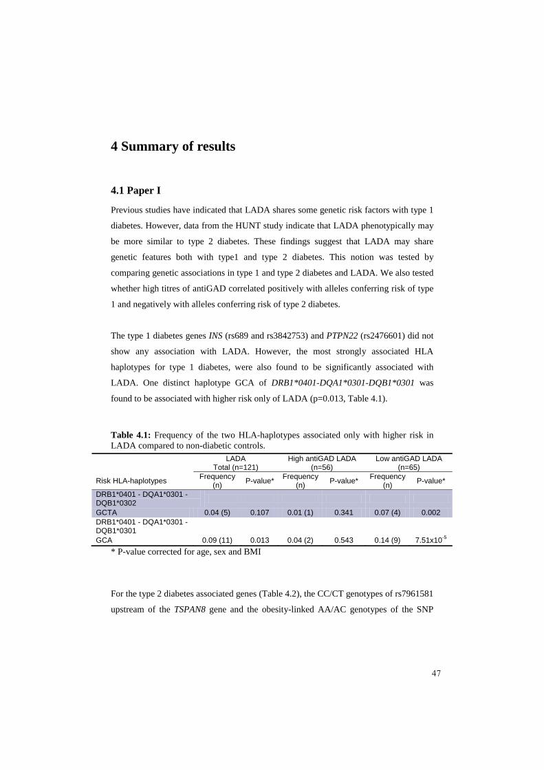
4 Summary of results
4.1 Paper I
Previous studies have indicated that LADA shares some genetic risk factors with type 1
diabetes. However, data from the HUNT study indicate that LADA phenotypically may
be more similar to type 2 diabetes. These findings suggest that LADA may share
genetic features both with type1 and type 2 diabetes. This notion was tested by
comparing genetic associations in type 1 and type 2 diabetes and LADA. We also tested
whether high titres of antiGAD correlated positively with alleles conferring risk of type
1 and negatively with alleles conferring risk of type 2 diabetes.
The type 1 diabetes genes INS (rs689 and rs3842753) and PTPN22 (rs2476601) did not
show any association with LADA. However, the most strongly associated HLA
haplotypes for type 1 diabetes, were also found to be significantly associated with
LADA. One distinct haplotype GCA of DRB1*0401-DQA1*0301-DQB1*0301 was
found to be associated with higher risk only of LADA (p=0.013, Table 4.1).
Table 4.1: Frequency of the two HLA-haplotypes associated only with higher risk in LADA compared to non-diabetic controls.
* P-value corrected for age, sex and BMI
For the type 2 diabetes associated genes (Table 4.2), the CC/CT genotypes of rs7961581
upstream of the TSPAN8 gene and the obesity-linked AA/AC genotypes of the SNP
LADATotal (n=121)
High antiGAD LADA(n=56)
Low antiGAD LADA(n=65)
Risk HLA-haplotypes Frequency(n) P-value* Frequency
(n) P-value* Frequency(n) P-value*
DRB1*0401 - DQA1*0301 -DQB1*0302GCTA 0.04 (5) 0.107 0.01 (1) 0.341 0.07 (4) 0.002DRB1*0401 - DQA1*0301 -DQB1*0301GCA 0.09 (11) 0.013 0.04 (2) 0.543 0.14 (9) 7.51x10⁻⁵
47

rs8050126 in the FTO gene were associated with LADA. The TT/TC genotypes within
the strongly type 2 diabetes associated TCF7L2 gene (rs7903146) were not obviously
associated with LADA.
Table 4.2: Genotypes of known type 2 diabetes associated loci in LADA compared to non-diabetic controls.
Gene name, SNP Genotype LADA vs. controls Low antiGAD LADA vs. controls
OR (95% CI) P-value* OR (95% CI) P-value*
TCF7L2, rs7903146 CT/TT vs. CC 1.25 (0.85-1.83) 0.141 1.32 (0.78-2.24) 0.162
TSPAN8/LGR5, rs7961581 CC/CT vs. TT 1.68 (1.15-2.46) 0.01 2.17 (1.28-3.69) 0.006
FTO, rs8050136 AA/AC vs. CC 1.94 (1.22-3.09) 0.005 2.85 (1.39-5.84) 0.003
FTO, rs1861866 CT/TT vs. CC 2.55 (1.45-4.50) 0.003 3.85 (1.52-10.0) 0.004
FTO, rs9931494 CG/GG vs. CC 2.72 (1.63-4.53) 2.43x10⁻⁴ 3.29 (1.56-6.95) 0.001
* Adjusted p-value from logistic regression for age, sex and BMI.
After dichotomising into high and low antiGAD titre the type 2 diabetes associated
genes (Table 4.3) FTO (rs8050136, rs1861866 and rs9931491) and TSPAN8
(rs7961581) were associated only with low-antiGAD titre LADA. The strongly
associated type 2 diabetes gene (TCF7L2, rs7903146) which was not associated with
LADA in general, was neither associated with sub-groups of antiGAD. The HLA
haplotypes (Table 4.1) were found to be mainly associated with high antiGAD LADA
patients, except the GCA-haplotype for DRB1*04:01-DQA1*03:01-DQB1*03:01 which
was associated with higher risk in low antiGAD LADA patients. Interestingly, yet
another distinct haplotype was found, the GCTA-haplotype for DRB1*04:01-
DQA1*03:01-DQB1*03:02, which was only associated with higher risk in low
antiGAD LADA and not with type 1 or type 2 diabetes.
48
4.2 Paper II
LADA comprises a significant proportion of all diabetic cases. Yet, the etiology of
LADA assessed by autoimmune markers is not completely clarified, nor is the
distinction from “classical” type 1 diabetes. We investigated cross-sectionally the
prevalence and prospectively the pre-diabetic and post-diabetic presence of antiGAD,
antiIA-2 and antiZnT8 in LADA and in type 1 diabetes.
Cross-sectional
LADA was positive for only one antibody in 90% of the cases (10% were multiple-
antibody-positive). Doubly and triply positive LADA patients vs. singly positive had
lower systolic blood pressure (p=0.005), higher non-fasting blood glucose (p=0.011)
and higher antiGAD titer (p<0.001). Age at onset was not associated with the number of
antibodies.
Comparing LADA with adult onset type 1 diabetes (age >24 years, n=103), LADA
patients were less frequently positive for more than one antibody (p<0.001). Those with
LADA displayed higher levels of C-peptide (p<0.001) and antiGAD titer (p<0.001) but
lower antiIA-2 titer (p=0.002) compared to type 1 diabetic subjects.
Prospectively
Fifty-nine percent of antiGAD-positive LADA patients in HUNT2 were no longer
positive in HUNT3 (Table 4.3). LADA patients who became negative possessed less
frequently risk HLA haplotypes and were phenotypically more akin to those with type 2
diabetes than those who stayed positive. The former patients were also older at onset
(p=0.001), more obese with both higher BMI and waist circumference (p=0.013 and
p=0.009, respectively) and displayed higher levels of C-peptide (p=0.003). Still, those
losing positivity differed from those with type 2 diabetes by lower C-peptide levels
(p=0.009). Persistent antibody positivity was associated with earlier requirement of
insulin treatment (p=0.001).
49

Table 4.3: Comparison of clinical characteristics from the HUNT2 survey between LADA cases who participated in both HUNT2 and HUNT3 and either became antibody negative or stayed antibody positive during follow-up (10-13 years between HUNT2and HUNT3)
Data presented as percentages (n) or median (min-max-value)*Unadjusted p-value.
Of incident LADA cases in HUNT3, 21 out of 31 (68%) were already antibody-positive
10-13 years earlier at HUNT2, i.e. before diabetes diagnosis (Table 4.4). Incident
LADA cases who displayed antibody positivity before diagnosis were phenotypically
more akin to type 1 diabetes than those who were antibody negative in HUNT2.
Antibody positive patients at HUNT2 were diagnosed at younger age (p=0.001) and
were associated with higher antiGAD-titer (p=0.006) and higher fasting blood glucose
(p=0.013) at HUNT3 than antibody negative patients.
Thirteen out of 24 incident type 1 diabetic cases (54%) were antibody positive already
at HUNT2. There were no distinctive differences between antibody positive and
antibody negative type 1 diabetic patients either at pre-diabetes (HUNT2) or at overt
diabetes (HUNT3).
LADAClinical characteristics HUNT2 Ab negative HUNT3 Ab positive HUNT3 P-value*N 26 18Age at onset (yr) 53.5 (42-75) 44.5 (21-60) 0.001Waist circumference (cm) 96 (78-122) 88.5 (71-103) 0.009
BMI (kg/m2
) 27.9 (24.6-44.8) 25.7 (21.9-36.9) 0.013HDL cholesterol (mmol/l) 1.1 (0.6-2.4) 1.75 (0.7-3.3) 0.017Triacylglycerol (mmol/l) 1.6 (0.86-4.42) 1.12 (0.44-4.48) 0.002C-peptide (pmol/l) 492 (30-1,384) 118.5 (30-588) 0.003Glucose, fasting (mmol/l) 7.55 (2.9-11.5) 9.0 (3.9-16.9) 0.111GADA titer (ai) 0.11 (0.08-0.46) 0.51 (0.07-2.43) <0.001Numbers of abs positivity 0.0231 Ab 65% (26) 37% (14)2-3 Abs - 100% (4)
50
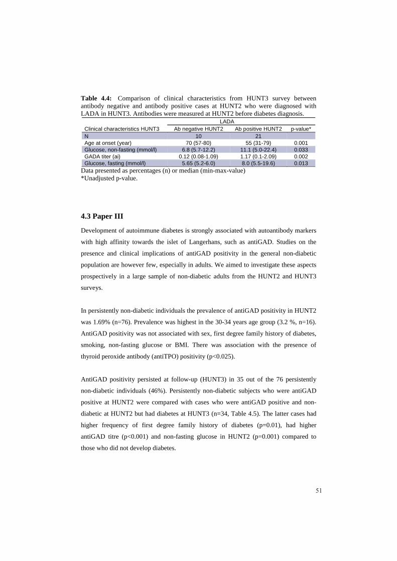
Table 4.4: Comparison of clinical characteristics from HUNT3 survey between antibody negative and antibody positive cases at HUNT2 who were diagnosed with LADA in HUNT3. Antibodies were measured at HUNT2 before diabetes diagnosis.
LADAClinical characteristics HUNT3 Ab negative HUNT2 Ab positive HUNT2 p-value*N 10 21Age at onset (year) 70 (57-80) 55 (31-79) 0.001Glucose, non-fasting (mmol/l) 6.8 (5.7-12.2) 11.1 (5.0-22.4) 0.033GADA titer (ai) 0.12 (0.08-1.09) 1.17 (0.1-2.09) 0.002Glucose, fasting (mmol/l) 5.65 (5.2-6.0) 8.0 (5.5-19.6) 0.013
Data presented as percentages (n) or median (min-max-value)*Unadjusted p-value.
4.3 Paper III
Development of autoimmune diabetes is strongly associated with autoantibody markers
with high affinity towards the islet of Langerhans, such as antiGAD. Studies on the
presence and clinical implications of antiGAD positivity in the general non-diabetic
population are however few, especially in adults. We aimed to investigate these aspects
prospectively in a large sample of non-diabetic adults from the HUNT2 and HUNT3
surveys.
In persistently non-diabetic individuals the prevalence of antiGAD positivity in HUNT2
was 1.69% (n=76). Prevalence was highest in the 30-34 years age group (3.2 %, n=16).
AntiGAD positivity was not associated with sex, first degree family history of diabetes,
smoking, non-fasting glucose or BMI. There was association with the presence of
thyroid peroxide antibody (antiTPO) positivity (p<0.025).
AntiGAD positivity persisted at follow-up (HUNT3) in 35 out of the 76 persistently
non-diabetic individuals (46%). Persistently non-diabetic subjects who were antiGAD
positive at HUNT2 were compared with cases who were antiGAD positive and non-
diabetic at HUNT2 but had diabetes at HUNT3 (n=34, Table 4.5). The latter cases had
higher frequency of first degree family history of diabetes (p=0.01), had higher
antiGAD titre (p<0.001) and non-fasting glucose in HUNT2 (p=0.001) compared to
those who did not develop diabetes.
51
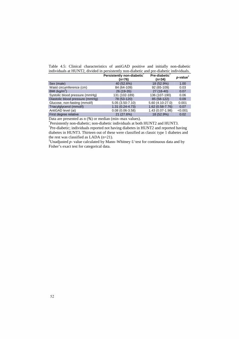
Table 4.5: Clinical characteristics of antiGAD positive and initially non-diabetic individuals at HUNT2, divided in persistently non-diabetic and pre-diabetic individuals.
Persistently non-diabetic*
(n=76)Pre-diabetic†
(n=34) p-value‡
Sex (male) 40 (52.6%) 18 (52.9%) 1.00Waist circumference (cm) 84 (64-109) 92 (65-109) 0.03BMI (kg/m2) 26 (19-35) 27 (18-40) 0.07Systolic blood pressure (mmHg) 131 (102-189) 136 (107-190) 0.06Diastolic blood pressure (mmHg) 78 (53-120) 85 (58-122) 0.09Glucose, non-fasting (mmol/l) 5.05 (3.50-7.10) 5.60 (4.10-27.0) 0.001Triacylglycerol (mmol/l) 1.31 (0.24-4.73) 1.62 (0.58-7.76) 0.07AntiGAD level (ai) 0.08 (0.06-3.58) 1.43 (0.07-1.98) <0.001First degree relative 21 (27.6%) 18 (52.9%) 0.02Data are presented as n (%) or median (min–max values).*Persistently non-diabetic; non-diabetic individuals at both HUNT2 and HUNT3.†Pre-diabetic; individuals reported not having diabetes in HUNT2 and reported having diabetes in HUNT3. Thirteen out of these were classified as classic type 1 diabetes and the rest was classified as LADA (n=21).‡Unadjusted p- value calculated by Mann–Whitney U test for continuous data and by Fisher’s exact test for categorical data.
52

5 Discussion
5.1 Methodological considerations
The HUNT study
This study was based on subjects selected from the second and third HUNT surveys in
the HUNT study which encompasses the population of the Nord-Trøndelag County.
This county consists of mainly rural areas and is thereby sparsely populated; however,
the county is fairly demographically representative of Norway with regard to
geography, economy, age distribution, morbidity and mortality. The population is stable
and ethnically homogenous120. The HUNT study is population-based and can provide a
long observation time. These features make the HUNT study very suitable for genetic
and epidemiological studies on slowly developing diseases such as diabetes. However,
there are important concerns regarding the HUNT study that needs to be addressed.
Selection bias
There has been a noticeable decrease in the attendance rate from the HUNT1 survey to
the HUNT3 survey raising the question if non-responders could lead to increased
selection bias over time. Because of this concern a non-responder study was carried out
both in HUNT1 and HUNT3130,131. The attendance rate was lowest in the youngest (20-
29 years old) and oldest (80+ years old) age groups both in HUNT1 and HUNT3. The
main reasons for not attending had not changed between the two surveys. “Had no time
/ absent” was the most frequently reason given for not attending, especially in the
youngest age groups whereas “too ill to attend” was the main reason amongst the oldest
people (80+).
In the HUNT1 survey one could not find any differences in health status between
participants and non-participants in the youngest age groups, however there was a
selection on marital status and social network. Among the elderly the non-participants
showed a higher frequency in mortality and morbidity indicating a more “healthy”
population among those who participated.
53

In the HUNT3 survey the prevalence of reported diabetes diagnosis (assessed by
questionnaire) was higher among non-participants than among participants. Also the
prevalence of diabetes was higher when based on records from the general practitioners
compared to reported diabetes diagnosis among the participants. This difference was
most marked among those over 60 years old.
This indicates that the prevalence and incidence of diabetic cases in paper I and II may
be underestimated. The decreasing prevalence among participants above 60 years old
may also explain why the prevalence of LADA in HUNT3 is lower than in HUNT2
(paper II).
Self-reported information (information bias)
As described, the presence of diabetes in this study is based on answering affirmative to
the question “Do you have/or have had diabetes?”. Also, the use of insulin (or use of
any other treatment for diabetes), which was used in the classification of type of
diabetes, was based on self-reporting. One may question the reliability in self-reported
answers and the possibility of misclassification. Midthjell et al. performed a validation
study regarding these aspects of the HUNT1 survey132. Correct diagnosis diabetes/no
diabetes was verified in 96.4% of those reporting having diabetes and in 99.7% in those
reporting not having diabetes. Regarding treatment this was verified as correct in 95%
of those reporting using insulin and in 100% of those reporting using oral agents. A
negative answer regarding treatment was verified in 100% and 99%, respectively. This
indicated a very good correlation between the self-reported answers from the
questionnaires and the medical records and a minimized possibility of misclassification
of diabetic cases.
Antibody measurements (information bias)
By setting a cut-off of antibody positivity, it is possible that we have “recruited” falsely
antibody positive cases. However, in paper I and II the cut-off was the same as the cut-
off used by the Aker Hormone Laboratory, which was based on data from participation
54
in DASP. The laboratory wanted highest possible specificity to ensure that the patients
who were found to be antibody positive were true positive (correspondence with Peter
Torjesen, Aker Hormone Laboratory, Oslo, Norway). The cut-offs in both the antiGAD
and antiZnT8 assays had a 100% specificity and the antiIA-2 had 99% specificity
according to DASP. All assays had however a somewhat low sensitivity which will
rather underestimate than overestimate the prevalence of antibody positive individuals.
This may have influenced the classification of LADA and type 2 diabetes, leaving an
underestimation of the LADA group. However, having this high cut-off will also lead to
a more clearly defined LADA phenotype because we are more certain that these patients
are truly antibody positive. Since the LADA group is much smaller than the type 2
diabetic group, it is more important that the LADA cases are identified are true LADA
than if a few LADA cases are misdiagnosed as type 2 diabetes.
Storage time of serum samples
Could long-term storage at low temperatures influence the antibody levels in serum?
Very few studies have addressed this potential problem. One study reported that long-
term storage (>14 years) of serum at -25oC markedly increased the antiTPO level in the
samples133. These results may indicate that there is a probability of evaporation of water
over time from the stored serum samples that could affect the antibody level in samples.
If evaporation of water from serum samples occurs, this may lead to increased antibody
titer and consequently false positive antibody results. This could affect the prevalence of
LADA cases (paper I and II) and the prevalence of antiGAD positivity in the non-
diabetic population (paper III) in our study. The serum samples used in this study,
which were collected from the HUNT2 survey, have been stored for up to 16 years
(paper III), a time span which could potentially lead to evaporation. However, the
HUNT serum samples have been stored at -70oC and have been exposed to a limited
number of freeze and thaw cycles, which most likely will be better for the quality of the
serum samples and minimize the probability of evaporation compared to storage at -
25oC.
55

Classification criteria of LADA
Age, antibody positivity and independence of insulin treatment have been used in the
past to diagnose LADA.
The age limit (usually <35 years) is to some extent arbitrary. We agree with the view of
Rolandsson and Palmer that there should be no age limit in the classification of
LADA134.
Antibody positivity (usually anti-GAD) is regarded as a sine qua non for diagnosis of
LADA. Is it possible to have LADA without being antibody positive? Data, from this
study (paper II) do indicate so. About 50% of individuals with diabetes who were found
to have LADA at HUNT2 because they were antibody positive, lost anti-GAD positivity
when they were re-examined 10-13 years later at HUNT3. Hence, if diabetes had been
diagnosed for the first time at HUNT3 these individuals would have been classified as
having type 2 diabetes. If one accepts that even fleeting positivity for autoimmune
markers in diabetes does have clinical importance, then one must conclude that “mild”
autoimmunity plays a role for developing diabetes not only for those individuals that are
diagnosed as LADA at a given time point of examination but also for a significant part
of those individuals who are currently diagnosed as having type 2 diabetes.
The last criterion for LADA is independence of insulin treatment at diagnosis and for a
significant period of time thereafter. Whether an individual with new-onset diabetes is
in need of insulin treatment is open to subjective evaluation and to current norms of
treatment. Clearly, the insulin independence criterion is the fussiest one when defining
LADA. Accordingly, it has been proposed to merge LADA with type 1 diabetes into a
single category of autoimmune diabetes in adults134. Still, the insulin independence
feature separates LADA patients phenotypically and genetically from those with adult
onset “classical” type 1 diabetes (papers I and II).
56
Candidate gene studies
The candidate gene approach was chosen for several reasons. First: GWAS are effective
for identification of common variants; however the effect sizes have been small with
low odds ratios. Correction for hundreds of thousands of variants (multiple testing) is
also essential in GWAS leading to the requirement of large sample sizes. The LADA
group was therefore too small to consider GWAS. When this study was started there
were already established several well-known loci associated with either type 1 diabetes
or type 2 diabetes, identified from both candidate gene analysis and GWAS, and which
had not been tested for associations in LADA. Candidate gene studies where therefore
at the time of investigation the most suitable approach for testing for the type 1 and type
2 diabetes associated SNPs in the LADA patients.
5.2 Genetic and autoimmune markers associated with risk of
developing LADA
Paper I was concerned with two important issues 1) evaluating the genetic risk factors
for LADA and 2) assessing if genetic risk factors were dependent on the degree of
autoimmunity (assessed by anti-GAD titre) and/or insulin resistance (assessed by BMI).
We did, as others, find that the type 1 diabetes high risk HLA haplotypes were
associated with LADA, but still with a lower frequency compared to type 1 diabetes66,76.
The HLA risk haplotypes were also found to be mainly associated with high antiGAD
titer LADA patients. It is well known that associations of risk HLA haplotypes diminish
with increasing age of type 1 diabetes135. However, this does not explain altogether the
difference in strength of association between LADA and type 1 diabetes.
Regarding type 2 diabetes genes we found two novel associations with LADA, the FTO
and TSPAN8 genes. Both genes were associated with only low antiGAD LADA,
however, the FTO gene was associated with obese LADA cases whereas the TSPAN8
gene was associated with non-obese LADA.
57
Because of the small sample size and consequently low statistical power, we may have
failed to detect previously reported genetic associations like the non-HLA type 1
diabetes genes INS, PTPN22, CTLA4 and the type 2 diabetes gene TCF7L276,77. Apart
from the sample size in our study the discrepancies may also be due to the differences in
classification criteria between ours and other studies.
Paper II was mainly concerned with one important issue; the pre- and post-diabetic
presence of different beta-cell antibodies in patients with LADA. We found that about
50% of our LADA patients seroconverted to antibody negative during 10-13 years of
follow-up (from HUNT2 to HUNT3), a percentage which was higher than previously
reported136,137. Cases who reverted to negative were found to be phenotypically more
similar to cases with type 2 diabetes. However, the patients with LADA displayed lower
levels of C-peptide compared to patients with type 2 diabetic which indicates that even a
temporary appearance of the GADA marker of autoimmunity is of clinical importance.
We have also documented that a large proportion of the LADA patients displayed
antiGAD positivity several years before their diagnosis. This is in line with one earlier
study that has shown that antiGAD positivity can predict development of “type 2
diabetes” in adults114.
The existence of LADA as a subgroup of autoimmune diabetes has been
debated118,134,138,139. In this study we have documented that the genetic risk factors for
LADA includes both type 1 and type 2 diabetes associated genes. These differences
indicate important etiological differences between LADA and type 1 diabetes.
5.3 The significance of autoimmunity in a general adult non-diabetic
population
We found an antiGAD positivity prevalence of 1.7% in our adult persistently non-
diabetic population, a prevalence which is in agreement with previous
reports95,113,114,140. We could not find any clinical parameters conferring risk of diabetes
that were associated with antiGAD positivity in the non-diabetic subjects. However,
58
having high risk HLA-DQA1/DQB1 haplotypes was associated with the presence of
GAD antibodies. This associations is in line with another recent study119. Given the
etiological importance of these haplotypes it is reasonable to conclude that even minor
and fleeting antiGAD positivity is coupled to autoimmunity. A comparison of antiGAD
positive persistent non-diabetic individuals with pre-diabetic antiGAD positive
individuals revealed higher titers of antiGAD, higher BMI and age in HUNT2 in the
latter group. This could indicate higher autoimmune activity in the pre-diabetic group as
a cause of precipitating overt diabetes in these individuals. However, also risk factors
for type 2 diabetes (such as age and obesity) could have contributed to the development
to diabetes.
If antiGAD or also other antibodies are to be used in classification of patients with
diabetes as proposed by the World Health Organization and the American Diabetes
Association, it is necessary to know the prevalence of these antibodies in the adult non-
diabetic population. The presence of antibody positivity, in particular antiGAD has been
argued as being non-specific, not only in the non-diabetic population but also in diabetic
cases who display low titres of antiGAD118. However, our data show that antiGAD
positivity in long term (>10 years) non-diabetic individuals associate to risk HLA
haplotypes and also to other markers of autoimmunity (antiTPO). Hence, the antiGAD
findings in non-diabetic individuals cannot easily be dismissed as false positives.
59

60
6 Conclusions
I Genetic predisposition in LADA is a mixture of both type 1 and type 2 diabetes
associated genes. The type 1 diabetes genes are associated with LADA with
higher degree of autoimmunity (high antiGAD titer) and the type 2 diabetes
genes are associated with LADA with lower degree of autoimmunity (low
antiGAD titer).
II The dynamic patterns of antibody positivity in patients classified as LADA
influence the phenotype of disease.
III AntiGAD positivity in adult persistently non-diabetic individuals from the
general population is partly persistent over a long time period and is not
associated with clinical parameters related to diabetes, but associated with high
risk HLA haplotypes known to be associated with type 1 diabetes.
61

62

7 Future perspectives
Since we started this study a new generation of DNA sequencing techniques have
developed which makes it possible to identify new variants and loci contributing to
disease risk141. Next-generation sequencing like exome sequencing and whole genome
sequencing are now being used to detect low-frequent (<0.5%) variants. Some causal
rare variants may have large effect sizes relative to the typically small effect sizes seen
for common variants genotyped by current GWAS, and it is hypothesized that multiple
rare variants, each occurring at low frequency but with larger effect size, may contribute
to common diseases. The larger effect size of such variants also make re-sequencing
approaches more suitable for diseases with low sample sizes like LADA, and in this
regard it would have been interesting to carry out exome or whole genome sequencing
of the diabetic population from HUNT and particularly of the LADA population. This
approach would gather genetic information beyond what can be obtained by
conventional GWAS and might contribute to define the genetic architecture of the
LADA phenotype.
In this study (paper II and III) antiGAD was analyzed in pre-diabetic state (at HUNT2)
only in cases found to have autoimmune diabetes at HUNT3. It would also have been
interesting to explore the antiGAD status at a pre-diabetic state in those cases who
developed diabetes between HUNT2 and HUNT3 but who were antiGAD negative in
HUNT3. Another point that we did not address but which would have been interesting
to study further is the appearance of antiGAD positivity in the entire cohort at the visit
in HUNT3 and not only in those being antiGAD positive in HUNT2 (paper III). One
may expect that some of the antiGAD negative individuals in HUNT2 should turn
antiGAD positive during the follow-up. A third point would have been to screen for not
only antiGAD but also antiIA-2, antiZnT8 and antiIA in the non-diabetic population.
All of these points would add important and very interesting information, and clearly is
something we will focus on in the nearest future.
63

It will also be interesting to follow the as of now persistently non-diabetic individuals
with antiGAD for possible development of diabetes in coming years (hopefully in a
HUNT4).
64
8 References1. Whiting DR, Guariguata L, Weil C, Shaw J. IDF diabetes atlas: global estimates of the prevalence of diabetes for 2011 and 2030. Diabetes Res Clin Pract 2011;94:311-21.2. Report of the expert committee on the diagnosis and classification of diabetes mellitus. Diabetes Care 2003;26 Suppl 1:S5-20.3. Alberti KG, Zimmet PZ. Definition, diagnosis and classification of diabetes mellitus and its complications. Part 1: diagnosis and classification of diabetes mellitus provisional report of a WHO consultation. Diabet Med 1998;15:539-53.4. Atkinson MA, Maclaren NK. The pathogenesis of insulin-dependent diabetes mellitus. N Engl J Med 1994;331:1428-36.5. Aamodt G, Stene LC, Njolstad PR, Sovik O, Joner G. Spatiotemporal trends and age-period-cohort modeling of the incidence of type 1 diabetes among children aged <15 years in Norway 1973-1982 and 1989-2003. Diabetes Care 2007;30:884-9.6. Onkamo P, Vaananen S, Karvonen M, Tuomilehto J. Worldwide increase in incidence of Type I diabetes--the analysis of the data on published incidence trends. Diabetologia 1999;42:1395-403.7. Patterson CC, Dahlquist GG, Gyurus E, Green A, Soltesz G. Incidence trends for childhood type 1 diabetes in Europe during 1989-2003 and predicted new cases 2005-20: a multicentre prospective registration study. Lancet 2009;373:2027-33.8. Steck AK, Rewers MJ. Genetics of type 1 diabetes. Clin Chem 2011;57:176-85.9. Lempainen J, Tauriainen S, Vaarala O, et al. Interaction of enterovirus infection and cow's milk-based formula nutrition in type 1 diabetes-associated autoimmunity. Diabetes Metab Res Rev 2012;28:177-85.10. Honeyman M. How robust is the evidence for viruses in the induction of type 1 diabetes? Curr Opin Immunol 2005;17:616-23.11. Oikarinen M, Tauriainen S, Oikarinen S, et al. Type 1 diabetes is associated with enterovirus infection in gut mucosa. Diabetes 2012;61:687-91.12. Akerblom HK, Vaarala O, Hyoty H, Ilonen J, Knip M. Environmental factors in the etiology of type 1 diabetes. Am J Med Genet 2002;115:18-29.13. Stene LC, Oikarinen S, Hyoty H, et al. Enterovirus infection and progression from islet autoimmunity to type 1 diabetes: the Diabetes and Autoimmunity Study in the Young (DAISY). Diabetes 2010;59:3174-80.14. Hypponen E, Laara E, Reunanen A, Jarvelin MR, Virtanen SM. Intake of vitamin D and risk of type 1 diabetes: a birth-cohort study. Lancet 2001;358:1500-3.15. Gale EA. A missing link in the hygiene hypothesis? Diabetologia 2002;45:588-94.16. Noble JA, Valdes AM, Cook M, Klitz W, Thomson G, Erlich HA. The role of HLA class II genes in insulin-dependent diabetes mellitus: molecular analysis of 180 Caucasian, multiplex families. Am J Hum Genet 1996;59:1134-48.17. Redondo MJ, Fain PR, Eisenbarth GS. Genetics of type 1A diabetes. Recent Prog Horm Res 2001;56:69-89.18. Thomson G. HLA disease associations: models for insulin dependent diabetes mellitus and the study of complex human genetic disorders. Annu Rev Genet 1988;22:31-50.
65

19. Ounissi-Benkalha H, Polychronakos C. The molecular genetics of type 1 diabetes: new genes and emerging mechanisms. Trends Mol Med 2008;14:268-75.20. Lambert AP, Gillespie KM, Thomson G, et al. Absolute risk of childhood-onset type 1 diabetes defined by human leukocyte antigen class II genotype: a population-based study in the United Kingdom. J Clin Endocrinol Metab 2004;89:4037-43.21. Pugliese A, Kawasaki E, Zeller M, et al. Sequence analysis of the diabetes-protective human leukocyte antigen-DQB1*0602 allele in unaffected, islet cell antibody-positive first degree relatives and in rare patients with type 1 diabetes. J Clin Endocrinol Metab 1999;84:1722-8.22. Barratt BJ, Payne F, Lowe CE, et al. Remapping the insulin gene/IDDM2 locus in type 1 diabetes. Diabetes 2004;53:1884-9.23. Kawasaki E, Awata T, Ikegami H, et al. Systematic search for single nucleotide polymorphisms in a lymphoid tyrosine phosphatase gene (PTPN22): association between a promoter polymorphism and type 1 diabetes in Asian populations. Am J Med Genet A 2006;140:586-93.24. Bottini N, Musumeci L, Alonso A, et al. A functional variant of lymphoid tyrosine phosphatase is associated with type I diabetes. Nat Genet 2004;36:337-8.25. Ueda H, Howson JM, Esposito L, et al. Association of the T-cell regulatory gene CTLA4 with susceptibility to autoimmune disease. Nature 2003;423:506-11.26. Vella A, Cooper JD, Lowe CE, et al. Localization of a type 1 diabetes locus in the IL2RA/CD25 region by use of tag single-nucleotide polymorphisms. Am J Hum Genet 2005;76:773-9.27. Lowe CE, Cooper JD, Brusko T, et al. Large-scale genetic fine mapping and genotype-phenotype associations implicate polymorphism in the IL2RA region in type 1 diabetes. Nat Genet 2007;39:1074-82.28. Barrett JC, Clayton DG, Concannon P, et al. Genome-wide association study and meta-analysis find that over 40 loci affect risk of type 1 diabetes. Nat Genet 2009;41:703-7.29. Smyth DJ, Cooper JD, Bailey R, et al. A genome-wide association study of nonsynonymous SNPs identifies a type 1 diabetes locus in the interferon-induced helicase (IFIH1) region. Nat Genet 2006;38:617-9.30. Todd JA, Walker NM, Cooper JD, et al. Robust associations of four new chromosome regions from genome-wide analyses of type 1 diabetes. Nat Genet 2007;39:857-64.31. Hakonarson H, Grant SF, Bradfield JP, et al. A genome-wide association study identifies KIAA0350 as a type 1 diabetes gene. Nature 2007;448:591-4.32. Cooper JD, Smyth DJ, Smiles AM, et al. Meta-analysis of genome-wide association study data identifies additional type 1 diabetes risk loci. Nat Genet 2008;40:1399-401.33. Pociot F, Akolkar B, Concannon P, et al. Genetics of type 1 diabetes: what's next? Diabetes 2010;59:1561-71.34. Pugliese A. The insulin gene in type 1 diabetes. IUBMB Life 2005;57:463-8.35. Baker PR, Eisenbarth GS. Type 1 Diabetes—Pathogenesis, Prediction, and Prevention. US Endocrinology 2009;5:79-84.36. Baekkeskov S, Aanstoot HJ, Christgau S, et al. Identification of the 64K autoantigen in insulin-dependent diabetes as the GABA-synthesizing enzyme glutamic acid decarboxylase. Nature 1990;347:151-6.
66
37. Wenzlau JM, Juhl K, Yu L, et al. The cation efflux transporter ZnT8 (Slc30A8) is a major autoantigen in human type 1 diabetes. Proc Natl Acad Sci U S A 2007;104:17040-5.38. Bonifacio E, Lampasona V, Genovese S, Ferrari M, Bosi E. Identification of protein tyrosine phosphatase-like IA2 (islet cell antigen 512) as the insulin-dependent diabetes-related 37/40K autoantigen and a target of islet-cell antibodies. J Immunol 1995;155:5419-26.39. Palmer JP, Asplin CM, Clemons P, et al. Insulin antibodies in insulin-dependent diabetics before insulin treatment. Science 1983;222:1337-9.40. Lyssenko V, Almgren P, Anevski D, et al. Predictors of and longitudinal changes in insulin sensitivity and secretion preceding onset of type 2 diabetes. Diabetes 2005;54:166-74.41. Tuomilehto J, Lindstrom J, Eriksson JG, et al. Prevention of type 2 diabetes mellitus by changes in lifestyle among subjects with impaired glucose tolerance. N Engl J Med 2001;344:1343-50.42. van Dam RM. The epidemiology of lifestyle and risk for type 2 diabetes. Eur J Epidemiol 2003;18:1115-25.43. Wild SH, Byrne CD. ABC of obesity. Risk factors for diabetes and coronary heart disease. BMJ 2006;333:1009-11.44. Knowler WC, Barrett-Connor E, Fowler SE, et al. Reduction in the incidence of type 2 diabetes with lifestyle intervention or metformin. N Engl J Med 2002;346:393-403.45. Hales CN, Barker DJ, Clark PM, et al. Fetal and infant growth and impaired glucose tolerance at age 64. BMJ 1991;303:1019-22.46. Hales CN, Barker DJ. The thrifty phenotype hypothesis. Br Med Bull 2001;60:5-20.47. Frayling TM, Hattersley AT. The role of genetic susceptibility in the association of low birth weight with type 2 diabetes. Br Med Bull 2001;60:89-101.48. Horikawa Y, Oda N, Cox NJ, et al. Genetic variation in the gene encoding calpain-10 is associated with type 2 diabetes mellitus. Nat Genet 2000;26:163-75.49. Grant SF, Thorleifsson G, Reynisdottir I, et al. Variant of transcription factor 7-like 2 (TCF7L2) gene confers risk of type 2 diabetes. Nat Genet 2006;38:320-3.50. Gloyn AL, Weedon MN, Owen KR, et al. Large-scale association studies of variants in genes encoding the pancreatic beta-cell KATP channel subunits Kir6.2 (KCNJ11) and SUR1 (ABCC8) confirm that the KCNJ11 E23K variant is associated with type 2 diabetes. Diabetes 2003;52:568-72.51. Altshuler D, Hirschhorn JN, Klannemark M, et al. The common PPARgamma Pro12Ala polymorphism is associated with decreased risk of type 2 diabetes. Nat Genet 2000;26:76-80.52. Sandhu MS, Weedon MN, Fawcett KA, et al. Common variants in WFS1 confer risk of type 2 diabetes. Nat Genet 2007;39:951-3.53. Sladek R, Rocheleau G, Rung J, et al. A genome-wide association study identifies novel risk loci for type 2 diabetes. Nature 2007;445:881-5.54. Scott LJ, Mohlke KL, Bonnycastle LL, et al. A genome-wide association study of type 2 diabetes in Finns detects multiple susceptibility variants. Science 2007;316:1341-5.
67
55. Zeggini E, Scott LJ, Saxena R, et al. Meta-analysis of genome-wide association data and large-scale replication identifies additional susceptibility loci for type 2 diabetes. Nat Genet 2008;40:638-45.56. Perry JR, Frayling TM. New gene variants alter type 2 diabetes risk predominantly through reduced beta-cell function. Curr Opin Clin Nutr Metab Care 2008;11:371-7.57. McCarthy MI. Genomics, type 2 diabetes, and obesity. N Engl J Med 2010;363:2339-50.58. Bonnefond A, Froguel P, Vaxillaire M. The emerging genetics of type 2 diabetes. Trends Mol Med 2010;16:407-16.59. Frayling TM, Timpson NJ, Weedon MN, et al. A common variant in the FTOgene is associated with body mass index and predisposes to childhood and adult obesity. Science 2007;316:889-94.60. Dina C, Meyre D, Gallina S, et al. Variation in FTO contributes to childhood obesity and severe adult obesity. Nat Genet 2007;39:724-6.61. Lango H, Palmer CN, Morris AD, et al. Assessing the combined impact of 18 common genetic variants of modest effect sizes on type 2 diabetes risk. Diabetes 2008;57:3129-35.62. Wilcox G. Insulin and insulin resistance. Clin Biochem Rev 2005;26:19-39.63. Macor C, Ruggeri A, Mazzonetto P, Federspil G, Cobelli C, Vettor R. Visceral adipose tissue impairs insulin secretion and insulin sensitivity but not energy expenditure in obesity. Metabolism 1997;46:123-9.64. Groop LC, Bottazzo GF, Doniach D. Islet cell antibodies identify latent type I diabetes in patients aged 35-75 years at diagnosis. Diabetes 1986;35:237-41.65. Tuomi T, Groop LC, Zimmet PZ, Rowley MJ, Knowles W, Mackay IR. Antibodies to glutamic acid decarboxylase reveal latent autoimmune diabetes mellitus in adults with a non-insulin-dependent onset of disease. Diabetes 1993;42:359-62.66. Tuomi T, Carlsson A, Li H, et al. Clinical and genetic characteristics of type 2 diabetes with and without GAD antibodies. Diabetes 1999;48:150-7.67. Buzzetti R, Di Pietro S, Giaccari A, et al. High titer of autoantibodies to GAD identifies a specific phenotype of adult-onset autoimmune diabetes. Diabetes Care 2007;30:932-8.68. Szepietowska B, Glebocka A, Puch U, Gorska M, Szelachowska M. Latent autoimmune diabetes in adults in a population-based cohort of Polish patients with newly diagnosed diabetes mellitus. Arch Med Sci 2012;8:491-5.69. Davies H, Brophy S, Fielding A, et al. Latent autoimmune diabetes in adults (LADA) in South Wales: incidence and characterization. Diabet Med 2008;25:1354-7.70. Lampasona V, Petrone A, Tiberti C, et al. Zinc transporter 8 antibodies complement GAD and IA-2 antibodies in the identification and characterization of adult-onset autoimmune diabetes: Non Insulin Requiring Autoimmune Diabetes (NIRAD) 4. Diabetes Care 2010;33:104-8.71. Carlsson S, Midthjell K, Tesfamarian MY, Grill V. Age, overweight and physical inactivity increase the risk of latent autoimmune diabetes in adults: results from the Nord-Trondelag health study. Diabetologia 2007;50:55-8.72. Juneja R, Hirsch IB, Naik RG, Brooks-Worrell BM, Greenbaum CJ, Palmer JP. Islet cell antibodies and glutamic acid decarboxylase antibodies, but not the clinical
68

phenotype, help to identify type 1(1/2) diabetes in patients presenting with type 2 diabetes. Metabolism 2001;50:1008-13.73. Radtke MA, Midthjell K, Nilsen TI, Grill V. Heterogeneity of patients with latent autoimmune diabetes in adults: linkage to autoimmunity is apparent only in those with perceived need for insulin treatment: results from the Nord-Trondelag Health (HUNT) study. Diabetes Care 2009;32:245-50.74. Desai M, Zeggini E, Horton VA, et al. An association analysis of the HLA gene region in latent autoimmune diabetes in adults. Diabetologia 2007;50:68-73.75. Olsson L, Ahlbom A, Grill V, Midthjell K, Carlsson S. Sleep disturbances and low psychological well-being are associated with an increased risk of autoimmune diabetes in adults. Results from the Nord-Trondelag Health Study. Diabetes Res Clin Pract 2012;98:302-11.76. Cervin C, Lyssenko V, Bakhtadze E, et al. Genetic similarities between latent autoimmune diabetes in adults, type 1 diabetes, and type 2 diabetes. Diabetes 2008;57:1433-7.77. Haller K, Kisand K, Pisarev H, et al. Insulin gene VNTR, CTLA-4 +49A/G and HLA-DQB1 alleles distinguish latent autoimmune diabetes in adults from type 1 diabetes and from type 2 diabetes group. Tissue Antigens 2007;69:121-7.78. Desai M, Zeggini E, Horton VA, et al. The variable number of tandem repeats upstream of the insulin gene is a susceptibility locus for latent autoimmune diabetes in adults. Diabetes 2006;55:1890-4.79. Zampetti S, Spoletini M, Petrone A, et al. Association of TCF7L2 gene variants with low GAD autoantibody titre in LADA subjects (NIRAD Study 5). Diabet Med 2010;27:701-4.80. Bakhtadze E, Cervin C, Lindholm E, et al. Common variants in the TCF7L2 gene help to differentiate autoimmune from non-autoimmune diabetes in young (15-34 years) but not in middle-aged (40-59 years) diabetic patients. Diabetologia 2008;51:2224-32.81. Lukacs K, Hosszufalusi N, Dinya E, Bakacs M, Madacsy L, Panczel P. The type 2 diabetes-associated variant in TCF7L2 is associated with latent autoimmune diabetes in adult Europeans and the gene effect is modified by obesity: a meta-analysis and an individual study. Diabetologia 2012;55:689-93.82. Turner R, Stratton I, Horton V, et al. UKPDS 25: autoantibodies to islet-cell cytoplasm and glutamic acid decarboxylase for prediction of insulin requirement in type 2 diabetes. UK Prospective Diabetes Study Group. Lancet 1997;350:1288-93.83. Chiu HK, Tsai EC, Juneja R, et al. Equivalent insulin resistance in latent autoimmune diabetes in adults (LADA) and type 2 diabetic patients. Diabetes Res Clin Pract 2007;77:237-44.84. Bottazzo GF, Florin-Christensen A, Doniach D. Islet-cell antibodies in diabetes mellitus with autoimmune polyendocrine deficiencies. Lancet 1974;2:1279-83.85. MacCuish AC, Irvine WJ, Barnes EW, Duncan LJ. Antibodies to pancreatic islet cells in insulin-dependent diabetics with coexistent autoimmune disease. Lancet 1974;2:1529-31.86. Torn C, Mueller PW, Schlosser M, Bonifacio E, Bingley PJ. Diabetes Antibody Standardization Program: evaluation of assays for autoantibodies to glutamic acid decarboxylase and islet antigen-2. Diabetologia 2008;51:846-52.
69

87. Lampasona V, Schlosser M, Mueller PW, et al. Diabetes antibody standardization program: first proficiency evaluation of assays for autoantibodies to zinc transporter 8. Clin Chem 2011;57:1693-702.88. Pihoker C, Gilliam LK, Hampe CS, Lernmark A. Autoantibodies in diabetes. Diabetes 2005;54 Suppl 2:S52-61.89. Decochez K, Tits J, Coolens JL, et al. High frequency of persisting or increasing islet-specific autoantibody levels after diagnosis of type 1 diabetes presenting before 40 years of age. The Belgian Diabetes Registry. Diabetes Care 2000;23:838-44.90. Andersson C, Larsson K, Vaziri-Sani F, et al. The three ZNT8 autoantibody variants together improve the diagnostic sensitivity of childhood and adolescent type 1 diabetes. Autoimmunity 2011;44:394-405.91. Gorsuch AN, Spencer KM, Lister J, et al. Evidence for a long prediabetic period in type I (insulin-dependent) diabetes mellitus. Lancet 1981;2:1363-5.92. Tarn AC, Smith CP, Spencer KM, Bottazzo GF, Gale EA. Type I (insulin dependent) diabetes: a disease of slow clinical onset? Br Med J (Clin Res Ed) 1987;294:342-5.93. Kukko M, Kimpimaki T, Korhonen S, et al. Dynamics of diabetes-associated autoantibodies in young children with human leukocyte antigen-conferred risk of type 1 diabetes recruited from the general population. J Clin Endocrinol Metab 2005;90:2712-7.94. LaGasse JM, Brantley MS, Leech NJ, et al. Successful prospective prediction of type 1 diabetes in schoolchildren through multiple defined autoantibodies: an 8-year follow-up of the Washington State Diabetes Prediction Study. Diabetes Care 2002;25:505-11.95. Knip M, Korhonen S, Kulmala P, et al. Prediction of type 1 diabetes in the general population. Diabetes Care 2010;33:1206-12.96. Reddy S, Wu D, Poole CA. Glutamic acid decarboxylase 65 and 67 isoforms in fetal, neonatal and adult porcine islets: predominant beta cell co-localization by light and confocal microscopy. J Autoimmun 1996;9:21-7.97. Leslie RD, Atkinson MA, Notkins AL. Autoantigens IA-2 and GAD in Type I(insulin-dependent) diabetes. Diabetologia 1999;42:3-14.98. Bu DF, Erlander MG, Hitz BC, et al. Two human glutamate decarboxylases, 65-kDa GAD and 67-kDa GAD, are each encoded by a single gene. Proc Natl Acad Sci U S A 1992;89:2115-9.99. Hagopian WA, Michelsen B, Karlsen AE, et al. Autoantibodies in IDDM primarily recognize the 65,000-M(r) rather than the 67,000-M(r) isoform of glutamic acid decarboxylase. Diabetes 1993;42:631-6.100. Tuomilehto J, Zimmet P, Mackay IR, et al. Antibodies to glutamic acid decarboxylase as predictors of insulin-dependent diabetes mellitus before clinical onset of disease. Lancet 1994;343:1383-5.101. Hagopian WA, Sanjeevi CB, Kockum I, et al. Glutamate decarboxylase-,insulin-, and islet cell-antibodies and HLA typing to detect diabetes in a general population-based study of Swedish children. J Clin Invest 1995;95:1505-11.102. Vandewalle CL, Falorni A, Svanholm S, Lernmark A, Pipeleers DG, Gorus FK. High diagnostic sensitivity of glutamate decarboxylase autoantibodies in insulin-dependent diabetes mellitus with clinical onset between age 20 and 40 years. The Belgian Diabetes Registry. J Clin Endocrinol Metab 1995;80:846-51.
70
103. Saeki K, Zhu M, Kubosaki A, Xie J, Lan MS, Notkins AL. Targeted disruption of the protein tyrosine phosphatase-like molecule IA-2 results in alterations in glucose tolerance tests and insulin secretion. Diabetes 2002;51:1842-50.104. Gorus FK, Goubert P, Semakula C, et al. IA-2-autoantibodies complement GAD65-autoantibodies in new-onset IDDM patients and help predict impending diabetes in their siblings. The Belgian Diabetes Registry. Diabetologia 1997;40:95-9.105. Chimienti F, Devergnas S, Favier A, Seve M. Identification and cloning of a beta-cell-specific zinc transporter, ZnT-8, localized into insulin secretory granules. Diabetes 2004;53:2330-7.106. Chimienti F, Favier A, Seve M. ZnT-8, a pancreatic beta-cell-specific zinc transporter. Biometals 2005;18:313-7.107. Nielsen LB, Vaziri-Sani F, Porksen S, et al. Relationship between ZnT8Ab, the SLC30A8 gene and disease progression in children with newly diagnosed type 1 diabetes. Autoimmunity 2011;44:616-23.108. Wenzlau JM, Liu Y, Yu L, et al. A common nonsynonymous single nucleotide polymorphism in the SLC30A8 gene determines ZnT8 autoantibody specificity in type 1 diabetes. Diabetes 2008;57:2693-7.109. Kawasaki E, Nakamura K, Kuriya G, et al. Differences in the humoral autoreactivity to zinc transporter 8 between childhood- and adult-onset type 1 diabetes in Japanese patients. Clin Immunol 2011;138:146-53.110. Trabucchi A, Faccinetti NI, Guerra LL, et al. Detection and characterization of ZnT8 autoantibodies could help to screen latent autoimmune diabetes in adult-onset patients with type 2 phenotype. Autoimmunity 2012;45:137-42.111. Trabucchi A, Faccinetti NI, Guerra LL, et al. Detection and characterization of ZnT8 autoantibodies could help to screen latent autoimmune diabetes in adult-onset patients with type 2 phenotype. Autoimmunity 2011.112. Rolandsson O, Hagg E, Janer M, et al. High GAD65 autoantibody levels in nondiabetic adults are associated with HLA but not with CTLA-4 or INS VNTR. JIntern Med 2003;253:447-53.113. Ruige JB, Batstra MR, Aanstoot HJ, et al. Low prevalence of antibodies to GAD65 in a 50- to 74-year-old general Dutch population. The Hoorn Study. Diabetes Care 1997;20:1108-10.114. Lundgren VM, Isomaa B, Lyssenko V, et al. GAD antibody positivity predicts type 2 diabetes in an adult population. Diabetes 2010;59:416-22.115. Schlosser M, Strebelow M, Rjasanowski I, Kerner W, Wassmuth R, Ziegler M. Prevalence of diabetes-associated autoantibodies in schoolchildren: the Karlsburg Type 1 Diabetes Risk Study. Ann N Y Acad Sci 2004;1037:114-7.116. Rolandsson O, Hagg E, Nilsson M, Hallmans G, Mincheva-Nilsson L, Lernmark A. Prediction of diabetes with body mass index, oral glucose tolerance test and islet cell autoantibodies in a regional population. J Intern Med 2001;249:279-88.117. Bosi EP, Garancini MP, Poggiali F, Bonifacio E, Gallus G. Low prevalence of islet autoimmunity in adult diabetes and low predictive value of islet autoantibodies in the general adult population of northern Italy. Diabetologia 1999;42:840-4.118. Gale EA. Latent autoimmune diabetes in adults: a guide for the perplexed. Diabetologia 2005;48:2195-9.119. Andersen MK, Lundgren V, Isomaa B, Groop L, Tuomi T. Association of variants in HLA-DQA1-DQB1, PTPN22, INS, and CTLA4 with GAD autoantibodies
71
and insulin secretion in nondiabetic adults of the Botnia Prospective Study. Eur J Endocrinol 2012;167:27-33.120. Holmen J, Midthjell,K, Krüger,Ø, Langhammer,A, Holmen,TL, Bratberg,GH, Vatten,L, Lund-Larsen,PG. The Nord-Trøndelag Health Study 1995–97 (HUNT2): objectives, contents, methods and participation. Norsk Epidemiologi 2003;13:19-32.121. Krokstad S, Langhammer A, Hveem K, et al. Cohort Profile: The HUNT Study, Norway. Int J Epidemiol 2012.122. Damcott CM, Ott SH, Pollin TI, et al. Genetic variation in adiponectin receptor 1 and adiponectin receptor 2 is associated with type 2 diabetes in the Old Order Amish. Diabetes 2005;54:2245-50.123. Vaxillaire M, Dechaume A, Vasseur-Delannoy V, et al. Genetic analysis of ADIPOR1 and ADIPOR2 candidate polymorphisms for type 2 diabetes in the Caucasian population. Diabetes 2006;55:856-61.124. Zeggini E, Weedon MN, Lindgren CM, et al. Replication of genome-wide association signals in UK samples reveals risk loci for type 2 diabetes. Science 2007;316:1336-41.125. Meyre D, Bouatia-Naji N, Tounian A, et al. Variants of ENPP1 are associated with childhood and adult obesity and increase the risk of glucose intolerance and type 2 diabetes. Nat Genet 2005;37:863-7.126. Grarup N, Urhammer SA, Ek J, et al. Studies of the relationship between the ENPP1 K121Q polymorphism and type 2 diabetes, insulin resistance and obesity in 7,333 Danish white subjects. Diabetologia 2006;49:2097-104.127. Aulchenko YS, Pullen J, Kloosterman WP, et al. LPIN2 is associated with type 2 diabetes, glucose metabolism, and body composition. Diabetes 2007;56:3020-6.128. de Bakker PI, McVean G, Sabeti PC, et al. A high-resolution HLA and SNP haplotype map for disease association studies in the extended human MHC. Nat Genet 2006;38:1166-72.129. Purcell S, Neale B, Todd-Brown K, et al. PLINK: a tool set for whole-genome association and population-based linkage analyses. Am J Hum Genet 2007;81:559-75.130. Holmen J, Midthjell K, Forsén L, Skjerve K, Gorseth M, Oseland A. [Attendance and comparative analysis of attendants and non-attendants]. Tidsskr Nor Laegeforen 1990;110:1973-7.131. Langhammer A, Krokstad S, Romundstad P, Heggland J, Holmen J. The HUNT study: participation is associated with survival and depends on socioeconomic status, diseases and symptoms. BMC Med Res Methodol 2012;12:143.132. Midthjell K, Holmen J, Bjorndal A, Lund-Larsen G. Is questionnaireinformation valid in the study of a chronic disease such as diabetes? The Nord-Trondelag diabetes study. J Epidemiol Community Health 1992;46:537-42.133. Mannisto T, Surcel HM, Bloigu A, et al. The effect of freezing, thawing, and short- and long-term storage on serum thyrotropin, thyroid hormones, and thyroid autoantibodies: implications for analyzing samples stored in serum banks. Clin Chem 2007;53:1986-7.134. Rolandsson O, Palmer JP. Latent autoimmune diabetes in adults (LADA) is dead: long live autoimmune diabetes! Diabetologia 2010;53:1250-3.135. Okruszko A, Szepietowska B, Wawrusiewicz-Kurylonek N, Gorska M, Kretowski A, Szelachowska M. HLA-DR, HLA-DQB1 and PTPN22 gene
72
polymorphism: association with age at onset for autoimmune diabetes. Arch Med Sci 2012;8:874-8.136. Desai M, Cull CA, Horton VA, et al. GAD autoantibodies and epitope reactivities persist after diagnosis in latent autoimmune diabetes in adults but do not predict disease progression: UKPDS 77. Diabetologia 2007;50:2052-60.137. Borg H, Gottsater A, Fernlund P, Sundkvist G. A 12-year prospective study of the relationship between islet antibodies and beta-cell function at and after the diagnosis in patients with adult-onset diabetes. Diabetes 2002;51:1754-62.138. Fourlanos S, Dotta F, Greenbaum CJ, et al. Latent autoimmune diabetes in adults (LADA) should be less latent. Diabetologia 2005;48:2206-12.139. Groop L, Tuomi T, Rowley M, Zimmet P, Mackay IR. Latent autoimmune diabetes in adults (LADA)--more than a name. Diabetologia 2006;49:1996-8.140. Siljander HT, Veijola R, Reunanen A, Virtanen SM, Akerblom HK, Knip M. Prediction of type 1 diabetes among siblings of affected children and in the general population. Diabetologia 2007;50:2272-5.141. Mohlke KL, Scott LJ. What will diabetes genomes tell us? Curr Diab Rep 2012;12:643-50.
73


Paper I

Is not included due to copyright


Paper II

Is not included due to copyright


Paper III


1
Presence of antiGAD; its clinical influence in a non-diabetic
population of adults. Results from the HUNT study.
Running head:
AntiGAD in a non-diabetic population
Elin P. Sørgjerd1, Frank Skorpen2, Kirsti Kvaløy3, Valdemar Grill4,5.1Department of Cancer Research and Molecular Medicine, Faculty of Medicine, The
Norwegian University of Science and Technology, Levanger, Norway.2Department of Laboratory Medicine, Children’s and Women’s Health, Faculty of Medicine,
The Norwegian University of Science and Technology, Trondheim, Norway.3HUNT Research Centre, Department of Public Health and General Practice, Faculty of Medicine, The Norwegian University of Science and Technology, Levanger, Norway.4Department of Cancer Research and Molecular Medicine, Faculty of Medicine, The
Norwegian University of Science and Technology, Trondheim, Norway.5Department of Endocrinology, St. Olav University Hospital, Trondheim, Norway.
Corresponding author:
Elin Pettersen Sørgjerd
Department of Cancer Research and Molecular Medicine, DMF, NTNU
HUNT Research Centre
Forskningsveien 2
7600 LEVANGER, Norway
Phone work: 0047 74019247
Mobile: 0047 40230422
E-mail: [email protected]
Fax: Do not have fax
Word Count:
Manuscript: 3043 words (without ref.)
Abstract: 200
Tables: 3
Figures: 2
Supplement: 3

2
Abstract
The presence and clinical implications of antiGAD positivity in non-diabetic populations are
poorly elucidated. We investigated these aspects prospectively in an all-population based
cohort from the Nord-Trøndelag health study (HUNT). We selected 4496 individuals
(randomly from different age groups, 50% men/women) who were non-diabetic in two
consecutive surveys, HUNT2 (1995-97) and HUNT3 (2006-08). AntiGAD positive subjects
at HUNT2 were followed up at HUNT3. HLA-DQA1/DQB1 was genotyped in antiGAD
positive individuals and in matched antiGAD negative controls. In persisting non-diabetic
individuals, prevalence of antiGAD positivity at HUNT2 was 1.69% (n=76). Positivity was
not associated with gender, first degree family history of diabetes (FHD), smoking, glucose or
BMI. However, HLA-DQA1/DQB1, a risk-haplotype for autoimmune diabetes was
associated with antiGAD positivity as was thyroid peroxidase antibody positivity. AntiGAD
positivity was lost at follow-up (HUNT3) in 41 out of 76 individuals (54%). Results in the
non-diabetic cohort were compared with pre-diabetic subjects (autoimmune diabetes at
HUNT3 but non-diabetic and antiGAD positive at HUNT2, n=34). Pre-diabetic subjects had
higher frequency of FHD, higher antiGAD levels and glucose levels. AntiGAD positivity in
persistently non-diabetic individuals is partly consistent, is not associated with clinical
parameters related to diabetes, but associated with HLA risk and thyroid autoimmunity.

3
Introduction
Development of autoimmune diabetes is strongly associated with the presence of
autoantibody markers. The commonly used antibody for diagnostic purpose is against
Glutamic Acid Decarboxylase (antiGAD). This antibody is present in over 80% of the young-
onset type 1 diabetic patients (1).
AntiGAD and other antibodies to pancreatic antigens can be detected many years before the
clinical onset of autoimmune mediated diabetes, indicating a long pre-diabetic phase of
autoimmune activity (2, 3). Studies which identify subjects at risk for autoimmune diabetes by
antibodies are however mainly based on relatives of patients with type 1 diabetes (4, 5) who
comprises only about 10% of all type 1 diabetic cases (6). There are few data available on
antibody profiling in the remaining 90% of the subjects from the general population. Previous
studies based on adults (7-9) and school children (10, 11) have shown that antibodies, in
particular antiGAD, are present in a proportion of non-diabetic subjects who do not have close
relatives with autoimmune diabetes. The frequency and clinical importance of antiGAD in
non-diabetic individuals is however still unclear and debated. It has been argued that
positivity under these conditions do not predict developing of diabetes (12, 13), that it is
unspecific (particularly if it is weak), and should be regarded as falsely positive (14). More
large and prospective epidemiological studies which are population-based are needed to
clarify this issue.
The human leukocyte antigene (HLA) genes are strongly associated with risk of developing
type 1 diabetes (15). HLA DQA1*0301-DQB1*0302 (DQ8) and DQA1*0501-DQB1*0201
(DQ2) are two high risk haplotypes possessed by about 90% of type 1 diabetic children (16).
It is still not fully clarified whether these haplotypes are associated or not with antiGAD

4
positivity in adult non-diabetic individuals from the general population. To our knowledge
only one study has investigated an association in persistently non-diabetic subjects and found
that antiGAD positivity was associated with the type 1 diabetic HLA DQA-DQB high risk
haplotype (17).
The primary aim of this study was to investigate the prevalence, persistence and the potential
clinical impact of antiGAD positivity together with HLA genotypes in a non-diabetic
population of adults. To this end we have analysed relevant data prospectively in a large non-
diabetic adult population based cohort collected from the Norwegian Health Study in Nord-
Trøndelag (HUNT). Our analysis also included tests for association between antiGAD
positivity and other autoimmune diseases.
Material and methods
Study population
The study is based on the second and third health surveys (HUNT2 and HUNT3) in Nord-
Trøndelag county located in Norway. HUNT2 was performed in 1995-1997 and HUNT3 in
2006-2008 Details about the HUNT study have been published (18). For our analysis we
collected a random sample from the 37,059 who took part in both
HUNT2 and HUNT3 surveys, comprising 40% of all adults in the county.
Equal numbers of men and women who were non-diabetic at both surveys (in the following
termed persistently non-diabetic individuals), were randomly selected from different age
groups: 500 individuals aged 20-29, 500 aged 30-34 etc. up to the last age group with 500
individuals aged from 65 years and above. Altogether we sampled a total of 4,500 persistently
non-diabetic individuals to represent the general adult population in Norway (Figure 1).

5
Serum specimens to be used for antiGAD measurements were available for 4,496 of the 4,500
selected individuals from both HUNT2 and HUNT3 surveys. All individuals who were found
to be antiGAD positive in HUNT2 were analysed for positivity in HUNT3.
We also included 55 cases with autoimmune diabetes (including both type 1 diabetes and
LADA, Figure 1) who developed diabetes between HUNT2 and HUNT3 (i.e. reported not
having diabetes in HUNT2 but reported having diabetes in HUNT3). Thirty-four of these
cases were antiGAD positive already at HUNT2, i.e. several years before diagnosis. Some
results on these cases have been published earlier (3). Diabetic cases were classified as having
type 1 diabetes (n=13) if they started insulin treatment within 12 months of diagnosis and
were antibody positive, or antibody negative but in addition had fasting C-peptide levels <150
pmol/l. Cases were classified as having Latent Autoimmune Diabetes in the Adult (LADA,
n=21) if they were antibody positive and had not been treated with insulin within 12 months
of diagnosis. No age limit was set for LADA. Data from the patients who with time developed
diabetes and were antiGAD positive prior to diabetes diagnosis (here called pre-diabetic
cases, n=34) were compared with data from the population that was antiGAD positive and
persistently non-diabetic.
Assays of antiGAD
The serum samples to be used were stored at the HUNT Biobank, Levanger, Norway, at
minus 80oC until analysed. AntiGAD was measured at the Aker Hormone Laboratory, Oslo
University Hospital, Oslo, Norway in 2011. AntiGAD was measured by immune precipitation
using 3H leucine translation labelled GAD65 as reagent (Novo Nordisk, Bagsværd, Denmark).
Separation of bound antiGAD and free labelled GAD65 was done by protein A coupled to
6
Sepharose. Antibody levels were expressed as an antibody index (ai) relative to a standard
serum given by the formula [(counts per minute (cpm) in the patients sample – cpm from
negative reference sample) / (cpm of a positive reference sample – cpm from negative
reference sample)]. Subjects above the 98.5th percentile of the antiGAD levels in the total
cohort were considered antiGAD “positive”. This corresponded to a value >0.05ai. Based on
Aker Hormone Laboratory’s participation in the Diabetes Autoantibody Standardization
Program (DASP) 2010, this corresponded with an 82% DASP-sensitivity and a 99% DASP-
specificity. The intra assay coefficient variation (CV) was 14% in the lowest (0.11ai), 8% in
the middle (0.22ai) and 17% in the highest (2.0ai) range of measurements. The total CV was
19% in the lowest (0.21ai) and 23% in the highest (0.66ai) measurements range.
HLA-typing
Persistently non-diabetic individuals who were antiGAD positive at HUNT2 as well as a
control group of antiGAD negative non-diabetic individuals were typed for HLA- DQA1 and
HLA- DQB1. The control group was age and gender matched to the antiGAD positive, non-
diabetic group. Two controls were selected per antiGAD positive individual. The same HLA
genotypes were also analysed in individuals who developed autoimmune diabetes during the
interval between HUNT2 and HUNT3.
HLA-DQA1 and HLA-DQB1 genotypes were analysed at the Unit for Immunology, St Olavs
Hospital, Trondheim, Norway by sequence-specific oligonucleotide probes (SSO) using
LABTypeR SSO DQA1/DQB1 Typing Tests (One Lambda Inc., CA, USA) as described by
the manufacturer. In brief, the target DNA was amplified by polymerase chain reaction (PCR)
using HLA-locus specific target primers. The amplified target DNA was denatured and re-
hybridized with complementary DNA probes bound to fluorescently coded microspheres,

7
which allowed detection using R-Phycoerytrin-bound (PE) Strepavidin. The flow analyser
Luminex 100 (Luminex Corporation, Texas, USA) was used to measure the fluorescent signal
of PE and the HLA fusion 2.0 (One Lambda Inc., CA, USA) software was used to process the
results.
The HLA DQA1-DQB1 haplotypes were divided into five groups based on known risk for
type 1 diabetes: 1) very high risk, having both DQA1*0301-DQB1*0302 and DQA1*0501-
DQB1*0201; 2) high risk, having one of the DQA1*0301-DQB1*0302, DQA1*X-
DQB1*0302 or DQA1*0501-DQB1*0201 haplotypes (homozygosity not excluded); 3)
moderate risk, having DQA1*X-DQB1*0201, DQA1*0101-DQB1*0501 and DQA1*0401-
DQB1*0402; 4) neutral/low-risk HLA having DQA1*X-DQB1*X; 5) very-low-risk, having
DQA1*X-DQB1*0602 and DQA1*X-DQB1*0603. For group 2-5, X indicates a non-defined
allele and homozygosity was not excluded.
Statistical analysis
Data are given as numbers and percentage for categorical data and as median (min-max value)
for continuous data. All statistical analyses were performed by the PASW Statistics (SPSS,
2 test or Fisher exact test (when appropriate) was used to compare
differences in categorical data. Mann Whitney U test was used to test differences in
continuous data between two groups. Kruskal-Wallis test was used to test differences in
continuous data between more than two groups. Logistic regression models were used to
examine whether HLA haplotypes were associated with antiGAD after adjusting for other
confounding factors such as age, gender and BMI. A two-tailed p-value of 0.05 was
considered to be significant.
8
Ethics
All participants gave their written consent. The study was approved by the Regional
Committee for Ethics in Medical Research.
Results
Prevalence of antiGAD in persistently non-diabetic individuals
Seventy-six out of 4496 persistently non-diabetic participants (1.69%) tested positive for
antiGAD in HUNT2 (median level 0.08ai with min-max value of 0.06-3.58ai). The age group
with highest prevalence was 30-34 years (16 out of 499, 3.2%). The age group with the lowest
prevalence was 45-49 years (2 out of 499, 0.4%, Figure 2).
AntiGAD positivity in relation to clinical data
Positivity for antiGAD was not associated with first degree family history of diabetes
(overall), smoking, non-fasting glucose or BMI (Table 1). When splitting first degree family
history of diabetes into parents, children and siblings, there tended to be a higher risk of being
antiGAD positive in those who had siblings with diabetes (OR [95% CI]: 1.96 [0.83-4.51],
p=0.14, 8.2% (6 of 73 individuals) vs. 4.4% (190 of 4292 individuals)].
AntiGAD conversion amongst persistently non-diabetic participants
A follow-up serum sample was obtained 9-13 years later (HUNT3) from the persistently non-
diabetic participants who initially (at HUNT2) were positive for antiGAD. Of the 76 initially
antiGAD positive adults 41 (54%) converted to negative in the follow-up sample.
There were no phenotypic differences between antiGAD positive persisters compared to
converters (Supplementary table 1). However, the initial antiGAD levels in HUNT2 was
9
higher in those who stayed positive compared to those who converted to negative, with a
median level (min-max value) of 0.27ai (0.06-3.58 ai) vs. 0.06ai (0.06-1.34 ai) (p<0.001 for
difference).
AntiGAD positive non-diabetic persisters compared to antiGAD positive pre-diabetic cases
Persistently non-diabetic subjects who were antiGAD positive at HUNT2 (n=76) were
compared with pre-diabetic cases who were antiGAD positive and non-diabetic at HUNT2
but had developed diabetes at HUNT3 (n=34). The pre-diabetic cases (i.e. those that
developed diabetes) had higher frequency of first degree family history of diabetes compared
to those who did not develop diabetes (53% (n=18) vs. 28% (n=21), p=0.02, Table 2). This
difference was significant also after adjusting (by logistic regression) for HLA genotypes, sex,
age and BMI (OR 2.86 [1.12-7.34], p=0.03). The individuals who turned diabetic also had
higher antiGAD levels at HUNT2 (1.43ai [0.07-1.98 ai] vs. 0.08ai [0.06-3.58 ai], p<0.001)
and higher non-fasting glucose (5.60mmol/l [4.10-27.0 mmol/l] vs. 5.05mmol/l [3.50-7.10
mmol/l], p=0.001) compared to the persistently non-diabetic individuals. Removing the non-
diabetic non-persistent antiGAD positive cases (i.e. in those who became antiGAD negative
in HUNT3, n=41) from the analysis, did not affect the results (data not shown).
HLA risk haplotypes and antiGAD
Seventy-five antiGAD positive and 151 antiGAD negative persistently non-diabetic
participants as well as 34 antiGAD positive individuals (who were pre-diabetic at HUNT2 and
diabetic at HUNT3 were successfully genotyped for HLA DQA1-DQB1. Comparing
antiGAD positive persistently non-diabetic individuals with antiGAD negative ones, we found
that having the very high risk genotype DQA1*0301-DQB1*0302/DQA1*0501-DQB1*0201
(OR [95% CI]: 9.6 [2.2-42], p=0.003) or having one of the DQA1*0301-DQB1*0302,
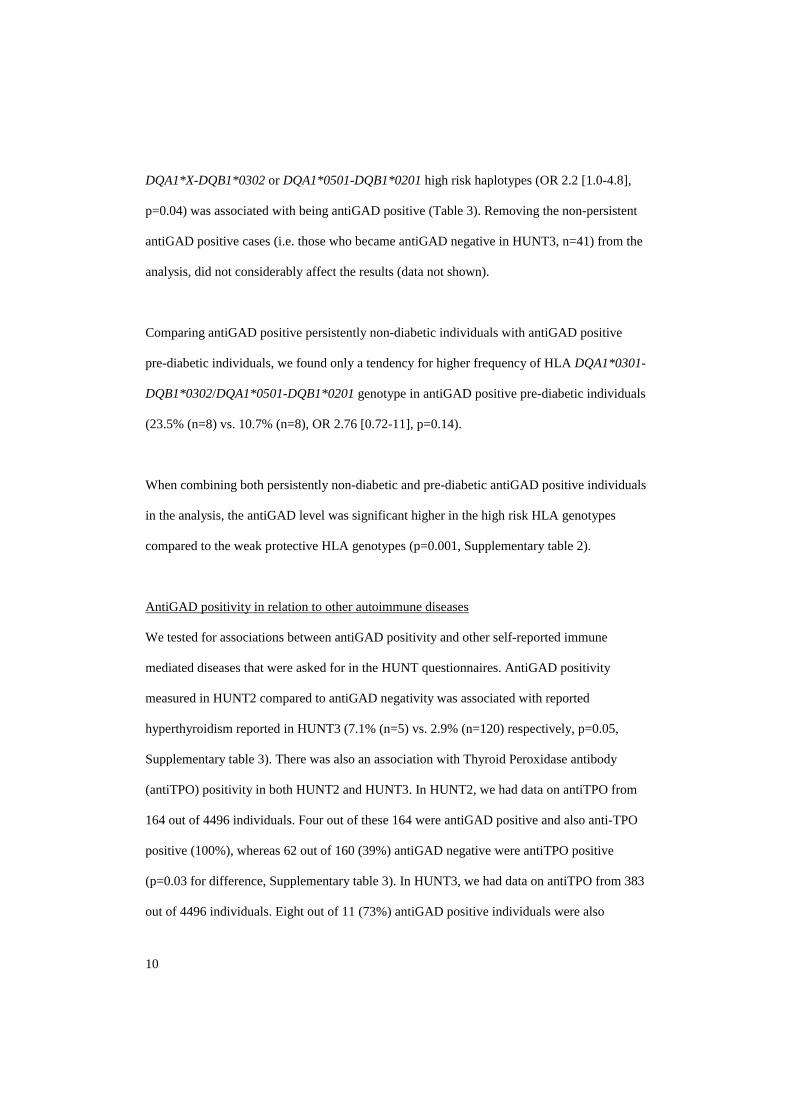
10
DQA1*X-DQB1*0302 or DQA1*0501-DQB1*0201 high risk haplotypes (OR 2.2 [1.0-4.8],
p=0.04) was associated with being antiGAD positive (Table 3). Removing the non-persistent
antiGAD positive cases (i.e. those who became antiGAD negative in HUNT3, n=41) from the
analysis, did not considerably affect the results (data not shown).
Comparing antiGAD positive persistently non-diabetic individuals with antiGAD positive
pre-diabetic individuals, we found only a tendency for higher frequency of HLA DQA1*0301-
DQB1*0302/DQA1*0501-DQB1*0201 genotype in antiGAD positive pre-diabetic individuals
(23.5% (n=8) vs. 10.7% (n=8), OR 2.76 [0.72-11], p=0.14).
When combining both persistently non-diabetic and pre-diabetic antiGAD positive individuals
in the analysis, the antiGAD level was significant higher in the high risk HLA genotypes
compared to the weak protective HLA genotypes (p=0.001, Supplementary table 2).
AntiGAD positivity in relation to other autoimmune diseases
We tested for associations between antiGAD positivity and other self-reported immune
mediated diseases that were asked for in the HUNT questionnaires. AntiGAD positivity
measured in HUNT2 compared to antiGAD negativity was associated with reported
hyperthyroidism reported in HUNT3 (7.1% (n=5) vs. 2.9% (n=120) respectively, p=0.05,
Supplementary table 3). There was also an association with Thyroid Peroxidase antibody
(antiTPO) positivity in both HUNT2 and HUNT3. In HUNT2, we had data on antiTPO from
164 out of 4496 individuals. Four out of these 164 were antiGAD positive and also anti-TPO
positive (100%), whereas 62 out of 160 (39%) antiGAD negative were antiTPO positive
(p=0.03 for difference, Supplementary table 3). In HUNT3, we had data on antiTPO from 383
out of 4496 individuals. Eight out of 11 (73%) antiGAD positive individuals were also

11
antiTPO positive compared to 134 out of 238 (36%) antiGAD negative ones (p=0.02 for
difference, Supplementary table 3).
Discussion
We investigated the prevalence, evolution and clinical impact of antiGAD in a non-diabetic
population recruited from all adults in a cohort from Norway. We also evaluated the
associations of high risk type 1 diabetes associated HLA genotypes to the presence of
antiGAD positivity in this non-diabetic population.
In our adult persistently non-diabetic population we find an antiGAD positivity prevalence of
1.7% which increases to 2.0% when including also pre-diabetic individuals. This is in fair
agreement with previous reports in which the prevalence ranges from 1-4% in both children
and adults from the general population (5, 8, 9, 19). However, in previous reports a
distinction was usually not made as to whether diabetes developed over time or not, nor to
phenotypic characteristics described here.
We observed that antiGAD in the non-diabetic population frequently changed from positivity
to negativity over a 9-13 years period, giving a conversion rate of 54%. Seroconversion from
antibody positive to antibody negative was associated with lower levels of antiGAD. Such
evolution may lead one to question the clinical importance of low and fleeting antibody
positivity. However, as elaborated below, the HLA associations constitute tangible evidence
of importance. Further, fleeting antiGAD positivity in LADA patients was previously shown
to impact on insulin secretion, as assessed from fasting levels of C-peptide (3).

12
In the persistently non-diabetic individuals we could not find any clinical parameters
conferring risk of diabetes that were associated with antiGAD positivity. Neither did the
presence or absence of stability of antiGAD positivity (positivity or not both in HUNT2 and
HUNT3) affect the clinical parameters. However, having high risk HLA DQA1-DQB1
haplotypes were associated with presence of GAD antibodies. This is in line whit a recent
study mainly based one adult relatives of type 2 diabetic cases which found that antiGAD was
associated with HLA high risk haplotypes regardless of family history of type 1 diabetes and
evolution of diabetes (17).
As mentioned above, given the etiological importance of these haplotypes it is reasonable to
conclude that even minor and fleeting antiGAD positivity is coupled to autoimmunity.
This notion is reinforced by finding associations between antiGAD positivity and
hyperthyroidism (in this population bound to be Graves’ disease) as well as presence of
antibody towards thyroid peroxidase. An association between type 1 diabetes and thyroid
autoimmune disease is well documented (20-22); however, an association with presence of
antiGAD in persistently non-diabetic individuals has, to our knowledge, not been described.
A comparison of antiGAD positive persistent non-diabetic individuals with pre-diabetic
individuals (i.e. antiGAD positive but non-diabetic at HUNT 2, diabetic at HUNT3) revealed
higher titers of antiGAD in HUNT2 in the latter. This could signify higher autoimmune
activity which in turn could precipitate overt diabetes in these individuals. However, also risk
factors for type 2 diabetes, such as BMI and age were higher and could participate in
evolution to diabetes. In this context it will be of great interest to follow those as of now
persistent non-diabetic individuals with antiGAD for possible evolution of diabetes in coming
years.

13
The antiGAD level (in only antiGAD positive individuals pooled with those who also
progressed to diabetes) was correlated with genetic risk as assessed by HLA haplotypes. This
finding agrees with that of the Botnia Prospective Study (17). The mentioned correlation
highlights the potential clinical importance of the risk HLA haplotypes for autoimmunity in
adults.
We did not find an association of antiGAD positivity and overall first degree family history of
diabetes in the non-diabetic subjects. In contrast such an association was found in the Botnia
study (17). This difference between studies may be explained by our questionnaire data not
being able to distinguish between type 1 and type 2 family histories of diabetes. It has been
shown in earlier studies that risk of developing type 1 diabetes and LADA is higher in
siblings of diabetic patients compared to having parents with diabetes (23, 24). We did see a
tendency for higher risk of being antiGAD positive in those having siblings with diabetes.
A limitation of this study is the sample size of the antiGAD positive individuals (both non-
diabetic and pre-diabetic) and should be considered when interpreting these results. One may
also argue one should confirm the positivity by performing competition assays. However, this
type of analysis has been done before and did not detect unspecific influence between anyone
of the three different antiGAD assays tested (25). A further limitation concerns the small
subset of the studied subjects on which the anti-TPO results are based. Therefor studies in
other populations should be performed to confirm the present results on autoimmune thyroid
diseases.

14
We conclude that antiGAD positivity in adult non-diabetic individuals from the general
population is 1) partly persistent over a long time period 2) not associated with clinical
parameters related to diabetes, but associated with increased frequency of high risk HLA
haplotypes known to be associated with type 1 diabetes and 3) associated with
hyperthyroidism and thyroid autoimmunity.
Acknowledgment
The HUNT study was performed as collaboration between the HUNT Research Centre
(Faculty of Medicine, Norwegian University of Science and Technology (NTNU)), Nord-
Trøndelag County Council, Central Norway Health Authority and the Norwegian Institute of
Public Health. Our study was funded by the liaison committee of the Central Norway
Regional Health Authority and the NTNU, and the liaison committee of St Olav’s Hospital
Trust and the Faculty of Medicine, NTNU. The authors would also thank Ms Oddrun Storrø,
Ms Marit Aarhaug and Ms Anne Kristin Lysakerrud at Unit for Immunology at St Olavs
Hospital, Trondheim, Norway for their appreciated technical assistance.
E.P.S. did the statistical analyses and wrote the manuscript. K.K. and F.S. participated in the
interpretation of data and reviewed/edited the manuscript. V.G. designed the study,
participated in the interpretation of data, contributed to the discussion and reviewed/edited the
manuscript.
References
1. Baekkeskov S, Aanstoot HJ, Christgau S, Reetz A, Solimena M, Cascalho M, Folli F, Richter-Olesen H, De Camilli P. Identification of the 64K autoantigen in insulin-dependent diabetes as the GABA-synthesizing enzyme glutamic acid decarboxylase. Nature 1990;347:151-156 2. Tuomilehto J, Zimmet P, Mackay IR, Koskela P, Vidgren G, Toivanen L, Tuomilehto-Wolf E, Kohtamaki K, Stengard J, Rowley MJ. Antibodies to glutamic acid decarboxylase as predictors of insulin-dependent diabetes mellitus before clinical onset of disease. Lancet 1994;343:1383-1385

15
3. Sorgjerd EP, Skorpen F, Kvaloy K, Midthjell K, Grill V. Time dynamics of autoantibodies are coupled to phenotypes and add to the heterogeneity of autoimmune diabetes in adults: the HUNT study, Norway. Diabetologia 2012;4. Vermeulen I, Weets I, Costa O, Asanghanwa M, Verhaeghen K, Decochez K, Ruige J, Casteels K, Wenzlau J, Hutton JC, Pipeleers DG, Gorus FK. An important minority of prediabetic first-degree relatives of type 1 diabetic patients derives from seroconversion to persistent autoantibody positivity after 10 years of age. Diabetologia 2012;55:413-4205. Siljander HT, Veijola R, Reunanen A, Virtanen SM, Akerblom HK, Knip M. Prediction of type 1 diabetes among siblings of affected children and in the general population. Diabetologia 2007;50:2272-22756. Veijola R, Reijonen H, Vahasalo P, Sabbah E, Kulmala P, Ilonen J, Akerblom HK, Knip M. HLA-DQB1-defined genetic susceptibility, beta cell autoimmunity, and metabolic characteristics in familial and nonfamilial insulin-dependent diabetes mellitus. Childhood Diabetes in Finland (DiMe) Study Group. J Clin Invest 1996;98:2489-24957. Rolandsson O, Hagg E, Janer M, Rutledge E, Gaur LK, Nilsson M, Hallmans G, Lernmark A. High GAD65 autoantibody levels in nondiabetic adults are associated with HLA but not with CTLA-4 or INS VNTR. J Intern Med 2003;253:447-4538. Ruige JB, Batstra MR, Aanstoot HJ, Bouter LM, Bruining GJ, De Neeling JN, Heine RJ. Low prevalence of antibodies to GAD65 in a 50- to 74-year-old general Dutch population. The Hoorn Study. Diabetes Care 1997;20:1108-11109. Lundgren VM, Isomaa B, Lyssenko V, Laurila E, Korhonen P, Groop LC, Tuomi T. GAD antibody positivity predicts type 2 diabetes in an adult population. Diabetes 2010;59:416-42210. LaGasse JM, Brantley MS, Leech NJ, Rowe RE, Monks S, Palmer JP, Nepom GT, McCulloch DK, Hagopian WA. Successful prospective prediction of type 1 diabetes in schoolchildren through multiple defined autoantibodies: an 8-year follow-up of the Washington State Diabetes Prediction Study. Diabetes Care 2002;25:505-51111. Schlosser M, Strebelow M, Rjasanowski I, Kerner W, Wassmuth R, Ziegler M. Prevalence of diabetes-associated autoantibodies in schoolchildren: the Karlsburg Type 1 Diabetes Risk Study. Ann N Y Acad Sci 2004;1037:114-11712. Rolandsson O, Hagg E, Nilsson M, Hallmans G, Mincheva-Nilsson L, Lernmark A. Prediction of diabetes with body mass index, oral glucose tolerance test and islet cell autoantibodies in a regional population. J Intern Med 2001;249:279-28813. Bosi EP, Garancini MP, Poggiali F, Bonifacio E, Gallus G. Low prevalence of islet autoimmunity in adult diabetes and low predictive value of islet autoantibodies in the general adult population of northern Italy. Diabetologia 1999;42:840-84414. Gale EA. Latent autoimmune diabetes in adults: a guide for the perplexed. Diabetologia 2005;48:2195-219915. Howson JM, Stevens H, Smyth DJ, Walker NM, Chandler KA, Bingley PJ, Todd JA. Evidence that HLA class I and II associations with type 1 diabetes, autoantibodies to GAD and autoantibodies to IA-2, are distinct. Diabetes 2011;60:2635-264416. Lambert AP, Gillespie KM, Thomson G, Cordell HJ, Todd JA, Gale EA, Bingley PJ. Absolute risk of childhood-onset type 1 diabetes defined by human leukocyte antigen class II genotype: a population-based study in the United Kingdom. J Clin Endocrinol Metab 2004;89:4037-404317. Andersen MK, Lundgren V, Isomaa B, Groop L, Tuomi T. Association of variants in HLA-DQA1-DQB1, PTPN22, INS, and CTLA4 with GAD autoantibodies and insulin secretion in nondiabetic adults of the Botnia Prospective Study. Eur J Endocrinol 2012;167:27-33

16
18. Holmen J, Midthjell,K, Krüger,Ø, Langhammer,A, Holmen,TL, Bratberg,GH, Vatten,L, Lund-Larsen,PG. The Nord-Trøndelag Health Study 1995–97 (HUNT2): objectives, contents, methods and participation. Norsk Epidemiologi 2003;13:19-32 19. Knip M, Korhonen S, Kulmala P, Veijola R, Reunanen A, Raitakari OT, Viikari J, Akerblom HK. Prediction of type 1 diabetes in the general population. Diabetes Care 2010;33:1206-1212 20. Hansen D, Bennedbaek FN, Hansen LK, Hoier-Madsen M, Jacobsen BB, Hegedus L. Thyroid function, morphology and autoimmunity in young patients with insulin-dependent diabetes mellitus. Eur J Endocrinol 1999;140:512-518 21. Barova H, Perusicova J, Hill M, Sterzl I, Vondra K, Masek Z. Anti-GAD-positive patients with type 1 diabetes mellitus have higher prevalence of autoimmune thyroiditis than anti-GAD-negative patients with type 1 and type 2 diabetes mellitus. Physiol Res 2004;53:279-286 22. Kordonouri O, Charpentier N, Hartmann R. GADA positivity at onset of type 1 diabetes is a risk factor for the development of autoimmune thyroiditis. Pediatr Diabetes 2011;12:31-33 23. Carlsson S, Midthjell K, Grill V. Influence of family history of diabetes on incidence and prevalence of latent autoimmune diabetes of the adult: results from the Nord-Trondelag Health Study. Diabetes Care 2007;30:3040-3045 24. Steck AK, Rewers MJ. Genetics of type 1 diabetes. Clin Chem 2011;57:176-185 25. Daka B, Svensson MK, Lernmark K, Mincheva-Nilsson L, Hallmans G, Rolandsson O. Low agreement between radio binding assays in analyzing glutamic acid decarboxylase (GAD65Ab) autoantibodies in patients classified with type 2 diabetes. Autoimmunity 2009;42:507-514

17
Table 1: Clinical characteristics of persistently non-diabetic adult individuals stratified by being antiGAD negative and antiGAD positive in HUNT2.
AntiGAD negative AntiGAD positive p-valueN 4420 76 (1.7%)Sex (male) 2208 (50%) 40 (52.6%) 0.64Age at attendance 47.6 (19.4-85.1) 41.9 (21.3-77.1) 0.22Waist circumference (cm) 86 (55-131) 84 (64-109) 0.69BMI (kg/m2) 25.6 (16.2-52.8) 26.0 (19.4-34.8) 0.70Systolic blood pressure (mmHg) 133 (82-225) 132 (102-189) 0.82Diastolic blood pressure (mmHg) 79 (44-136) 78 (52-120) 0.85Glucose, non-fasting (mmol/l) 5.1 (0-14.4) 5.1 (3.5-7.5) 0.89Cholesterol (mmol/l) 5.8 (1.9-13.2) 5.7 (3.4-8.2) 0.35HDL cholesterol (mmol/l) 1.4 (0.5-3.8) 1.3 (0.6-2.4) 0.62Triacylglycerol b (mmol/l) 1.42 (0.31-18.6) 1.31 (0.24-4.73) 0.30Family history of diabetes
No 3229 55Yes 1181 (26.8%) 21 (27.6%) 0.86
SmokingNo 3224 59Yes 1052 (24.9%) 17 (22.4%) 0.65
Data is given as numbers (%) and median (min-max value).Unadjusted p- value calculated by Mann–Whitney U test for continuous data and by 2 test for categorical data.
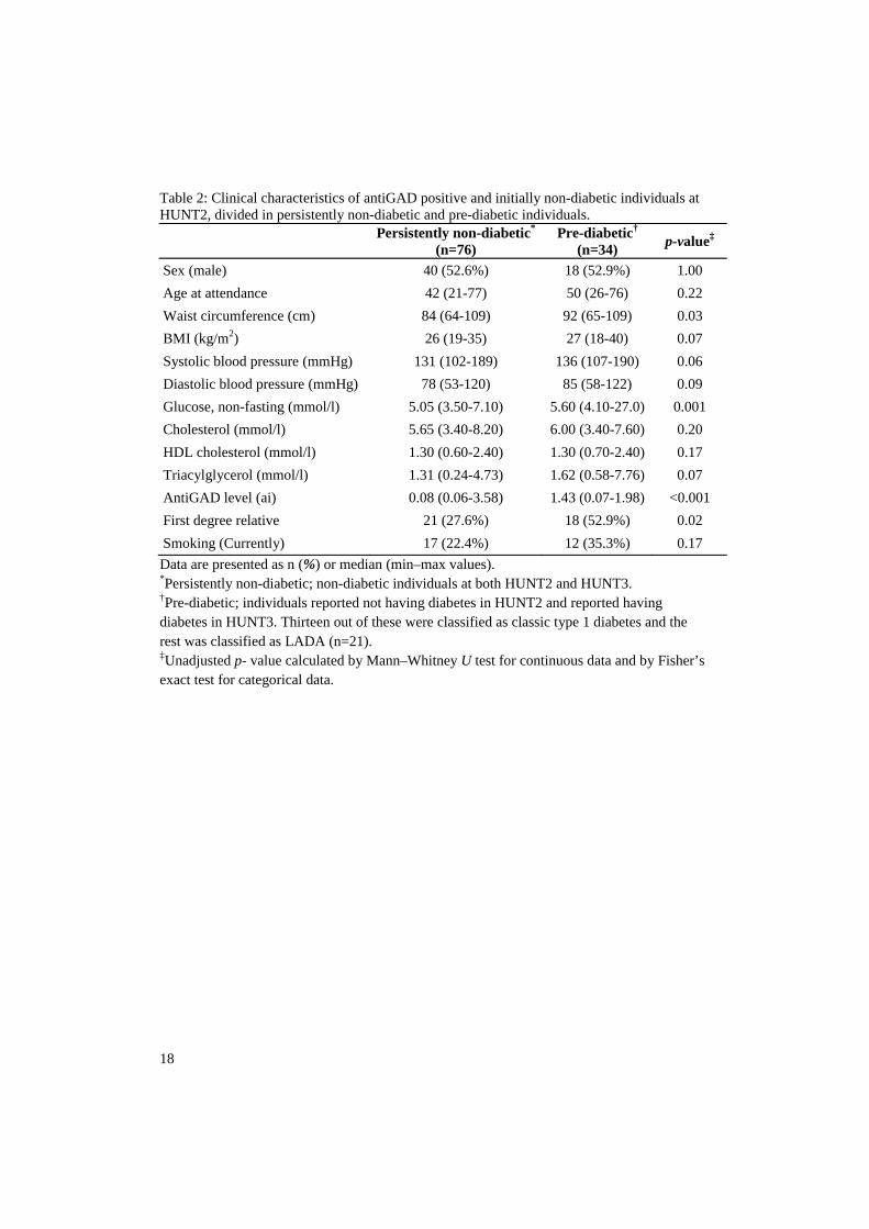
18
Table 2: Clinical characteristics of antiGAD positive and initially non-diabetic individuals at HUNT2, divided in persistently non-diabetic and pre-diabetic individuals.
Persistently non-diabetic*
(n=76)Pre-diabetic†
(n=34) p-value‡
Sex (male) 40 (52.6%) 18 (52.9%) 1.00Age at attendance 42 (21-77) 50 (26-76) 0.22Waist circumference (cm) 84 (64-109) 92 (65-109) 0.03BMI (kg/m2) 26 (19-35) 27 (18-40) 0.07Systolic blood pressure (mmHg) 131 (102-189) 136 (107-190) 0.06Diastolic blood pressure (mmHg) 78 (53-120) 85 (58-122) 0.09Glucose, non-fasting (mmol/l) 5.05 (3.50-7.10) 5.60 (4.10-27.0) 0.001Cholesterol (mmol/l) 5.65 (3.40-8.20) 6.00 (3.40-7.60) 0.20HDL cholesterol (mmol/l) 1.30 (0.60-2.40) 1.30 (0.70-2.40) 0.17Triacylglycerol (mmol/l) 1.31 (0.24-4.73) 1.62 (0.58-7.76) 0.07AntiGAD level (ai) 0.08 (0.06-3.58) 1.43 (0.07-1.98) <0.001 First degree relative 21 (27.6%) 18 (52.9%) 0.02Smoking (Currently) 17 (22.4%) 12 (35.3%) 0.17Data are presented as n (%) or median (min–max values). *Persistently non-diabetic; non-diabetic individuals at both HUNT2 and HUNT3. †Pre-diabetic; individuals reported not having diabetes in HUNT2 and reported having diabetes in HUNT3. Thirteen out of these were classified as classic type 1 diabetes and the rest was classified as LADA (n=21).‡Unadjusted p- value calculated by Mann–Whitney U test for continuous data and by Fisher’s exact test for categorical data.
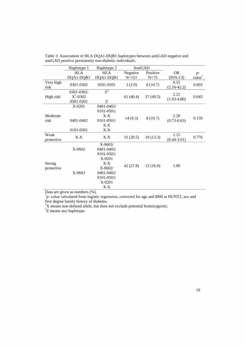
19
Table 3: Association of HLA DQA1-DQB1 haplotypes between antiGAD negative and antiGAD positive persistently non-diabetic individuals.
Data are given as numbers (%).*p- value calculated from logistic regression, corrected for age and BMI at HUNT2, sex and first degree family history of diabetes.†X means non-defined allele, but does not exclude potential homozygosity.‡Z means any haplotype.
Haplotype 1 Haplotype 2 AntiGADHLA
DQA1-DQB1HLA
DQA1-DQB1NegativeN=151
PositiveN=75
OR (95% CI)
p-value*
Very high risk 0301-0302 0501-0201 3 (2.0) 8 (10.7) 9.55
(2.16-42.2) 0.003
High risk0301-0302/
X†-03020501-0201
Z‡
Z61 (40.4) 37 (49.3) 2.22
(1.03-4.80) 0.043
Moderaterisk
X-0201
0401-0402
0101-0501
0401-0402/0101-0501/
X-X0101-0501/
X-XX-X
14 (9.3) 8 (10.7) 2.20(0.73-6.63) 0.159
Weak protective X-X X-X 31 (20.5) 10 (13.3) 1.15
(0.44-3.01) 0.776
Strong protective
X-0602
X-0603
X-0603/0401-0402/0101-0501/
X-0201X-X
X-0602/0401-0402/0101-0501/
X-0201X-X
42 (27.8) 12 (16.0) 1.00

20
Legends to the figures
Figure 1: Flow sheet over included cases followed from HUNT2 until HUNT3
Figure 2: Prevalence of antiGAD positivity amongst the adult population in the HUNT2 survey distributed across different age categories.

21
FIGURE 1
Selected participants from HUNT2
Non-diabetic HUNT2N=4496
Pre-diabetic HUNT2N=55
N=76AntiGAD pos
HUNT2
N=4420AntiGAD neg
HUNT2
N=35Anti-GAD pos
HUNT3
Autoimmune diabetes in
HUNT3Persistently non-diabetic
HUNT3
N=41Anti-GAD neg
HUNT3
N=34AntiGAD pos
HUNT2
N=21AntiGAD neg
HUNT2

22
FIGURE 2

Supplementary table 1: Clinical characteristics from HUNT2 between antiGAD positivity persisters and converters in persistently non-diabetic participants.
AntiGAD negative AntiGAD positive p-value N=79 41 35 Sex (male) 19 (46%) 21 (60%) 0.24 Age at attendance 44.3 (21.7-77.1) 39.7 (21.3-65.9) 0.24 Waist circumference (cm) 82 (64-120) 85 (64-106) 0.24 BMI (kg/m2) 25.6 (19.4-34.2) 26.3 (19.4-34.8) 0.32 Systolic blood pressure (mmHg) 131 (102-188) 132 (104-189) 0.89 Diastolic blood pressure (mmHg) 78 (60-120) 79 (53-105) 0.81 Glucose, non-fasting (mmol/l) 5.2 (3.5-7.5) 5.0 (3.7-7.1) 0.90 Cholesterol (mmol/l) 5.5 (3.4-8.2) 5.8 (4.1-7.8) 0.74 HDL cholesterol (mmol/l) 1.3 (0.7-2.4) 1.4 (0.6-2.2) 0.84 Triacylglycerol (mmol/l) 1.3 (0.2-4.0) 1.3 (0.6-4.7) 0.73 AntiGAD titre (ai) 0.06 (0.06-1.34) 0.27 (0.06-3.58) <0.001 Family history of diabetes No 29 26 Yes 12 (29.3%) 9 (25.7%) 0.80 Smoking (Yes) No 30 29 Yes 11 (26.8%) 6 (17.1%) 0.41 Data presented as numbers (%) and median (min-max) value. Unadjusted p- value calculated by Mann–Whitney U test for continuous data and by Fisher’s exact test for categorical data.

Supplementary table 2: AntiGAD titre in initially antiGAD positive participants in HUNT2 stratified by HLA genotype risk and in persistently non-diabetic individuals in both HUNT2 and HUNT3 and pre-diabetic individuals who developed diabetes during follow-up (between HUNT2 and HUNT3).
Haplotype 1 Haplotype 2 AntiGAD titre*
HLA DQA1-DQB1
HLA DQA1-DQB1
Non-diabeticN=75
Pre-diabeticN=34
Very high risk 0301-0302 0501-0201 1.11
(0.06-3.58)1.49
(0.7-1.74)
High risk0301-0302/
X†-03020501-0201
Z‡
Z
0.08(0.06-1.82)
1.46(0.34-1.98)
Moderaterisk
X-0201
0401-0402
0101-0501
0401-0402/0101-0501/
X-X0101-0501/
X-XX-X
0.08(0.06-2.05)
0.74(0.25-1.36)
Weak protective X-X X-X 0.09
(0.06-049)0.82 (0.07-
1.79)
Strong protective
X-0602
X-0603
X-0603/0401-0402/0101-0501/
X-0201X-X
X-0602/0401-0402/0101-0501/
X-0201X-X
0.065(0.06-1.57 No subjects
Data are given as median (min-max value).*AntiGAD titre was significant higher in the vhigh risk HLA genotypes compared to the weakprotective HLA genotypes (p=0.001). Data analysed by Kruskal-Wallis Test and the non-diabetic and pre-diabetic combined.†X means non-defined allele, but does not exclude potential homozygosity.‡Z means any haplotype.
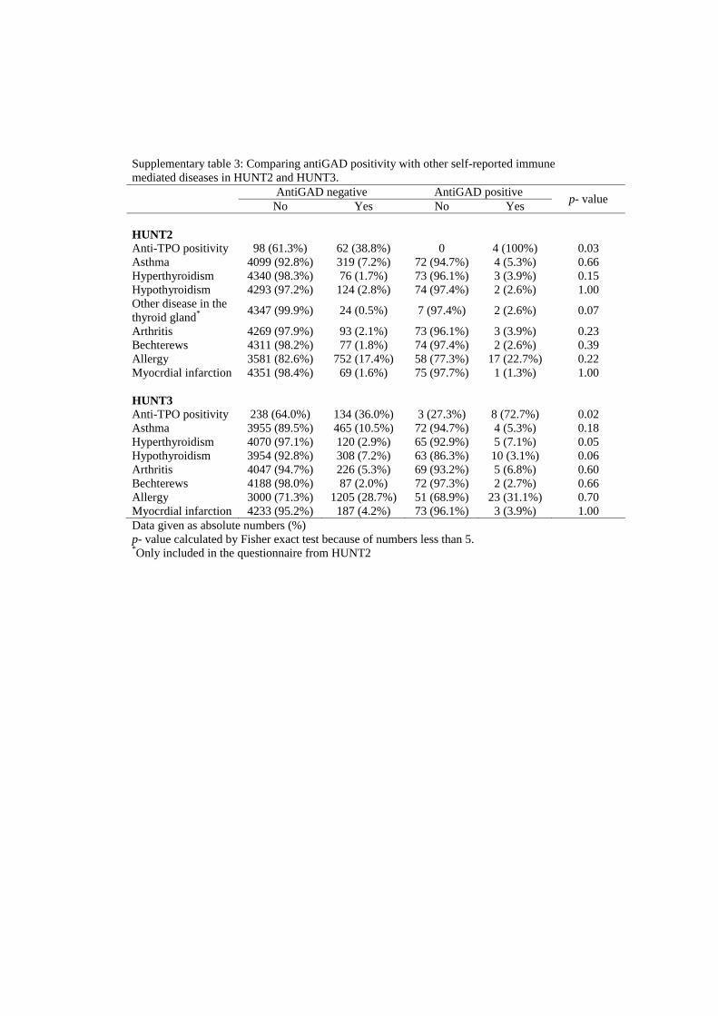
Supplementary table 3: Comparing antiGAD positivity with other self-reported immune mediated diseases in HUNT2 and HUNT3.
AntiGAD negative AntiGAD positive p- valueNo Yes No Yes
HUNT2Anti-TPO positivity 98 (61.3%) 62 (38.8%) 0 4 (100%) 0.03Asthma 4099 (92.8%) 319 (7.2%) 72 (94.7%) 4 (5.3%) 0.66Hyperthyroidism 4340 (98.3%) 76 (1.7%) 73 (96.1%) 3 (3.9%) 0.15Hypothyroidism 4293 (97.2%) 124 (2.8%) 74 (97.4%) 2 (2.6%) 1.00Other disease in the thyroid gland* 4347 (99.9%) 24 (0.5%) 7 (97.4%) 2 (2.6%) 0.07
Arthritis 4269 (97.9%) 93 (2.1%) 73 (96.1%) 3 (3.9%) 0.23Bechterews 4311 (98.2%) 77 (1.8%) 74 (97.4%) 2 (2.6%) 0.39Allergy 3581 (82.6%) 752 (17.4%) 58 (77.3%) 17 (22.7%) 0.22Myocrdial infarction 4351 (98.4%) 69 (1.6%) 75 (97.7%) 1 (1.3%) 1.00
HUNT3Anti-TPO positivity 238 (64.0%) 134 (36.0%) 3 (27.3%) 8 (72.7%) 0.02Asthma 3955 (89.5%) 465 (10.5%) 72 (94.7%) 4 (5.3%) 0.18Hyperthyroidism 4070 (97.1%) 120 (2.9%) 65 (92.9%) 5 (7.1%) 0.05Hypothyroidism 3954 (92.8%) 308 (7.2%) 63 (86.3%) 10 (3.1%) 0.06Arthritis 4047 (94.7%) 226 (5.3%) 69 (93.2%) 5 (6.8%) 0.60Bechterews 4188 (98.0%) 87 (2.0%) 72 (97.3%) 2 (2.7%) 0.66Allergy 3000 (71.3%) 1205 (28.7%) 51 (68.9%) 23 (31.1%) 0.70Myocrdial infarction 4233 (95.2%) 187 (4.2%) 73 (96.1%) 3 (3.9%) 1.00Data given as absolute numbers (%)p- value calculated by Fisher exact test because of numbers less than 5.*Only included in the questionnaire from HUNT2


Appendix I
Q1-HUNT2


146

147

148

Appendix II
Q1-HUNT3


Du inviteres herved til å delta i den tredje store Helseundersøkelsen i Nord-Trøndelag (HUNT 3). Ved å delta får du en enkel undersøkelse av din egen helse, og du gir samtidig et viktig bidrag til medisinsk forskning.
Hver deltaker er like viktig, enten du er ung eller gammel, frisk eller syk, er HUNT-veteran eller møter for første gang. Tilsvarende undersøkelse er tidligere gjennom-ført i 1984-86 (HUNT 1) og 1995-97 (HUNT 2 og Ung-HUNT). For å kunne studereårsaker til sykdom, er det viktig at også de som tidligere har deltatt møter fram.
Vennligst fyll ut spørreskjemaet, og ta det med når du møter til undersøkelse.
Undersøkelsen tar vanligvis ca 1/2 time. Du vil få brev med resultater fra dineprøver etter noen uker. Dersom noen av resultatene er utenom det normale, vil dubli anbefalt undersøkelse hos fastlegen din.
Du kan lese mer om HUNT 3 i den vedlagte brosjyren eller på www.hunt.ntnu.no.Har du spørsmål, kan du også ringe til HUNT forskningssenter, tlf 74075180.
Vel møtt til undersøkelsen!
Vennlig hilsen
Steinar Krokstad Jostein Holmen Stig A. SlørdahlFørsteamanuensis Professor, daglig leder Professor, dekanus
Prosjektleder HUNT 3 HUNT forskningssenter Det medisinske fakultet, NTNU
Dersom det foreslåtte tidspunktet ikke passer for deg, behøver du ikkebestille ny time. Du kan møte når det passer deg innenfor åpningstiden,men det kan da bli noe ventetid. Du kan også møte i en annen kommune,hvis det skulle passe bedre. Takk for at du deltar!
Åpningstida:
Invitasjon til HUNT 3
Tid og sted for oppmøte
En ti
me
for
bedr
e fo
lkeh
else
Viktig
EnkeltGra
tis
VE
ST
VIK
RE
KLA
ME
AS
. F
OT
O:
HA
RA
LD S
ÆT
ER
ØY
OG
JO
HA
N A
RN
TN
ES
GÅ
RD
.

Slik fyller du ut skjemaet
Rett Galt ��X
• Skjemaet vil bli lest maskinelt.
• Det er derfor viktig at du krysser av riktig:
• Krysser du feil sted, retter du ved å fylle boksen slik:
• Skriv tydelige tall:
• Bruk bare svart eller blå penn. Ikke bruk blyant eller tusj.

1SIDE
HE
LSE
UN
DE
RSØ
KE
LSE
N I
NO
RD
-TR
ØN
DE
LAG
HELSE OG DAGLIGLIV SYKDOMMER OG PLAGER
Hvordan er helsa di nå?1
Dårlig
Har du noen langvarig (minst 1 år)sykdom, skade eller lidelse av fysiskeller psykisk art som nedsetter dinefunksjoner i ditt daglige liv?
Hvor mye vil du si at dine funksjoner er nedsatt?
2
Ikke helt god God Svært god
Ja Nei
Har du i løpet av de siste 12 måneder vært hos:6
Ja Nei
Har du vært innlagt i sykehusi løpet av de siste 12 måneder?
7 Ja Nei
Har du kroppslige smerter nå somhar vart mer enn 6 måneder?
3
Hvor sterke kroppslige smerter har du hatt i løpetav de siste 4 uker?
4
Ja Nei
Er bevegelseshemmet..................
Har nedsatt syn .............................
Har nedsatt hørsel ........................
Hemmet pga. kroppslig sykdom.
Hemmet pga. psykisk sykdom.....
Hjerteinfarkt ...................................
Angina pectoris (hjertekrampe) ...
Hjertesvikt ......................................
Annen hjertesykdom.....................
Hjerneslag/hjerneblødning ..........
Nyresykdom...................................
Astma .............................................
Kronisk bronkitt, emfysem, KOLS
Diabetes (sukkersyke)....................
Psoriasis..........................................
Eksem på hendene .......................
Kreftsykdom...................................
Epilepsi...........................................
Leddgikt (reumatoid artritt) ..........
Bechterews sykdom ......................
Sarkoidose .....................................
Beinskjørhet (osteoporose) ..........
Fibromyalgi ....................................
Slitasjegikt (artrose) .......................
Psykiske plager som duhar søkt hjelp for ...........................
Fastlege/allmennlege .....................................
Annen legespesialist utenfor sykehus ...........
Konsultasjon uten innleggelse
- ved psykiatrisk poliklinikk.........................
- ved annen poliklinikk i sykehus ...............
Kiropraktor .......................................................
Homøopat, akupunktør, soneterapeut, hånds-
pålegger eller annen alternativ behandler ...
Har du hatt noe anfall med pipendeeller tung pust de siste 12 måneder?
8 Ja Nei
Har du noen gang de siste 5 årbrukt medisiner for astma, kroniskbronkitt, emfysem eller KOLS?
9Ja Nei
Bruker du, eller har du brukt,medisin mot høyt blodtrykk?
10 Ja Nei
Har du noen gang fått påvist forhøyt blodsukker?
12 Ja Nei
Har du, eller har du noengang hatt, noen av dissesykdommene/plagene: (Sett ett kryss pr. linje)
11
Mode-rate Sterke
MegetsterkeIngen
Megetsvake Svake
I hvilken grad har din fysiske helse eller følelses-messige problemer begrenset deg i din vanligesosiale omgang med familie eller venner i løpet avde siste 4 uker?
5
Mye
Kunne ikkeha sosialomgang
Ikke i dethele tatt En del Litt
Hvis ja:
Hvis ja: I hvilken situasjon første gang?
Hvis ja, hvor gammelvar du første gang?
Littnedsatt
Middelsnedsatt
Myenedsatt
Ja Nei
Eksempel:
Ved helseundersøkelse... Under sykdom .............
Under svangerskap ......... Annet ............................
HELSETJENESTER
år gammel
år gammel
år gammel
år gammel
år gammel
år gammel
år gammel
år gammel
år gammel
år gammel
år gammel
år gammel
år gammel
år gammel
år gammel
år gammel
år gammel
år gammel
år gammel
år gammel
år gammel
SIDE
HE
LSE
UN
DE
RSØ
KE
LSE
N I
NO
RD
-TR
ØN
DE
LAG
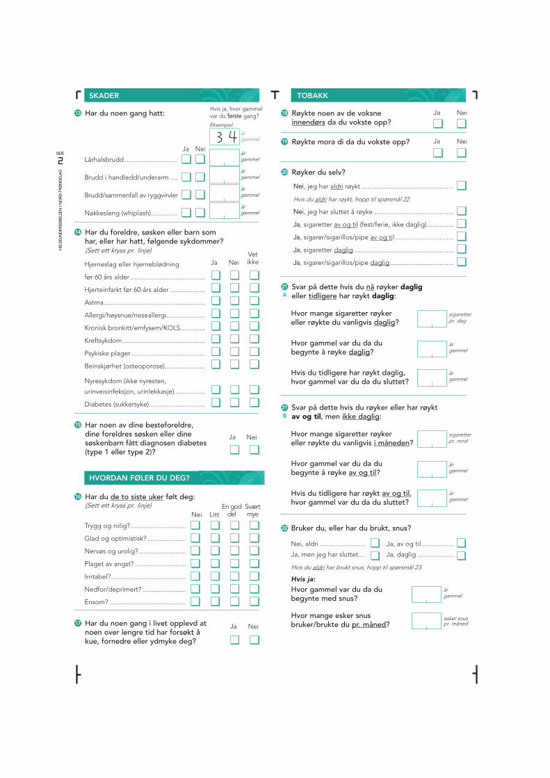
2SIDE
HE
LSE
UN
DE
RSØ
KE
LSE
N I
NO
RD
-TR
ØN
DE
LAG
Lårhalsbrudd ..............................
Brudd i handledd/underarm ....
Brudd/sammenfall av ryggvirvler
Nakkesleng (whiplash)...............
Har du noen gang hatt: 13
Ja Nei
Hjerneslag eller hjerneblødning
før 60 års alder...........................................
Hjerteinfarkt før 60-års alder ....................
Astma..........................................................
Allergi/høysnue/neseallergi......................
Kronisk bronkitt/emfysem/KOLS..............
Kreftsykdom ...............................................
Psykiske plager ..........................................
Beinskjørhet (osteoporose).......................
Nyresykdom (ikke nyresten,
urinveisinfeksjon, urinlekkasje) .................
Diabetes (sukkersyke)................................
Har du foreldre, søsken eller barn somhar, eller har hatt, følgende sykdommer?(Sett ett kryss pr. linje)
14
Ja NeiVetikke
Trygg og rolig?................................
Glad og optimistisk? ......................
Nervøs og urolig?...........................
Plaget av angst? .............................
Irritabel?...........................................
Nedfor/deprimert? .........................
Ensom? ............................................
Nei, jeg har aldri røykt .....................................................
Hvis du aldri har røykt, hopp til spørsmål 22.
Nei, jeg har sluttet å røyke..............................................
Ja, sigaretter av og til (fest/ferie, ikke daglig) ...............
Ja, sigarer/sigarillos/pipe av og til .................................
Ja, sigaretter daglig .........................................................
Ja, sigarer/sigarillos/pipe daglig ....................................
Har du de to siste uker følt deg:(Sett ett kryss pr. linje)
16
LittNeiSværtmye
En goddel
Har noen av dine besteforeldre,dine foreldres søsken eller dinesøskenbarn fått diagnosen diabetes(type 1 eller type 2)?
15
Ja Nei
Røykte noen av de voksne innendørs da du vokste opp?
18 Ja Nei
Røykte mora di da du vokste opp?19 Ja Nei
Røyker du selv? 20
Svar på dette hvis du nå røyker daglig eller tidligere har røykt daglig:
Hvor mange sigaretter røykereller røykte du vanligvis daglig?
21
Har du noen gang i livet opplevd atnoen over lengre tid har forsøkt åkue, fornedre eller ydmyke deg?
17 Ja Nei
sigaretterpr. dag
Hvor gammel var du da dubegynte å røyke daglig?
Hvis du tidligere har røykt daglig,hvor gammel var du da du sluttet?
Svar på dette hvis du røyker eller har røykt av og til, men ikke daglig:
Hvor mange sigaretter røykereller røykte du vanligvis i måneden?
Hvor gammel var du da du begynte å røyke av og til?
Hvis du tidligere har røykt av og til,hvor gammel var du da du sluttet?
Bruker du, eller har du brukt, snus?22
Hvor gammel var du da dubegynte med snus?
Hvor mange esker snusbruker/brukte du pr. måned?
esker snuspr. måned
A
21B
Hvis ja:
Nei, aldri .......................... Ja, av og til...................
Ja, men jeg har sluttet.... Ja, daglig .....................
Hvis du aldri har brukt snus, hopp til spørsmål 23.
SKADER
HVORDAN FØLER DU DEG?
TOBAKK
SIDE
HE
LSE
UN
DE
RSØ
KE
LSE
N I
NO
RD
-TR
ØN
DE
LAG
Hvis ja, hvor gammelvar du første gang?Eksempel:
år gammel
år gammel
år gammel
år gammel
år gammel
år gammel
år gammel
sigaretterpr. mnd
år gammel
år gammel
år gammel
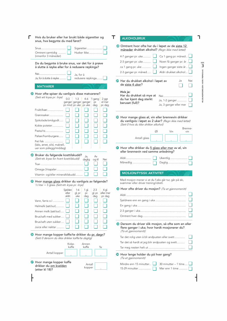
3SIDE
HE
LSE
UN
DE
RSØ
KE
LSE
N I
NO
RD
-TR
ØN
DE
LAG
Hvis du bruker eller har brukt både sigaretter og snus, hva begynte du med først?
Omtrent hvor ofte har du i løpet av de siste 12 måneder drukket alkohol? (Regn ikke med lettøl)
28
Hvis ja:Har du drukket så mye atdu har kjent deg sterktberuset (full)?
Da du begynte å bruke snus, var det for å prøve å slutte å røyke eller for å redusere røykinga?
Hvor ofte spiser du vanligvis disse matvarene?(Sett ett kryss pr. linje)
23
2 ggrel merpr. dag
1 gangpr.
dag
4-6gangerpr. uke
1-3gangerpr. uke
0-3gangerpr. mnd
Vann, farris o.l ...............
Helmelk (søt/sur)...........
Annen melk (søt/sur) ....
Brus/saft med sukker....
Brus/saft uten sukker....
Juice eller nektar ..........
Antall kopper
Koke-kaffe
Hvor mange glass drikker du vanligvis av følgende?1/2 liter = 3 glass (Sett ett kryss pr. linje)
25
Aldri ..................................................................................
Sjeldnere enn en gang i uka ..........................................
En gang i uka ...................................................................
2-3 ganger i uka...............................................................
Omtrent hver dag............................................................
4 gl.eller merpr. dag
2-3gl. pr.dag
1 gl.pr.
dag
1-6gl. pruke
Sjeldenelleraldri
Tran ..........................................................
Omega-3-kapsler ....................................
Vitamin- og/eller mineraltilskudd..........
Bruker du følgende kosttilskudd?(Sett ett kryss for hvert kosttilskudd)
24
NeiAv
og tilJa,
daglig
ALKOHOLBRUK
MOSJON/FYSISK AKTIVITET
Hvor ofte driver du mosjon? (Ta et gjennomsnitt)32
Tar det rolig uten å bli andpusten eller svett ..............
Tar det så hardt at jeg blir andpusten og svett...........
Tar meg nesten helt ut ..................................................
Dersom du driver slik mosjon, så ofte som en eller flere ganger i uka; hvor hardt mosjonerer du? (Ta et gjennomsnitt)
33
Hvor mange kopper kaffe/te drikker du pr. døgn?(Sett 0 dersom du ikke drikker kaffe/te daglig)
26
Hvor mange kopper kaffedrikker du om kvelden(etter kl 18)?
27Antall
kopper
Annenkaffe Te
Antall glass
Hvor mange glass øl, vin eller brennevin drikkerdu vanligvis i løpet av 2 uker? (Regn ikke med lettøl)(Sett 0 hvis du ikke drikker alkohol)
30
VinBrenne-
vin
Har du drukket alkohol i løpet avde siste 4 uker?
29 Ja Nei
Med mosjon mener vi at du f.eks går tur, går på ski,svømmer eller driver trening/idrett.
31
Hvor lenge holder du på hver gang? (Ta et gjennomsnitt)
34
Snus.................................. Sigaretter......................
Omtrent samtidig .......... Husker ikke...................(innenfor 3 måneder)
Nei.............................................. Ja, for å Ja, for å slutte å røyke ........... redusere røykinga........
Frukt/bær......................
Grønnsaker...................
Sjokolade/smågodt .....
Kokte poteter...............
Pasta/ris ........................
Pølser/hamburgere......
Fet fisk .........................(laks, ørret, sild, makrell,uer som pålegg/middag)
4-7 ganger pr. uke........... Ca 1 gang pr. måned ..
2-3 ganger pr. uke........... Noen få ganger pr. år .
ca 1 gang pr. uke ............ Ingen ganger siste år ..
2-3 ganger pr. måned..... Aldri drukket alkohol...
Nei................................
Ja, 1-2 ganger .............
Ja, 3 ganger eller mer
Hvor ofte drikker du 5 glass eller mer av øl, vineller brennevin ved samme anledning?
Aldri.................................. Ukentlig ........................
Månedlig ......................... Daglig...........................
Mindre enn 15 minutter.. 30 minutter – 1 time....
15-29 minutter ................. Mer enn 1 time ............
SIDE
HE
LSE
UN
DE
RSØ
KE
LSE
N I
NO
RD
-TR
ØN
DE
LAG
MATVARER
Øl

4SIDE
HE
LSE
UN
DE
RSØ
KE
LSE
N I
NO
RD
-TR
ØN
DE
LAG
Har du vanligvis minst 30 minutterfysisk aktivitet daglig på arbeidog/eller i fritida?
35 Ja Nei
Omtrent hvor mange timer sitterdu i ro på en vanlig hverdag? (Regn med både jobb og fritid)
36
Antall timer
cm Husker ikke
For det meste stillesittende arbeid (f.eks skrivebordsarbeid, montering) ...............................
Arbeid som krever at du går mye (f.eks ekspeditørarbeid, lett industriarb.,undervisning) .
Arbeid hvor du går og løfter mye(f.eks postbud, pleier, bygningsarbeid)...........................
Tungt kroppsarbeid (f.eks skogsarbeid, tungtjordbruksarbeid, tungt bygningsarbeid) .........................
Hvis du er i lønnet eller ulønnet arbeid, hvordan vildu beskrive arbeidet ditt? (Sett ett kryss)
37
Omtrent hva var din høyde da du var 18 år? 38
kg Husker ikke
Omtrent hva var din kroppsvekt da du var 18 år?39
Nei, for tung
Er du fornøyd med vekta di nå?40
Nei, for lettJa
Er din kroppsvekt minst 2 kg lavere nåenn for 1 år siden?
42 Ja Nei
Ja, mange ganger
Har du forsøkt å slanke deg i løpet av de siste 10 år? 41
Ja, noen gangerNei
Vet ikke
Hva er grunnen til dette?Sykdom/stressSlanking
Hvis ja:
Har det vært dødsfall i nær familie?(barn, ektefelle/samboer, søsken ellerforeldre)
43
Har du vært i overhengende livsfarepga. alvorlig ulykke, katastrofe,voldssituasjon eller krig?
44
Har du hatt samlivsbrudd i ekteskapeller i lengre samboerforhold?
45
46
47
Vokste du opp på gård med husdyr?52
Hvis du har svart ja på et eller flere av spm 43, 44eller 45; i hvilken grad har du hatt reaksjoner pådette de siste 7 dager?
Hvem vokste du opp sammen med?
49 Døde noen av dineforeldre da du var barn?
50 Vokste du opp med kjæledyr?
48
53
Svært fornøyd ...................
Meget fornøyd..................
Ganske fornøyd ................
Både/og ............................
Nokså misfornøyd.........
Meget misfornøyd ........
Svært misfornøyd..........
Når du tenker på barndommen/oppveksten din, vil du beskrive den som:
Ble dine foreldre skilt, ellerflyttet de fra hverandre, dadu var barn?
Hvor mye melk eller yoghurt drakk du vanligvis?51
2-3 gl.pr. dag
Mer enn3 glasspr. dag
Sjelden/aldri
1-6 gl.pr. uke
1 glasspr. dag
Når du tenker på hvordan du har det for tida, er dustort sett fornøyd med tilværelsen eller er du stortsett misfornøyd? (Sett ett kryss)
54
Ja Nei
Ja Nei
Ja Nei
Ja Nei
Ikke i det hele tatt........... I moderat grad.............
Litt..................................... I høy grad.....................
Mor ................................... Andre slektninger ........
Far..................................... Adoptivforeldre ...........
Stemor/stefar................... Foster-/pleieforeldre ...
Nei ............................
Ja, før jeg var 7 år....
Ja, da jeg var 7-18 år
Nei .............................
Ja, før jeg var 7 år ....
Ja, da jeg var 7-18 år
Nei ................................
Ja, katt.............................. Ja, hund........................
Ja, hest............................. Ja, annet levende dyr .
Svært god ........................ Vanskelig ......................
God .................................. Svært vanskelig............
Middels ............................
SIDE
HE
LSE
UN
DE
RSØ
KE
LSE
N I
NO
RD
-TR
ØN
DE
LAG
ARBEID
HØYDE/VEKT
ALVORLIGE LIVSHENDELSER SISTE 12 MÅNEDER
OPPVEKST - DA DU VAR 0-18 ÅR
ALT I ALT

Appendix III
Diabetes questionnaire HUNT2

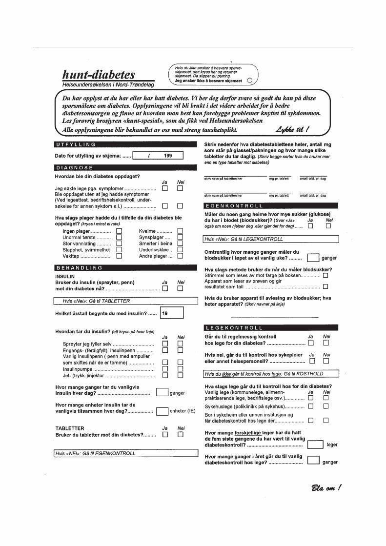

Appendix IV
Diabetes questionnaire HUNT3


����
����
�������������� ��������������� ������� ���������� ��������������� � ������������������������� ������������������������������������������������������������������������������������������������������������������������������������!��������������������� �
"�������������������������������#�������������������
��� ����������������� ������ �� �� �������
���� ���� ���
������������ �� �� ���������� ��
�����������������������������������
��������������� ���"�����������#��� ���������� ���
���������� ����� ���
��$��������������� ���� #�%�����&���������� ����� �������
�������� �������������%%���'�
��� ���#��� �� ������������%%���'�
�� ���
�������� ���������������������������������������
� �������������������������������������������������� ����������������������������������������������������������������������������
����� ���� �����#����� ������ �*����������������'�
�� ������ ������ ����������������� �����
������� ���������������������������������������������������������
������ ��������������������������������������������������
��������������� �����������������������!�������������� ���� ������� �������������������������
$�"�%$�$������������ ���+�%�����"�%���"%��%�<���������������#'������������������
���������������������������
�� ���
�&'%(��()$���������� ����������������'�
�� ���
� ������
=# ���������������*�����*��������������+� �����<���*������ ���+� �>������<'��!���"#$���������������%�����������������%�������������� ������������������������������
��
�� ���
������������
@#����� ����� ������������ �*��� �����������������' �������������������������
� �� ���
@#����� ������ �*�������% ������ ��������*� ��%������ ' ����������������������������������
������������������������������������&�'��������������*+�
�� ���
������������
������� ��
C�����*�����������������# ������ ���������� �%���������� �����G���'�!������������������������ ���
��
��� ���#��� ���������������� ��'�
�
��� ���#��� �������������� �������������'�
�
���������������� ��'���!�����������������%��
��� ���
"�� ������ ����������������������������������������������������������
"�� ��������������������������������������������������������������
���#$�����#%�������� �����������������������������������������������
�������������������������� ��>�������� ���*������'
�������
�������������
�������������
������������������ ��� �����*����*����������������������*����M��� ���� �������������� '
��
���$�%
��������������������#����#����� ��� �������������� �*��� ���'
��������
�������������*�������� ����������� ������� �������*������'
� �������$"&%
Hvis nei:
������ �� ��!"!��#���
P������ ������Q������������������'
�� �� ���
����������������� ��%#������� ���������������'
�� �� ���
W#�����M�������������� ��������������*���������'
�� �� ���
�����*������#���������������������������'-������������������������������������1�&
��
Hvis ja: C������#��� ������ ��'�
'��*���� ������������������������������������������������������������������������
��� ����������� ��� �������������������������������������������������������
����� �����$���������������� ����% �������������������������������
+��������� ��������������������������������������������������������
����*� ����*������������������������������������������������������������
"�������������������������������������������������������������������������������������
:�� ;!���
G YZ<�
���� ��� ����������
4���&
4���&
4���&
4���&
90900000019
´zzÆÆ!¶96¨
[�����������������������������������������������*� ����*���� ��� ���
�
>� �����
���������
��������
>� ����
��������
������%��� ����������������� ���*���������� ������������'
�� ���
$��%�����&��
P�����%���������������� �#���� ������'
�
�������#� ������*�� �������������%������������>������������������� >���������'
��� ���
�'�
�������!���!��!���(
�� ���
@#����� ����� ������������������ ��+�����*����G�����������<�%#������������������'�
�� ���
��������������������������������������������������� ����
����������������!��!������������
������������������� ���������!�������������������� � ����������������
:��� �������������������������!�����!������������ ���������
�������������������������������
>������
@#Q���������
W#X���������
Y#Z���������
��� ���*� ���
�����[� ����������������������������
&����*�����* ����[� �������
>��������[ �����������������������
\�[ ����������������������������������
����� ������������� �������� > ���*������������ ����������'
�!�����
Hvis ja:
����������������*������ ���� �>������'��"������$��"��������%���$��
� �� ���
����������������*���#� ���� �>�������+\�����]<������#��*��*�� %������������#�������������'
�
�
�� ���
������������������*�����*��������������'
������
������������������*���� �����%#�����*���������������������'
������
Hvis ja:
������*���#��%#�����������*������������^������%#�#����'
�����������������#������������������������ �������������� �*��� ���'
�
�� ���
C�����*������������������������#�������'������������������������������������������ ����
��������#������ ����������������� ����� ���G�����%��G����% ����_������*���� ������������ ���*��������'
�!��#���
Hvis ja:
������������������� ���#�*��������'�!����������
�
��������� �������������� ������������ �����������������������
���������������������!����������������������������������������������������
������������ ������������������������������������������������������������������������
������� ��������������������������������������������������������������������������
����������������������� �����!���� ��������������������������������
� ������������������������������������������������
�������*��� �����%#�����*���������������������"�*���*����� �����������'���!����������������������
�
����������%���������� �������
\����� �������*��� �������� ������������������ �����
>���� �������*]����� ��] ����������������������������������������
>�����*���������$��������������������������� ������% �
��������� ����������������������������������������������������������������������
+����������������������������������������������������������������������������
�������#���%����+���M�����<����� ������ ����������������������� ���!�����������������%���������������������������
�^�*���[ ���������������������������������
���*���[������������������������������
!�[�����������������������������������������
��������%% ���������������������#����� ����� ����������'
�
��^�������� ������������������� \�����������������������������������
_���� �� ��������������������������� ��^��� �������������������������
�!��*�� ����������������������������
[����������������������� �����������!����������������������
�
�
\�������������������������������������� +��������������������������������
�����������*���� ���� ������� :���� � �����������������������
����� ����*��������� ����
�� ���
�� �������� ����������� >���������������
������ ��������������������������� ������������ �����`���������� �
���� ��� ���"�#
�$#
<���
