elucidating the formation of 6-deoxyheptose: biochemical...
TRANSCRIPT
pubs.acs.org/Biochemistry Published on Web 07/17/2009 r 2009 American Chemical Society
7764 Biochemistry 2009, 48, 7764–7775
DOI: 10.1021/bi901065t
Elucidating the Formation of 6-Deoxyheptose: Biochemical Characterization of theGDP-D-glycero-D-manno-heptose C6 Dehydratase, DmhA, and Its Associated C4
Reductase, DmhB†
Frank D. Butty,‡ Monique Aucoin,‡ Leslie Morrison,‡ Nathan Ho,‡ Gary Shaw,§ and Carole Creuzenet*,‡
‡Department of Microbiology and Immunology, Infectious Diseases Research Group and §Department of Biochemistry, University ofWestern Ontario, London, Ontario N6A 5C1, Canada
Received June 23, 2009; Revised Manuscript Received July 17, 2009
ABSTRACT: 6-Deoxyheptose is found within the surface polysaccharides of several bacterial pathogens. InYersinia pseudotuberculosis, it is important for the barrier function of the O-antigen in vitro and for bacterialdissemination in vivo. The putative C6 dehydratase DmhA and C4 reductase DmhB, that were identified asresponsible for 6-deoxyheptose synthesis based on genetics data, represent potential therapeutical targets.Their detailed biochemical characterization is presented herein. The substrate, GDP-D-glycero-D-manno-heptose, was synthesized enzymatically from sedoheptulose 7-phosphate using overexpressed and purifiedGmhA/B/C/D enzymes from Aneurinibacillus thermoaerophilus. Overexpressed and purified DmhA used thissubstrate with high efficiency, as indicated by its Km of 0.23 mM and kcat of 1.1 s-1. The mass spectrometry(MS) analysis of the reaction product was consistent with a C6 dehydration reaction. DmhB could readilyreduce this compound in the presence of NAD(P)H to produce GDP-6-deoxy-D-manno-heptose, as indicatedbyMS andNMR analyses. DmhA also used GDP-mannose as a substrate with aKm of 0.32 mM and a kcat of0.25 min-1. This kinetic analysis indicates that although the Km values for GDP-mannose and GDP-manno-heptose were similar, the genuine substrate for DmhA is GDP-manno-heptose. DmhB was also able to reducethe GDP-4-keto-6-deoxymannose produced by DmhA, although with poor efficiency and exclusively in thepresence of NADPH. This study is the first complete biochemical characterization of the 6-deoxyheptosebiosynthesis pathway. Also, it allows the screening for inhibitors, the elucidation of substrate specificitydeterminants, and the synthesis of carbohydrate antigens of therapeutic relevance.
6-Deoxyheptose has been found so far within the surfacepolysaccharides of several pathogens including Yersinia pseudo-tuberculosis, Campylobacter jejuni, and Burkholderia species(1-8). The modified heptose is present as part of the lipopoly-saccharide (LPS)1 or of the capsule. Although its role on bacterialvirulence is not known in most cases, a recent study hasdemonstrated that 6-deoxyheptose was important for the barrier
function of the Y. pseudotuberculosis O-antigen against a varietyof components of the innate immune system in vitro (9). It wasalso important for dissemination of the bacteria to deep organs ina mouse model of infection, although it was not important forcolonization (9). This validated the genes and enzymes respon-sible for the biosynthesis of 6-deoxyheptose as antimicrobialtargets.
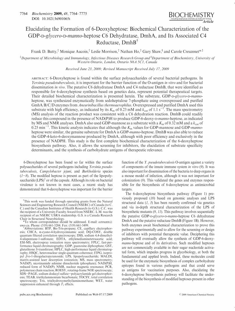
The 6-deoxyheptose biosynthesis pathway (Figure 1) pre-viously proposed (10) based on genomic analyses and LPSstructural data (1, 3) has been recently confirmed via geneticsand via in-depth structural characterization of the LPS ofbiosynthetic mutants (9, 11). This pathway involves sequentiallythe putative GDP-D-glycero-D-manno-heptose C6 dehydrataseDmhA and the putative reductase DmhB (Figure 1B). However,both enzymes await biochemical characterization to prove thispathway experimentally and to allow for the screening or designof inhibitors with potential therapeutic value. Deciphering thispathway will eventually allow the synthesis of GDP-6-deoxy-manno-heptose and of its derivatives. Such modified heptosesare not commercially available in their sugar nucleotide activa-ted form, which impedes progress in glycobiology, at both thefundamental and applied levels. Indeed, these molecules couldbe used for the enzymatic biosynthesis of complex carbohydrateepitopes found in various pathogens and that could serveas antigens for vaccination purposes. Also, elucidating the6-deoxyheptose biosynthesis pathway will facilitate the under-standing of the biosynthesis ofmodified heptoses present in otherpathogens.
†This work was funded through operating grants from the NaturalSciences and Engineering Research Council (NSERC) of Canada (to C.C.) and the Canadian Institutes of Health Research (to G.S.). C.C. wasthe recipient of aUniversity FacultyAward fromNSERC. L.M.was therecipient of an NSERC USRA studentship. G.S. is a Canada ResearchChair in Structural Neurobiology.*To whom correspondence should be addressed. E-mail: ccreuzen@
uwo.ca. Phone: (519) 661-3204. Fax: (519) 661-3499.1Abbreviations: BTP, Bis-Tris-propane; CE, capillary electrophor-
esis; CHCA, R-cyano-4-hydroxycinnamic acid; DQ-COSY, doublequantum filtered correlation spectroscopy; DSS, sodium 4,4-dimethyl-4-silapentane-1-sulfonate; EDTA, ethylenediaminetetraacetic acid;ESI-MS, electrospray ionization mass spectrometry; FPLC, fast-per-formance liquid chromatography; GDP, guanosine diphosphate; GST,glutathione S-transferase; HPLC, high-performance liquid chromatog-raphy; HSQC, heteronuclear single-quantum coherence; ITPG, isopro-pyl β-D-1-thiogalactopyranoside; LPS, lipopolysaccharide; MALDI,matrix-assisted laser desorption ionization; MS, mass spectrometry;NAD(P), nicotinamide adenine dinucleotide (phosphate); NAD(P)H,reduced form of NAD(P); NMR, nuclear magnetic resonance; PCR,polymerase chain reaction; ROESY, rotating-frameNOE spectroscopy;SDS-PAGE, sodium dodecyl sulfate-polyacrylamide gel electrophor-esis; TEAB, triethylammonium bicarbonate; TOCSY, total correlationspectroscopy; Tris, tris(hydroxymethyl)aminomethane; WET, watersuppression enhanced through T1 effects.
Article Biochemistry, Vol. 48, No. 32, 2009 7765
As mentioned above, the lack of a commercially availablenucleotide-activated heptose-based substrate is a problem forglycobiology and has rendered the biochemical characterizationofDmhA andDmhB very difficult. The chemical synthesis of theputative substrate GDP-D-glycero-D-manno-heptose (referred toasGDP-manno-heptose thereafter) was described earlier (12), butit cannot be implemented easily on a large scale outside ofspecialized chemistry laboratories. A four-step enzymatic path-way leading to GDP-manno-heptose starting from sedoheptulose7-phosphate was recently described (Figure 1A) (13).
In this report, the four enzymes mentioned above were used togenerate a stock of GDP-manno-heptose suitable for the enzy-matic characterization of DmhA and DmhB, which was doneusing mostly capillary electrophoresis (CE), mass spectrometry(MS), and NMR. We investigated the GDP-manno-heptose C6dehydratase activity of DmhA and the C4 reductase activity ofDmhB, determined the optimal reaction conditions, and ana-lyzed the reaction products by MS and NMR. We also investi-gated the ability of DmhA andDmhB to use GDP-mannose andits 4-keto-6-deoxy derivative as substrates. To our knowledge,this is the first biochemical characterization of a GDP-manno-heptose C6 dehydratase and its associated reductase. The dataobtained open the way to screening of inhibitors, for the analysisof other putative heptose dehydratases, and for the elucidation ofsubstrate specificity determinants via structure/function studies.
MATERIALS AND METHODS
Preparation of ExpressionConstructs forGmhA,GmhB,GmhC, and GmhD. Plasmids containing the gmhA, gmhB,gmhC, and gmhD genes from Aneurinibacillus thermoaerophiluscloned into the Gateway pDONR201 vector (Invitrogen) werekindly provided by Dr. Messner (Vienna, Austria) (13, 14).The gmhA, gmhB, and gmhC genes were transferred intothe pDEST17 vector (Invitrogen) to allow protein expression
as N-terminally histidine-tagged proteins. The gmhD gene wastransferred into pDEST15 to allow the expression of GmhD as aN-terminally glutathione S-transferase (GST) tagged protein.The transfer to the pDEST vectors was performed following theGateway procedure (Invitrogen) as recommended by the manu-facturer, using Escherichia coli Novablue and ampicillin selec-tion (100 μg/mL). The clones were screened by restrictiondigestion using EcoRI, by PCR using T7 promoter and T7terminator primers, and finally byDNA sequencing. The sequen-cing was performed at the Robarts Institute Sequencing Facility(London, Canada).Cloning of dmhA and dmhB in the pETVector. The dmhA
and dmhB genes from Y. pseudotuberculosis O:2a were PCRamplified from pUC-dmhB/A (9) using the primers YPDMHAP4 (AGGGTCCATGGGCATGAATAATGTTTTAATTAC-AGGT) andYPDMHAP5 (GCGACGGATCCTTAGCGATT-TAATGGAATGCG) for dmhA and YPDMHB P4 (AGG-GTCCATGGGCATGACAAAGGTGTTTATATTAGG) andYPDMHB P5 (GCGTCGGATCCTTATTCCTTAATTACTT-CCAGATATGG) for dmhB. The PCR was performed usingExpand Long Range DNA polymerase (Roche Diagnostics) asrecommended by the manufacturer. The PCR products weredigested with NcoI and BamHI and were cloned into the pET23derivative (15) with an N-terminal histidine tag. The constructswere transformed into E. coli JM109 with ampicillin selection(100 μg/mL). The resulting plasmids pET-dmhA and pET-dmhBwere then purified using the GFX kit (GE-Healthcare) andverified by DNA sequencing.Cloning of HP0044 as a GST-Tagged Protein.HP0044 is
a previously characterized GDP-mannose dehydratase fromHelicobacter pylori that was used as a control in this study.The gene for HP0044 was PCR amplified from chromosomalDNA from H. pylori strain 26695, using primers HP0044 P1(ATGTCAGCTTGAATCAAAC) and HP0044 P2 (GCGTC-GGATCCTCATTCATAAAAATTCCTTA).ThePCRwasdone
FIGURE 1: Biosynthetic pathways leading to the production of GDP-6-deoxy-manno-heptose from sedoheptulose phosphate. Panel A:Biosynthetic pathway showing the four successive enzymatic reactions catalyzed by GmhA/B/C/D, allowing the conversion of D-sedoheptulose7-phosphate into GDP-manno-heptose. Panel B: The two-step pathway that allows conversion of GDP-manno-heptose into GDP-6-deoxy-manno-heptose via action of DmhA and DmhB (13).
7766 Biochemistry, Vol. 48, No. 32, 2009 Butty et al.
using Expand Long Range DNA polymerase. The PCR productwas cleaved with BamHI and EcoRV and cloned into the vectorpBluescript SK that had been cut with the same enzymes. Thegene was then subcloned into the vector pGEX-2T by PCR usingprimers HP0044 P5 (AAGACGGATCCATGAAAGAAAAA-ATCGCTTTAATCAC) and HP0044 P6 (GCCTCGAATTCT-CATTCATAAA AATTCCTTAAAATATAAC) and digestionwithBamHI and EcoRI. After transformation intoE. coliDH5Rand ampicillin selection (100 μg/mL), the plasmid was extractedand sequenced.Protein Expression and Purification. For all proteins
except HP0044, protein expression was performed in E. coliBL21(DE3)pLys, using Luria-Bertani broth (LB) or media I, II,or III supplemented with 100 μg/mL ampicillin and 34 μg/mLchloramphenicol. Media I contained 0.6% Na2HPO4, 0.3%KH2PO4, 0.05% NaCl, 0.1% NH4Cl, 0.05% MgSO4 3 7H2O,0.0015%CaCl2, 0.2% casamino acids, and 0.2% glucose. MediaII contained 1% bactotryptone, 0.5% yeast extract, 0.5% NaCl,and 0.2% glucose. Media III contained 2% bactotryptone, 0.2%Na2HPO4, 1% KH2PO4, 0.8% NaCl, 1.5% yeast extract, and0.2% glucose (16). Protein expression was induced by theaddition of 0.15 mM isopropyl β-D-1-thiogalactopyranoside(IPTG). For GmhA, GmhB, and GmhC, expression was carriedout in 1 L (for GmhA and GmhC) or 2 L (for GmhB) of LB at25 �Cwith 3 h induction. For GmhD, expression was carried outin 2 L of LB at 15 �C or room temperature or inmedia I, II, or IIIat room temperature for ∼16 h. For DmhA, overexpression ofthe protein was performed in LB for 3 h at 37 �C. For DmhB,overexpression of the protein was performed in LB for 18 h atroom temperature. For HP0044, protein expression was carriedout in DH5R in LB with induction overnight at 30 �C with0.1 mM IPTG. For all proteins, at the end of the inductionperiod, the cells were harvested by centrifugation (15300g) andstored at -20 �C until needed.
To determine whether the proteins were overexpressed in asoluble form, a cell pellet obtained from 1.5 mL of inducedculture was treated with 150 μL of 2 mM ethylenediaminete-traacetate (EDTA), 0.1 mg/mL lysozyme, 0.1% Triton X-100,20000 units of DNase, and 10 mM MgCl2 in 50 mM Tris-HCl,pH 8.0, for 30 min at 30 �C. The soluble proteins were separatedfrom cellular debris and insoluble proteins by centrifugation at15300g for 15 min at 4 �C. The pellets containing the insolubleproteins were resuspended in 50 mM Tris-HCl, pH 8.0, contain-ing 2 mM EDTA. The fractions were run on 10% SDS-PAGEgels and analyzed by Coomassie blue staining or, when applic-able, by anti-histidine tag Western blotting using a mouse anti-His IgG antibody (Sigma-Aldrich) and an Alexa Fluor 680labeled goat anti-mouse IgG antibody (Molecular Probes).Detection was performed using a Li-Cor Odyssey infraredimaging system.Purification of GmhD and HP0044 by GST Affinity
Chromatography. The cell pellet obtained after overexpressionof HP0044 or of GmhD was resuspended in 30 mL of PBSbinding buffer, pH 8 (140 mM NaCl, 2.7 mM KCl, 10 mMNa2HPO4, 1.8 mM KH2PO4). Lysozyme was added (150 μg/mL), and the samplewas incubated on ice for 15min before beingpassed through a French press three times. Cellular debris andinsoluble proteins were removed by centrifugation for 20 min at15300g and by filtration through a 0.2 μm filter. GST affinitypurification was completed on an FPLC system using a 1 mLGSTrap FF column (GE Healthcare) that had been equilibratedin PBS binding buffer. The protein was loaded twice, and the
column was washed with 10 column volumes (CV) of bindingbuffer. The GmhD protein was eluted with 3 CV of 0.01 Mreduced glutathione. The glutathione was removed from thesample by overnight dialysis (cutoff of 3500 Da) in 50 mM Tris-HCl, pH 8, at 4 �C. For HP0044, the GST tag was removed byon-column incubation with thrombin for 2 h at 37 �C, and theprotein was eluted in 5 CV of 50 mMTris-HCl buffer, pH 8. Thepurified proteins were analyzed by SDS-PAGE and Coomassiestaining and were stored in 50% glycerol at -20 �C.Purification of Histidine-Tagged Proteins (GmhA,
GmhB, GmhC, DmhA, and DmhB) by Nickel Chelation.The induced cell pellets were resuspended in 30 mL of bindingbuffer, pH 7.5 (20 mM imidazole, 20 mM Tris-HCl, and 0.1 MNaCl). Lysozyme addition, cell lysis, centrifugation and filteringsteps were completed as described above. The histidine-taggedproteins were purified by FPLC (Akta purifier) using a 1.6 mLPoros MC 20 column (4.6 mm � 100 mm; Applied Biosystems)that had been loaded with nickel sulfate and equilibrated with10 CV of binding buffer. The sample was passed through thecolumn twice before the column was washed with 10 CV ofbinding buffer. The proteins of interest were eluted by a lineargradient of imidazole from a concentration of 50 mM to 1 M in30 CV. The fractions that contained the pure protein of interestwere pooled anddialyzed (cutoff of 3500Da) in 50mMTris-HCl,pH 7.5, overnight at 4 �C. The purified proteins were analyzed bySDS-PAGE as described above, quantitated using the Bio-Radprotein determination reagent and a standard curve of bovineserum albumin following the manufacturer’s instructions, andwere stored in 50% glycerol at -20 �C.Preparation of GDP-manno-heptose using GmhA/B/C/
D. Sedoheptulose 7-phosphate was obtained from GlycOTeamGmbH (Germany). To monitor the stepwise conversion ofsedoheptulose 7-phosphate into GDP-manno-heptose 1-phos-phate, 30 μL reactions containing 0.5 mM sedoheptulose 7-phosphate, 10mMMgCl2, and 1 mMATP in 200mMTris-HCl,pH 8, with either no enzyme, GmhA alone, GmhA and GmhB,GmhA, GmhB, and GmhC, or all four enzymes were incubatedfor 5 h at 37 �C.Approximately 8, 24, 13, and 20 pmol of GmhA,GmhB, GmhC, and GmhD, respectively, were used in eachreaction as needed. These reactions were analyzed by HPLC asdescribed below.
A large-scale reaction of 2.5 mLwas set up to produce heptose1-phosphate. It contained 1.25 mM sedoheptulose 7-phosphate,50 mMMgCl2, 1.3 mMATP, 1.68 nmol of GmhA, 0.75 nmol ofGmhB, and 0.65 nmol of GmhC in 400 mM Tris-HCl, pH 9.0.The reaction was incubated 5 h at 37 �C. This reaction was thenused to generateGDP-manno-heptose via the addition of GTP to0.6 mM and 1.44 nmol of GmhD in a final volume of 4 mL. Thereaction was incubated further for 5 h at 37 �C. The finalconversion into GDP-manno-heptose by GmhD was monitoredby capillary electrophoresis (CE) with UV detection as describedbelow.HPLC Analysis of Various Sugars. HPLC analysis was
performed on a Dionex ICS 3000 instrument equipped with anelectrochemical detector with a gold electrode and used in theintegrated amperometric detection mode essentially as reportedpreviously (13). The samples were analyzed on a CarboPac PA1column (4 � 250 mm; Dionex) that had been equilibrated in100 mM NaOH for 10 min at 1 mL/min. Ten microliters ofsample diluted to 0.1 mM in 100 mMNaOH was injected, and a40 min linear gradient of 100-500 mM NaOAc in 100 mMNaOH was applied at 1 mL/min.
Article Biochemistry, Vol. 48, No. 32, 2009 7767
Capillary Electrophoresis of Sugar Nucleotides. CE wasperformed on a Beckman Gold instrument using the 32 Karatsoftware and a 57 cmbare silica capillary. The initial conditioningof the capillary was performed by washing the capillary with 0.1N HCl for 30 min at 20 psi, followed by 10 min of water. Forsample analyses, the capillarywas washed for 2minwith 200mMborax, pH 9, buffer, the sample was injected by pressure for 4 s,and separation was performed by applying 26 kV to both ends ofthe capillary that were maintained in the borax buffer. Migratingcompounds were monitored at 254 nm in a window placed at50 cm from the beginning of the capillary. The capillary waswashed 2 min with water, 2 min with 0.1 M NaOH, and 2 minagain with water between each run. Substrate conversion wasestimated by integration of the surface areas under the substrateand product peaks using the 32 Karat software.Purification of Sugar Nucleotides. All sugar nucleotides
were purified by anion-exchange chromatography using a HighQEconopac 1mL column (Bio-Rad) (17) and a linear gradient of20 column volumes of triethylammonium bicarbonate (TEAB),pH 8.5 (50 mM to 1 M), at 1 mL/min with UV detection(260 and 214 nm). The fractions containing the product ofinterest were pooled and lyophilized twice with resuspensionin water between both lyophilization steps. They were thenresuspended in water for assessment of purity by CE and ofidentity by mass spectrometry (MS) analysis. Quantitationwas performed using a Nanodrop spectrophotometer and usingεGTP = 12000 mol-1 L cm-1.Enzyme Assays with DmhA, DmhB, and HP0044. Typi-
cally, reactions were performed by incubating between 0.1 and 22pmol of DmhA, HP0044, and/or DmhB with 0.1-0.2 mMsubstrate (GDP-manno-heptose prepared above or commercialGDP-mannose (Sigma)) in 200 mM Tris-HCl buffer, pH 9.5 or8.5 (as specified in the legends to the figures), in a final volume of10 μL unless stated otherwise. Cofactors NAD(P)þ and NAD-(P)H were added to 0.2 mM, when necessary. The reactions wereincubated for as little as 2 min to up to overnight at 37 �C.Whenappropriate, the buffer was replaced by 200 mM TEAB, pH 8.5.Specifics for each experiment are stated in the legends to thefigures. The reaction products were analyzed by CE as describedabove. Large-scale reactions were prepared by direct propor-tional increase of all components for anion-exchange purificationfollowed by MS or NMR.
For the temperature study for DmhA, the reactions contained0.44 pmol of DmhA and 0.1 mMGDP-manno-heptose in a totalvolumeof 10 μLof 200mMTris-HCl, pH9.Theywere incubatedfor 10min at temperatures ranging from 4 to 65 �C. ForDmhB, astock reaction containing 4.4 pmol of DmhA, 0.1 mM GDP-manno-heptose, and 0.1 mMNADH in a total volume of 100 μLof 200 mMTris-HCl, pH 9, was incubated for 20 min at 37 �C toensure 100% conversion into the 4-keto derivative. Then 0.6pmol of DmhB was added to 7 μL of this reaction stock, the finalvolumewas brought up to 10 μL, and the samples were incubatedfor 20 min at temperatures ranging from 4 to 65 �C.
For the pH studies, the DmhA reactions contained 0.89 pmolof DmhA and 0.1 mM substrate in a total volume of 10 μL. Thereactions were performed in 200 mMNaOAc buffer for pH 5-7and in 200 mM Bis-Tris-propane (BTP) for pH 7-10.5. Theywere incubated at 37 �C for 10 min. To determine the pHoptimum for DmhB, a stock reaction of 4.4 pmol of DmhA,0.1 mM GDP-manno-heptose, and 0.1 mM NADH in a totalvolume of 100 μL of 200 mM Tris-HCl, pH 8, was incubated for20 min at 37 �C to ensure 100% conversion into the 4-keto
derivative. Aliquots of 7 μL were withdrawn, and their pH wasadjusted to the appropriate value by addition of 1MTris-HCl forpH 7-8.0 or BTP for pH 8.0-10.5. Then 0.6 pmol of DmhBwasadded, the volume was brought to 10 μL, and the samples wereincubated for 20 min at 37 �C.
To test for cofactor dependence, NAD(P)þ and NAD(P)Hwere added to a final concentration of 0.1 mM in DmhA andDmhB reactions, respectively.
For determination of Km, Vmax, and turnover parameters, 11reactions with varying concentrations of substrate (0.1-0.5 mM)were set up in duplicates and incubated for 2 min (for GDP-manno-heptose series) or 60 min (for GDP-mannose series) at37 �C,which ensured less than 10% substrate conversion over thewhole range of substrate concentrations tested. The data are theaverage of two independent experiments and were calculatedusing the Lineweaver equation. All reactions were flash frozen ina dry ice-ethanol bath and ran individually immediately afterthawing.
To decipher whether all reaction products observed in DmhAreactions were the result of enzymatic activity or not, a 70 μLreaction containing 62 pmol ofDmhA and 0.1mMGDP-manno-heptose in 200 mMTEAB, pH 8.5, was incubated at 37 �C for 30min. The reaction was quenched by flash freezing in dry ice-ethanol. The enzyme was then removed by ultrafiltration(Nanosep centrifugal device, 10 kDa cutoff (Pall, Life Sciences)).The filtrate was analyzed by CE. A 10 μL aliquot of theultrafiltrate was supplemented with 0.1mM (final concentration)GDP-manno-heptose and was further incubated at 37 �C for 30or 60 min. Also, aliquots (10 μL each) of the ultrafiltrate werefurther incubated at 37 �C for 30, 45, 60, or 90 min and analyzedby CE. A complete (unfiltered) reaction was also incubated at37 �C for 60 min and served as a positive control.Mass Spectrometry Analyses.Mass spectrometry analyses
of the sugar nucleotides and cofactors were performed at the Dr.Don Rix Protein Identification Facility of the University ofWestern Ontario. All sugar nucleotides and cofactors wereanalyzed by ESI-MS on a Micromass Qtof spectrometerequipped with a Z-spray source operating in the negative ionmode (40 V, 80 �C). Calibration was performed with NaI, andacquisition was performed using the software MassLynx 4.0(Micromass). GDP-manno-heptose and products P2 and P20
were purified by anion-exchange chromatography before analy-sis. The DmhA reaction product (product P1) was analyzedsimilarly except that, due to its instability, it was generateddirectly in TEAB buffer (100mM, pH 8.5), flash frozen, partiallylyophilized, and resuspended in ice-cold water twice, before finalresuspension in ice-cold water for MS analysis. The sample wasmaintained frozen or on ice at all times to avoid degradation. TheDmhB product (P3) was also produced in TEAB for MSanalyses.
To identify the cofactor present within DmhA, 5 nmol ofDmhA resuspended in 1 mL of 50 mM ammonium bicarbonate,pH 8, was boiled for 5 min, filtered through an Amiconultracentrifugation device (3 kDa cutoff), concentrated downto 20 μL by lyophilization, and analyzed by MS as indicatedabove.
Purified DmhA and DmhB proteins were analyzed by MAL-DImass spectrometry using a 4700 Proteomics analyzer (AppliedBiosystems, Foster City, CA) in the linear positive ion mode toconfirm their size. The MALDI matrix, R-cyano-4-hydroxycin-namic acid (CHCA), was prepared as 5 mg/mL in 6 mMmonobasi ammonium phosphatec, 50% acetonitrile, and 0.1%
7768 Biochemistry, Vol. 48, No. 32, 2009 Butty et al.
trifluoroacetic acid and mixed with the sample 1:1 (v/v). Dataacquisition and data processing were respectively done using4000 Series Explorer and Data Explorer (both from AppliedBiosystems). The analysis was performed at the MALDI massspectrometry facility of the University of Western Ontario(London, Ontario, Canada).NMRAnalysis of GDP-manno-heptose and of theDmhB
Reaction Product.A 3mL reaction containing 0.33 mMGDP-manno-heptose, 220 pmol of DmhA, 304 pmol of DmhB, and 0.5mM NADPH in 200 mM TEAB, pH 8.5, was incubated over-night at 30 �C. The reaction progress was assessed by CE. Oncethe reaction had reached completion, the product was purified byanion-exchange chromatography using a 5mLHighQEconopaccolumn as described above. The fractions containing the pureproduct (as assessed by CE) were pooled, lyophilized, resus-pended in 350 μL of D2O, lyophilized again, and resuspended inD2O twice more before being analyzed by NMR spectroscopy.The final sample (1.7 mM final) was dissolved in 350 μL of D2Oand placed in a 5mmShigemiNMR tube having 5 μMDSS as aninternal standard. An identical sample of GDP-glycero-manno-heptose was prepared for comparison.
All 1H NMR data were collected with a Varian Inova 600MHz NMR spectrometer at 25 �C. One-dimensional 1H NMRspectra were collected using the WET water suppression se-quence. Two-dimensional 1HTOCSY spectra (18), using a 5 kHzspinlock and DQ-COSY spectra (19), were collected for 256complex increments. Natural abundance 1H-13C HSQC experi-ments (20, 21) were also used to confirm all assignments. Allspectra were processed using VnmrJ, baseline corrected, andreferenced to DSS at 0.00 ppm.
RESULTS
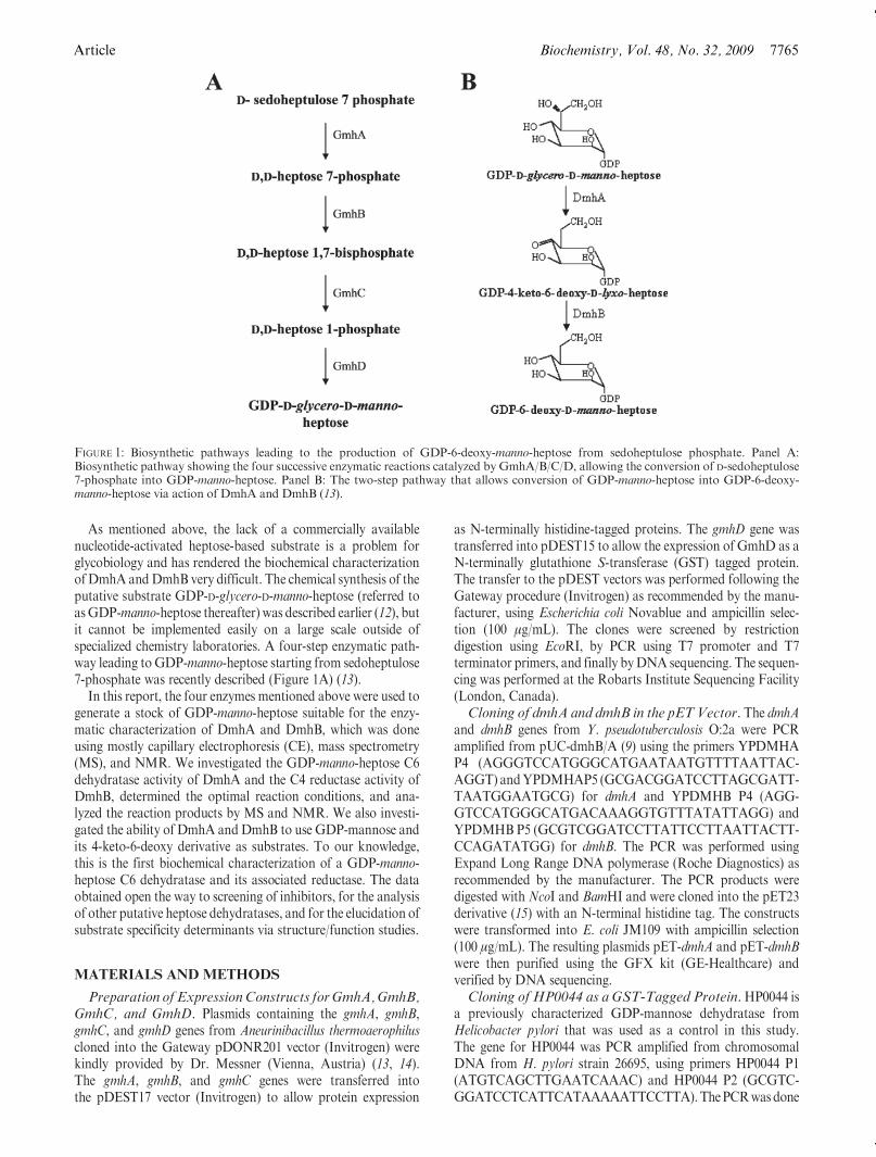
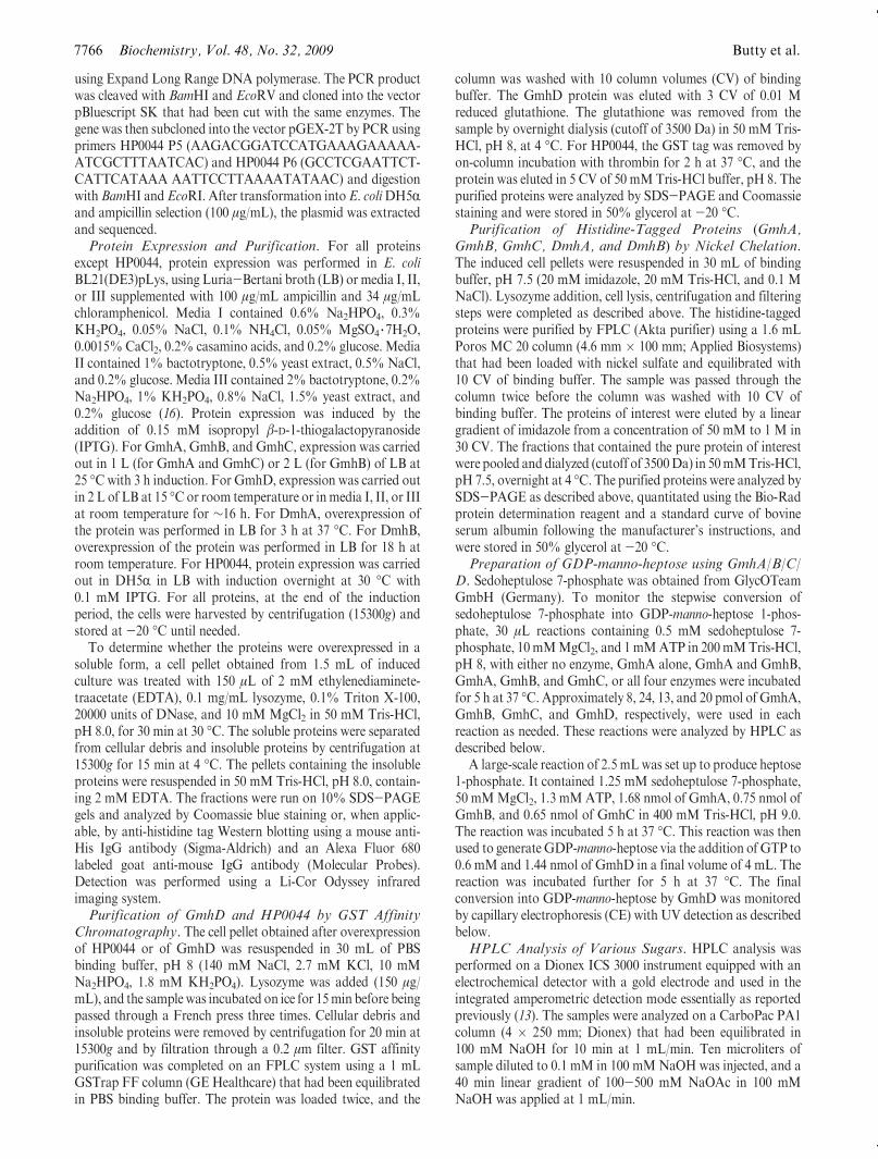
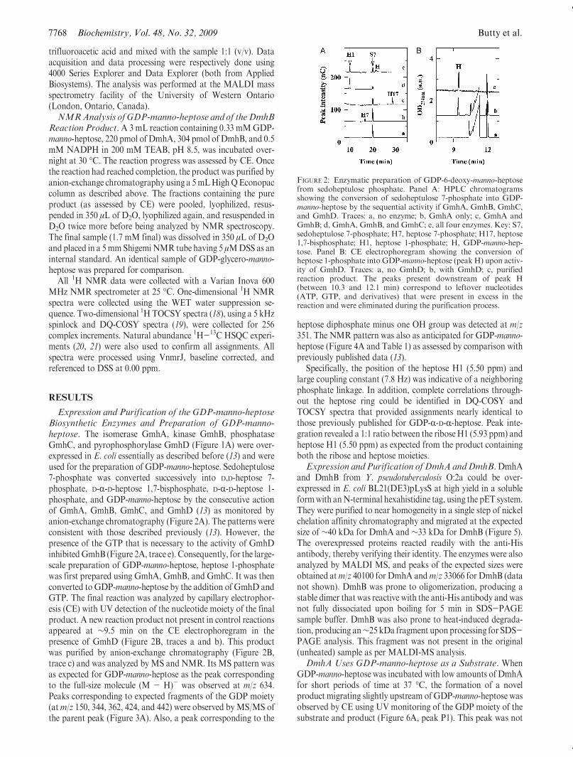
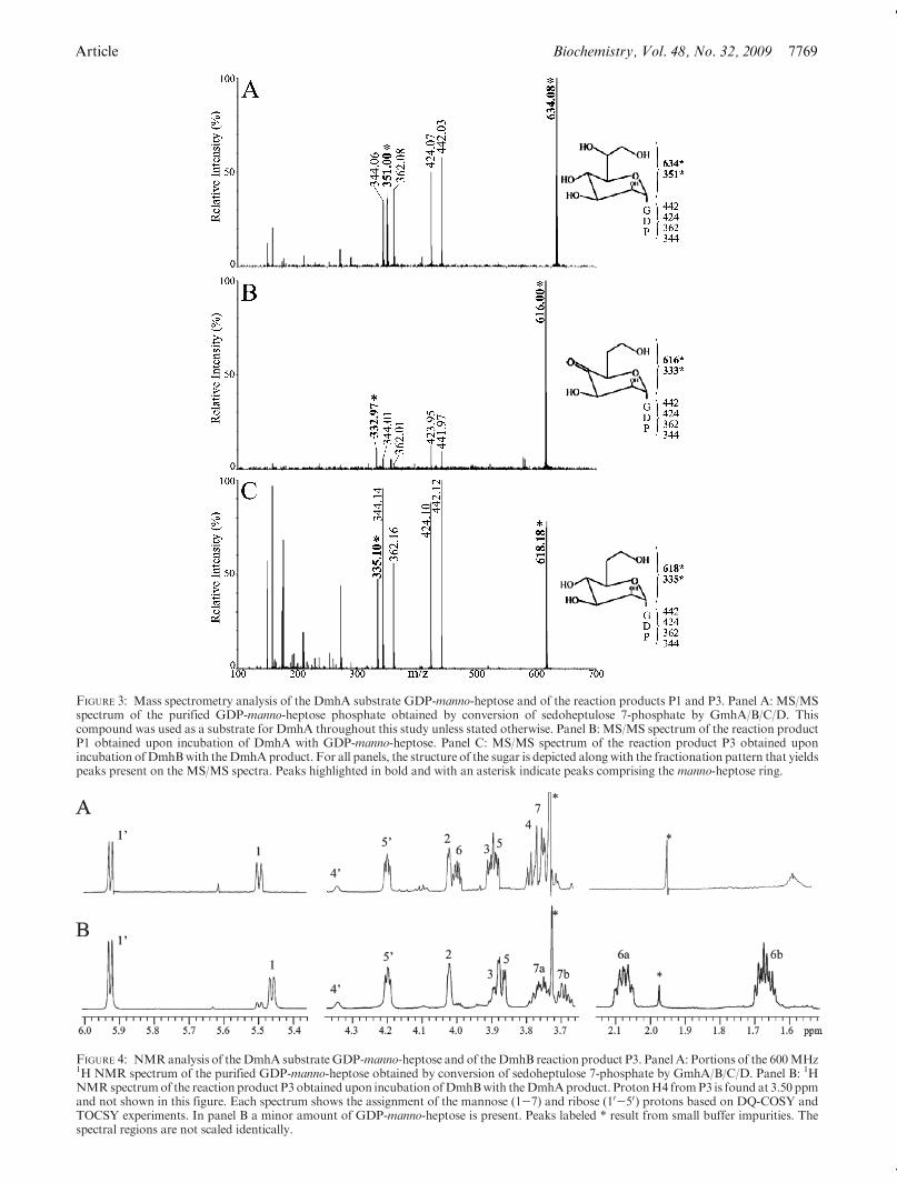
Expression and Purification of the GDP-manno-heptoseBiosynthetic Enzymes and Preparation of GDP-manno-heptose. The isomerase GmhA, kinase GmhB, phosphataseGmhC, and pyrophosphorylase GmhD (Figure 1A) were over-expressed in E. coli essentially as described before (13) and wereused for the preparation of GDP-manno-heptose. Sedoheptulose7-phosphate was converted successively into D,D-heptose 7-phosphate, D-R-D-heptose 1,7-bisphosphate, D-R-D-heptose 1-phosphate, and GDP-manno-heptose by the consecutive actionof GmhA, GmhB, GmhC, and GmhD (13) as monitored byanion-exchange chromatography (Figure 2A). The patterns wereconsistent with those described previously (13). However, thepresence of the GTP that is necessary to the activity of GmhDinhibitedGmhB (Figure 2A, trace e). Consequently, for the large-scale preparation of GDP-manno-heptose, heptose 1-phosphatewas first prepared using GmhA, GmhB, and GmhC. It was thenconverted to GDP-manno-heptose by the addition of GmhD andGTP. The final reaction was analyzed by capillary electrophor-esis (CE) with UV detection of the nucleotide moiety of the finalproduct. A new reaction product not present in control reactionsappeared at ∼9.5 min on the CE electrophoregram in thepresence of GmhD (Figure 2B, traces a and b). This productwas purified by anion-exchange chromatography (Figure 2B,trace c) and was analyzed by MS and NMR. Its MS pattern wasas expected for GDP-manno-heptose as the peak correspondingto the full-size molecule (M - H)- was observed at m/z 634.Peaks corresponding to expected fragments of the GDP moiety(atm/z 150, 344, 362, 424, and 442) were observed byMS/MS ofthe parent peak (Figure 3A). Also, a peak corresponding to the
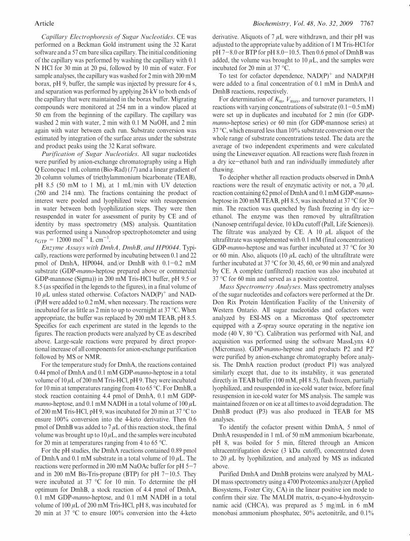
heptose diphosphate minus one OH group was detected at m/z351. The NMR pattern was also as anticipated for GDP-manno-heptose (Figure 4A and Table 1) as assessed by comparison withpreviously published data (13).
Specifically, the position of the heptose H1 (5.50 ppm) andlarge coupling constant (7.8 Hz) was indicative of a neighboringphosphate linkage. In addition, complete correlations through-out the heptose ring could be identified in DQ-COSY andTOCSY spectra that provided assignments nearly identical tothose previously published for GDP-R-D-R-heptose. Peak inte-gration revealed a 1:1 ratio between the riboseH1 (5.93 ppm) andheptose H1 (5.50 ppm) as expected from the product containingboth the ribose and heptose moieties.Expression and Purification of DmhA andDmhB.DmhA
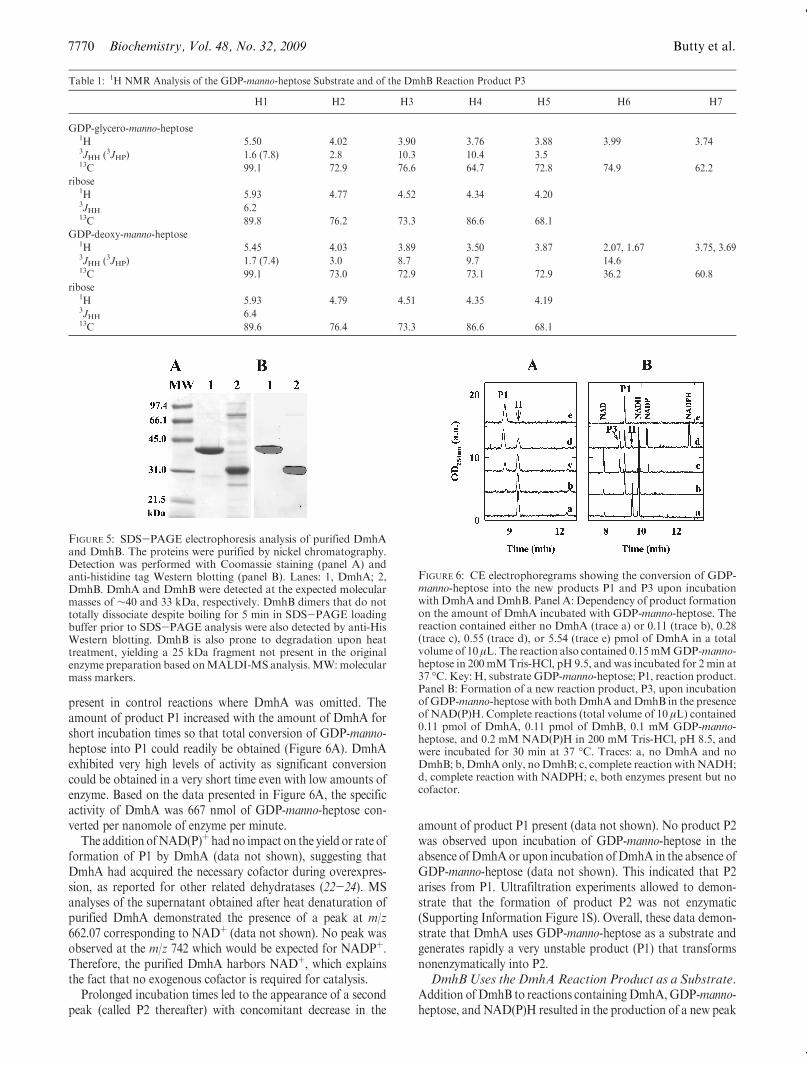
and DmhB from Y. pseudotuberculosis O:2a could be over-expressed in E. coli BL21(DE3)pLysS at high yield in a solubleformwith anN-terminal hexahistidine tag, using the pET system.They were purified to near homogeneity in a single step of nickelchelation affinity chromatography and migrated at the expectedsize of ∼40 kDa for DmhA and ∼33 kDa for DmhB (Figure 5).The overexpressed proteins reacted readily with the anti-Hisantibody, thereby verifying their identity. The enzymes were alsoanalyzed by MALDI MS, and peaks of the expected sizes wereobtained atm/z 40100 for DmhA andm/z 33066 for DmhB (datanot shown). DmhB was prone to oligomerization, producing astable dimer that was reactive with the anti-His antibody and wasnot fully dissociated upon boiling for 5 min in SDS-PAGEsample buffer. DmhB was also prone to heat-induced degrada-tion, producing an∼25 kDa fragment upon processing for SDS-PAGE analysis. This fragment was not present in the original(unheated) sample as per MALDI-MS analysis.DmhA Uses GDP-manno-heptose as a Substrate. When
GDP-manno-heptose was incubated with low amounts of DmhAfor short periods of time at 37 �C, the formation of a novelproduct migrating slightly upstream ofGDP-manno-heptose wasobserved by CE using UV monitoring of the GDP moiety of thesubstrate and product (Figure 6A, peak P1). This peak was not
FIGURE 2: Enzymatic preparation of GDP-6-deoxy-manno-heptosefrom sedoheptulose phosphate. Panel A: HPLC chromatogramsshowing the conversion of sedoheptulose 7-phosphate into GDP-manno-heptose by the sequential activity if GmhA, GmhB, GmhC,and GmhD. Traces: a, no enzyme; b, GmhA only; c, GmhA andGmhB; d, GmhA, GmhB, and GmhC; e, all four enzymes. Key: S7,sedoheptulose 7-phosphate; H7, heptose 7-phosphate; H17, heptose1,7-bisphosphate; H1, heptose 1-phosphate; H, GDP-manno-hep-tose. Panel B: CE electrophoregram showing the conversion ofheptose 1-phosphate into GDP-manno-heptose (peakH) upon activ-ity of GmhD. Traces: a, no GmhD; b, with GmhD; c, purifiedreaction product. The peaks present downstream of peak H(between 10.3 and 12.1 min) correspond to leftover nucleotides(ATP, GTP, and derivatives) that were present in excess in thereaction and were eliminated during the purification process.
Article Biochemistry, Vol. 48, No. 32, 2009 7769
FIGURE 3: Mass spectrometry analysis of the DmhA substrate GDP-manno-heptose and of the reaction products P1 and P3. Panel A: MS/MSspectrum of the purified GDP-manno-heptose phosphate obtained by conversion of sedoheptulose 7-phosphate by GmhA/B/C/D. Thiscompound was used as a substrate for DmhA throughout this study unless stated otherwise. Panel B: MS/MS spectrum of the reaction productP1 obtained upon incubation of DmhA with GDP-manno-heptose. Panel C: MS/MS spectrum of the reaction product P3 obtained uponincubation ofDmhBwith theDmhAproduct. For all panels, the structure of the sugar is depicted alongwith the fractionation pattern that yieldspeaks present on the MS/MS spectra. Peaks highlighted in bold and with an asterisk indicate peaks comprising the manno-heptose ring.
FIGURE 4: NMRanalysis of theDmhA substrate GDP-manno-heptose and of theDmhB reaction product P3. Panel A: Portions of the 600MHz1H NMR spectrum of the purified GDP-manno-heptose obtained by conversion of sedoheptulose 7-phosphate by GmhA/B/C/D. Panel B: 1HNMRspectrumof the reaction product P3 obtained upon incubation ofDmhBwith theDmhAproduct. ProtonH4 fromP3 is found at 3.50 ppmand not shown in this figure. Each spectrum shows the assignment of the mannose (1-7) and ribose (10-50) protons based on DQ-COSY andTOCSY experiments. In panel B a minor amount of GDP-manno-heptose is present. Peaks labeled * result from small buffer impurities. Thespectral regions are not scaled identically.
7770 Biochemistry, Vol. 48, No. 32, 2009 Butty et al.
present in control reactions where DmhA was omitted. Theamount of product P1 increased with the amount of DmhA forshort incubation times so that total conversion of GDP-manno-heptose into P1 could readily be obtained (Figure 6A). DmhAexhibited very high levels of activity as significant conversioncould be obtained in a very short time even with low amounts ofenzyme. Based on the data presented in Figure 6A, the specificactivity of DmhA was 667 nmol of GDP-manno-heptose con-verted per nanomole of enzyme per minute.
The addition ofNAD(P)þ had no impact on the yield or rate offormation of P1 by DmhA (data not shown), suggesting thatDmhA had acquired the necessary cofactor during overexpres-sion, as reported for other related dehydratases (22-24). MSanalyses of the supernatant obtained after heat denaturation ofpurified DmhA demonstrated the presence of a peak at m/z662.07 corresponding to NADþ (data not shown). No peak wasobserved at the m/z 742 which would be expected for NADPþ.Therefore, the purified DmhA harbors NADþ, which explainsthe fact that no exogenous cofactor is required for catalysis.
Prolonged incubation times led to the appearance of a secondpeak (called P2 thereafter) with concomitant decrease in the
amount of product P1 present (data not shown). No product P2was observed upon incubation of GDP-manno-heptose in theabsence ofDmhAor upon incubation ofDmhA in the absence ofGDP-manno-heptose (data not shown). This indicated that P2arises from P1. Ultrafiltration experiments allowed to demon-strate that the formation of product P2 was not enzymatic(Supporting Information Figure 1S). Overall, these data demon-strate that DmhA uses GDP-manno-heptose as a substrate andgenerates rapidly a very unstable product (P1) that transformsnonenzymatically into P2.DmhB Uses the DmhAReaction Product as a Substrate.
Addition of DmhB to reactions containing DmhA,GDP-manno-heptose, andNAD(P)H resulted in the production of a new peak
Table 1: 1H NMR Analysis of the GDP-manno-heptose Substrate and of the DmhB Reaction Product P3
H1 H2 H3 H4 H5 H6 H7
GDP-glycero-manno-heptose1H 5.50 4.02 3.90 3.76 3.88 3.99 3.743JHH (3JHP) 1.6 (7.8) 2.8 10.3 10.4 3.513C 99.1 72.9 76.6 64.7 72.8 74.9 62.2
ribose1H 5.93 4.77 4.52 4.34 4.203JHH 6.213C 89.8 76.2 73.3 86.6 68.1
GDP-deoxy-manno-heptose1H 5.45 4.03 3.89 3.50 3.87 2.07, 1.67 3.75, 3.693JHH (3JHP) 1.7 (7.4) 3.0 8.7 9.7 14.613C 99.1 73.0 72.9 73.1 72.9 36.2 60.8
ribose1H 5.93 4.79 4.51 4.35 4.193JHH 6.413C 89.6 76.4 73.3 86.6 68.1
FIGURE 5: SDS-PAGE electrophoresis analysis of purified DmhAand DmhB. The proteins were purified by nickel chromatography.Detection was performed with Coomassie staining (panel A) andanti-histidine tag Western blotting (panel B). Lanes: 1, DmhA; 2,DmhB. DmhA and DmhB were detected at the expected molecularmasses of ∼40 and 33 kDa, respectively. DmhB dimers that do nottotally dissociate despite boiling for 5 min in SDS-PAGE loadingbuffer prior to SDS-PAGE analysis were also detected by anti-HisWestern blotting. DmhB is also prone to degradation upon heattreatment, yielding a 25 kDa fragment not present in the originalenzyme preparation based onMALDI-MS analysis.MW:molecularmass markers.
FIGURE 6: CE electrophoregrams showing the conversion of GDP-manno-heptose into the new products P1 and P3 upon incubationwithDmhA andDmhB. Panel A: Dependency of product formationon the amount of DmhA incubated with GDP-manno-heptose. Thereaction contained either no DmhA (trace a) or 0.11 (trace b), 0.28(trace c), 0.55 (trace d), or 5.54 (trace e) pmol of DmhA in a totalvolume of 10 μL. The reaction also contained 0.15mMGDP-manno-heptose in 200mMTris-HCl, pH 9.5, and was incubated for 2 min at37 �C.Key: H, substrate GDP-manno-heptose; P1, reaction product.Panel B: Formation of a new reaction product, P3, upon incubationofGDP-manno-heptose with bothDmhA andDmhB in the presenceof NAD(P)H. Complete reactions (total volume of 10 μL) contained0.11 pmol of DmhA, 0.11 pmol of DmhB, 0.1 mM GDP-manno-heptose, and 0.2 mM NAD(P)H in 200 mM Tris-HCl, pH 8.5, andwere incubated for 30 min at 37 �C. Traces: a, no DmhA and noDmhB; b, DmhAonly, noDmhB; c, complete reactionwithNADH;d, complete reaction with NADPH; e, both enzymes present but nocofactor.
Article Biochemistry, Vol. 48, No. 32, 2009 7771
that migrated upstream of P1 (Figure 6B, product P3), con-comitantly with the disappearance of P1 and the formation ofNAD(P)þ. Formation of this peak was directly proportional tothe amount of enzyme present in the reaction and to the reactiontime (as long as the substrate was not limiting) and was notobserved in the absence of DmhA or of exogenous cofactor. Thefact that greater substrate conversion was obtained in thepresence of NADPH than with NADH indicates that thepreferred cofactor for DmhB is NADPH (Figure 6B) as seen inrelated reductases (25). These data indicate that DmhB uses thereaction product of DmhA as a substrate in an oxidoreductionreaction that oxidizes the NAD(P)H cofactor provided.MS Analysis of the Reaction Products Generated upon
Incubation ofDmhAandDmhBwithGDP-manno-heptose.TheMS pattern of product P1 (generated by DmhA) showed thepresence of products atm/z 616 and atm/z 638 and 660. TheMS/MS patterns of the parent peaks atm/z 616 (Figure 3B), 638, and660 (not shown) demonstrated that they were sodium adducts ofone another as they yielded similar MS/MS spectra where themain peaks differed in size by 22 mass units (i.e., the mass of thesodium atom) from one spectrum to the other. The product atm/z 616 exhibited a loss of 18 mass units compared with the originalsubstrate (detected at m/z 634), which is consistent with the C6dehydration reaction and formation of the 4-keto-6-deoxy inter-mediate.
The MS pattern of product P3 (generated by DmhB) showedthe presence of a product atm/z 618 (Figure 3C), which exhibiteda gain of 2mass units comparedwith P1 (detected atm/z 616) anda loss of 16 mass units compared with the original substrate(detected atm/z 634). This is consistent with the C4 reduction ofthe 4-keto-6-deoxy intermediate produced by DmhA.
TheMS/MS fragmentation patterns of the substrate and of P1andP3 all contained common fragments atm/z 344, 362, 424, and442, which correspond to the GDP moiety of the molecule asobserved previously (26). This indicates that products P1 and P3arise from the GDP-manno-heptose substrate and that DmhAand DmhB did not catalyze any reaction on the GDP moiety, asexpected. Importantly, the peak corresponding to the heptosediphosphate fragment of the molecule, which was detected atm/z351 in the MS/MS pattern of the original substrate, was shiftedby 18 mass units tom/z 333 in the DmhA product P1 and shiftedback to m/z 335 in the MS/MS spectrum of the DmhB productP3. This is indicating that the dehydration and reduction reac-tions occurred on the heptose moiety.
In conclusion, these MS data support the hypothesis thatDmhA carried out a dehydration reaction on the manno-heptosemoiety of GDP-manno-heptose and that P1 may be the 4-keto-6-deoxy derivative of the substrate, while DmhB carries out the C4reduction of this 4-keto intermediate and product P3 may beGDP-6-deoxy-manno-heptose.NMR Analysis of the DmhB Reaction Product P3. In
contrast to product P1, theDmhB reaction product P3was stableenough to be purified by anion-exchange chromatography andanalyzed by NMR spectroscopy to ascertain its identity. Whencompared with the original GDP-manno-heptose substrate, newsignals were observed at 2.07 and 1.67 ppm. DQ-COSY and1H-13C HSQC analysis indicated these protons resided on theC6 carbon and were coupled to protons on the C5 and C7positions. The chemical shifts for the C6 methylene signals (H6a,H6b) were also similar with those described before for 6-deoxy-manno-heptose that had been extracted directly from Y. pseudo-tuberculosis LPS (9, 11) (Figure 4B, Table 1). In addition, the
removal of the hydroxyl from C6 caused the obvious loss of theH6 resonances near 3.99 ppm observed in the original substrate.Correspondingly, H4 from the substrate shifted from 3.76 to 3.50ppm in P3, consistent with a change in the environment of H4from removal of the hydroxyl group from C6. The remainingportions of the spectra for P3 were very similar to the substrate,including the coupling patterns around the heptose ring. Inparticular, large coupling constants were measured betweenH3-H4 and H4-H5 (Table 1), indicating the C3-C4-C5configuration of the heptose ring appears to be unchanged uponformation of P3. In addition, an NOE between H4 and H6a,bcould be identified in two-dimensional 1H ROESY spectraindicating the configuration of the heptose ring appeared to bethe same in the P3 product as the starting GDP-manno-heptosesubstrate.DmhA Also Uses GDP-mannose as a Substrate. To test
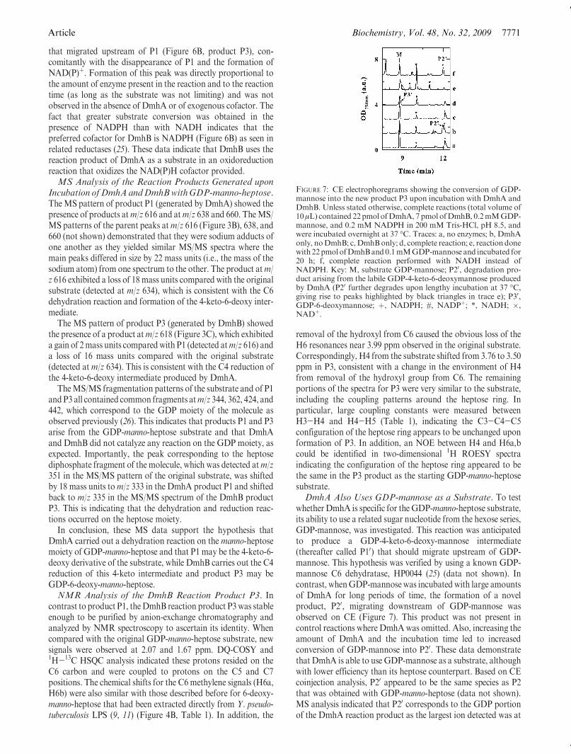
whether DmhA is specific for the GDP-manno-heptose substrate,its ability to use a related sugar nucleotide from the hexose series,GDP-mannose, was investigated. This reaction was anticipatedto produce a GDP-4-keto-6-deoxy-mannose intermediate(thereafter called P10) that should migrate upstream of GDP-mannose. This hypothesis was verified by using a known GDP-mannose C6 dehydratase, HP0044 (25) (data not shown). Incontrast, whenGDP-mannose was incubated with large amountsof DmhA for long periods of time, the formation of a novelproduct, P20, migrating downstream of GDP-mannose wasobserved on CE (Figure 7). This product was not present incontrol reactions where DmhAwas omitted. Also, increasing theamount of DmhA and the incubation time led to increasedconversion of GDP-mannose into P20. These data demonstratethat DmhA is able to use GDP-mannose as a substrate, althoughwith lower efficiency than its heptose counterpart. Based on CEcoinjection analysis, P20 appeared to be the same species as P2that was obtained with GDP-manno-heptose (data not shown).MS analysis indicated that P20 corresponds to the GDP portionof the DmhA reaction product as the largest ion detected was at
FIGURE 7: CE electrophoregrams showing the conversion of GDP-mannose into the new product P3 upon incubation with DmhA andDmhB. Unless stated otherwise, complete reactions (total volume of10μL) contained 22pmolofDmhA, 7 pmol ofDmhB, 0.2mMGDP-mannose, and 0.2 mM NADPH in 200 mM Tris-HCl, pH 8.5, andwere incubated overnight at 37 �C. Traces: a, no enzymes; b, DmhAonly, noDmhB; c,DmhBonly; d, complete reaction; e, reaction donewith22pmolofDmhBand0.1mMGDP-mannose and incubated for20 h; f, complete reaction performed with NADH instead ofNADPH. Key: M, substrate GDP-mannose; P20, degradation pro-duct arising from the labile GDP-4-keto-6-deoxymannose producedby DmhA (P20 further degrades upon lengthy incubation at 37 �C,giving rise to peaks highlighted by black triangles in trace e); P30,GDP-6-deoxymannose; þ, NADPH; #, NADPþ; *, NADH; �,NADþ.
7772 Biochemistry, Vol. 48, No. 32, 2009 Butty et al.
m/z 442 (data not shown). Attempts at detecting the P10 productby MS have been unsuccessful, probably because of its lowabundance and labile character. This is consistent with the factthat 4-keto intermediates are notoriously unstable (27, 28).DmhB Can Also Use the Labile GDP-4-keto-6-deoxy-
mannose Formed by DmhA as a Substrate. As mentionedabove, incubation of DmhA with GDP-mannose leads to theformation of P20, which probably arises from degradation of alabile GDP-4-keto-6-deoxymannose produced by DmhA. WhenDmhB was added to the DmhA/GDP-mannose reaction in thepresence of NADPH, the formation of the degradation productP20 was not observed, but a new product was observed as a rightshoulder on theGDP-mannose peak (Figure 7, peak P30, trace d).Formation of this product could be enhanced via increasing theDmhB/GDP-mannose ratio, and increasing the incubation time(trace e). Coinjection of this productwithGDP-mannose demon-strated that this product is indeed distinct from GDP-mannose(data not shown). Overall, formation of P30 was poorly efficientand exacerbated the preference of DmhB for its NADPHcofactor, as no new product was observed in the presence ofNADH (trace f) or in the absence of cofactor (data not shown).Also, no new product was formed upon incubation of DmhBwith GDP-mannose in the absence of DmhA (trace c), therebyconfirming that the activity of DmhB is dependent on theformation of the 4-keto intermediate by DmhA.
MS analysis performed on a complete reaction revealed theexistence of a peak at m/z 588 (full product is 589), i.e., 16 massunits smaller than GDP-mannose. The MS/MS fragmentationpattern of this peak contained a fragment unique to the DmhBreaction that was detected atm/z 305 (data not shown). This was16mass units smaller than the hexose ring peak that was detectedat m/z 321 in the parent substrate. The MS/MS pattern alsocontained peaks corresponding to the unaffectedGDPmoiety (atm/z 273, 344, 362, 424, and 442; data not shown). These data areconsistent with the occurrence of a C6 dehydration followed by aC4 reduction reaction on the mannose ring, as observed for theheptose substrate.Physicokinetic Parameters for the DmhA and DmhB
Activities. As determined using GDP-manno-heptose as a sub-strate, the activity of DmhA was optimal at pH 9-9.5 and at37 �C, and that of DmhB was optimal at pH 8.5-9 and 30 �C(data not shown).
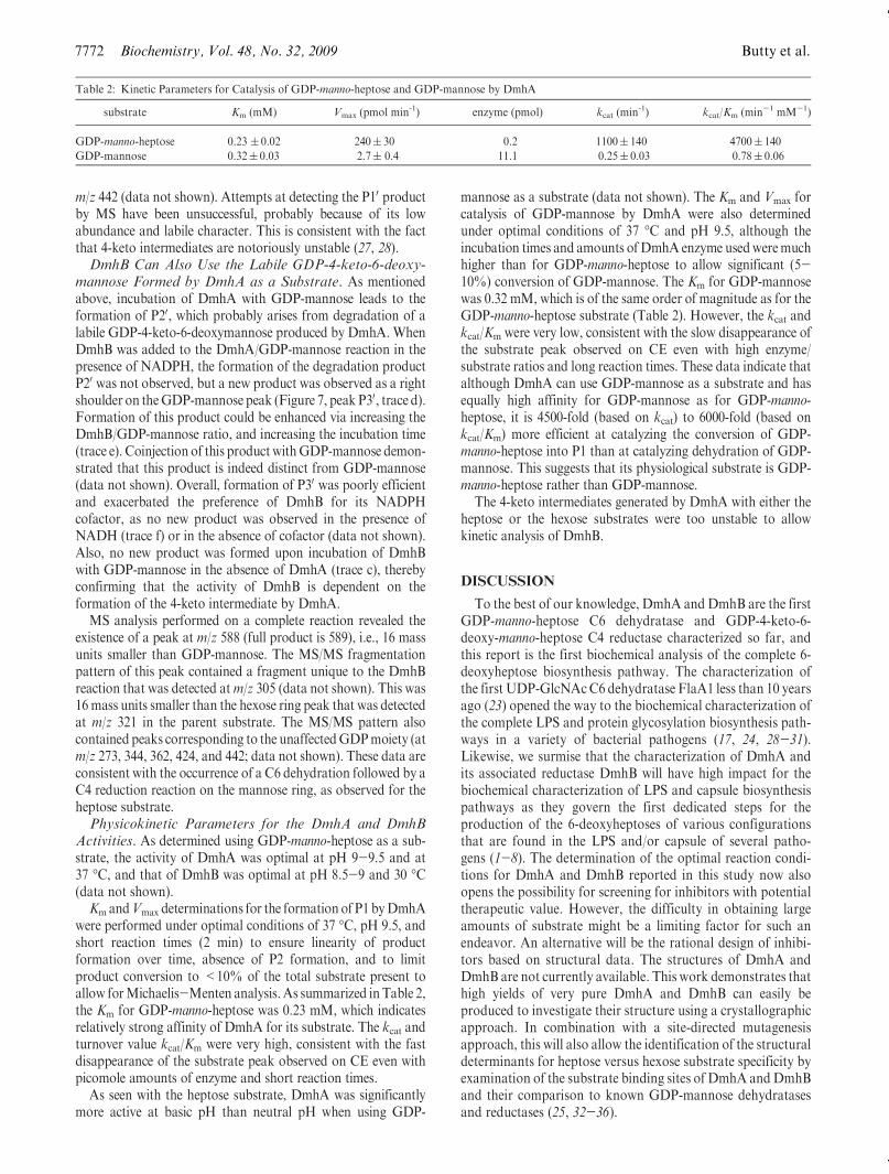
Km andVmax determinations for the formation of P1 byDmhAwere performed under optimal conditions of 37 �C, pH 9.5, andshort reaction times (2 min) to ensure linearity of productformation over time, absence of P2 formation, and to limitproduct conversion to <10% of the total substrate present toallow forMichaelis-Menten analysis.As summarized in Table 2,the Km for GDP-manno-heptose was 0.23 mM, which indicatesrelatively strong affinity of DmhA for its substrate. The kcat andturnover value kcat/Km were very high, consistent with the fastdisappearance of the substrate peak observed on CE even withpicomole amounts of enzyme and short reaction times.
As seen with the heptose substrate, DmhA was significantlymore active at basic pH than neutral pH when using GDP-
mannose as a substrate (data not shown). The Km and Vmax forcatalysis of GDP-mannose by DmhA were also determinedunder optimal conditions of 37 �C and pH 9.5, although theincubation times and amounts of DmhA enzyme usedweremuchhigher than for GDP-manno-heptose to allow significant (5-10%) conversion of GDP-mannose. The Km for GDP-mannosewas 0.32 mM, which is of the same order of magnitude as for theGDP-manno-heptose substrate (Table 2). However, the kcat andkcat/Km were very low, consistent with the slow disappearance ofthe substrate peak observed on CE even with high enzyme/substrate ratios and long reaction times. These data indicate thatalthough DmhA can use GDP-mannose as a substrate and hasequally high affinity for GDP-mannose as for GDP-manno-heptose, it is 4500-fold (based on kcat) to 6000-fold (based onkcat/Km) more efficient at catalyzing the conversion of GDP-manno-heptose into P1 than at catalyzing dehydration of GDP-mannose. This suggests that its physiological substrate is GDP-manno-heptose rather than GDP-mannose.
The 4-keto intermediates generated by DmhA with either theheptose or the hexose substrates were too unstable to allowkinetic analysis of DmhB.
DISCUSSION
To the best of our knowledge, DmhA and DmhB are the firstGDP-manno-heptose C6 dehydratase and GDP-4-keto-6-deoxy-manno-heptose C4 reductase characterized so far, andthis report is the first biochemical analysis of the complete 6-deoxyheptose biosynthesis pathway. The characterization ofthe first UDP-GlcNAcC6 dehydratase FlaA1 less than 10 yearsago (23) opened the way to the biochemical characterization ofthe complete LPS and protein glycosylation biosynthesis path-ways in a variety of bacterial pathogens (17, 24, 28-31).Likewise, we surmise that the characterization of DmhA andits associated reductase DmhB will have high impact for thebiochemical characterization of LPS and capsule biosynthesispathways as they govern the first dedicated steps for theproduction of the 6-deoxyheptoses of various configurationsthat are found in the LPS and/or capsule of several patho-gens (1-8). The determination of the optimal reaction condi-tions for DmhA and DmhB reported in this study now alsoopens the possibility for screening for inhibitors with potentialtherapeutic value. However, the difficulty in obtaining largeamounts of substrate might be a limiting factor for such anendeavor. An alternative will be the rational design of inhibi-tors based on structural data. The structures of DmhA andDmhB are not currently available. This work demonstrates thathigh yields of very pure DmhA and DmhB can easily beproduced to investigate their structure using a crystallographicapproach. In combination with a site-directed mutagenesisapproach, this will also allow the identification of the structuraldeterminants for heptose versus hexose substrate specificity byexamination of the substrate binding sites of DmhA andDmhBand their comparison to known GDP-mannose dehydratasesand reductases (25, 32-36).
Table 2: Kinetic Parameters for Catalysis of GDP-manno-heptose and GDP-mannose by DmhA
substrate Km (mM) Vmax (pmol min-1) enzyme (pmol) kcat (min-1) kcat/Km (min-1 mM-1)
GDP-manno-heptose 0.23 ( 0.02 240( 30 0.2 1100( 140 4700( 140
GDP-mannose 0.32( 0.03 2.7( 0.4 11.1 0.25( 0.03 0.78( 0.06
Article Biochemistry, Vol. 48, No. 32, 2009 7773
The biochemical characterization of DmhB and completecharacterization of its reaction product were important as theycan now serve as a reference for the study of related reductasespresent in other species. This is significant as the reductases donot exhibit quite as high similarities as the dehydratases, whichcould imply functional differences. For example, the DmhAhomologue (WcbK) found inC. jejuni strain 81-176 that harbors6-deoxy-altro-heptose in its capsule (8, 37) is 78% identical and87% similar to the Y. pseudotuberculosisDmhA. In contrast, thebest hit for the candidate reductase in this strain, WcaG, onlyexhibits 23% identity and 43% similarity to the Y. pseudotuber-culosis DmhB. It will be interesting to investigate whether thereductase WcaG is involved in changes in ring configuration,using DmhB and its characterized product as references.
MS analyses support the hypothesis that the reaction productof DmhA (P1) is GDP-4-keto-6-deoxy-lyxo-heptose. Althoughno analysis of product P1 was possible beyond MS due to itsextreme lability, the identity of P1 is consistent with the fact thatthe modified heptose present in the O-antigen of Y. pseudotu-berculosis is also in the same, unaltered manno configuration asthat of the substrate. In some cases, C6 dehydration can beaccompanied by C5 epimerization (27, 28) or C4/C5 enoliza-tion (22), resulting in inversion of the ring configuration. How-ever, such a step is unnecessary for the synthesis of the manno-heptose present in the O-antigen of Y. pseudotuberculosis. Also,modified heptoses of altered configuration are found in thecapsule or exopolysaccharide of a variety of Campylobacterisolates (4, 8, 37-42). The corresponding gene clusters alsoencode putative epimerases that could be responsible for the ringconfiguration change (37, 43). TheO-antigen cluster that encodesDmhA and DmhB does not encode for any candidate epimerasethat could restore the original configuration (10, 44). All of thissuggests that the configuration of the heptose ring is retainedduring C6 dehydration. The identity of P1 as GDP-4-keto-6-deoxy-lyxo-heptose was further supported by MS and NMRanalyses of its derivative P3, obtained from catalysis of P1 byDmhB (P3).
It is somewhat surprising that the optimal activity of DmhA is37 �C since O-antigen production is downregulated at 37 �C (45).This suggests that O-antigen regulation in Y. pseudotuberculosismight not encompass the biosynthesis of required precursors butrather might concern their assembly and export to the bacterialsurface. Also, the basic optimal pH and the slight differences ofoptimal conditions observed between DmhA and DmhB wereunexpected since the optimal pHs are higher than the cytoplasmicpH that the enzymes are exposed to, and both enzymes would beanticipated to function optimally under the same conditions sincethey belong to the same biochemical pathway.
DmhA and DmhB can perform their activity on both theheptose and the hexose substrates. C6 dehydration and reductionof GDP-mannose is usually associated with the formation ofGDP-L-fucose or ofGDP-perosamine (34, 36).MSdata obtainedfor the reduced product P30 are consistent with the formation ofeither compound, but we could not generate enough material todistinguish between both compounds by NMR. Also, neitherfucose nor perosamine have been reported in the surface carbo-hydrates ofY. pseudotuberculosis, but theymay be part of proteinglycosylation motifs. Importantly though, although DmhA hassimilar Km values for GDP-manno-heptose and GDP-mannose,and although theseKm are comparable to theKmof knownGDP-mannose (or other sugar nucleotide) C6 dehydratases (34, 35, 46),the kinetic analysis performed in this study suggests that the
yields for such compounds would be extremely small comparedwith the production of 6-deoxyheptose dedicated to LPS synth-esis. Indeed, the kcat of DmhA for GDP-mannose is 5-10-foldsmaller than that of the H. pylori or human GMD (33, 34). Nokinetic analysis of DmhB was possible for the lack of stablesubstrate, but very limited conversion of the 4-keto-6-deoxy-manno intermediate was obtained after lengthy incubation withabundant DmhB, as opposed to the very rapid and completeconversion of the 4-keto-6-deoxy-manno-heptose obtained withtrace amounts of DmhB. Hence, the physiological substrate forDmhA/DmhB is not GDP-mannose but is indeed GDP-manno-heptose, which the enzymes use to generate 6-deoxyheptose forO-antigen synthesis as demonstrated genetically previously (9).
Several Gram-negative bacteria, including Y. pseudotubercu-losis and C. jejuni, harbor two biosynthetic pathways to generatenucleotide-activated manno-heptose (43, 47). One uses ADP-L-glycero-β-D-manno-heptose and is, among others, utilized forLPS core synthesis, whereas the other uses GDP-D-glycero-R-D-manno-heptose (14, 48). The reason for the existence of twopathways is not clear inmost cases. If a single form of nucleotide-activated manno-heptose was shared between various pathways,it would be difficult for the cell to control the level of activity ofpotent dehydratases to generate enough dehydrated heptose forO-antigen or capsule synthesis without depleting the intracellularpool of primary heptose and ensure appropriate levels of lipid A-core synthesis which are necessary for membrane stability. Thekinetic analysis that shows the extremely high catalytic efficiencyof DmhA for conversion of GDP-manno-heptose into GDP-4-keto-6-deoxy-lyxo-heptose brings some rationale for the exis-tence of both pathways: the high activity of DmhA on the GDP-based precursor should not compromise the stock of ADP-basedprecursor dedicated to lipid-A core synthesis. The activity ofDmhA on ADP-glycero-manno-heptose was not tested in thisstudy for lack of substrate, but sugar nucleotide C6 dehydratasesusually use the nucleotide as a fixed handle while catalysisproceeds on the sugar moiety. Consequently, they are usuallyvery specific and exclusive for a given nucleotide, and no activitywould be expected for DmhA with ADP-bound sugars.
Prior genetics studies suggested that DmhA might interactwith DmhB to be active in vivo, as it was not possible tocomplement a dmhA mutant in the absence of functional DmhBencoded in cis from dmhA (9). This biochemical study clearlydemonstrates that DmhA functions with very high efficiency onits own in vitro, in the absence of anyDmhB. It is possible that aninteraction between DmhA and DmhB is actually needed in vivoto allow the completion of deoxy-manno-heptose synthesis, notfor DmhA activity per se, but rather for channeling of itsextremely unstable reaction product (P1) to the reductaseDmhB.This would avoid unproductive degradation of P1 into P2. Also,this would be important to prevent DmhA from sequesteringavailable GDP-mannose for which it has high affinity and whichmight then be lacking from other pathways important for cellsurvival or virulence. Such a channelingmechanism has also beenproposed for related enzymes involved in protein glycosylationbiosynthetic pathways (24).
In summary, this study is the first characterization of a heptoseC6 dehydratase and its associated reductase and opens up manypossibilities to decipher the modified heptose biosynthetic path-ways that support the production of virulence factors in bacterialpathogens. This work has practical implications as it will allowadvances for the development of therapeutic inhibitors, and itwill also allow the synthesis of complexmodified heptoses in their
7774 Biochemistry, Vol. 48, No. 32, 2009 Butty et al.
nucleotide-activated forms that can then be used in the enzymaticsynthesis of carbohydrate epitopes for vaccination purposes. Thiswork also has fundamental significance as it will allow theelucidation of the structural determinants that govern the sub-strate specificity and segregation of heptose versus hexose C6dehydratases and associated reductases.
ACKNOWLEDGMENT
We thank Dr. P. Reeves for providing the sequence for DmhAbefore its release on-line at the onset of this project.We thankDr.P. Messner (Vienna, Austria) for providing the starterpDONR201 plasmids for GmhA/B/C/D and M. D’Elia (Dr.Brown’s laboratory, Mc Master University) for transferring theGmhA/B/C/D genes into the pDEST vectors. We thank V. Soofor cloning HP0044 in the GST-tag vector system. Finally, wethank P. Hopf for assistance with softwares and computer issuesand S. Kichler for assistance with Western blotting.
SUPPORTING INFORMATION AVAILABLE
Figure 1S containing data demonstrating that the formation ofproduct P2, that occurs over time after incubation of DmhA andGDP-manno-heptose, is nonenzymatic. This material is availablefree of charge via the Internet at http://pubs.acs.org.
REFERENCES
1. Samuelsson, K., Lindberg, B., and Brubaker, R. R. (1974) Structureof O-specific side chains of lipopolysaccharides from Yersinia pseu-dotuberculosis. J. Bacteriol. 117, 1010–1016.
2. Skurnik, M., and Zhang, L. (1996) Molecular genetics and biochem-istry of Yersinia lipopolysaccharide. APMIS 104, 849–872.
3. Komandrova,N.A.,Gorshkova,R. P., Isakov, V.V., andOvodov Iu,S. (1984) Structure of O-specific polysaccharide isolated from theYersinia pseudotuberculosis serotype 1A lipopolysaccharide. Bioorg.Khim. 10, 232–237.
4. St. Michael, F., Szymanski, C. M., Li, J., Chan, K. H., Khieu, N. H.,Larocque, S.,Wakarchuk,W.W., Brisson, J. R., andMonteiro,M.A.(2002) The structures of the lipooligosaccharide and capsule poly-saccharide of Campylobacter jejuni genome sequenced strain NCTC11168. Eur. J. Biochem. 269, 5119–5136.
5. Knirel, Y. A., Paramonov, N. A., Shashkov, A. S., Kochetkov, N. K.,Yarullin, R. G., Farber, S. M., and Efremenko, V. I. (1992) Structureof the polysaccharide chains of Pseudomonas pseudomallei lipopoly-saccharides. Carbohydr. Res. 233, 185–193.
6. Perry, M. B., MacLean, L. L., Schollaardt, T., Bryan, L. E., and Ho,M. (1995) Structural characterization of the lipopolysaccharide Oantigens of Burkholderia pseudomallei. Infect. Immun. 63, 3348–3352.
7. DeShazer, D., Waag, D. M., Fritz, D. L., and Woods, D. E. (2001)Identification of a Burkholderia mallei polysaccharide gene cluster bysubtractive hybridization and demonstration that the encoded capsuleis an essential virulence determinant. Microb. Pathog. 30, 253–269.
8. Aspinall, G. O., McDonald, A. G., and Pang, H. (1992) Structures ofthe O chains from lipopolysaccharides of Campylobacter jejuni ser-otypes O:23 and O:36. Carbohydr. Res. 231, 13–30.
9. Ho, N., Kondakova, A. N., Knirel, Y. A., and Creuzenet, C. (2008)The biosynthesis and biological role of 6-deoxyheptose in the lipopo-lysaccharide O-antigen of Yersinia pseudotuberculosis. Mol. Micro-biol. 68, 424–447.
10. Pacinelli, E., Wang, L., and Reeves, P. R. (2002) Relationship ofYersinia pseudotuberculosisO antigens IA, IIA, and IVB: the IIA genecluster was derived from that of IVB. Infect. Immun. 70, 3271–3276.
11. Kondakova, A. N., Ho, N., Bystrova, O. V., Shashkov, A. S.,Lindner, B., Creuzenet, C., andKnirel, Y. A. (2008) Structural studiesof the O-antigens of Yersinia pseudotuberculosis O:2a and mutantsthereof with impaired 6-deoxy-D-manno-heptose biosynthesis path-way. Carbohydr. Res. 343, 1383–1389.
12. Graziani, A., Zamyatina, A., and Kosma, P. (2004) A convenientsynthesis of GDP D-glycero-alpha-D-manno-heptopyranose. Carbo-hydr. Res. 339, 147–1451.
13. Kneidinger, B., Graninger, M., Puchberger, M., Kosma, P., andMessner, P. (2001) Biosynthesis of nucleotide-activated D-glycero-D-manno-heptose. J. Biol. Chem. 276, 20935–20944.
14. Kneidinger, B., Marolda, C., Graninger, M., Zamyatina, A.,McArthur, F., Kosma, P., Valvano, M. A., and Messner, P. (2002)Biosynthesis pathway of ADP-L-glycero-beta-D-manno-heptose inEscherichia coli. J. Bacteriol. 184, 363–369.
15. Newton,D. T., andMangroo,D. (1999)Mapping the active site of theHaemophilus influenzaemethionyl-tRNA formyltransferase: residuesimportant for catalysis and tRNA binding. Biochem. J. 339 (Part 1),63–69.
16. Moore, J. T., Uppal, A., Maley, F., and Maley, G. F. (1993) Over-coming inclusion body formation in a high-level expression system.Protein Expression Purif. 4, 160–163.
17. Obhi, R.K., andCreuzenet, C. (2005) Biochemical characterization ofthe Campylobacter jejuni Cj1294, a novel UDP-4-keto-6-deoxy-GlcNAc aminotransferase that generates UDP-4-amino-4,6-di-deoxy-GalNAc. J. Biol. Chem. 280, 20902–20908.
18. Bax, A., and Davis, D. G. (1985) MLEV-17-based two-dimensionalhomonuclear magnetization transfer spectroscopy. J. Magn. Reson.65, 355–360.
19. Piantini, U., Sorensen, O. W., and Ernst, R. R. (1982) Multiplequantum filters for elicidatingNMRcoupling networks. J.Am.Chem.Soc. 104, 6800–6801.
20. Kay, L. E., Keifer, P., and Saarinen, T. (1992) Pure absorptiongradient enhanced heteronuclear single quantum correlation spec-troscopy with improved sensitivity. J. Am. Chem. Soc. 114, 10663–10665.
21. John, B. K., Plant, D., and Hurd, R. E. (1992) Improved proton-detected heteronuclear correlation using gradient-enhanced z and zzfilters. J. Magn. Reson. A101, 113–117.
22. Ishiyama, N., Creuzenet, C., Miller, W. L., Demendi, M., Anderson,E. M., Harauz, G., Lam, J. S., and Berghuis, A. M. (2006) Structuralstudies of FlaA1 from Helicobacter pylori reveal the mechanism forinverting 4,6-dehydratase activity. J. Biol. Chem. 281, 24489–24495.
23. Creuzenet, C., Schur, M. J., Li, J., Wakarchuk,W.W., and Lam, J. S.(2000) FlaA1, a new bifunctional UDP-GlcNAc C6 Dehydratase/C4
reductase from Helicobacter pylori. J. Biol. Chem. 275, 34873–34880.24. Creuzenet, C. (2004) Characterization of CJ1293, a new UDP-
GlcNAc C6 dehydratase from Campylobacter jejuni. FEBS Lett.559, 136–140.
25. Wu, B., Zhang, Y., and Wang, P. G. (2001) Identification andcharacterization of GDP-d-mannose 4,6-dehydratase and GDP-l-fucose snthetase in a GDP-l-fucose biosynthetic gene cluster fromHelicobacter pylori. Biochem. Biophys. Res. Commun. 285, 364–371.
26. Ramm, M., Wolfender, J. L., Queiroz, E. F., Hostettmann, K., andHamburger, M. (2004) Rapid analysis of nucleotide-activated sugarsby high-performance liquid chromatography coupled with diode-array detection, electrospray ionizationmass spectrometry and nucle-ar magnetic resonance. J. Chromatogr. A 1034, 139–148.
27. McNally, D. J., Schoenhofen, I. C., Mulrooney, E. F., Whitfield, D.M., Vinogradov, E., Lam, J. S., Logan, S. M., and Brisson, J. R.(2006) Identification of labile UDP-ketosugars inHelicobacter pylori,Campylobacter jejuni and Pseudomonas aeruginosa: key metabolitesused to make glycan virulence factors. ChemBioChem 7, 1865–1868.
28. Schoenhofen, I. C., McNally, D. J., Vinogradov, E., Whitfield, D.,Young, N. M., Dick, S., Wakarchuk, W. W., Brisson, J. R., andLogan, S. M. (2006) Functional characterization of dehydratase/aminotransferase pairs from Helicobacter and Campylobacter: en-zymes distinguishing the pseudaminic acid and bacillosamine biosyn-thetic pathways. J. Biol. Chem. 281, 723–732.
29. Creuzenet, C., and Lam, J. S. (2001) Topological and functionalcharacterization of WbpM, an inner membrane UDP-GlcNAc C6
dehydratase essential for lipopolysaccharide biosynthesis in Pseudo-monas aeruginosa. Mol. Microbiol. 41, 1295–1310.
30. Schoenhofen, I. C., McNally, D. J., Brisson, J. R., and Logan, S. M.(2006) Elucidation of the CMP-pseudaminic acid pathway in Helico-bacter pylori: synthesis from UDP-N-acetylglucosamine by a singleenzymatic reaction. Glycobiology 16, 8C–14C.
31. Vijayakumar, S., Merkx-Jacques, A., Ratnayake, D., Gryski, I.,Obhi, R. K., Houle, S., Dozois, C., and Creuzenet, C. (2006)Cj1121c, a novel UDP-4-keto-6-deoxy-GlcNAc C4 aminotransferaseessential for protein glycosylation and virulence in Campylobacterjejuni. J. Biol. Chem. 281, 27733–27743.
32. Bisso,A., Sturla, L., Zanardi,D.,DeFlora, A., andTonetti,M. (1999)Structural and enzymatic characterization of human recombinantGDP-D-mannose-4,6-dehydratase. FEBS Lett. 456, 370–374.
33. Sullivan, F. X., Kumar, R., Kriz, R., Stahl, M., Xu, G. Y., Rouse, J.,Chang, X. J., Boodhoo, A., Potvin, B., and Cumming, D. A. (1998)Molecular cloning of human GDP-mannose 4,6-dehydratase andreconstitution of GDP-fucose biosynthesis in vitro. J. Biol. Chem.273, 8193–8202.
Article Biochemistry, Vol. 48, No. 32, 2009 7775
34. Wu, B., Zhang, Y., and Wang, P. G. (2001) Identification andcharacterization of GDP-D-mannose 4,6-dehydratase and GDP-l-fucose synthetase in a GDP-l-fucose biosynthetic gene cluster fromHelicobacter pylori. Biochem. Biophys. Res. Commun. 285, 364–371.
35. Sturla, L., Bisso, A., Zanardi, D., Benatti, U., De Flora, A., andTonetti, M. (1997) Expression, purification and characterization ofGDP-D-mannose 4,6-dehydratase from Escherichia coli. FEBS Lett.412, 126–130.
36. Albermann, C., and Piepersberg, W. (2001) Expression and identifica-tion of the RfbE protein from Vibrio cholerae O1 and its use for theenzymatic synthesis of GDP-D-perosamine. Glycobiology 11, 655–661.
37. Karlyshev, A. V., Champion, O. L., Churcher, C., Brisson, J. R.,Jarrell, H. C., Gilbert, M., Brochu, D., St. Michael, F., Li, J.,Wakarchuk, W. W., Goodhead, I., Sanders, M., Stevens, K., White,B., Parkhill, J.,Wren, B.W., and Szymanski, C.M. (2005) Analysis ofCampylobacter jejuni capsular loci reveals multiple mechanisms forthe generation of structural diversity and the ability to form complexheptoses. Mol. Microbiol. 55, 90–103.
38. Aspinall, G. O., McDonald, A. G., Pang, H., Kurjanczyk, L. A., andPenner, J. L. (1993) An antigenic polysaccharide fromCampylobactercoli serotype O:30. Structure of a teichoic acid-like antigenic poly-saccharide associated with the lipopolysaccharide. J. Biol. Chem. 268,18321–18329.
39. Aspinall, G. O., McDonald, A. G., Pang, H., Kurjanczyk, L. A., andPenner, J. L. (1993) Lipopolysaccharide of Campylobacter coli ser-otype O:30. Fractionation and structure of liberated core oligosac-charide. J. Biol. Chem. 268, 6263–6268.
40. Aspinall, G. O., Monteiro, M. A., and Pang, H. (1995) Lipo-oligo-saccharide of the Campylobacter lari type strain ATCC 35221.Structure of the liberated oligosaccharide and an associated extra-cellular polysaccharide. Carbohydr. Res. 279, 245–264.
41. Hanniffy, O. M., Shashkov, A. S., Moran, A. P., Prendergast, M.M.,Senchenkova, S. N., Knirel, Y. A., and Savage, A. V. (1999) Chemicalstructure of a polysaccharide from Campylobacter jejuni 176.83
(serotype O:41) containing only furanose sugars. Carbohydr. Res.319, 124–132.
42. Chen, Y. H., Poly, F., Pakulski, Z., Guerry, P., and Monteiro, M. A.(2008) The chemical structure and genetic locus of Campylobacterjejuni CG8486 (serotype HS:4) capsular polysaccharide: the identifi-cation of 6-deoxy-D-ido-heptopyranose. Carbohydr. Res. 343, 1034–1040.
43. Parkhill, J., Wren, B. W., Mungall, K., Ketley, J. M., Churcher, C.,Basham, D., Chillingworth, T., Davies, R. M., Feltwell, T., Holroyd,S., Jagels, K., Karlyshev, A. V., Moule, S., Pallen, M. J., Penn, C. W.,Quail, M. A., Rajandream, M. A., Rutherford, K. M., van Vliet, A.H., Whitehead, S., and Barrell, B. G. (2000) The genome sequence ofthe food-borne pathogen Campylobacter jejuni reveals hypervariablesequences. Nature 403, 665–668.
44. Skurnik, M., Venho, R., Toivanen, P., and al-Hendy, A. (1995) Anovel locus of Yersinia enterocolitica serotype O:3 involved in lipo-polysaccharide outer core biosynthesis. Mol. Microbiol. 17, 575–594.
45. Straley, S. C., and Perry, R. D. (1995) Environmental modulation ofgene expression and pathogenesis in Yersinia. Trends Microbiol. 3,310–317.
46. Sullivan, F. X., Kumar, R., Kriz, R., Stahl, M., Xu, G. Y., Rouse, J.,Chang, X. J., Boodhoo, A., Potvin, B., and Cumming, D. A. (1998)Molecular cloning of human GDP-mannose 4,6-dehydratase andreconstitution of GDP-fucose biosynthesis in vitro. J. Biol. Chem.273, 8193–8202.
47. Skurnik,M., Peippo,A., andErvela, E. (2000) Characterization of theO-antigen gene clusters of Yersinia pseudotuberculosis and the crypticO-antigen gene cluster ofYersinia pestis shows that the plague bacillusis most closely related to and has evolved from Y. pseudotuberculosisserotype O:1b. Mol. Microbiol. 37, 316–330.
48. Valvano, M. A., Messner, P., and Kosma, P. (2002) Novel pathwaysfor biosynthesis of nucleotide-activated glycero-manno-heptose pre-cursors of bacterial glycoproteins and cell surface polysaccharides.Microbiology 148, 1979–1989.