elucidation of the expression and role of …
TRANSCRIPT

ELUCIDATION OF THE EXPRESSION AND ROLE
OF CAROTENOGENIC GENES IN ASTAXANTHIN
PRODUCTION IN A HYPER-PRODUCING
MUTANT OF Xanthophyllomyces dendrorhous
ANG FONG SIM
UNIVERSITI SAINS MALAYSIA
2018

ELUCIDATION OF THE EXPRESSION AND ROLE
OF CAROTENOGENIC GENES IN ASTAXANTHIN
PRODUCTION IN A HYPER-PRODUCING
MUTANT OF Xanthophyllomyces dendrorhous
by
ANG FONG SIM
Thesis submitted in fulfilment of requirements
for the degree of
Master of Science
May 2018
ii
ACKNOWLEDGEMENT
Above all, it would have been impossible to write this thesis without the many wonderful
people who supported me throughout this challenging period of my MSc. study. First and
foremost, I would like to express my sincere gratitude to my supervisor, Dr. Chew Ai Lan
for her constant guidance, patience and support throughout the duration of my study.
Without her knowledge and constructive suggestions, this task would not have been
completed. I would also like to thank my co-supervisor, Assoc. Prof. Few Ling Ling for
her endless support and guidance.
Heartfelt thanks to Prof. Dr Tan Soo Choon also for his valuable advice and guidance. I
am grateful to have all the kind and helpful friends and colleagues who are friendly and
always give me a helping hand. Special thanks to Tan Wee Yee, Chang Chiat Han, Christy
Khaw, Michelle Kong, Ng Woei Kean, Kuah Vee May, Ron Siah, Hannah, Richard Tan,
Jason Chin, Carlos Silvester, Mei Jin and other members of Institute for Research in
Molecular Medicine (INFORMM), who were involved directly or indirectly in this
research. Thank you for all the support and motivation, and never tired of guiding me
during my study. I would also like to extend my sincere appreciation to all the scientific
officers and administration staffs of INFORMM.
My special appreciation goes to the Ministry of Higher Education Malaysia (MoHE) for
the financial support through MyBrain15 (MyMaster), in the early phase of my study, and
USM for giving me the Graduate Assistantship in the later part of my study. Thanks to the

iii
Ministry of Higher Education Malaysia for providing the Fundamental Research Grant
Scheme (FRGS) under grant 203/CIPPM/6711337 to support this study.
Last but not least, I would like to thank my family for their encouragement and support in
my pursuits. I would also like to express my deepest gratitude to my wife, Low Wei Ling,
for her understanding and love all these years. I would not have such achievement in this
path of research without all of them and I wish that I can continue to make them proud.
Finally, I would like to extend my token of appreciation to everyone else who helped me
throughout my journey.
iv
TABLE OF CONTENT
ACKNOWLEDGEMENT ......................................................................................... ii
TABLE OF CONTENTS .......................................................................................... iv
LIST OF TABLES .................................................................................................. viii
LIST OF FIGURES .................................................................................................. ix
LIST OF SYMBOLS, ABBREVIATIONS AND ACRONYMNS ....................... xii
ABSTRAK ............................................................................................................... xvi
ABSTRACT ........................................................................................................... xviii
CHAPTER 1: INTRODUCTION ............................................................................. 1
1.1 Background ......................................................................................................... 1
1.2 Significance of the Study .................................................................................... 5
1.3 Objectives ........................................................................................................... 8
CHAPTER 2: LITERATURE REVIEW ................................................................. 9
2.1 Carotenoids ......................................................................................................... 9
2.1.1 Background........................................................................................... 9
2.1.2 Astaxanthin ......................................................................................... 11
2.1.2(a) Structures and Chemicals Forms of Astaxanthin ............ 11
2.1.2(b) Analysis of Astaxanthin................................................... 17
2.1.2(c) Biological Properties of Astaxanthin and Its Health
Benefits ............................................................................ 21
2.2 Astaxanthin Biosynthesis in Xanthophyllomyces dendrorhous ........................ 28
2.2.1 Xanthophyllomyces dendrorhous ....................................................... 28
2.2.2 Biosynthesis of Astaxanthin ............................................................... 30
2.2.3 Astaxanthin Biosynthesis Genes in X. dendrorhous .......................... 33
2.2.3(a) idi Gene............................................................................ 33
2.2.3(b) crtE Gene ......................................................................... 34
2.2.3(c) crtYB Gene ....................................................................... 34
2.2.3(d) crtI Gene .......................................................................... 34
2.2.3(e) crtS Gene ......................................................................... 35
2.2.3(f) crtR Gene ......................................................................... 35

v
2.3 Improvement of Astaxanthin Production.......................................................... 36
2.3.1 Ethyl Methanesulfonate ...................................................................... 38
2.3.2 N-Methyl-N’-Nitro-N-Nitrosoguadinine ............................................ 39
2.3.3 β-Ionone .............................................................................................. 40
CHAPTER 3: MATERIALS AND METHODS ................................................... 41
3.1 Materials ........................................................................................................... 41
3.1.1 Chemicals, Reagents and Kits ............................................................ 41
3.1.2 Glasswares and Consumables ............................................................. 41
3.1.3 Instruments ......................................................................................... 41
3.1.4 Softwares ............................................................................................ 41
3.1.5 Buffers ................................................................................................ 41
3.2 Methods ............................................................................................................ 45
3.2.1 Culture Conditions and Isolation of Overproducing Mutants
through Chemical Mutagenesis .......................................................... 46
3.2.1(a) Yeast Strain and Culture Condition ................................. 46
3.2.1(b) Random Mutagenesis and Screening ............................... 46
3.2.2 Carotenoid Extraction and Analysis ................................................... 48
3.2.2(a) Carotenoid Extraction ...................................................... 48
3.2.2(b) Stability Test .................................................................... 49
3.2.2(c) High Performance Liquid Chromatography Profiling of
Carotenoid Extract ........................................................... 49
3.2.2(d) Construction of Growth and Carotenoid Production
Profile .............................................................................. 50
3.2.2(d)(i) Growth and Carotenoid Production Profile .. 50
3.2.2(d)(ii) Construction of Standard Curve ................... 50
3.2.3 Gene Expression Analysis .................................................................. 51
3.2.3(a) Total RNA Extraction ...................................................... 51
3.2.3(b) Validation of RNA Quality.............................................. 52
3.2.3(c) Validation of RNA Integrity ............................................ 53
3.2.3(c)(i) Tris-Borate-EDTA (TBE) Buffer, 0.5× ........ 53
3.2.3(c)(ii) 8 mg/mL of Ethidium Bromide (EtBr) ......... 53
3.2.3(c)(iii) 1% Agarose Gel ............................................ 53
3.2.3(c)(iv) Gel Electrophoresis ....................................... 54

vi
3.2.3(d) cDNA Synthesis by Reverse Transcription (RT
reaction) ........................................................................... 54
3.2.3(d)(i) Genomic DNA Elimination .......................... 55
3.2.3(d)(ii) cDNA Synthesis............................................ 55
3.2.3(e) Determination of The Relative Expression of
Carotenogenic Gene Transcripts by Real-Time PCR ...... 56
3.2.3(e)(i) Primer Design ............................................... 56
3.2.3(e)(ii) Reference Gene Validation ........................... 57
3.2.3(e)(iii) Target Gene Validation................................. 58
3.2.3(e)(iv) Determination of Target Gene mRNA
Expression..................................................... 59
3.2.4 Gene Sequence Study ......................................................................... 60
3.2.4(a) Genomic DNA Purification ............................................. 60
3.2.4(b) PCR Amplification of Target Genes ............................... 61
3.2.4(b)(i) Primer Design ............................................... 61
3.2.4(b)(ii) PCR Amplification ....................................... 63
3.2.4(c) Extraction of DNA Fragment from Gel and Analysis ..... 63
3.3 Data Collection, Results and Statistical Analysis ............................................. 65
CHAPTER 4: RESULTS ......................................................................................... 66
4.1 Mutagenesis and Isolation of Hyperproducing Strain ...................................... 66
4.1.1 Random Mutagenesis of X. dendrorhous ........................................... 66
4.1.2 β-Ionone Screening ............................................................................. 71
4.2 Total Carotenoid Determination ....................................................................... 73
4.2.1 Total Carotenoid Extraction and Measurement .................................. 73
4.2.2 Stability Test ....................................................................................... 74
4.2.3 HPLC Profiling of Carotenoid Extract ............................................... 78
4.3 Gene Expression ............................................................................................... 88
4.3.1 Total RNA Extraction and Determination .......................................... 88
4.3.1(a) Analysis of RNA Quality and Quantity ........................... 88
4.3.1(b) Determination of RNA Intergrity ................................... 89
4.3.2 Real-Time PCR .................................................................................. 90
4.3.2(a) Validation of Reference Gene ......................................... 90
4.3.2(b) Optimization of Template Concentration ........................ 91
vii
4.3.2(c) Validation of Target Gene ............................................... 93
4.3.2(d) Study of mRNA Expression ............................................ 94
4.3.3 Study of mRNA Expression Levels of Carotenogenic Genes in
Different Carbon Sources ................................................................... 98
4.3.3(a) Analysis of RNA Quality and Quantity ........................... 99
4.3.3(b) Determination of RNA Integrity ...................................... 99
4.3.3(c) Study of mRNA Expression .......................................... 100
4.4 DNA Sequence Analysis of Carotenoid Biosynthesis Genes ......................... 105
4.4.1 Extraction of DNA Fragments.......................................................... 105
4.4.2 DNA Sequences Analysis................................................................. 112
CHAPTER 5: DISCUSSION ................................................................................ 115
CHAPTER 6: CONCLUSION .............................................................................. 142
6.1 Conclusion of The Study ................................................................................ 142
6.2 Recommendations for Future Studies ............................................................. 145
REFERENCES ....................................................................................................... 147
APPENDICES
viii
LIST OF TABLES
Page
Table 1.1 Sustainability comparison of different astaxanthin production
methods................................................................................................. 3
Table 3.1 List of reagents and kits used in this study ......................................... 42
Table 3.2 List of glasswares and consumables used in this study ...................... 43
Table 3.3 List of instruments used in this study ................................................. 44
Table 3.4 List of softwares used in this study .................................................... 44
Table 3.5 List of buffers used in this study ........................................................ 44
Table 3.6 List of primers for Real-Time PCR .................................................... 57
Table 3.7 Step and formula for relative quantification of gene expression ........ 60
Table 3.8 List of primers used for PCR reaction in this study ........................... 62
Table 4.1 Growth and carotenoid production of X. dendrorhous DSM 5626
and its mutants .................................................................................... 74
Table 4.2 The concentration and purity of extracted total RNA from wild
type strain and M34 mutant strain ...................................................... 89
Table 4.3 Optimization of cDNA concentration for Real-Time PCR using
β-actin ................................................................................................. 93
Table 4.4 The concentration and purity of extracted total RNA from M34
mutant (grown with sucrose as carbon source) .................................. 99
Table 4.5 Mutations of carotenoid biosynthesis genes in M34 mutant
compared to wild type strain of X. dendrorhous .............................. 113
ix
LIST OF FIGURES
Page
Figure 2.1 Chemical structure of astaxanthin ..................................................... 12
Figure 2.2 Geometrical isomers of astaxanthin ................................................... 13
Figure 2.3 Optical stereoisomers of astaxanthin. ................................................. 15
Figure 2.4 Astaxanthin and its esters from various sources................................. 16
Figure 2.5 UV-visible spectrum of X. dendrorhous carotenoid extracts
............................................................................................................ 18
Figure 2.6 Spectra of astaxanthin geometrical isomers ....................................... 21
Figure 2.7 Metabolic pathway from glucose to astaxanthin in X.
dendrorhous ....................................................................................... 31
Figure 2.8 Modified astaxanthin biosynthetic pathway in X. dendrorhous ........ 32
Figure 2.9 Enzymatic reaction catalyzed by IPP isomerase in X. dendrorhous
............................................................................................................ 33
Figure 2.10 Chemical structure of ethyl methanesulfonate .................................. 38
Figure 2.11 Chemical structure of N-methyl-N’-nitro-N-nitrosoguanidine ......... 39
Figure 2.12 Chemical structure of β-ionone .......................................................... 40
Figure 3.1 Flow chart of methodology ................................................................ 45
Figure 4.1 Survival rate of chemical mutagen treated X. dendrorhous ............... 67
Figure 4.2 Colour phenotype of wild type and mutant strains cultured on
solid yeast malt agar ........................................................................... 68
Figure 4.3 Cultures of X. dendrorhous strains ..................................................... 69
Figure 4.4 Colonies of wild type and chemical mutagen treated X.
dendrorhous on agar plates ................................................................ 70
Figure 4.5 Selected X. dendrorhous mutants cultured on screening media
with and without β-ionone. ................................................................. 72
x
Figure 4.6 Stability of carotenoid production in mutants M34, M35 and M38
for 10 generations ............................................................................... 75
Figure 4.7 Growth and carotenoid production of wild type strain and M34
mutant of X. dendrorhous in glucose medium ................................... 76
Figure 4.8 Growth and carotenoid production of M34 mutant of X.
dendrorhous in medium with sucrose and glucose as carbon
sources ................................................................................................ 77
Figure 4.9 HPLC chromatograms of astaxanthin standard for the first three
repeated analyses ................................................................................ 79
Figure 4.10 HPLC chromatogram of astaxanthin standard eluted with
methanol/dichloromethane/acetonitrile/water (67.5/17.5/9.5/5.5) ..... 80
Figure 4.11 HPLC chromatogram of astaxanthin standard eluted with
methanol/dichloromethane/acetonitrile/water
(67.5/12.5/9.5/10.5) ............................................................................ 81
Figure 4.12 HPLC chromatogram of astaxanthin standard eluted with
methanol/dichloromethane/acetonitrile/water (72.5/17.5/9.5/0.5) ..... 81
Figure 4.13 HPLC chromatogram of astaxanthin standard eluted with
methanol/dichloromethane/acetonitrile/water (77.5/12.5/9.5/0.5) ..... 82
Figure 4.14 HPLC chromatogram of astaxanthin standard eluted with Solvent
A [methanol/dichloromethane/acetonitrile/water (77.5/12.5/9.5/
0.5)] and Solvent B (UHQ water) at 1:1 ratio .................................... 83
Figure 4.15 HPLC chromatogram of astaxanthin standard eluted with Solvent
A [methanol/dichloromethane/acetonitrile/water (77.5/12.5/9.5/
0.5)] and Solvent B (UHQ water) at 3:1 ratio .................................... 83
Figure 4.16 HPLC chromatogram of astaxanthin standard eluted with Solvent
A [methanol/dichloromethane/acetonitrile/water (77.5/12.5/9.5/
0.5)] and Solvent B (UHQ water) at 9:1 ratio .................................... 84
Figure 4.17 HPLC chromatogram of astaxanthin standard eluted with Solvent
A [methanol/dichloromethane/acetonitrile/water (77.5/12.5/9.5/
0.5)] and Solvent B (UHQ water) at 19:1 ratio .................................. 85
Figure 4.18 HPLC chromatogram of (a) β-carotene standard; (b) astaxanthin
standard; (c) carotenoid extract from wild type strain; (d)
carotenoid extract from M34 mutant; (e) MeOH eluted with
Solvent A [methanol/dichloromethane/acetonitrile/water (77.5
/12.5/9.5/0.5)] and Solvent B (UHQ water) at 9:1 ratio ..................... 87

xi
Figure 4.19 Total RNA extracted from wild type strain and M34 mutant at 24
h, 40 h, 60 h and 90 h ......................................................................... 90
Figure 4.20 Real-Time PCR standard curve for reference gene (β-actin) ............. 91
Figure 4.21 Melt curves of cDNA templates at different concentrations. ............. 92
Figure 4.22 Amplification of β-actin gene and astaxanthin biosynthesis genes
in X. dendrorhous. .............................................................................. 94
Figure 4.23 Real-Time PCR analysis of six carotenoid biosynthesis genes in
wild type strain and M34 mutant strain of X. dendrorhous ................ 96
Figure 4.24 Total RNA extracted from M34 mutant culture with sucrose as
carbon source .................................................................................... 100
Figure 4.25 Real-Time PCR analysis of six carotenoid biosynthesis genes in
wild type strain and M34 mutant strain of X. dendrorhous in
different carbon sources ................................................................... 102
Figure 4.26 Amplification of idi gene in genomic DNA of M34 mutant via
conventional PCR ............................................................................. 106
Figure 4.27 Amplification of crtE gene in genomic DNA of M34 mutant via
conventional PCR ............................................................................. 107
Figure 4.28 Amplification of crtYB gene in genomic DNA of M34 mutant via
conventional PCR ............................................................................. 108
Figure 4.29 Amplification of crtI gene in genomic DNA of M34 mutant via
conventional PCR ............................................................................. 109
Figure 4.30 Amplification of crtS gene in genomic DNA of M34 mutant via
conventional PCR ............................................................................. 110
Figure 4.31 Amplification of crtR gene in genomic DNA of M34 mutant via
conventional PCR ............................................................................. 111

xii
LIST OF SYMBOLS, ABBREVIATIONS AND ACRONYMNS
ºC Degree Celcius
% Percent
TM Trademark
> More Than
- Negative
~ Approximately
0.5× 0.5 Time
×g Gravitational Force
µg Microgram (s)
µM Micromolar (s)
µL Microlitre (s)
A260 Absorbance at 260 nm Wavelength
A280 Absorbance at 280 nm Wavelength
AcaT Acetyl-CoA Acetyltransferase,
A:T Adenosine : Thymine
ATCC American Type Culture Collection
ATM Ataxia Relangiectasia Mutated
ATR Ataxia Telangiectasia and Rad3-related
Au Absorbance Unit (s)
BLAST Basic Local Alignment Search Tool
bp Base Pair (s)
C Carbon
C:G Cytosine : Guanine
cDNA Complementary DNA
CPR Cytochrome P450 Reductase
Ct Cycle Threshold
Da Dalton (s)
DCW Dry Cell Weight
ddH2O Double Distilled Water

xiii
DMAPP Dimethylallyl Pyrophosphate
DMSO Dimethyl Sulfoxide
DNA Deoxyribonucleic Acid
dNTPs Deoxyribonucleoside Triphosphates
dsDNase Double-Strand Specific DNase
dT Deoxythymine
EDTA Ethylenediaminetetraacetic Acid
EMS Ethyl Methanesulfonate
FAD Flavin Adenine Dinucleotide
FMN Flavin Mononucleotide
g Gram (s)
GFP Green Fluorescent Protein
GGPP Geranylgeranyl Pyrophosphate
h Hour (s)
H2O Water
HCl Hydrogen Chloride
HDCO 3-hydroxy-4-ketotorulene
HDL High Density Lipoprotein
HmgS 3-Hydroxy-3-Methylglutaryl Coenzyme-A Synthase
HmgR 3-Hydroxy-3-Methylglutaryl Coenzyme-A Reductase
HPLC High Performance Liquid Chromatography
H. pluvialis Haematococcus pluvialis
H. pylori Helicobacter pylori
IPP Isopentenyl-Pyrophosphate
kb Kilobase (s)
kDa Kilodalton (s)
L Litre (s)
LDL Low Density Lipoprotein
M Molar (s)
MeOH Methanol
mg Milligram (s)
xiv
mg/g Milligram per Gram (s)
mg/mL Milligram (s) per Millilitre (s)
MgCl2 Magnesium Chloride
Mig1 MADS-box Transcription Factor Mig1
min Minute (s)
mL Millilitre (s)
mM Millimolar (s)
mm Millimeter (s)
MNNG N-Methyl-N′-Nitro-N-Nitrosoguanidine
mRNA Messenger RNA
NaCl Sodium Chloride
NAD+ Nicotinamide Adenine Dinucleotide
NADPH Dihydronicotinamide-adenine Dinucleotide Phosphate
NaOH Sodium Hydroxide
NCBI National Center for Biotechnology Information
ng Nanogram (s)
ng/µL Nanogram (s) per Microlitre (s)
nm Nanometer (s)
NTC No Template Control
OD Optical Density
ORF Open Reading Frame
PCR Polymerase Chain Reaction
pH Power of Hydrogen
P. rhodozyma Phaffia rhodozyma
pmol Picomole (s)
qPCR Quantitative Polymerase Chain Reaction
RNA Ribonucleic Acid
RNase Ribonuclease
rpm Revolutions per Minute
RT Reverse Transcription
SD Standard Deviation

xv
sec Second (s)
TBE Tris-Borate-EDTA
TCA Tri-Carboxylic Acid
U Unit (s)
UHQ Ultra-high Q
USA United States of America
UV Ultraviolet
UVA Ultraviolet A
UV-vis Ultraviolet-visible
V Volt (s)
v/v Volume per Volume
w/v Weight per Volume
X. dendrorhous Xanthophyllomyces dendrorhous
YM Yeast Malt
µg Microgram
µg/g Microgram per Gram
µg/mL Microgram per Millilitre
µL Microlitre
µm Micrometer
µM Micromolar
A (amino acid) Alanine
D (amino acid) Aspartic Acid/ Aspartat
E (amino acid) Glutamic Acid/ Glutamate
H (amino acid) Histidine
K (amino acid) Lysine
P (amino acid) Proline
Q (amino acid) Glutamine
R (amino acid) Arginine
S (amino acid) Serine
V (amino acid) Valine

xvi
ELUSIDASI PENGEKSPRESAN DAN PERANAN GEN-GEN
KAROTENOGENIK DALAM PENGHASILAN ASTAXANTHIN OLEH MUTAN
PENGHASIL HIPER Xanthophyllomyces dendrorhous
ABSTRAK
Astaxanthin merupakan satu pigmen xantofil merah yang digunakan secara
komersil dalam akuakultur sebagai aditif makanan haiwan dan dalam industri
farmaseutikal berdasarkan sifat antioksidannya yang kuat. Astaxanthin sintetik bukan
sahaja dibataskan oleh ciri-cirinya, malah keperluan proses penghasilan dan juga
peraturan keselamatan yang ketat terhadap bahan kimia sintetik sebagai aditif makanan.
Ini telah mendorong penghasilan astaxanthin semulajadi secara mikrobial.
Xanthophyllomyces dendrorhous mempunyai ciri-ciri yang diingini dan nilai komersial
sebagai sumber diet bagi astaxanthin semulajadi, tetapi ia mempunyai kandungan
astaxanthin yang rendah secara relatif. Pelbagai cara telah digunakan untuk meningkatkan
hasil astaxanthin termasuk pengoptimuman keadaan pertumbuhan, mutagenesis secara
rawak dan kejuruteraan genetik. Tujuan kajian ini adalah untuk menjana satu mutan
penghasil hiper X. dendrorhous DSM 5626 melalui mutagenesis kimia menggunakan etil
metansulfonat (EMS) dan N-metil-N’-nitro-N-nitrosoguanidin (MNNG). Tahap ekspresi
dan perubahan jujukan nukleotida bagi gen-gen karotenogenik dalam mutan dikaji dengan
Real-Time PCR dan PCR masing-masing, dan dikaitkan dengan penghasilan karotenoid.
Satu mutan dengan kebolehan penghasilan astaxanthin yang lebih tinggi berjaya
dipencilkan daripada mutagenesis MNNG melalui pengskrinan β-ionone, dengan jumlah

xvii
kandungan karotenoid sebanyak 602.36 µg/g berbanding dengan 285.71 µg/g bagi strain
jenis liar, iaitu peningkatan sebanyak 110.83%. Berbanding EMS, rawatan dengan
MNNG menghasilkan kadar kematian yang lebih tinggi dalam sel-sel penerima dan lebih
efektif dalam menginduksikan mutan berpigmen dengan warna merah gelap dan saiz
koloni yang lebih besar menunjukkan kandungan astaxanthin yang lebih tinggi dan
pertumbuhan sel yang lebih baik, dengan kadar mutasi terbalik yang lebih rendah. Tiada
mutasi terbalik diperhatikan dalam mutan M34 dalam kajian kestabilan dan M34 didapati
stabil dalam pertumbuhan sel dan penghasilan astaxanthin sepanjang seluruh tempoh
penyelidikan ini. Dalam HPLC fasa terbalik, puncak utama bagi ekstrak M34 telah
dikenalpasti sebagai astaxanthin berdasarkan masa retensi dan spektrum penyerapan
bebanding dengan standard sahih astaxanthin. Keputusan pengekspresan gen
menggunakan Real-Time PCR menunjukkan bahawa kedua-dua gen crtE and crtS
memberikan pengekspresan yang lebih tinggi berbanding dengan strain jenis liar
sepanjang kitaran penghasilan, yang paling tinggi sebanyak 1.3 and 3.8 kali ganda masing-
masing. Sementara gen-gen idi, crtYB, crtI and crtR tidak menunjukkan korelasi bermakna
antara tahap transkripsi mRNA dengan penghasilan karotenoid. Corak pengekspresan gen
adalah serupa apabila M34 dikultur dengan sama ada glukosa atau sukrosa sebagai sumber
karbon untuk kesemua enam gen-gen crt. Selain itu, kesemua enam gen-gen
karotenogenik mempamerkan sejumlah 38 perubahan nukleotida selepas mutasi.
Walaupun kebanyakan perubahan nukleotida membawa kepada mutasi senyap, satu
mutasi missense didapati di gen idi (EA) dan gen crtI (HQ), manakala dua mutasi
missense dijumpai dalam gen crtE (KR; SD) dan gen crtR (SV; QP). Kajian ini
akan membantu dalam memahami peranan gen-gen carotenogenik dalam
penambahbaikan penghasilan karotenoid.
xviii
ELUCIDATION OF THE EXPRESSION AND ROLE OF CAROTENOGENIC
GENES IN ASTAXANTHIN PRODUCTION IN A HYPER-PRODUCING
MUTANT OF Xanthophyllomyces dendrorhous
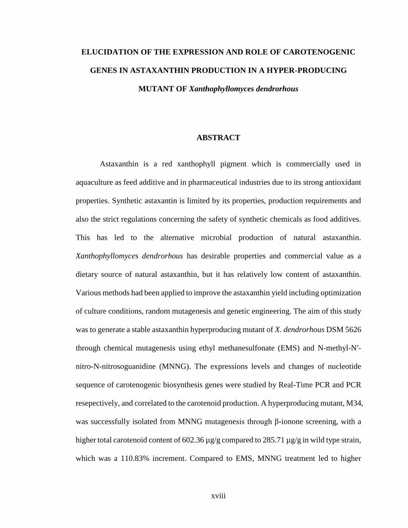
ABSTRACT
Astaxanthin is a red xanthophyll pigment which is commercially used in
aquaculture as feed additive and in pharmaceutical industries due to its strong antioxidant
properties. Synthetic astaxantin is limited by its properties, production requirements and
also the strict regulations concerning the safety of synthetic chemicals as food additives.
This has led to the alternative microbial production of natural astaxanthin.
Xanthophyllomyces dendrorhous has desirable properties and commercial value as a
dietary source of natural astaxanthin, but it has relatively low content of astaxanthin.
Various methods had been applied to improve the astaxanthin yield including optimization
of culture conditions, random mutagenesis and genetic engineering. The aim of this study
was to generate a stable astaxanthin hyperproducing mutant of X. dendrorhous DSM 5626
through chemical mutagenesis using ethyl methanesulfonate (EMS) and N-methyl-N′-
nitro-N-nitrosoguanidine (MNNG). The expressions levels and changes of nucleotide
sequence of carotenogenic biosynthesis genes were studied by Real-Time PCR and PCR
resepectively, and correlated to the carotenoid production. A hyperproducing mutant, M34,
was successfully isolated from MNNG mutagenesis through β-ionone screening, with a
higher total carotenoid content of 602.36 µg/g compared to 285.71 µg/g in wild type strain,
which was a 110.83% increment. Compared to EMS, MNNG treatment led to higher
xix
lethality in recipient cells and was more effective in inducing the pigmented mutants with
intense red colour and bigger colony indicating higher astaxanthin content and cell growth,
with lower rate of reverse mutation. No reversion was observed in M34 mutant during
stability test and M34 mutant was found to be stable in its cell growth and astaxanthin
production throughout the whole research period. In reversed-phase HPLC, the major
peak in M34 mutant extract was identified as astaxanthin based on the retention time and
absorption spectrum compared to authentic astaxanthin standard. Gene expression results
by Real-Time PCR showed that both crtE and crtS genes showed significantly higher
expressions relative to wild type throughout the production cycle, the highest being 1.3
and 3.8 folds, respectively. Meanwhile idi, crtYB, crtI and crtR genes did not show
meaningful correlation between their mRNA transcription levels and carotenoid
production. The gene expression patterns were similar when M34 mutant was cultured
with either glucose or sucrose as carbon source for all six crt genes. Besides, all six
carotenogenic genes exhibited a total of 38 nucleotide changes after mutation. Although
most of the nucleotide changes led to silent mutations, there was a missense mutation
found in idi gene (EA) and crtI gene (HQ), while two missense mutations occurred
in crtE (KR; SD) and crtR genes (SV; QP). This study will help in understanding
the role of the carotenogenic genes in further improvement of carotenoid production.

1
CHAPTER 1
INTRODUCTION
1.1 Background
Astaxanthin, also known as 3,3’-dihydroxy-β,β-carotene-4,4’-dione, is a ketocarotenoid
synthesized by several species of plants and microorganisms including algae, bacteria, and
fungi. It is a valuable red xanthophyll pigment with a high biotechnological interest,
predominantly in the aquaculture, poultry industry and cosmetology, as a pigmentation
source and in the food and pharmaceutical industries mainly for its outstanding antioxidant
properties and other beneficial effects on human health (Ranga Rao et al., 2014).
Astaxanthin is one of the most important pigments in the nature as it is responsible for the
orange-red colour in microorganisms and marine species such as salmon, trout, lobster,
shrimp and other seafood besides bird species including flamingo and quail (Chimsung et
al., 2014). However, these marine species cannot produced the astaxanthin de novo, but
obtain it from their diet (Bon et al., 1997; Kamath et al., 2008). Thus, astaxanthin is
conventionally used as feed additives for aquaculture and poultry industry. In aquaculture
and poultry industry, it is used as a colouring agent to increase the organoleptic value and
characteristics preferred by the consumer especially in lobster, shrimp, salmon, fish eggs
and egg yolk, since the bright colour could be more attractive. It was later found that
astaxanthin possesses strong antioxidant properties, which has been reported to outclass
other carotenoids, including β-carotene and α-tocopherol (Lauritano & Ianora, 2016).
Many studies have shown that astaxanthin is capable in promoting human health through
2
the prevention and treatment of various diseases including cancers, inflammatory diseases,
diabetes, cardiovascular diseases, gastrointestinal diseases, neurodegenerative diseases,
eye diseases and skin diseases. Consequently, astaxanthin is used as an ingredient in
nutraceutical, medicinal and food products (Rodríguez-Sáiz et al., 2010; Tanaka et al.,
2012). Due to its biological functions in various aspects, astaxanthin obtains significant
economic value and a growing global commercial market, thus attracts the interest and
attention from researchers and industries to put in more efforts in astaxanthin-related
research.
Generally, astaxanthin production has been performed through chemical synthesis and this
synthetic astaxanthin covers most of the world markets (Britton et al., 1996). Compared
to natural astaxanthin, the chemical synthesis is recognized as the lowest cost production
process of astaxanthin, which very likely to be its advantage to remain as the primary
pigment source as animal feed (Schmidt et al., 2011). However, the production processes
rely on total synthesis approaches, involving a rather long series of chemical steps ending
with a C20+C20 reaction or a C19+C2+C19 reaction. Chemical synthesis of astaxanthin
is dependent on petrochemicals as raw material which is not environmental friendly and
sustainable. Synthetic carotenoids cannot be absorbed effectively by the body compared
with other natural sources due to difference in isomer content, where astaxanthin from
natural sources has optically pure chirality, while synthetic astxanthin consists of a
mixture of enantiomers (3R,3'R and 3S,3'S) and mesoform (3R,3'S and 3S,3’R) (Lorenz
& Cysewski, 2000). Furthermore, the chemical product cannot be labeled as a natural
product, and thus does not penetrate the higher value and fastgrowing market segments.
The consumption of synthetic pigments at high dose ranges has eventually been warned
3
by Food and Drug Administration since it might cause precarious effects to human health
(Kläui & Bauernfeind, 1981). Only the astaxanthin obtained from algal and yeast are
approved for human consumption, while the synthetic form is used only for animal feed
purpose. This may be due to the insecure usage of petrochemicals for astaxanthin
production since it was reported to be cancer-causing (Newsome, 1986). All these coupled
with increasingly stringent regulations regarding the safety of chemicals to be used as food
additives results in the preference for natural carotenoid products over synthetic pigments.
However, there are also limitations for the production of astaxanthin by biological systems
in industries such as the low astaxanthin yields in wild type strains and extraction methods
which are costly compared to synthetic astaxanthin (Johnson & Schroeder, 1996;
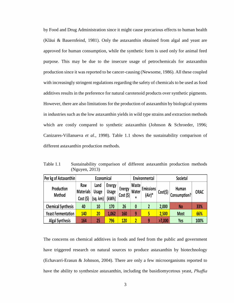
Canizares-Villanueva et al., 1998). Table 1.1 shows the sustainability comparison of
different astaxanthin production methods.
Table 1.1 Sustainability comparison of different astaxanthin production methods
(Nguyen, 2013)
The concerns on chemical additives in foods and feed from the public and government
have triggered research on natural sources to produce astaxanthin by biotechnology
(Echavarri-Erasun & Johnson, 2004). There are only a few microorganisms reported to
have the ability to synthesize astaxanthin, including the basidiomycetous yeast, Phaffia

4
rhodozyma; the green alga, Haematococcus pluvialis and the bacteria, Agrobacterium
aurantiacum, Paracoccus sp., Chlorococcum, Chlorella zofingiensis, Brevibaterium and
Mycobaterium lacticola (Goswami et al., 2010). The heterobasidiomycetous yeast, X.
dendrorhous is currently known as one of the most potential microorganism for synthesis
of astaxanthin at industrial scale besides H. pluvialis (Ranga Rao et al., 2010).
Currently the H. pluvialis is the primary contributor to astaxanthin market for human
consumption (Kidd, 2011). However, H. pluvialis has limitations in its own
characteristics, such as its tough cell wall that traps the astaxanthin, complicating the
extraction process and yield of production. At the same time, disadvantages such as long
growing period, low cell densities, ease of contamination by other bacteria and even
protozoa and finally susceptibility to adverse weather conditions, have magnified the
weakness of the algae, especially during the scale up for mass industrial production
(Retamales et al., 1998; Ukibe et al., 2008; Rodríguez-Sáiz et al., 2010).
Due to the shortcomings of the algae, researchers see the potential of X. dendrorhous to
replace H. pluvialis as the main source, as the yeast produces astaxanthin as its principle
carotenoid while having high metabolism and high cell densities can be achieved in
fermenters. Besides rapid breeding, short growth cycle, and fully developed fermentation
process (Bhatt et al., 2013), X. dendrorhous is also able to grow on molasses, enzymatic
wood hydrolysates, corn wet-milling co-products, corn syrup, grape juice and date juice,
which were by-products from agricultural activities (An et al., 2001; Ramirez et al., 2006;
Zheng et al., 2006). This gives the yeast more advantages compared to H. pluvialis.

5
However, the disadvantage of X. dendrorhous compared to H. pluvialis is its rather low
yield of astaxanthin in wild type strains with specific production at 200–400 mg/g of dry
yeast. The commercialized grown alga cells contains astaxanthin up to 3% of their dry
cell mass, while astaxanthin in wild type strains of X. dendrorhous is below 0.1% (Johnson
& Schroeder, 1996). Various methods had been attempted to improve the yield of
astaxanthin in X. dendrorhous, including optimization of culture condition such as
modifying the concentration of glucose or carbon/nitrogen ratio, the use of low cost raw
materials, temperature, pH, oxygen content, selective addition of chemical precursors such
as mevalonic acid, ethanol, acetic acid and lycopene, illumination with white and UV light,
strain improvement through genetic engineering and random mutagenesis (Schmidt et al.,
2011). This will help to make X. dendrorhous more competitive in becoming the main
source of astaxanthin for large scale production in industrial processes.
1.2 Significance of the Study
Astaxanthin is a valuable red xanthophyll pigment with a high biotechnological interest,
mainly in the aquaculture, poultry industry and cosmetology, as a pigmentation source
and as an antioxidant agent in the food and pharmaceutical industries due to its potent
antioxidant properties and other beneficial effects on human health. This contributes to its
high market value and growing demand. Traditionally this colourant and anti-oxidant in
the market has been produced by chemical synthesis using petroleum-derived raw
materials. Chemical synthesis of astaxanthin is disadvantaged by its dependence on
petrochemical which is not environmental friendly and sustainable as raw material, other
than involving a rather long series of chemical steps in the synthesis reaction. Apart from

6
that, strict regulations regarding the safety of synthetic chemical as food additives have
been implemented from time to time and the poor absorption of synthetic astaxanthin
compared with that from biological sources may give preference on natural astaxanthin
products over synthetic pigments.
The current growing demand for natural pigments favours the use of natural sources. Thus,
H. pluvialis and X. dendrorhous become best choices as the main source for astaxanthin.
However, H. pluvialis has its limitation during mass production such as its tough cell wall
complicating the extraction process and yields. It is also disadvantaged by slow growing
rate, low cell densities, ease of contamination by other bacteria and protozoa, and
vulnerability to the unfavorable weather conditions. As an alternative, X. dendrorhous
produces astaxanthin as the principle carotenoid, while having high metabolism, short
growth cycle, high cell density and mature fermentation process as well as being able to
grow on by-products from agriculture. This gives the yeast more advantages compared to
H. pluvialis but it is limited by its rather low yield of astaxanthin in wild type strains with
specific production at 200–400mg/g of dry yeast. Various approaches have been attempted
to increase the astaxanthin production of X. dendrorhous including optimization of
fermentation methodologies, application of chemical stimulants, genetic engineering and
mutagenesis.
Despite the rapid developments in the field of carotenoid biosynthesis, there are only few
reports on the genes or enzymes that mediate astaxanthin metabolism, especially the

7
carotenogenic genes. It is of utmost importance to generate knowledge on the astaxanthin
biosynthetic pathway at the level of biosynthetic genes and understand the role of these
carotenogenic genes in improving and/or modifying astaxanthin biosynthetic pathway.
Therefore, it is our interest to obtain a hyperproducing X. dendrorhous mutant by chemical
mutagenesis, determine the possible mutations in its carotenogenic genes involved in the
biosynthesis of astaxanthin and their transcription levels in the mutant and subsequently
the possible influence of carbon sources on their transcription levels. By characterizing
the possible alteration in these carotenogenic genes in a mutant and elucidating the
transcription levels of these carotenogenic genes in relationship to astaxanthin
biosynthesis, we aim to identify gene targets that can be genetically manipulated to
improve astaxanthin production. The effect of different carbon sources on the mRNA
expression of the carotenogenic genes was studied. Maximum carotenoid biosynthesis
may be achieved through over-expression of crt genes in combination with advantageous
cultivation conditions. The mutant crt genes can be used to develop new strains of X.
dendrorhous with enhanced astaxanthin content or for the production of new xanthophylls.
The findings may allow a rational amplification of astaxanthin production, either in X.
dendrorhous or in alternative microbial systems for large scale carotenoid production.
This all-round genetic approach constitutes the crucial steps towards the development of
X. dendrorhous as a cell factory for carotenoid production which may be competitive
economically with chemical synthesis.
8
1.3 Objectives
The aim of this study is to understand and identify the role of carotenogenic genes
involved in the production of astaxanthin in a hyperproducing mutant of X. dendrorhous
and the objectives of the study are:
1. To generate a X. dendrorhous mutant with higher astaxanthin production capacity
by chemical mutagenesis.
2. To elucidate the transcription levels of the carotenogenic genes in relationship to
astaxanthin biosynthesis.
3. To study the effect of mutagenesis on the nucleotide sequences of carotenogenic
genes in the mutant.
9
CHAPTER 2
LITERATURE REVIEW
2.1 Carotenoids
2.1.1 Background
Carotenoids are lipid-soluble pigments with colours, usually in orange, red or yellow
colour that are able to absorb light in the range of 300-600 nm of wavelength. They are
important natural pigments occur in nature throughout the photosynthetic systems of
plants and phototrophic microorganisms including algae, yeast, cyanobacteria and fungi,
which are the main sources for isolation and production of carotenoids at big scale (Blanco
et al., 2007; Nelis & DeLeenheer, 1991; Bourgaud et al., 2001; Olaizola, 2003). Animals
can only obtain carotenoids from their diet since they cannot synthesize carotenoids de
novo (Britton et al., 2009).
Carotenoids consist of isoprene residues and a C40 hydrocarbon polyene chain of
conjugated double bonds (Sandmann & Misawa, 2002; Alcaíno et al., 2016a). The
polyene system not only gives carotenoids their distinctive molecular structure, but also
their chemical properties and light-absorption characteristics. The number of conjugated
double bonds and functional groups present in the structure of the carotenoids are
contributing to their light absorption ability. The presence of double bonds in the polyene
chain allows the existence of two configurations in carotenoids; as geometric isomers cis
or trans, where trans-isomers are more stable than the cis-isomers in the aspect of
10
thermodynamic. Thus, most carotenoids in nature are found typically in all trans-isomers
(Britton, 1995; Miller et al., 2014). Structurally and functionally, carotenoids can be
classified into carotenes (hydrocarbon derivative) and xanthophylls (oxygenated
derivative by addition of oxygen-containing functional groups to obtain cyclic or acyclic-
xanthophylls). In xanthophyll, oxygen can be present as OH groups, which can be found
in zeaxanthin, or as oxy-groups, which are found in cantaxanthin, or combination of both
such as in astaxanthin. β-carotene is an example of carotene carotenoid and astaxanthin is
a xanthopyll carotenoid (Schwab, 2011). Phytoene is produced as the first carotenoid in
the carotenoid biosynthesis pathway, formed by condensation of two geranyl-geranyl
diphosphate (GGDP) molecules (C20 hydrocarbon), which results in a basic symmetrical
acyclic C40 hydrocarbon backbone structure. This carotenoid is important as the precursor
to undergo enzyme-mediated biochemical reactions for the synthesis of other natural
carotenoids.
The presence of polyene system in carotenoids contributes to its distinctive characteristics
in molecular structure, chemical properties and ability to absorb light. The molecule of
carotenoid provides antioxidant properties when it introduces a ring structure joined to its
carbon chain (DellaPenna & Pogson, 2006). Other than their significant antioxidant
activities and important role in inhibiting the onset of chronic diseases such as
cardiovascular disease, diabetes and age related macular degeneration, carotenoids are
involved in numerous other biological functions including vitamin A synthesis, gap
junction communication, immune system modulation and antitumor activity (Nishino et
al., 2002; Minatel et al., 2017). The discoveries of beneficial properties associated to these
11
bioactive phytonutrients have brought great interest in production of carotenoids in
diverse structures for various applications. The interest in carotenoids has caused in the
global carotenoid market value to increase considerably from $1.5 billion in 2014 to an
expected $1.8 billion in 2019, with a compound annual growth rate (CAGR) of 3.9%
(BCC Research, 2015).
Approximately 700 naturally occurring carotenoids are identified and over 600 different
chemical structures have been described, but just a few of them are of economical
importance and utilized by industry commercially as colourants or health and dietary
supplements (Takaichi et al., 1996; Amorim-Carrilho et al., 2014; Arvayo-Enriquez et al.,
2013; Merhan, 2017). Only a few carotenoids can be produced commercially, such as β-
carotene, lycopene, astaxanthin, canthaxanthin, capsanthin, lutein, β-apo-8-carotenal, β-
apo-8-carotenal-ester, through processes such as chemical synthesis and fermentation
(Johnson & Schroeder, 1996). Among them, astaxanthin is the most valuable carotenoid
to be studied due to its outstanding antioxidant and pigmentation properties.
2.1.2 Astaxanthin
2.1.2(a) Structures and Chemical Forms of Astaxanthin
Astaxanthin is a carotenoid with a chemical formula C40H52O4, with a molecular mass of
596 Da (Goswami et al., 2010). It is a xanthophyll because of the presence of oxygen
atoms besides carbon and hydrogen atoms. Astaxanthin is composed of eight isoprenoid
units in the polyene chain, with two terminal rings that contain oxygen (Ducrey Sanpietro
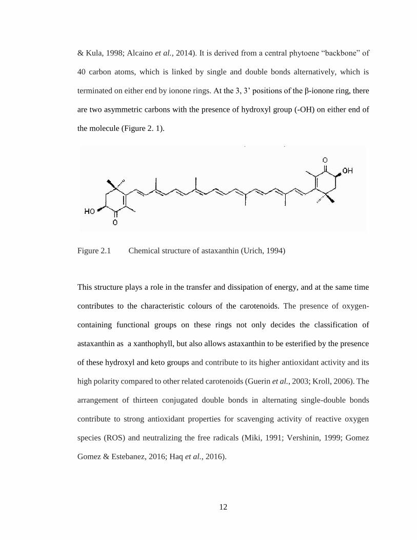
12
& Kula, 1998; Alcaino et al., 2014). It is derived from a central phytoene “backbone” of
40 carbon atoms, which is linked by single and double bonds alternatively, which is
terminated on either end by ionone rings. At the 3, 3’ positions of the β-ionone ring, there
are two asymmetric carbons with the presence of hydroxyl group (-OH) on either end of
the molecule (Figure 2. 1).
Figure 2.1 Chemical structure of astaxanthin (Urich, 1994)
This structure plays a role in the transfer and dissipation of energy, and at the same time
contributes to the characteristic colours of the carotenoids. The presence of oxygen-
containing functional groups on these rings not only decides the classification of
astaxanthin as a xanthophyll, but also allows astaxanthin to be esterified by the presence
of these hydroxyl and keto groups and contribute to its higher antioxidant activity and its
high polarity compared to other related carotenoids (Guerin et al., 2003; Kroll, 2006). The
arrangement of thirteen conjugated double bonds in alternating single-double bonds
contribute to strong antioxidant properties for scavenging activity of reactive oxygen
species (ROS) and neutralizing the free radicals (Miki, 1991; Vershinin, 1999; Gomez
Gomez & Estebanez, 2016; Haq et al., 2016).
13
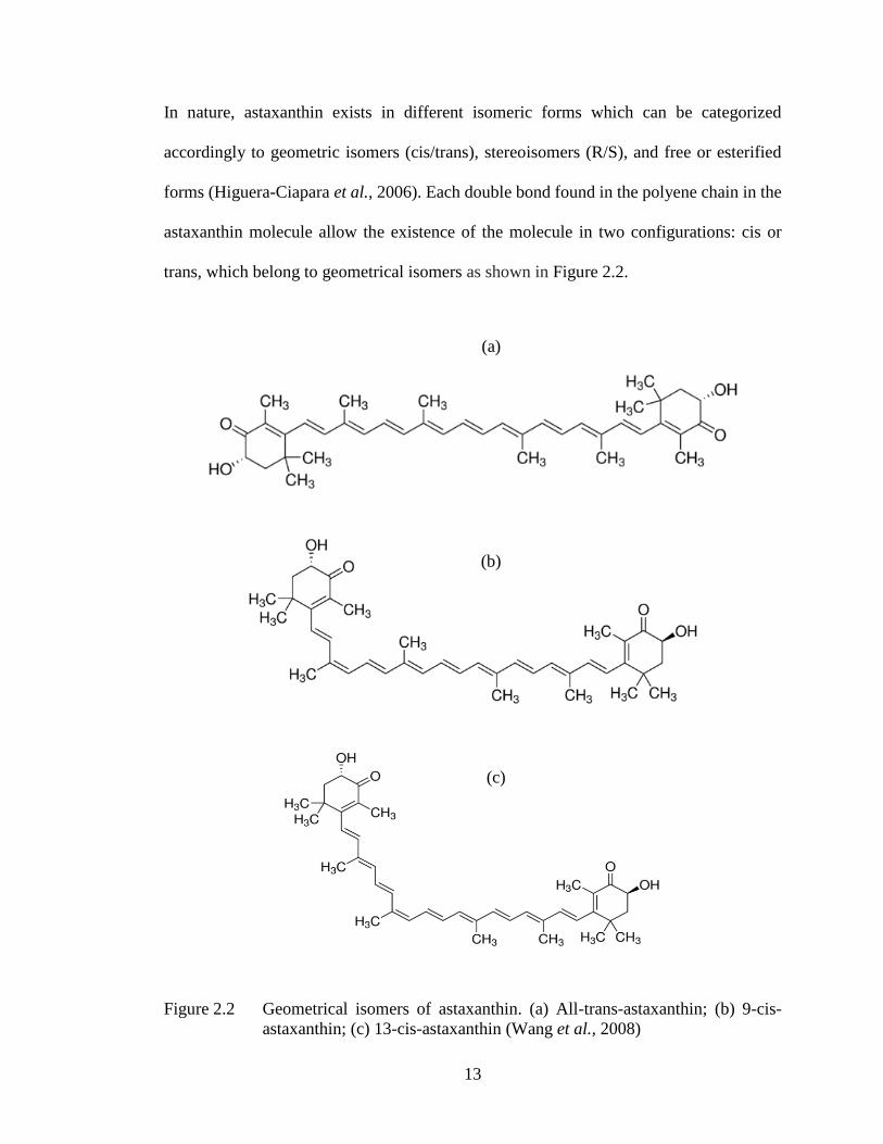
In nature, astaxanthin exists in different isomeric forms which can be categorized
accordingly to geometric isomers (cis/trans), stereoisomers (R/S), and free or esterified
forms (Higuera-Ciapara et al., 2006). Each double bond found in the polyene chain in the
astaxanthin molecule allow the existence of the molecule in two configurations: cis or
trans, which belong to geometrical isomers as shown in Figure 2.2.
Figure 2.2 Geometrical isomers of astaxanthin. (a) All-trans-astaxanthin; (b) 9-cis-
astaxanthin; (c) 13-cis-astaxanthin (Wang et al., 2008)
(a)
(b)
(c)
14
Majority of natural carotenoids are principally all trans-isomers. Trans-isomer of
astaxanthin is the predominant geometric isomer found in H. pluvialis (Yuan & Chen,
1998). Because of the steric reason, the isomerization of trans-astaxanthin into cis-trans
mixtures, especially 9-cis and 13-cis, usually cannot be prevented. Among the geometric
isomers, it was found that astaxanthin isomer in cis form, especially 9-cis astaxanthin, has
a higher antioxidant capability than that of the all-trans isomer, which can be arranged in
the order of 9-cis > 13-cis > all-trans (Liu & Osawa, 2007). In addition, compared to trans-
astaxanthin that consists of shorter chain length, the cis-astaxanthin is able to accumulate
especially in blood plasma (Bohn, 2008).
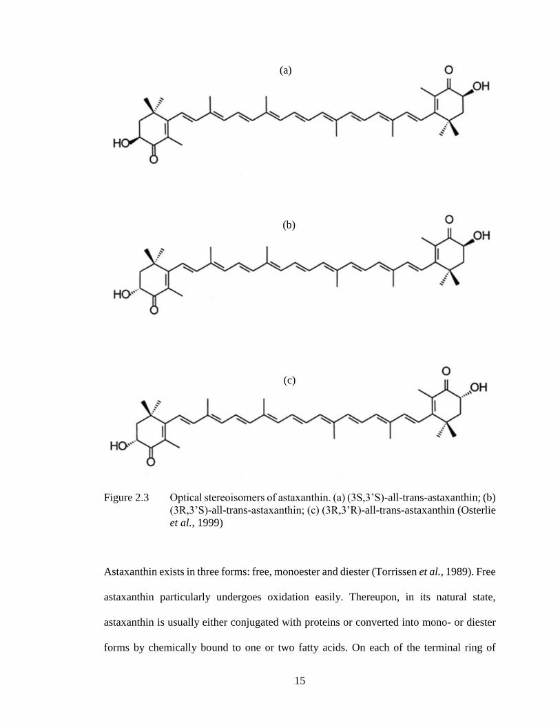
Isomers of astaxanthin molecule may present in three optical stereo configurations: two
enantiomers (3R, 3′ R and 3S, 3′S) and a meso form (3R, 3′ S), as each astaxanthin
molecule consists of two chiral centers in C-3 and C-3′, as shown in Figure 2.3.
The 3S, 3′ S is the most abundant form in nature. >98% of astaxanthin produced by X.
dendrorhous is in (3R,3’R) form and microalgae H. pluvialis synthesizes only (3S, 3’S)
isoform (Hussein et al., 2006). Meanwhile synthetic astaxanthin like Carophyll Pink (La
Roche) consists of a racemic mixture with the two enantiomers (3R,3’R) and (3S, 3’S)
equally at 25% and 50% of the meso form.
15
Figure 2.3 Optical stereoisomers of astaxanthin. (a) (3S,3’S)-all-trans-astaxanthin; (b)
(3R,3’S)-all-trans-astaxanthin; (c) (3R,3’R)-all-trans-astaxanthin (Osterlie
et al., 1999)
Astaxanthin exists in three forms: free, monoester and diester (Torrissen et al., 1989). Free
astaxanthin particularly undergoes oxidation easily. Thereupon, in its natural state,
astaxanthin is usually either conjugated with proteins or converted into mono- or diester
forms by chemically bound to one or two fatty acids. On each of the terminal ring of
(a)
(b)
(c)

16
astaxanthin, there is a free (unreacted) hydroxyl (OH) groups. These allow astaxanthin
molecule to exist in free form, or can be esterified to form an ester. Addition of a fatty
acid to form an ester leads to higher hydrophobicity on the esterified end of the molecule,
where the strength of hydrophobicity (difficulty in dissolving in water) in different form
can be observed in the sequence of diesters>monoesters>free. Astaxanthin from different
organisms exhibited different compositions of the free form and esterified forms as shown
in Figure 2.4.
Figure 2.4 Astaxanthin and its esters from various sources (Lorenz, 1999)
Although xanthophyll esters seem to be of low bioavailability, but contradicting findings
were also reported. Sugawara et al. (2009) suggested that the presence of enzymatic
activity after intestinal absorption led to the esterification of consumed xanthophylls and
followed by the incorporation of xanthophyll esters into the lipid core in chylomicron
before being carried into various tissues of the body. The esterification of xanthophylls
17
into highly nonpolar products could also contribute in the protection of intestinal cells
from the cytotoxic effects. Thus, the accumulation of astaxanthin in H. pluvialis in ester
form might be an added advantage to provide positive influence to the higher
bioavailability of astaxanthin (Ranga Rao et al., 2010).
2.1.2(b) Analysis of Astaxanthin
Multiple techniques, including spectrophotometry, thin layer chromatography (TLC),
high performance liquid chromatography (HPLC) and nuclear magnetic resonance
spectroscopy (NMR), have been developed to analyze and characterize the carotenoids
profile, along with the chemical structures, for various specific species of organisms.
Among them, UV spectrophotometry and HPLC are commonly employed for routine
analysis of bulk samples.
Spectrometric analysis of X. dendrorhous extracts showed that it has a characteristic UV–
Vis spectrum. The spectrum showed a straight peak with a maximum absorbance at 309-
310 nm, and a distinctive broad spectrum band with no obvious absorption peaks and a
maximum at 474-477 nm, corresponding to mycosporine-glutaminol-glucoside (MGG)
and astaxanthin respectively (Figure 2.5) (Tognetti et al., 2013).
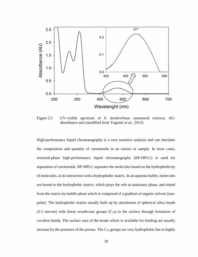
18
Figure 2.5 UV-visible spectrum of X. dendrorhous carotenoid extracts; AU:
absorbance unit (modified from Tognetti et al., 2013)
High-performance liquid chromatography is a very sensitive analysis and can elucidate
the composition and quantity of carotenoids in an extract or sample. In most cases,
reversed-phase high-performance liquid chromatography (RP-HPLC) is used for
separation of carotenoids. RP-HPLC separates the molecules based on the hydrophobicity
of molecules, in its interaction with a hydrophobic matrix. In an aqueous buffer, molecules
are bound to the hydrophobic matrix, which plays the role as stationary phase, and eluted
from the matrix by mobile phase which is composed of a gradient of organic solvent (non-
polar). The hydrophobic matrix usually built up by attachment of spherical silica beads
(3-5 micron) with linear octadecane groups (C18) to the surface through formation of
covalent bonds. The surface area of the beads which is available for binding are usually
increase by the presence of the porous. The C18 groups are very hydrophobic but in highly

19
polar solvent such as water can bind to polar molecules such as charged peptides. The
name "reversed phase" is originated from the opposite technique of "normal phase"
chromatography which involves the separation of molecules through their interaction with
a polar matrix (silica beads without octadecane groups attached) in the presence of a non-
polar solvent.
Two conditions can be applied during the elution process, either isocratic conditions
where the organic solvent has constant concentration throughout the experiment, or by
gradient elution where there is an increment in the amount of organic solvent over a period
of time. Hence, the solutes are eluted in the order of low to high molecular hydrophobicity
strength (Aguilar, 2004). RP-HPLC is an excellent technique especially for the analysis
of proteins and peptides with the advantages of its forceful resolution that can be achieved
under a wide range of conditions in chromatographic for very closely related molecules
as well as structurally different molecules. In addition, the experiment could be done with
the manipulation of chromatographic selectivity through the modification in mobile phase
characteristics. Moreover, it also has high recoveries that lead to high productivity. RP-
HPLC also has sorbent materials which are stable under a wide range of mobile phase
conditions that leads to excellent reproducibility for repeated separations carried out over
a long period of time (Aguilar & Hearn, 1996; Mant & Hodges, 1996).
Absorbance spectra of carotenoids are important criteria for their identification. The
properties of spectra of each carotenoids are determined by the basic structure of
carotenoids and some of the substituents (Britton, 1995). For astaxanthin, the spectrum is
20
not affected by 3-OH groups. On the other hand, a 4-keto group in conjugation with the
polyene chain causes a shifting of the bell-shaped spectrum towards higher absorbance
wavelength. The presence of second keto group at position 4’ resulted in a maximum
absorbance of astaxanthin in the range of 470-477 nm in various reports. In X.
dendrorhous, most of the carotenoids have a 3-HO-4-keto-β-ionone end group. Increment
in the polarity will lead to decrease of the mobility of a compound in absorption
chromatography on stationary phases like silica or reduce the retention time in reversed-
phase HPLC. Astaxanthin normally appears first in HPLC chromatograms since it is a
xanthophyll with high polarity. During RP-HPLC process, the expected order of elution
is not affected by the presence of the hydrogen bond between the HO and keto group.
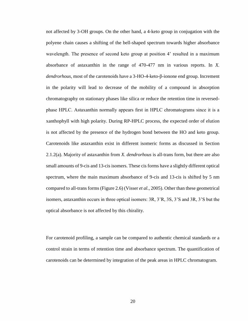
Carotenoids like astaxanthin exist in different isomeric forms as discussed in Section
2.1.2(a). Majority of astaxanthin from X. dendrorhous is all-trans form, but there are also
small amounts of 9-cis and 13-cis isomers. These cis forms have a slightly different optical
spectrum, where the main maximum absorbance of 9-cis and 13-cis is shifted by 5 nm
compared to all-trans forms (Figure 2.6) (Visser et al., 2005). Other than these geometrical
isomers, astaxanthin occurs in three optical isomers: 3R, 3’R, 3S, 3’S and 3R, 3’S but the
optical absorbance is not affected by this chirality.
For carotenoid profiling, a sample can be compared to authentic chemical standards or a
control strain in terms of retention time and absorbance spectrum. The quantification of
carotenoids can be determined by integration of the peak areas in HPLC chromatogram.
21
Figure 2.6 Spectra of astaxanthin geometrical isomers (Visser et al., 2005)
2.1.2(c) Biological Properties of Astaxanthin and Its Health Benefits
Astaxanthin is a high-value ketocarotenoid mainly synthesized by several species of
microalgae, plants, bacteria, and fungi. In X. dendrorhous and H. pluvialis, astaxanthin is
built up through the stimulation of the environmental stress, and protects cellular DNA
from photodynamic damage (Hagen et al., 1993). In general, animals do not have the
ability to produce carotenoids on their own and have to obtain these compounds from their
diets. Astaxanthin is responsible for the colour of the flesh for large amount of marine
species through consumption of microorganisms that synthesize the pigment (Gu et al.,
1997; Olaizola, 2003; Schroeder & Johnson, 1995a; Alvarez et al., 2006). It plays a
conventional role in poultry and aquaculture industry as the useful feed additive in
providing characteristic pigmentation of the tissues that influences organoleptic values
22
and consumer preferences. It is also widely used as colourants in the food industry to
ensure proper pigmentation and enhance acceptability of many foods. Astaxanthin has
been approved by the United States Food and Drug Administration (USFDA) to be added
in to animal and fish feed for colouration purpose (Pashkow et al., 2008), while the
European Commission has considered natural astaxanthin as a safe food dye (Roche,
1987).
Other than foods and feeds, the use of astaxanthin in nutritional supplement has been
growing fast in nutraceuticals and pharmaceuticals as it was discovered that the
consumption of astaxanthin can reduce or prevent risk of various diseases in humans and
animals (Kidd, 2011; Yang et al., 2013; Yuan et al., 2011; Dhankhar et al., 2012). The
oxidative molecules, such as free radicals, including hydroxyls and peroxides, and reactive
oxygen species are generated during aerobic metabolism in organisms. Excess quantities
of such compounds might cause oxidative damage which lead to various human diseases.
Human body can generate superoxide dismutase catalase and peroxidase as their own
enzymatic antioxidants to control and reduce these oxidative effects. But these compounds
are not enough most of the time, thus extra consumption of antioxidants is required.
Astaxanthin plays the role as a strong antioxidant by reacting with free radicals through
donation of electrons to convert them to more stable products and suspends free radical
chain reaction in living organisms (Ranga Rao et al., 2010). Astaxanthin shows strong
scavenging activity on free radicals and protects LDL cholesterol, cell membranes, cells
and tissues from lipid peroxidation and oxidative damage. It is a strong antioxidant that
protects the phospholipid cell membrane and other lipid components by removing singlet

23
oxygen and inhibiting free radicals, thus shields the plants and organisms from oxidative
damage caused by active oxygen species (Schroeder & Johnson, 1995b). Many studies
have revealed that astaxanthin has powerful antioxidant activity, which may be ten times
stronger than that of other carotenoids such as β-carotene, lutein, zeaxanthin,
canthaxanthin, and lipophilic antioxidants like vitamin E (α-tocopherol) in scavenging of
singlet oxygen (1O2) and peroxyl radicals (H2O2). The unique structure of astaxanthin
allow it to stay both inside and outside of the cell membrane and terminate free radical
chain reaction on both sides of the membrane, which gives better protection when
compared to vitamin C and β-carotene (McNulty et al., 2007; Lauritano & Ianora, 2016).
Besides mitigating the damaging effects of oxidative stress, astaxanthin modulates
stimulation of inflammation and immune response. The antioxidation properties of
astaxanthin make it among the bioactive phytochemicals credited for reducing the risks of
human diseases such as cancer, cardiovascular diseases and diabetes besides protection
against UVA-induced oxidative stress (Maldonade et al., 2008).
Clinical and epidemiological data showed that there might be protection effect against
cardiovascular disease through consumption of dietary antioxidants. In human body, high
level of Low Density Lipoprotein (LDL), the bad cholesterol, may lead to development of
cardiovascular diseases (Maher, 2000). Astaxanthin is carried by VLDL, LDL and HDL
in the human blood and it can improve heart health by altering levels of LDL and HDL
cholesterol in bloods (Guerin et al., 2003). Astaxanthin supplementation prevented LDL-
cholesterol from stimulation of in vitro oxidation and induced the increment of HDL in
24
blood levels, the form of blood cholesterol negatively correlated with coronary heart
disease (Iwamoto et al., 2000). Other than that, astaxanthin may reduce the risk of
cardiovascular disease by lowering elevated blood pressure, inhibiting oxidation of LDL,
stabilizing atherosclerotic plaque, lessening the risk of myocardial infarction and
preventing atherosclerosis. The possibility of involvement of astaxanthin in nitric oxide-
related mechanism and modulation of the blood fluidity might contribute to
antihypertensive effect in human. It can also benefit the heart health by reducing
inflammation most likely correlated with the increasing risk of coronary heart disease
(Tracy, 1999). Astaxanthin has the potential to become therapeutic agent against
atherosclerotic cardiovascular disease as it is a potent antioxidant agent against
inflammation and oxidative stress, which are the pathophysiological properties of
atherosclerotic cardiovascular disease (Fassett & Coombes, 2011).
Antioxidant compounds such as astaxanthin are capable to reduce the risk of mutagenesis
and carcinogenesis by prohibiting cell damage caused by oxidation. Besides having higher
antitumor activity when compared to other carotenoids such as β-carotene and
cantaxanthin (Chew & Park, 2004), it also acts as an inhibitor for the development of
breast and prostate cancer cells, fibrosarcoma and embryonic fibroblasts (Palozza et al.,
2009), cell death, cell proliferation and chemically induced mammary tumors (Jyonouchi
et al., 2000; Prabhu et al., 2009; Nakao et al., 2010). Moaka et al. (2012) found that
Epstein-Barr virus and carcinogenesis in mouse skin-papillomas were effectively
prohibited after astaxanthin treatment. Astaxanthin may unleash anti-tumor activities
through the promotion of immune response by preventing stress-stimulated lipid