elucidation of the molecular envenomation strategy of the cone
TRANSCRIPT
Hu et al. BMC Genomics 2012, 13:284http://www.biomedcentral.com/1471-2164/13/284
RESEARCH ARTICLE Open Access
Elucidation of the molecular envenomationstrategy of the cone snail Conus geographusthrough transcriptome sequencing of itsvenom ductHao Hu1, Pradip K Bandyopadhyay2, Baldomero M Olivera2 and Mark Yandell1*
Abstract
Background: The fish-hunting cone snail, Conus geographus, is the deadliest snail on earth. In the absence ofmedical intervention, 70% of human stinging cases are fatal. Although, its venom is known to consist of a cocktailof small peptides targeting different ion-channels and receptors, the bulk of its venom constituents, their sites ofmanufacture, relative abundances and how they function collectively in envenomation has remained unknown.
Results: We have used transcriptome sequencing to systematically elucidate the contents the C. geographus venomduct, dividing it into four segments in order to investigate each segment’s mRNA contents. Three different types ofcalcium channel (each targeted by unrelated, entirely distinct venom peptides) and at least two different nicotinicreceptors appear to be targeted by the venom. Moreover, the most highly expressed venom component is notparalytic, but causes sensory disorientation and is expressed in a different segment of the venom duct from venomsbelieved to cause sensory disruption. We have also identified several new toxins of interest for pharmaceutical andneuroscience research.
Conclusions: Conus geographus is believed to prey on fish hiding in reef crevices at night. Our data suggest thatdisorientation of prey is central to its envenomation strategy. Furthermore, venom expression profiles also suggest asophisticated layering of venom-expression patterns within the venom duct, with disorientating and paralyticvenoms expressed in different regions. Thus, our transcriptome analysis provides a new physiological framework forunderstanding the molecular envenomation strategy of this deadly snail.
Keywords: Conus geographus, Conotoxins, RNA-seq, Venom duct compartmentalization
BackgroundCone snails are venomous predators that rapidlyimmobilize their prey using a complex cocktail of shortpeptides (10–40 AA long) collectively known as cono-toxins. Most of these peptides, are targeted with exquis-ite specificity to receptors, ion channels, andtransporters in the nervous system [1-4]. Conotoxins areimportant pharmacological reagents and potential drugleads [5-8]. Each snail is believed to synthesize 50–200such compounds [1,2,9], and with approximately 700
* Correspondence: [email protected] institute of Human Genetics, University of Utah, and School ofMedicine, Salt Lake City, UT 84112, USAFull list of author information is available at the end of the article
© 2012 Hu et al.; licensee BioMed Central Ltd.Commons Attribution License (http://creativecreproduction in any medium, provided the or
species [1,10], the venoms of the genus Conus constitutean exceptionally rich pharmacological resource.We have analyzed the venom-duct of Conus geogra-
phus using a transcriptomics approach. This species,widely known as the geography cone, is well known asthe deadliest of all cone snail species, responsible formost of the human fatalities recorded in the medical lit-erature. In the absence of medical intervention, 70% ofhuman stinging cases are fatal [11]. The venom of thiscone snail species was the first that was comprehensivelyanalyzed; it was the characterization of Conus geographusvenom peptides which established that most of thebiologically-active components of Conus venoms, weresmall, disulfide rich peptides [12].
This is an Open Access article distributed under the terms of the Creativeommons.org/licenses/by/2.0), which permits unrestricted use, distribution, andiginal work is properly cited.
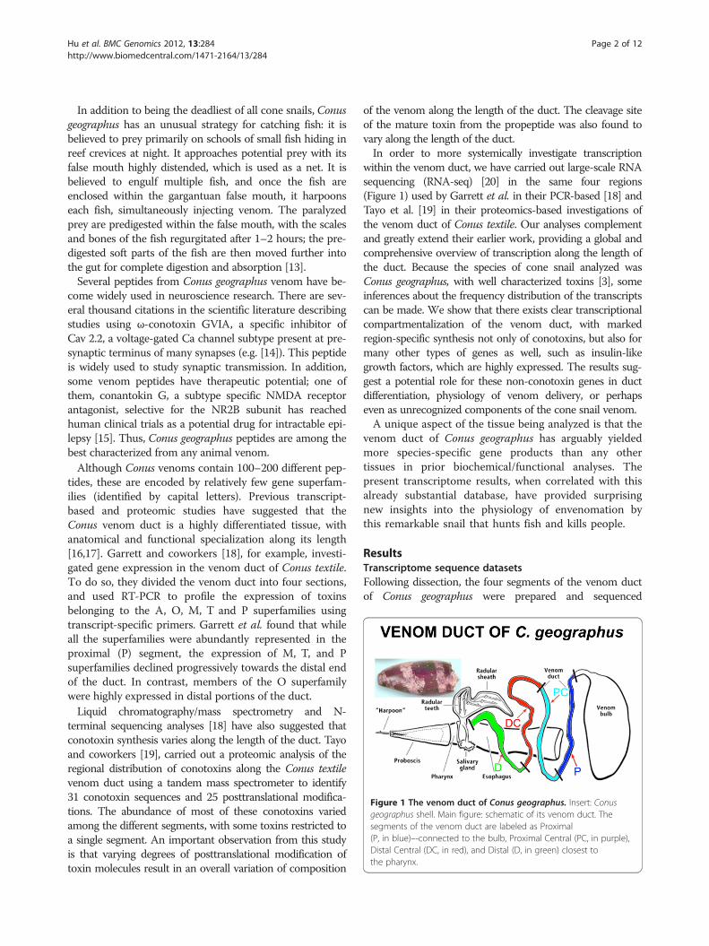
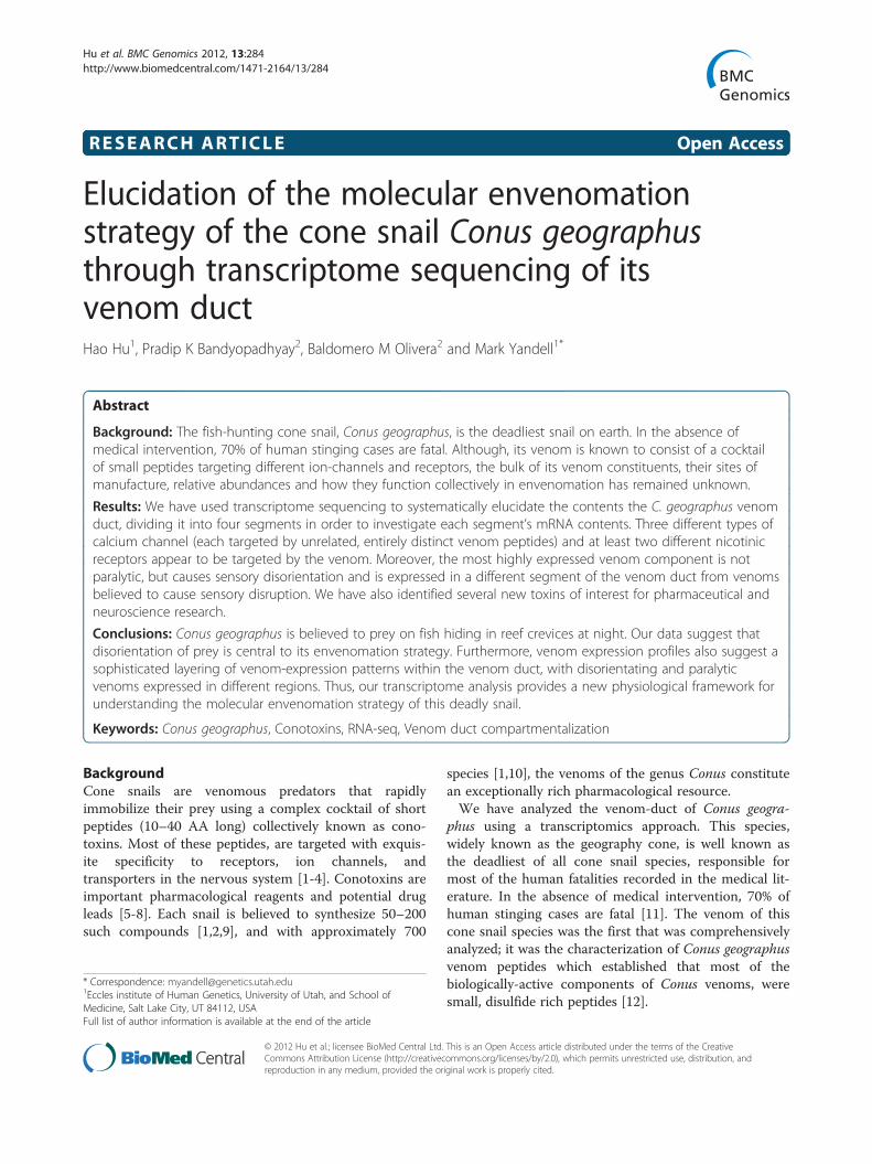
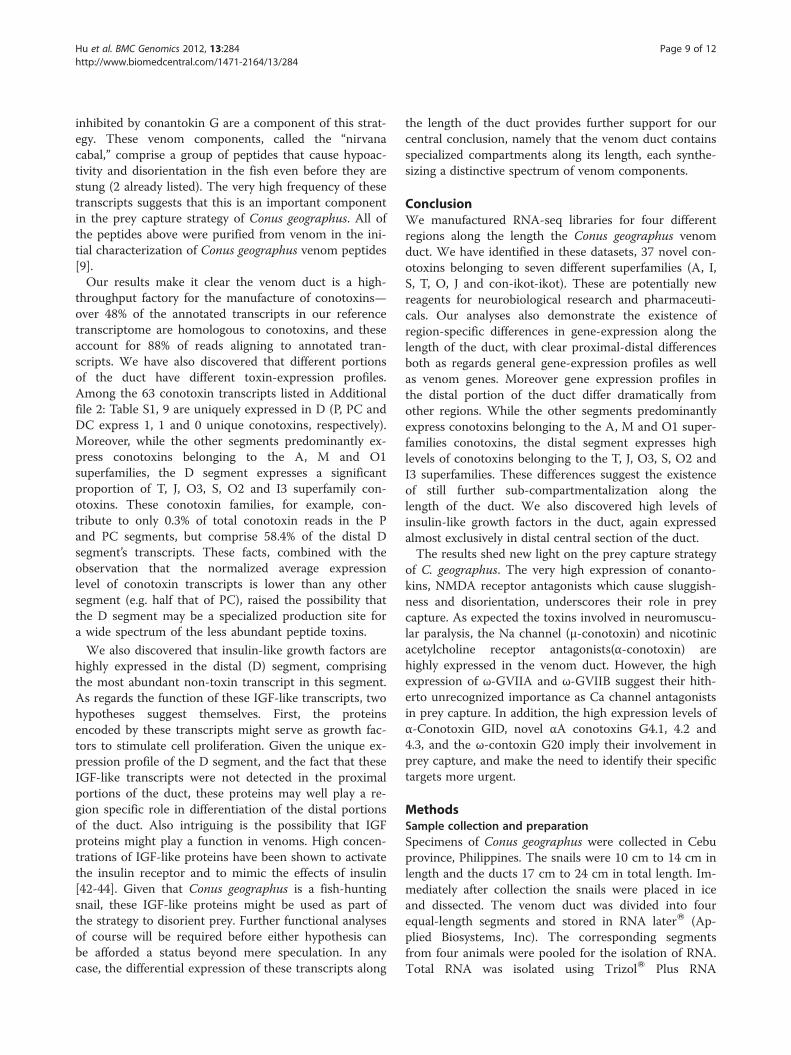
Figure 1 The venom duct of Conus geographus. Insert: Conusgeographus shell. Main figure: schematic of its venom duct. Thesegments of the venom duct are labeled as Proximal(P, in blue)–-connected to the bulb, Proximal Central (PC, in purple),Distal Central (DC, in red), and Distal (D, in green) closest tothe pharynx.
Hu et al. BMC Genomics 2012, 13:284 Page 2 of 12http://www.biomedcentral.com/1471-2164/13/284
In addition to being the deadliest of all cone snails, Conusgeographus has an unusual strategy for catching fish: it isbelieved to prey primarily on schools of small fish hiding inreef crevices at night. It approaches potential prey with itsfalse mouth highly distended, which is used as a net. It isbelieved to engulf multiple fish, and once the fish areenclosed within the gargantuan false mouth, it harpoonseach fish, simultaneously injecting venom. The paralyzedprey are predigested within the false mouth, with the scalesand bones of the fish regurgitated after 1–2 hours; the pre-digested soft parts of the fish are then moved further intothe gut for complete digestion and absorption [13].Several peptides from Conus geographus venom have be-
come widely used in neuroscience research. There are sev-eral thousand citations in the scientific literature describingstudies using ω-conotoxin GVIA, a specific inhibitor ofCav 2.2, a voltage-gated Ca channel subtype present at pre-synaptic terminus of many synapses (e.g. [14]). This peptideis widely used to study synaptic transmission. In addition,some venom peptides have therapeutic potential; one ofthem, conantokin G, a subtype specific NMDA receptorantagonist, selective for the NR2B subunit has reachedhuman clinical trials as a potential drug for intractable epi-lepsy [15]. Thus, Conus geographus peptides are among thebest characterized from any animal venom.Although Conus venoms contain 100–200 different pep-
tides, these are encoded by relatively few gene superfam-ilies (identified by capital letters). Previous transcript-based and proteomic studies have suggested that theConus venom duct is a highly differentiated tissue, withanatomical and functional specialization along its length[16,17]. Garrett and coworkers [18], for example, investi-gated gene expression in the venom duct of Conus textile.To do so, they divided the venom duct into four sections,and used RT-PCR to profile the expression of toxinsbelonging to the A, O, M, T and P superfamilies usingtranscript-specific primers. Garrett et al. found that whileall the superfamilies were abundantly represented in theproximal (P) segment, the expression of M, T, and Psuperfamilies declined progressively towards the distal endof the duct. In contrast, members of the O superfamilywere highly expressed in distal portions of the duct.Liquid chromatography/mass spectrometry and N-
terminal sequencing analyses [18] have also suggested thatconotoxin synthesis varies along the length of the duct. Tayoand coworkers [19], carried out a proteomic analysis of theregional distribution of conotoxins along the Conus textilevenom duct using a tandem mass spectrometer to identify31 conotoxin sequences and 25 posttranslational modifica-tions. The abundance of most of these conotoxins variedamong the different segments, with some toxins restricted toa single segment. An important observation from this studyis that varying degrees of posttranslational modification oftoxin molecules result in an overall variation of composition
of the venom along the length of the duct. The cleavage siteof the mature toxin from the propeptide was also found tovary along the length of the duct.In order to more systemically investigate transcription
within the venom duct, we have carried out large-scale RNAsequencing (RNA-seq) [20] in the same four regions(Figure 1) used by Garrett et al. in their PCR-based [18] andTayo et al. [19] in their proteomics-based investigations ofthe venom duct of Conus textile. Our analyses complementand greatly extend their earlier work, providing a global andcomprehensive overview of transcription along the length ofthe duct. Because the species of cone snail analyzed wasConus geographus, with well characterized toxins [3], someinferences about the frequency distribution of the transcriptscan be made. We show that there exists clear transcriptionalcompartmentalization of the venom duct, with markedregion-specific synthesis not only of conotoxins, but also formany other types of genes as well, such as insulin-likegrowth factors, which are highly expressed. The results sug-gest a potential role for these non-conotoxin genes in ductdifferentiation, physiology of venom delivery, or perhapseven as unrecognized components of the cone snail venom.A unique aspect of the tissue being analyzed is that the
venom duct of Conus geographus has arguably yieldedmore species-specific gene products than any othertissues in prior biochemical/functional analyses. Thepresent transcriptome results, when correlated with thisalready substantial database, have provided surprisingnew insights into the physiology of envenomation bythis remarkable snail that hunts fish and kills people.
ResultsTranscriptome sequence datasetsFollowing dissection, the four segments of the venom ductof Conus geographus were prepared and sequenced
Hu et al. BMC Genomics 2012, 13:284 Page 3 of 12http://www.biomedcentral.com/1471-2164/13/284
independently. Using the Roche Genome Sequencer FLXTitanium platform, we generated 167,211, 238,682, 186,398and 199,680 high-quality reads for the Proximal, Proximal-central, Distal-central and Distal segments, respectively.The average read length was 425.8 bp with an N50 readlength of 580 bp.
Transcriptome assemblyTo generate a reference transcriptome for Conus geogra-phus, we pooled the reads and then assembled them withMira3 [21]. This generated 60,305 contigs totaling34Mbp in length. We used cd-hit-est [22] to prune re-dundant contigs from the assembly arising from sequen-cing errors. See Methods for details. This produced areference transcriptome assembly consisting of 49,515contigs totaling 20.8 MB with an N50 of 576 bp. Themedian depth of coverage is 3.6x and the average depthis 26.7x. By aligning the raw reads in each segment backto the reference assembly with bwa [23], 98.7%, 99.3%,99.1% and 99.2% of reads for the four segments aligned.
AnnotationWe annotated our reference transcriptome usingBLASTX [24] and InterProScan [25]. A total of 8,252contigs have a significant homology (BLASTX, E< 1e-4)to proteins in the Uniprot protein database [26] and/orthe Conoserver conotoxin collection [27]. Among these8,252 geographus contigs, 48.6% (4,010) are significantlyhomologous to known conotoxins.InterProScan [25] search identified 5,420 contigs with pro-
tein domains, 2,216 of which (40.9%) are annotated as cono-toxins. Among non-conotoxin contigs with InterProScanhits, the most abundant Gene Ontology [28] category istranslation (GO:0006412) accounting for 1.1% of the totaltranscriptome assembly, followed by cellular iron ionhemostasis (GO:0006879), cell redox homeostasis(GO:0045454), electron transport chain (GO:0022900) andproteolysis (GO:0006508). The high level of transcriptsrelated to translation and metabolism is consistent withvenom duct physiology, as it is an organ engaged in intensiveprotein synthesis and processing [29,30]. This is also true forthe GO terms related to redox homeostasis and proteolysis,as extensive post-translational modification of conotoxins ashas been reported previously[19]. Current models of venom-duct physiology less easily explain the high level of iron ionhemostasis related GO-terms. Most of the transcripts fallinginto this GO category have significant homology to ferritin,which stores and regulates the release of iron. Further inves-tigation will be required to explain the purpose underlyingthe high level of ferritin transcription in the duct.
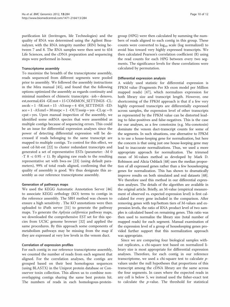
Functional overview of transcriptome assemblyWe used iPath explorer [31] to produce a high-level over-view of the contents of our venom duct transcriptome
(Figure 2A); see Methods for details. These provide a high-level summary of the metabolic, regulatory and secondary-metabolites biosynthesis pathways present within thetranscriptome assembly (Additional file 1: Figure S1). Wealso generated the metabolic pathway map for Aplysiacalifornica [32] (Figure 2B), using available all ESTs forthis organism. As expected, we observed most compo-nents of the energy metabolism pathways in both maps,including the citrate cycle pathways, fatty acid metabol-ism pathways, carbohydrate metabolism pathways. How-ever, by comparison to A. californica, the C. geographusvenom duct transcriptome is less comprehensive, missingfor example, 1) fatty acid synthesis pathways; 2) aminosugar metabolism pathways; 3) galactose metabolismpathways; 4) vitamin metabolism pathways and etc. Thesefindings suggest that the above pathways are either absentfrom venom duct or at least being too inactive to bedetected by RNA-seq approach, consistent with the ducthaving a highly streamlined metabolism geared at toxinsecretion.
Conotoxins are the most abundant class of transcriptConotoxins comprised 48.6% of total annotated tran-scripts in the reference transcriptome or 88% of totalaligned reads within annotated transcripts, suggestingthat venom duct is a very high-throughput factory forthe manufacture of conotoxins. To better understand therepertory of conotoxins present in the venom duct, weused a BLAST-based pipeline [33] to assign transcrip-tome contigs with homology to conotoxins to conotoxinsuperfamilies. In previous analyses [33] of the transcrip-tome in Conus bullatus this process is 98.7% accurate. Intotal, we were able to assign 1685 out of 4014 (42%) pu-tative conotoxins a superfamily.Figure 3 shows the proportion of each superfamily in
the duct transcriptome, by reads (3a) and by contig num-bers (3b). Consistent with Conoserver’s [27] contents, wefound A-superfamily conotoxins to be the most abun-dantly expressed conotoxin superfamily in the C. geogra-phus transcriptome assembly. However, while Conoservercurrently only contains representatives from the A, M,O1, S and T superfamilies for Conus geographus, ourtranscriptome also contains J, O2, O3, I3 and Z conotox-ins. For a list of toxins that we were able to retrieve fullcoding sequences, see Additional file 2: Table S1.
Duct segments synthesize different spectra of conotoxinsAmong the 49,515 contigs in our reference assembly,3089 (6.2%) are differentially expressed among the foursegments (p< 0.05/50,000 = 1E-6; see Methods fordetails). However, among conotoxin contigs, a muchhigher proportion are differentially expressed along thelength of the duct: 1626 out of 3803 contigs or 42.8%. Ifwe further restrict this analysis to conotoxin contigs
Figure 2 (See legend on next page.)
Hu et al. BMC Genomics 2012, 13:284 Page 4 of 12http://www.biomedcentral.com/1471-2164/13/284
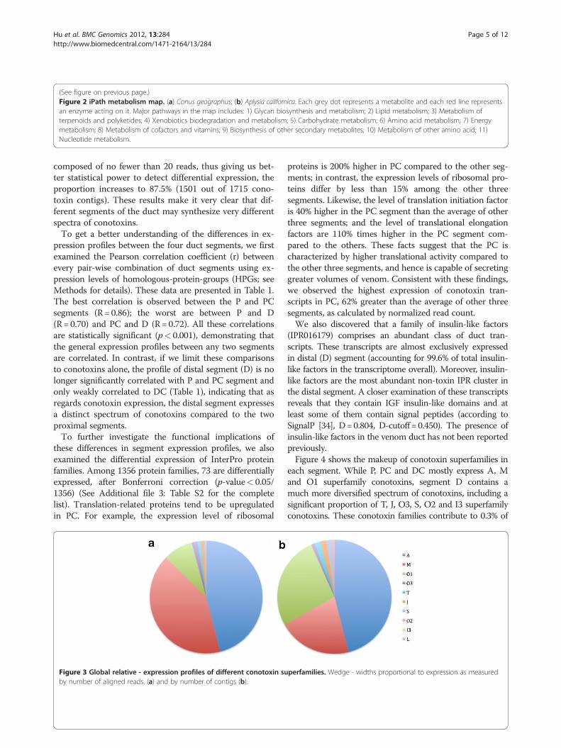
(See figure on previous page.)Figure 2 iPath metabolism map. (a) Conus geographus; (b) Aplysia californica. Each grey dot represents a metabolite and each red line representsan enzyme acting on it. Major pathways in the map includes: 1) Glycan biosynthesis and metabolism; 2) Lipid metabolism; 3) Metabolism ofterpenoids and polyketides; 4) Xenobiotics biodegradation and metabolism; 5) Carbohydrate metabolism; 6) Amino acid metabolism; 7) Energymetabolism; 8) Metabolism of cofactors and vitamins; 9) Biosynthesis of other secondary metabolites; 10) Metabolism of other amino acid; 11)Nucleotide metabolism.
Hu et al. BMC Genomics 2012, 13:284 Page 5 of 12http://www.biomedcentral.com/1471-2164/13/284
composed of no fewer than 20 reads, thus giving us bet-ter statistical power to detect differential expression, theproportion increases to 87.5% (1501 out of 1715 cono-toxin contigs). These results make it very clear that dif-ferent segments of the duct may synthesize very differentspectra of conotoxins.To get a better understanding of the differences in ex-
pression profiles between the four duct segments, we firstexamined the Pearson correlation coefficient (r) betweenevery pair-wise combination of duct segments using ex-pression levels of homologous-protein-groups (HPGs; seeMethods for details). These data are presented in Table 1.The best correlation is observed between the P and PCsegments (R= 0.86); the worst are between P and D(R= 0.70) and PC and D (R= 0.72). All these correlationsare statistically significant (p< 0.001), demonstrating thatthe general expression profiles between any two segmentsare correlated. In contrast, if we limit these comparisonsto conotoxins alone, the profile of distal segment (D) is nolonger significantly correlated with P and PC segment andonly weakly correlated to DC (Table 1), indicating that asregards conotoxin expression, the distal segment expressesa distinct spectrum of conotoxins compared to the twoproximal segments.To further investigate the functional implications of
these differences in segment expression profiles, we alsoexamined the differential expression of InterPro proteinfamilies. Among 1356 protein families, 73 are differentiallyexpressed, after Bonferroni correction (p-value< 0.05/1356) (See Additional file 3: Table S2 for the completelist). Translation-related proteins tend to be upregulatedin PC. For example, the expression level of ribosomal
Figure 3 Global relative - expression profiles of different conotoxin suby number of aligned reads. (a) and by number of contigs (b).
proteins is 200% higher in PC compared to the other seg-ments; in contrast, the expression levels of ribosomal pro-teins differ by less than 15% among the other threesegments. Likewise, the level of translation initiation factoris 40% higher in the PC segment than the average of otherthree segments; and the level of translational elongationfactors are 110% times higher in the PC segment com-pared to the others. These facts suggest that the PC ischaracterized by higher translational activity compared tothe other three segments, and hence is capable of secretinggreater volumes of venom. Consistent with these findings,we observed the highest expression of conotoxin tran-scripts in PC, 62% greater than the average of other threesegments, as calculated by normalized read count.We also discovered that a family of insulin-like factors
(IPR016179) comprises an abundant class of duct tran-scripts. These transcripts are almost exclusively expressedin distal (D) segment (accounting for 99.6% of total insulin-like factors in the transcriptome overall). Moreover, insulin-like factors are the most abundant non-toxin IPR cluster inthe distal segment. A closer examination of these transcriptsreveals that they contain IGF insulin-like domains and atleast some of them contain signal peptides (according toSignalP [34], D=0.804, D-cutoff = 0.450). The presence ofinsulin-like factors in the venom duct has not been reportedpreviously.Figure 4 shows the makeup of conotoxin superfamilies in
each segment. While P, PC and DC mostly express A, Mand O1 superfamily conotoxins, segment D contains amuch more diversified spectrum of conotoxins, including asignificant proportion of T, J, O3, S, O2 and I3 superfamilyconotoxins. These conotoxin families contribute to 0.3% of
perfamilies. Wedge - widths proportional to expression as measured
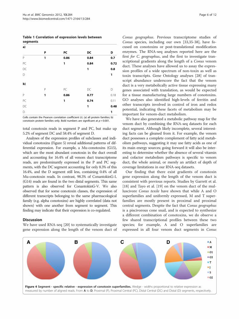
Table 1 Correlation of expression levels betweensegments
a)
P PC DC D
P 1 0.86 0.84 0.7
PC 1 0.84 0.72
DC 1 0.8
D 1
b)
P PC DC D
P 1 0.86 0.77 0.18
PC 1 0.74 0.11
DC 1 0.48
D 1
Cells contain the Pearson correlation coefficient (r). (a) all protein families; b)conotoxin protein families only. Bold numbers are significant at p=0.001.
Hu et al. BMC Genomics 2012, 13:284 Page 6 of 12http://www.biomedcentral.com/1471-2164/13/284
total conotoxin reads in segment P and PC, but make up5.2% of segment DC and 58.4% of segment D.Analyses of the expression profiles of subclasses and indi-
vidual conotoxins (Figure 5) reveal additional patterns of dif-ferential expression. For example, a Mu-conotoxins (G15),which are the most abundant conotoxin in the duct overalland accounting for 16.4% of all venom duct transcriptomereads, are predominantly expressed in the P and PC seg-ments, with the DC segment accounting for only 8.3% of this16.4%, and the D segment still less, containing 0.4% of allMu-conotoxin reads. In contrast, 98.3% of ConantokinG-L(G14) reads are found in the two distal segments. This samepattern is also observed for ConantokinG-V. We alsoobserved that for some conotoxin classes, the expression ofdifferent transcripts belonging to the same pharmacologicalfamily (e.g. alpha conotoxins) are highly correlated (data notshown) with one another from segment to segment. Thisfinding may indicate that their expression is co-regulated.
DiscussionWe have used RNA-seq [20] to systematically investigategene expression along the length of the venom duct of
Figure 4 Segment - specific relative - expression of conotoxin superfameasured by number of aligned reads. From A to D, Proximal (P), Proximal
Conus geographus. Previous transcriptome studies ofConus species, including our own [33,35-38], have fo-cused on conotoxins or post-translational modificationenzymes. The RNA-seq analyses reported here are thefirst for C. geographus, and the first to investigate tran-scriptional gradients along the length of a Conus venomduct. These analyses have allowed us to assay the expres-sion profiles of a wide spectrum of non-toxin as well astoxin transcripts. Gene Ontology analyses [28] of tran-script abundance underscore the fact that the venomduct is a very metabolically active tissue expressing manygenes associated with translation, as would be expectedfor a tissue manufacturing large numbers of conotoxins.GO analyses also identified high-levels of ferritin andother transcripts involved in control of iron and redoxpotential, indicating these facets of metabolism may beimportant for venom-duct metabolism.We have also generated a metabolic pathway map for the
venom duct by combining the RNA-seq datasets for eachduct segment. Although likely incomplete, several interest-ing facts can be gleaned from it. For example, the venomduct possesses a complete complement of fatty acid metab-olism pathways, suggesting it may use fatty acids as one ofits main energy sources; going forward it will also be inter-esting to determine whether the absence of several vitaminand cofactor metabolism pathways is specific to venomduct, the whole animal, or merely an artifact of depth ofcoverage limitations in our RNA-seq datasets.Our finding that there exist gradients of conotoxin
gene expression along the length of the venom duct isconsistent with previous reports. Studies by Garrett et al.[18] and Tayo et al. [19] on the venom duct of the mul-luscivore Conus texile have shown that while A and Osuperfamilies and uniformly expressed, M and T super-families are mostly present in proximal and proximalcentral segments. Despite the fact that Conus geographusis a piscivorous cone snail, and is expected to synthesizea different combination of conotoxins, we do observe afew shared transcriptional profiles between these twospecies; for example, A and O superfamilies areexpressed in all four venom duct segments in Conus
milies. Wedge - widths proportional to relative expression asCentral (PC), Distal Central (DC) and Distal (D) segments, respectively.
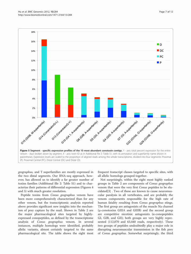
Figure 5 Segment - specific expression profiles of the 10 most abundant conotoxin contigs. Y - axis: total percent expression for the entirevenom - duct broken down by segment. X - axis: toxin ID as in Additional file 2: Table S1, with its annotation and superfamily name shown inparentheses. Expression levels are scaled to the proportion of aligned reads among the whole transcriptome, divided into four segments: Proximal(P), Proximal Central (PC), Distal Central (DC) and Distal (D).
Hu et al. BMC Genomics 2012, 13:284 Page 7 of 12http://www.biomedcentral.com/1471-2164/13/284
geographus, and T superfamilies are mostly expressed inthe two distal segments. Our RNA-seq approach, how-ever, has allowed us to identify a far greater number oftoxins families (Additional file 2: Table S1) and to char-acterize their patterns of differential expression (Figures 4and 5) with much greater resolution.Peptide toxins from Conus geographus venom have
been more comprehensively characterized than for anyother venom, but the transcriptomic analysis reportedabove provides significant new insights into the mechan-ism of prey capture by the snail. Shown in Table 2 arethe major pharmacological sites targeted by highly-expressed conopeptides, as defined by the transcriptomeanalysis of Conus geographus venom. In severalinstances, multiple homologs were identified, probablyallelic variants, almost certainly targeted to the samepharmacological site. The table shows the eight most
frequent transcript classes targeted to specific sites, withall allelic homologs grouped together.Not surprisingly, within the eight most highly ranked
groups in Table 2 are components of Conus geographusvenom that were the very first Conus peptides to be elu-cidated[3] . Two of these are known to cause neuromus-cular paralysis in all vertebrates, and are probably thevenom components responsible for the high rate ofhuman fatality resulting from Conus geographus stings.The first group are antagonists of the muscle Na channel(μ-conotoxins GIIIA and GIIIB) and the second groupare competitive nicotinic antagonists (α-conopeptidesGI, GIB, and GII); both groups are very highly repre-sented (112,070 and 63,440 reads, respectively). Thesetwo groups of peptides undoubtedly play a major role indisrupting neuromuscular transmission in the fish preyof Conus geographus. Somewhat surprisingly, the third
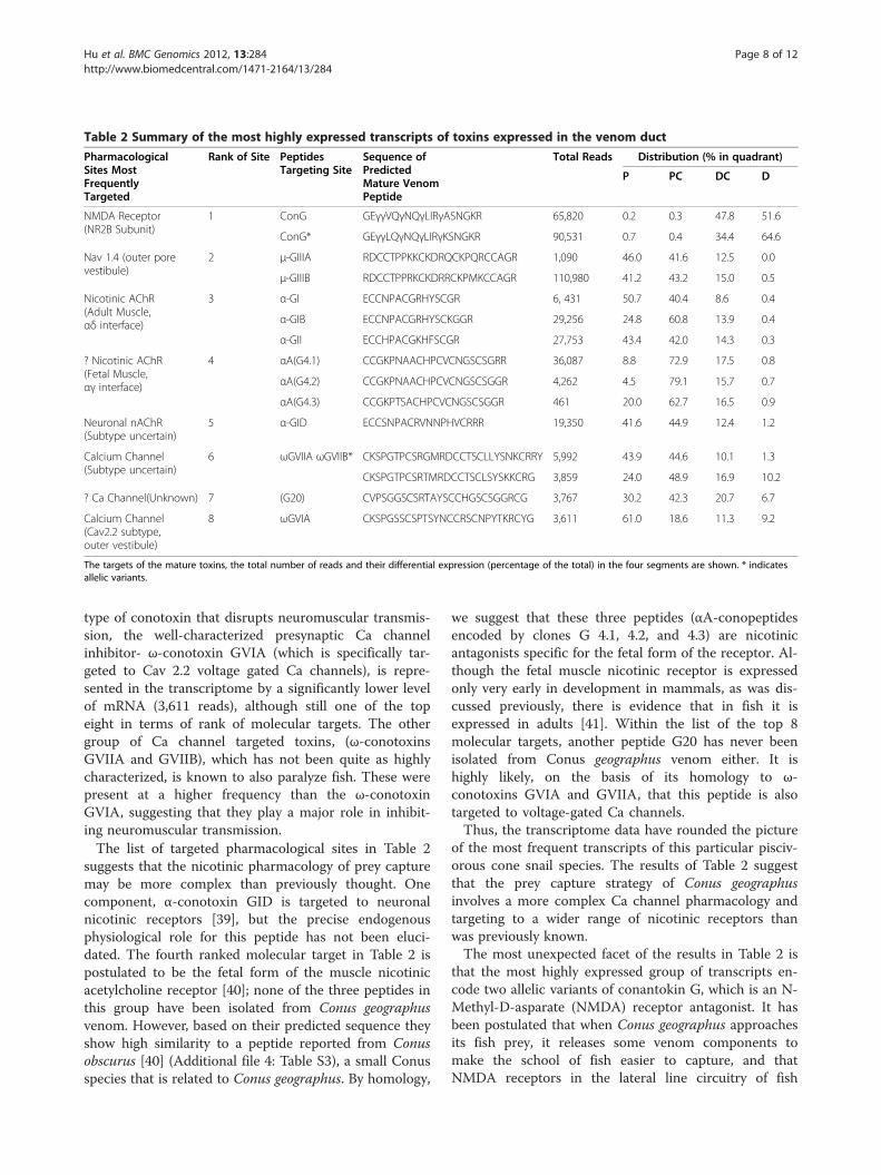
Table 2 Summary of the most highly expressed transcripts of toxins expressed in the venom duct
PharmacologicalSites MostFrequentlyTargeted
Rank of Site PeptidesTargeting Site
Sequence ofPredictedMature VenomPeptide
Total Reads Distribution (% in quadrant)
P PC DC D
NMDA Receptor(NR2B Subunit)
1 ConG GEγγVQγNQγLIRγASNGKR 65,820 0.2 0.3 47.8 51.6
ConG* GEγγLQγNQγLIRγKSNGKR 90,531 0.7 0.4 34.4 64.6
Nav 1.4 (outer porevestibule)
2 μ-GIIIA RDCCTPPKKCKDRQCKPQRCCAGR 1,090 46.0 41.6 12.5 0.0
μ-GIIIB RDCCTPPRKCKDRRCKPMKCCAGR 110,980 41.2 43.2 15.0 0.5
Nicotinic AChR(Adult Muscle,αδ interface)
3 α-GI ECCNPACGRHYSCGR 6, 431 50.7 40.4 8.6 0.4
α-GIB ECCNPACGRHYSCKGGR 29,256 24.8 60.8 13.9 0.4
α-GII ECCHPACGKHFSCGR 27,753 43.4 42.0 14.3 0.3
? Nicotinic AChR(Fetal Muscle,αγ interface)
4 αA(G4.1) CCGKPNAACHPCVCNGSCSGRR 36,087 8.8 72.9 17.5 0.8
αA(G4.2) CCGKPNAACHPCVCNGSCSGGR 4,262 4.5 79.1 15.7 0.7
αA(G4.3) CCGKPTSACHPCVCNGSCSGGR 461 20.0 62.7 16.5 0.9
Neuronal nAChR(Subtype uncertain)
5 α-GID ECCSNPACRVNNPHVCRRR 19,350 41.6 44.9 12.4 1.2
Calcium Channel(Subtype uncertain)
6 ωGVIIA ωGVIIB* CKSPGTPCSRGMRDCCTSCLLYSNKCRRY 5,992 43.9 44.6 10.1 1.3
CKSPGTPCSRTMRDCCTSCLSYSKKCRG 3,859 24.0 48.9 16.9 10.2
? Ca Channel(Unknown) 7 (G20) CVPSGGSCSRTAYSCCHGSCSGGRCG 3,767 30.2 42.3 20.7 6.7
Calcium Channel(Cav2.2 subtype,outer vestibule)
8 ωGVIA CKSPGSSCSPTSYNCCRSCNPYTKRCYG 3,611 61.0 18.6 11.3 9.2
The targets of the mature toxins, the total number of reads and their differential expression (percentage of the total) in the four segments are shown. * indicatesallelic variants.
Hu et al. BMC Genomics 2012, 13:284 Page 8 of 12http://www.biomedcentral.com/1471-2164/13/284
type of conotoxin that disrupts neuromuscular transmis-sion, the well-characterized presynaptic Ca channelinhibitor- ω-conotoxin GVIA (which is specifically tar-geted to Cav 2.2 voltage gated Ca channels), is repre-sented in the transcriptome by a significantly lower levelof mRNA (3,611 reads), although still one of the topeight in terms of rank of molecular targets. The othergroup of Ca channel targeted toxins, (ω-conotoxinsGVIIA and GVIIB), which has not been quite as highlycharacterized, is known to also paralyze fish. These werepresent at a higher frequency than the ω-conotoxinGVIA, suggesting that they play a major role in inhibit-ing neuromuscular transmission.The list of targeted pharmacological sites in Table 2
suggests that the nicotinic pharmacology of prey capturemay be more complex than previously thought. Onecomponent, α-conotoxin GID is targeted to neuronalnicotinic receptors [39], but the precise endogenousphysiological role for this peptide has not been eluci-dated. The fourth ranked molecular target in Table 2 ispostulated to be the fetal form of the muscle nicotinicacetylcholine receptor [40]; none of the three peptides inthis group have been isolated from Conus geographusvenom. However, based on their predicted sequence theyshow high similarity to a peptide reported from Conusobscurus [40] (Additional file 4: Table S3), a small Conusspecies that is related to Conus geographus. By homology,
we suggest that these three peptides (αA-conopeptidesencoded by clones G 4.1, 4.2, and 4.3) are nicotinicantagonists specific for the fetal form of the receptor. Al-though the fetal muscle nicotinic receptor is expressedonly very early in development in mammals, as was dis-cussed previously, there is evidence that in fish it isexpressed in adults [41]. Within the list of the top 8molecular targets, another peptide G20 has never beenisolated from Conus geographus venom either. It ishighly likely, on the basis of its homology to ω-conotoxins GVIA and GVIIA, that this peptide is alsotargeted to voltage-gated Ca channels.Thus, the transcriptome data have rounded the picture
of the most frequent transcripts of this particular pisciv-orous cone snail species. The results of Table 2 suggestthat the prey capture strategy of Conus geographusinvolves a more complex Ca channel pharmacology andtargeting to a wider range of nicotinic receptors thanwas previously known.The most unexpected facet of the results in Table 2 is
that the most highly expressed group of transcripts en-code two allelic variants of conantokin G, which is an N-Methyl-D-asparate (NMDA) receptor antagonist. It hasbeen postulated that when Conus geographus approachesits fish prey, it releases some venom components tomake the school of fish easier to capture, and thatNMDA receptors in the lateral line circuitry of fish
Hu et al. BMC Genomics 2012, 13:284 Page 9 of 12http://www.biomedcentral.com/1471-2164/13/284
inhibited by conantokin G are a component of this strat-egy. These venom components, called the “nirvanacabal,” comprise a group of peptides that cause hypoac-tivity and disorientation in the fish even before they arestung (2 already listed). The very high frequency of thesetranscripts suggests that this is an important componentin the prey capture strategy of Conus geographus. All ofthe peptides above were purified from venom in the ini-tial characterization of Conus geographus venom peptides[9].Our results make it clear the venom duct is a high-
throughput factory for the manufacture of conotoxins—over 48% of the annotated transcripts in our referencetranscriptome are homologous to conotoxins, and theseaccount for 88% of reads aligning to annotated tran-scripts. We have also discovered that different portionsof the duct have different toxin-expression profiles.Among the 63 conotoxin transcripts listed in Additionalfile 2: Table S1, 9 are uniquely expressed in D (P, PC andDC express 1, 1 and 0 unique conotoxins, respectively).Moreover, while the other segments predominantly ex-press conotoxins belonging to the A, M and O1superfamilies, the D segment expresses a significantproportion of T, J, O3, S, O2 and I3 superfamily con-otoxins. These conotoxin families, for example, con-tribute to only 0.3% of total conotoxin reads in the Pand PC segments, but comprise 58.4% of the distal Dsegment’s transcripts. These facts, combined with theobservation that the normalized average expressionlevel of conotoxin transcripts is lower than any othersegment (e.g. half that of PC), raised the possibility thatthe D segment may be a specialized production site fora wide spectrum of the less abundant peptide toxins.
We also discovered that insulin-like growth factors arehighly expressed in the distal (D) segment, comprisingthe most abundant non-toxin transcript in this segment.As regards the function of these IGF-like transcripts, twohypotheses suggest themselves. First, the proteinsencoded by these transcripts might serve as growth fac-tors to stimulate cell proliferation. Given the unique ex-pression profile of the D segment, and the fact that theseIGF-like transcripts were not detected in the proximalportions of the duct, these proteins may well play a re-gion specific role in differentiation of the distal portionsof the duct. Also intriguing is the possibility that IGFproteins might play a function in venoms. High concen-trations of IGF-like proteins have been shown to activatethe insulin receptor and to mimic the effects of insulin[42-44]. Given that Conus geographus is a fish-huntingsnail, these IGF-like proteins might be used as part ofthe strategy to disorient prey. Further functional analysesof course will be required before either hypothesis canbe afforded a status beyond mere speculation. In anycase, the differential expression of these transcripts along
the length of the duct provides further support for ourcentral conclusion, namely that the venom duct containsspecialized compartments along its length, each synthe-sizing a distinctive spectrum of venom components.
ConclusionWe manufactured RNA-seq libraries for four differentregions along the length the Conus geographus venomduct. We have identified in these datasets, 37 novel con-otoxins belonging to seven different superfamilies (A, I,S, T, O, J and con-ikot-ikot). These are potentially newreagents for neurobiological research and pharmaceuti-cals. Our analyses also demonstrate the existence ofregion-specific differences in gene-expression along thelength of the duct, with clear proximal-distal differencesboth as regards general gene-expression profiles as wellas venom genes. Moreover gene expression profiles inthe distal portion of the duct differ dramatically fromother regions. While the other segments predominantlyexpress conotoxins belonging to the A, M and O1 super-families conotoxins, the distal segment expresses highlevels of conotoxins belonging to the T, J, O3, S, O2 andI3 superfamilies. These differences suggest the existenceof still further sub-compartmentalization along thelength of the duct. We also discovered high levels ofinsulin-like growth factors in the duct, again expressedalmost exclusively in distal central section of the duct.The results shed new light on the prey capture strategy
of C. geographus. The very high expression of conanto-kins, NMDA receptor antagonists which cause sluggish-ness and disorientation, underscores their role in preycapture. As expected the toxins involved in neuromuscu-lar paralysis, the Na channel (μ-conotoxin) and nicotinicacetylcholine receptor antagonists(α-conotoxin) arehighly expressed in the venom duct. However, the highexpression of ω-GVIIA and ω-GVIIB suggest their hith-erto unrecognized importance as Ca channel antagonistsin prey capture. In addition, the high expression levels ofα-Conotoxin GID, novel αA conotoxins G4.1, 4.2 and4.3, and the ω-contoxin G20 imply their involvement inprey capture, and make the need to identify their specifictargets more urgent.
MethodsSample collection and preparationSpecimens of Conus geographus were collected in Cebuprovince, Philippines. The snails were 10 cm to 14 cm inlength and the ducts 17 cm to 24 cm in total length. Im-mediately after collection the snails were placed in iceand dissected. The venom duct was divided into fourequal-length segments and stored in RNA laterW (Ap-plied Biosystems, Inc). The corresponding segmentsfrom four animals were pooled for the isolation of RNA.Total RNA was isolated using TrizolW Plus RNA
Hu et al. BMC Genomics 2012, 13:284 Page 10 of 12http://www.biomedcentral.com/1471-2164/13/284
purification kit (Invitrogen, life Technologies) and thequality of RNA was determined using the Agilent Bioa-nalyzer, with the RNA integrity number (RIN) being be-tween 7 and 8. The RNA samples were then sent to 454Life Sciences, and the cDNA preparation and sequencingsteps were performed in-house.
Transcriptome assemblyTo maximize the breadth of the transcriptome assembly,reads sequenced from different segments were pooledprior to assembly. We followed the assembly instructionsin the Mira manual [45], and found that the followingoptions optimized the assembly as regards continuity andminimal numbers of chimeric transcripts: -job = denovo,est,normal,454 -GE:not = 15 COMMON_SETTINGS -CL:ascdc = 1 -SK:not = 15 -AS:nop = 4 454_SETTINGS -ED:ace = 1 -AS:mrl = 30:mrpc = 1 -OUT:sssip = yes -CL:qc = 0:cpat = yes. Upon manual inspection of the assembly, weidentified some mRNA species that were assembled asmultiple contigs because of sequencing errors. This couldbe an issue for differential expression analyses since thepower of detecting differential expression will be de-creased if reads belonging to the same transcript aremapped to multiple contigs. To control for this effect, weused cd-hit-est [22] to cluster redundant transcripts andgenerated a set of representative ESTs (parameter: -M 0-T 8 -c 0.95 -r 1). By aligning raw reads to the resultingrepresentative set with bwa-sw [23] (using default para-meters), 99% of total reads aligned, confirming that thequality of assembly is good. We thus designate this as-sembly as our reference transcriptome assembly.
Generation of pathways mapsWe used the KEGG Automatic Annotation Server [46]to associate KEGG orthology (KO) terms to contigs inthe reference assembly. The SBH method was chosen toensure a high sensitivity . The KO annotations were thenuploaded to iPath server [31] to generate the pathwaymaps. To generate the Aplysia californica pathway maps,we downloaded the comprehensive EST set for this spe-cies from UCSC genome browser [32] and applied thesame procedures. By this approach some components ofmetabolism pathways may be missing from the map ifthey are expressed at very low levels in the venom duct.
Correlation of expression profilesFor each contig in our reference transcriptome assembly,we counted the number of reads from each segment thataligned. For the correlation analyses, the contigs aregrouped based on their best homologous sequences(using BLASTX) in the Uniprot protein database or Con-oserver toxin collection. This allows us to combine non-overlapping contigs sharing the same best protein hit.The numbers of reads in each homologous-protein-
group (HPG) were then calculated by summing the num-bers of reads aligned to each contig in this group. Thesecounts were converted to log10 scale (log normalized) toavoid bias toward very highly expressed transcripts. Wethen calculated Pearson’s correlation coefficient (R) usingthe read counts for each HPG between every two seg-ments. The significance levels for these correlations werecalculated by permutation.
Differential expression analysisA widely used statistic for differential expression isFPKM value (Fragments Per Kb exon model per Millionmapped reads) [47], which normalizes expression forboth library size and transcript length. However, oneshortcoming of the FPKM approach is that if a few veryhighly expressed transcripts are differentially expressedacross samples, the expression level of other transcriptsas represented by the FPKM value can be distorted lead-ing to false-positives and false-negatives. This is the casefor our analyses, as a few conotoxins (e.g. Mu-conotoxin)dominate the venom duct-transcript counts for some ofthe segments. In such situations, one alternative to FPKMis to use a house-keeping gene for normalization, but herethe concern is that using just one house-keeping gene maylead to inaccurate normalizations. Thus, we used a moreappropriate approach for normalization. The trimmedmean of M-values method as developed by Mark D.Robinson and Alicia Oshlack [48] uses the median propor-tion of all expressed genes rather than a few housekeepinggenes for normalization. This has shown to dramaticallyimprove results on both simulated and real datasets [48].We therefore used this method in our differential expres-sion analyses. The details of the algorithm are available inthe original article. Briefly, an M-value (empirical measure-ment of observed vs. expected expression level) is first cal-culated for every gene included in the comparison. Afterremoving genes with top/bottom tiers of M-values and ex-pression levels, the ratio of RNA product level of two sam-ples is calculated based on remaining genes. This ratio wasthen used to normalize the library size (total number ofmapped reads) for each segment. Manual examination ofthe expression level of a group of housekeeping genes pro-vided further support that this normalization approachwas appropriate.Since we are comparing four biological samples with-
out replicates, a chi-square test based on normalized li-brary size is most appropriate for differential expressionanalyses. Therefore, for each contig in our referencetranscriptome, we used a chi-square test to calculate p-values under the null hypotheses that proportions of thistranscript among the cDNA library are the same acrossthe four segments. In cases where the expected reads inany cell is below 5, we instead used the fisher-exact testto calculate the p-value. The threshold for statistical
Hu et al. BMC Genomics 2012, 13:284 Page 11 of 12http://www.biomedcentral.com/1471-2164/13/284
significance was set to 0.05/49515 = 1e-6, to control formultiple tests. For the differential expression of HPG orInterPro protein families, the same procedures are ap-plied and Bonferroni-corrected alpha were set accordingto the number of comparisons performed.
Data availabilityThe sequence data from this study are submitted tothe NCBI Sequence Read Archive (http://www.ncbi.nlm.nih.gov/Traces/sra/sra.cgi) under accession numbersSRR503413, SRR503414, SRR503415 and SRR503416.
Additional files
Additional file 1: Figure S1. iPath (a) regulatory pathway map and(b) secondary-metabolite biosynthesis pathway map. Each grey dotrepresents a substrate and each red line represents an enzyme.
Additional file 2: Table S1. A list of complete conotoxins sequencesidentified in the venom duct. The expression levels are shown for eachconotoxin in each segment, represented as number of reads aligned tothe toxin. Toxins are numbered G1-63, and have been listed according totheir superfamilies. In the A superfamily, (X,Y) refers to the number ofamino acid residues in the first and second disulfide loops. G4.x is thecommonly used nomenclature of alpha-A family of conotoxins identifiedfrom Conus geographus. Other designations in parenthesis adjacent to GXindicate previously used nomenclature in the literature [3,4,12,49-55].
Additional file 3: Table S2. InterPro protein families differentiallyexpressed among the four segments Proximal (P), Proximal Central(PC), Distal Central (DC) and Distal (D), showing the proportion ofaligned reads among whole transcriptome [3,4,12,49-55].
Additional file 4: Table S3. Comparison of toxin sequences of αA-OIVA, a fetal muscle nicotinic acetylcholine receptor antagonist,with G10 4.1 and G11 4.2 [3,4,12,49-55].
AbbreviationsP: Proximal; PC: Proximal-central; DC: Distal-central; D: Distal;HPG: Homologous protein group; NMDA: N-Methyl-D-asparate; IGF: Insulingrowth factor; FPKM: Fragments Per Kb exon model per Million mapped reads.
Competing interestsThe authors declare that they have no competing interests.
Authors’ contributionsHH designed, performed the bioinformatic analysis and drafted themanuscript. PB prepared cDNA samples for sequencing, analyzed conotoxinsequences and drafted the manuscript. MY and BO conceived, supervisedthe study and draft the manuscript. All authors read and approved the finalmanuscript.
AcknowledgementsThis work was partially supported by a program project grant GM48677 fromthe National Institute of General Medical Sciences (PB, BMO) andR01GM099939 to MY. Specimen collection was carried out in conjunctionwith a field expedition supported by ICBG grant, 1U01TW008163 (M.Haygood, Director; G.Concepcion, PI). We would like to thank Frank M.Heralde III, Noel Saguil, Meljune Chicote and Alexander Fedosov forcollection and dissection of the specimens. We thank Dr. Chinnappa Kodirafor helping to sequence the transcriptome with a grant from 454 LifeSciences, A Roche Company.
Author details1Eccles institute of Human Genetics, University of Utah, and School ofMedicine, Salt Lake City, UT 84112, USA. 2Department of Biology, Universityof Utah, Salt Lake City, UT 84112, USA.
Received: 29 February 2012 Accepted: 7 June 2012Published: 28 June 2012
References1. Olivera BM: Conus venom peptides, receptor and ion channel targets and
drug design: 50 million years of neuropharmacology (E.E. Just Lecture,1996). Mol Biol Cell 1997, 8:2101–2109.
2. Terlau H, Olivera BM: Conus venoms: a rich source of novel ion channel-targeted peptides. Physiol Rev 2004, 84:41–68.
3. Olivera BM, Gray WR, Zeikus R, McIntosh JM, Varga J, Rivier J, de Santos V,Cruz LJ: Peptide neurotoxins from fish-hunting cone snails. Science 1985,230(4732):1338–1343.
4. Cruz LJ, Gray WR, Olivera BM, Zeikus RD, Kerr L, Yoshikami D, MoczydlowskiE: Conus geographus toxins that discriminate between neuronal andmuscle sodium channels. J Biol Chem 1985, 260(16):9280–9288.
5. Teichert RW, Olivera BM: Natural products and ion channel pharmacology.Future Med Chem 2010, 2(5):731–744.
6. Han TS, Teichert RW, Olivera BM, Bulaj G: Conus venoms - a rich source ofpeptide-based therapeutics. Curr Pharm Des 2008, 14(24):2462–2479.
7. Lewis RJ, Garcia ML: Therapeutic potential of venom peptides. Nat RevDrug Discov 2003, 2(10):790–802.
8. Olivera BM, Teichert RW: Diversity of the neurotoxic Conus peptides: a model forconcerted pharmacological discovery. Mol Interv 2007, 7(5):251–260.
9. Olivera BM, Rivier J, Clark C, Ramilo CA, Corpuz GP, Abogadie FC, Mena EE,Woodward SR, Hillyard DR, Cruz LJ: Diversity of Conus neuropeptides.Science 1990, 249(4966):257–263.
10. Röckel D, Korn W, Kohn AJ: Manual of the living Conidae. Wiesbaden: VerlagChrista Hemmen; 1995.
11. Cruz LJ, White J: Clinical Toxicology of Conus Snail Stings. In Handbook ofClinical Toxicology of Animal Venoms and Poisons. Boca Raton: CRC-press;1995:117–127.
12. Gray WR, Luque A, Olivera BM, Barrett J, Cruz LJ: Peptide toxins from Conusgeographus venom. J Biol Chem 1981, 256(10):4734–4740.
13. Johnson CR, Stablum W: Observations on the Feeding Behavior of Conusgeographus (Gastropoda: Toxoglossa). Pac Sci 1971, 25(1):109–111.
14. Olivera BM, Miljanich GP, Ramachandran J, Adams ME: Calcium channeldiversity and neurotransmitter release: the omega-conotoxins andomega-agatoxins. Annu Rev Biochem 1994, 63:823–867.
15. McIntosh JM, Jones RM: Cone venom–from accidental stings to deliberateinjection. Toxicon 2001, 39(10):1447–1451.
16. Endean R, Duchemin C: The venom apparatus of Conus magus. Toxicon1967, 4(4):275–284.
17. Bingham JJA, Alewood PF, Lewis RJ: Conus venom peptides(conopeptides): inter-species, intra-species and within individualvariation revealed by ionspray mass spectrometry. In Biomedical Aspects ofMarine Pharmacology. Edited by Lazarovici E, Spira ME, Zlotkin.Fort Collins: CO: Alaken Inc; 1996.
18. Garrett JE, Buczek O, Watkins M, Olivera BM, Bulaj G: Biochemical and geneexpression analyses of conotoxins in Conus textile venom ducts. BiochemBiophys Res Commun 2005, 328(1):362–367.
19. Tayo LL, Lu B, Cruz LJ, Yates JR 3rd: Proteomic analysis provides insightson venom processing in Conus textile. J Proteome Res 2010,9(5):2292–2301.
20. Morin R, Bainbridge M, Fejes A, Hirst M, Krzywinski M, Pugh T, McDonald H,Varhol R, Jones S, Marra M: Profiling the HeLa S3 transcriptome usingrandomly primed cDNA and massively parallel short-read sequencing.Biotechniques 2008, 45(1):81–94.
21. Chevreux B, Pfisterer T, Drescher B, Driesel AJ, Muller WE, Wetter T, Suhai S:Using the miraEST assembler for reliable and automated mRNAtranscript assembly and SNP detection in sequenced ESTs. Genome Res2004, 14(6):1147–1159.
22. Russell DJ, Way SF, Benson AK, Sayood K: A grammar-based distancemetric enables fast and accurate clustering of large sets of 16 Ssequences. BMC Bioinforma 2010, 11:601–624.
23. Li H, Durbin R: Fast and accurate short read alignment with Burrows-Wheeler transform. Bioinformatics 2009, 25(14):1754–1760.
24. Altschul SF, Gish W, Miller W, Myers EW, Lipman DJ: Basic local alignmentsearch tool. J Mol Biol 1990, 215(3):403–410.
25. Zdobnov EM, Apweiler R: InterProScan–an integration platform for thesignature-recognition methods in InterPro. Bioinformatics 2001,17(9):847–848.

Hu et al. BMC Genomics 2012, 13:284 Page 12 of 12http://www.biomedcentral.com/1471-2164/13/284
26. Apweiler R, Bairoch A, Wu CH: Protein sequence databases. Curr OpinChem Biol 2004, 8(1):76–80.
27. Kaas Q, Westermann JC, Halai R, Wang CK, Craik DJ: ConoServer, a databasefor conopeptide sequences and structures. Bioinformatics 2008,24(3):445–446.
28. Ashburner M, Ball CA, Blake JA, Botstein D, Butler H, Cherry JM, Davis AP,Dolinski K, Dwight SS, Eppig JT, et al: Gene ontology: tool for theunification of biology. The Gene Ontology Consortium. Nat Genet 2000,25(1):25–29.
29. Craig AG, Bandyopadhyay P, Olivera BM: Post-translationally modifiedneuropeptides from Conus venoms. Eur J Biochem 1999, 264(2):271–275.
30. Buczek O, Bulaj G, Olivera BM: Conotoxins and the posttranslationalmodification of secreted gene products. Cell Mol Life Sci 2005,62(24):3067–3079.
31. Yamada T, Letunic I, Okuda S, Kanehisa M, Bork P: iPath2.0: interactivepathway explorer. Nucleic Acids Res 2011,39(Web Server issue):W412–W415.
32. UCSC genome browser sea hare. http://hgdownload.cse.ucsc.edu/downloads.html#seahare.
33. Hu H, Bandyopadhyay PK, Olivera BM, Yandell M: Characterization of theConus bullatus genome and its venom-duct transcriptome. BMCGenomics 2011, 12:60.
34. Petersen TN, Brunak S, von Heijne G, Nielsen H: SignalP 4.0: discriminatingsignal peptides from transmembrane regions. Nat Methods 2011,8(10):785–786.
35. Terrat Y, Biass D, Dutertre S, Favreau P, Remm M, Stocklin R, Piquemal D,Ducancel F: High-resolution picture of a venom gland transcriptome:Case study with the marine snail Conus consors. Toxicon 2011,59(1):34–46.
36. Pi C, Liu J, Peng C, Liu Y, Jiang X, Zhao Y, Tang S, Wang L, Dong M, Chen S,et al: Diversity and evolution of conotoxins based on gene expressionprofiling of Conus litteratus. Genomics 2006, 88(6):809–819.
37. Pi C, Liu Y, Peng C, Jiang X, Liu J, Xu B, Yu X, Yu Y, Wang L, Dong M, et al:Analysis of expressed sequence tags from the venom ducts of Conusstriatus: focusing on the expression profile of conotoxins. Biochimie 2006,88(2):131–140.
38. Lluisma AO, Milash BA, Moore B, Olivera BM, Bandyopadhyay PK: Novelvenom peptides from the cone snail Conus pulicarius discoveredthrough next-generation sequencing of its venom duct transcriptome.Marine Genomics 2012, 5:43–51.
39. Millard EL, Nevin ST, Loughnan ML, Nicke A, Clark RJ, Alewood PF, Lewis RJ,Adams DJ, Craik DJ, Daly NL: Inhibition of neuronal nicotinic acetylcholinereceptor subtypes by alpha-Conotoxin GID and analogues. J Biol Chem2009, 284(8):4944–4951.
40. Teichert RW, Lopez-Vera E, Gulyas J, Watkins M, Rivier J, Olivera BM:Definition and characterization of the short alphaA-conotoxins: a singleresidue determines dissociation kinetics from the fetal muscle nicotinicacetylcholine receptor. Biochemistry 2006, 45(4):1304–1312.
41. Jones AK, Elgar G, Sattelle DB: The nicotinic acetylcholine receptor genefamily of the pufferfish, Fugu rubripes. Genomics 2003, 82(4):441–451.
42. Parrizas M, Plisetskaya EM, Planas J, Gutierrez J: Abundant insulin-likegrowth factor-1 (IGF-1) receptor binding in fish skeletal muscle. GenComp Endocrinol 1995, 98(1):16–25.
43. Baudry A, Lamothe B, Bucchini D, Jami J, Montarras D, Pinset C, Joshi RL:IGF-1 receptor as an alternative receptor for metabolic signaling ininsulin receptor-deficient muscle cells. FEBS Lett 2001, 488(3):174–178.
44. Di Cola G, Cool MH, Accili D: Hypoglycemic effect of insulin-like growthfactor-1 in mice lacking insulin receptors. J Clin Invest 1997,99(10):2538–2544.
45. Mira online manual. http://creativecommons.org/licenses/by/2.0.46. Moriya Y, Itoh M, Okuda S, Yoshizawa AC, Kanehisa M: KAAS: an automatic
genome annotation and pathway reconstruction server. Nucleic Acids Res2007, 35(Web Server issue):W182–W185.
47. Trapnell C, Williams BA, Pertea G, Mortazavi A, Kwan G, van Baren MJ,Salzberg SL, Wold BJ, Pachter L: Transcript assembly and quantification byRNA-Seq reveals unannotated transcripts and isoform switching duringcell differentiation. Nat Biotechnol 2010, 28(5):511–515.
48. Robinson MD, Oshlack A: A scaling normalization method for differentialexpression analysis of RNA-seq data. Genome Biol 2010, 11(3):R25.
49. Nicke A, Loughnan ML, Millard EL, Alewood PF, Adams DJ, Daly NL, Craik DJ,Lewis RJ: Isolation, structure, and activity of GID, a novel alpha
4/7-conotoxin with an extended N-terminal sequence. J Biol Chem 2003,278(5):3137–3144.
50. McIntosh JM, Olivera BM, Cruz LJ, Gray WR: Gamma-carboxyglutamate in aneuroactive toxin. J Biol Chem 1984, 259(23):14343–14346.
51. Olivera BM, McIntosh JM, Cruz LJ, Luque FA, Gray WR: Purification andsequence of a presynaptic peptide toxin from Conus geographusvenom. Biochemistry 1984, 23(22):5087–5090.
52. Yanagawa Y, Abe T, Satake M, Odani S, Suzuki J, Ishikawa K: A novel sodiumchannel inhibitor from Conus geographus: purification, structure, andpharmacological properties. Biochemistry 1988, 27(17):6256–6262.
53. Walker CSR, Olivera BM, Hooper D, Jacobsen R, Steele D, Jones RM:Tau-conotoxin peptides. U.S: Patent No. 6630573.
54. England LJ, Imperial J, Jacobsen R, Craig AG, Gulyas J, Akhtar M, Rivier J,Julius D, Olivera BM: Inactivation of a serotonin-gated ion channel by apolypeptide toxin from marine snails. Science 1998, 281(5376):575–578.
55. Craig AG, Norberg T, Griffin D, Hoeger C, Akhtar M, Schmidt K, Low W,Dykert J, Richelson E, Navarro V, et al: Contulakin-G, an O-glycosylatedinvertebrate neurotensin. J Biol Chem 1999, 274(20):13752–13759.
doi:10.1186/1471-2164-13-284Cite this article as: Hu et al.: Elucidation of the molecular envenomationstrategy of the cone snail Conus geographus through transcriptomesequencing of its venom duct. BMC Genomics 2012 13:284.
Submit your next manuscript to BioMed Centraland take full advantage of:
• Convenient online submission
• Thorough peer review
• No space constraints or color figure charges
• Immediate publication on acceptance
• Inclusion in PubMed, CAS, Scopus and Google Scholar
• Research which is freely available for redistribution
Submit your manuscript at www.biomedcentral.com/submit