elucidation sequence-specific tat, tycg, g-ta
TRANSCRIPT
Proc. Nail. Acad. Sci. USAVol. 89, pp. 3840-3844, May 1992Biochemistry
Elucidation of the sequence-specific third-strand recognition of fourWatson-Crick base pairs in a pyrimidine triple-helix motif: TAT,C-GC, TYCG, and G-TAKYONGGEUN YOON*t, CHERYL A. HOBBS*, JULIE KOCH*, MARK SARDAROt, ROSTYSLAW KUTNYI,AND ALEXANDER L. WEIS§¶Departments of *Molecular Biology, tMolecular Characterization, and §Medicinal Chemistry, Sterling Winthrop Pharmaceuticals Research Division, 25 GreatValley Parkway, Malvern, PA 19355
Communicated by I. Tinoco, Jr., January 24, 1992
ABSTRACT We report a specific pattern of recognition bythird-strand bases for each ofthe four Watson-Crick base pairswithin a pyrimidine triple-helix motif as determined by PAGE:TEAT, C-GC, T-CG, and G-TA. Our recognition scheme forbase triplets is in agreement with previous studies. In addition,we identified another triplet, T-CG, under physiological con-ditions, in which formaIJon of triple helix was observed atequimolar ratios of the third strand and duplex target. Al-though different nearest-neighbor effects are expected, thisrmding extends the base-recognition code to all 4 base pairs indouble-stranded DNA under physiological conditions. Base-composition analysis of putative triplex species provided inde-pendent evidence for the formation of triplex and confirmed thebase-recognition code determined by PAGE. Moreover, theformation of triplex, as detected by gel electrophoresis, wasseen to be an all-or-none phenomenon, dependent upon asingle-base mismatch among 21 nucleotides. This result sug-gests a high specificity for the recognition of double-strandedDNA by a third strand. In addition, we report the surprisingrnding that triplex stability depends on the length and sequenceof the target duplex DNA.
Approaches at gene regulation via triple-helix formation havebeen hindered by limited information regarding specific third-strand base recognition of all 4 Watson-Crick base pairs.Current information on triplex base recognition is limited tohomopurine or homopyrimidine sequences. Pyrimidine oli-gonucleotides bind in the major groove of Watson-Crickdouble-stranded DNA (1-3). Specificity is due to Hoogsteenhydrogen bonding, in which thymine in the third strandrecognizes ART base pairs in double-stranded DNA (TEATtriplet) and protonated cytosine in the third strand recognizesG-C base pairs (C+-GC triplet) (1-6). Purine oligonucleotidesin the third strand have also been shown to bind to purines induplex DNA (A-AT and G-GC triplets) (7-10). Recently,several nonstandard base triplets (C+-AT, U-GC, G-GC, andA-AT) as well as standard base triplets (TEAT and C-GC)were determined by using a combinatorial approach (11).This method involves the identification of DNA or RNAsequences for their ability to recognize double-helical DNAvia the synthesis of random RNA sequences and subsequentselection for triple-helix formation. To target oligonucleo-tides to any given sequence of the genome, it is important to.determine the specificity of the base recognition of the thirdstrand for all four combinations of Watson-Crick base pairs.We examined the relative affinities of natural bases for all
four Watson-Crick base pairs within a pyrimidine triple-helixmotif, in which 1 base of the third strand involved in duplexrecognition is flanked by 10 TEAT triplets on either side. We
report the detection of triplex formation involving one spe-cific base in the third strand for each Watson-Crick base-paircombination with PAGE: TEAT, C-GC, T-CG, and G&TA.Although different nearest-neighbor effects are expected,this finding extends specific recognition within a pyrimidine-triplex motif to all 4 possible base pairs in double-strandedDNA. Moreover, the all-or-none phenomenon seen in triple-strand formation contingent upon a single-base mismatchamong 21 bases, suggests a high specificity of base recogni-tion in triple-stranded DNA.
MATERIALS AND METHODSDeoxyoligonucleotides. All deoxyoligonucleotides were
purchased from Midland Certified Reagent (Midland, TX).The concentration of oligonucleotides was determined spec-trophotometrically. The reported extinction coefficient forpoly(dA) [6257 = 8600 cm-l (mol of base/liter)-'] was usedfor A21, A1oXA10, and CGA1oXAOGC, where X = A, G, C,or T (12). The extinction coefficient for poly(dT) [E265 = 8700cm-1 (mol of base/1)-1] was used for T21, TjoYT,0, andGCTOYTOCG, where Y = A, G, T, or C (13).
Triplex Formation and Electrophoretic Analysis. Duplexwas made by combining equimolar amounts of oligonucleo-tides in a 0.15 M NaCl/10mM MgCI2/5 mM Tris acetate (pH7.0) buffer, heating at 70'C for 15 min, and slow cooling toroom temperature. Triplex DNA was made by adding anequimolar amount of the third strand to the duplex and thenincubating at various temperatures overnight. The concen-tration of each strand was 14 /.M in a total vol of 10 ,ul.Electrophoresis was done at 40C, 230C, and 370C with 12%polyacrylamide gels (acrylamide/bisacrylamide, 19:1; 20 x20 x 0.15 cm) in a 50 mM Tris borate/5 mM MgCI2 (pH 8.3)buffer. In general, electrophoresis was at a constant 10 V/cmwith a current of 20-30 mA. After electrophoresis, gels werestained by using a Bio-Rad silver-staining kit according tomanufacturer's specifications.
TriplexDNA Isolation for HPLC Analysis. The formation oftriplex was as described above. The concentration of eachstrand was 81 AM in a total vol of 60 A.l. The entire DNAsample was loaded on a 12% polyacrylamide gel and sepa-rated according to mobility. After gel electrophoresis, DNAbands were visualized by UV shadowing and dissected fromthe gel. Gel slices were cut into smaller pieces and placed in1 ml of buffer containing 0.1 x standard saline citrate and 2mM EDTA (pH 8.0) for 2 hr with gentle rocking. The bufferwas removed and stored at 40C. Gel slices were incubatedovernight in an additional 1 ml of buffer. The combined 2 mlof sample was filtered through a Microfilter of 0.45-gm-poresize (Schleicher & Schuell).
Abbreviation: RP, reverse phase.tTo whom reprint requests should be addressed.Present address: CancerTherapy and Research Center, Departmentof Research, 8121 Data Point Drive, San Antonio, TX 78229.
3840
The publication costs of this article were defrayed in part by page chargepayment. This article must therefore be hereby marked "advertisement"in accordance with 18 U.S.C. §1734 solely to indicate this fact.
Dow
nloa
ded
by g
uest
on
Janu
ary
1, 2
022
Proc. Natl. Acad. Sci. USA 89 (1992) 3841
Strand Separation of Triplex by Reverse-Phase (RP)-HPLC.A Waters 600E HPLC system (Millipore) was used in con-junction with a Kratos Spectroflow 783 variable-wavelengthdetector (Applied Biosystems) set at 260 nm. For strandseparation of triplex, the DNA sample was injected onto aRP-18 column (5-gim-particle size, 150 x 4.6 mm i.d.) fromSupelco. DNA was eluted with a binary gradient at aflow rateof 1.0 ml/min. The mobile phase consisted of a mixture ofsolvents A and B: solvent A was 0.01 M triethylamine acetateat pH 7.5; solvent B was 80% (vol/vol) acetonitrile/20%(vol/vol) solvent A. Elution was accomplished by using alinear gradient up to 20% solvent B in 30 min.
Base-Composition Analysis. Base composition was analyzedon a Hewlett-Packard 1090M HPLC system with a diode-array detector fixed at 260 nm. DNA was digested to com-pletion by a mixture of snake venom phosphodiesterase andbacterial alkaline phosphatase (Pharmacia), at 1 unit/ml and 8units/ml, respectively, in 15 mM MgCl2/33 mM Tris HCI, pH7.5, buffer for 16 hr at 370C (14). After digestion, protein wasremoved by ethanol precipitation. Nucleosides were recon-stituted in 50 1A of HPLC-grade water and injected onto aRP-18 column (Spheri 5; 5-,um-particle size, 250 x 4.6mm i.d.)from Applied Biosystems. The mobile phase consisted of amixture of solvents C and D: solvent C was 0.1 M triethyl-amine acetate, pH 7.0; solvent D was 90%o acetonitrile plus10% solvent C. Elution was done by using an initial 10-minisocratic period in 100%o solvent C followed with a lineargradient to 10%o solvent D in 30 min with a flow rate of 1.0ml/min. Data were recorded and integrated for peak heightand area using a Perkin-Elmer-Nelson analytical chromatog-raphy system (Perkin-Elmer). For standards, each nucleosidewas analyzed by using conditions identical to those describedabove. Three different amounts of each nucleoside-0.3, 3,and 30 nmol-assayed in triplicate, were used to generate astandard curve. Base composition was calculated by normal-ization of each peak calibrated against the standard curve.
RESULTSGel Mobility and Stoichiometry of Triplex. To establish the
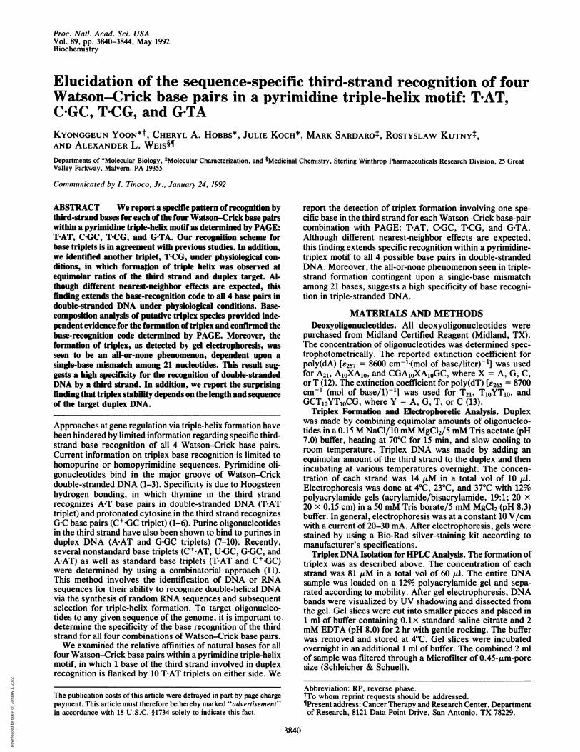
stoichiometry of triplex DNA, mixtures of different molarratios of the deoxyoligonucleotides A21 and T21 were incu-bated at 4°C, separated by gel electrophoresis according tomobility, and visualized by silver staining. The mixturecontaining an equimolar ratio of A21 and T21 was detected asa duplex, whereas the mixtures containing a molar excess ofT21 with respect to A21 resulted in the conversion of duplexto a complex of slower mobility, as shown in Fig. la. Themobility of this complex was invariant among mixturescontaining a molar excess of T21, indicating a constantstoichiometry of 2:1, (2T*A)21. In contrast, mixtures contain-ing molar ratios in which A21 exceeded T21 did not form anyhigher-molecular-weight complex, as shown in Fig. lb. Thus,under the conditions of this experiment, a TAT triplet wasseen, but an A-AT triplet was not seen.
Specificity and Stability for Recognition of Double-StrandedDNA by a Third Strand. Third-strand base-specific interac-tions with all 4 Watson-Crick base pairs were examined.Deoxyoligonucleotides, 21 nucleotides in length (A1oXA1o andT1oYT10, where X Y = A-T, G-C, C-G, or T-A), were mixed atan equimolar ratio to form each of the four combinations ofduplex. An equimolar amount of the third strand (T1oZT1,Owhere Z = T, A, G, or C) was added to each duplexcombination to achieve a stoichiometric conversion ofduplex to triplex at equilibrium, at near physiological condi-tions:
T10Z T10
AloX AloT10Y T10.
a
MA-
b 1 2 3 4 5 6 8 9 10
MA- _ _ 4"OW
FIG. 1. (a) Silver-stained 12% polyacrylanide gel showing com-plexes of different mobilities formed by combining a fixed amount ofA21 with increased amounts of T21. Ratios of A21 to T21 are 1:0.5(lanes 1, 2); 1:1 (lanes 3, 4); 1:1.5 (lanes 5, 6); 1:2 (lanes 7, 8); and 1:3(lanes 9, 10). The species are labeled triplex (T), duplex (D), orsingle-stranded A21 (MA) according to mobilities. Single-stranded T21was not detectable by silver staining but was detected by a UVshadowing technique. The formation of complex and gel electropho-resis were done as described. (b) Silver-stained 12% polyacrylamidegel showing complexes formed by combining a fixed amount of T21with increased amounts of A21. Ratios of A21 to T21 are 0.5:1 (lanes1, 2); 1:1 (lanes 3, 4); 1.5:1 (lanes 5, 6); 2:1 (lanes 7, 8); and 3:1 (lanes9, 10).
The different molecular species-monomer, duplex, andtriplex-were separated by PAGE and detected by silverstaining, as described. To measure the stability of the triplexcontaining variation at a single base (Z) among 21 bases, gelelectrophoresis was done at three temperatures-40C, 23TC,and 37TC.At 40C, all 16 combinations of equimolar mixtures (Z'XY)
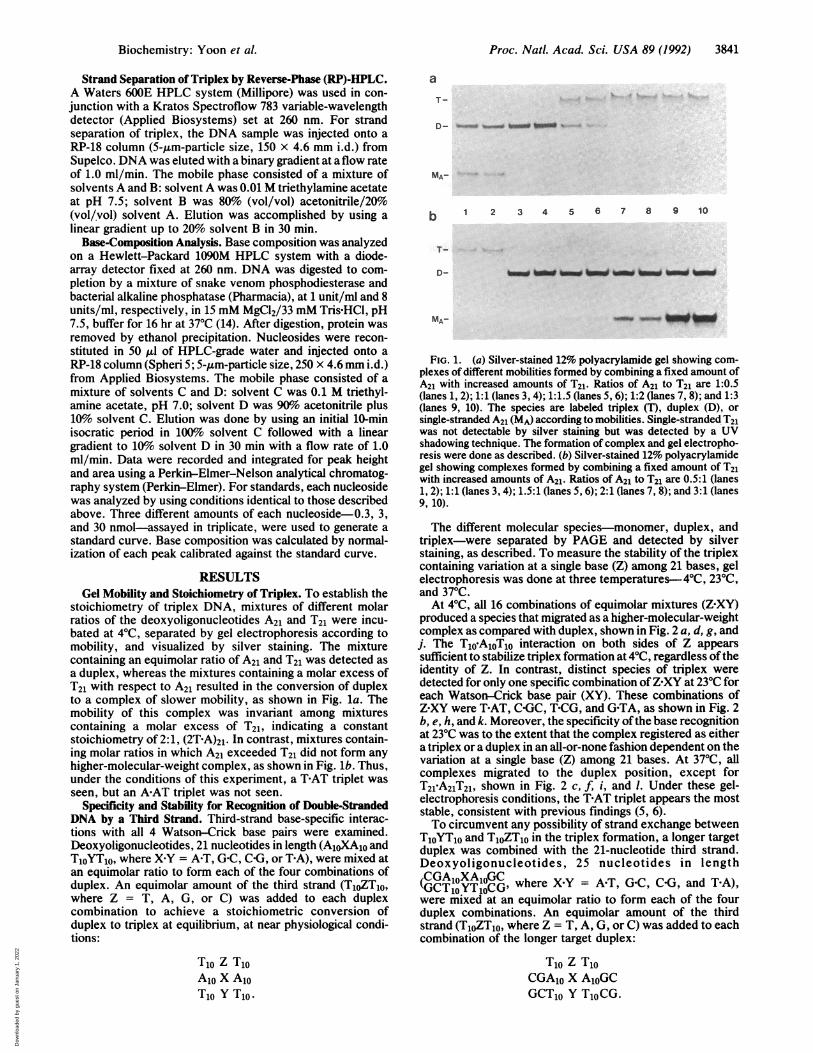
produced a species that migrated as a higher-molecular-weightcomplex as compared with duplex, shown in Fig. 2 a, d, g, andj. The T1o-A1OT1O interaction on both sides of Z appearssufficient to stabilize triplex formation at 40C, regardless oftheidentity of Z. In contrast, distinct species of triplex weredetected for only one specific combination ofZ'XY at 23°C foreach Watson-Crick base pair (XY). These combinations ofZ XY were T-AT, C GC, T-CG, and G-TA, as shown in Fig. 2b, e, h, and k. Moreover, the specificity ofthe base recognitionat 23°C was to the extent that the complex registered as eithera triplex or a duplex in an all-or-none fashion dependent on thevariation at a single base (Z) among 21 bases. At 37°C, allcomplexes migrated to the duplex position, except forT21 A21T21, shown in Fig. 2 c, f, i, and 1. Under these gel-electrophoresis conditions, the TAT triplet appears the moststable, consistent with previous findings (5, 6).To circumvent any possibility of strand exchange between
T1oYT10 and T1oZT10 in the triplex formation, a longer targetduplex was combined with the 21-nucleotide third strand.Deoxyoligonucleotides, 25 nucleotides in length(CGAOXA1OC where X-Y = AT, G-C, C-G, and T-A),were mixed at an equimolar ratio to form each of the fourduplex combinations. An equimolar amount of the thirdstrand (T1oZT1,O where Z = T, A, G, or C) was added to eachcombination of the longer target duplex:
T10 Z T10CGA10 X A1OGCGCT10 Y TjoCG.
Biochemistry: Yoon et al.
Dow
nloa
ded
by g
uest
on
Janu
ary
1, 2
022
Proc. Natl. Acad. Sci. USA 89 (1992)
a 9| j
b h
c
T-h i i
1 2 3 4 5 6 7 8 9 10
d
e
&---- :..A&. -Aba A-
1 2 3 4 5 6 7. 8 9 10
T-
k
*__
f
0-
1 2 3 4 5 6 7 8 9 10 1 2 3 4 5 6 7 8 9 10
FIG. 2. Temperature profile of triplex formation between a duplex,IT1OT41, and an equimolar amount of a third strand, TjoZT10, in whichZ = T, A, G, or C, analyzed on a 12% polyacrylamide gel. (a, b, and c) bels run at 40C, 230C, and 370C, respectively, in which X Y = A-T.(d, e, and f) Gels run at 40C, 230C, and 370C, in which XKY = GAC. (g, h, and i) Gels run at 40C, 230C, and 370C, in which X Y = C-G. (j, k,and l) Gels run at 40C, 230C, and 370C, in which XKY = T-A. The species exhibiting different mobilities are labeled as either triplex (T) or duplex(D). Lanes 1 and 2 contain only the duplex. The remaining lanes are as follows: Z = T (lanes 3, 4); Z = A (lanes 5, 6); Z = G (lanes 7, 8); andZ = C (lanes 9, 10). The formation of complex and electrophoresis were done as described.
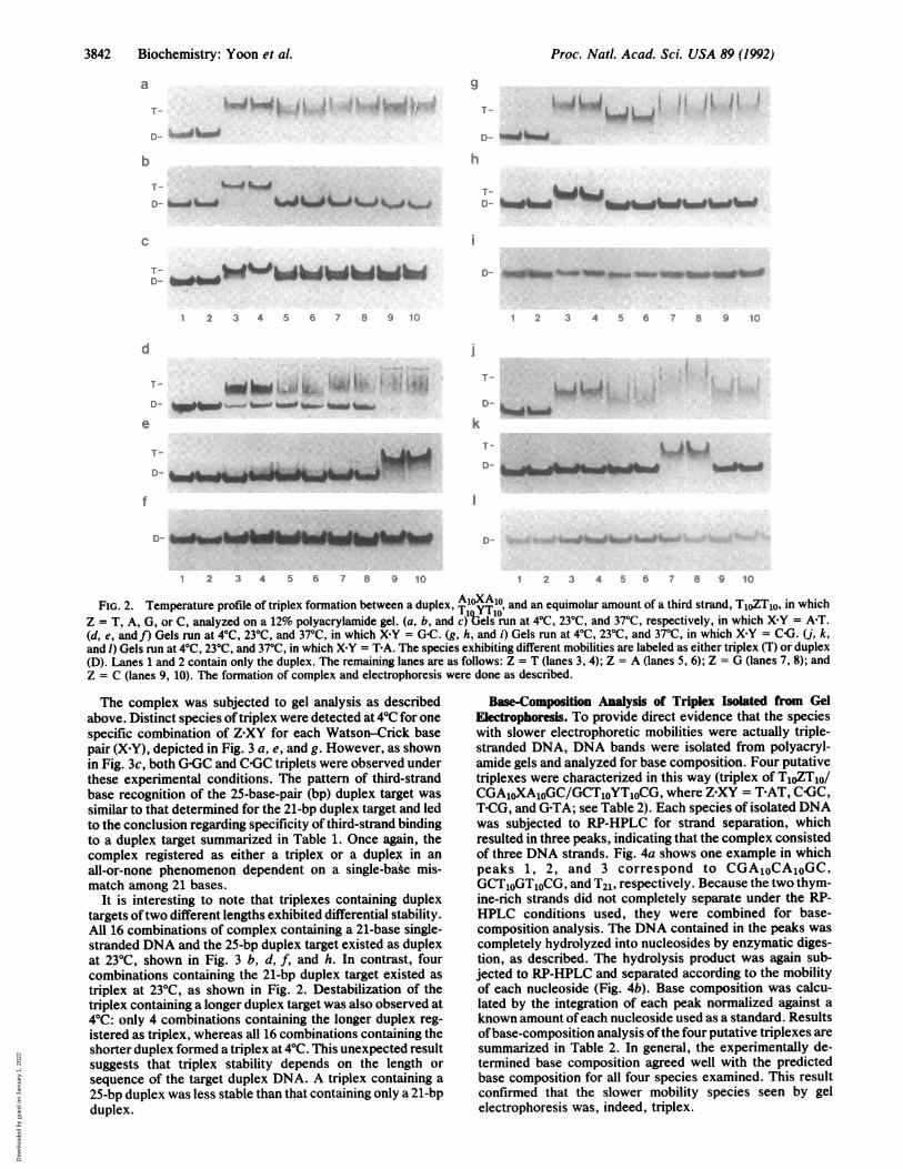
The complex was subjected to gel analysis as describedabove. Distinct species of triplex were detected at 40C for onespecific combination of Z XY for each Watson-Crick basepair (X Y), depicted in Fig. 3 a, e, and g. However, as shownin Fig. 3c, both G-GC and C-GC triplets were observed underthese experimental conditions. The pattern of third-strandbase recognition of the 25-base-pair (bp) duplex target wassimilar to that determined for the 21-bp duplex target and ledto the conclusion regarding specificity of third-strand bindingto a duplex target summarized in Table 1. Once again, thecomplex registered as either a triplex or a duplex in anall-or-none phenomenon dependent on a single-bake mis-match among 21 bases.
It is interesting to note that triplexes containing duplextargets oftwo different lengths exhibited differential stability.All 16 combinations of complex containing a 21-base single-stranded DNA and the 25-bp duplex target existed as duplexat 230C, shown in Fig. 3 b, d, f, and h. In contrast, fourcombinations containing the 21-bp duplex target existed astriplex at 230C, as shown in Fig. 2. Destabilization of thetriplex containing a longer duplex target was also observed at40C: only 4 combinations containing the longer duplex reg-istered as triplex, whereas all 16 combinations containing theshorter duplex formed a triplex at 40C. This unexpected resultsuggests that triplex stability depends on the length orsequence of the target duplex DNA. A triplex containing a25-bp duplex was less stable than that containing only a 21-bpduplex.
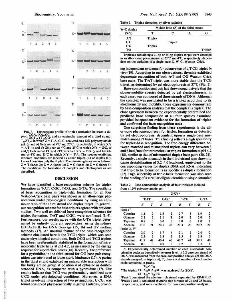
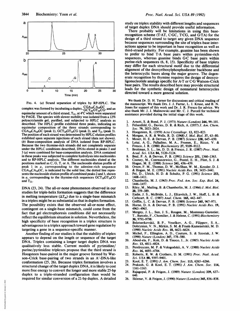
Base-Composition Analysis of Triplex Isolated from GelElectrophoresis. To provide direct evidence that the specieswith slower electrophoretic mobilities were actually triple-stranded DNA, DNA bands were isolated from polyacryl-amide gels and analyzed for base composition. Four putativetriplexes were characterized in this way (triplex of T10ZT1o/CGA1oXAOGC/GCTOYToCG, where Z XY = THAT, COGC,T-CG, and G-TA; see Table 2). Each species of isolated DNAwas subjected to RP-HPLC for strand separation, whichresulted in three peaks, indicating that the complex consistedof three DNA strands. Fig. 4a shows one example in whichpeaks 1, 2, and 3 correspond to CGA1OCAOGC,GCToGTOCG, and T21, respectively. Because the two thym-ine-rich strands did not completely separate under the RP-HPLC conditions used, they were combined for base-composition analysis. The DNA contained in the peaks wascompletely hydrolyzed into nucleosides by enzymatic diges-tion, as described. The hydrolysis product was again sub-jected to RP-HPLC and separated according to the mobilityof each nucleoside (Fig. 4b). Base composition was calcu-lated by the integration of each peak normalized against aknown amount ofeach nucleoside used as a standard. Resultsofbase-composition analysis ofthe four putative triplexes aresummarized in Table 2. In general, the experimentally de-termined base composition agreed well with the predictedbase composition for all four species examined. This resultconfirmed that the slower mobility species seen by gelelectrophoresis was, indeed, triplex.
AL k.&- -JEL-- Albgk Adb- Am-
AL bdL- AML-n aloomm"m ..NOW,
3842 Biochemistry: Yoon et al.
i ,tj .,4x-. ... ,6vi L--j
i
i
Dow
nloa
ded
by g
uest
on
Janu
ary
1, 2
022
Proc. Natl. Acad. Sci. USA 89 (1992) 3843
a
T- ___ -
D_ _t %__ _=ow%
1 2 3 4 5
C
b
1 2 3 4 5
d
Table 1. Triplex detection by silver staining
W-C duplex Middle base (Z) of the third strand(X Y) T C A G
A-T TriplexG-C TriplexCOG TriplexT-A Triplex
Triplexes containing a 21-bp or 25-bp duplex target were detectedin an all-or-none phenomenon at 230C and 40C, respectively, depen-dent on the variation of a single base Z. W-C, Watson-Crick.
1 2 3 4 5 1 2 3 4 5
f
1 2 3 4 5
9
1 2 3 4 5
1 2 3 4 5
h
Dw- _-
1 2 3 4 5
FIG. 3. Temperature profile of triplex formation between a du-CGA1OXAJ3Cplex, GCTO YT10CG, and an equimolar amount of a third strand,
T10ZT10, in which Z = T, A, G, C, analyzed on a 12% polyacrylamidegel. (a and b) Gels run at 40C and 230C, respectively, in which X-Y= A-T. (c and d) Gels run at 40C and 230C in which X Y = G-C. (eandf) Gels run at 40C and 230C in which X-Y = C(G. (g and h) Gelsrun at 40C and 230C in which X Y = T-A. The species exhibitingdifferent mobilities are labeled as either triplex (T) or duplex (D).Lanes 1 contains only the duplex. The remaining lanes are as follows:Z = T (lanes 2); Z = A (lanes 3); Z = G (lanes 4); Z = C (lanes 5).The conditions for formation of complex and electrophoresis aredescribed.
DISCUSSIONWe have identified a base-recognition scheme for triplexformation as T-AT, CG(C, T-CG, and G-TA. The specificityof base recognition in triple-helix formation for all fourWatson-Crick base pairs was shown as an all-or-noie phe-nomenon under physiological conditions by using an equi-molar ratio of the third strand and duplex target. In general,our recognition scheme for base triplets agreed with previousstudies. Two well-established base-recognition schemes fortriplex formation, T-AT and C-GC, were confirmed (1-6).Furthermore, our results agree with the G-TA triplet deter-mined by entirely different approaches, using thymidine/EDTA/Fe(II) for DNA cleavage (15, 16) and UV meltingmethods (17). An unusual feature of the base-recognitionscheme elucidated here is the T-CG triplet, which was seenunder physiological conditions. Both C(CG and T-CG tripletshave been preferentially stabilized in the formation of intra-molecular triple helix at pH 4.2, as measured by the energyrequired for superhelical stress (18). Recently, the preferencefor a pyrimidine in the third strand for COG base-pair recog-nition was attributed to lower steric hindrance (17). A purinein the third strand exhibited an unfavorable interaction withthe bulky amino group at position 4 of cytosine in double-stranded DNA, as compared with a pyrimidine (17). Ourresults indicate that T*CG was preferentially stabilized overC-CG under physiological conditions. Moreover, a RNAtriplet involving interaction of two pyrimidines, U-CG, wasfound conserved phylogenetically in group I introns, provid-
ing independent evidence for occurrence of a T-CG triplet invivo (19). According to our observations, thymine exhibiteddegenerate recognition of both A-T and C-G Watson-Crickbase pairs. The THAT triplet was more stable than the T-CGtriplet, as determined by gel electrophoresis at 370C (Fig. 2).
Base-composition analysis has shown conclusively that theslower-mobility species detected by gel electrophoresis, ineach case, was composed of three strands ofDNA. Althoughthe complex was postulated to be a triplex according to itsstoichiometry and mobility, these experiments demonstrateby base-composition analysis that the complex is triplex. Theclose agreement between the experimentally determined andpredicted base composition of all four species examinedprovided independent evidence for the formation of triplexand confirmed the base-recognition code.One surprising finding from these experiments is the all-
or-none phenomenon seen for triplex formation as detectedby gel electrophoresis, dependent upon a single-base mis-match among 21 bases. This finding reflects a high specificityfor triplex-base recognition. The free energy difference be-tween matched and mismatched triplets can vary between 3and 6 kcal/mol for intramolecular triplex formation at pH 4.2(18), similar to that of mismatched duplex base pairs (20, 21).Recently, a single mismatch in the third strand was shown tocause destabilization of 3.2-4.0 kcal/mol, equivalent to thecorresponding values for duplex DNA and RNA, indicatingthat triple helix formation is as specific as duplex formation(22). High selectivity of triple-helix formation was also seenin the binding of a circular oligonucleotide to single-stranded
Table 2. Base-composition analysis of four triplexes isolatedfrom a 12% polyacrylamide gel.
Z.XY*
T-AT C GC T-CG G-TA
E T E T E T E T
Peak ltCytosine 1.1 2 1.8 2 2.7 3 1.9 2Guanine 2.1 2 3.1 3 2.0 2 2.0 2Thymine 0.8 0 0.0 0 0.0 0 1.0 1Adenine 21.0 21 20.1 20 20.3 20 20.2 20
Peaks 2, 3*Cytosine 2.0 2 3.7 4 2.1 2 2.0 2Guanine 2.3 2 1.8 2 3.5 3 3.3 3Thymine 41.7 42 40.4 40 40.7 41 39.5 40Adenine 0.0 0 0.0 0 0.2 0 1.2 1
E, Experimentally determined number of each nucleoside con-tained in peaks as described (the error level, +0.2 base per strand ofDNA, was measured from the base-composition analysis of six DNAstrands assayed, in triplicate); T, theoretical number of each nucle-oside contained in peaks.
T10Z T10*The triplex CG A1oX AioGC was analyzed for Z XY.
GC T1oY Tj0CGtPeak 1 contained the adenine-rich strand separated by RP-HPLC.tPeaks 2 and 3 contained thymine-rich strands of 21 and 25 bases,respectively, and were combined for base-composition analysis.
e
Biochemistry: Yoon et al.
Dow
nloa
ded
by g
uest
on
Janu
ary
1, 2
022
Proc. Natl. Acad. Sci. USA 89 (1992)
b
study on triplex stability with different lengths and sequencesof target duplex DNA should provide useful information.There probably will be limitations in using this base-
2 3 recognition scheme (T-AT, C-GC, T-CG, and G-TA) for theI0 ^ \ A A design of a third strand to target any given DNA sequence
because sequences surrounding the site of triplex-base inter-actions appear to be important in base recognition as well asthird-strand polarity. For example, guanine has been shown
20 25 30 to prefer to bind T-A base pairs within pyrimidine-richsequences, whereas guanine binds G-C base pairs within
/
LOTA purine-rich sequences (6, 8, 15). Specificity of base tripletsmay differ for each structural motif due to the differentialalignment of the deoxyribosylphosphodiester backbone and
so l the heterocyclic bases along the major groove. The degen-,C /G I erate recognition by thymine requires the design of deoxyo-
. ; ....................... .............. ...... ligonucleotide analogs specific for APT or G Watson-Crickbase pairs. The results described here may provide structural
P10 15 20 25 30 leads for the synthetic design of nonnatural heterocycles0510 15 20 25 30 directed toward a more general solution.
Time (min)
FIG. 4. (a) Strand separation of triplex by RP-HPLC. Thecomplex was formed by incubating a duplex, CTOGTOCG, and anequimolar amount of a third strand, T21, at 40C which were separatedby PAGE. The species with slower mobility was isolated from a 12%polyacrylamide gel, purified, and subjected to HPLC analysis asdescribed. The HPLC profile exhibited three peaks, indicating anequimolar composition of the three strands corresponding toCGA1OCAOGC (peak 1), GCT1OGTOCG (peak 2), and T21 (peak 3).The position ofeach strand was determined by HPLC elution profilesexhibited upon separate injections of each strand (data not shown).(b) Base-composition analysis of DNA isolated from RP-HPLC.Because the two thymine-rich strands did not completely separateunder the HPLC conditions described, DNAs eluted in peaks 2 and3 (a) were combined for base-composition analysis. DNA containedin these peaks was subjected to complete hydrolysis into nucleosidesand to RP-HPLC analysis. The different nucleosides eluted at thepositions marked as C, G, T, or A. The nucleoside elution profile ofpeak 1 in a, corresponding to the adenine-rich sequenceCGA1OCAOGC, is indicated by the solid line; the dotted line repre-sents the nucleoside elution profile ofcombined peaks 2 and 3, shownin a, corresponding to the thymine-rich sequences GCT1OGT1OCGand T21.
DNA (23, 24). The all-or-none phenomenon observed in ourstudies for triple-helix formation suggests that the differencein melting temperature (AT.) due to a single-base mismatchin a triplex might be as substantial as that in duplex formation.The possibility exists that the observed all-or-none effect,contingent on a single-base mismatch, could come from thefact that gel electrophoresis conditions did not necessarilyreflect the equilibrium situation in solution. Nevertheless, thehigh specificity of base recognition in a triplex structure isadvantageous to a triplex approach toward gene regulation bytargeting a gene in a sequence-specific manner.Another finding of our studies is that the stability of triplex
appears to depend on the length or sequence of the targetDNA. Triplex containing a longer target duplex DNA wasqualitatively less stable. Current models of pyrimidine/purine/pyrimidine triplexes propose that the third strand isHoogsteen base-paired in the major groove formed by Wat-son-Crick base-pairing of two strands in an A'-DNA-likeconformation (25, 26). Because triplex formation involves astructural change ofthe target duplex DNA, it is likely to costmore free energy to convert the longer and more stable 25-bpduplex to a triple-stranded configuration than would berequired for similar conversion of a 21-bp duplex. A detailed
We thank Dr. D. H. Turner for discussions and critical reading ofthe manuscript. We thank Drs. J. J. Farrar, L. I. Kruse, and B. N.Jones for support of this work and Dr. J. R. Fresco for advice. Wealso thank Mr. J. J. Malinowski and Mr. C. J. Purcell for technicalassistance provided during the initial stage of this work.
1. Arnott, S. & Bond, P. J. (1973) Nature (London) 244, 99-101.2. Felsenfeld, G., Davies, D. R. & Rich, A. (1957) J. Am. Chem.
Soc. 79, 2023-2024.3. Hoogsteen, K. (1959) Acta Crystallogr. 12, 822-823.4. Morgan, A. R. & Wells, R. D. (1968) J. Mol. Biol. 37, 63-80.5. Moser, H. E. & Dervan, P. B. (1987) Science 238, 645-650.6. Letai, A. G., Palladino, M. A., Fromm, E., Rizzo, V. &
Fresco, J. R. (1988) Biochemistry 27, 9109-9112.7. Broitman, S. L., Im, D. D. & Fresco, J. R. (1987) Proc. Natl.
Acad. Sci. USA 84, 5120-5124.8. Beal, P. A. & Dervan, P. B. (1991) Science 251, 1360-1363.9. Cooney, M., Czemuszewicz, G., Postel, E. H., Flint, S. J. &
Hogan, M. E. (1988) Science 241, 456-459.10. Orson, F. M., Thomas, D. W., McShan, W. M., Kessler, D. J.
& Hogan, M. E. (1991) Nucleic Acids Res. 19, 3435-3441.11. Pei, D., Ulrich, H. D. & Schultz, P. G. (1991) Science 253,
1408-1411.12. Chamberlin, M. J. (1965) Proc. Fed. Am. Soc. Exp. Biol. 24,
1446-1457.13. Riley, M., Mailing, B. & Chamberlin, M. J. (1966) J. Mol. Biol.
20, 359-389.14. Eadie, J. S., McBride, L. J., Efcavitch, J. W., Hoff, L. B. &
Cathcart, R. C. (1987) Anal. Chem. 165, 442-447.15. Griffin, L. C. & Dervan, P. B. (1989) Science 245, 967-971.16. Home, D. A. & Dervan, P. B. (1991) Nucleic Acids Res. 19,
4963-4965.17. Mergny, J. L., Sun, J. S., Rougee, M., Montenay-Garestier,
T., Barcelo, F., Chomilier, J. & Helene, C. (1991) Biochemistry30, 9791-9798.
18. Belotserkovskii, B. P., Veselkov, A. G., Filippov, S. A.,Dobryniun, V. N., Mirkin, S. M. & Frank-Kamenetskii, M. D.(1990) Nucleic Acids Res. 18, 6621-6624.
19. Michel, F., Ellington, A. D., Couture, S. & Szostak, J. W.(1990) Nature (London) 347, 578-580.
20. Aboul-ela, F., Koh, D. & Tinoco, I., Jr. (1985) Nucleic AcidsRes. 13, 4811-4824.
21. Perelroyzen, M. P. & Vologodskii, A. V. (1988) Nucleic AcidsRes. 16, 4693-4704.
22. Roberts, R. W. & Crothers, D. M. (1991) Proc. Natl. Acad.Sci. USA 88, 9397-9401.
23. Kool, E. T. (1991) J. Am. Chem. Soc. 113, 6265-6266.24. Prakash, G. & Kool, E. T. (1991) J. Am. Chem. Soc. 114,
1161-1163.25. Rajagopal, P. & Feigon, J. (1989) Nature (London) 339, 637-
640.26. Sklenar, V. & Feigon, J. (1990) Nature (London) 345, 836-838.
3844 Biochemistry: Yoon et al.
Dow
nloa
ded
by g
uest
on
Janu
ary
1, 2
022