embryology of the eye the eye is formed from both ectoderm and mesenchyme. the neuroectoderm that is...
TRANSCRIPT

Embryology of the eyeEmbryology of the eyeThe eye is formed from both eThe eye is formed from both ectodctoderm and erm and mesenchyme. The mesenchyme. The neuroectodermneuroectoderm that is derived that is derived from the neural tube gives rise to (the from the neural tube gives rise to (the retinaretina, the , the fibers of thefibers of the optic nerve optic nerve, and the, and the smooth muscle of smooth muscle of the iris)the iris). The . The surface ectodermsurface ectoderm on the side of the on the side of the head forms( thehead forms( the corneal and conjunctival corneal and conjunctival epitheliumepithelium, the, the lens lens, and the , and the lacrimal and tarsallacrimal and tarsal glands). Theglands). The mesenchyme mesenchyme forms( the forms( the corneal corneal stromastroma, the, the sclera sclera, the, the choroid choroid, the , the irisiris, the , the ciliary ciliary musculaturemusculature, part of the, part of the vitreous body vitreous body, and, and the cells the cells lining the anterior chamber)lining the anterior chamber)..

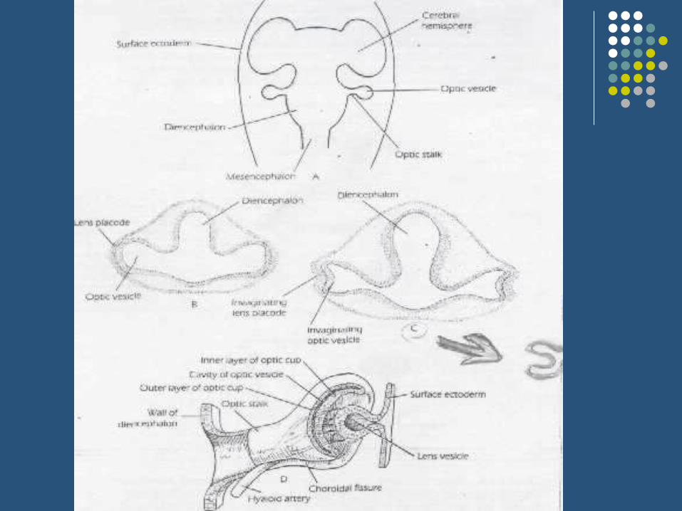
The eyeballThe eyeballThe rudimentary eyeball develops as an The rudimentary eyeball develops as an ectodermal diverticulum from the lateral aspect ectodermal diverticulum from the lateral aspect of the forebrain. The diverticulum grows out of the forebrain. The diverticulum grows out laterally toward the side of the head, and the end laterally toward the side of the head, and the end becomes slightly dilated to form the becomes slightly dilated to form the optic optic vesiclevesicle, , while the proximal portion becomes while the proximal portion becomes constricted to form the constricted to form the optic stalkoptic stalk. At the same . At the same time, a small area of surface ectoderm overlying time, a small area of surface ectoderm overlying the optic vesicle thickens to form thethe optic vesicle thickens to form the lens lens placodeplacode. . The lens placode invaginates and sinks The lens placode invaginates and sinks below the surface ectoderm to become the below the surface ectoderm to become the lens lens vesiclevesicle, the optic vesicle becomes invaginated to , the optic vesicle becomes invaginated to form the double-layered form the double-layered optic cupoptic cup..


Orbit and Ocular AdnexaDuring infancy and childhood, the orbital volume increases, the shape of the orbital opening becomes less circular and more like a horizontal oval, the lacrimal fossa becomes more superficial, and the angle formed by the axes of the 2 orbits assumes a less divergent position.The palpebral fissure measures approximately 18 mm horizontall y and 8 mm vertically at birth and changes very little during the first year of life, but a rapid increase in palpebral fissure length occurs during the first decade. causing the round infant eye to acquire an elliptical adult shape.

Iris, Pupil, and Anterior ChamberMost iris color changes occur over the first 6 to 12 months of life. as pigment accumulatesin the iris stroma and melanocytes, but iris pigmentation may continue.

The optic nerveThe optic nerveThe ganglion cells of the retina develop The ganglion cells of the retina develop axons that converge to a point where the axons that converge to a point where the optic stalk leaves the posterior surface of optic stalk leaves the posterior surface of the optic cup. This site will later become the optic cup. This site will later become the the optic discoptic disc. . The axons now pass The axons now pass among the cells that form the inner layer among the cells that form the inner layer of the stalk. Gradually, the inner layer of the stalk. Gradually, the inner layer encroaches on the cavity of the stalk until encroaches on the cavity of the stalk until the inner and outer layers fusethe inner and outer layers fuse..


Developmental Malformations and AnomaliesEyelidsEpicanthic folds Signs- bilateral vertical folds of skinthat extend from the upper or lowerLids towards the medial canthi;

Coloboma1. Definition- partial or full-thicknessEyelid defect occurring when eyelidDevelopment is incomplete.2. Pathogenesis failure of migrationof lid ectoderm to fuse the lid folds,or mechanical forces such as Amniotic bands.3. Upper lid - at junction of the middleand inner thirds.4. Lower lid - at junction of the middleand outer thirds

GlobeMicrophthalmos1. Definition- total axial length atleast2 standard deviations belowage-similar controls usuallyunilateral.2. Pathogenesis non-specific growthFailure in response to a variety ofPrenatal insults.3. Classificationa. Simple- isolated.b. Complex(colobomatous\

Anophthalmos1. Pathogenesis either acompleteFailure of budding of the optic vesicleor early developmental arrest.2. Simple- associated with absence ofExtraocular muscles,short conjunctivalsac. and microbleoharon.3. Anophthalmos with cyst (congenitalcystic eyeball) - globe is replacedby a cyst

Iris, Pupil, and Anterior ChamberMost iris color changes occur over the first 6 to 12 months of li fe. as pigment accumulatesin the iris stroma and melanocytes, but iris pigmentation may continue. Compared with


ReferencesReferences1. Lecture notes in 1. Lecture notes in ophthalmology ophthalmology 2. Parson’s disease of the 2. Parson’s disease of the eyeeye