embryonic stem cells/induced pluripotent stem cells
TRANSCRIPT
EMBRYONIC STEM CELLS/INDUCED PLURIPOTENT STEM CELLS
SOX15 and SOX7 Differentially Regulate the Myogenic Program
in P19 Cells
JOSEE SAVAGE,aANDREW J. CONLEY,
bALEXANDRE BLAIS,
a,cILONA S. SKERJANC
a
aDepartment of Biochemistry, Microbiology, and Immunology, University of Ottawa, Ottawa, Ontario, Canada;bDepartment of Biology, Biological and Geological Sciences Building, University of Western Ontario, London,
Ontario, Canada; cOttawa Institute of Systems Biology, University of Ottawa, Ottawa, Ontario, Canada
Key Words. P19 cells • Gene expression • Transcription • Myogenesis • Sox15 • Sox7 • Mesoderm
ABSTRACT
In this study, we have identified novel roles for Sox15 andSox7 as regulators of muscle precursor cell fate in P19cells. To examine the role of Sox15 and Sox7 during skele-
tal myogenesis, we isolated populations of P19 cells witheither gene stably integrated into the genome, termed
P19[Sox15] and P19[Sox7]. Both SOX proteins were suffi-cient to upregulate the expression of the muscle precursormarkers Pax3/7, Meox1, and Foxc1 in aggregated cells. In
contrast to the P19[Sox7] cell lines, which subsequentlydifferentiated into skeletal muscle, myogenesis failed to
progress past the precursor stage in P19[Sox15] cell lines,shown by the lack of MyoD and myosin heavy chain(MHC) expression. P19[Sox15] clones showed elevated and
sustained levels of the inhibitory factors Msx1 and Id1,which may account for the lack of myogenic progression
in these cells. Stable expression of a Sox15 dominant-nega-tive protein resulted in the loss of Pax3/7 and Meox1 tran-scripts, as well as myogenic regulatory factor (MRF) and
MHC expression. These results suggest that Sox15, orgenes that are bound by Sox15, are necessary and suffi-
cient for the acquisition of the muscle precursor cell fate.On the other hand, knockdown of endogenous Sox15caused a decrease in Pax3 and Meox1, but not MRF
expression, suggesting that other factors can compensatein the absence of Sox15. Taken together, these results
show that both Sox7 and Sox15 are able to induce theearly stages of myogenesis, but only Sox7 is sufficient toinitiate the formation of fully differentiated skeletal myo-
cytes. STEM CELLS 2009;27:1231–1243
Disclosure of potential conflicts of interest is found at the end of this article.
INTRODUCTION
During the process of vertebrate skeletal myogenesis, signal-ing molecules such as Wnts, bone morphogenetic proteins,and Sonic Hedgehog (Shh) are secreted from tissues surround-ing the somite [1–3]. These extracellular cues activate a cas-cade of transcription factors that leads to the expression ofthe myogenic regulatory factors (MRFs) MyoD, Myf5, myo-genin, and MRF4, which control the end-stages of myogenesis[4]. Induction of MRF expression in response to these signalsappears to be mediated by many transcription factors includ-ing Pax3, Meox1/2, Six1, and Gli2 [5–12].
Pax3 and Meox1 are part of a regulatory network of tran-scription factors that regulate myogenesis [9, 11, 13]. Pax3 isexpressed in the mediolateral dermomyotome and migratinglimb precursors [14, 15], while the closely related Pax7, isalso expressed in the dermomyotome and may have redundantfunctions with Pax3 [16]. Cell culture studies reveal a role forPax3 in regulating the commitment of cells to the myogeniclineage. Ectopic expression of Pax3 in embryonic tissues is
sufficient to activate MyoD and Myf5 expression [10, 17],and overexpression in P19 cells also leads to the induction ofmyogenesis and MRF expression [11]. P19 cells are pluripo-tent embryonal carcinoma (EC) cells, derived from mouseembryonic stem cells, that can differentiate into skeletal mus-cle in a dimethyl sulfoxide (DMSO)- and aggregation-depend-ent manner [18]. Identification of a Pax3-binding site withinthe Myf5 regulatory region suggests direct activation of Myf5by Pax3 in developing murine limbs [19]. Furthermore, P19cells expressing a dominant-negative form of Pax3, termedPax3/EnR, are unable to undergo myogenesis or expressMyoD [11]. Expression of Pax3/EnR and Pax7/EnR alsoinhibits MyoD expression in the mouse embryo [5] and ani-mals lacking both Pax3 and Pax7 form myotomal muscle, butdo not develop primary or secondary muscle fibers [20].Pax3/Pax7 are essential for the formation of satellite cells,suggesting that the Pax3/Pax7 pool of progenitor cells is re-sponsible for nearly all muscle formation [20, 21].
A role for Meox1 in regulating skeletal myogenesis andPax3 expression has been demonstrated [8, 9]. Both Meox1and Meox2 homeobox family members are initially expressed
Author contributions: J.S.: conception and design, collection and/or assembly of data, creation of all figures in manuscript, data analysisand interpretation, manuscript writing; A.C.: collection and/or assembly of data; A.B.: collection and/or assembly of data; I.S.S.:conception and design, financial support, data analysis and interpretation, manuscript writing, final approval of manuscript.
Correspondence: Ilona S. Skerjanc, Ph.D., Department of Biochemistry, Microbiology, and Immunology, University of Ottawa, 451Smyth Road, Ottawa, Ontario, Canada K1H 8M5. Telephone: (613) 562-5800, ext. 8669; Fax: (613) 562-5452; e-mail: [email protected] Received January 12, 2009; accepted for publication March 5, 2009; first published online in STEM CELLS EXPRESS March 5,2009. VC AlphaMed Press 1066-5099/2009/$30.00/0 doi: 10.1002/stem.57
STEM CELLS 2009;27:1231–1243 www.StemCells.com
in the somites, and their expression becomes localized to thedermomyotome and the developing limb bud, respectively, asdevelopment proceeds [7, 22]. Mice carrying null mutationsfor both Meox genes show a loss of Pax3 and Pax7, in thesomite [8]. These results place Meox1 before Pax transcrip-tion factors in the molecular hierarchy controlling myogene-sis. Consistent with this, a dominant-negative Meox1 tran-scription factor, Meox1/EnR, downregulates Pax3 expressionand ablates myogenesis in P19 cells [9]. Further, Wnt signal-ing can activate Pax3 expression, although it is unclearwhether this activation is direct or indirect [23–26]. Althoughmuch is known about the downstream effects of Pax3 andMeox1 during myogenesis, less is understood about the fac-tors that regulate their gene expression.
Sox transcription factors have been characterized for theirinvolvement in muscle development. Sox8 has been identifiedas a marker of adult satellite stem cells, and has the ability toinhibit differentiation of cultured primary myoblasts whenectopically expressed [27]. Studies have implicated anotherfactor, Sox15, as a regulator of myogenesis. Although Sox15expression has not yet been examined in the somite, it isexpressed at high levels in the mouse blastocyst, embryonicstem cells, and in satellite cells [28–31]. Murine Sox15 isdownregulated during C2C12 differentiation and can blockmyotube formation when overexpressed [32]. Moreover, dis-ruption of Sox15 in mice resulted in the loss of satellite cellformation [30] and the attenuation of adult muscle regenera-tion with a decrease in MyoD levels [33]. Sox15, along withFhl3, binds and regulates Foxk1 expression, regulating adultmyogenic progenitor cells [30]. Sox7, another member of theSox family [34–37] is detected in the somite of mice as earlyas embryonic day 7.5 [35]. Studies in Xenopus have shownthat xSox7 can induce the expression of mesoderm inducinggenes Xnr1-6 and Mixer, suggesting a role for Sox7 in regu-lating the cell fate of mesodermal cells [38]. In addition,Sox7 has been extensively characterized for its role in con-trolling arteriovenous specification in zebrafish [39–41] andxenopus [42].
Based on the evidence in the literature showing thatSox15 and Sox7 are involved in controlling the cell fate ofmesodermal derivatives, the present study aims to furthercharacterize the role of SOX transcription factors in regulat-ing the expression of skeletal muscle precursor genes. TheP19 cell model was chosen because the differentiation ofthese cells follows early embryonic pathways [11, 26, 43] andthe DMSO-dependence of the differentiation allows for bothgain- and loss-of-function studies. Here we show that whileboth Sox15 and Sox7 can induce an early skeletal musclemesoderm phenotype, expressing Pax3/7, Meox1, and Foxc1,only Sox7 can initiate the entire pathway leading to skeletalmuscle formation.
MATERIALS AND METHODS
DNA Constructs
Expression constructs of PGK-Puro, PGK-Lacz, B17, PGK-Pax3, PGK-Meox1, CMV-Gli2, and activated CMV–b-cateninhave been described previously [9, 11, 26, 44]. CMV-Sox15was kindly provided by F. Beranger [32]. PGK-Sox7 was cre-ated by excision of the Sox7 ORF from the pCMVScript vec-tor (kind gift from Y. Hayashi, Japanese Science and Tech-nology Agency). The ends of the excised insert were bluntedto form compatible ends for cloning into the SmaI site of thePGK vector, which has been described previously [45].
The dominant-negative Sox15/EnR fusion protein was cre-ated by polymerase chain reaction (PCR) amplification of theengrailed (EN-2) repressor domain using the following oligo-nucleotides: EnR-F 50-AACTCGAGAGAGGAGAAGGATTC-CAAGCCC and EnR-R 50-TTGAATTCCTAGCCCA-GAGTGGCGCTGGCTT. Restriction sites for XhoI andEcoRI were introduced for cloning purposes and are italicizedin the sequences. The activation domain of Sox15 wasremoved by digesting the pRK5-Sox15 vector using therestriction enzymes XhoI and EcoRI (removes nucleotides376–696 from the Sox15 cDNA) and replaced by ligationwith the engrailed repressor domain. DNA sequencing wasperformed to ensure that the resulting chimeric protein wasin-frame.
The 1.6 kb fragment upstream of the Pax3 transcriptionalstart site [46] was amplified by PCR from 50 ng of genomicDNA, isolated from P19 cells [47]. To amplify the promoterregion, the 50 oligonucleotide utilized was AAAGCTAGC-GAGCTCTAATGCTCCTCC and the 30 oligonucleotide wasAAACTCGAGCACCGAGTGCAGGGATCC, at 0.25 lMeach. Amplification (30 cycles) was performed with annealingat 56�C, in the presence of Q solution (Qiagen, Mississauga,ON). The amplified fragment was cloned into the luciferasereporter vector pGL3-basic (Promega, Madison, WI, http://www.promega.com) via the Nhe1 and Xho1 sites, respectively(italicized in the oligonucleotide sequences) and sequenced.
Cell Culture and Transfections
P19 EC cells were cultured as described previously [48] in a-minimal essential media (Invitrogen, Burlington, ON, Canada)supplemented with 5% cosmic calf serum (Hyclone, Logan,UT, http://www.hyclone.com) and 5% fetal bovine serum(FBS) (CanSera, Rexdale, ON, Canada). P19[control],P19[Sox15], P19[Sox15/EnR], and P19[Sox7] cell lines werecreated as described previously [9, 11, 26]. Cells were differ-entiated in the presence or absence of DMSO as describedpreviously [44]. RNA was harvested on the days indicated forquantitative (Q)-PCR analysis and cells were fixed forimmuofluorescence. Seven clonal populations were examinedfor the P19[Sox15] cell line, three for P19[Sox15/EnR], fourfor P19[Sox7], and four for P19[control] cells. Little variabili-ty was observed between clonal populations for a given cellline.
Luciferase Reporter Assays
For reporter analysis, 200,000 cells/well were plated into 6-well plates and transiently transfected 24 hours post-plating.Transfection mixtures were composed of 0.2 lg Renilla lucif-erase, 0.5 lg Pax3 promoter-luciferase, and 0.5 lg CMV-Sox15 and/or 2.5 lg CMV-Sox15/EnR, as indicated. Cellswere harvested 20–24 hours following transfection using theDual Luciferase Kit, according to the manufacturer’s protocol(Promega) and samples were analyzed using an LmaxII384luminometer (Molecular Devices). All transfections weredone in duplicate, and results shown are from four independ-ent experiments. To determine the statistical differencesamong means, statistical analysis was done using the student’st test.
Immunofluorescence
Myosin heavy chain (MHC) expression was detected usingthe MF20 monoclonal antibody supernatant as described pre-viously [44]. A 1:100 dilution of goat anti-mouse Cy3-linkedantibody (Jackson ImmunoResearch Laboratories, WestGrove, PA, http://www.jacksonimmuno.com) in phosphate-buffered saline (PBS) was used as a secondary antibody. ForPax3 and Meox1 detection, coverslips were fixed at �20�C in
1232 Sox15 and Sox7 Differentially Regulate Myogenesis
acetone for 10 minutes before rehydration in PBS. Sox15 andMyc staining were performed on cells fixed in 3.7% parafor-maldehyde for 20 minutes at room temperature. Followingpermeabilization in 0.5% Triton X-100/PBS, cells wereblocked using a 10% FBS, 0.1% bovine serum albumin, and0.1% Triton X-100 solution in PBS at room temperature for 1hour. The coverslips were incubated overnight using a 1:50dilution of Meox1 antibody (sc-10185, Santa Cruz Biotech-nology, Santa Cruz, CA, http://www.scbt.com), a 1:50 dilutionof Pax3 antibody (clone 274212, R&D Systems, Minneapolis,MN, http://www.rndsystems.com), a 1:100 dilution of aSox15 antibody (kind gift from Ibrahim M. Adham) or a1:100 dilution of a myc antibody (clone 9E10, Santa CruzBiotechnology, Santa Cruz, CA) followed by a 1 hour roomtemperature incubation with a 1:100 dilution of donkey anti-goat Cy3-linked antibody (Jackson ImmunoResearch Labora-tories) in PBS, or a 1:100 dilution of goat anti-mouse Cy3-linked antibody (Jackson ImmunoResearch Laboratories) inPBS. Immunofluorescence was detected using a Zeiss Axio-scope microscope. Images were captured on a Sony 3CCDcamera and processed with Axiovision, Adobe Photoshop7and Canvas 8. For cell counting experiments, 10 fields ofview per coverslip were counted using the 20� objective.Two coverslips for each treatment were counted, and theresults shown are the average of three independent differentia-tions � SEM.
Reverse Transcription and Q-PCR
RNA was isolated using the RNeasy Mini Kit, according tothe manufacturer’s protocol (Qiagen, Mississauga, ON, Can-ada). One microgram of the purified RNA was used duringthe first strand DNA synthesis reaction using the QuantitectReverse Transcription Kit, as per the manufacturer’s specifica-tions (Qiagen, Mississauga, ON, Canada). For the PCR reac-tion, 1/20th of the reverse transcription was used as a tem-plate for quantitative PCR amplification, using the FastStartSYBR Green kit from Roche (Roche Applied Sciences, Laval,QC, Canada). All reactions were performed and analyzedusing the ABI 7,300 system and SDS analysis software(Applied Biosystems, Streetsville, ON, Canada). Primers usedfor quantitative detection of gene expression are listed in sup-porting information Table 1.
Microarray Analysis
For microarray analysis using the mouse genome 430 v2.0Affymetrix array, P19[Sox15] and P19[control] cells were dif-ferentiated in the absence of DMSO, and RNA was extractedon day 2 using the RNeasy Mini Kit (Qiagen, Mississauga,Canada), according to the manufacturer’s instructions. Allprocedures including labeling, hybridization, and scanningwere performed at the Ottawa Genome Centre (Ottawa, ON,Canada). Data analysis was performed with the dChIP pro-gram [49], using the PM-only method and filtering out probeswith near-background expression levels in both samples. Aprobe representing the Sox15 mRNA was among the mostinduced genes in this analysis.
RNA Interference
Complementary DNA sequences targeting nucleotides 269–287 (50-TTTGGATGAAGAGAAGCGACCCTTTCAAGAGAAGGGTCGCTTCTCTTCATCGCTTTTT-30 and 50-CTAGAAAAAGCGATGAAGAGAAGCGACCCTTCTCTTGAAAGGGTCGCTTCTCTTCATC-30) and nucleotides 514-533 (50-TTTGCCTGGCAGTTACACCTCTTCTCAAGAGAAAGAGGTGTAACTGCCAGGCATTTTT-30 and 50-CTAGAAAAATGCCTGGCAGTTACACCTCTTTCTCTTGAGAAGAGGTGTAACTGCCAGG-30 of mouse Sox15 (NM_009235) were
annealed and cloned into mU6pro using Bbs1 and XbaIrestriction enzymes. A control vector was created using ascrambled sequence that is not complementary to any sequen-ces in the mouse genome (50-TTTGGCTAAGCGAGCTGCTAGAGTTCAAGAGACTCTAGCAGCTCGCTTAGCTTTTT-30and 50-CTAGAAAAAGCTAAGCGAGCTGCTAGAGTCTCTTGAACTCTAGCAGCTCGCTTAGC-30). The mU6pro vectorwas a generous gift from Dave Turner (University of Michigan,Ann Arbor, MI) and has been described previously [50]. Forthe generation of stable cell lines expressing the short hairpinconstructs, 0.8 lg of either shSox15 or shScrambled was trans-fected into P19 cells as described previously [44]. Followingpuromycin selection, individual clones were either picked andexpanded for further analysis or pooled together and furtheranalyzed.
Chromatin Immunoprecipitation
Five 150 mm dishes of day 5 P19[Sox15] aggregates werefixed using 1% formaldehyde at room temperature for 45minutes, and quenched by adding 0.125 M glycine. Cellswere washed in ice-cold PBS and resuspended in lysis bufferone (50 mM HEPES-KOH pH 7.4, 140 mM NaCl, 1 mMEDTA, 10% glycerol, 0.5% NP-40, 0.25% Triton X-100 andcomplete protease inhibitor cocktail; Roche Applied Sciences,Laval, QC) and incubated at 4�C for 10 minutes with rocking.Cells were then resuspended and incubated for an additional10 minutes at room temperature in lysis buffer two (200 mMNaCl, 1 mM EDTA [pH 8.0], 0.5 mM, EGTA [pH 8.0], 10mM Tris [pH 8.0], and protease inhibitor cocktail) beforeresuspension in sonication buffer (1 mM EDTA [pH 8.0], 0.5mM EGTA [pH 8.0], 10 mM Tris [pH 8.0], and proteaseinhibitors). Cells were sonicated using a Sonic Dismembrator(Fisher Scientific International, Hampton, NH, http://www.fisherscientific.com) for a total of 15 � 30-secondpulses (1-minute rest between pulses) and lysates were clearedby centrifugation at 13,000 rpm for 30 minutes at 4�C. Thelysates were pre-cleared by incubation with protein-G sephar-ose beads for 1 hour, and the chromatin was separated intothree aliquots supplemented with 1% TritonX-100, 0.1% so-dium deoxycholate, 1 mM EDTA, and 1 mM AEBSF. Aninput sample of 1% of the total chromatin was set aside. Forimmunoprecipitation, 5 lg of Sox15 antibody (sc-17354,Santa Cruz Biotechnology, Santa Cruz, CA) or 5 lg of IgGantiserum (Zymed Laboratories, CA) was used. Followingovernight incubation with the antibodies, the immune com-plexes were captured by addition of protein-G sepharosebeads for 1 hour, followed by 10-minute washes in each ofthe following buffers: low-salt (0.1% SDS, 1% Triton X-100,2 mM EDTA, 20 mM Tris-HCl pH 8.1, 150 mM NaCl),high-salt (low-salt buffer with 500 mM NaCl) and LiCl (0.25M LiCl, 1% IGEPAL-CA630, 1% deoxycholic acid, 1 mMEDTA, 10 mM Tris pH 8.1), and TE. Protein/DNA complexeswere eluted using 50 mM Tris pH 8.0, 1 mM EDTA, 1%SDS, 50 mM NaHCO3 and crosslinks reversed overnight at65�C by the addition of 200 mM NaCl. Samples were treatedwith 20 lg RNase A and 40 lg Proteinase K and DNA waspurified using Qiagen’s PCR Purification Kit (Qiagen, Missis-sauga, ON). Relative enrichment of binding sites comparedwith the IgG negative control immunoprecipitation were ana-lyzed using SYBR Green real-time PCR, as described earlier(supporting information Table 1).
Statistical Analysis
Statistical differences between means were calculated usingthe Student’s t test. p values of at least p < .05 were consid-ered significant.
Savage, Conley, Blais et al. 1233
www.StemCells.com
RESULTS
Sox15 Expression Is Downregulated in a Populationof Cells During Myogenesis in P19 cells
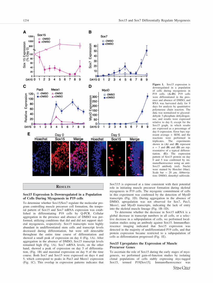
To determine whether Sox15/Sox7 regulate the molecular pro-gram controlling muscle precursor cell formation, the tempo-ral pattern of Sox15 and Sox7 mRNA expression was estab-lished in differentiating P19 cells by Q-PCR. Cellularaggregation in the presence and absence of DMSO was per-formed, utilizing conditions that did and did not support skel-etal myogenesis, respectively. Sox15 transcripts were highlyabundant in undifferentiated stem cells and transcript levelsdecreased during differentiation, but were still detectablethroughout the entire time course of differentiation andshowed a small peak of expression on day 4 (Fig. 1A). Afteraggregation in the absence of DMSO, Sox15 transcript levelsremained high (Fig. 1A). Sox7 mRNA levels, on the otherhand, showed a peak of expression on day 5 of differentia-tion, (Fig. 1B) and maximal expression on day 9 of the timecourse. Both Sox7 and Sox15 were expressed on days 4 and5, which correspond to peaks in Pax3 and Meox1 expression(Fig. 1C). This overlap in expression patterns indicates that
Sox7/15 is expressed at a time consistent with their potentialrole in initiating muscle precursor formation during skeletalmyogenesis in P19 cells. The myogenic commitment of cellsin this experiment was confirmed by the detection of MyoDtranscripts (Fig. 1D). During aggregation in the absence ofDMSO, upregulation was not observed for Sox7, Pax3,Meox1, and MyoD transcripts, indicating the lack of entryinto the skeletal muscle lineage (Fig. 1B–1D).
To determine whether the decrease in Sox15 mRNA is aglobal decrease in transcript numbers in all cells, or a selec-tive decrease in a subpopulation of cells, we performed local-ization studies using an antibody against Sox15. Immunofluo-rescence imaging indicated that Sox15 expression wasdetected in the majority of undifferentiated P19 cells, and thatprotein expression became restricted to a subpopulation ofcells as differentiation progressed (Fig. 1E).
Sox15 Upregulates the Expression of MusclePrecursor Genes
To ascertain the role of Sox15 during the early stages of myo-genesis, we performed gain-of-function studies by isolatingclonal populations of cells stably expressing myc-taggedSox15, termed P19[Sox15]. Immunofluorescence was
Figure 1. Sox15 expression isdownregulated in a populationof cells during myogenesis inP19 cells. (A–D): P19 cellswere differentiated in the pres-ence and absence of DMSO, andRNA was harvested daily for 9days for analysis by quantitativepolymerase chain reaction. Thedata was normalized to glyceral-dehyde 3-phosphate dehydrogen-ase, and results were expressedrelative to day 0, except for theSox15 graph, in which resultsare expressed as a percentage ofday 0 expression. Error bars rep-resent average � SEM, and thereactions were performed intriplicates. The experimentsshown in (A) and (B) representn ¼ 3 and (B) and (D) are rep-resentative of a typical differen-tiation. (E): The expressionpattern of Sox15 protein on day0 and 5 was confirmed by im-munofluorescence using an anti-Sox15 antibody (red). Nucleiwere stained by Hoechst (blue).Scale bar ¼ 20 lm. Abbrevia-tion: DMSO, dimethyl sulfoxide.
1234 Sox15 and Sox7 Differentially Regulate Myogenesis
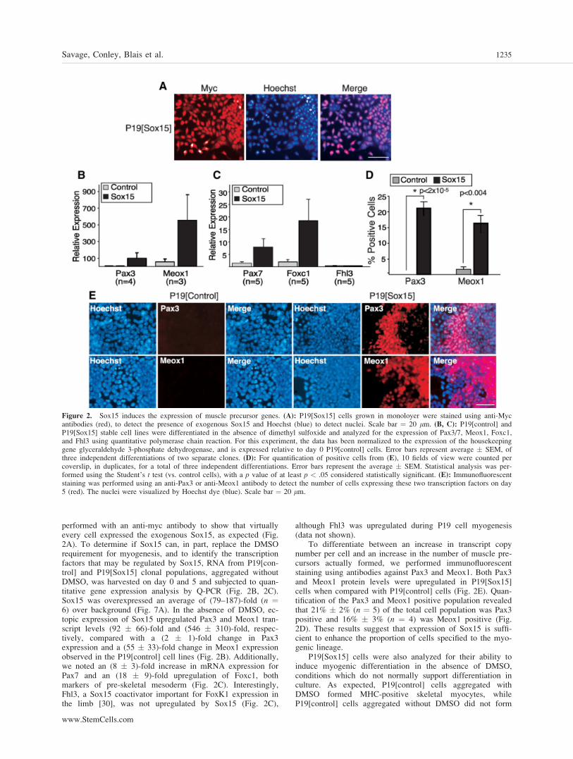
performed with an anti-myc antibody to show that virtuallyevery cell expressed the exogenous Sox15, as expected (Fig.2A). To determine if Sox15 can, in part, replace the DMSOrequirement for myogenesis, and to identify the transcriptionfactors that may be regulated by Sox15, RNA from P19[con-trol] and P19[Sox15] clonal populations, aggregated withoutDMSO, was harvested on day 0 and 5 and subjected to quan-titative gene expression analysis by Q-PCR (Fig. 2B, 2C).Sox15 was overexpressed an average of (79–187)-fold (n ¼6) over background (Fig. 7A). In the absence of DMSO, ec-topic expression of Sox15 upregulated Pax3 and Meox1 tran-script levels (92 � 66)-fold and (546 � 310)-fold, respec-tively, compared with a (2 � 1)-fold change in Pax3expression and a (55 � 33)-fold change in Meox1 expressionobserved in the P19[control] cell lines (Fig. 2B). Additionally,we noted an (8 � 3)-fold increase in mRNA expression forPax7 and an (18 � 9)-fold upregulation of Foxc1, bothmarkers of pre-skeletal mesoderm (Fig. 2C). Interestingly,Fhl3, a Sox15 coactivator important for FoxK1 expression inthe limb [30], was not upregulated by Sox15 (Fig. 2C),
although Fhl3 was upregulated during P19 cell myogenesis(data not shown).
To differentiate between an increase in transcript copynumber per cell and an increase in the number of muscle pre-cursors actually formed, we performed immunofluorescentstaining using antibodies against Pax3 and Meox1. Both Pax3and Meox1 protein levels were upregulated in P19[Sox15]cells when compared with P19[control] cells (Fig. 2E). Quan-tification of the Pax3 and Meox1 positive population revealedthat 21% � 2% (n ¼ 5) of the total cell population was Pax3positive and 16% � 3% (n ¼ 4) was Meox1 positive (Fig.2D). These results suggest that expression of Sox15 is suffi-cient to enhance the proportion of cells specified to the myo-genic lineage.
P19[Sox15] cells were also analyzed for their ability toinduce myogenic differentiation in the absence of DMSO,conditions which do not normally support differentiation inculture. As expected, P19[control] cells aggregated withDMSO formed MHC-positive skeletal myocytes, whileP19[control] cells aggregated without DMSO did not form
Figure 2. Sox15 induces the expression of muscle precursor genes. (A): P19[Sox15] cells grown in monoloyer were stained using anti-Mycantibodies (red), to detect the presence of exogenous Sox15 and Hoechst (blue) to detect nuclei. Scale bar ¼ 20 lm. (B, C): P19[control] andP19[Sox15] stable cell lines were differentiated in the absence of dimethyl sulfoxide and analyzed for the expression of Pax3/7, Meox1, Foxc1,and Fhl3 using quantitative polymerase chain reaction. For this experiment, the data has been normalized to the expression of the housekeepinggene glyceraldehyde 3-phosphate dehydrogenase, and is expressed relative to day 0 P19[control] cells. Error bars represent average � SEM, ofthree independent differentiations of two separate clones. (D): For quantification of positive cells from (E), 10 fields of view were counted percoverslip, in duplicates, for a total of three independent differentiations. Error bars represent the average � SEM. Statistical analysis was per-formed using the Student’s t test (vs. control cells), with a p value of at least p < .05 considered statistically significant. (E): Immunofluorescentstaining was performed using an anti-Pax3 or anti-Meox1 antibody to detect the number of cells expressing these two transcription factors on day5 (red). The nuclei were visualized by Hoechst dye (blue). Scale bar ¼ 20 lm.
Savage, Conley, Blais et al. 1235
www.StemCells.com
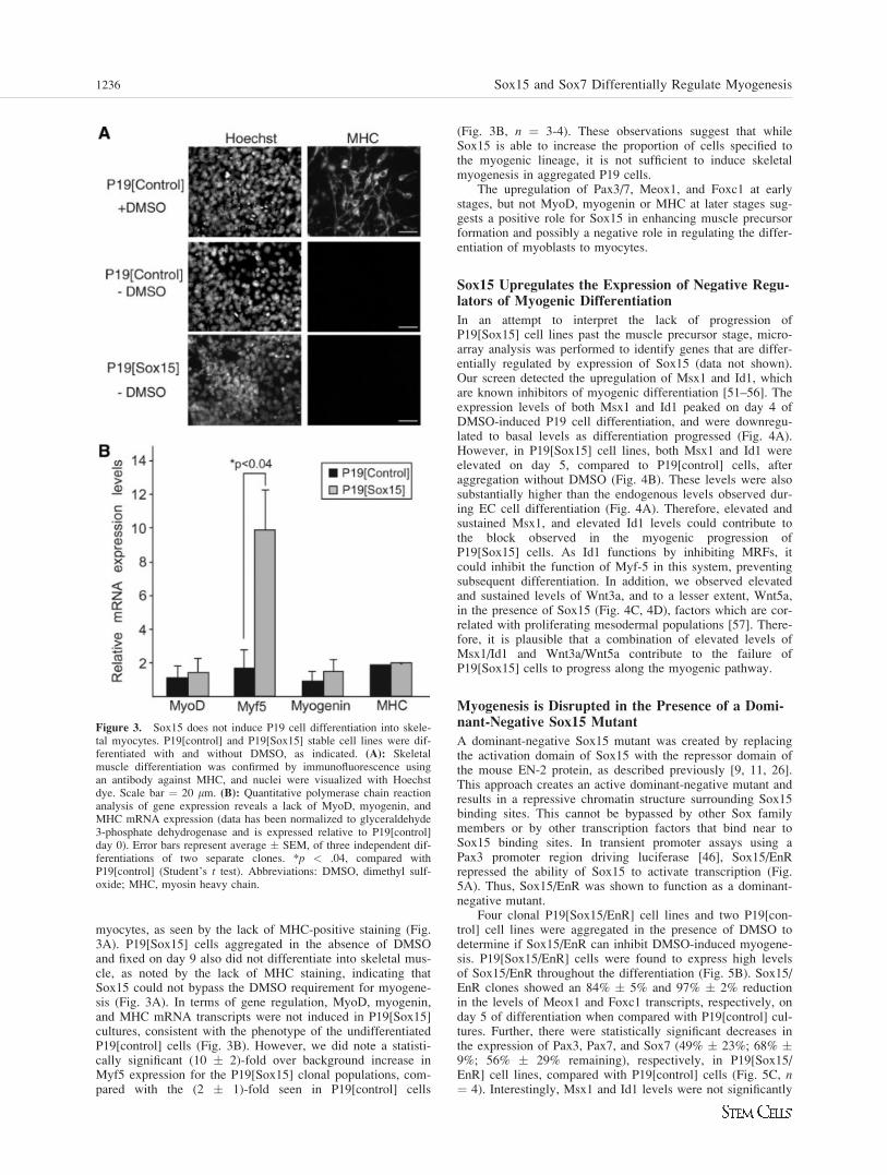
myocytes, as seen by the lack of MHC-positive staining (Fig.3A). P19[Sox15] cells aggregated in the absence of DMSOand fixed on day 9 also did not differentiate into skeletal mus-cle, as noted by the lack of MHC staining, indicating thatSox15 could not bypass the DMSO requirement for myogene-sis (Fig. 3A). In terms of gene regulation, MyoD, myogenin,and MHC mRNA transcripts were not induced in P19[Sox15]cultures, consistent with the phenotype of the undifferentiatedP19[control] cells (Fig. 3B). However, we did note a statisti-cally significant (10 � 2)-fold over background increase inMyf5 expression for the P19[Sox15] clonal populations, com-pared with the (2 � 1)-fold seen in P19[control] cells
(Fig. 3B, n ¼ 3-4). These observations suggest that whileSox15 is able to increase the proportion of cells specified tothe myogenic lineage, it is not sufficient to induce skeletalmyogenesis in aggregated P19 cells.
The upregulation of Pax3/7, Meox1, and Foxc1 at earlystages, but not MyoD, myogenin or MHC at later stages sug-gests a positive role for Sox15 in enhancing muscle precursorformation and possibly a negative role in regulating the differ-entiation of myoblasts to myocytes.
Sox15 Upregulates the Expression of Negative Regu-lators of Myogenic Differentiation
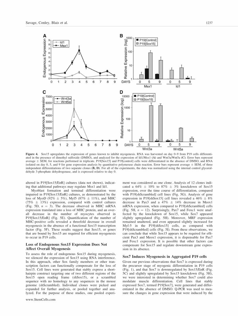
In an attempt to interpret the lack of progression ofP19[Sox15] cell lines past the muscle precursor stage, micro-array analysis was performed to identify genes that are differ-entially regulated by expression of Sox15 (data not shown).Our screen detected the upregulation of Msx1 and Id1, whichare known inhibitors of myogenic differentiation [51–56]. Theexpression levels of both Msx1 and Id1 peaked on day 4 ofDMSO-induced P19 cell differentiation, and were downregu-lated to basal levels as differentiation progressed (Fig. 4A).However, in P19[Sox15] cell lines, both Msx1 and Id1 wereelevated on day 5, compared to P19[control] cells, afteraggregation without DMSO (Fig. 4B). These levels were alsosubstantially higher than the endogenous levels observed dur-ing EC cell differentiation (Fig. 4A). Therefore, elevated andsustained Msx1, and elevated Id1 levels could contribute tothe block observed in the myogenic progression ofP19[Sox15] cells. As Id1 functions by inhibiting MRFs, itcould inhibit the function of Myf-5 in this system, preventingsubsequent differentiation. In addition, we observed elevatedand sustained levels of Wnt3a, and to a lesser extent, Wnt5a,in the presence of Sox15 (Fig. 4C, 4D), factors which are cor-related with proliferating mesodermal populations [57]. There-fore, it is plausible that a combination of elevated levels ofMsx1/Id1 and Wnt3a/Wnt5a contribute to the failure ofP19[Sox15] cells to progress along the myogenic pathway.
Myogenesis is Disrupted in the Presence of a Domi-nant-Negative Sox15 Mutant
A dominant-negative Sox15 mutant was created by replacingthe activation domain of Sox15 with the repressor domain ofthe mouse EN-2 protein, as described previously [9, 11, 26].This approach creates an active dominant-negative mutant andresults in a repressive chromatin structure surrounding Sox15binding sites. This cannot be bypassed by other Sox familymembers or by other transcription factors that bind near toSox15 binding sites. In transient promoter assays using aPax3 promoter region driving luciferase [46], Sox15/EnRrepressed the ability of Sox15 to activate transcription (Fig.5A). Thus, Sox15/EnR was shown to function as a dominant-negative mutant.
Four clonal P19[Sox15/EnR] cell lines and two P19[con-trol] cell lines were aggregated in the presence of DMSO todetermine if Sox15/EnR can inhibit DMSO-induced myogene-sis. P19[Sox15/EnR] cells were found to express high levelsof Sox15/EnR throughout the differentiation (Fig. 5B). Sox15/EnR clones showed an 84% � 5% and 97% � 2% reductionin the levels of Meox1 and Foxc1 transcripts, respectively, onday 5 of differentiation when compared with P19[control] cul-tures. Further, there were statistically significant decreases inthe expression of Pax3, Pax7, and Sox7 (49% � 23%; 68% �9%; 56% � 29% remaining), respectively, in P19[Sox15/EnR] cell lines, compared with P19[control] cells (Fig. 5C, n¼ 4). Interestingly, Msx1 and Id1 levels were not significantly
Figure 3. Sox15 does not induce P19 cell differentiation into skele-tal myocytes. P19[control] and P19[Sox15] stable cell lines were dif-ferentiated with and without DMSO, as indicated. (A): Skeletalmuscle differentiation was confirmed by immunofluorescence usingan antibody against MHC, and nuclei were visualized with Hoechstdye. Scale bar ¼ 20 lm. (B): Quantitative polymerase chain reactionanalysis of gene expression reveals a lack of MyoD, myogenin, andMHC mRNA expression (data has been normalized to glyceraldehyde3-phosphate dehydrogenase and is expressed relative to P19[control]day 0). Error bars represent average � SEM, of three independent dif-ferentiations of two separate clones. *p < .04, compared withP19[control] (Student’s t test). Abbreviations: DMSO, dimethyl sulf-oxide; MHC, myosin heavy chain.
1236 Sox15 and Sox7 Differentially Regulate Myogenesis
altered in P19[Sox15/EnR] cultures (data not shown), indicat-ing that additional pathways may regulate Msx1 and Id1.
Myoblast formation and terminal differentiation wereimpaired in P19[Sox15/EnR] cultures, as demonstrated by theloss of MyoD (92% � 5%), Myf5 (87% � 11%), and MHC(75% � 13%) expression, compared with control cultures(Fig. 5D, n ¼ 3). The decrease observed in MHC mRNAexpression translated into a loss of MHC protein, and an over-all decrease in the number of myocytes observed inP19[Sox15/EnR] (Fig. 5E). Quantification of the number ofMHC-positive cells revealed a threefold decrease in overallmyogenesis in the absence of a functional Sox15 transcriptionfactor (Fig. 5F). These results suggest that Sox15, or genesthat are bound by Sox15 are required for efficient myogenesisto occur in P19 cells.
Loss of Endogenous Sox15 Expression Does NotAffect Overall Myogenesis
To assess the role of endogenous Sox15 during myogenesis,we silenced the expression of Sox15 using RNA interference.In this approach, other Sox family members or other tran-scription factors can functionally compensate for the loss ofSox15. Cell lines were generated that stably express a short-hairpin construct targeting one of two different regions of theSox15 open reading frame (shSox15), or a scrambledsequence with no homology to any sequences in the mousegenome (shScrambled). Individual clones were picked andexpanded for further analysis, or pooled together and ana-lyzed. For the purpose of these studies, one pooled experi-
ment was considered as one clone. Analysis of 12 clones indi-cated a 64% � 10% to 87% � 3% knockdown of Sox15expression, over the time course of differentiation, comparedwith P19[shScrambled] cell lines (Fig. 5G). Analysis of geneexpression in P19[shSox15] cell lines revealed a 46% � 8%decrease in Pax3 and a 47% � 14% decrease in Meox1mRNA expression, when compared to P19[shScrambled] cells(Fig. 5H, n ¼ 12). Surprisingly, Pax7 and Foxc1 were unaf-fected by the knockdown of Sox15, while Sox7 appearedslightly upregulated (Fig. 5H). Moreover, MRF expressionremained unaltered, and even appeared slightly increased forMyf-5 in the P19[shSox15] cells, as compared withP19[shScrambled] cells (Fig. 5I). From these observations, wecan conclude that while Sox15 appears to be required for effi-cient Pax3 and Meox1 expression, it is dispensable for Pax7and Foxc1 expression. It is possible that other factors cancompensate for Sox15 and regulate downstream gene expres-sion in its absence.
Sox7 Induces Myogenesis in Aggregated P19 cells
Given our previous observations that Sox7 is expressed duringthe precursor stage of myogenic differentiation in P19 cells(Fig. 1), and that Sox7 is downregulated by Sox15/EnR (Fig.5C) and slightly upregulated by Sox15 knockdown (Fig. 5H),we were interested in determining whether Sox7 could alsomodulate muscle differentiation. Cell lines that stablyexpressed Sox7, termed P19[Sox7], were generated and differ-entiated in the absence of DMSO. Q-PCR was used to mea-sure the changes in gene expression that were induced by the
Figure 4. Sox15 upregulates the expression of genes known to inhibit myogenesis. RNA was harvested on day 0–9 from P19 cells differenti-ated in the presence of dimethyl sulfoxide (DMSO), and analyzed for the expression of Id1/Msx1 (A) and Wnt3a/Wnt5a (C). Error bars representaverage � SEM, for reactions performed in triplicate. P19[Sox15] and P19[control] cells were differentiated in the absence of DMSO, and RNAisolated on day 0, 5, and 9 for gene expression analysis by quantitative polymerase chain reaction. Error bars represent average � SEM, of threeindependent differentiations of two separate clones (B, D). For all of the experiments, the data was normalized using the internal control glyceral-dehyde 3-phosphate dehydrogenase, and is expressed relative to day 0.
Savage, Conley, Blais et al. 1237
www.StemCells.com
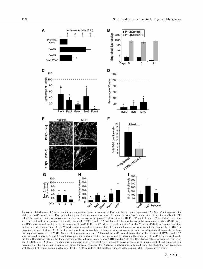
Figure 5. Interference of Sox15 function and expression causes a decrease in Pax3 and Meox1 gene expression. (A): Sox15/EnR repressed theability of Sox15 to activate a Pax3 promoter region. Pax3-luciferase was transfected alone or with Sox15 and/or Sox15/EnR, transiently into P19cells. The resulting luciferase activity was expressed relative to the promoter alone (n ¼ 4). (B–F): P19[control] and P19[Sox15/EnR] cell lineswere differentiated in the presence of dimethyl sulfoxide (DMSO) and RNA was harvested for quantitative polymerase chain reaction (PCR) analy-sis. RNA was isolated on day 5 for the detection of Sox15/EnR, Pax3/7, Meox1, Foxc1, and Sox7 on day 9 for Sox15/EnR, myogenic regulatoryfactors, and MHC expression (B–D). Myocytes were detected in these cell lines by immunofluorescence using an antibody against MHC (E). Thepercentage of cells that was MHC-positive was quantified by counting 10 fields of view per coverslip from two independent differentiations. Errorbars represent average � SEM (F). Stable cell lines expressing shRNA targeted to Sox15 were differentiated in the presence of DMSO, and RNAwas harvested on day 0, 5, and 9. Quantitative polymerase chain reaction was performed to determine the efficiency of Sox15 knockdown through-out the differentiation (G) and for the expression of the indicated genes on day 5 (H) and day 9 (I) of differentiation. The error bars represent aver-age � SEM, n ¼ 12 clones. The data was normalized using glyceraldehyde 3-phosphate dehydrogenase as an internal control and expressed as apercentage of the expression in control cell lines, for each respective day. Statistical analysis was performed using the Student’s t test (comparedwith the control group), with a p value of at least p < .05 considered statistically significant. Abbreviation: MHC, myosin heavy chain.
1238 Sox15 and Sox7 Differentially Regulate Myogenesis
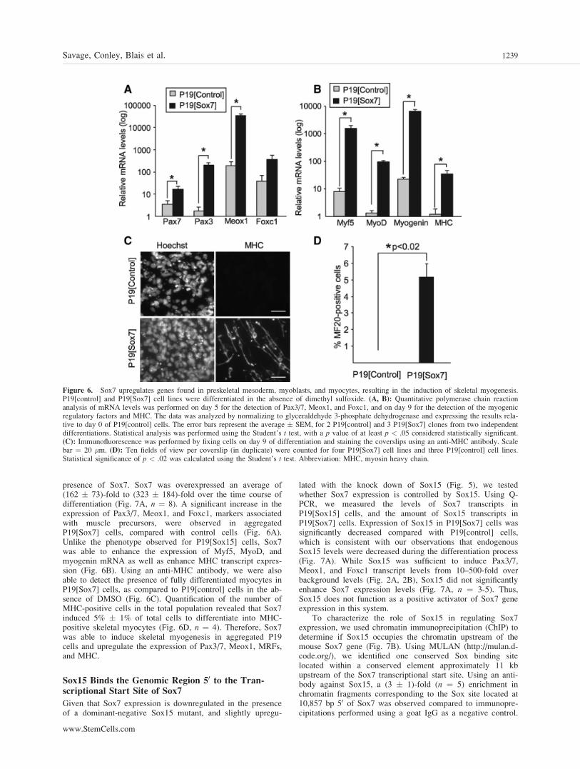
presence of Sox7. Sox7 was overexpressed an average of(162 � 73)-fold to (323 � 184)-fold over the time course ofdifferentiation (Fig. 7A, n ¼ 8). A significant increase in theexpression of Pax3/7, Meox1, and Foxc1, markers associatedwith muscle precursors, were observed in aggregatedP19[Sox7] cells, compared with control cells (Fig. 6A).Unlike the phenotype observed for P19[Sox15] cells, Sox7was able to enhance the expression of Myf5, MyoD, andmyogenin mRNA as well as enhance MHC transcript expres-sion (Fig. 6B). Using an anti-MHC antibody, we were alsoable to detect the presence of fully differentiated myocytes inP19[Sox7] cells, as compared to P19[control] cells in the ab-sence of DMSO (Fig. 6C). Quantification of the number ofMHC-positive cells in the total population revealed that Sox7induced 5% � 1% of total cells to differentiate into MHC-positive skeletal myocytes (Fig. 6D, n ¼ 4). Therefore, Sox7was able to induce skeletal myogenesis in aggregated P19cells and upregulate the expression of Pax3/7, Meox1, MRFs,and MHC.
Sox15 Binds the Genomic Region 50 to the Tran-scriptional Start Site of Sox7
Given that Sox7 expression is downregulated in the presenceof a dominant-negative Sox15 mutant, and slightly upregu-
lated with the knock down of Sox15 (Fig. 5), we testedwhether Sox7 expression is controlled by Sox15. Using Q-PCR, we measured the levels of Sox7 transcripts inP19[Sox15] cells, and the amount of Sox15 transcripts inP19[Sox7] cells. Expression of Sox15 in P19[Sox7] cells wassignificantly decreased compared with P19[control] cells,which is consistent with our observations that endogenousSox15 levels were decreased during the differentiation process(Fig. 7A). While Sox15 was sufficient to induce Pax3/7,Meox1, and Foxc1 transcript levels from 10–500-fold overbackground levels (Fig. 2A, 2B), Sox15 did not significantlyenhance Sox7 expression levels (Fig. 7A, n ¼ 3-5). Thus,Sox15 does not function as a positive activator of Sox7 geneexpression in this system.
To characterize the role of Sox15 in regulating Sox7expression, we used chromatin immunoprecipitation (ChIP) todetermine if Sox15 occupies the chromatin upstream of themouse Sox7 gene (Fig. 7B). Using MULAN (http://mulan.d-code.org/), we identified one conserved Sox binding sitelocated within a conserved element approximately 11 kbupstream of the Sox7 transcriptional start site. Using an anti-body against Sox15, a (3 � 1)-fold (n ¼ 5) enrichment inchromatin fragments corresponding to the Sox site located at10,857 bp 50 of Sox7 was observed compared to immunopre-cipitations performed using a goat IgG as a negative control.
Figure 6. Sox7 upregulates genes found in preskeletal mesoderm, myoblasts, and myocytes, resulting in the induction of skeletal myogenesis.P19[control] and P19[Sox7] cell lines were differentiated in the absence of dimethyl sulfoxide. (A, B): Quantitative polymerase chain reactionanalysis of mRNA levels was performed on day 5 for the detection of Pax3/7, Meox1, and Foxc1, and on day 9 for the detection of the myogenicregulatory factors and MHC. The data was analyzed by normalizing to glyceraldehyde 3-phosphate dehydrogenase and expressing the results rela-tive to day 0 of P19[control] cells. The error bars represent the average � SEM, for 2 P19[control] and 3 P19[Sox7] clones from two independentdifferentiations. Statistical analysis was performed using the Student’s t test, with a p value of at least p < .05 considered statistically significant.(C): Immunofluorescence was performed by fixing cells on day 9 of differentiation and staining the coverslips using an anti-MHC antibody. Scalebar ¼ 20 lm. (D): Ten fields of view per coverslip (in duplicate) were counted for four P19[Sox7] cell lines and three P19[control] cell lines.Statistical significance of p < .02 was calculated using the Student’s t test. Abbreviation: MHC, myosin heavy chain.
Savage, Conley, Blais et al. 1239
www.StemCells.com
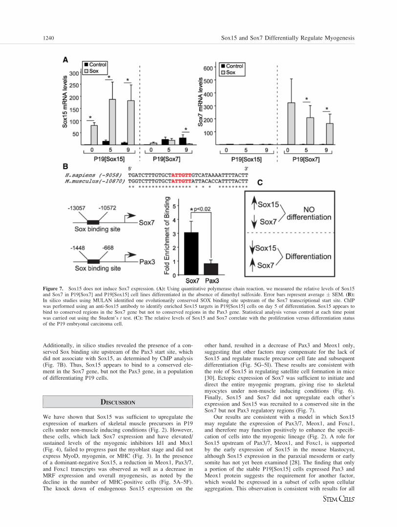
Additionally, in silico studies revealed the presence of a con-served Sox binding site upstream of the Pax3 start site, whichdid not associate with Sox15, as determined by ChIP analysis(Fig. 7B). Thus, Sox15 appears to bind to a conserved ele-ment in the Sox7 gene, but not the Pax3 gene, in a populationof differentiating P19 cells.
DISCUSSION
We have shown that Sox15 was sufficient to upregulate theexpression of markers of skeletal muscle precursors in P19cells under non-muscle inducing conditions (Fig. 2). However,these cells, which lack Sox7 expression and have elevated/sustained levels of the myogenic inhibitors Id1 and Msx1(Fig. 4), failed to progress past the myoblast stage and did notexpress MyoD, myogenin, or MHC (Fig. 3). In the presenceof a dominant-negative Sox15, a reduction in Meox1, Pax3/7,and Foxc1 transcripts was observed as well as a decrease inMRF expression and overall myogenesis, as noted by thedecline in the number of MHC-positive cells (Fig. 5A–5F).The knock down of endogenous Sox15 expression on the
other hand, resulted in a decrease of Pax3 and Meox1 only,suggesting that other factors may compensate for the lack ofSox15 and regulate muscle precursor cell fate and subsequentdifferentiation (Fig. 5G–5I). These results are consistent withthe role of Sox15 in regulating satellite cell formation in mice[30]. Ectopic expression of Sox7 was sufficient to initiate anddirect the entire myogenic program, giving rise to skeletalmyocytes under non-muscle inducing conditions (Fig. 6).Finally, Sox15 and Sox7 did not upregulate each other’sexpression and Sox15 was recruited to a conserved site in theSox7 but not Pax3 regulatory regions (Fig. 7).
Our results are consistent with a model in which Sox15may regulate the expression of Pax3/7, Meox1, and Foxc1,and therefore may function positively to enhance the specifi-cation of cells into the myogenic lineage (Fig. 2). A role forSox15 upstream of Pax3/7, Meox1, and Foxc1, is supportedby the early expression of Sox15 in the mouse blastocyst,although Sox15 expression in the paraxial mesoderm or earlysomite has not yet been examined [28]. The finding that onlya portion of the stable P19[Sox15] cells expressed Pax3 andMeox1 protein suggests the requirement for another factor,which would be expressed in a subset of cells upon cellularaggregation. This observation is consistent with results for all
Figure 7. Sox15 does not induce Sox7 expression. (A): Using quantitative polymerase chain reaction, we measured the relative levels of Sox15and Sox7 in P19[Sox7] and P19[Sox15] cell lines differentiated in the absence of dimethyl sulfoxide. Error bars represent average � SEM. (B):In silico studies using MULAN identified one evolutionarily conserved SOX binding site upstream of the Sox7 transcriptional start site. ChIPwas performed using an anti-Sox15 antibody to identify enriched Sox15 targets in P19[Sox15] cells on day 5 of differentiation. Sox15 appears tobind to conserved regions in the Sox7 gene but not to conserved regions in the Pax3 gene. Statistical analysis versus control at each time pointwas carried out using the Student’s t test. (C): The relative levels of Sox15 and Sox7 correlate with the proliferation versus differentiation statusof the P19 embryonal carcinoma cell.
1240 Sox15 and Sox7 Differentially Regulate Myogenesis
of the other stable P19 cell lines tested, including MyoD,Pax3, ß-catenin, and Gli2 [9, 11, 26, 43]. It is also consistentwith the idea that factors, including Sox15, may have oppos-ing functions, such as maintaining the stem cell phenotypeand enhancing differentiation, depending on the cellular con-text in which they are found. For example, Oct4 and Wnt3acan perform both functions, depending on their context [26,58, 59]. It is likely that each transcription factor is influencedby the combination of factors, the signaling pathways, and thecurrent chromatin structure available in each cell type.
In agreement with previous results in C2C12 myoblasts[32], we observed that P19 cells with high levels of Sox15failed to progress past the precursor/myoblast stage (Fig. 3).Further, P19[Sox15] cell lines did not enhance Sox7 expressionlevels, and knock down of Sox15 slightly upregulated Sox7(Fig. 7). It is possible that Sox15 is acting as a repressor ofSox7 expression under these circumstances. The loss of Sox7in the P19[Sox15/EnR] cells (Fig. 5) is consistent with ourfinding that Sox15 may bind to the Sox7 regulatory sequences,shown by the ChIP assay (Fig. 7B), but it does not indicatewhether Sox15 activates or represses Sox7 expression. Thus,cell lines expressing high levels of Sox15 and low levels ofSox7 fail to terminally differentiate, whereas cell lines express-ing low levels of Sox15 and high levels of Sox7 exhibit normalmyogenesis (Fig. 7C). Although we have established thatSox15 does not activate Sox7 expression, a more detailed ex-amination of the Sox7 regulatory sequences and the role ofSox15 is required to confirm if Sox15 represses Sox7.
Our finding that Sox15 knock down results in the loss ofPax3 and Meox1 expression, but not overall myogenesis (Fig.5G–5I), is consistent with the phenotype of mice lackingSox15. These mice develop relatively normal muscle but lackefficient satellite cell formation [30] and efficient adult muscleregeneration, shown by a loss of MyoD but not Myf-5 expres-sion [30, 33]. As Pax3 is an important determinant of satellitecell formation and MyoD regulation, our results in P19 cellsare consistent with the Sox15�/� mouse phenotype [5, 13].Interestingly, embryonic expression of Meox1 or Pax3 wasnot examined in Sox15�/� mice [33]. We would predict adownregulation of Pax3 and Meox1 expression in the devel-oping myotome.
Gene expression analysis revealed that Wnt3a expressionis elevated and sustained in P19[Sox15] cell lines (Fig. 4),and that these cells failed to progress through the myogenicpathway. Interestingly, a constitutively active ß-cateninexpressed in satellite cells caused an increase in the propor-tion of cells expressing Pax7, without a concomitant increasein MyoD [57]. These results suggest that ß-catenin promotesself-renewal of muscle precursors, with fewer cells under-going myogenesis. It is tempting to speculate that the elevatedlevels of Wnt3a observed in our system are contributing tothe maintenance of muscle precursors at the expense ofdifferentiation.
Our finding that Sox15 failed to induce myogenesis inP19[Sox15] cells aggregated in the absence of DMSO (Fig. 3)is consistent with previous findings showing that ectopicexpression of Sox15 is not sufficient to initiate the myogenicprogram [33]. Stable expression of Sox15 in 3T3 fibroblastsdid not result in positive MHC staining, or any detectableMyoD/Myf5 expression. In other studies, ectopic expressionof Sox15 in C2C12 proliferating myoblasts antagonized dif-ferentiation and decreased MyoD expression, whereas a C-ter-minal truncation of Sox15 showed no adverse effects on myo-genesis [32]. Finally, Sox15 expression decreased duringmyoblast differentiation. Therefore, in these studies, increasedlevels of Sox15 appeared inhibitory toward myoblast mainte-nance and differentiation [32].
Similarly, in our experiments, Sox15 seemed to inhibitMyoD expression, since an increase in Pax3 expression didnot result in a concomitant increase in MyoD expression(Figs. 2, 3), as would be predicted from the phenotype ofP19[Pax3] cells [11]. Our results support a model in whichSox15 enhances Pax3 and Meox1 expression during musclespecification and then inhibits myoblast maintenance and/ordifferentiation if not subsequently downregulated. A dual pos-itive/negative function for stem cell transcription factors isalso observed during melanocyte stem cell differentiation. Inthis case, Pax3 acts early to activate expression of Mitf, whilesimultaneously inhibiting expression of melanin-synthesizinggenes. These cells are thus committed, yet will not differenti-ate until the Pax3-mediated inhibition is relieved [60]. Simi-larly, upregulation of Id1 and Msx1 (Fig. 4), may contributeto the lack of differentiation in P19[Sox15] cells. In summary,the lack of upregulation of MyoD in this study may be due tothe presence of inhibitors of differentiation, such as Id1 andMsx1, and/or the absence of an essential activator, such asSox7.
As knock down of Sox15 resulted in a decrease of Meox1and Pax3 expression (Fig. 5), it is possible that these twogenes are immediate downstream targets of Sox15. Attemptsto determine Pax3 and Meox1 promoter occupancy by Sox15using chromatin immunoprecipitation (ChIP) were unsuccess-ful (Fig. 7, data not shown). In this case, Pax3 or Meox1 arelikely indirect targets, although regions outside the 25 kb pro-moter region were not examined. It is possible that othermembers of the Sox family may be regulating Pax3 directly,given the conserved cis-acting site identified, and given thatSox15 is able to activate the Pax3 promoter (Fig. 5). Severalother Sox factors are upregulated during P19 cell myogenesis,including Sox8, Sox11, and Sox17, which may bind Pax3/Meox1 directly. In contrast, Sox7 appears to be a direct targetof Sox15 and expression analysis suggests that Sox15 is not apositive regulator of Sox7 expression. Further studies arerequired to elucidate the network of direct and indirect factorsregulated by Sox15 and Sox7, and other family members,ideally using high throughput methods.
SUMMARY
In summary, we have provided evidence that Sox15 and Sox7may be regulators of early muscle precursor cell fate by func-tioning as upstream regulators of Pax3/7, Meox1, and Foxc1expression. This regulation may enhance the specification ofcells into the muscle lineage. Furthermore, Sox7, but notSox15, regulates myoblast formation and differentiation byupregulating MRF and MHC expression. Thus, we proposedifferential roles for Sox15 and Sox7 during myogenesis.These findings could have implications for future stem celltherapy, which requires the ability to control differentiationversus proliferation and efficient satellite cell repopulation.
ACKNOWLEDGMENTS
We wish to thank Dr. Ashraf Al-Madhoun, Jennifer Dawson,Virja Mehta, and Anastassia Voronova for critically reading themanuscript. We acknowledge the technical assistance of SophieBoisvenue and Flavia Sendi-Mukasa. We are grateful to Dr.Alain Stintzi for his expertise and assistance with quantitativepolymerase chain reaction analysis. We thank Dr. Valerie Wal-lace for helpful discussions. J.S. was supported by a NSERC
Savage, Conley, Blais et al. 1241
www.StemCells.com
studentship as well as a Canadian Institute of Health DoctoralResearch Award (Institute of Aging). I.S.S. was supported by aCanadian Institute of Aging Investigator Award. This work wassupported by grant MOP-84458 (to I.S.S.) from the CanadianInstitutes of Health Research.
DISCLOSURE OF POTENTIAL CONFLICTS
OF INTEREST
The authors indicate no potential conflicts of interest.
REFERENCES
1 Cossu G, Borello U. Wnt signaling and the activation of myogenesisin mammals. EMBO J 1999;18:6867–6872.
2 Currie PD, Ingham PW. The generation and interpretation of posi-tional information within the vertebrate myotome. Mech Dev 1998;73:3–21.
3 Tajbakhsh S, Cossu G. Establishing myogenic identity during somi-togenesis. Curr Opin Genet Dev 1997;7:634–641.
4 Arnold HH, Braun T. Genetics of muscle determination and develop-ment. Curr Top Dev Biol 2000;48:129–164.
5 Relaix F, Montarras D, Zaffran S et al. Pax3 and Pax7 have distinctand overlapping functions in adult muscle progenitor cells. J CellBiol 2006;172:91–102.
6 Laclef C, Hamard G, Demignon J et al. Altered myogenesis in Six1-deficient mice. Development 2003;130:2239–2252.
7 Candia AF, Hu J, Crosby J et al. Mox-1 and Mox-2 define a novelhomeobox gene subfamily and are differentially expressed duringearly mesodermal patterning in mouse embryos. Development 1992;116:1123–1136.
8 Mankoo BS, Skuntz S, Harrigan I et al. The concerted action ofMeox homeobox genes is required upstream of genetic pathwaysessential for the formation, patterning and differentiation of somites.Development 2003;130:4655–4664.
9 Petropoulos H, Gianakopoulos PJ, Ridgeway AG et al. Disruption ofMeox or Gli activity ablates skeletal myogenesis in P19 cells. J BiolChem 2004;279:23874–23881.
10 Maroto M, Reshef R, Munsterberg AE et al. Ectopic Pax-3 activatesMyoD and Myf-5 expression in embryonic mesoderm and neural tis-sue. Cell 1997;89:139–148.
11 Ridgeway AG, Skerjanc IS. Pax3 is essential for skeletal myogenesisand the expression of Six1 and Eya2. J Biol Chem 2001;276:19033–19039.
12 Borycki AG, Mendham L, Emerson CP Jr. Control of somite pat-terning by Sonic hedgehog and its downstream signal responsegenes. Development 1998;125:777–790.
13 Buckingham M, Relaix F. The role of Pax genes in the developmentof tissues and organs: Pax3 and Pax7 regulate muscle progenitor cellfunctions. Annu Rev Cell Dev Biol 2007;23:645–673.
14 Goulding M, Lumsden A, Paquette AJ. Regulation of Pax-3 expres-sion in the dermomyotome and its role in muscle development. De-velopment 1994;120:957–971.
15 Williams BA, Ordahl CP. Pax-3 expression in segmental mesodermmarks early stages in myogenic cell specification. Development1994;120:785–796.
16 Relaix F, Rocancourt D, Mansouri A et al. Divergent functions ofmurine Pax3 and Pax7 in limb muscle development. Genes Dev2004;18:1088–1105.
17 Reshef R, Maroto M, Lassar AB. Regulation of dorsal somitic cellfates: BMPs and Noggin control the timing and pattern of myogenicregulator expression. Genes Dev 1998;12:290–303.
18 Skerjanc IS. Cardiac and skeletal muscle development in P19 embry-onal carcinoma cells. Trends Cardiovasc Med 1999;9:139–143.
19 Bajard L, Relaix F, Lagha M et al. A novel genetic hierarchy func-tions during hypaxial myogenesis: Pax3 directly activates Myf5 inmuscle progenitor cells in the limb. Genes Dev 2006;20:2450–2464.
20 Relaix F, Rocancourt D, Mansouri A et al. A Pax3/Pax7-dependentpopulation of skeletal muscle progenitor cells. Nature 2005;435:948–953.
21 Gros J, Manceau M, Thome V et al. A common somitic origin forembryonic muscle progenitors and satellite cells. Nature 2005;435:954–958.
22 Candia AF, Wright CV. Differential localization of Mox-1 and Mox-2 proteins indicates distinct roles during development. Int J DevBiol 1996;40:1179–1184.
23 Capdevila J, Tabin C, Johnson RL. Control of dorsoventral somitepatterning by Wnt-1 and beta-catenin. Dev Biol 1998;193:182–194.
24 Wagner J, Schmidt C, Nikowits W Jr et al. Compartmentalization ofthe somite and myogenesis in chick embryos are influenced by wntexpression. Dev Biol 2000;228:86–94.
25 Fan CM, Lee CS, Tessier-Lavigne M. A role for WNT proteins ininduction of dermomyotome. Dev Biol 1997;191:160–165.
26 Petropoulos H, Skerjanc IS. Beta-catenin is essential and sufficientfor skeletal myogenesis in P19 cells. J Biol Chem 2002;277:15393–15399.
27 Schmidt K, Glaser G, Wernig A et al. Sox8 is a specific marker formuscle satellite cells and inhibits myogenesis. J Biol Chem 2003;278:29769–29775.
28 Yoshikawa T, Piao Y, Zhong J et al. High-throughput screen forgenes predominantly expressed in the ICM of mouse blastocysts bywhole mount in situ hybridization. Gene Expr Patterns 2006;6:213–224.
29 Sarraj MA, Wilmore HP, McClive PJ et al. Sox15 is up regulated inthe embryonic mouse testis. Gene Expr Patterns 2003;3:413–417.
30 Meeson AP, Shi X, Alexander MS et al. Sox15 and Fhl3 transcrip-tionally coactivate Foxk1 and regulate myogenic progenitor cells.EMBO J 2007;26:1902–1912.
31 Maruyama M, Ichisaka T, Nakagawa M et al. Differential roles forSox15 and Sox2 in transcriptional control in mouse embryonic stemcells. J Biol Chem 2005;280:24371–24379.
32 Beranger F, Mejean C, Moniot B et al. Muscle differentiation isantagonized by SOX15, a new member of the SOX protein family. JBiol Chem 2000;275:16103–16109.
33 Lee HJ, Goring W, Ochs M et al. Sox15 is required for skeletalmuscle regeneration. Mol Cell Biol 2004;24:8428–8436.
34 Shiozawa M, Hiraoka Y, Komatsu N et al. Cloning and characteriza-tion of Xenopus laevis xSox7 cDNA. Biochim Biophys Acta 1996;1309:73–76.
35 Takash W, Canizares J, Bonneaud N et al. SOX7 transcription fac-tor: Sequence, chromosomal localisation, expression, transactivationand interference with Wnt signalling. Nucleic Acids Res 2001;29:4274–4283.
36 Taniguchi K, Hiraoka Y, Ogawa M et al. Isolation and characteriza-tion of a mouse SRY-related cDNA, mSox7. Biochim Biophys Acta1999;1445:225–231.
37 Katoh M. Expression of human SOX7 in normal tissues and tumors.Int J Mol Med 2002;9:363–368.
38 Zhang C, Basta T, Fawcett SR et al. SOX7 is an immediate-earlytarget of VegT and regulates Nodal-related gene expression in Xeno-pus. Dev Biol 2005;278:526–541.
39 Pendeville H, Winandy M, Manfroid I et al. Zebrafish Sox7 andSox18 function together to control arterial-venous identity. Dev Biol2008;317:405–416.
40 Cermenati S, Moleri S, Cimbro S et al. Sox18 and Sox7 play redun-dant roles in vascular development. Blood 2008;111:2657–2666.
41 Herpers R, van de Kamp E, Duckers HJ et al. Redundant roles forsox7 and sox18 in arteriovenous specification in zebrafish. Circ Res2008;102:12–15.
42 Zhang C, Basta T, Klymkowsky MW. SOX7 and SOX18 are essen-tial for cardiogenesis in Xenopus. Dev Dyn 2005;234:878–891.
43 Skerjanc IS, Slack RS, McBurney MW. Cellular aggregation enhan-ces MyoD-directed skeletal myogenesis in embryonal carcinomacells. Mol Cell Biol 1994;14:8451–8459.
44 Ridgeway AG, Petropoulos H, Wilton S et al. Wnt signaling regu-lates the function of MyoD and myogenin. J Biol Chem 2000;275:32398–32405.
45 Skerjanc IS, Petropoulos H, Ridgeway AG et al. Myocyte enhancer fac-tor 2C and Nkx2–5 up-regulate each other’s expression and initiate car-diomyogenesis in P19 cells. J Biol Chem 1998;273:34904–34910.
46 Natoli TA, Ellsworth MK, Wu C et al. Positive and negative DNAsequence elements are required to establish the pattern of Pax3expression. Development 1997;124:617–626.
47 Sambrook J, Fritsch EF, Maniatis T. Molecular Cloning, A LaboratoryManual. New York: Cold Spring Harbor Laboratory Press, 1989.
48 Wilton S, Skerjanc H. Factors in serum regulate muscle developmentin P19 cells. In Vitro Cell Dev Biol Anim 1999;35:175–177.
49 Li C, Hung Wong W. Model-based analysis of oligonucleotidearrays: Model validation, design issues and standard error applica-tion. Genome Biol 2001;2:RESEARCH0032.
50 Yu JY, DeRuiter SL, Turner DL. RNA interference by expression ofshort-interfering RNAs and hairpin RNAs in mammalian cells. ProcNatl Acad Sci USA 2002;99:6047–6052.
51 Thompson-Jaeger S, Raghow R. Exogenous expression of Msx1 ren-ders myoblasts refractory to differentiation into myotubes and elicitsenhanced biosynthesis of four unique mRNAs. Mol Cell Biochem2000;208:63–69.
1242 Sox15 and Sox7 Differentially Regulate Myogenesis
52 Lee H, Habas R, Abate-Shen C. MSX1 cooperates with histone H1bfor inhibition of transcription and myogenesis. Science 2004;304:1675–1678.
53 Miller KA, Barrow J, Collinson JM et al. A highly conservedWnt-dependent TCF4 binding site within the proximal enhancer ofthe anti-myogenic Msx1 gene supports expression within Pax3-expressing limb bud muscle precursor cells. Dev Biol 2007;311:665–678.
54 Bendall AJ, Ding J, Hu G et al. Msx1 antagonizes the myogenic activ-ity of Pax3 in migrating limb muscle precursors. Development 1999;126:4965–4976.
55 Melnikova IN, Bounpheng M, Schatteman GC et al. Differential bio-logical activities of mammalian Id proteins in muscle cells. Exp CellRes 1999;247:94–104.
56 Vinals F, Ventura F. Myogenin protein stability is decreased byBMP-2 through a mechanism implicating Id1. J Biol Chem 2004;279:45766–45772.
57 Perez-Ruiz A, Ono Y, Gnocchi VF et al. beta-Catenin promotes self-renewal of skeletal-muscle satellite cells. J Cell Sci 2008;121:1373–1382.
58 Pan G, Thomson JA. Nanog and transcriptional networks in embry-onic stem cell pluripotency. Cell Res 2007;17:42–49.
59 Zeineddine D, Papadimou E, Chebli K et al. Oct-3/4 dose depend-ently regulates specification of embryonic stem cells toward a car-diac lineage and early heart development. Dev Cell 2006;11:535–546.
60 Lang D, Lu MM, Huang L et al. Pax3 functions at a nodal point inmelanocyte stem cell differentiation. Nature 2005;433:884–887.
See www.StemCells.com for supporting information available online.
Savage, Conley, Blais et al. 1243