emergency department clinical decision unit cdu... · 4/1/2014 page 2 table of contents: contact...
TRANSCRIPT
4/1/2014 Page 1
EMORY UNIVERSITY SCHOOL OF MEDICINE –
DEPARTMENT OF EMERGENCY MEDICINE
2014
EMERGENCY DEPARTMENT
CLINICAL DECISION UNIT EMORY MIDTOWN HOSPITAL
EMORY UNIVERSITY HOSPITAL
GRADY MEMORIAL HOSPITAL

4/1/2014 Page 2
TABLE OF CONTENTS:
CONTACT INFORMATION
GENERAL GUIDELINES FOR CDU OPERATION
GUIDELINES FOR HOLDING PATIENTS IN THE CDU OBSERVATION UNIT QUALITY ASSURANCE PROGRAM
OBSERVATION UNIT UTILIZATION REVIEW PROGRAM
GUIDELINES FOR STRESS TESTING CDU PATIENTS
CMS AND ACEP POLICY ON OBSERVATION SERVICES
CDU STATISTICS
CONDITION SPECIFIC GUIDELINES
4/1/2014 Page 3
CONTACT INFORMATION
Medical Director, Observation Medicine: Michael Ross MD Clinical Decision Units: Emory University:
CDU phone number: 404-712-2908
Medical Director: Michael Ross, MD [email protected] (office: 404-778-2643, PIC # 13951)
Associate Providers: Andrea Brown, NP [email protected]
Sheria Brantley NP [email protected] (office: 404-778-2908, CDU phone: 404-938-0164)
Unit Director, EM : Elaina Hall, RN
Nurse Manager: Sharon Friday, RN sharon.friday @emory.net (office: 404-712-7576; 404-712-2908 )
Emory Midtown:
CDU phone number: 404-686-3154
Medical Director: Anwar Osborne, MD ([email protected] (PIC #17548)
Associate Providers: Barbara Swartzberg, NP [email protected] (office: 404-686-3845, PIC #20393)
Unit Director, EM: Michael Flake, RN ([email protected])
Grady Memorial Hospital:
CDU phone number: (404) 616-6448
Medical Director: Matthew Wheatley, MD ([email protected] Office: 404-616-6419; PIC # 12807)
Associate Providers: Tryphena (Triffy) Pacitti NP [email protected]
VP of nursing Michelle Wallace, RN [email protected]
Unit Director: Mulikat “Lola” Amuda , RN ([email protected])
Mission statement – The Clinical Decision Unit strives to provide high quality patient care to those emergency department patients needing further management to determine their need for inpatient admission. The unit will do this by providing active management of specific conditions using the best available clinical evidence. We will provide this in a setting that is both efficient for health care providers and pleasant for our patients. We strive for the unit to be a nationally recognized center of excellence in this area of patient care, teaching, and research.
Scope of services – The “Clinical Decision Unit”, or CDU, is an emergency department (ED)
observation unit which provides physician and hospital “observation services” as defined by CMS, AMA-CPT, and ACEPs policy on the management of observation units. The unit provides services to emergency patients who require care that goes beyond their initial evaluation and management in the emergency department. The goal of observation is to determine the need for inpatient admission. The scopes of these services are outlined in this document.
Management – The CDU is administratively part of the emergency department (ED) and

4/1/2014 Page 4
therefore it is under the ED nursing and medical administration.
Nursing leadership – The charge nurse for the CDU is supervised by the nursing director of the ED.
Physician leadership – The Medical Director of Observation medicine, who reports to the Chair of the Department of Emergency Medicine, shall oversee the observation medicine service line at all Emory School of Medicine adult emergency departments. Each ED shall have a CDU site director who shall work under the direction of the medical director of observation medicine. CDU Associate Provider leaders will work under the direction of the CDU site director on CDU issues.
Physician accountability - The ED PHYSICIAN WILL ACT AS THE "GATEKEEPER" FOR ALL ADMISSIONS TO THE CDU. THE PHYSICIAN ASSIGNED TO COVER THE CDU IS THE “ACCOUNTABLE” PHYSICIAN FOR ALL CDU PATIENTS. This means that admission to and discharge from the unit can only be made by the ED physician (or His or Her designee). Other services may not “bypass” the ED physician and admit directly to the CDU. However they may admit their patients for observation services to hospital inpatient beds as dictated by hospital policy. Consultants and Private Attendings may recommend discharge or admission; however the final disposition order must come from the ED physician.
Associate Providers (NP or PA) in the CDU - The associate provider (AP) works under the direct supervision of the ED attending physician. The AP will facilitate patient care in the CDU as detailed below. Work activities outside the CDU may vary by setting and will occur following completion of CDU activities.
Unit operation - Patients are managed in the CDU based on the guidelines detailed in this manual. These guidelines are developed through research and internal consensus. Their goal is to facilitate optimal patient care and consistency. Guidelines detail what is felt to be reasonable care for most patients with the specified condition most of the time, with the understanding that appropriate exceptions may occur. Prudent judgment may allow care outside these guidelines. There will be a monthly meeting to review unit utilization, quality, and clinical issues – attended by the CDU medical director, AP, and nursing representative.
Unstable patients –As detailed below, clinically unstable ED patients are excluded from the CDU based on general unit guidelines and condition specific guidelines. If a patient becomes unstable while in the CDU then the patient should be evaluated by the CDU attending physician and / or CDU Associate Provider. Unstable patients should be moved back to the ED for acute stabilization and admission. If a CDU patient experiences a cardiac or respiratory arrest, the staff will notify ED staff immediately (either press the code button in the patient’s room which in turn will notify ED staff or place an overhead page to the ED for “Doctor to the CDU STAT”).
4/1/2014 Page 5
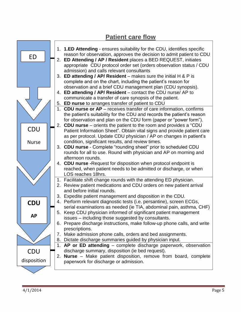
Patient care flow 1. 1.ED Attending - ensures suitability for the CDU, identifies specific
reason for observation, approves the decision to admit patient to CDU 2. ED Attending / AP / Resident places a BED REQUEST, initiates
appropriate CDU protocol order set (orders observation status / CDU admission) and calls relevant consultants
3. ED attending / AP/ Resident – makes sure the initial H & P is complete and on the chart, including the patient’s reason for observation and a brief CDU management plan (CDU synopsis).
4. ED attending / AP/ Resident – contact the CDU nurse/ AP to communicate a transfer of care synopsis of the patient.
5. ED nurse to arranges transfer of patient to CDU
1. CDU nurse or AP – receives transfer of care information, confirms the patient’s suitability for the CDU and records the patient’s reason for observation and plan on the CDU form (paper or “power form”).
2. CDU nurse – orients the patient to the room and provides a “CDU Patient Information Sheet”. Obtain vital signs and provide patient care as per protocol. Update CDU physician / AP on changes in patient’s condition, significant results, and review times.
3. CDU nurse - Complete “rounding sheet” prior to scheduled CDU rounds for all to use. Round with physician and AP on morning and afternoon rounds.
4. CDU nurse -Request for disposition when protocol endpoint is reached, when patient needs to be admitted or discharge, or when LOS reaches 18hrs.
1. Facilitate shift change rounds with the attending ED physician. 2. Review patient medications and CDU orders on new patient arrival
and before initial rounds. 3. Expedite patient management and disposition in the CDU. 4. Perform relevant diagnostic tests (i.e. persantine), screen ECGs,
serial examinations as needed (ie TIA, abdominal pain, asthma, CHF) 5. Keep CDU physician informed of significant patient management
issues – including those suggested by consultants. 6. Prepare discharge instructions, make follow-up phone calls, and write
prescriptions. 7. Make admission phone calls, orders and bed assignments. 8. Dictate discharge summaries guided by physician input.
1. AP or ED attending – complete discharge paperwork, observation discharge summary, disposition (ie bed request).
2. Nurse – Make patient disposition, remove from board, complete paperwork for discharge or admission.
ED
CDU
Nurse
CDU
AP
CDU disposition
4/1/2014 Page 6
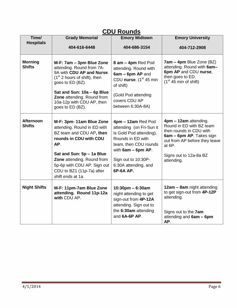
CDU Rounds Time/
Hospitals Grady Memorial
404-616-6448
Emory Midtown
404-686-3154
Emory University
404-712-2908
Morning Shifts
M-F: 7am – 3pm Blue Zone attending. Round from 7A-9A with CDU AP and Nurse. (1st 2 hours of shift), then goes to ED (BZ). Sat and Sun: 10a – 6p Blue Zone attending. Round from 10a-12p with CDU AP, then goes to ED (BZ).
8 am – 4pm Red Pod
attending. Round with
6am – 6pm AP and
CDU nurse. (1st 45 min
of shift)
(Gold Pod attending
covers CDU AP
between 6:30A-8A)
7am – 4pm Blue Zone (BZ) attending. Round with 6am–6pm AP and CDU nurse, then goes to ED. (1st 45 min of shift)
Afternoon Shifts
M-F: 3pm- 11am Blue Zone
attending. Round in ED with
BZ team and CDU AP, then
rounds in CDU with CDU
AP.
Sat and Sun: 5p – 1a Blue
Zone attending. Round from
5p-6p with CDU AP. Sign out
CDU to BZ1 (11p-7a) after
shift ends at 1a.
4pm – 12am Red Pod
attending (on Fri-Sun it
is Gold Pod attending).
Rounds in ED with
team, then CDU rounds
with 6am – 6pm AP.
Sign out to 10:30P-
6:30A attending, and
6P-6A AP.
4pm – 12am attending. Round in ED with BZ team then rounds in CDU with 6am – 6pm AP. Takes sign out from AP before they leave at 6P. Signs out to 12a-8a BZ attending.
Night Shifts
M-F: 11pm-7am Blue Zone attending. Round 11p-12a with CDU AP.
10:30pm – 6:30am
night attending to get
sign-out from 4P-12A
attending. Sign out to
the 6:30am attending
and 6A-6P AP.
12am – 8am night attending to get sign-out from 4P-12P attending.
Signs out to the 7am attending and 6am – 6pm AP.
4/1/2014 Page 7
Rounding principles: 1. Round at the beginning of each shift - CDU rounds are comparable to having a patient signed out
to you at shift change. The beginning of a shift is the time to get “sign out”, examine the patient, give
orders, make dispositions, and assign your name to each patient on the CDU tracking board. The
compelling question should be “why is this patient still here?” Patient’s who have not clinically
“declared themselves” by 15-18 hours are unlikely to leave and a disposition should be made.
Morning rounds are busiest, afternoon are lightest (average census is lowest) and evening rounds
may be chart review only unless a patient is likely to be discharged.
2. Who to round on - Round on all patients that are new or have not had a disposition made. However,
if a patient has already been seen, discharged or admitted, and all observation discharge paperwork
completed then that patient does not need to be seen.
3. What to do – review the chart (ie. ED H/P, transfer of care paperwork, labs, x-ray reports, consults,
test results), take report from the CDU nurse / AP, examine the patient (focused on why they are in
the CDU), and document / communicate your findings and plan with the CDU team. Discharge /
admit patients as needed (with AP if present).
4. CDU (observation) discharge summary - This must cover all four CPT documentation elements: a. Clinical course in the unit b. A final examination (focused) c. Instructions for continuing care (outpatient or inpatient) d. Preparation of discharge (or admission) records
Patient Selection
Overview – The CDU manages patients for up to 18-24 hours, after which time a disposition should be
made. Care beyond this time frame may occasionally occur if it is clear that as short term disposition
is likely to occur (ie. stress test in the morning). The goal is to provide accelerated care while
decreasing inappropriate ED discharges. Patients will first have been managed in the ED and found
to need further management to determine their need for admission. If a patient can be discharged
within 4-6 hours placement in the CDU may not be the best use of available resources. Based on
clinical judgment, and the best scientific evidence, patients should have at least a 70% probability of
discharge within 18 hours - if managed actively. Patients will be managed in the unit using the
guidelines and principles detailed in this document.
General principles of CDU patient selection
Focused patient care goal - The Physician’s note should document the specific reason for admission to the CDU. Generally there should be only one specific problem that requires acute management. When multiple problems require acute management, the likelihood of discharge is much lower. “Focused Goals” fall into three broad categories:
Diagnostic evaluation of critical symptom – i.e. chest pain, syncope, etc.
Short term treatment of an emergency condition – i.e. asthma, dehydration, etc.
Management of psychosocial needs – i.e. need for home support services or placement (if feasible)

4/1/2014 Page 8
Limited intensity of service and severity of illness – based on available resources, such as nurse to patient ratios, higher acuity patients will need to be placed in the hospital for management. This is defined for each condition for several conditions in this document, however conditions outside this list may be observed if they meet the general principles outlined here.
General EXCLUSIONS from the CDU PATIENTS WITH AN INCOMPLETE CHART – A missing or poorly documented ED history,
physical, and medical decision making, a single concise diagnosis, a clear plan, and appropriate orders. This makes is very difficult to efficiently and safely manage the patient.
HIGH SEVERITY OF ILLNESS – Such as patients requiring more nursing care than can be offered in the unit. For example, patients with acutely unstable vitals signs, unstable cardiac, pulmonary, or neurological conditions. These patients should be managed in the initial Emergency Center treatment area until deemed to be stable for at least one hour or admitted.
HIGH INTENSITY OF SERVICE – Such as patients that are too unstable or ill to be observed. For example difficult intoxicated or suicidal psychiatric patients, patients requiring frequent vital signs or treatments. This includes patients on intravenous vasoactive drip infusions of nitroglycerin, labetalol, cardiazem (diltiazem), dopamine or dobutamine, epoprostenol (flolan), or treprostinil (remodulin).
PATIENTS FOR WHOM INPATIENT ADMISSION IS CLEARLY NEEDED - If the ED physician identifies the need for a traditional inpatient admission, the patient should not be admitted to the CDU. However, when appropriate, patients that are “holds” may be temporarily boarded in the unit based on criteria below.
AGE LESS THAN 15 YEARS OLD - Younger patients will be managed in a pediatric CHOA hospital based on general pediatric transfer practices. Pediatric CDU patients over the age of 15 should NOT have significant underlying illness or co-morbidities (such as underlying heart disease, sickle cell disease, etc) requiring skilled pediatric nursing care. Children in the CDU should have a legally responsible adult stay with them while in the CDU.
OBSTETRIC PATIENTS OVER 20 WEEKS PREGNANT - These patients should be managed on the Labor and Delivery unit according to hospital and ED practices. If they have already been evaluated and cleared by the obstetric service (either on L & D and sent back to ED, or cleared by an obstetrician) for CDU management of a non-obstetrical condition (i.e. asthma), then they may be managed in the CDU.
PATIENTS AT RISK OF SELF HARM. Specifically suicidal patients, acutely psychotic patients, or patients with significant inebriation due to alcohol or illicit drugs. The unit is not physically designed to closely monitor these patients for their safety. Patients determined to be at risk of self harm should have their clothing held and be moved to the ED for closer psychiatric monitoring (consistent with ED practices).
ANTICIPATED CDU LENGTH OF STAY LESS THAN 4 HOURS OR OVER 24 HOURS. The work of transferring, admitting, and discharging patient whose stay is under 4 hours is not the best use of these resources. On the other hand, since most patients are discharged from the unit in 15 hours - patients that clearly require more than 18 hours of care are more likely to be admitted and unlikely to benefit from the CDU. Reasons for staying beyond 18 hours should be documented in the chart.
PATIENTS WITH (1) AN ACUTE GAIT DISTURBANCE, (2) “RULE OUT HIP FRACTURE”, OR (3) OVER AGE 65 WITH BACK PAIN, (4) TRANSPLANT PATIENTS, (5) HEMODIALYSIS PATIENTS - These patients have been found to have a very high admit rate and often require more than 24 hours of care.

4/1/2014 Page 9
CDU Quality Assurance and Utilization Review
The CDU committee for each hospital will meet on a monthly basis to review CDU utilization, CDU quality reports, clinical and administrative issues. Meetings will ideally be attended by the site director of the CDU and leadership representatives of CDU nursing, CDU Associate Providers, and an administrative assistant. Utilization Review Monitors – to be reviewed by month and for prior 12 months (as available)
1. Case mix for CDU – by diagnosis (with “total” composite value as well), volume, admit rates, ED LOS, and CDU LOS.
2. Arrival and departure volumes - by hour of day 3. Unit occupancy (where available) – by hour of day 4. CDU LOS by time of arrival in the CDU – by hour of the day
Quality Assurance Monitors – to be reviewed monthly – when available or needed
1. Concerns voiced by staff, patients, or consultants - Reviewed monthly. 2. Return to ED or hospital within 14 days of CDU discharge – Reviewed when available. 3. Death or cardio-respiratory arrest in the unit – Reviewed monthly. 4. Length of stay over 36 hours – Reviewed periodically as needed. 5. Protocol compliance – Reviewed periodically as needed. 6. Protocol failure characteristics – Reviewed periodically as needed. 7. Documentation and transfer of care compliance – missing or incomplete charts
Guidelines for “holds” or “boarders” in the CDU General principle - A “Hold” applies to a patient who is awaiting a prearranged action such as traditional
inpatient admission, transfer to another facility, surgery, discharge home, etc. This is in contrast to a patient whose status is “observation” – where a patient is actively managed to determine the need for inpatient admission. “Holds” are often a manifestation of hospital overcrowding, or inefficiencies of patient care (i.e. prolonged waits to go to the O.R. or a bed). They have no limit on length of stay, acuity, or clinical condition. The CDU helps to address the problem of “holds” by avoiding admission and keeping inpatient beds open. Alternatively, filling the CDU with holds will exacerbate a bed shortage and enables inefficiencies of care to continue.
Guidelines for “holds” in the CDU - A patient who is awaiting admission to an inpatient bed or transfer
may be held in the CDU provided that: 1. All efforts have been made to expedite inpatient admission or transfer (i.e. charge nurse has
spoken with pre-op waiting, etc). All other options have been explored. 2. It is estimated that the bed or procedure will not be available for 3 hours or more. It is otherwise
not worth the work of transferring twice in less than 3 hours. 3. “Holds” may not constitute more than half of the CDU bed capacity. The last available CDU bed
may not be used for a hold.

4/1/2014 Page 10
EUH CDU low census staffing Principle: The 8 bed CDU provides service to selected emergency department patients needing observation services. The CDU always has an 8 bed capacity. The unit is generally staffed with two RNs for eight beds. With variations in CDU nurses or CDU patients, some flexibility is required to maximize resource utilization: 1. When the CDU has less than 4 patients, AND the ED needs nursing support – the CDU nurse
may be flexed to assist in the ED. HOWEVER, the CDU capacity remains at 8 and patients will
continue to be assigned to the CDU regardless of this staffing shift. When the number of CDU
patients (either in the CDU or in the ED awaiting a CDU bed) reaches 5 patients, then the second
“flexed” CDU nurse will return to the CDU to assume patient care.
2. When the CDU has no patients, AND the ED needs nursing support - If both CDU nurses are flexed
then the CDU nurse will return to the CDU when there are two CDU patients in the ED. The ED shift
nurse manager has 60 minutes to transition the ED patient assignment.
3. When the CDU has less than 2 nurses (which may occur for portions of a shift due to staffing
issues) – an ED nurse may be flexed to cover the CDU until the CDU has 2 nurses. If there is not a
nurse available to assist CDU staffing then patients assigned to the CDU will be held in the ED until
staff are available.
Recommend Guidelines - CDU nurses will not be assigned ED critical patient unless one has been
properly oriented to acute care. The charge nurse will work with the CDU nurse to give an assignment
that is manageable for the nurse. The CDU nurse should actively seek potential CDU patients and make
suggestions to ED attending for CDU admissions - in accordance with the CDU guidelines. To assure
success, flexibility and cooperation is required.
GUIDELINES FOR STRESS TESTING OBSERVATION UNIT CHEST PAIN PATIENTS
The purpose of stress testing CDU chest pain patients is to identify those with severe coronary
artery stenosis, or unstable angina (USA). Initial ECG or cardiac markers in this population do not
adequately detect USA. Subendocardial myocardial infarction, or “NSTEMI” must first be ruled out
before a stress test can be performed safely. This is done with ECGs and serial cardiac marker
testing.
1. The ACC/AHA Guidelines for the Management of Patients with Unstable Angina/Non-ST-
Elevation Myocardial Infarction are most recently updated in 2011 and recommend certain
goals of care. Patients in whom Acute Coronary Syndrome (ACS) is considered to be probable
or possible, an admission to an observation unit is acceptable in those with a non-diagnostic
EKG, negative cardiac biomarkers, and a history of present illness that is not highly suggestive of
ACS. Stress testing and imaging at all locations is done in accordance with ACC/AHA guidelines
for stress testing and imaging. Stress imaging (nuclear, echo, MRI, coronary CTA) will be
interpreted by those trained and credentialed to interpret each modality in accordance with
hospital standards and national guidelines for each imaging modality.
4/1/2014 Page 11
2. Chest Pain Protocol - the ACC/AHA Guidelines recommend serial EKG’s, and serial cardiac
biomarkers for appropriate low risk patients. For selected low risk patients, it is acceptable to
discharge them with arrangements for a stress test within 72 hours. When this occurs, strict
patient instructions should be given for when to return to the ED, along with aspirin therapy if not
contraindicated .
3. Vasodilator stress injections (dipyrimadole or lexiscan) – may be performed by associate
providers (NP or PA) who have completed training in this area and have performed at least 10
supervised injections. This includes compliance with persantine / lexiscan patient selection,
monitoring and documenting patient condition during drug infusions, identifying and treating both
minor and major vasodilator side effects, coordinating testing with other departments,
understanding imaging results which are reported by nuclear cardiology. Credentialing in this
area will be renewed each year based on performance skills and knowledge in this area. These
injections will be supervised by the attending physician working with the associate provider.
4. The following variables are considered in choosing an appropriate stress test
a. What is available
b. Patient characteristics:
i. Initial probability of acute coronary ischemia in the patient (Bayes’ theorem) –higher
probability of disease warrant a more sensitive test, lower probability patients benefit
from a less sensitive test (i.e. where the false positive rate is less than disease
prevalence).
ii. The patients’ ability to exercise.
iii. Contraindications to various stress tests (Persantine - severe asthma; cCTA - high
BMI/CRF/CAD; cCTA/MPI – child bearing age females (radiation - relative issue)).
c. Test characteristics
i. Sensitivity and specificity of the stress test – More sensitive tests produce more
false positives. More specific tests may yield more false negatives.
ii. PET – ideal for high BMI, known CAD/prior MI
iii. DSE or MRI – ideal for child bearing age females
d. The cost of the stress test
Why stress imaging after MI has been ruled out? We do stress imaging determine if the patient’s symptoms (ie chest pain) are due to unstable angina
once AMI has been ruled out. In other words, we are asking if there is >70% coronary occlusion (by
plaque or ruptured plaque + clot) causing myocardial ischemia. Stress testing with imaging identifies 3/5
of true positive ACS cases in this population, while serial markers and ECGs identify only 1/5 of cases.
Stress testing / imaging options can be broken down as follows:
4/1/2014 Page 12
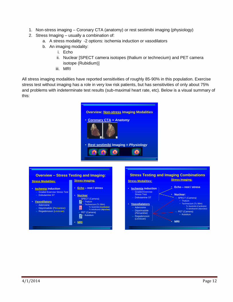
1. Non-stress imaging – Coronary CTA (anatomy) or rest sestimibi imaging (physiology)
2. Stress Imaging – usually a combination of:
a. A stress modality -2 options: ischemia induction or vasodilators
b. An imaging modality:
i. Echo
ii. Nuclear [SPECT camera isotopes (thalium or technecium) and PET camera
isotope (Rubidium)]
iii. MRI
All stress imaging modalities have reported sensitivities of roughly 85-90% in this population. Exercise
stress test without imaging has a role in very low risk patients, but has sensitivities of only about 75%
and problems with indeterminate test results (sub-maximal heart rate, etc). Below is a visual summary of
this:
Overview: Non-stress Imaging Modalities
• Coronary CTA = Anatomy
• Rest sestimibi Imaging = Physiology
Overview – Stress Testing and Imaging:
Stress Modalities:
• Ischemia induction– Graded Exercise Stress Test
– Dobutamine ST
• Vasodilators– Adenosine
– Dipyrimadole (Persantine)
– Regadenoson (Lexiscan)
Stress Imaging:
• Echo – rest / stress
• Nuclear– SPECT (Camera)
• Thalium
• Technecium (Tc 99m)
– Tc Sestimibi (Cardiolyte)
– Tc tetrofosmin (Myoview)
– PET (Camera)
• Rubidium
• MRI
Stress Testing and Imaging Combinations
Stress Modalities:
• Ischemia induction– Graded Exercise
Stress Test
– Dobutamine ST
• Vasodialators– Adenosine
– Dipyrimadole (Persantine)
– Regadenoson(Lexiscan)
Stress Imaging:
• Echo – rest / stress
• Nuclear:– SPECT (Camera)
• Thalium
• Technecium (Tc 99m)
– Tc Sestimibi (Cardiolyte)
– Tc tetrofosmin (Myoview)
– PET (Camera)
• Rubidium
• MRI
4/1/2014 Page 13
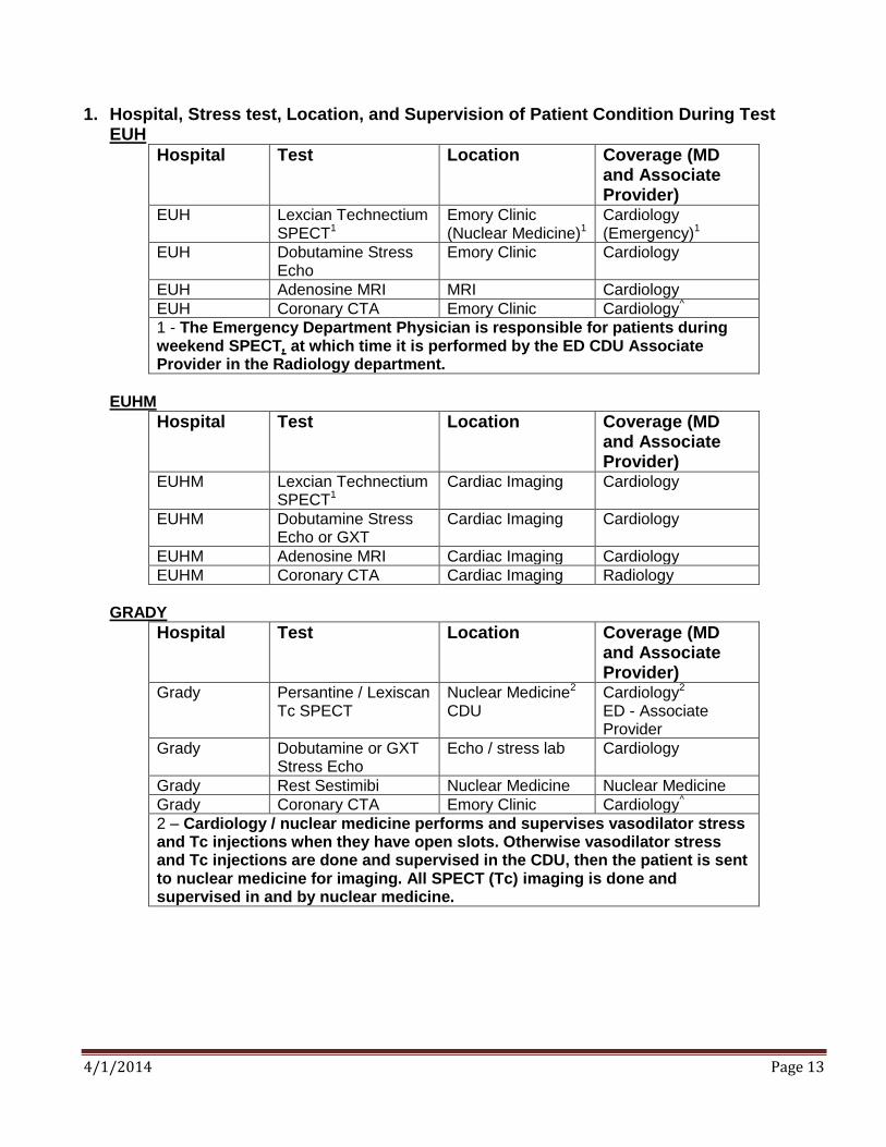
1. Hospital, Stress test, Location, and Supervision of Patient Condition During Test EUH
Hospital Test Location Coverage (MD and Associate Provider)
EUH Lexcian Technectium SPECT1
Emory Clinic (Nuclear Medicine)1
Cardiology (Emergency)1
EUH Dobutamine Stress Echo
Emory Clinic Cardiology
EUH Adenosine MRI MRI Cardiology
EUH Coronary CTA Emory Clinic Cardiology^
1 - The Emergency Department Physician is responsible for patients during weekend SPECT, at which time it is performed by the ED CDU Associate Provider in the Radiology department.
EUHM
Hospital Test Location Coverage (MD and Associate Provider)
EUHM Lexcian Technectium SPECT1
Cardiac Imaging Cardiology
EUHM Dobutamine Stress Echo or GXT
Cardiac Imaging Cardiology
EUHM Adenosine MRI Cardiac Imaging Cardiology
EUHM Coronary CTA Cardiac Imaging Radiology
GRADY
Hospital Test Location Coverage (MD and Associate Provider)
Grady Persantine / Lexiscan Tc SPECT
Nuclear Medicine2
CDU Cardiology2 ED - Associate Provider
Grady Dobutamine or GXT Stress Echo
Echo / stress lab Cardiology
Grady Rest Sestimibi Nuclear Medicine Nuclear Medicine
Grady Coronary CTA Emory Clinic Cardiology^
2 – Cardiology / nuclear medicine performs and supervises vasodilator stress and Tc injections when they have open slots. Otherwise vasodilator stress and Tc injections are done and supervised in the CDU, then the patient is sent to nuclear medicine for imaging. All SPECT (Tc) imaging is done and supervised in and by nuclear medicine.
4/1/2014 Page 14
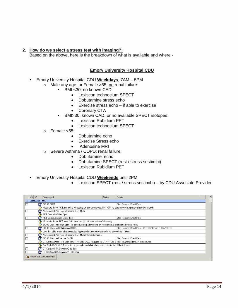
2. How do we select a stress test with imaging?: Based on the above, here is the breakdown of what is available and where -
Emory University Hospital CDU
Emory University Hospital CDU Weekdays, 7AM – 5PM o Male any age, or Female >55; no renal failure:
BMI <30, no known CAD:
Lexiscan technecium SPECT
Dobutamine stress echo
Exercise stress echo – if able to exercise
Coronary CTA BMI>30, known CAD, or no available SPECT isotopes:
Lexiscan Rubidium PET
Lexiscan technecium SPECT o Female <55:
Dobutamine echo
Exercise Stress echo
Adenosine MRI o Severe Asthma / COPD; renal failure:
Dobutamine echo
Dobutamine SPECT (rest / stress sestimibi)
Lexiscan Rubidium PET
Emory University Hospital CDU Weekends until 2PM
Lexiscan SPECT (rest / stress sestimibi) – by CDU Associate Provider
4/1/2014 Page 15
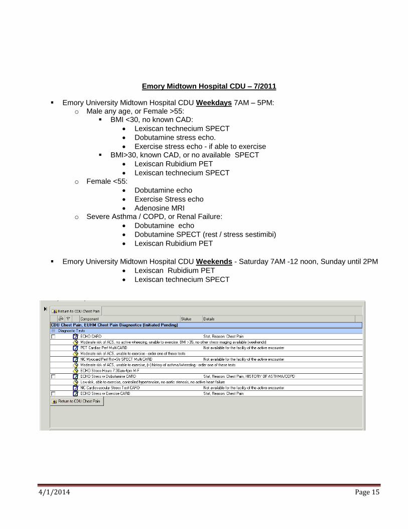
Emory Midtown Hospital CDU – 7/2011
Emory University Midtown Hospital CDU Weekdays 7AM – 5PM: o Male any age, or Female >55:
BMI <30, no known CAD:
Lexiscan technecium SPECT
Dobutamine stress echo.
Exercise stress echo - if able to exercise BMI>30, known CAD, or no available SPECT
Lexiscan Rubidium PET
Lexiscan technecium SPECT o Female <55:
Dobutamine echo
Exercise Stress echo
Adenosine MRI o Severe Asthma / COPD, or Renal Failure:
Dobutamine echo
Dobutamine SPECT (rest / stress sestimibi)
Lexiscan Rubidium PET
Emory University Midtown Hospital CDU Weekends - Saturday 7AM -12 noon, Sunday until 2PM
Lexiscan Rubidium PET
Lexiscan technecium SPECT

4/1/2014 Page 16
Grady Memorial Hospital CDU – 2/2014
Grady Memorial Hospital CDU Weekdays 7AM – 2PM: o BMI <30, no known CAD:
Exercise Treadmill (ETT) – if able to exercise Coronary CT Angiogram (cCTA)
Must be currently in sinus rhythm (no Atrial fibrillation/flutter)
Resting HR <80 (must be below 60 after beta blockers)
Able to get IV dye o No dye allergy o GFR > 50 o 18 or 20g AC or forearm IV
No Beta-blocker allergy
No active wheezing or history of COPD
No history of CHF (EF > 45%) o BMI>30, known CAD, or not candidate for cCTA or ETT
Persantine or Adenosine Technetium SPECT
Persantine if performed in CDU
Adenosine if performed in Stress lab Consider Cardiology consult for recs on stress vs cath/admission
o Severe Asthma / COPD: Regadenoson (Lexiscan®) technetium SPECT
Grady Memorial Hospital CDU Weekends - Saturday and Sunday until 2PM Persantine/Lexiscan technetium SPECT
4/1/2014 Page 17
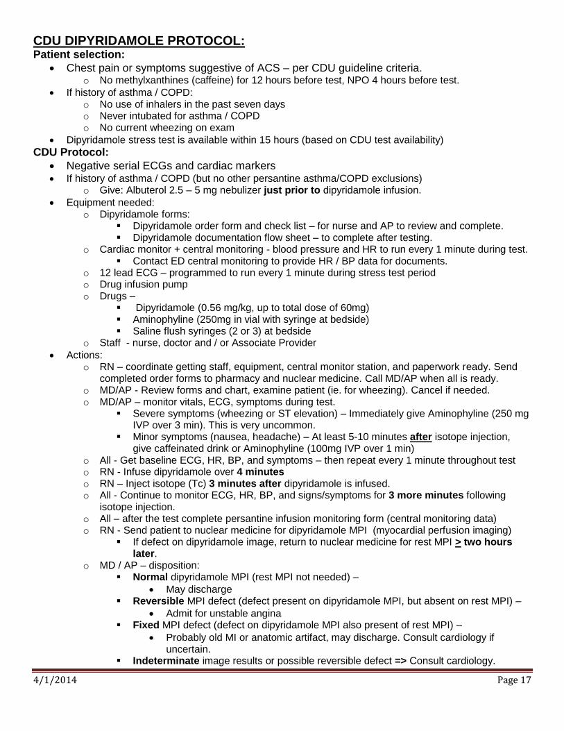
CDU DIPYRIDAMOLE PROTOCOL: Patient selection:
Chest pain or symptoms suggestive of ACS – per CDU guideline criteria. o No methylxanthines (caffeine) for 12 hours before test, NPO 4 hours before test.
If history of asthma / COPD: o No use of inhalers in the past seven days o Never intubated for asthma / COPD o No current wheezing on exam
Dipyridamole stress test is available within 15 hours (based on CDU test availability)
CDU Protocol:
Negative serial ECGs and cardiac markers If history of asthma / COPD (but no other persantine asthma/COPD exclusions)
o Give: Albuterol 2.5 – 5 mg nebulizer just prior to dipyridamole infusion.
Equipment needed: o Dipyridamole forms:
Dipyridamole order form and check list – for nurse and AP to review and complete. Dipyridamole documentation flow sheet – to complete after testing.
o Cardiac monitor + central monitoring - blood pressure and HR to run every 1 minute during test. Contact ED central monitoring to provide HR / BP data for documents.
o 12 lead ECG – programmed to run every 1 minute during stress test period o Drug infusion pump o Drugs –
Dipyridamole (0.56 mg/kg, up to total dose of 60mg) Aminophyline (250mg in vial with syringe at bedside) Saline flush syringes (2 or 3) at bedside
o Staff - nurse, doctor and / or Associate Provider
Actions: o RN – coordinate getting staff, equipment, central monitor station, and paperwork ready. Send
completed order forms to pharmacy and nuclear medicine. Call MD/AP when all is ready. o MD/AP - Review forms and chart, examine patient (ie. for wheezing). Cancel if needed. o MD/AP – monitor vitals, ECG, symptoms during test.
Severe symptoms (wheezing or ST elevation) – Immediately give Aminophyline (250 mg IVP over 3 min). This is very uncommon.
Minor symptoms (nausea, headache) – At least 5-10 minutes after isotope injection, give caffeinated drink or Aminophyline (100mg IVP over 1 min)
o All - Get baseline ECG, HR, BP, and symptoms – then repeat every 1 minute throughout test o RN - Infuse dipyridamole over 4 minutes o RN – Inject isotope (Tc) 3 minutes after dipyridamole is infused. o All - Continue to monitor ECG, HR, BP, and signs/symptoms for 3 more minutes following
isotope injection. o All – after the test complete persantine infusion monitoring form (central monitoring data) o RN - Send patient to nuclear medicine for dipyridamole MPI (myocardial perfusion imaging)
If defect on dipyridamole image, return to nuclear medicine for rest MPI > two hours later.
o MD / AP – disposition: Normal dipyridamole MPI (rest MPI not needed) –
May discharge Reversible MPI defect (defect present on dipyridamole MPI, but absent on rest MPI) –
Admit for unstable angina Fixed MPI defect (defect on dipyridamole MPI also present of rest MPI) –
Probably old MI or anatomic artifact, may discharge. Consult cardiology if uncertain.
Indeterminate image results or possible reversible defect => Consult cardiology.
4/1/2014 Page 18
4/1/2014 Page 19
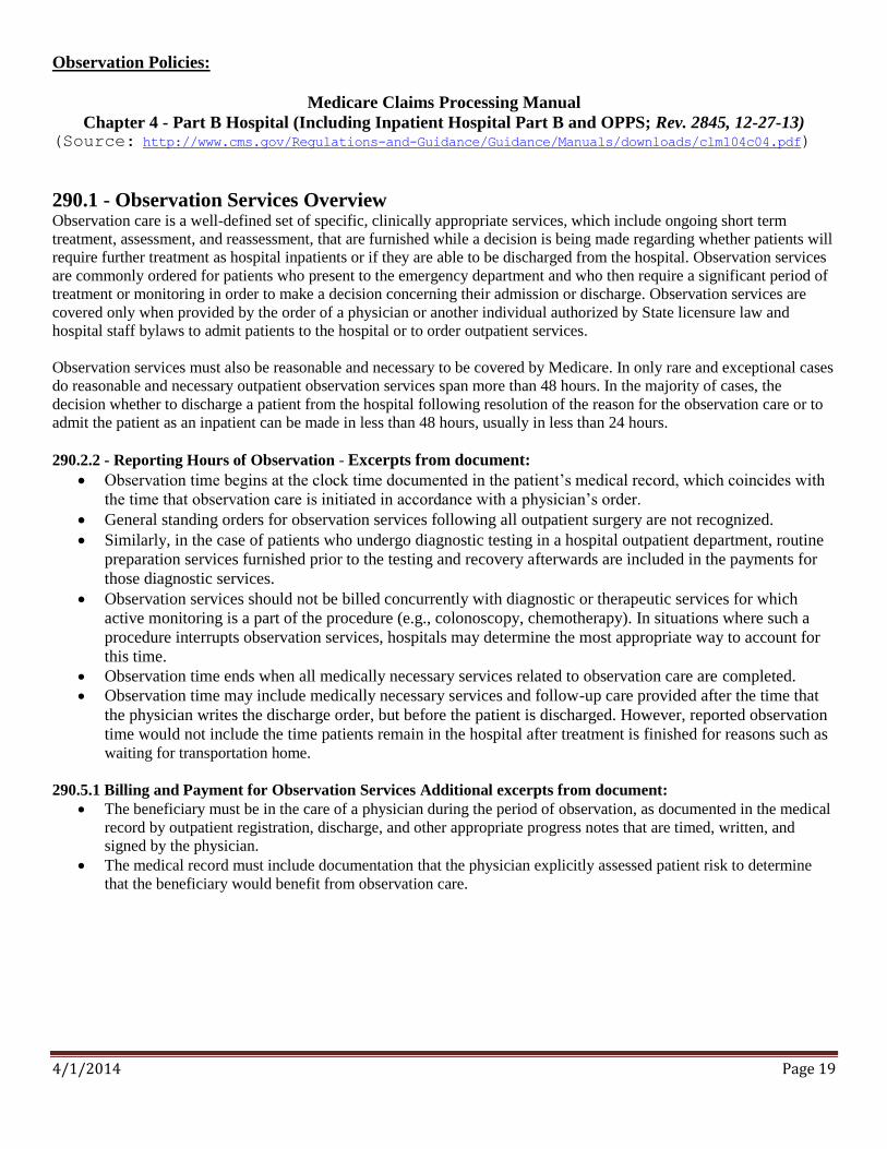
Observation Policies:
Medicare Claims Processing Manual
Chapter 4 - Part B Hospital (Including Inpatient Hospital Part B and OPPS; Rev. 2845, 12-27-13) (Source: http://www.cms.gov/Regulations-and-Guidance/Guidance/Manuals/downloads/clm104c04.pdf)
290.1 - Observation Services Overview Observation care is a well-defined set of specific, clinically appropriate services, which include ongoing short term
treatment, assessment, and reassessment, that are furnished while a decision is being made regarding whether patients will
require further treatment as hospital inpatients or if they are able to be discharged from the hospital. Observation services
are commonly ordered for patients who present to the emergency department and who then require a significant period of
treatment or monitoring in order to make a decision concerning their admission or discharge. Observation services are
covered only when provided by the order of a physician or another individual authorized by State licensure law and
hospital staff bylaws to admit patients to the hospital or to order outpatient services.
Observation services must also be reasonable and necessary to be covered by Medicare. In only rare and exceptional cases
do reasonable and necessary outpatient observation services span more than 48 hours. In the majority of cases, the
decision whether to discharge a patient from the hospital following resolution of the reason for the observation care or to
admit the patient as an inpatient can be made in less than 48 hours, usually in less than 24 hours.
290.2.2 - Reporting Hours of Observation - Excerpts from document:
Observation time begins at the clock time documented in the patient’s medical record, which coincides with
the time that observation care is initiated in accordance with a physician’s order.
General standing orders for observation services following all outpatient surgery are not recognized.
Similarly, in the case of patients who undergo diagnostic testing in a hospital outpatient department, routine
preparation services furnished prior to the testing and recovery afterwards are included in the payments for
those diagnostic services.
Observation services should not be billed concurrently with diagnostic or therapeutic services for which
active monitoring is a part of the procedure (e.g., colonoscopy, chemotherapy). In situations where such a
procedure interrupts observation services, hospitals may determine the most appropriate way to account for
this time.
Observation time ends when all medically necessary services related to observation care are completed.
Observation time may include medically necessary services and follow-up care provided after the time that
the physician writes the discharge order, but before the patient is discharged. However, reported observation
time would not include the time patients remain in the hospital after treatment is finished for reasons such as waiting for transportation home.
290.5.1 Billing and Payment for Observation Services Additional excerpts from document:
The beneficiary must be in the care of a physician during the period of observation, as documented in the medical
record by outpatient registration, discharge, and other appropriate progress notes that are timed, written, and
signed by the physician.
The medical record must include documentation that the physician explicitly assessed patient risk to determine
that the beneficiary would benefit from observation care.

4/1/2014 Page 20
ACEP POLICY –

4/1/2014 Page 21
Clinical Decision Unit – EUH and EUHM (7/12 – 6/13)
Condition %
census # visits
% Discharge
CDU LOS (hrs)
Total LOS (ED+CDU)
(hrs)
CDU Admit
LOS (hrs)
CDU Discharge LOS (hrs)
Chest Pain 45% 2,839 81% 16.2 21.6 16.7 16.2
Dehydration/vomiting 7% 437 72% 15.6 22.2 17.8 14.8
Syncope 6% 369 83% 16.8 22.2 16.5 16.7
TIA 6% 345 71% 16.1 22.2 13.9 17
Other 5% 312 73% 14.4 21 13.2 15
Cellulitis 4% 264 73% 17 22.5 17 17
Abd pain 4% 242 71% 14.6 22.3 15.8 14
Asthma 3% 186 61% 17.6 23.2 20.8 16.4
CHF 2% 134 74% 18.1 23.6 16.7 18.6
Electrolyte abnormality 2% 116 80% 14.5 20.4 13.6 14.7
Transfusion of blood/products 2% 96 86% 13.9 19.1 14.7 13.8
COPD exacerbation 2% 95 55% 16.4 22.8 17.8 15.7
Pyelonephritis 2% 95 67% 16.5 22.8 16.2 16.7
Pneumonia 1% 79 70% 17.2 23 18.3 16.3
Hyperglycemia 1% 76 82% 17.2 23.4 20.1 17
Allergic rxn 1% 66 89% 12.5 16.8 12.1 12.7
GI bleed 1% 60 78% 16.1 22.5 12.9 17.2
Headache 1% 54 76% 14.7 22.4 13.7 15
Back pain 1% 42 79% 15.8 23.2 14.5 16.2
Vertigo 1% 42 86% 15.3 22.1 13.8 15.6
DVT 1% 41 85% 10.8 16.5 14.5 10.2
Atrial fibrillation 1% 37 57% 14.4 19.2 13.3 15.3
Renal colic <1% 29 76% 14.8 20.8 20.2 13
Hypoglycemia <1% 27 48% 16.6 21.9 17.3 15.9
Hypertensive urgency <1% 23 87% 14.2 19.9 18.7 13.5
Hyperemesis gravidarium <1% 22 86% 16.4 23.7 19.2 16
Anemia <1% 12 83% 12.1 16.7 11.1 12.4
Heart Failure <1% 12 0% 16.4 26.1 N/A N/A
Supraventricular tachycardia <1% 9 100% 8.5 15.2 N/A 8.5
Vaginal bleeding <1% 9 100% 12.4 18.4 N/A 12.4
Grand Total 100% 6,255 77% 16.0 21.8 16.3 15.9
4/1/2014 Page 22

4/1/2014 Page 23
Index of CDU protocols
1. ABDOMINAL INJURY ......................................................................24
2. ABDOMINAL PAIN ...........................................................................25
3. ACUTE HEART FAILURE ...............................................................26
4. ALLERGIC REACTION.....................................................................27
5. ASTHMA .............................................................................................28
6. ATRIAL FIBRILLATION – ACUTE ONSET ...................................29
7. BACK PAIN ........................................................................................30
8. CELLULITIS .......................................................................................31
9. CHEST INJURY ..................................................................................32
10. CHEST PAIN – POSSIBLE ACS .......................................................33
11. COPD EXACERBATION ...................................................................34
12. DEEP VEIN THROMBOSIS ..............................................................35
13. DEHYDRATION OR VOMITING .....................................................36
14. DILANTIN TOXICITY.......................................................................37
15. ELECTROLYTE ABNORMALITY ...................................................38
16. GASTROINTESTINAL BLEED ........................................................39
17. HEADACHE........................................................................................40
18. HEAD INJURY ...................................................................................41
19. HYPEREMESIS GRAVIDARUM ......................................................42
20. HYPOGLYCEMIA..............................................................................43
21. HYPERGLYCEMIA ...........................................................................44
22. PNEUMONIA......................................................................................45
23. PYELONEPHRITIS ............................................................................46
24. RENAL COLIC ...................................................................................47
25. SEIZURES ...........................................................................................48
26. SOCIAL ADMISSIONS ......................................................................49
27. SYNCOPE ...........................................................................................50
28. TOXICOLOY OBSERVATION .........................................................51
29. TRANSFUSION OF BLOOD AND BLOOD PRODUCTS ...............52
30. TRANSIENT ISCHEMIC ATTACK ..................................................53
31. VAGINAL BLEEDING ......................................................................54
32. VERTIGO ............................................................................................55

4/1/2014 Page 24
ABDOMINAL INJURY (NON-PENETRATING)
TRANSFER CRITERIA Cooperative patient with stable vital signs (RR>8 or <24, SBP>100 P>60 or <110)
No Peritoneal Signs
Negative initial imaging studies (i.e. CT)
Pertinent lab results acceptable (e.g., Hgb)
Surgery consult documented
EXCLUSION CRITERIA
Uncooperative patient, patients requiring restraints
Impending alcohol withdrawal syndrome
ETOH estimated >200 mg/dL at transfer
Pregnancy >20 weeks
Abnormal vital signs (above)
CT scan not done or significant acute abnormality
POTENTIAL INTERVENTIONS
NPO initially, advance per physician
Repeat Hct q 4-6 hours (if pertinent to patient’s management)
Serial abdominal examinations (e.g. q 4 hours)
If indicated by physician, serial ultrasounds
Immediate reevaluation by ED physician or surgeon if patient develops: - Significant vomiting - Increasing abdominal pain - Increased tenderness - Worsening vital signs: Decreased BP, increased HR, fever
DISCHARGE CRITERIA:
Patient is ambulatory
Serial abdominal exams essentially negative
Repeat labs reviewed and stable (Specifically any Hb drop?)
Vital signs reviewed and stable
Patient able to tolerate PO
Appropriate follow-up established
Surgery agrees with disposition

4/1/2014 Page 25
ABDOMINAL PAIN
TRANSFER CRITERIA
Stable VS
Ancillary Signs/Sx - anorexia, N&V, fever, elevated WBC
Negative pregnancy test
Non-surgical abdomen
High likelihood (~70%) of discharge within 15 hours
EXCLUSION CRITERIA
Unstable VS (HR >110, SBP<100, RR > 22)
Immunocompromised patient (T-cells < 200, chemo, transplant)
Pregnant patient
Bowel obstruction (even partial) or ileus
Cholecystitis (sonographic Murphy, pericholecystic fluid, GB wall thickening>4mm, or dilated CBD)
Surgical abdomen - free air, rigidity, rebound tenderness
Hx of frequent ED visits for abdominal pain – suspected habitual patient / narcotic abuse
POTENTIAL INTERVENTIONS
Analgesics
NPO, IV hydration, repeat CBC
Imaging studies as indicated (i.e. CT abd / pelvis, ultrasound, MRI)
Serial VS
Serial exams Q2-4 hours while awake and as indicated
Surgical or GI consultation as needed
DISPOSITION Home
Pain and / or tenderness resolved or significantly improved
VS acceptable
No diagnosis requiring hospitalization Admit
Persistent vomiting
Pain not resolving or worsening
Unstable VS
Clinical condition or positive testing that merits hospitalization
Consultant preference
Surgical abdomen

4/1/2014 Page 26
ACUTE HEART FAILURE
TRANSFER CRITERIA
Previous history of CHF
Acceptable VS: SBP >100, R < 32, HR <130
Pulse-ox >90 on room air after initial treatment, correctable to > 92 on Oxygen by NC.
High likelihood of correction to baseline status within 24 hours with good home support
No acute co-morbidities
EXCLUSION CRITERIA
New onset CHF
Acute cardiac ischemia (EKG changes, positive troponin, ongoing ischemic chest pain, unstable angina) or new arrhythmias
Unstable VS after treatment (HR>130, SBP<85 or >180, RR>32, Pox<92 on O2 by NC)
Acute co-morbidities - sepsis, pneumonia, new murmur, confusion
Abnormal labs to consider - Severe anemia (Hb<8), renal failure (BUN>40 or Cr>3), Na<135, BNP > 840
Patient requiring vasoactive drips, invasive or noninvasive ventilation (bipap)
Evidence of poor perfusion (confusion, cool extremity, weakness, N/V)
Patients requiring provocative stress tests
POTENTIAL INTERVENTION
Cardiac monitoring, strict Intake/Output, vital signs Q4hr, weight on arrival
Oxygen per respiratory guidelines with pulse oximetry (continuous or q4hours)
Serial EKGs, and cardiac markers (TnI) - 2,4, and 6hrs from 1st lab draw.
Medication as indicated – IV diuretics (home dose), nitroglycerine paste, ACE Inhibitors, ASA
Repeat electrolytes q6 hours or prn
Echocardiography (if not done within 30d) and cardiology consultation - as indicated
CHF education and smoking cessation education
DISPOSITION Home
Subjective improvement – no chest pain, orthopnea, or exertional dyspnea above baseline
Acceptable VS (O2 sat at baseline or >94%, RR <20HR<100, SBP >100 or baseline,).
Negative serial ECGs and cardiac markers, good electrolytes, acceptable echo if done
Evidence of adequate diuresis – 1L urine, decrease in weight, decrease in JVD
CHF discharge checklist (ACEi, β-blocker, HF/ diet/ smoking education, close followup) Hospital
New ischemic EKG changes, arrhythmia, cardiac markers, or evidence of cardiac ischemia
Persistent hypoxia, rales, dyspnea
Poor response to therapy - Failure to improve subjectively
Poor home support
Physician judgment

4/1/2014 Page 27
ALLERGIC REACTION
TRANSFER CRITERIA
Response to therapy in the ED
Erythroderma, urticaria, or angioedema present
If airway angioedema present, need surgical airway judged to be highly unlikely
Minimum 2-hours of stability or improvement in ED after treatment
EXCLUSION CRITERIA
Hypotension (SBP <100), tachycardia > 110
O2 saturation consistently < 94% on room air
Suspicion of acute coronary syndrome
Stridor, respiratory distress, hoarseness at the time of transfer
IV vasopressors required
POTENTIAL INTERVENTIONS
IV fluids as needed
Frequent rechecks and documentation of clear airway
Antihistamines, corticosteroids
Cardiac monitoring (if indicated)
Inhaler or nebulizer treatments (if indicated)
Pulse oximetry
Repeat doses of SQ epinephrine DISPOSITION Home
Resolution or improvement in clinical condition
Stable VS Hospital
Delayed worsening of allergic symptoms
Persistent wheezing or stridor
Inadequate response to therapy during observation
Inability to take oral medications
Abnormal vital signs: SBP < 100mm or RR > 24/min or hypoxia
4/1/2014 Page 28
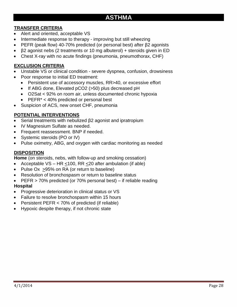
ASTHMA
TRANSFER CRITERIA
Alert and oriented, acceptable VS
Intermediate response to therapy - improving but still wheezing
PEFR (peak flow) 40-70% predicted (or personal best) after β2 agonists
β2 agonist nebs (2 treatments or 10 mg albuterol) + steroids given in ED
Chest X-ray with no acute findings (pneumonia, pneumothorax, CHF)
EXCLUSION CRITERIA
Unstable VS or clinical condition - severe dyspnea, confusion, drowsiness
Poor response to initial ED treatment:
Persistent use of accessory muscles, RR>40, or excessive effort
If ABG done, Elevated pCO2 (>50) plus decreased pH
O2Sat < 92% on room air, unless documented chronic hypoxia
PEFR* < 40% predicted or personal best
Suspicion of ACS, new onset CHF, pneumonia
POTENTIAL INTERVENTIONS
Serial treatments with nebulized β2 agonist and ipratropium
IV Magnesium Sulfate as needed.
Frequent reassessment. BNP if needed.
Systemic steroids (PO or IV)
Pulse oximetry, ABG, and oxygen with cardiac monitoring as needed
DISPOSITION Home (on steroids, nebs, with follow-up and smoking cessation)
Acceptable VS – HR <100, RR <20 after ambulation (if able)
Pulse Ox >95% on RA (or return to baseline)
Resolution of bronchospasm or return to baseline status
PEFR > 70% predicted (or 70% personal best) – if reliable reading Hospital
Progressive deterioration in clinical status or VS
Failure to resolve bronchospasm within 15 hours
Persistent PEFR < 70% of predicted (if reliable)
Hypoxic despite therapy, if not chronic state
4/1/2014 Page 29
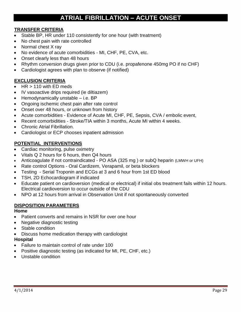
ATRIAL FIBRILLATION – ACUTE ONSET
TRANSFER CRITERIA
Stable BP, HR under 110 consistently for one hour (with treatment)
No chest pain with rate controlled
Normal chest X ray
No evidence of acute comorbidities - MI, CHF, PE, CVA, etc.
Onset clearly less than 48 hours
Rhythm conversion drugs given prior to CDU (i.e. propafenone 450mg PO if no CHF)
Cardiologist agrees with plan to observe (if notified) EXCLUSION CRITERIA
HR > 110 with ED meds
IV vasoactive drips required (ie diltiazem)
Hemodynamically unstable – i.e. BP
Ongoing ischemic chest pain after rate control
Onset over 48 hours, or unknown from history
Acute comorbidities - Evidence of Acute MI, CHF, PE, Sepsis, CVA / embolic event,
Recent comorbidities - Stroke/TIA within 3 months, Acute MI within 4 weeks.
Chronic Atrial Fibrillation.
Cardiologist or ECP chooses inpatient admission POTENTIAL INTERVENTIONS
Cardiac monitoring, pulse oximetry
Vitals Q 2 hours for 6 hours, then Q4 hours Anticoagulate if not contraindicated - PO ASA (325 mg ) or subQ heparin (LMWH or UFH)
Rate control Options - Oral Cardizem, Verapamil, or beta blockers
Testing - Serial Troponin and ECGs at 3 and 6 hour from 1st ED blood
TSH, 2D Echocardiogram if indicated
Educate patient on cardioversion (medical or electrical) if initial obs treatment fails within 12 hours. Electrical cardioversion to occur outside of the CDU
NPO at 12 hours from arrival in Observation Unit if not spontaneously converted DISPOSITION PARAMETERS Home
Patient converts and remains in NSR for over one hour
Negative diagnostic testing
Stable condition
Discuss home medication therapy with cardiologist Hospital
Failure to maintain control of rate under 100
Positive diagnostic testing (as indicated for MI, PE, CHF, etc.)
Unstable condition

4/1/2014 Page 30
BACK PAIN
TRANSFER CRITERIA
Inability to adequately control pain in ED with analgesics
Normal neurological function and temperature.
No risk of metastatic disease or vertebral or epidural abscess
Back pain without severe trauma
Normal imaging (if obtained)
Inability to ambulate because of pain
EXCLUSION CRITERIA
Frequent ED visits for back pain – suspected habitual patient
Age over 65 years old
Acute motor deficit (i.e. foot drop, loss of extension of foot or 1st toe, loss of control of bowel or bladder)
Abnormal x-rays if obtained (burst fracture, spine canal involvement)
High suspicion of cord compression, metastatic disease, epidural bleed or abscess, discitis.
Fever
POTENTIAL INTERVENTIONS
Narcotic analgesics (+ NSAIDs if appropriate)
Serial exams
Physical therapy assessment
Consultation as needed – PMR, Ortho, social service
Imaging (CT or MRI) if acute surgical disease or cancer is suspected
DISPOSITION CRITERIA Home
Ability to ambulate and care for self at home with oral analgesics
Pain at a tolerable level for discharge home
No worsening in neurologic exam Hospital
Inability to tolerate pain on oral medications
Inability to ambulate or care for self at home
Worsening neurological exam
Abnormal imaging warranting inpatient admission

4/1/2014 Page 31
CELLULITIS
TRANSFER CRITERIA
Serial exams needed to exclude rapidly progressive cellulitis
Cellulitis which requires > 1 dose antibiotics in the ED
Temp < 40.C, WBC < 16,000 and WBC >4,000.
Cellulitis with a drained abscess which requires a brief period of observation and wound care
EXCLUSION CRITERIA
Septic or toxic patients – clinical appearance, evidence of severe sepsis (Temp >40, SBP<100, RR>22, HR>100, acute organ dysfunction, lactate >4mmol/L )
Immunocompromized patients – neutropenia, HIV, transplant patients, ESRD/hemodialyisis patients, patients on immunosuppressants or chemotherapy, post-splenectomy patients.
High risk infections – diabetic foot infections; infections proximate to a prosthesis, percutaneous catheter or indwelling device; infections of the orbit or upper lip/nose, neck; infections of >9% TBSA; extensive tissue sloughing; suspicion of osteomyelitis or deep wound infection.
Poorly controlled diabetes
Patient unable to care for self at home
Patient who can be discharged after 1 dose of antibiotics in the ED
POTENTIAL INTERVENTIONS
Mark edges of cellulitis with indelible marker to monitor progression
Antibiotics based on contemporary local guidelines and sensitivities
IV antibiotics - MRSA coverage as indicated (Vancomycin X >2, Bactrim, Clinda, or Doxycycline)
Pertinent labs (CBC, glucose, blood or wound cultures PRN)
DISPOSITION Home
Improvement or no progression of cellulitis
Improved and good clinical condition (ie. No fever, good VS) for 8 hrs.
Able to perform cellulitis care at home and take oral medications
Admit
Increase in skin involvement
Clinical condition worse or not better (i.e. rising temp, poor vitals)
Unable to take oral medications
Unable to care for wound at home, home care unavailable
4/1/2014 Page 32
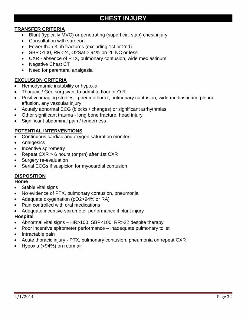
CHEST INJURY
TRANSFER CRITERIA
Blunt (typically MVC) or penetrating (superficial stab) chest injury
Consultation with surgeon
Fewer than 3 rib fractures (excluding 1st or 2nd)
SBP >100, RR<24, O2Sat > 94% on 2L NC or less
CXR - absence of PTX, pulmonary contusion, wide mediastinum
Negative Chest CT
Need for parenteral analgesia
EXCLUSION CRITERIA
Hemodynamic instability or hypoxia
Thoracic / Gen surg want to admit to floor or O.R.
Positive imaging studies - pneumothorax, pulmonary contusion, wide mediastinum, pleural effusion, any vascular injury
Acutely abnormal ECG (blocks / changes) or significant arrhythmias
Other significant trauma - long bone fracture, head injury
Significant abdominal pain / tenderness
POTENTIAL INTERVENTIONS
Continuous cardiac and oxygen saturation monitor
Analgesics
Incentive spirometry
Repeat CXR > 6 hours (or prn) after 1st CXR
Surgery re-evaluation
Serial ECGs if suspicion for myocardial contusion
DISPOSITION Home
Stable vital signs
No evidence of PTX, pulmonary contusion, pneumonia
Adequate oxygenation (pO2>94% or RA)
Pain controlled with oral medications
Adequate incentive spirometer performance if blunt injury Hospital
Abnormal vital signs – HR>100, SBP<100, RR>22 despite therapy
Poor incentive spirometer performance – inadequate pulmonary toilet
Intractable pain
Acute thoracic injury - PTX, pulmonary contusion, pneumonia on repeat CXR
Hypoxia (<94%) on room air

4/1/2014 Page 33
CHEST PAIN – POSSIBLE ACS
TRANSFER CRITERIA
ACS risk is low based on Reilly / Goldman criteria
Chest discomfort is potentially due to cardiac ischemia
No acute ECG changes of ACS, negative initial troponin (<0.04 or <0.15 if very low suspicion of ACS)
Acceptable vital signs
EXCLUSION CRITERIA
Moderate to high risk criteria by Reilly / Goldman criteria (Pain worse than usual angina or like prior
MI, recent revascularization, SBP<110, rales above both bases).
New ECG changes consistent with ischemia
Positive troponin (>0.10) not known to be chronic
Stress test or cardiac imaging needed - but NOT available while in the CDU
Chest pain is clearly not cardiac ischemia (consider: no NACPR criteria; or HART score <3)
Recent normal cardiac catheterization (no coronary stenosis)
Private attending chooses hospital admission
POTENTIAL INTERVENTIONS:
Continue saline lock, O2, cardiac and ST segment monitor, nitrates prn, daily aspirin, and NO CAFFIENE if persantine is planned, NPO six hours before stress test.
Serial Troponin I and ECGs at 3 and 6 hour from first ED blood draw
No 6-hour level needed if negative provocative test done after 3hr draw
6 hour lab needed if positive “delta” (normal, but >50% rise) between 1st two labs
Repeat EKG based on symptoms or ST monitor alert – show to CDU physician STAT
Stress testing and cardiac Imaging - if initial and 3 hour TnI is negative:
EUH, EMH, GMH - Stress test based on selection algorithm If no stress test is available – admit if indicated, otherwise discharge on appropriate medications (i.e. aspirin, ntg) with short term follow up and instructions.
DISPOSITION Home
Acceptable VS, stable symptoms, no serious cause of symptoms identified
Normal serial cardiac markers and EKGs
Negative provocative test or cardiac imaging for ACS – no ischemic or reversible defects identified.
Hospital
Unstable VS
Positive cardiac markers or EKGs
Positive provocative test – ischemic or reversible perfusion defect
CDU or personal physician discretion
Serious alternative diagnosis, e.g. PE, aortic dissection

4/1/2014 Page 34
COPD EXACERBATION
TRANSFER CRITERIA
Good response to initial therapy (β-agonists, ipratropium, steroids).
No acute process on chest Xray (required)
Acceptable VS (PO2>90, HR<100, RR<24, SBP>100)
Alert and oriented
No indication of impending respiratory fatigue
EXCLUSION CRITERIA
Concurrent acute co-morbidities - Pneumonia, CHF, cardiac ischemia
Unstable VS or clinical condition
Acute confusion / lethargy or other evidence of CO2 narcosis; elevated pCO2 (if drawn)
Poor response to initial therapy
O2 sat < 85 on 2 L O2 after 5 mg aerosolized Albuterol
Persistent use of accessory muscles, RR>28 after initial treatment
Estimated likelihood of discharge from observation unit is less than 70%
POTENTIAL INTERVENTION
Serial treatments: β-agonists Q2-4hr, ipratropium Q6hr, and steroids
Hydration, antibiotics if indicated
Pulse oximetry (continuous or q4hr), ABG if indicated
Supplemental oxygen as indicated
Reassessment Q4 hours
Cardiac monitoring, cardiac markers, ECGs, and BNP - as needed
DISPOSITION Home
Acceptable VS
Resolution of exacerbation or return to baseline status
Pulse-ox > 90% on room air or home FIO2, back to patient’s baseline Hospital
Progressive deterioration in status, Unstable VS
Failure to resolve exacerbation within 18 hours
Co-existent pneumonia or CHF
Uncompensated pCO2 Retention
O2 sat < 90 % on room air or home FIO2
4/1/2014 Page 35
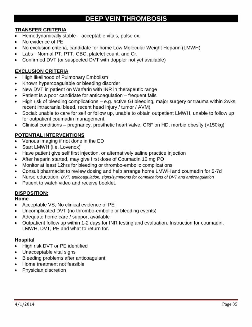
DEEP VEIN THROMBOSIS
TRANSFER CRITERIA
Hemodynamically stable – acceptable vitals, pulse ox.
No evidence of PE
No exclusion criteria, candidate for home Low Molecular Weight Heparin (LMWH)
Labs - Normal PT, PTT, CBC, platelet count, and Cr.
Confirmed DVT (or suspected DVT with doppler not yet available) EXCLUSION CRITERIA
High likelihood of Pulmonary Embolism
Known hypercoagulable or bleeding disorder
New DVT in patient on Warfarin with INR in therapeutic range
Patient is a poor candidate for anticoagulation – frequent falls
High risk of bleeding complications – e.g. active GI bleeding, major surgery or trauma within 2wks, recent intracranial bleed, recent head injury / tumor / AVM)
Social: unable to care for self or follow up, unable to obtain outpatient LMWH, unable to follow up for outpatient coumadin management.
Clinical conditions – pregnancy, prosthetic heart valve, CRF on HD, morbid obesity (>150kg)
POTENTIAL INTERVENTIONS
Venous imaging if not done in the ED
Start LMWH (i.e. Lovenox)
Have patient give self first injection, or alternatively saline practice injection
After heparin started, may give first dose of Coumadin 10 mg PO
Monitor at least 12hrs for bleeding or thrombo-embolic complications
Consult pharmacist to review dosing and help arrange home LMWH and coumadin for 5-7d Nurse education: DVT, anticoagulation, signs/symptoms for complications of DVT and anticoagulation
Patient to watch video and receive booklet.
DISPOSITION: Home
Acceptable VS, No clinical evidence of PE
Uncomplicated DVT (no thrombo-embolic or bleeding events)
Adequate home care / support available
Outpatient follow up within 1-2 days for INR testing and evaluation. Instruction for coumadin, LMWH, DVT, PE and what to return for.
Hospital
High risk DVT or PE identified
Unacceptable vital signs
Bleeding problems after anticoagulant
Home treatment not feasible
Physician discretion
4/1/2014 Page 36
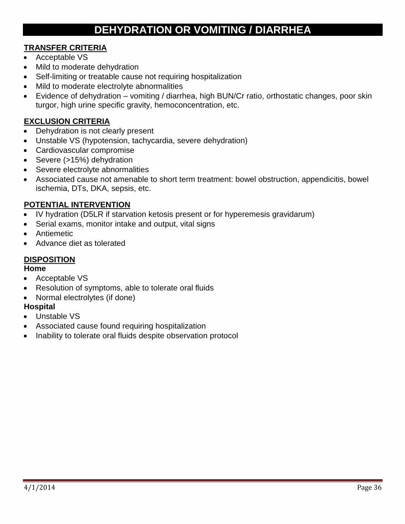
DEHYDRATION OR VOMITING / DIARRHEA
TRANSFER CRITERIA
Acceptable VS
Mild to moderate dehydration
Self-limiting or treatable cause not requiring hospitalization
Mild to moderate electrolyte abnormalities
Evidence of dehydration – vomiting / diarrhea, high BUN/Cr ratio, orthostatic changes, poor skin turgor, high urine specific gravity, hemoconcentration, etc.
EXCLUSION CRITERIA
Dehydration is not clearly present
Unstable VS (hypotension, tachycardia, severe dehydration)
Cardiovascular compromise
Severe (>15%) dehydration
Severe electrolyte abnormalities
Associated cause not amenable to short term treatment: bowel obstruction, appendicitis, bowel ischemia, DTs, DKA, sepsis, etc.
POTENTIAL INTERVENTION
IV hydration (D5LR if starvation ketosis present or for hyperemesis gravidarum)
Serial exams, monitor intake and output, vital signs
Antiemetic
Advance diet as tolerated
DISPOSITION Home
Acceptable VS
Resolution of symptoms, able to tolerate oral fluids
Normal electrolytes (if done) Hospital
Unstable VS
Associated cause found requiring hospitalization
Inability to tolerate oral fluids despite observation protocol

4/1/2014 Page 37
DILANTIN TOXICITY TRANSFER CRITERIA
Dilantin toxicity secondary to unintentional overmedication
Unsteady gait
CBC, chem-7, dilantin level
Toxicology consult on ALL patients: document time of call to Poison Control Center
IV access and cardiac monitor
Charcoal if indicated EXCLUSION CRITERIA
Suicide attempt or gesture
Multiple acute co-ingestants
Significant inebriation or intoxication
Unstable vital signs
Arrhythmmias
Other acute comorbidities or complicating illnesses
Dilantin level greater than 35 µ/cc POTENTIAL INTERVENTIONS
Cardiac monitoring
Serial dilantin levels
Repeat charcoal if needed
Initial bed rest, then advance activity as tolerated
Follow up with Poison Control Center call / consult DISPOSITION CRITERIA Home
Steady gait
Understanding of discharge instructions
Poison center or toxicology consult completed Hospital
Persistent symptoms after 24 hours
Increasing dilantin level after second dose of charcoal
Unstable vital signs or clinical condition
Physician discretion based on other complications or comorbidities

4/1/2014 Page 38
ELECTROLYTE ABNORMALITY
TRANSFER CRITERIA
Acceptable VS
Cause of electrolyte disturbance does not require hospitalization
No co-moribidity requiring more prolonged hospitalization
Mild and rapidly correctable electrolyte abnormality Hypokalemia > 2.5 mEq/L, with no ventricular ectopy on ED monitoring for >1 hour. Hyponatremia >120 mEq/L with normal mentation and a reversible etiology (eg dilutional, drug-
induced, gastroenteritis, hyperemesis). Not psychogenic polydipsia, SIADH Hypernatremia < 155 mEq/L with normal mentation and rapidly reversible etiology (e.g. NH
patient with infection) Hypercalcemia < 7.0 mEq/L (ionized) rapidly correctible etiology Hypocalcemia > 1.0 mEq/L (ionized), e.g. renal failure Hypomagnesemia >2.0 mEq/L associated with other electrolyte abnormalities
EXCLUSION CRITERIA
Unstable VS or cardiovascular compromise
Severe dehydration or severe electrolyte abnormalities (K >6.0, K <2.5, Na >155, Na <120, iCa >7.0, iCa <1.0, Mg <2.0)
Mental status changes, seizure, lethargy, neuro deficit, or other sign of cerebral edema
Associated cause not amenable to short term treatment: bowel obstruction, appendicitis, bowel ischemia, DTs, DKA, sepsis, some drug effects, etc.
Unlikely to be corrected within 15 hours
More than two acute electrolyte disturbances
POTENTIAL INTERVENTIONS
IV therapy (Normal saline for most) therapy targeting the specific disorder, per CDU physician.
Electrolyte replacement / correction, and repeat labs as ordered by CDU physician
Serial vital signs and repeat clinical examination
DISPOSITION Home
Acceptable VS
Resolution of symptoms, able to tolerate oral fluids
Improved electrolytes Hospital
Unstable VS
Associated cause found requiring hospitalization Inability to tolerate oral fluids

4/1/2014 Page 39
GASTROINTESTINAL BLEED (UPPER)
ADMISSION CRITERIA History of dark stool (not bright red) in last 24-48 hours
No more than 2 episodes of bright red blood
GI or surgery consulted for evaluation (or endoscopy) within 24hr
Normal PT/INR, Hgb >10, normal Cr.
Rectal exam for guiac and orthostatics vitals done in the ED
EXCLUSION CRITERIA
Unstable VS (HR>100, SBP<100, RR>22) or fever (T>38)
Significant orthostatic changes ( SBP>20); standing pulse >110
More than 2 episodes of bright red bleeding
Bowel prep and endoscpopy can not be completed within 18-24 hours (i.e. both EGD and colonscopy planned).
Active bleeding = fresh voluminous hematemesis, multiple episodes of melena on day of arrival, or a significant amount of bright red bowel movement per rectum
Hgb <8.0, or a drop of Hct >10 in 4 hours (if repeated in the ED)
History of end stage liver disease, coagulopathy, portal hypertension, esophageal varices, or coumadin
EKG Changes
Social issues = inadequate home support
POTENTIAL INTERVENTIONS
Serial Hct / Hgb Q6 hr
Guaiac stools / emesis prn.
IV Hydration, PPI or H2 blockers IV
Frequent VS – Q2 hours X3, then Q4hrs
NPO, I & O, clotting studies
GI Consult for endoscopy
DISPOSITION Home
Normal or stable serial exams
Stable VS
No deterioration in clinical condition
If endoscopy - no active bleeding, and follow-up arranged on PPI Hospital
Continual decrease in Hct/Hg
Recurrence of bleeding
Deterioration in clinical condition
Active bleeding by endoscopy

4/1/2014 Page 40
HEADACHE
TRANSFER CRITERIA
Persistent pain in tension or migraine headache
Hx of migraine with same aura, onset, location and pattern
Drug related headache
No focal neurological signs
Normal CT scan (if done)
If LP is needed, then it must be done and normal (unless failed attempt and IR consult for LP arranged in ED BEFORE transfer to CDU, and low risk patient)
Neurology, Neurosurgery, Neuro-ophthalmology consult completed in ED for complicated cases
EXCLUSION CRITERIA
Focal neurologic signs
Meningismus or high suspicion of meningitis, encephalitis, or subarachnoid hemorrhage
Elevated intraocular pressure as cause (i.e. glaucoma)
Abnormal CT scan
Abnormal LP (if performed)
Hypertensive emergency (diastolic BP > 120 with symptoms)
Suspected temporal arteritis
Blocked VP shunt
Frequent ED visits – suspected habitual patient, narcotic seeking behavior
POTENTIAL INTERVENTIONS
Serial exams including vital signs,
Neuro checks: level of alertness, speech, motor function
Analgesics, analgesics appropriate for a headache
Neurology consult as indicated
MRI/MRA/MRV Imaging as indicated
Retina scan if available
DISPOSITION CRITERIA Home
Resolution of pain
Other to take patient home
No deterioration in clinical course Hospital
No resolution in pain
Deterioration in clinical course
Rule in of exclusionary causes

4/1/2014 Page 41
HEAD INJURY
TRANSFER CRITERIA
Patients on anticoagulation with a normal head CT who need serial neuro exams or repeat head imaging
Patients not on anticoagulation with mildly abnormal CT head (questionable punctate hyperdensity, small SAH or SDH with no mass effect/shift) who need serial neuro exams or repeat CT imaging
Acceptable Vital Signs
Headache, dizziness, transient vomiting, transient amnesia are acceptable
Ethanol level <100 for intoxicated patients
Patients mentating clearly, able to ambulate and perform self-care
Otherwise cleared from a trauma standpoint
Lacerations repaired prior to CDU admission
Trauma and Neurosurgery consults initiated in the ED, as appropriate
EXCLUSION CRITERIA
Unstable VS
Abnormal CT Scan of brain in the setting of a coagulopathy
Depressed skull fracture
Penetrating skull injury
Focal neurologic abnormality or significant confusion
Uncooperative patient, restraints, or sitter required
Other traumatic injuries requiring further work-up or close monitoring
Patients who require ongoing spine precautions
Acute psychiatric disorder, suicidal patient
POTENTIAL INTERVENTION
Serial neurologic exams including vital signs every 2-4 hours, as ordered
Analgesics
Antiemetics
Neurosurgical consultation if indicated
Repeat CT scan or MRI as indicated
DISPOSITION Home
Acceptable VS
Normal serial neurologic exams
Hospital
Deterioration in clinical condition
Development of any exclusion criteria
4/1/2014 Page 42
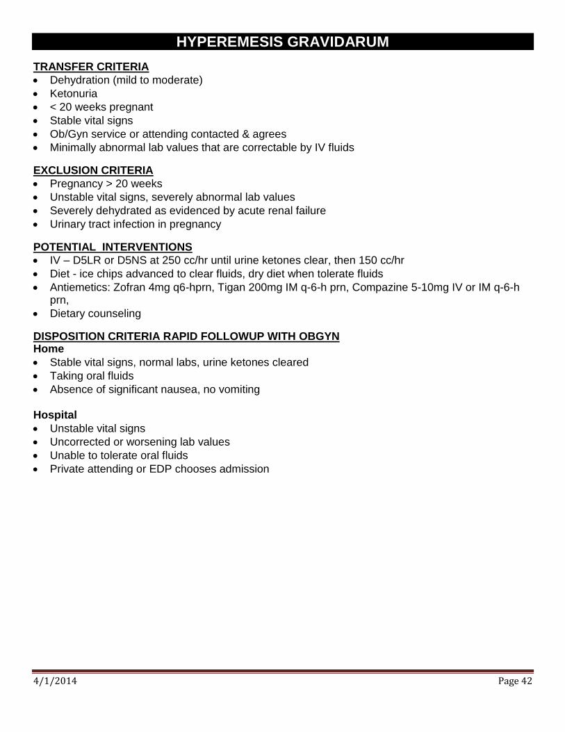
HYPEREMESIS GRAVIDARUM
TRANSFER CRITERIA
Dehydration (mild to moderate)
Ketonuria
< 20 weeks pregnant
Stable vital signs
Ob/Gyn service or attending contacted & agrees
Minimally abnormal lab values that are correctable by IV fluids
EXCLUSION CRITERIA
Pregnancy > 20 weeks
Unstable vital signs, severely abnormal lab values
Severely dehydrated as evidenced by acute renal failure
Urinary tract infection in pregnancy
POTENTIAL INTERVENTIONS
IV – D5LR or D5NS at 250 cc/hr until urine ketones clear, then 150 cc/hr
Diet - ice chips advanced to clear fluids, dry diet when tolerate fluids
Antiemetics: Zofran 4mg q6-hprn, Tigan 200mg IM q-6-h prn, Compazine 5-10mg IV or IM q-6-h prn,
Dietary counseling
DISPOSITION CRITERIA RAPID FOLLOWUP WITH OBGYN Home
Stable vital signs, normal labs, urine ketones cleared
Taking oral fluids
Absence of significant nausea, no vomiting
Hospital
Unstable vital signs
Uncorrected or worsening lab values
Unable to tolerate oral fluids
Private attending or EDP chooses admission
4/1/2014 Page 43
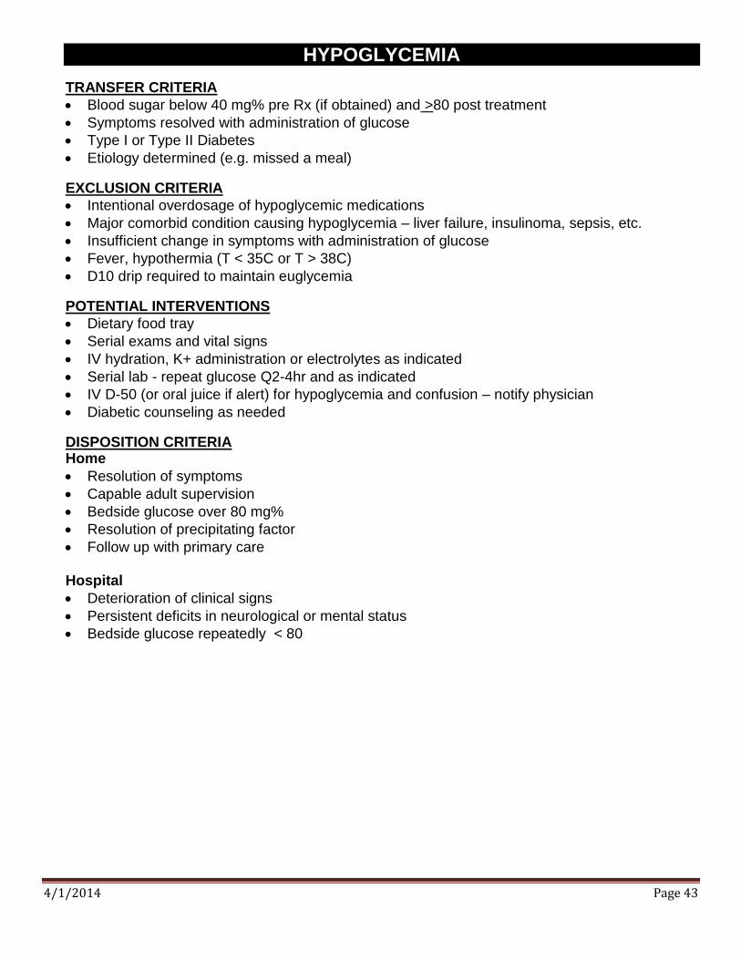
HYPOGLYCEMIA
TRANSFER CRITERIA
Blood sugar below 40 mg% pre Rx (if obtained) and >80 post treatment
Symptoms resolved with administration of glucose
Type I or Type II Diabetes
Etiology determined (e.g. missed a meal)
EXCLUSION CRITERIA
Intentional overdosage of hypoglycemic medications
Major comorbid condition causing hypoglycemia – liver failure, insulinoma, sepsis, etc.
Insufficient change in symptoms with administration of glucose
Fever, hypothermia (T < 35C or T > 38C)
D10 drip required to maintain euglycemia
POTENTIAL INTERVENTIONS
Dietary food tray
Serial exams and vital signs
IV hydration, K+ administration or electrolytes as indicated
Serial lab - repeat glucose Q2-4hr and as indicated
IV D-50 (or oral juice if alert) for hypoglycemia and confusion – notify physician
Diabetic counseling as needed
DISPOSITION CRITERIA Home
Resolution of symptoms
Capable adult supervision
Bedside glucose over 80 mg%
Resolution of precipitating factor
Follow up with primary care Hospital
Deterioration of clinical signs
Persistent deficits in neurological or mental status
Bedside glucose repeatedly < 80

4/1/2014 Page 44
HYPERGLYCEMIA
ADMISSION CRITERIA
Blood sugar > 300 & < 600 after ED treatment
Normal to near normal pH and total CO2 level
Readily treatable cause (e.g. non-compliance, UTI, abscess)
New onset hyperglycemia / suspect undiagnosed DM
EXCLUSION CRITERIA
DKA (pH <7.20, total CO2 <18, elevated serum acetone, anion gap >18)
Hyperosmolar non-ketotic coma (or AMS)
Blood glucose > 600
Precipitating cause unknown or not readily treatable
Social issues – precluding adequate outpatient management
POTENTIAL INTERVENTIONS
IV hydration, 0.9NS at 150-250 cc/hr
Bedside glucose q 2 hours until level < 300, then q 4 hours
Sliding scale insulin (see sliding scale guidelines)
Treat precipitating cause (antibiotics, I&D abscess, etc.)
Diabetic counseling
Repeat electrolytes q4hours until labs stable
Initiate Metformin if new onset per CDU MD
DISPOSITION CRITERIA Home
Blood glucose < 250
Resolution of symptoms
Stable vital signs
Successful treatment of precipitating cause
Tolerating PO fluids
PCP or Endocrine follow up within 48 hours if new onset
Patient education materials: includes BG monitor, lancets, strips, education video /book Hospital
Worsening symptoms
Unstable vital signs
Blood glucose remains > 250
Development of DKA
Unable to tolerate PO fluids
Poor candidate for home management

4/1/2014 Page 45
PNEUMONIA
TRANSFER CRITERIA
History, exam, and CXR consistent with acute pneumonia
PORT score class <3
O2 saturation >92 % on room air at the time of CDU admission
Able to return to previous living environment when discharged (outpatient support is present)
Initial dose of antibiotics given in the ED
EXCLUSION CRITERIA
Persistently abnormal vitals – after ED treatment (O2 saturation <92% on RA, HR >120, SBP<100, RR >30, T<35 or >40 C)
Significantly abnormal ABG – if done (pCO2>45, pH<7.35)
Potential respiratory failure
Multi-lobar pneumonia
Unlikely to be discharged in 24 hours, poor candidate for outpatient therapy
Immunocompromised patients: AIDS, PCP pneumonia, chemotherapy, chronic corticosteroid use, active cancer, sickle cell disease, asplenic patients.
High risk patients: Nursing home patient, cancer, cirrhosis, ESRD, altered mental status, nosocomial etiology, aspiration risk (ie. bulbar stroke)
High suspicion of – DVT/PE, SARS, H1N1, or TB (HIV/AIDS, institutionalized, recent prison, native of endemic region, history of pulmonary TB, apical disease on CXR)
POTENTIAL INTERVENTIONS
Antibiotics based on contemporary hospital guidelines for pneumonia
Supplemental oxygen and bronchodilator therapy as needed. Steroids and indicated.
Analgesics as needed for pain, myalgias, or cough/sputum
Serial vital signs, cardiac and oxygen saturation monitoring (continuous or intermittent)
Assistance with activities of daily living as needed
DISPOSITION Home
Subjective and clinical improvement during CDU stay
Acceptable vital signs during observation period
Patient able to tolerate oral medications and diet Hospital
Patient not subjectively improved enough to go home
Lack of clinical progress or clinical deterioration.
Unable to safely discharge for outpatient management
Physician discretion

4/1/2014 Page 46
PYELONEPHRITIS
TRANSFER CRITERIA
Acceptable vital signs and normal mentation
Clinical evidence of pyelonephritis (flank pain, urgency, frequency, dysuria)
UA evidence of pyelonephritis (significant pyuria, nitrates, and/or leukocyte esterase)
Not suitable for discharge from the ED
Urine cultures obtained
EXCLUSION CRITERIA
Male patients
Pregnant females
Abnormal VS after ED treatment (SBP <90, HR >120, T<35 or >40 C)
Mental status changes
Significant comorbidities – diabetes, renal failure, sickle cell disease
Immunosuppressed patients - HIV, transplant patients, chronic high dose steroids, asplenic
Urinary tract anatomic abnormality (solitary kidney, reflux, or indwelling device)
Urethral or ureteral obstruction (ie. kidney stones, urinary retention)
Poor candidate for outpatient treatment of pyelonephritis (ie poor home support)
POTENTIAL INTERVENTIONS
IV hydration, antiemetics, antipyretic
IV antibiotics based on contemporary guidelines for pyelonephritis
Advance to oral antibiotics, antiemetics, and analgesics – as tolerated
Imaging as needed (CT or ultrasound)
DISPOSITION CRITERIA Home
Resolution or improvement of systemic symptoms
Ability to take po medications
Stable vital signs
PCP follow up within 72 hours for culture results and repeat exam. Hospital
Clinical deterioration or lack of adequate improvement
Inability to tolerate oral meds or hydration
Unstable vital signs or evidence of septic shock
Abnormal imaging (ureteral obstruction or emphysematous pyelonephritis, solitary kidney)

4/1/2014 Page 47
RENAL COLIC
TRANSFER CRITERIA
Diagnosis of renal colic established by helical CT, IVP or ultrasound
Uncomplicated stone
Persistent pain or vomiting despite medication
Acceptable VS
Urology resident notified
EXCLUSION CRITERIA
Unstable VS
Clinical evidence of a UTI (fever, significant pyuria on a cath specimen)
Solitary kidney
Relative large proximal stone (>6 mm) with high grade obstruction
Acute renal failure
POTENTIAL INTERVENTION
IV Hydration
As needed - IV narcotics, IV ketolorac, IV antiemetics
Medical Expulsive therapy as indicated (ie Flomax / tamsulosin, steroids)
Diagnostic tests as needed - Delayed IVP films, ultrasound, CT
Serial exams and vital signs
Strain urine for stone capture and analysis, U/A if not yet done
Urology consultation as needed.
DISPOSITION Home
Acceptable VS
Pain and nausea resolved or controlled
Passage of stone Hospital
Persistent vomiting or uncontrolled pain after 14 hours
Diagnosis of coexistent infection or significant abnormality
Change in diagnosis requiring further therapy or workup
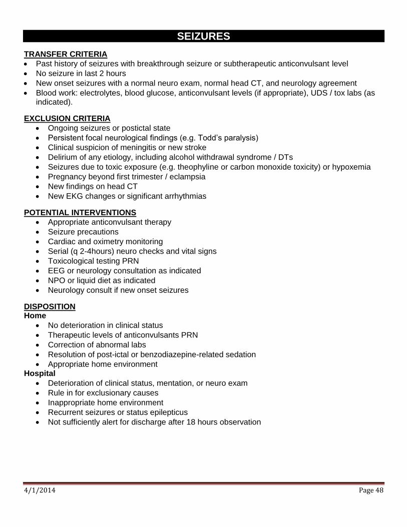
4/1/2014 Page 48
SEIZURES
TRANSFER CRITERIA
Past history of seizures with breakthrough seizure or subtherapeutic anticonvulsant level
No seizure in last 2 hours
New onset seizures with a normal neuro exam, normal head CT, and neurology agreement
Blood work: electrolytes, blood glucose, anticonvulsant levels (if appropriate), UDS / tox labs (as indicated).
EXCLUSION CRITERIA
Ongoing seizures or postictal state
Persistent focal neurological findings (e.g. Todd’s paralysis)
Clinical suspicion of meningitis or new stroke
Delirium of any etiology, including alcohol withdrawal syndrome / DTs
Seizures due to toxic exposure (e.g. theophyline or carbon monoxide toxicity) or hypoxemia
Pregnancy beyond first trimester / eclampsia
New findings on head CT
New EKG changes or significant arrhythmias
POTENTIAL INTERVENTIONS
Appropriate anticonvulsant therapy
Seizure precautions
Cardiac and oximetry monitoring
Serial (q 2-4hours) neuro checks and vital signs
Toxicological testing PRN
EEG or neurology consultation as indicated
NPO or liquid diet as indicated
Neurology consult if new onset seizures
DISPOSITION Home
No deterioration in clinical status
Therapeutic levels of anticonvulsants PRN
Correction of abnormal labs
Resolution of post-ictal or benzodiazepine-related sedation
Appropriate home environment Hospital
Deterioration of clinical status, mentation, or neuro exam
Rule in for exclusionary causes
Inappropriate home environment
Recurrent seizures or status epilepticus
Not sufficiently alert for discharge after 18 hours observation
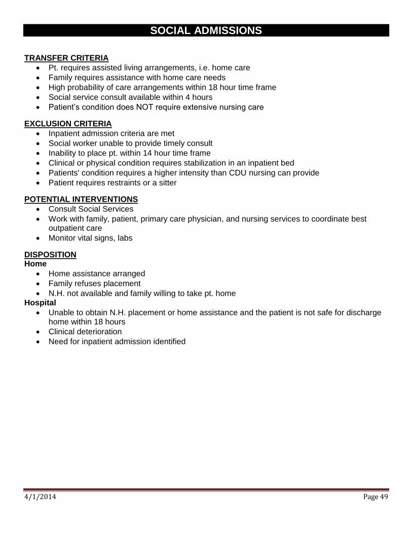
4/1/2014 Page 49
SOCIAL ADMISSIONS
TRANSFER CRITERIA
Pt. requires assisted living arrangements, i.e. home care
Family requires assistance with home care needs
High probability of care arrangements within 18 hour time frame
Social service consult available within 4 hours
Patient’s condition does NOT require extensive nursing care
EXCLUSION CRITERIA
Inpatient admission criteria are met
Social worker unable to provide timely consult
Inability to place pt. within 14 hour time frame
Clinical or physical condition requires stabilization in an inpatient bed
Patients' condition requires a higher intensity than CDU nursing can provide
Patient requires restraints or a sitter
POTENTIAL INTERVENTIONS
Consult Social Services
Work with family, patient, primary care physician, and nursing services to coordinate best outpatient care
Monitor vital signs, labs
DISPOSITION Home
Home assistance arranged
Family refuses placement
N.H. not available and family willing to take pt. home Hospital
Unable to obtain N.H. placement or home assistance and the patient is not safe for discharge home within 18 hours
Clinical deterioration
Need for inpatient admission identified
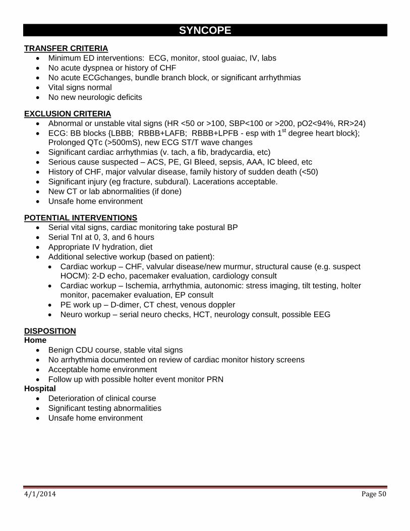
4/1/2014 Page 50
SYNCOPE
TRANSFER CRITERIA
Minimum ED interventions: ECG, monitor, stool guaiac, IV, labs
No acute dyspnea or history of CHF
No acute ECGchanges, bundle branch block, or significant arrhythmias
Vital signs normal
No new neurologic deficits
EXCLUSION CRITERIA
Abnormal or unstable vital signs (HR <50 or >100, SBP<100 or >200, pO2<94%, RR>24)
ECG: BB blocks {LBBB; RBBB+LAFB; RBBB+LPFB - esp with 1st degree heart block}; Prolonged QTc (>500mS), new ECG ST/T wave changes
Significant cardiac arrhythmias (v. tach, a fib, bradycardia, etc)
Serious cause suspected – ACS, PE, GI Bleed, sepsis, AAA, IC bleed, etc
History of CHF, major valvular disease, family history of sudden death (<50)
Significant injury (eg fracture, subdural). Lacerations acceptable.
New CT or lab abnormalities (if done)
Unsafe home environment
POTENTIAL INTERVENTIONS
Serial vital signs, cardiac monitoring take postural BP
Serial TnI at 0, 3, and 6 hours
Appropriate IV hydration, diet
Additional selective workup (based on patient):
Cardiac workup – CHF, valvular disease/new murmur, structural cause (e.g. suspect HOCM): 2-D echo, pacemaker evaluation, cardiology consult
Cardiac workup – Ischemia, arrhythmia, autonomic: stress imaging, tilt testing, holter monitor, pacemaker evaluation, EP consult
PE work up – D-dimer, CT chest, venous doppler
Neuro workup – serial neuro checks, HCT, neurology consult, possible EEG
DISPOSITION Home
Benign CDU course, stable vital signs
No arrhythmia documented on review of cardiac monitor history screens
Acceptable home environment
Follow up with possible holter event monitor PRN Hospital
Deterioration of clinical course
Significant testing abnormalities
Unsafe home environment
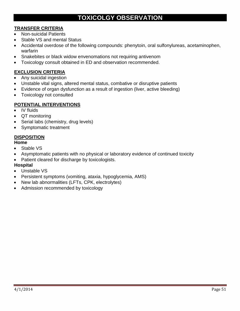
4/1/2014 Page 51
TOXICOLGY OBSERVATION
TRANSFER CRITERIA
Non-suicidal Patients
Stable VS and mental Status
Accidental overdose of the following compounds: phenytoin, oral sulfonylureas, acetaminophen, warfarin
Snakebites or black widow envenomations not requiring antivenom
Toxicology consult obtained in ED and observation recommended.
EXCLUSION CRITERIA
Any suicidal ingestion
Unstable vital signs, altered mental status, combative or disruptive patients
Evidence of organ dysfunction as a result of ingestion (liver, active bleeding)
Toxicology not consulted
POTENTIAL INTERVENTIONS
IV fluids
QT monitoring
Serial labs (chemistry, drug levels)
Symptomatic treatment
DISPOSITION Home
Stable VS
Asymptomatic patients with no physical or laboratory evidence of continued toxicity
Patient cleared for discharge by toxicologists. Hospital
Unstable VS
Persistent symptoms (vomiting, ataxia, hypoglycemia, AMS)
New lab abnormalities (LFTs, CPK, electrolytes)
Admission recommended by toxicology

4/1/2014 Page 52
TRANSFUSION OF BLOOD AND BLOOD PRODUCTS
TRANSFER CRITERIA
Symptomatic anemia or thrombocytopenia
Deficiency correctable by transfusion
Stable vital signs with recent labs verifying need for transfusion
EXCLUSION CRITERIA
Unstable vital signs
Active bleeding present unless transfusing platelets for thrombocytopenia and patient stable
End stage renal failure, dialysis patients
Pregnant patients
Hgb <5
POTENTIAL INTERVENTIONS
IV started, Pre-medicate and IV hydration as needed
Type and Cross match sent if not previously done
Transfuse only leukocyte-reduced red cells or platelets per Nursing protocol –Repeat CBC at least 2 hours following transfusion.
Obtain and record vital signs according to blood bank policy:
Within 30 minutes prior to the transfusion (baseline)
15 minutes after the start of the transfusion:
1 hour after the start of the transfusion
Post transfusion
DISPOSITION Home
Stable vital signs
Symptoms improved
No fever for 1 hour after 1 unit PRBC's or 1 dose of platelets for 2 hours after 2 units PRBC's
No evidence of fluid overload or CHF
No evidence of transfusion reaction per Nursing protocol
Satisfactory increase in hemoglobin following transfusion Hospital
Transfusion reaction
Unstable vital signs
Fluid overload, CHF
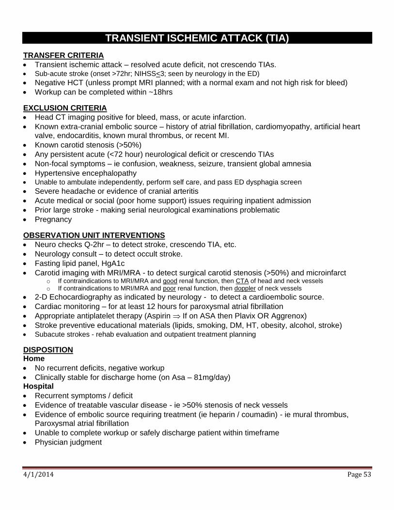
4/1/2014 Page 53
TRANSIENT ISCHEMIC ATTACK (TIA)
TRANSFER CRITERIA
Transient ischemic attack – resolved acute deficit, not crescendo TIAs. Sub-acute stroke (onset >72hr; NIHSS<3; seen by neurology in the ED)
Negative HCT (unless prompt MRI planned; with a normal exam and not high risk for bleed)
Workup can be completed within ~18hrs
EXCLUSION CRITERIA
Head CT imaging positive for bleed, mass, or acute infarction.
Known extra-cranial embolic source – history of atrial fibrillation, cardiomyopathy, artificial heart valve, endocarditis, known mural thrombus, or recent MI.
Known carotid stenosis (>50%)
Any persistent acute (<72 hour) neurological deficit or crescendo TIAs
Non-focal symptoms – ie confusion, weakness, seizure, transient global amnesia
Hypertensive encephalopathy Unable to ambulate independently, perform self care, and pass ED dysphagia screen
Severe headache or evidence of cranial arteritis
Acute medical or social (poor home support) issues requiring inpatient admission
Prior large stroke - making serial neurological examinations problematic
Pregnancy
OBSERVATION UNIT INTERVENTIONS
Neuro checks Q-2hr – to detect stroke, crescendo TIA, etc.
Neurology consult – to detect occult stroke.
Fasting lipid panel, HgA1c
Carotid imaging with MRI/MRA - to detect surgical carotid stenosis (>50%) and microinfarct o If contraindications to MRI/MRA and good renal function, then CTA of head and neck vessels o If contraindications to MRI/MRA and poor renal function, then doppler of neck vessels
2-D Echocardiography as indicated by neurology - to detect a cardioembolic source.
Cardiac monitoring – for at least 12 hours for paroxysmal atrial fibrillation
Appropriate antiplatelet therapy (Aspirin If on ASA then Plavix OR Aggrenox)
Stroke preventive educational materials (lipids, smoking, DM, HT, obesity, alcohol, stroke) Subacute strokes - rehab evaluation and outpatient treatment planning
DISPOSITION Home
No recurrent deficits, negative workup
Clinically stable for discharge home (on Asa – 81mg/day) Hospital
Recurrent symptoms / deficit
Evidence of treatable vascular disease - ie >50% stenosis of neck vessels
Evidence of embolic source requiring treatment (ie heparin / coumadin) - ie mural thrombus, Paroxysmal atrial fibrillation
Unable to complete workup or safely discharge patient within timeframe
Physician judgment
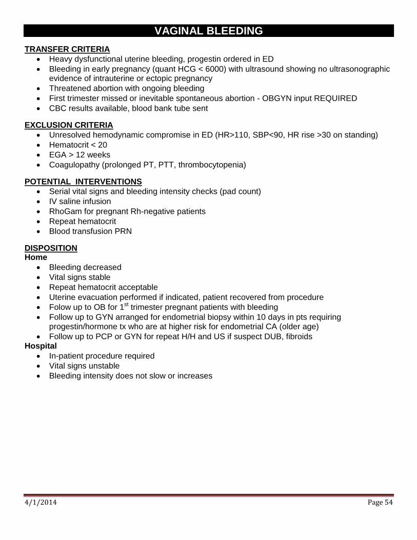
4/1/2014 Page 54
VAGINAL BLEEDING
TRANSFER CRITERIA
Heavy dysfunctional uterine bleeding, progestin ordered in ED
Bleeding in early pregnancy (quant HCG < 6000) with ultrasound showing no ultrasonographic evidence of intrauterine or ectopic pregnancy
Threatened abortion with ongoing bleeding
First trimester missed or inevitable spontaneous abortion - OBGYN input REQUIRED
CBC results available, blood bank tube sent
EXCLUSION CRITERIA
Unresolved hemodynamic compromise in ED (HR>110, SBP<90, HR rise >30 on standing)
Hematocrit < 20
EGA > 12 weeks
Coagulopathy (prolonged PT, PTT, thrombocytopenia)
POTENTIAL INTERVENTIONS
Serial vital signs and bleeding intensity checks (pad count)
IV saline infusion
RhoGam for pregnant Rh-negative patients
Repeat hematocrit
Blood transfusion PRN
DISPOSITION Home
Bleeding decreased
Vital signs stable
Repeat hematocrit acceptable
Uterine evacuation performed if indicated, patient recovered from procedure
Folow up to OB for 1st trimester pregnant patients with bleeding
Follow up to GYN arranged for endometrial biopsy within 10 days in pts requiring progestin/hormone tx who are at higher risk for endometrial CA (older age)
Follow up to PCP or GYN for repeat H/H and US if suspect DUB, fibroids Hospital
In-patient procedure required
Vital signs unstable
Bleeding intensity does not slow or increases
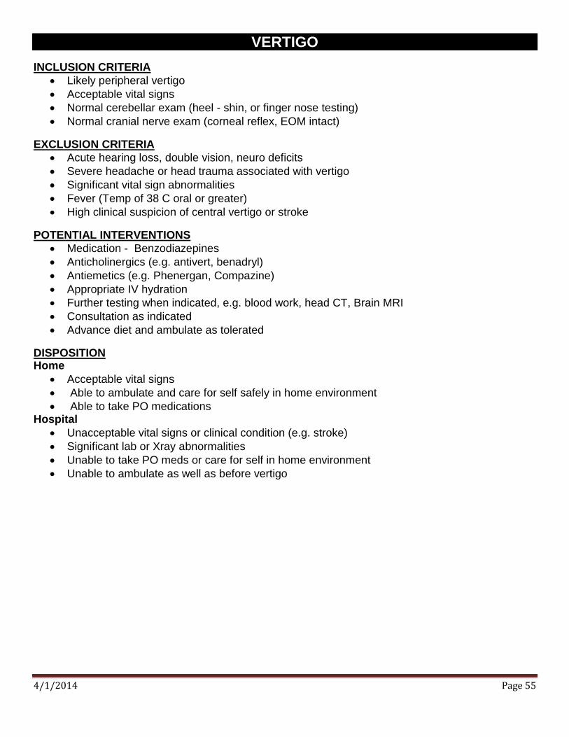
4/1/2014 Page 55
VERTIGO
INCLUSION CRITERIA
Likely peripheral vertigo
Acceptable vital signs
Normal cerebellar exam (heel - shin, or finger nose testing)
Normal cranial nerve exam (corneal reflex, EOM intact)
EXCLUSION CRITERIA
Acute hearing loss, double vision, neuro deficits
Severe headache or head trauma associated with vertigo
Significant vital sign abnormalities
Fever (Temp of 38 C oral or greater)
High clinical suspicion of central vertigo or stroke
POTENTIAL INTERVENTIONS
Medication - Benzodiazepines
Anticholinergics (e.g. antivert, benadryl)
Antiemetics (e.g. Phenergan, Compazine)
Appropriate IV hydration
Further testing when indicated, e.g. blood work, head CT, Brain MRI
Consultation as indicated
Advance diet and ambulate as tolerated
DISPOSITION Home
Acceptable vital signs
Able to ambulate and care for self safely in home environment
Able to take PO medications Hospital
Unacceptable vital signs or clinical condition (e.g. stroke)
Significant lab or Xray abnormalities
Unable to take PO meds or care for self in home environment
Unable to ambulate as well as before vertigo