emergency war surgery chp16thoracicinjuries
TRANSCRIPT

8/6/2019 Emergency War Surgery Chp16ThoracicInjuries
http://slidepdf.com/reader/full/emergency-war-surgery-chp16thoracicinjuries 1/16
16.1
Thoracic Injuries
Chapter 16
Thoracic Injuries
IntroductionAbout 15% of war injuries involve the chest. Of those, 10% aresuperficial (soft tissue only) requiring only basic wound
treatment. The remaining 90% of chest injuries are almost allpenetrating.
Those injuries involving the central column of the chest (heart,great vessels, pulmonary hilum) are generally fatal on the battlefield. Injuries of the lung parenchyma (the vast majority)can be managed by the insertion of a chest tube and basic wound
treatment. Although penetrating injuries are most common, blunt chest trauma may occur and can result in disruption of thecontents of the thorax as well as injury to the chest wall itself.Blast injuries can result in the rupture of air-filled structures(the lung) as well as penetrating injuries from fragments.
The immediate recognition and treatment of tensionpneumothorax is the single most important and life-
saving intervention in the treatment of chest injuries incombat. Distended neck veins, tracheal shift, decreasedbreath sounds, and hyperresonance in the affectedhemithorax, and hypotension are the cardinal signs.None or all may be present. Immediate decompressionis lifesaving.
With the advent of body armor, it is hoped that the majority of thoracic injuries seen in past conflicts will be avoided.Unfortunately, there will be individuals who will not have suchprotection, as well as others who will sustain chest injuriesdespite protection.

8/6/2019 Emergency War Surgery Chp16ThoracicInjuries
http://slidepdf.com/reader/full/emergency-war-surgery-chp16thoracicinjuries 2/16
16.2
Emergency War Surgery
Anatomic Considerations Superior border is at the level of the clavicles anteriorly and
the junction of the C7-T1 vertebral bodies posteriorly. Thethoracic inlet at that level contains major arteries (commoncarotids, vertebrals), veins (anterior and internal jugulars),trachea, esophagus, and spinal cord.
Within or traversing the container of the chest itself are foundthe heart and coronary vessels, great vessels including arteries(aorta, arch, inominate, right subclavian, common carotid,left subclavian, and descending aorta), veins (superior andinferior vena cava, azygous vein, brachiocephalic vein),
pulmonary arteries and veins, distal trachea and main stem bronchi, lungs, and esophagus.
The inferior border is described by the diaphragm, attachedanteriorly at the T6 level and gradually sloping posteriorlyto the T12 level.
Penetrating thoracic injuries below the T4 level (nippleline) have a high probability of involving abdominalstructures (Fig. 16-1).
Evaluation and DiagnosisKnowledge of the mechanism of injury (eg, blast, fragment, amongothers) may increase the index of suspicion for a particular injury. A
complete and accurate diagnosis isusually not possible because of thelimited diagnostic tools available inthe setting of combat trauma. None-theless, because injuries to the chestcan profoundly affect breathing andcirculation (and on rare occasion, theairway), a complete and rapid assess-ment of each injury is mandatory. If the casualty is able to talk, there is reasonable assurance
that the airway is intact.
Fig. 16-1. Thoracic incisionof abdominal contents.
T4T4

8/6/2019 Emergency War Surgery Chp16ThoracicInjuries
http://slidepdf.com/reader/full/emergency-war-surgery-chp16thoracicinjuries 3/16
16.3
Thoracic Injuries
Life-Threatening Injuries
Injuries not immediately obvious, yet requiring urgentattention, include tension pneumothorax, massivehemothorax, and cardiac tamponade.
Tension Pneumothorax.! A patient with a known chest injury presenting with an
open airway and difficulty breathing has a tensionpneumothorax until proven otherwise and requires rapiddecompression and the insertion of a chest tube.
Massive Hemothorax.! The return of blood may indicate a significant intrathoracic
injury. Generally, the immediate return of 1,500 cc of bloodmandates thoracotomy (especially if the wound wassustained within the past hour). With less blood initially, but a continued loss of 200 cc/hour for over 4 hours ,thoracotomy is indicated.
!
Casualties with massive thoracic hemorrhage requiredamage control techniques (see Chapter 12, DamageControl Surgery).
Cardiac Tamponade.! Distended neck veins (may be absent with significant
blood loss) in the presence of clear breath sounds andhypotension indicate the possibility of life-threateningcardiac tamponade.
! Fluid resuscitation may temporarily stabilize a patient intamponade.
! Perform an ultrasound (US) with a stable patient." If positive, proceed to the OR (pericardial window,
sternotomy, thoracotomy). Any pericardial bloodmandates median sternotomy/thoracotomy.
" A negative US requires either repeat US or pericardial
window, depending on level of clinical suspicion.! Pericardiocentesis is only a stopgap measure on the wayto definitive surgical repair.

8/6/2019 Emergency War Surgery Chp16ThoracicInjuries
http://slidepdf.com/reader/full/emergency-war-surgery-chp16thoracicinjuries 4/16
16.4
Emergency War Surgery
Open pneumothorax (hole in chest wall) is treated by placinga chest tube and sealing the hole. Alternatives include one-way valve chest dressings or a square piece of plastic dressingtaped to the chest on three sides.
Flail chest (entire segment of the chest wall floating due tofractures of a block of ribs, with two fractures on each rib)will require treatment (either airway intubation or obser-vation) based on the severity of the underlying lung injury.In cases where intubation is not required, repeated intercostalnerve blocks with a long-acting local anesthetic such asMarcaine may be very helpful in relieving pain and limiting
atelectasis and other pulmonary complications.
Surgical Management
Most penetrating chest injuries reaching medicalattention are adequately treated with tube thoracostomy(chest tube) alone.
Tube thoracostomy (chest tube). Indications.! Known or suspected tension pneumothorax.! Pneumothorax (including open).! Hemothorax.! Any penetrating chest injury requiring transport (manda-
tory in case of aeromedical evacuation).
Procedure (Fig. 16-2).! In cases of tension pneumothorax, immediate decom-
pression with a large bore needle is lifesaving. An IVcatheter (14/16/18 gauge at least 2–3 inches in length) isinserted in the midclavicular line in the second interspace(approximately 2 fingerbreadths below the clavicle on theadult male). Entry is confirmed by the sound of air passing
through the catheter. This must be rapidly followed bythe insertion of a chest tube.! In a contaminated environment, a single gram of IV
cefazolin (Ancef) is recommended.! If time allows, prep the anterior and lateral chest on the
affected side with povidone-iodine.

8/6/2019 Emergency War Surgery Chp16ThoracicInjuries
http://slidepdf.com/reader/full/emergency-war-surgery-chp16thoracicinjuries 5/16
16.5
Thoracic Injuries
! Identify the incision site along the anterior axillary line,intersecting the 5th or 6th rib.
! Inject a local anesthetic in the awake patient, if conditionsallow.
! Make a transverse incision, 3–4 cm in length, along andcentered over the rib, carrying it down to the bone.
12
3
4
5
6 7
8
9
0
7
6
5
Lungs
4 5
6
AnteriorAxillary Line
c
d
e
4 5 6 7
89
1012 3
b
4 5
6
6th Rib Site
Chest tube
Suction/ Open
Heimlich valve
OR
a
Fig. 16-2. Procedure for tube thoracostomy.
8/6/2019 Emergency War Surgery Chp16ThoracicInjuries
http://slidepdf.com/reader/full/emergency-war-surgery-chp16thoracicinjuries 6/16
16.6
Emergency War Surgery
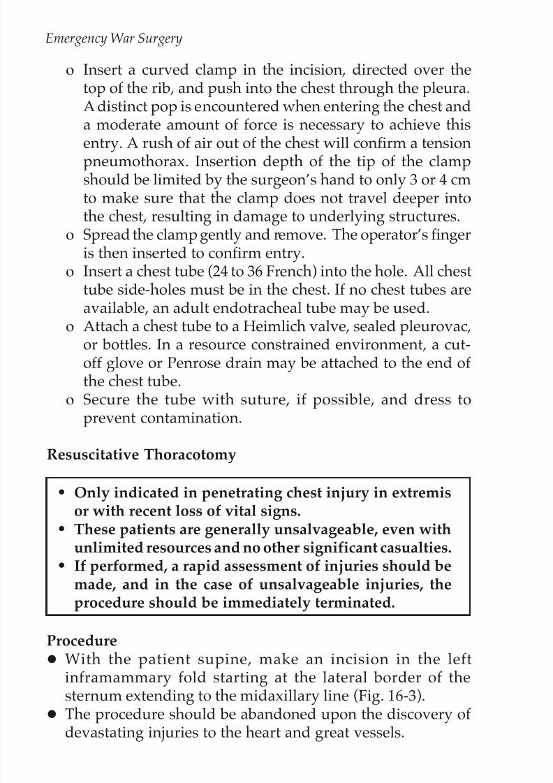
! Insert a curved clamp in the incision, directed over thetop of the rib, and push into the chest through the pleura.A distinct pop is encountered when entering the chest anda moderate amount of force is necessary to achieve thisentry. A rush of air out of the chest will confirm a tensionpneumothorax. Insertion depth of the tip of the clampshould be limited by the surgeon’s hand to only 3 or 4 cmto make sure that the clamp does not travel deeper intothe chest, resulting in damage to underlying structures.
! Spread the clamp gently and remove. The operator’s fingeris then inserted to confirm entry.
! Insert a chest tube (24 to 36 French) into the hole. All chesttube side-holes must be in the chest. If no chest tubes areavailable, an adult endotracheal tube may be used.
! Attach a chest tube to a Heimlich valve, sealed pleurovac,or bottles. In a resource constrained environment, a cut-off glove or Penrose drain may be attached to the end of the chest tube.
! Secure the tube with suture, if possible, and dress toprevent contamination.
Resuscitative Thoracotomy
••••• Only indicated in penetrating chest injury in extremisor with recent loss of vital signs.
••••• These patients are generally unsalvageable, even with
unlimited resources and no other significant casualties.••••• If performed, a rapid assessment of injuries should be
made, and in the case of unsalvageable injuries, theprocedure should be immediately terminated.
Procedure With the patient supine, make an incision in the left
inframammary fold starting at the lateral border of thesternum extending to the midaxillary line (Fig. 16-3). The procedure should be abandoned upon the discovery of
devastating injuries to the heart and great vessels.

8/6/2019 Emergency War Surgery Chp16ThoracicInjuries
http://slidepdf.com/reader/full/emergency-war-surgery-chp16thoracicinjuries 7/16
16.7
Thoracic Injuries
If no injury is found in leftchest, rapidly extend theincision across the midline,crossing through the sternumwith a Lebsche sternum knife,performing a mirror-imagethoracotomy (clamshell,Fig. 16-4). When doing thisprocedure you will cut across both internal mammaryarteries, which will be a sig-
nificant source of bleeding. Elevating the anterior chest
wall will expose virtually all mediastinal structures. Open the pericardium and assess the heart. Priorities are to stop bleeding and restore central perfusion.! Holes in the heart and/or great vessels should be
temporarily occluded." Temporary occlusion can be achieved with fingers, side-
biting clamps or Foley catheters with 30 cc balloons.Any other sterile device of opportunity is acceptable.
! Major pulmonary hilar injuries should be cross-clampeden masse.
! Descending aorta located, cross-clamped, and cardiacfunction restored via defibrillation or massage. (Make sureto open the mediastinal pleura over the aorta to securely
apply the vascular clamp.)! If unable to restore cardiac function rapidly, abandon the
operation. With successful restoration of cardiac function, injuries should
be more definitively repaired.
Subxiphoid Pericardial Window
Subxiphoid pericardial window should not be attemptedin an unstable patient. Unstable patients with penetratinginjuries suspicious for cardiac injury should undergoimmediate median sternotomy/thoracotomy.
Fig. 16-3. Incision for resuscitative
thoracotomy.

8/6/2019 Emergency War Surgery Chp16ThoracicInjuries
http://slidepdf.com/reader/full/emergency-war-surgery-chp16thoracicinjuries 8/16
16.8
Emergency War Surgery
Procedure
With the patient supine, make a 4–5 cm longitudinal incision just on and below the xiphoid process through the skin andfascia.
Bluntly dissect superiorly toward the heart exposing thephrenopericardial membrane below the heart.
Fig. 16-4.Supraclavicular approach.
Supraclavicular Extension

8/6/2019 Emergency War Surgery Chp16ThoracicInjuries
http://slidepdf.com/reader/full/emergency-war-surgery-chp16thoracicinjuries 9/16
16.9
Thoracic Injuries
Place two stay sutures into the membrane and sharply incise between them, with care to avoid the heart, opening thepericardial sac and exposing the underlying beating heart.
Median Sternotomy Indications.! Suspected cardiac injury in an unstable patient.! Positive pericardiocentesis/subxiphoid pericardial
window.! Suspected injury to the great vessels in the chest.! Suspected distal tracheal injury.
Procedure.! In the supine position, make a midline skin incision from
the sternal notch to just below the xiphoid.! Through blunt/sharp dissection, develop a plane for
several centimeters both superiorly and inferiorly beneaththe sternum.
! Divide the sternum with a sternal saw or Lebsche knife.Keep the foot of the knife/saw tilted up toward theundersurface of the sternum to avoid cardiac injury. Bonewax can be used to decrease bleeding on the cut edges of the sternum.
! Separate the halves of the sternum using a chest retractor.! Carefully divide the pericardium superiorly, avoiding the
innominate vein, exposing the heart and base of the greatvessels.
In general, exposure to the heart and great vessels is bestachieved through a median sternotomy. For proximal leftsubclavian artery injuries, additional exposure (trapdoor) may be necessary.
! Close with wire suture directly through the halves of thesternum, approximately 2 cm from the edge, or aroundthe sternum through the costal interspaces using wiresutures.
! Place one or two mediastinal tubes for drainage, exitingthrough a midline stab wound inferior to the mediastinalskin incision.

8/6/2019 Emergency War Surgery Chp16ThoracicInjuries
http://slidepdf.com/reader/full/emergency-war-surgery-chp16thoracicinjuries 10/16
16.10
Emergency War Surgery
Trap Door Extension
Other Approaches Supraclavicular.! Indication." Mid to distal subclavian artery injury.
! Procedure." Make an incision 2 cm above and parallel to the clavicle,
beginning at the sternal notch and extending laterally 8 cm.Trap door (Fig. 16-5).! Indication." Proximal left subclavian artery injury.
! Procedure.
Fig. 16-5. Trap door procedure.

8/6/2019 Emergency War Surgery Chp16ThoracicInjuries
http://slidepdf.com/reader/full/emergency-war-surgery-chp16thoracicinjuries 11/16
16.11
Thoracic Injuries
" Perform supraclavicular approach as above." Perform a partial median sternotomy to the fourth
intercostal space." At the fourth intercostal interspace, incise the skin
laterally in the submammary fold to the anterior axillaryline.
" Divide the sternum laterally and continue in the 4th
intercostal space (ICS) to the anterior axillary line. Theinternal mammary artery will be divided and must becontrolled.
" It may be necessary to either fracture or remove a section
of clavicle to gain adequate exposure of the proximalleft subclavian artery.
" Approach distal left subclavian artery injuries througha supraclavicular incision.
Thoracoabdominal.! Indication." Combined thoracic and abdominal injuries.
! Procedure." The resuscitative thoracotomy can be continued
medially and inferiorly across the costal margin intothe abdominal midline to complete a thoracoabdominalincision.
" Alternatively, a separate abdominal incision can bemade.
" With right-sided lower chest injuries, the liver and
retrohepatic vena cava can be exposed well using a rightthoracoabdominal approach.
Specific Injuries Vascular.! Initially, holes in vessels should be digitally occluded.
Stopgap measures include placing Fogarty or Foleycatheters, side-biting clamps, or in the case of venousinjuries, sponge sticks.
! Total occlusion or clamping may temporarily be necessaryto allow resuscitation to continue and restore cardiacfunction.

8/6/2019 Emergency War Surgery Chp16ThoracicInjuries
http://slidepdf.com/reader/full/emergency-war-surgery-chp16thoracicinjuries 12/16
16.12
Emergency War Surgery
! If cardiac function cannot be restored within 5 to 10minutes, the procedure should be abandoned (on-the-tabletriage).
! Repair of vessels should follow the principles detailed invascular repair: attempting primary repair if possible, withthe use of prosthetics if primary repair is not feasible.Consider shunting as an alternative.
Heart.
The usual result of high-velocity injuries to the heart is
irreparable destruction of the muscle.
! Isolated punctures of the heart should be exposed (openingthe pericardium) and occluded by finger pressure. Othermethods include the use of a Foley catheter or skin staples.
! Use pledgeted horizontal mattress sutures (2-0 prolene)on a tapered needle for definitive repair. Care must be
taken to avoid additional injury to coronary vessels.Extreme care must be taken to avoid tearing the cardiacmuscle.
! Atrial repairs may include simple ligature, stapled repair,or running closures (Fig. 16-6).
! Temporary inflow occlusion may prove helpful in repair.! More complex repairs are impractical without cardiac
bypass. Lung.! Tube thoracostomy alone is adequate treatment for most
simple lung parenchymal injuries.! Large air leaks not responding to chest tubes or that do
not allow adequate ventilation will require open repair (seetracheobronchial tree below).
! Posterolateral thoracotomy is preferred for isolated lung
injuries. Anterior thoracotomy may also be used.! Control simple bleeding with absorbable suture on a
tapered needle. Alternatively, staples (TA-90) may be usedfor bleeding lung tears.

8/6/2019 Emergency War Surgery Chp16ThoracicInjuries
http://slidepdf.com/reader/full/emergency-war-surgery-chp16thoracicinjuries 13/16
16.13
Thoracic Injuries
! Tractotomy: Open any bleeding tracts (through andthrough lung penetrations) with a GIA stapler and ligate bleeding points.
Do not simply close the entrance and exit points ofpenetrating tracts in the lung. With positive pressureventilation, the risk is air embolism. The more centralthe injury, the higher the risk.
! Resection for bleeding may be indicated with severeparenchymal injury. Anatomic resections are not indicated
and simple stapled wedge excisions recommended.! Uncontrolled parenchymal/hilar bleeding, or complex
hilar injuries with massive air leak should be controlled withhilar clamping and repair attempted. Pneumonectomy isperformed as a last resort (90% mortality).
Fig. 16-6. Repair of penetrating cardiac injury.

8/6/2019 Emergency War Surgery Chp16ThoracicInjuries
http://slidepdf.com/reader/full/emergency-war-surgery-chp16thoracicinjuries 14/16
16.14
Emergency War Surgery
Tracheobronchial tree.! Suspect the diagnosis with massive air leak, frothy
hemoptysis, and pneumomediastinum.! Confirm by bronchoscopy.! Airway control is paramount.! Median sternotomy is best approach.! Repair over endotracheal tube with absorbable suture –-
may require segmental resection. Bolster with pleural orintercostal muscle flap.
! Temporizing measures include:" Single lung ventilation.
" Control the airway through the defect.
Esophagus.! Isolated thoracic esophageal injuries are exceedingly rare.
They will usually be diagnosed incidentally associatedwith other intrathoracic injuries.
! Diagnostic clues include pain, fever, leucocytosis, cervical
emphysema, Hamman’s sign, chest X-ray (CXR) evidenceof pneumothorax, mediastinal air, and pleural effusion.Contrast swallow may confirm the diagnosis.
! Start IV antibiotics as soon as the diagnosis is suspected,and continue post-op until fever and leucocytosis resolve.This is an adjunctive measure only. Surgery is thedefinitive treatment.
! For stable patients in a forward location, chest tube
drainage and a nasogastric tube placed above the level of injury is a temporizing measure. Ideally, primary repair isperformed within 6–12 hours of injury. Beyond 12 hours,isolation of the injured segment may be necessary.
The preferred approach for intrathoracic esophagealinjuries is posterolateral thoracotomy; right for upper
esophagus and left for lower esophagus.
! Locate the injury by mobilizing the esophagus. Primarilyrepair with a single layer of 3-0 absorbable suture and coverwith pleural or intercostal muscle flap.

8/6/2019 Emergency War Surgery Chp16ThoracicInjuries
http://slidepdf.com/reader/full/emergency-war-surgery-chp16thoracicinjuries 15/16
16.15
Thoracic Injuries
! Drainage with chest tubes (one apical, one posterior) isrecommended.
! If unable to primarily repair (as with a large segmentalloss or severely contaminated/old injury), staple aboveand below the injury, place a nasogastric (NG) tube intothe upper pouch and place a gastrostomy tube into thestomach. Drain the chest as indicated above. Complexexclusion procedures are not indicated in a forwardoperative setting.
! An alternative when the esophageal injury is too old forprimary repair is to close the injury over a large T-tube,
which converts the injury to a controlled fistula. Themediastinum is then widely drained using chest tubes orclosed suction catheters placed nearby. After a maturefistula tract is established, slowly advance the T-tube andlater the mediastinal drains can be slowly advanced.
Diaphragm.!
All injuries of the diaphragm should be closed." Simple small lacerations (< 2 cm) should be reapprox-imated with interrupted nonabsorbable 0 or 1-0horizontal mattress sutures.
" Lacerations larger than 2 cm should be approximatedas above, then reinforced with a running suture to assurean airtight closure.
" Care should be exercised in the central tendon area to
avoid inadvertent cardiac injury during the repair.! If there is significant contamination of the pleural space
by associated enteral injuries, anterior thoracotomy andplueral irrigation and drainage with two well-placed chesttubes should strongly be considered." Inadequate irrigation and drainage leads to a high
incidence of empyema, especially of the fungal variety.

8/6/2019 Emergency War Surgery Chp16ThoracicInjuries
http://slidepdf.com/reader/full/emergency-war-surgery-chp16thoracicinjuries 16/16
16.16
Emergency War Surgery