emma donnelly, daniel lewis - in practicein ractice focs november 2016 7 table 1: animal trauma...
TRANSCRIPT
6 In Practice FOCUS November 2016
Triage of the veterinary patient
Emma Donnelly, Daniel Lewis
TRIAGE
Triage is a concept that dates back to the Napoleonic Wars. It is the process by which the stability of patients is determined, allowing those that are the most unstable to receive an examination, the implementation of essential treatment and further investigations as soon as possible. Well-executed triage should allow the rapid ‘processing’ and initial assessment of multiple casualties to maximise the efficiency of the emergency team. This article discusses the aims of the triage process, the preparation required when waiting for the arrival of an emergency patient, and how to undertake primary and secondary surveys of the sick animal.
HistoryThe formal concept of triage has its roots in the late 1700s when two French surgeons – Baron Dominique Jean Larrey and Baron François Percy – faced with the massive numbers of casualties resulting from the European wars of this period, devised systems to assess patients according to the severity of their injuries (trier is the French verb ‘to sort’). While they appear to have evolved their approaches independently, these two surgeons both divided patients into three groups:
■■ The walking wounded;
■■ Those requiring surgical intervention;
■■ Those beyond salvation given the resources and techniques of the time.
Integral to their schemes were other, still extant, principles:
■■ Casualties should be delivered to the most appropriate facility in prior order (Larrey also originated the idea of the modern ambulance);
■■ Definitive treatment should be administered with minimal delay (this, long before another military surgeon, Cowley, popularised the idea of ‘the golden hour’ in the 1970s).
Triage continues to be used to this day by medical personnel on the battlefield, as well as in emergency departments around the world (Kennedy and others 1996).
Different systemsFollowing observations made during mass casualty events (in particular Hurricane
Emma Donnelly, Daniel Lewis, Vets Now Referrals, 123-145 North Street, Glasgow G3 7DA, UKe-mail: [email protected]
Katrina in 2005), a continuous integrated triage system has been widely adopted involving a three-tier process of global then individual triage prehospital, followed by a hospital-based triage.
Various systems are used in human medicine, such as the Australasian triage scale (ATS), Canadian triage and acuity scale (CTAS) and the Manchester triage system (MTS) (Rochar and others 1994). Although there are some differences, most of these systems invariably divide patients into five categories:
■■ Those requiring immediate care;
■■ Imminently life-threatening cases;
■■ Potentially life-threatening cases;
■■ Potentially serious cases;
■■ Less urgent cases.
Some of these triage systems (the ATS and CTAS) also specify time limits for treatment in each category.
Although mass casualty events are rare in veterinary medicine, practitioners are frequently presented with multiple emergencies at one time, often with limited staff resources. It would therefore seem logical that having a ‘veterinary triage scale’ would help to ensure patients are treated in the most appropriate time frame. So far, much of the work in developing such a system has been research based and is not practically useful, but such systems do show promise. Two that are currently used with our patients are the animal trauma triage (ATT) scoring system (Table 1) (Hayes and others 2011) and the acute patient physiological laboratory evaluation (APPLE) score (Hayes and others 2010, 2011). A five-point scoring system similar to the MTS has been proposed – the veterinary triage list – which may help with patient categorisation, although this study has yet to be validated widely (Ruys and others 2012).
The ATT scoring system was originally developed for use in research and assesses patients according to six categories:
■■ Perfusion;
■■ Cardiac;
■■ Respiratory;
■■ Eye/muscle/integument;
■■ Skeletal;
■■ Neurological.
Each section is assigned a score from 0 to 3, with a maximum score of 18 possible (Table 1). The higher the ATT score, the less likely the patient is to survive. However, there are limitations and this score can only be used as an aid in triage. It does not replace physical examination by an experienced veterinarian and a thorough history being taken, but it may be useful for less experienced team members to aid in their decision-making process.
The APPLE score provides either a full 10-point system or a reduced, more time-efficient five-point (APPLEFAST) system (Hayes 2010, 2011). These systems were designed to assess the severity of disease within the canine and feline populations presenting to an intensive care unit (ICU) and should help identify patients that require immediate, aggressive treatment. As such, this scoring system may have some utility within the emergency room setting; however, as the APPLE score requires blood results, it cannot be used at first presentation to determine whether immediate actions are required.
Triage systems should be designed to identify which patient requires help first and to help guide prognosis. As yet, there is no veterinary system that replaces a thorough clinical evaluation, so ultimately the decision as to which patient should be treated first has to be guided by the veterinary surgeon’s assessment and experience. Further development in this field is likely, although a future, universally applicable triage system ‘must remain rapid, reliable and must not make patient assessment cumbersome’ (Chan 2013).
PreparationTriage begins from the first contact an owner makes with the practice and it must be remembered that an owner may assume a situation is an emergency that we, as veterinary professionals, do not. From this perspective, it is worth considering a ‘worst-case scenario’ until an informed clinical judgement can be
6-11 Donnelly and LewisNEW.indd 6 02/11/2016 15:22
on June 12, 2020 by guest. Protected by copyright.
http://inpractice.bmj.com
/In P
ractice: first published as 10.1136/inp.i5803 on 10 Novem
ber 2016. Dow
nloaded from
7In Practice FOCUS November 2016
Table 1: Animal trauma triage scoring system
Perfusion Score
Mucous membranes pink/moist, CRT <2 seconds, temperature ≥37.8°C, strong or bounding femoral pulse quality
0
Mucous membranes hyperaemic or pale pink, tacky, CRT <2 seconds, temperature ≥37.8°C, fair femoral pulses
1
Mucous membranes very pale pink and tacky, CRT 2 to 3 seconds, temperature <37.8°C, detectable but poor pulses
2
Mucous membranes grey/blue/white, CRT >3 seconds, temperature <37.8°C, non-palpable femoral pulses
3
Cardiac Score
Heart rate: canine 60 to 140 bpm, feline 120 to 200 bpm; normal sinus rhythm 0
Heart rate: canine 140 to 180 bpm, feline 200 to 260 bpm; normal sinus rhythm or ventricular premature contraction <20/minute
1
Heart rate: canine >180 bpm, feline >260 bpm; consistent arrhythmia 2
Heart rate: canine <60 bpm, feline ≤120 bpm; erratic arrhythmia 3
Respiratory Score
Regular respiratory rate with no stridor, no abdominal component to respiration 0
Mild increase in respiratory rate and effort ± abdominal component, mild upper airway sounds
1
Moderately increased respiratory rate and effort, some abdominal component, elbow abduction, moderately increased upper airway noise
2
Marked respiratory effort or gasping/agonal respiration, little/no air passage 3
Eye/muscle/integument Score
Abrasion/laceration: none or partial thicknessEye: no fluorescein uptake
0
Abrasion/laceration: full thickness, no deep tissue involvedEye: corneal laceration, not perforated
1
Abrasion/laceration: full thickness, deep tissue involved, artery/nerve/muscle intactEye: corneal perforation, punctured globe or proptosis
2
Penetration of abdomen/thoraxAbrasion/laceration: full thickness, deep tissue involvement, artery/nerve/muscle compromised
3
Skeletal Score
Weight bearing on three or four limbs; no palpable fractures/joint laxity 0
Closed limb fracture/rib fracture or any mandibular fracture; single joint laxity/luxation (including sacroiliac); pelvic fracture with unilateral intact sacrum-ilium-acetabulum; single limb open/closed fracture at or below the carpus/tarsus
1
Multiple grade 1 conditions; single long bone open fracture above the carpus/tarsus with cortical bone preserved; non-mandibular skull fracture
2
Vertebral body fracture/luxation except coccygeal, multiple long bone open fracture above tarsus/carpus; single long bone open fracture above the tarsus/carpus with loss of cortical bone
3
Neurological Score
Central: alert to mildly dull, interested in surroundingsPeripheral: normal spinal reflexes, purposeful movement and nociception in all limbs
0
Central: dull/depressed/withdrawnPeripheral: abnormal spinal reflexes with purposeful movement and nociception intact in all four limbs
1
Central: unconscious, responds to noxious stimuliPeripheral: absent purposeful movement with intact nociception in two or more limbs or nociception absent in one limb, decreased anal or tail tone
2
Central: non-responsive to all stimuli, refractory seizuresPeripheral: absent nociception in two or more limbs, absent tail or perianal nociception
3
bpm Beats per minute, CRT Capillary refill timeTable adapted from www.k9tecc.org/assets/Animal_Trauma_Triage_Score.pdf
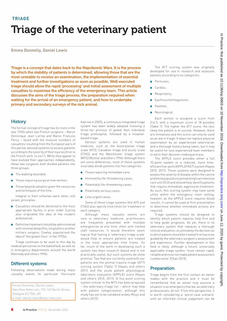
made in person. As most practices employ a variety of staff, it is important that the whole team, including non-clinical members, are trained in telephone triage and are able to identify those common emergencies that require an immediate appointment. The whole team should also be aware of certain phrases that give cause for real concern (Fig 1).
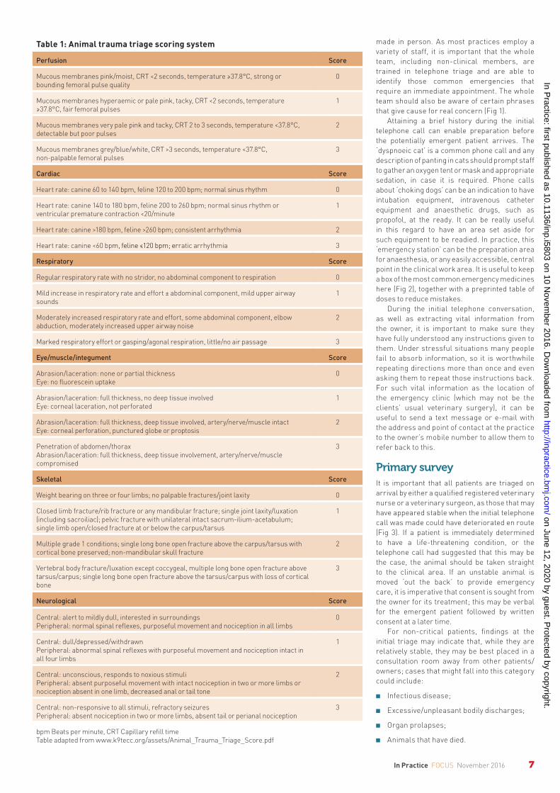
Attaining a brief history during the initial telephone call can enable preparation before the potentially emergent patient arrives. The ‘dyspnoeic cat’ is a common phone call and any description of panting in cats should prompt staff to gather an oxygen tent or mask and appropriate sedation, in case it is required. Phone calls about ‘choking dogs’ can be an indication to have intubation equipment, intravenous catheter equipment and anaesthetic drugs, such as propofol, at the ready. It can be really useful in this regard to have an area set aside for such equipment to be readied. In practice, this ‘emergency station’ can be the preparation area for anaesthesia, or any easily accessible, central point in the clinical work area. It is useful to keep a box of the most common emergency medicines here (Fig 2), together with a preprinted table of doses to reduce mistakes.
During the initial telephone conversation, as well as extracting vital information from the owner, it is important to make sure they have fully understood any instructions given to them. Under stressful situations many people fail to absorb information, so it is worthwhile repeating directions more than once and even asking them to repeat those instructions back. For such vital information as the location of the emergency clinic (which may not be the clients’ usual veterinary surgery), it can be useful to send a text message or e-mail with the address and point of contact at the practice to the owner’s mobile number to allow them to refer back to this.
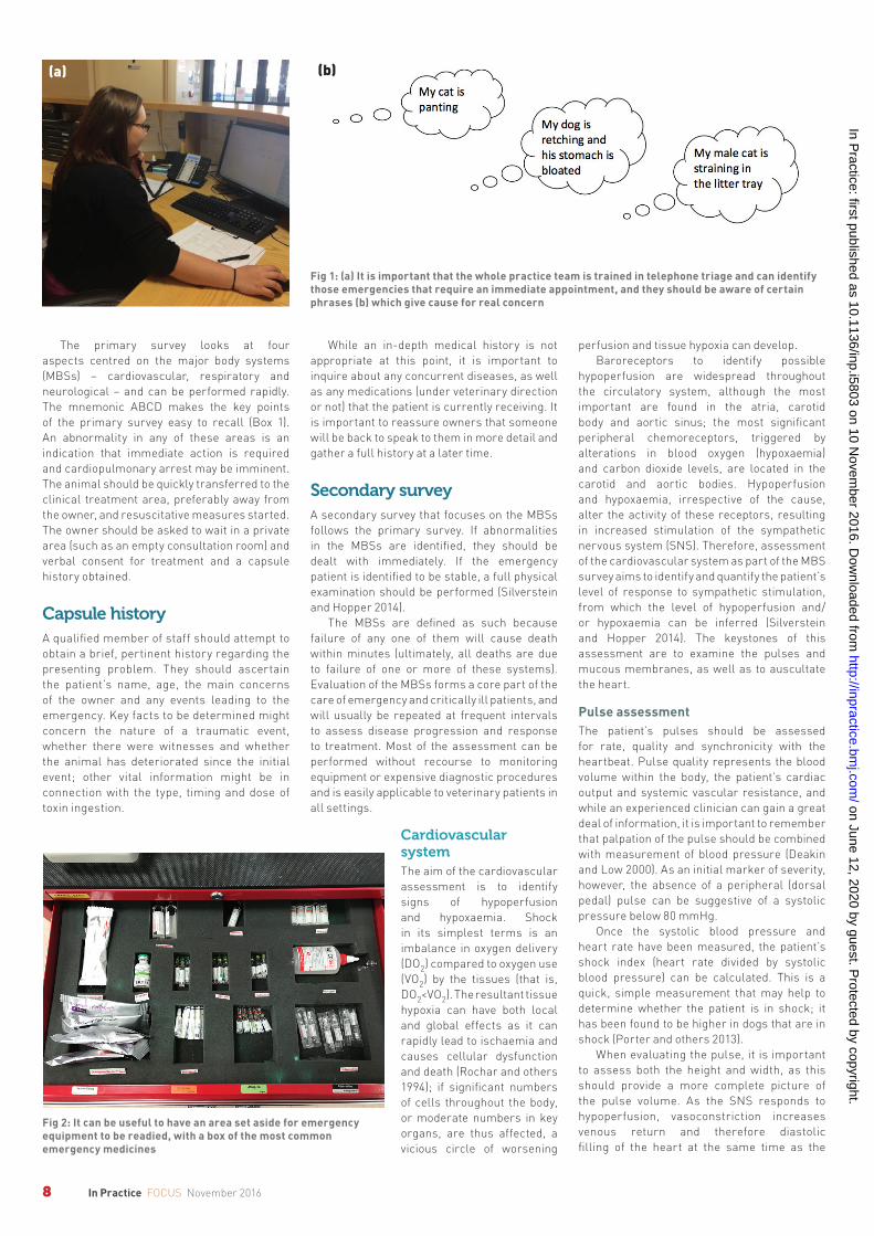
Primary surveyIt is important that all patients are triaged on arrival by either a qualified registered veterinary nurse or a veterinary surgeon, as those that may have appeared stable when the initial telephone call was made could have deteriorated en route (Fig 3). If a patient is immediately determined to have a life-threatening condition, or the telephone call had suggested that this may be the case, the animal should be taken straight to the clinical area. If an unstable animal is moved ‘out the back’ to provide emergency care, it is imperative that consent is sought from the owner for its treatment; this may be verbal for the emergent patient followed by written consent at a later time.
For non-critical patients, findings at the initial triage may indicate that, while they are relatively stable, they may be best placed in a consultation room away from other patients/owners; cases that might fall into this category could include:
■■ Infectious disease;
■■ Excessive/unpleasant bodily discharges;
■■ Organ prolapses;
■■ Animals that have died.
6-11 Donnelly and LewisNEW.indd 7 02/11/2016 15:22
on June 12, 2020 by guest. Protected by copyright.
http://inpractice.bmj.com
/In P
ractice: first published as 10.1136/inp.i5803 on 10 Novem
ber 2016. Dow
nloaded from
8 In Practice FOCUS November 2016
The primary survey looks at four aspects centred on the major body systems (MBSs) – cardiovascular, respiratory and neurological – and can be performed rapidly. The mnemonic ABCD makes the key points of the primary survey easy to recall (Box 1). An abnormality in any of these areas is an indication that immediate action is required and cardiopulmonary arrest may be imminent. The animal should be quickly transferred to the clinical treatment area, preferably away from the owner, and resuscitative measures started. The owner should be asked to wait in a private area (such as an empty consultation room) and verbal consent for treatment and a capsule history obtained.
Capsule historyA qualified member of staff should attempt to obtain a brief, pertinent history regarding the presenting problem. They should ascertain the patient’s name, age, the main concerns of the owner and any events leading to the emergency. Key facts to be determined might concern the nature of a traumatic event, whether there were witnesses and whether the animal has deteriorated since the initial event; other vital information might be in connection with the type, timing and dose of toxin ingestion.
While an in-depth medical history is not appropriate at this point, it is important to inquire about any concurrent diseases, as well as any medications (under veterinary direction or not) that the patient is currently receiving. It is important to reassure owners that someone will be back to speak to them in more detail and gather a full history at a later time.
Secondary survey A secondary survey that focuses on the MBSs follows the primary survey. If abnormalities in the MBSs are identified, they should be dealt with immediately. If the emergency patient is identified to be stable, a full physical examination should be performed (Silverstein and Hopper 2014).
The MBSs are defined as such because failure of any one of them will cause death within minutes (ultimately, all deaths are due to failure of one or more of these systems). Evaluation of the MBSs forms a core part of the care of emergency and critically ill patients, and will usually be repeated at frequent intervals to assess disease progression and response to treatment. Most of the assessment can be performed without recourse to monitoring equipment or expensive diagnostic procedures and is easily applicable to veterinary patients in all settings.
Cardiovascular systemThe aim of the cardiovascular assessment is to identify signs of hypoperfusion and hypoxaemia. Shock in its simplest terms is an imbalance in oxygen delivery (DO2) compared to oxygen use (VO2) by the tissues (that is, DO2<VO2). The resultant tissue hypoxia can have both local and global effects as it can rapidly lead to ischaemia and causes cellular dysfunction and death (Rochar and others 1994); if significant numbers of cells throughout the body, or moderate numbers in key organs, are thus affected, a vicious circle of worsening
perfusion and tissue hypoxia can develop.Baroreceptors to identify possible
hypoperfusion are widespread throughout the circulatory system, although the most important are found in the atria, carotid body and aortic sinus; the most significant peripheral chemoreceptors, triggered by alterations in blood oxygen (hypoxaemia) and carbon dioxide levels, are located in the carotid and aortic bodies. Hypoperfusion and hypoxaemia, irrespective of the cause, alter the activity of these receptors, resulting in increased stimulation of the sympathetic nervous system (SNS). Therefore, assessment of the cardiovascular system as part of the MBS survey aims to identify and quantify the patient’s level of response to sympathetic stimulation, from which the level of hypoperfusion and/or hypoxaemia can be inferred (Silverstein and Hopper 2014). The keystones of this assessment are to examine the pulses and mucous membranes, as well as to auscultate the heart.
Pulse assessmentThe patient’s pulses should be assessed for rate, quality and synchronicity with the heartbeat. Pulse quality represents the blood volume within the body, the patient’s cardiac output and systemic vascular resistance, and while an experienced clinician can gain a great deal of information, it is important to remember that palpation of the pulse should be combined with measurement of blood pressure (Deakin and Low 2000). As an initial marker of severity, however, the absence of a peripheral (dorsal pedal) pulse can be suggestive of a systolic pressure below 80 mmHg.
Once the systolic blood pressure and heart rate have been measured, the patient’s shock index (heart rate divided by systolic blood pressure) can be calculated. This is a quick, simple measurement that may help to determine whether the patient is in shock; it has been found to be higher in dogs that are in shock (Porter and others 2013).
When evaluating the pulse, it is important to assess both the height and width, as this should provide a more complete picture of the pulse volume. As the SNS responds to hypoperfusion, vasoconstriction increases venous return and therefore diastolic filling of the heart at the same time as the
Fig 1: (a) It is important that the whole practice team is trained in telephone triage and can identify those emergencies that require an immediate appointment, and they should be aware of certain phrases (b) which give cause for real concern
(a) (b)
Fig 2: It can be useful to have an area set aside for emergency equipment to be readied, with a box of the most common emergency medicines
6-11 Donnelly and LewisNEW.indd 8 02/11/2016 15:22
on June 12, 2020 by guest. Protected by copyright.
http://inpractice.bmj.com
/In P
ractice: first published as 10.1136/inp.i5803 on 10 Novem
ber 2016. Dow
nloaded from
9In Practice FOCUS November 2016
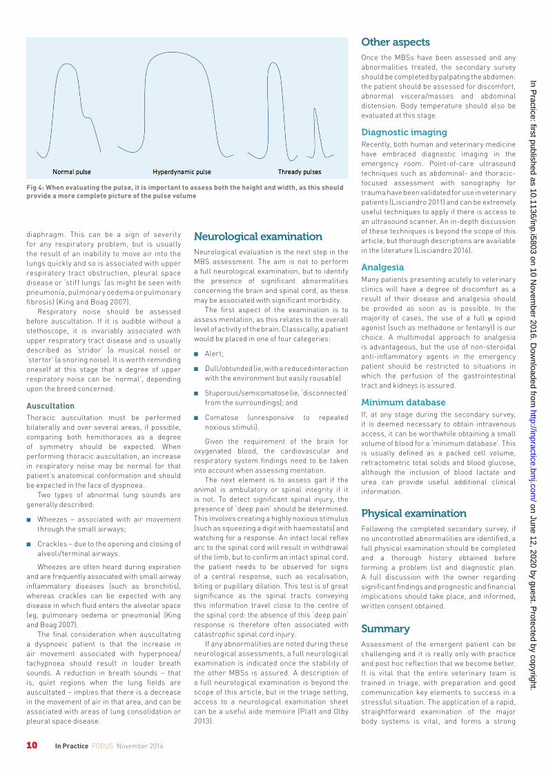
heart rate and contractility increase. This results in tachycardia, as well as taller and narrower pulses (which are often described as hyperdynamic). At this compensatory stage of shock, cardiac output is increased and this can be seen from the greater pulse volume (Fig 4). The change in pulse quality resulting from further volume depletion can also be seen clearly and is reflected clinically by the progression to thin, increasingly weak (‘thready’) pulses as the compensatory response becomes exhausted.
The pulses should also be assessed for rate and rhythm, and palpated while the heart is auscultated to identify any pulse deficits; any patient that has an arrhythmia or is identified as having pulse deficits should ideally be monitored using electrocardiography. At this stage, it is also worthwhile checking for the presence of jugular filling at rest – an early marker of right-sided cardiac insufficiency.
Mucous membrane examinationMucous membrane colour and capillary refill time (CRT) should also be assessed. Normal mucous membranes should be pink in dogs (paler in cats) and a normal CRT is one to two seconds. As the SNS response to hypoperfusion engages, the mucous membrane colour will initially become pinker with a shortened CRT; however, as the cardiovascular system decompensates, the mucous membranes will become increasingly pale and the CRT more prolonged. The pale hue of the mucous membranes is due to a lack of haemoglobin in the capillary beds and will also be seen in cases of anaemia. Identification of hyperaemic, red mucous membranes together with other signs of hypoperfusion indicates that the expected
A Alert – is the patient alert? B Breathing – is the patient ventilating
effectively? C Circulation – is there a palpable pulse
or heartbeat? Is there major external haemorrhage?
D Disability – is there evidence of significant neurological concern?
Box 1: Mnemonic for the primary survey
Fig 3: Animals that appear stable when the initial telephone call was made could have deteriorated on the way to the practice, so all patients should be triaged on arrival
peripheral vasoconstriction is not taking place and should alert the clinician to the likely presence of a widespread inflammatory response, such as that caused by sepsis or anaphylaxis.
Other colour changes in the mucous membranes can also provide useful information, with a yellow tint (icterus/jaundice) raising concerns of increased serum bilirubin levels and brown/’muddy’ coloration often being associated with the presence of methaemoglobin. The presence of petechiation is indicative of a primary haemostatic disorder and should prompt further investigation. Finally, the presence of more than 5 g/dl of deoxygenated haemoglobin will cause the mucous membranes to appear cyanotic; this is indicative of an extremely low level of oxygen in the blood and can rapidly progress to cardiopulmonary arrest (King and Boag 2009).
Respiratory systemThe importance of a hands-off examination of the respiratory system cannot be overemphasised: a wealth of information can be gained before auscultation such as body positioning, respiratory rate and effort. Patients with respiratory compromise commonly present in sternal recumbency (or sitting) with an open mouth, extended neck and abducted elbows (orthopnoeic stance). Patents that present in lateral recumbency or appear unable to adopt a comfortable position, together with evidence of dyspnoea, are frequently on the verge of generalised hypoxia and are at risk of imminent cardiopulmonary arrest.
For markedly dyspnoeic animals, the level of oxygen in the blood is often only just
enough to meet their requirements at rest; any additional oxygen demand (such as that caused by struggling or resisting restraint) can create an oxygen debt. The handling of these animals therefore needs to be as gentle, stress-free and considered as possible, with the provision of supplemental oxygen essential, to prevent their situation becoming even more precarious.
Observed changes in cats can be subtle, so extra care should be taken as they might only be sitting quietly with a mild tachypnea but have significant disease and decompensate quickly when handled.
If dyspnoea is identified during any stage of triage or the MBS examination, it can be beneficial to provide oxygen and leave the patient for a short time. Profound dyspnoea is, by definition, distressing to the patient so the use of sedatives can be beneficial. Drugs such as acepromazine and medetomidine that can have profound cardiovascular effects should, however, be avoided in the emergent patient – a short-acting opioid such as butorphanol (0.1 to 0.3 mg/kg [Ramsay 2014]) is frequently our choice.
Hands-off assessmentRespiratory rate, effort, pattern and noise should be noted before handling the patient. A normal respiratory rate is 15 to 30 breaths/minute, with an increase in respiratory rate and effort part of the normal physiological response (mediated via the SNS) to hypoperfusion, hypoxaemia or stress.
Observing the pattern of respiration can aid in localising pulmonary pathology. An increased inspiratory effort/period indicates upper respiratory tract pathology, with increased effort during expiration indicating a lower respiratory tract problem affecting the bronchi or bronchioles.
In the normal patient, only very minimal chest and abdominal movement is observed. As respiratory effort increases, an increase in abdominal movement is seen initially, although as dyspnoea worsens, paradoxical abdominal movement can often be noted (King and Boag 2007). Paradoxical movement of the thorax and abdomen – that is, the inwards movement of the diaphragm immediately following the outwards expansion of the thorax – occurs when the patient’s intercostal muscles have been recruited to assist with ventilation, rather than relying upon the
6-11 Donnelly and LewisNEW.indd 9 02/11/2016 15:22
on June 12, 2020 by guest. Protected by copyright.
http://inpractice.bmj.com
/In P
ractice: first published as 10.1136/inp.i5803 on 10 Novem
ber 2016. Dow
nloaded from
10 In Practice FOCUS November 2016
diaphragm. This can be a sign of severity for any respiratory problem, but is usually the result of an inability to move air into the lungs quickly and so is associated with upper respiratory tract obstruction, pleural space disease or ‘stiff lungs’ (as might be seen with pneumonia, pulmonary oedema or pulmonary fibrosis) (King and Boag 2007).
Respiratory noise should be assessed before auscultation. If it is audible without a stethoscope, it is invariably associated with upper respiratory tract disease and is usually described as ‘stridor’ (a musical noise) or ‘stertor’ (a snoring noise). It is worth reminding oneself at this stage that a degree of upper respiratory noise can be ’normal’, depending upon the breed concerned.
AuscultationThoracic auscultation must be performed bilaterally and over several areas, if possible, comparing both hemithoraces as a degree of symmetry should be expected. When performing thoracic auscultation, an increase in respiratory noise may be normal for that patient’s anatomical conformation and should be expected in the face of dyspnoea.
Two types of abnormal lung sounds are generally described:
■■ Wheezes – associated with air movement through the small airways;
■■ Crackles – due to the opening and closing of alveoli/terminal airways.
Wheezes are often heard during expiration and are frequently associated with small airway inflammatory diseases (such as bronchitis), whereas crackles can be expected with any disease in which fluid enters the alveolar space (eg, pulmonary oedema or pneumonia) (King and Boag 2007).
The final consideration when auscultating a dyspnoeic patient is that the increase in air movement associated with hyperpnoea/tachypnoea should result in louder breath sounds. A reduction in breath sounds – that is, quiet regions when the lung fields are auscultated – implies that there is a decrease in the movement of air in that area, and can be associated with areas of lung consolidation or pleural space disease.
Neurological examinationNeurological evaluation is the next step in the MBS assessment. The aim is not to perform a full neurological examination, but to identify the presence of significant abnormalities concerning the brain and spinal cord, as these may be associated with significant morbidity.
The first aspect of the examination is to assess mentation, as this relates to the overall level of activity of the brain. Classically, a patient would be placed in one of four categories:
■■ Alert;
■■ Dull/obtunded (ie, with a reduced interaction with the environment but easily rousable)
■■ Stuporous/semicomatose (ie, ‘disconnect ed’ from the surroundings); and
■■ Comatose (unresponsive to repeated noxious stimuli).
Given the requirement of the brain for oxygenated blood, the cardiovascular and respiratory system findings need to be taken into account when assessing mentation.
The next element is to assess gait if the animal is ambulatory or spinal integrity if it is not. To detect significant spinal injury, the presence of ‘deep pain’ should be determined. This involves creating a highly noxious stimulus (such as squeezing a digit with haemostats) and watching for a response. An intact local reflex arc to the spinal cord will result in withdrawal of the limb, but to confirm an intact spinal cord, the patient needs to be observed for signs of a central response, such as vocalisation, biting or pupillary dilation. This test is of great significance as the spinal tracts conveying this information travel close to the centre of the spinal cord: the absence of this ‘deep pain’ response is therefore often associated with catastrophic spinal cord injury.
If any abnormalities are noted during these neurological assessments, a full neurological examination is indicated once the stability of the other MBSs is assured. A description of a full neurological examination is beyond the scope of this article, but in the triage setting, access to a neurological examination sheet can be a useful aide memoire (Platt and Olby 2013).
Other aspectsOnce the MBSs have been assessed and any abnormalities treated, the secondary survey should be completed by palpating the abdomen: the patient should be assessed for discomfort, abnormal viscera/masses and abdominal distension. Body temperature should also be evaluated at this stage.
Diagnostic imagingRecently, both human and veterinary medicine have embraced diagnostic imaging in the emergency room. Point-of-care ultrasound techniques such as abdominal- and thoracic-focused assessment with sonography for trauma have been validated for use in veterinary patients (Lisciandro 2011) and can be extremely useful techniques to apply if there is access to an ultrasound scanner. An in-depth discussion of these techniques is beyond the scope of this article, but thorough descriptions are available in the literature (Lisciandro 2016).
AnalgesiaMany patients presenting acutely to veterinary clinics will have a degree of discomfort as a result of their disease and analgesia should be provided as soon as is possible. In the majority of cases, the use of a full m opioid agonist (such as methadone or fentanyl) is our choice. A multimodal approach to analgesia is advantageous, but the use of non-steroidal anti-inflammatory agents in the emergency patient should be restricted to situations in which the perfusion of the gastrointestinal tract and kidneys is assured.
Minimum databaseIf, at any stage during the secondary survey, it is deemed necessary to obtain intravenous access, it can be worthwhile obtaining a small volume of blood for a ‘minimum database’. This is usually defined as a packed cell volume, refractometric total solids and blood glucose, although the inclusion of blood lactate and urea can provide useful additional clinical information.
Physical examinationFollowing the completed secondary survey, if no uncontrolled abnormalities are identified, a full physical examination should be completed and a thorough history obtained before forming a problem list and diagnostic plan. A full discussion with the owner regarding significant findings and prognostic and financial implications should take place, and informed, written consent obtained.
SummaryAssessment of the emergent patient can be challenging and it is really only with practice and post hoc reflection that we become better. It is vital that the entire veterinary team is trained in triage, with preparation and good communication key elements to success in a stressful situation. The application of a rapid, straightforward examination of the major body systems is vital, and forms a strong
Fig 4: When evaluating the pulse, it is important to assess both the height and width, as this should provide a more complete picture of the pulse volume
6-11 Donnelly and LewisNEW.indd 10 02/11/2016 15:22
on June 12, 2020 by guest. Protected by copyright.
http://inpractice.bmj.com
/In P
ractice: first published as 10.1136/inp.i5803 on 10 Novem
ber 2016. Dow
nloaded from
11In Practice FOCUS November 2016
Daniel Lewis graduated from the University of Cambridge in 1995, and worked in mixed practice for five years, where he gained the RCVS Certificate in Veterinary Anaesthesia. In 2000, he moved to Petmedics, a large hospital-based emergency clinic in Manchester, where he remained for eight years. In 2008, he embarked upon a residency at the RVC, obtaining his Diploma in Emergency and Critical Care in 2011. Since then, he has been in charge of the ICU at Bristol vet school before another period at Petmedics. He joined Vets Now Referrals in January 2015, joining its ECC team. He is interested in all aspects of critical care, but particularly in septic patients and poorly cats.
foundation for the ongoing assessment of critically ill veterinary patients.
ReferencesCHAN, D. L. (2013) Triage 2.0: re-evaluation of early patient assessment. Journal of Veterinary Emergency and Critical Care (San Antonio) 23, 487-488DEAKIN, C. D. & LOW, J. L. (2000) Accuracy of the advanced trauma life support guidelines for predicting systolic blood pressure using carotid, femoral, and radial pulses: observational study. BMJ 321, 673-674HAYES, G., MATTHEWS, K., DOIG, G., KRUTH, S., BOSTON, S., NYKAMP, S. & OTHERS (2010) The acute patient physiologic and laboratory evaluation (APPLE) score: a severity of illness stratification system for hospitalized dogs. Journal of Veterinary Internal Medicine 24, 1034-1047 HAYES, G., MATTHEWS, K., DOIG, G., KRUTH, S., NYKAMP, S., POLJAK, Z. & DEWEY, C. (2011) The Feline Acute Patient Physiologic and Laboratory Evaluation (Feline APPLE) Score: a severity of illness stratification system for
hospitalized cats. Journal of Veterinary Internal Medicine 25, 26-38 KENNEDY, K., AGHABABIAN, R. V., GANS, L. & LEWIS, C. P. (1996) Triage: techniques and applications in decision making. Annals of Emergency Medicine 28, 136-144KING, L. G. & BOAG, A. (2007) BSAVA Manual of Canine and Feline Emergency and Critical Care. 2nd edn. British Small Animal Veterinary Association. pp 85-113LISCIANDRO, G. R. (2011) Abdominal and thoracic focused assessment with sonography for trauma, triage, and monitoring in small animals. Journal of Veterinary Emergency and Critical Care (San Antonio) 21, 104-122LISCIANDRO, G. R. (2016) The use of diaphragmatico-hepatic (DH) views of the abdominal and thoracic focused assessment with sonography for triage (AFAST/TFAST) examinations for the detection of pericardial effusion in 24 dogs (2011-2012). Journal of Veterinary Emergency and Critical Care (San Antonio) 26, 125-131PLATT, S. & OLBY, N. (2013) BSAVA Manual of Canine and Feline Neurology. 4th edn. British Small
Animal Veterinary Association. pp 1-2PORTER, A. E., ROZANSKI, E. A., SHARP, C. R., DIXON, K. L., PRICE, L. L. & SHAW, S. P. (2013) Evaluation of the shock index in dogs presenting as emergencies. Journal of Veterinary Emergency and Critical Care (San Antonio) 23, 538-544RAMSAY, I. (2014) BSAVA Small Animal Formulary. 8th edn. British Small Animal Veterinary Association. pp 50-51ROCHAR, R. A., DROBATZ, K. S. & SHOFER, F. S. (1994) Development of a scoring system for the veterinary trauma patient. Journal of Veterinary Emergency and Critical Care (San Antonio) 4, 77-83RUYS, L. J., GUNNING, M., TESKE, E., ROBBEN, J. H. & SIGRIST, N. E. (2012) Evaluation of a veterinary triage list modified from a human five-point triage system in 485 dogs and cats. Journal of Veterinary Emergency and Critical Care (San Antonio) 22, 303-312SILVERSTEIN, D. C. & HOPPER, K (2014) Small Animal Critical Care Medicine. 2nd edn. Elsevier Saunders. pp 30-33
doi: 10.1136/inp.i5803
Emma Donnelly graduated from the University of Glasgow in 2003, and joined Vets Now to complete a rotating internship. After developing a passion for ECC during this year, she stayed with Vets Now to complete an ECC internship the following year before beginning her ECVECC residency in 2015. She enjoys all aspects of the emergency room and intensive care, but has a special interest in sepsis and respiratory distress.
6-11 Donnelly and LewisNEW.indd 11 02/11/2016 15:22
on June 12, 2020 by guest. Protected by copyright.
http://inpractice.bmj.com
/In P
ractice: first published as 10.1136/inp.i5803 on 10 Novem
ber 2016. Dow
nloaded from