emma kirke dissertation - osteomyology
TRANSCRIPT

1
A Study into Treatment Considerations for
the Osteomyologist; Systemic and
Pathological Conditions within the Clinical
Setting.
By Emma Kirke FAO
April 2010

2
Contents
1) Acknowledgement. 5
2) Abstract. 6
3) Introduction and Background. 7
4) Asthma. Allopathic Definition and Aetiology. 9
Alternative Definition, Aetiology and Pathology. 10
Allopathic Treatment. 12
Treatment and Management Considerations within
Osteomyology. 12
Exercises. 17
Structural and Physiological areas to consider within
Osteomyology Treatment. 17
5) Multiple Sclerosis. Allopathic Definition. 18
Allopathic Aetiology. 18
Alternative Aetiology. 19
Allopathic Treatment. 22
Treatment and Management Considerations within
Osteomyology. 23

3
Diet. 25
Exercise. 28
Structural and Physiological areas to consider within
Osteomyology Treatment. 28
6) Diabetes. Allopathic Definition. 31
Alternative Definition. 31
Allopathic Aetiology. 32
Alternative Aetiology and Pathology. 32
Allopathic Treatment. 36
Treatment and Management Considerations within
Osteomyology. 37
Diet. 39
Structural and Physiological areas to consider within
Osteomyology Treatment. 40
7) ADHD. Allopathic Definition and Aetiology. 42
Alternative Aetiology. 45
Dietary Aetiology. 47
Treatment and Management Considerations within
Osteomyology. 47
Structural and Physiological areas to consider within
Osteomyology Treatment. 51
8) Conclusion. 52

4
9) Appendices. 1) Definition of Somatic Dysfunction. 55
2) Exercises for the Asthmatic patient. 56
3) Asthma Case Study One. 62
4) Asthma Case Study Two. 71
5) Exercises for the Multiple Sclerosis Patient. 80
6) Multiple Sclerosis Case Study One. 83
7) Multiple Sclerosis Case Study Two. 90
8) Diabetes Diet. 99
9) Diabetes Case Study One. 101
10) Diabetes Case Study Two. 111
11) ADHD Case Study One. 120
12) Asthma – Physiological centres diagram. 141
13) MS – Physiological centres diagram. 144
14) Diabetes – Physiological centres diagram. 147
15) ADHD – Physiological centres diagram. 150
10) Bibliography 153
11) References 155

5
Acknowledgements
Thank you to my husband, family and friends; for all their support, encouragement and belief in me.
Thank you to Karen Harding, Alan Clemens, Chris Eedy, and Chris Gazely; for encouraging my professional
development and continued belief in me.
Thank you to Quentin Shaw and Howard Beardmore - inspirational tutors and mentors.
Thank you to Darren Bristowe friend and colleague, for his support, encouragement and his excellent
management of the personal training regimens for some of the patients used in this study.
6
Abstract
This study considers the possible aetiologies and thence treatment and management of four commonly
encountered pathologies for the Osteomyologist to use within their clinical practices. It shows research into
methods both past and present. It gives a guideline into structural and physiological areas for treatment
consideration. It highlights possible future research programmes that could be undertaken. It tries to enhance
the efficacy of the Osteomyologist beyond the fundamental structural practitioners that we can be associated
with.
The areas under consideration are Asthma, Multiple Sclerosis, Diabetes and Attention Deficit and
Hyperactivity Disorder. The components considered are aetiology and treatment, including structural,
emotional, dietary advice and possible prescription exercises.

7
Introduction and Background
In our profession, musculoskeletal pain and discomfort can become our ‘bread and butter’ and it is easy
to become complacent and ignore our duty to the patient and their well being as a whole. As practitioners of
Osteomyology Medicine, it is prudent to remain aware of our ability to not only affect the musculoskeletal
components of our patient, but that we are also able to affect viscera and the body’s natural homeostasis. To
quote “the body is its own medicine chest” (A T Still (1899) – Philosophy of Osteopathy). If we are able to
remove obstructions, we will allow the body to return to normal functioning, therefore removing imbalance and
the effects of this imbalance which may manifest as “Disease”. (Dis - ease – as the breakdown of the
terminology suggests the body has become in a state of unease or Disease.) We have the tools at our fingertips
to influence these systems and ease the body towards being a more functional unit allowing improvement to its
symptoms and hence quality of life.
I endeavour to maintain this focus, and to constantly remind myself that we can be more than “bone
crackers”. As Osteomyologists, we are fortunate in our position; unlike most professions we have such a great
diversity within our members and their backgrounds, for us to utilise and harness. We should therefore strive to
become as complete a practitioner as possible within our powers and abilities. Safety is paramount. However,
we should not become so safe that we negate our duty and skills.
I am keen therefore, to inspire others to be confident in practice and when faced with a patient that has
systemic disease, to try to address this alongside their musculoskeletal dysfunction and to not be wary or
dismissive of it. They should endeavour not to undertake the care of the patient solely for the palliative relief of
the symptoms of the condition. I wish to illuminate our members’ professional attitude, and I hope this work will
go some way to achieving that. I am going to discuss four conditions that I feel highlight the potential impact we
can have (given our strengths and abilities), and the possible management techniques we can utilise. Moreover,

8
we should be mindful that we are individuals, and that our patients should be treated as such. Each individual
should be assessed, and their lesions, both physical and emotional addressed, incorporated within a
management structure as a base plan. Therefore, I propose to discuss Multiple Sclerosis, Diabetes, Asthma and
ADHD and their management as used within my own practice. The case studies are derived over time from my
practice, research and practical application.
My personal interest in these conditions lead me to begin research some years ago. This is evident from
the case histories. These, on examination, give a good cross section of the differing aetiologies, management,
and condition types that we are encountering with increased frequency.

9
Asthma
Allopathic Definition and Aetiology
“Specific IgE antibodies or nonspecific inhaled irritants provoke mast cell degranulation; histamine,
leukotrienes, and other mediators are released to cause bronchospasm and bronchial mucus secretion” (Richard
C Harruff (1994); Pathology Facts)
“Asthma is a chronic relapsing inflammatory disorder characterised by hyper reactive airways, leading to
episodic, reversible bronchoconstriction, owing to increased responsiveness of the tracheobronchial tree to
various stimuli.” (Busse W, Holgate S (1995); Asthma and rhinitis. Boston: Blackwell Scientific)
Asthma is categorised into two types - extrinsic or allergic and intrinsic or non-allergic asthma.
(McFadden Jr ER. Diseases of the respiratory system: asthma. In: Fauci AS, Braunwald E, Isselbacher KJ et al
(1998); Harrison’s principles of internal medicine, 14th edn. New York: McGraw-Hill; 1419-1426.)
Allopathic physicians have noted that there is usually a family history of asthma or related diseases such
as eczema, showing increased levels of IgE in the serum. In non-allergic asthma there seems to be no family
history and no elevated IgE present in the serum. However, allopathic physicians have found that determining
which category a patient falls into is not always clear since both types display inflammation resulting in
increased airway irritation. This is the mechanism that creates the bronchospasm. The resultant further
reduction in airway diameter ensues to vascular congestion, oedema and mucus production. The patient
experiences chest tightening, wheezing and dyspnoea. During an attack they may experience a non productive
cough which later, on increased severity can produce mucus. The inability to breath leads to panic thus

10
exacerbating the symptoms. Status asthmaticus is life threatening and requires immediate emergency
intervention.
There are many precipitating factors including; climate, rapid changes of temperature and humidity. Air
pollution, certain chemicals, household pets and dust mites have also been implicated. Certain food types, food
additives and colourings, and some drugs can provoke attacks. E.g. nuts, grains, seafood and aspirin. Allopathic
physicians have notably increased their diagnosis of exercise induced asthma. It may however be environmental
aspects causing the symptoms.
Emotional stress, viral and bacterial infections when experienced by a patient with a history of asthmatic
episodes, may find that they become more susceptible to these episodes thus increasing the frequency and
severity.
Alternative Definition Aetiology and Pathology
“Bronchial or spasmodic asthma is a chronic affection, characterized by a paroxysmal dyspnoea due to a
spasmodic contraction of the muscles of the bronchial tubes or to swelling of their mucus membranes.”
(McConnell CP, Teall CC (1906); The Practice of Osteopathy: Disease of the Bronchi; Bronchial Asthma: 517-
521.) (Kuchera, ML, Kuchera, WA: Osteopathic Considerations in Systemic Dysfunction 2nd Edn 1994; Lower
Respiratory Disorders; Asthma: 48-51)
McConnell and Teall, state that the majority of lesions that are causative to bronchial asthma are from
D2-7 inclusive, either in the ribs posteriorly or anteriorly, or in the vertebrae. They state that these lesions
involve vasomotor nerves to the bronchioles, which produce the narrowing of the tubes and thus cause the
dyspnoea. During their research they have found that the lesion is usually at the 3rd, 4th, or 5th rib on the right or

11
in the corresponding vertebrae. They postulate that the lesions mainly occur on the right side due to the
majority of the population being right handed. Hence, the muscles on the right tend to be hypertrophic and
when contracted draw the ribs from their articulation. They also note that in a number of cases, a posterior
curve can be seen in the dorso-lumbar region; the resultant effect is that there will be catarrh and dilatation of
the stomach, congestion of the liver, and perhaps, intestinal indigestion and constipation. Therefore, addressing
the digestive organs is vital. They notice that occasional lesions at the atlas/axis complex can cause irritation to
the pneumogastric fibres that innervate those muscles of the bronchioles and consequently narrowing the tubes
causing paroxysms. They also note the onset of attacks from excitants as well as diseases of the upper
respiratory tract. However, they state that these are only causative of attacks if there is the pre existence of
lesions to the vaso-motor and motor nerves. This is noted by Laughlin in his work, “It is questionable whether
reflex causes alone are sufficient to produce genuine asthma without the existence of specific lesions affecting
the direct nerve connections of the part involved.” (Laughlin (1904); Asthma – Journal of the American
Osteopathic Association, Oct)
McConnell and Teall are confident in stating that true asthma is pure neurosis. In all cases, there are
vertebral and rib lesions affecting the spinal nerves at their exit and the sympathetic chain along the head of the
ribs. Irritating lesions to the vagi, constricting pulmonary vessels, and to the cervical sympathetics, causing the
same type of disturbance would be factors in the pathological chain. Reflex irritations can be found in various
lesions, but their findings concur with that of AT Still in that the primary osseous lesions are on the right side
from D2-6.
L Whooton and Q Shaw have both taken this concept of neurosis as the cause and have looked at the
developmental curvatures from birth into childhood. They note that a collapse of the dorsal arch, as a result of
birth trauma, can lead to pathological and systemic conditions such as asthma. Q Shaw pays particular attention
to this and suggests that between the ages of 0-7, the developmental milestones are paramount to erecting the

12
centre of gravity line successfully and the mechanisms for providing structure and function in relation to this
line. Shaw treats his patients accordingly seeking to restore the balance of the curves and therefore allowing the
body to restore overall balance and remove the neurosis relating to the asthmatic patient. “The body is its own
medicine chest” (AT STILL) (Shaw, Q – Spinal development and balance of children.) The institute of Classical
Osteopathy have also featured work on the subject of curvatures and their importance in 1985 and 2001 in their
yearbook publications but it does not specifically relate to asthma.
Allopathic Treatment
Allopathic treatment and management of this condition involves symptomatic treatment with
bronchodilators, sympathomimetics and corticosteroids. They are administered in the form of nebulised
inhalants that effect the dilation of the airway and/or prevent inflammation. If this method is unsuccessful it is
at this point that the corticosteroids may be administered, either by inhalation or orally.
Treatment and Management Considerations in Osteomyology
Balon J, Aker PD, Crowther ER et al (1998) published a study in “The New England Journal of Medicine”
(A comparison of active and stimulated chiropractic manipulation as adjunctive treatment for childhood
asthma. N Engl J Med; 339:1013-1020)
Both sets of treatments appeared to produce positive effects in the subjects, allowing the only
conclusion by the researchers to be that there was no significant difference between the two treatments. The

13
Chiropractors involved in the study used high velocity spinal manipulation but did not include an adequate
description of the soft tissue manipulation and gentle palpation to the spine and paraspinal muscles and
shoulders that were used on the second group. On further investigation, this second form of treatment is noted
as the basis for many osteopathic style treatments as standard practice. The chiropractic treatment was
simulated in the second type by using low velocity techniques to areas of the body that the researches felt were
insignificant. However, to Osteomyologists these areas are of great significance, notably the head and occipital
region - if for no other reason than the involvement of the vagus. In my opinion, they failed to note a difference
between the two groups having treatment because they are valid as treatment types in their own right. Classical
Osteopathic text books note these types of techniques in their management of asthma. In Pottinger’s
‘Symptoms of Visceral Disease’, the relation of the cranial nerves is noted, with clinicians reporting asthmatic
attacks arising from sinus inflammation. This can be explained through the reflex connection of the sensory
fibres of the 5th and the motor fibres of the 10th cranial nerves. Irritation of the nasal mucus membranes can
slow the heart rate, the reflex in the instance of an asthma attack travels over this pathway. However, it
manifests itself in the pulmonary instead of the cardiac branches of the vagus. It could also manifest in the
gastrointestinal branch, which would be noted as hyperacidity and spastic constipation as can sometimes be
noted in patients during attacks of hay fever. Kuchera and Kuchera note that asthma, cephalgia, and vertigo are
symptoms which can arise from nasal and sinus affections. (Kuchera, M.L., Kuchera, W.A.
(1994); Osteopathic Considerations in Systemic Dysfunction 2nd pp 2)
In this study we must consider the significant difference between the treatment and management of
asthma during an attack, and between attacks. Primarily techniques used during an attack should be inhibitory.
Practitioners working within Osteopathic hospitals i.e. Haifa Rambam Hospital in Haifa, and The Galil pain relief
hospital, which are both situated in Israel and run by Mervin Waldman, may well be experienced enough to
administer stimulatory treatment during an attack. Mervin Waldman states emphatically that a stimulatory
treatment during an attack will increase sympathetic activity and therefore produce bronchodilation. The

14
patient must have complete faith in the practitioner, who must, conversely, feel completely confident in his/her
treatment. In this country, we are not likely to have to deal with this scenario on a regular basis, as most
sufferers during an attack will proceed straight to A&E. However, it could be useful to be aware of techniques to
use as a patient may undergo an attack in your own clinic. DiGiovanna indicates that we should manage the
acute patient with medications such as epinephrine or nebulised albutenol. Then, as the breathing quiets, rib
raising is useful in easing the patient’s respiratory efforts and for loosening mucus plugs. (DiGiovanna EL,
Schiowitz S (1997); An Osteopathic Approach to Diagnosis and Treatment: The Pulmonary Patient: 466-467)
McConnell and Teall states that to relieve an attack, the practitioner must locate the lesion and correct
it as soon as possible. They discuss scenarios in which the patient displays severely contracted muscles
bilaterally, and within those scenarios how the practitioner should proceed, if they are rendered unable to
locate the lesion. They advise that the practitioner must apply deep inhibition to the muscles, prior to locating
the lesion, and consequently making an appropriate adjustment. This inhibition should be carried out in an
upward, outward movement over the angles of the ribs and lesions that are involved. The objective is to relieve
pressure or irritation to the nerves, so that the narrowed tubes may be relaxed. They suggest that a strong
inhibition such as placing the knee in the patient’s back and at the same time pulling back on the patient’s
shoulders, will have a temporary effect on the situation, but they still suggest finding the primary lesion and
reducing it to provide permanent stability within the condition. I think that perhaps this piece of advice is
outdated. However, these extreme techniques will reportedly aid the cessation of an asthma attack “In severe
cases dilatation of the rectum may relieve the paroxysm, and in a few instances it will be necessary to treat the
uterus locally” (McConnell CP, Teall CC (1906); The Practice of Osteopathy: Bronchial Asthma: pp 518)
In the treatment of the asthmatic patient between attacks, we should consider the thorax, not only the
intervertebral articulations but concurrently the costovertebral and costotransverse articulations. Any
restrictions here will limit the ability for full respiratory motion. The sympathetic ganglionic chain lies close to

15
these articulations. Therefore, mechanical dysfunction can also affect the nerves and their targets. D1-5/6
supplies the sympathetic outflow to the lungs and bronchi; therefore we should pay careful attention to this
region.
The mechanics of breathing, being more complex than merely movement of the ribs, means that we
need to consider the sternum, costal and vertebral attachments. Motor supply to the diaphragm comes from
the phrenic nerve originating from C3-5. Hence, somatic dysfunction at this level should be addressed. The
pulmonary plexus is made up of a mixture of sympathetics from D1-6 and parasympathetics from the vagus. The
autonomic nerves control the smooth muscle and consequently the diameter of the airways. The drainage
vessels from the lungs, both venous and lymphatic, travel through the mediastinum before reaching the
superior vena cava and the lymphatic ducts. A major contributing factor to asthma is congestion. Therein, the
practitioner should consider releasing tensions within the mediastinum. Gentle pumping techniques to the
thorax can assist lymphatic drainage. However, too much force can induce an attack, so this technique should be
used with care.
Respiration is controlled by the respiratory centre in the brain which is located in the upper medulla and
lower pontine region of the brain stem. Cranial techniques addressing lesions indicated in this area should also
be utilised.
Fascial tensions within the occiput, cervical fascia to the mediastinum should be considered as they
contain the vagus nerve. Over-activity of the vagus nerve can lead to an imbalance of the autonomic nervous
system, giving the patient the bronchospasm symptom. An under activity of the sympathetic nervous system will
also fail to balance this out. It is quite likely that the patient whilst, having an attack, will become anxious.
Therefore, their sympathetic nervous system will already be stimulated. They may well find that, although the
resultant affect of sympathetic stimulation is bronchodilation, in fact, there is reduced diameter within the
bronchus due to the increase in the production of mucus. Restoring the autonomics to normal function may

16
calm an attack. However, it is unlikely that an asthmatic patient would endure lying down to enable the
practitioner to administer the treatment. It is documented that AT Still would treat an asthmatic patient whilst
they were standing to avoid this. (Still AT (1910); Osteopathy – research and practice. Kirksville: Still: 174-177.)
He wrote that his treatment for asthma included treating all of the ribs and muscles of the thorax until he was
satisfied “this part of the work was completely normal”. He then advised practitioners to, “keep your hands off
the patient for at least a week”; this is sound advice as we should employ the philosophy of “find it, fix it and
leave it alone” in the instance. Considering the possibility of emotional well being as an aetiological component
of asthma, and the fear that can be elicited by the dyspnoea, the very act of placing the hands on the patient will
have beneficial effects and serve to calm as you treat. This alone can be a very significant therapeutic factor.
McConnell and Teall give some idea of treatment management timing in between attacks. They state
that the practitioner should use this time to locate the lesion without other structures interfering with the
diagnosis. “Many cases of asthma are cured in from one to three months treatment. One treatment a week is
sufficient, provided one is able each time to accomplish something toward a correction of the lesion and that the
patient does not suffer during the meantime. Too frequent treatments may simply act as an irritant to the
nervous lesions”. (McConnell CP, Teall CC; (1906) The Practice of Osteopathy: Bronchial Asthma: pp 520)
Wilson and Koch have managed to empirically prove that Osteopathic Manipulative Therapy (OMT)
between acute asthmatic episodes significantly decreases the frequency as well as the severity of attacks, and
possibly more importantly, the need for medications, therefore reducing the subsequent side effects of said
medications within the patient. They advocate that maximal thoracic, sternum, and costal motion is sought
between the attacks. (Wilson Perrin T, Koch RS; (1987) Osteopathic Research: Growth and Development. AOA)

17
Exercises
See Appendix two (2)
Structural and physiological areas to consider within Osteomyology treatment
Cranium – Upper medulla Lower pontine region of the brainstem
Occipital/Atlantal – freeing for vagal release
Atlas/Axis - pneumogastric
C3-5 – Phrenic nerve
D4 – Apex of the 2 polygons of force
D1-6 – General lung segment – physiological centre & Vasomotor to lungs
Thorax – Mediastinum, costal and intercostals,
Rib raising
Thoracic pump
D/L – Ascending colon, descending colon, micturition and defecation. Splanchnics.
Try to reduce stress and emotional strain where possible.
See Appendix 13

18
Multiple Sclerosis
Allopathic definition
“A chronic autoimmune inflammatory disease of the central nervous system marked by intermittent
damage to the myelin sheath that covers nerve cell axons. Visual changes and muscle weakness occur often and
have no consistent pattern; as the disease progresses, different nerves may be affected at different times,
exacerbating current symptoms or creating new problems. The disease may progress steadily, or acute attacks
may be followed by partial or complete temporary remission of symptoms. Most patients live a relatively normal
lifespan.” (Davis, FA; (1940) Taber’s cyclopedic medical dictionary 18th edn 1997 1st edn)
Allopathic Aetiology
“The current theory is that an autoimmune process involving T-lymphocytes and macrophages begins in
the periphery and somehow breaks through the blood-brain barrier, producing inflammation in the myelin
sheath. The damaged areas of myelin, called plaques, contain Bcells and macrophages that release cytokines,
proteolytic enzymes, and immunoglobulin G antibodies, which increase inflammatory damage.” (Davis, FA;
(1940) Taber’s cyclopedic medical dictionary 18th edn 1997 1st edn)

19
Alternative Aetiology
I have found it quite difficult to locate any literature regarding a possible osseous lesion that could cause
the onset of this particular disease. There is little in the way of evidence, or research to quantify how this
disease comes to be generally within any branch of primary care - including the allopathic fraternity who also
have little to offer. In fact, McConnell and Teall who can usually be relied upon to offer ideas regarding osseous
lesioning in respect to most common diseases can only be seen to say “The cause is not definitely known, but
probably derangements of the tissues, affecting the blood-vessels to degenerated areas, are the most common
cause. Thus osteopathic lesions, corresponding to the involved area, are found.” (McConnell CP, Teall CC; (1906)
The Practice of Osteopathy: pp 695)
It would seem that the classical and other likeminded manipulative therapists that employ the theory of
spinal disease causing visceral disease and vice versa, have few ideas as to why this particular disease ensues.
We can note that within patients displaying apparent symptomology, the osseous lesioning is deep seated.
Littlejohn in his ‘Fundamentals of Osteopathic Technique’, notes that the appearance of lesions is segmental
rather than singular. He believed that the lesioning was diffuse and combined the segments at a later date,
producing a multiple sclerosis effect.
Some alternative therapists hypothesise that infectious disease could be the precursor as well as the
possibility of emotions and stress. This particular disease is more frequent amongst younger female patients.
The disease usually shows itself in the lower extremity firstly and usually with the loss of power. However due to
its nature and its ability to affect any of the nerves and their functions, such symptoms as tremor, hyper-reflexia,
scanning speech, and nystagmus can also occur as the first symptom. Atrophy of the optic nerve, with tingling
numbness and vertigo are also common symptoms. A reduction in the mental acuity, epileptiform,
apoplectiform and occasionally coma has been infrequently seen. McConnell and Teall offer a disease course of

20
5 to 15 yrs with the outcome of death commonly resultant from some intercurrent disease. This is far from the
allopathic feeling that lifespan is little affected.
There are certain factions that believe MS is misdiagnosed regularly, and, in many cases, we are given
the diagnosis of MS as the less complicated, and time consuming alternative. In my opinion, many of the
conditions that have similar symptomology involve expensive testing procedures thus it is cost effective to give
the negative (No positive test is available) diagnosis of MS. It is also possible that there could be underlying
conditions that lead to similar symptoms and that if the underlying condition were being adequately controlled
then the progression at least of the symptoms may halt. Diabetes mellitus type 2, for example, shows similar
onset and progression of the neurological symptoms with the commencement of diabetic neuropathy. They are
almost indistinguishable from an MRI scan, and both show the same plaque formations in the spinal column and
brain tissue. Undiagnosed and thence untreated diabetes will see the progression of the symptoms of diabetic
neuropathy which can mimic multiple sclerosis.
Recent activity in the media with regards to MS shows an exciting re direction. ‘Zamboni Et Al’, used 65
subjects from a large cross section of MS sufferers containing all types of the disease in its progression and
diagnostic state. They compared their subjects against 235 people that were either in good health, or that were
suffering from differing types of neurological disorders. Their study showed a resounding correlation between
those suffering with MS and displaying the presence of venous insufficiency. They are postulating that the
abnormal venous return from the brain and spine may trigger the inflammation and perhaps the immune
mediated damage that we see with MS. Their research did not show any correlation between the extent of the
occlusion and the severity of the condition. Also, it would appear that the treatment and medication regimen to
date, at commencement of the study also showed no correlation to the extent of the occlusion within the
venous return. Although chronic cerebrospinal venous insufficiency (CCSVI) has shown some positive response
to having stents inserted, or the use of balloons to open up the occlusion, the procedures are still very

21
experimental. (Zamboni et al; (2009) Chronic cerebrospinal venous insufficiency (CCSVI) and MS; Neurology
Neurosurgery Psychiatry. 2009 Apr; 80 (4): 392-9. Epub. 2008 Dec 5.)
They cannot substantiate as to why this is an occurrence and suggest it is of little use clinically to our
own practices in the form of a surgical procedure. However, it may be prudent to consider the use of techniques
to free up the blood supply to the head and neck and the drainage of blood thereof through the venous return.
I have treated a number of patients within my clinic, and have formulated my own theory regarding the
onset. It has become evident to me that they each display a history of chronic constipation and digestive
dysfunction. The fatty acids that the body needs to maintain the myelin sheath are the same as those used to
produce hormones that perform essential functions within the body. If a person begins suffering with
malabsorption syndrome, they become unable to extract the fatty acids from the diet. Similarly, if they do not
have a balanced diet, they are not consuming appropriate acids in the first instance. They therefore have no
available fatty acids for hormone manufacture and immune system compounds. As these products are more
immediately essential than myelin sheath preservation that surrounds the neurons, the myelin sheaths are
broken down by ‘self’ in order to maintain the state of equilibrium and homeostasis. When ones ‘self’ has
produced sufficient hormones for the return to normal function, the fatty acids can then be replenished within
the myelin sheath. Hence, the progressive remissive nature of the disease. I have found it very beneficial to
address the digestive system, and to this end, include the treatment of it within my clinic. I have found that the
overall treatment becomes more successful when this is achieved.
22
Allopathic Treatment
“Until recently symptomatic management was the only treatment available. Beta interferon is the first
drug that actually influences the disease itself; it slows its progression by preventing or minimizing new plaque
formation. Another drug copolymer 1 is also available. Researchers are still investigating the use of
methotrexate, a cytotoxic drug that has been effective in treating other autoimmune disorders.” (Davis, FA;
(1940) Taber’s cyclopedic medical dictionary 18th edn 1997 1st edn)
Stem cell regeneration is an area that is under investigation and has been in the news recently. There
has been little in the way of published results in this area to date. It is also expensive, and is a long way from
being available to the general population. (Beta interferon is also expensive and has been limited in its
distribution, some patients that I have spoken to have reported being refused this treatment due to their
geographical location and the pool of money available within that area being in limited supply for treatment.)
“In 2009, the MS Society convened an International Consensus Meeting for stem cell therapies and MS
and a number of international experts put forward the view that MS is a condition that could benefit
greatly from targeted and increased stem cell research investment and the collaboration is in direct
response to that.
The UK is a recognised global leader in all aspects of stem cell research and in an ideal position to
advance stem cell techniques into the clinic for the benefit of billions of people around the globe.
Progress in this area is being hindered, however, by a critical gap between currently available
government and private funding and the countless promising research projects in need of financial
assistance.” (http://www.msrc.co.uk/index.cfm?fuseaction=show&pageid=1405)

23
Treatment and Management Considerations within Osteomyology
Treatment osteopathically is described as thorough and persistent, involving the entire spinal column,
especially the cervical spine. It is advised that the practitioner should correct all derangements located, relax the
muscles throughout, and stimulate the spine as a whole. McConnell and Teall talk about rest and easily
assimilated food being of value. A tepid bath has been found to be helpful. They view this as having a poor
prognosis as far as treatment is concerned. “In a few cases osteopathic treatment has improved the condition.”
(McConnell CP, Teall CC; (1940) The Practice of Osteopathy – 696)
Combinations of treatments have been studied using OMT and maximal-effort exercise, the
interpretation of the qualitative results were carried out to show the effects on strength, coordination,
endurance, and fatigue in female patients. The study has been carried out over a period of 12 weeks by Yates,
Vardy, Kuchera, Ripley and Johnson. Their research showed that this form of intervention had definite beneficial
results within the performance of daily activities. It showed that regular exercise can change the patient’s course
of life, by minimizing the deconditioning process and maintaining optimal levels of physical function.
Most research that I have found is based upon to the treatment theory of OMT on viscerosomatic and
somatic dysfunctions being the way forward. However, they do not mention specific treatment regimens. They
focus quite clearly on the benefits of treating somato dysfunctions which have occurred as the side effect of the
MS, as opposed to treating the cause of the condition. Hence, they seem to be focused on palliative treatment
and care. Although beneficial, this seems to be limited and interdependent on the individual. ‘Yates Et Al’
hypothesise that, theoretically, manipulative therapy could beneficially affect patients with MS through
viscerosomatic, endocrine, and psychoimmunologic pathways. They did not discover any somatic dysfunction
pattern within their subjects. They failed to specifically indicate the type of treatment administered. They only
mention that the practitioner should correct somatic (musculoskeletal) dysfunctions occurring during the study,

24
and the on-going compensatory problems resulting from the MS and the associated disabilities. Again, this is a
non-specific palliative course of treatment. They mention using myofascial techniques to reduce the muscle
spasm and inflammation; articulatory techniques to increase restricted range of motion, and multiple types of
direct and indirect spinal and rib techniques to lessen somatic and somaticovisceral dysfunctions. It would seem
that it is hoped that this method will enhance trophic and neurotrophic effects on the associated connective
tissues, although this particular hypothesis was not tested. (Yates Herbert A, Vardy Terence C, Kuchera Michael
L, Ripley Bret D, Johnson Jane C; (2002) Effects of osteopathic manipulative treatment and concentric and
eccentric maximal-effort exercise on women with multiple sclerosis: A pilot study – JAOA. vol 102. No 5. May
pp 267)
One other study that has been published on this subject unfortunately had no control group and only
used 6 subjects. Thus the research isn’t quantifiable hence, it is therefore invalid. However, they did not note
any improvements in the subjects’ functional capabilities. Vardy states that he is not determining a blanket
treatment for all MS sufferers, but states from his own experience that the treatment works. A large percentage
of patients treated within his practice have had positive responses to his treatment regimen. His own testimony
is that within his practice, he has discovered peripheral nerve blockages, and that although we should presume
that there is still the existence of the traditional CNS disease state, his direct manual techniques and palpatory
diagnosis shows areas of thickening and fibrous type blockages in the main arterial/nerve supply areas of the
pelvis and axilla. He suggests that the practitioner should free up these areas. This could possibly assist with
lymphatic drainage within the body. Normalising lymphatic flow could be beneficial for the motor function and
sensory reflexes of the upper and lower extremities. He states unequivocally that MS symptoms appear post
stressful incidents, emotional outbursts or periods of prolonged stress. He suggests that following each bout of
stress recorded in the history, or in fact each high emotional charge it will follow that further attacks will ensue. I
believe that perhaps this could be due to the pituitary glands involvement in the “fight or flight response” and
25
may actually have a hand in deprogramming the immune system. (Vardy TC; (1997) Enhancing homeostasis
using osteopathic techniques for multiple sclerosis. Aust J Osteopath; 8(2):20-26)
Tissue necrosis due to inadequate blood supply, or lack of particular amino acids and/or proteins (which
we know from the works of Irvin Korr are delivered by the nervous system) can occur post traumatically, as the
emotional stress can cause a temporary aberration to the blood/brain barrier. Still’s concept “the rule of the
artery is supreme” directs practitioners to the removal of all obstructions from arteries of any size, and would be
very apt to back up Vardy’s theory.
Diet
Diet is an area rarely discussed by allopathic or conventional practitioners with MS sufferers. However,
the role of diet within multiple sclerosis was proposed more than 50 yrs ago. It is an area that leaves most MS
sufferers and indeed most practitioners confused, but in itself it can be of great use and is an element that I
always discuss with my patients. Due to the high occurrence of dietary dysfunctions within most case histories
and considering my hypothesis for a possible aetiology, I consider it to be a vital contributory factor to gaining a
successful outcome. Diet and fatty acids may affect MS by interacting with the immune system. The body is
unable to reproduce polyunsaturated fatty acids; one such molecular compound is known as omega 6 which
relates to its structure. Once absorbed by the body from the diet, the fatty acid can be converted into other
forms by way of a chemical pathway.

26
Linoleic acid
Gamma-linolenic acid (GLA)
Dihomo-gamma-linolenic acid
Arachidonic acid
Prostaglandins and leukotrienes
Linoleic acid is present in a variety of foods, especially the oils of seeds and nuts. GLA can be found in
substances such as evening primrose oil. It is not in abundance within normal dietary components. Thus it needs
to be manufactured by the body. A supplement of evening primrose oil can assist the body to manufacture the
GLA. The final step in the chain converts the fatty acid into prostaglandins and other chemicals that may be
important for regulating the immune system. Since the fatty acids in the first two steps of the chain cannot be
replicated by the body, a source must be found within the body, if it is not available from the diet. Fatty acids
provide the necessary chemical structure for the outer surfaces or membranes of all cells in the body, including
the neuronal axons. Hence, if the body cannot replicate the fatty acids, it can release them from the membrane
its most abundant source, therefore causing myelin sheath destruction as seen in the disease MS.
Omega 3 is another required fatty acid, and its conversion follows a different pathway to omega 6. Fish,
other seafood, as well as green leafy vegetables and flaxseed oil are a well known source.
Alpha-linolenic acid
Eicosapentanoic acid (EPA)
Docosahexanoic acid (DHA)
Prostaglandins and leukotrienes

27
Consequently a diet low in saturated fats and high in polyunsaturated fats could be of benefit. In the
1950s, studies were conducted in several countries to evaluate the possible impact of food intake on MS. Some
studies suggested that MS was more common in areas where the consumption of saturated fats was relatively
high, and conversely relatively low where the consumption of polyunsaturated fats was fairly high. (Bowling
Allen C; (2007) Complementary and Alternative Medicine and Multiple Sclerosis: 2nd edn)
In 1948, Dr Roy Swank performed some of the original studies on dietary fats. From his findings, he
began treating his patients with a specific diet that he formulated. The Swank diet involves a very low intake of
saturated fats and a high intake of polyunsaturated fats as previously discussed. However, he specifies that
saturated fats should be decreased to 15 grams per day, and no red meat should be allowed in the first year.
After this, he suggests 3 ounces of red meat per week. He does not allow high fat dairy products or processed
foods that contain saturated fats. He recommends a high intake of polyunsaturated fats, cod liver oil
supplements, frequent meals incorporating fish and a multivitamin supplement. He observed his patients over a
17 year period and reported that those on the diet had less frequent and less severe attacks. The Swank diet is a
fairly extreme approach, and some patients may find it difficult to follow. There are some other low saturated
fat diets that are less extreme, and there is no current evidence to suggest that the Swank diet has any more
success than the others. (Bowling Allen C; (2007) Complementary and Alternative Medicine and Multiple
Sclerosis: 2nd edn)
With regards to other dietary considerations; there is a high incidence of constipation among sufferers
of the disease. Therefore, the MS sufferer should consider including some form of fibre. The most effective
being whole grain cereals and fruits. Vegetables are important as is an increased consumption of water (The
most beneficial quantity would be 6-8 glasses per day). Alcohol may worsen symptoms in the short term. There
is a hypothesis to suggest that grapefruit juice may increase the effect of medications including diazepam,
carbazepine, sildenafil and sertraline, this may or may not be of use to the patient.

28
Exercise
See Appendix 5
Structural and physiological areas to consider within Osteomyology treatment.
Treatment should be considered within a holistic programme in mind. We should treat the mind, body
and spirit; we should create a normalised homeostasis which, in turn, gives a normalised immune system and
circulation.
OMT – correct specific lesions found
Axilla and pelvis – deep tissue work to increase lymphatic flow and remove peripheral nerve blockages
improving the blood supply to the upper and lower extremities.
Stimulation of CSF – cranial therapy
Osteomyology tissue technique to head, neck and upper chest.
Stimulation of pituitary gland – C6-D1
C1-4 – flexors of arms
C4-7 – extensors of arms
C4-6 – shoulder motion
D2-4 – vaso motor to head

29
D5-6 – vaso motor to arms
D5-10 – vaso motor to stomach and small intestine
D10-12 – small splanchnics
D11-S1 – large splanchnics
L1-5 – ascending and descending colon
L3-5 – anterior and medial femoral muscles
L5-S3 – posterior femoral muscles
S1 – muscles of the gluteus
S4 – pelvic floor and perineal muscles
L2-3 – Micturition and defecation
L4-S1 - defecation
L5-S1 – circulation to lower body
Breathing exercises – deep breathing helps to drain the lymphatics, circular breathing can stimulate the pituitary
gland and endocrine system which in turn influences the CSF flow.
Deep tissue techniques should be applied to feet, upper thigh, Achilles tendon, outer rim of calf, hamstrings,
quadriceps, and the “frog” of the foot. Deep stimulatory friction to the plantar surface of the foot can also be
considered.
Reduce emotional stress – meditation, hypnotherapy and counselling are methods that can be utilised.

30
Exercise as above – static and dynamic core stability work and posture exercises, gentle swimming and walking
in the pool.
Plenty of sleep
Diet as above
Supplements – vitamin complex including vitamin b, calcium, vitamin d (time spent in the sun can be beneficial
and increase the feeling of well being) the vitamin d and calcium is integral to nerve function.
Avoidance of warm baths
Abdominal Massage and Colonic massage/irrigation
See appendix 14

31
Diabetes Mellitus
Allopathic Definition
“A chronic disorder of carbohydrate metabolism, marked by hyperglycaemia and glycosuria and resulting
from inadequate production or use of insulin. Persons fulfilling these conditions are not a homogenous group.
Diabetes mellitus is classified according to two syndromes: Type 1, or insulin-Dependent diabetes mellitus (IDDM)
and type 2, or non-insulin-dependent diabetes mellitus (NIDDM). In Type 1, the patient secretes no insulin. In type
2, insulin is produced, but exogenous insulin is needed to control hyperglycemia. Older terms for this form of
diabetes were mature onset, nonketotic, or stable diabetes. Type 2 diabetes occurs much more frequently than
type 1 and is the most common in individuals over 40 years of age.” (Davis, FA; (1940) Taber’s cyclopedic
medical dictionary 18th edn 1997 1st edn)
Alternative Definition
“A nutritional disorder in which there is an abnormal amount of sugar in the blood,
characterised by an excessive urinary discharge, in which grape sugar is constantly present, and by a
progressive loss of flesh and strength.” (McConnell CP, Teall CC; (1906) The Practice of Osteopathy p 365)

32
Allopathic Aetiology
“Type 1: autoimmune destruction of beta cells triggered by viral infection, predisposition linked to HLA-
DR on chromosome 6; Type 2: deficiency of insulin receptors on target cells, obesity.” (Harruff Richard C; (1994)
Pathology Facts: p 334)
Alternative Aetiology and Pathology
On examination of the diabetic patient, the practitioner will invariably find a posterior dorso-lumbar
curvature. At this point, it is also quite evident that the tissues of the spinal column are deeply contractured. It is
likely that there will be involvement of the sympathetic nervous system, vaso-motor and trophic fibres to the
liver pancreas and intestines in particular. There has been some research into the development of diabetes post
road traffic collision where, due to the forcible rotation of the body around the axis of the seat belt, the site of
most trauma is the D/L region. Unless this osseous lesion (by way of posteriorisation and rotation) is corrected,
then the somatic dysfunction that ensues leads to symptomatic diabetes. Unfortunately, I have been unable to
find any published research into this matter, as with most alternative therapy, it remains a plausible postulation.
Continuing with this theme, one of the most frequently cited causes of diabetes in the ‘Cayce’
information is spinal injury. He states that the nerve supply to the digestive organs is compromised by
impingement on the nerve centres in the thoracic spine. Here is an example from one of his case readings as he
describes the effects of spinal injury. “As indicated, the pressure is from an injury some time back that causes the
over activity. This lesion is lateral in nature, rather than circular; thus there is not the direct pancrean reaction.
But, as indicated, there are the symptoms; that is the liver activity, the excess at times and then again the

33
scantiness of the urine or the activities of the kidneys and the bladder, and the disturbance with the circulation as
to the blood pressure and the like, - all show the disturbance through the pancrean and liver area; affecting the
other conditions sympathetically.” (www.edgarcayce.org) He states that hence spinal adjustment is a primary
therapy in the case of diabetes.
McConnell and Teall discuss the hereditary influences on the disease and suggest that there is a genetic
predisposing factor. “It affects the better classes principally and especially those of a neurotic temperament. The
Hebrew race is especially predisposed. The coloured race is seldom affected.” (McConnell CP, Teall CC; (1906)
The Practice of Osteopathy – 365)
However, my clinics are mainly focused within the Huddersfield and Wakefield areas of West Yorkshire,
where a large percentage of the local population is Asian. I have found that the primary sufferers of diabetes
type 2 attending my clinics are the Asian population, specifically the Pakistani and Indian communities. It would
be interesting to investigate whether this is confined to Asians resident in the UK, or whether in fact those
Asians resident within Pakistan and India are equally affected.
There are certain conditions that predispose someone to diabetes; pregnancy, spinal cord or brain
injuries (especially those involving Bernard’s diabetic centre in the medulla) are common, with chronic
conditions such as malaria, gout and syphilis being infrequent. Surgical removal of the complete pancreas
without a doubt is followed by the onset of diabetes. However, there only needs a small portion to remain for
this to be avoided.
The islets of Langerhans, and the production of insulin have been studied with great interest in
connection with diabetes. Burns performed experiments using rabbits in an attempt to isolate and replicate a
spinal lesion that would affect the pancreas and islets of Langerhans reproducing diabetic symptoms in his
subjects. He was working from the testimony of osteopathic physicians that had written reports following case
studies within their own practices. Holt had disclosed that before the discovery of insulin, he was able to keep

34
his patients “sugar free” by correcting cervical lesions. He also reported that he was able to maintain this state
for the patient whilst the lesion remained absent. “The lesion is in the upper two joints of the cervical region, the
occipitoatlanto-axial joints” I cannot locate references referring to these reports, except the citations within
Burn’s Literature.
Burns created lesions within his subjects, namely a tenth thoracic osseous lesion, in which he diverted
the spinous process towards the right in nine rabbits, and in 10 rabbits he diverted the spinous process to the
left. He reported no significance between the results of the two groups. After three months, his rabbits with the
lesion at D10 showed weight loss, despite normal consumption of food comparative to the control group. He
killed and examined one of the lesioned rabbits after 3 months and found that the rabbit did indeed display
abnormalities within the pancreas. The control rabbit was found to have a normal pancreas. He then killed a
lesioned rabbit after four months, and discovered that the abnormalities had worsened comparatively to that of
the rabbit after three months. After two years he killed all of the rabbits and each one showed atrophic changes
to the pancreas, with a “palpable firmer quality to them”. In the Islets of Langerhans the fibrosis was uniform
throughout the lesioned rabbits, the cells of which appeared to be completely atrophic. He also suggested from
his findings that seventh, eighth, and probably sixth, ninth and tenth thoracic lesions were followed by
diminished strength, diminished elasticity and increased extensibility of the muscular walls of the stomach and
the intestines. This is similar to the changes also seen in the GI tract of patients suffering with MS. (Burns L;
Pathogenesis. Date unknown the information was emailed to me from a book I located on the internet which
is no longer in print or available to purchase. I located the only known book in the library archives at the
American Osteopathic Association, whom upon contact forwarded me an excerpt, entitled; Gastrointestinal
Changes Following Lesions: 226 – 233)
In 1905 Charles Hazzard produced a work entitled ‘The Practice and Applied Therapeutics of
Osteopathy’. His works were essentially an amalgamation of case studies from within his own practice. In his

35
chapter entitled constitutional diseases, he includes diabetes. He made observations upon the lesion patterns
that were present. He discussed 8 patients all of which had Diabetes Mellitus. D12, L2, L5, upper cervical
vertebra, D2, D3, D11 and atlas were specifically noted in some cases. However, all showed the presence of
lower dorsal upper lumbar vertebral disruption. He noted in one case that there was a specific rib lesion in the
region of the liver and splanchnics. He stated in this chapter that “It is established that pancreatic disease is
usually closely associated with diabetes; that a glycolytic ferment secreted by this gland is necessary to normal
metabolism. This being disturbed results in sugar in the urine. Such a result is doubtless affected by such lesions
as above, interfering with the innervations of the organ by way of the solar and splenic plexuses.” (Hazzard,
Charles; (1905) The Practice and Applied Therapeutics of Osteopathy: Chapter XIX)
He makes reference to Pavy, stating that his view of diabetes differs in the origination of the primary
lesioning; a disturbance to the cell function of the intestinal villi caused by an osseous lesion at the point of the
vaso-motor innervations to the portal vessels is the primary dysfunction. According to Pavy, the suggestion is to
look to the 5th and 9th dorsal vertebra as the main causative lesion. I have not been able to find how Hazzard
may have become privy to this information. I can find no reference to works by Pavy.
It is prudent for the practitioner to complete a liver examination, as frequently you will find that it will
be enlarged. Within the nervous system, there is the existence of multiple lesions, including sclerosis of the
brain, disturbance to the posterior cord, and sclerosis of the sympathetic ganglia (as with MS). As mentioned
previously, the involvement of the splanchnics leads to profound metabolic dysfunction.
A research paper published 8th February 2007 in the ‘Osteopathic Medicine and Primary Care’ journal
makes reference to the link between diabetes and osteopathic palpatory findings of spinal lesions. However, it is
firm in its belief that this is conjecture, and is not validated by adequate study. It sets out to prove or disprove
the statement. It appears that despite the use of 92 subjects, (controlled in their selection by their age, sex,
hypertension and clinical depression) they are fairly negative with regards to their findings. “the only significant

36
finding was an association between type 2 diabetes mellitus and tissue change at D11 – L2 on the right side.
Subgroup analyses of subjects with type 2 diabetes mellitus and hypertension demonstrated significant
associations with tissue changes at D11 – L2 bilaterally.” (Licciardone John C, Fulda Kimberly G, Stoll Scott T,
Gamber Russell G, Cage Clifton; (2007) A case-control study of osteopathic palpatory findings in type 2
diabetes mellitus: published 8th February 2007 – Osteopathic Medicine and Primary Care)
They postulated that it was a significant association but was not proof enough. They suggested that the
reason for the association included reflex viscerosomatic changes, directly related to the progression of type 2
diabetes mellitus; a “spurious” association attributable to most confounding visceral diseases. Or it seems, they
believed it could be a chance observation unrelated to the type 2 diabetes, and hence, make the suggestion that
further studies are still required which must include larger control groups. Their results make for interesting
reading despite their dismissal, and are held at the osteopathic research centre in the University of North Texas
Health Science Centre.
Allopathic Treatment
“This form of diabetes in non-obese patients can usually be controlled by diet and oral hypoglycaemic
agents, such as sulfonylurea or metformin, a nonsulfonylurea drug. Occasionally insulin therapy is required. In
obese patients the condition can be controlled by the avoidance of overfeeding.” (Davis, FA; (1940) Taber’s
cyclopedic medical dictionary 18th edn 1997 1st edn)

37
Treatment and Management Considerations within Osteomyology
As stated previously, most cases of diabetes mellitus display an underranged, posteriorised dorso-
lumbar curve. Correction of this spinal column lesion has been shown to be of benefit, giving positive results in
the majority of cases. The practitioner should attempt to mobilise the dorso-lumbar spine removing the
impingement upon the nerves. There are varying theories as to how exactly a lesion in this area causes the onset
of diabetes. McConnell and Teall postulate two of those theories; an unnatural acceleration of the portal
circulation may cause an increase in the amount of sugar that passes to the liver, which in turn may result in a
quantity of the sugar not being processed into glycogen and hence, passing unchanged into the circulation.
Alternatively, they postulate that impingement upon the vaso-motor nerves to the liver, which causes
congestion to accumulate, and consequently, a slowing of the blood stream is resultant. An accelerated
absorption of sugar from the intestines due to an abnormal state, or a disturbed nervous control of the pancreas
could also play a part. These focus on the dorso-lumbar area of the spine. However, there could also be hepatic
vaso-motor nerve disturbance. The vaso-motor centre is located in the floor of the fourth ventricle at the origin
of the vagi. Hence lesions, within the cervical spine, should also be corrected and particular attention should be
paid to the axis and atlas complex where the pneumogastric exists. (McConnell CP, Teall CC; (1906) The Practice
of Osteopathy)
As the disease continues along its course, without correct management, the heart, kidneys, lungs and
spleen undergo pathological changes. It is therefore necessary to give the areas of the spine that the
innervations arise from, equal attention depending upon the progression. The excretory systems should be of
concern, these assist with the elimination of sugar from the organs; the skin should be included in this thinking
due to its role within the elimination of toxins from the body. As with MS, there is often a history of constipation
and bowel dysfunction. The colon and intestines should both be stimulated and massaged to encourage
38
defecation. The body will require a complete and systemic treatment to reach equilibrium and assist the nervous
system similarly to MS. Progression of diabetes as an unmanaged disease can see neuropathy set in. Therefore,
neural stimulus is paramount to assist with this possibility. (Hazzard, Charles; (1905) The Practice and Applied
Therapeutics of Osteopathy: Chapter XIX)
Valitutto speaks of lifestyle modification being the practitioner’s first concern, and a major part of the
primary care management modality. Contrary to previous studies, he insists on the use of prescribed
medications in conjunction with medical nutritional therapy, and a physical fitness program of 30 minutes of
exercise five times per week, with weight loss if the patient is overweight. He believes that these lifestyle
changes will make the use of other therapies more effective. He does not seem to think that alternative
management can be sufficient.
1) Diagnosis of Type 2 Diabetes Mellitus
2) Lifestyle Intervention + Metformin
3) Add 2nd oral agent or basal insulin (titrate to goal)
4) Add 3rd oral agent or basal insulin; or intensify insulin
Steps 2 – 4 should take a maximum of 2 – 3 months to bring the condition under control.
His abstract states, that his paper will offer guidance to the osteopathic physician on the selection of
appropriate treatments for the management of type 2 diabetes. However, at no point does he mention selection
of osteopathic techniques. It appears that he is focusing on the use of conventional medical intervention, and is
mainly concerned with making suggestions, as to the appropriate point that the osteopathic physician should
hand the patient over to orthodox practitioners to continue the care. He does not suggest that the osteopathic
physician or any other manipulative profession would be able to treat the diabetic patient without external
support. (Valitutto Michael; (2008) Common crossroads in diabetes management: published 15th February
2008 – Osteopathic Medicine and Primary Care)

39
Diet
A regulated diet is essential but should be well balanced. Overloading any particular food types or,
conversely, eliminating whole food groups can be detrimental. A patient’s appetite is often inordinate and
therefore the practitioner should advise the patient on regulation of food quantities. The timing of the meals
may be essential. Van Noorden reported that some patients continued to secrete sugar in the urine despite a
strict ‘anti-diabetic’ diet. However, when he introduced oatmeal, vegetable and/or egg white protein, whilst
eliminating other carbohydrates these secretions arrested. (Van Noorden; (1905) Practical Medical Series)
Hazzard disagrees with the inclusion of carbohydrate to any degree, stating that it should be eliminated from
diet as thoroughly as possible - no sugars or starches to be allowed in any form. (Hazzard, Charles; (1905) The
Practice and Applied Therapeutics of Osteopathy: Chapter XIX)
Edgar Cayce advises to keep away from meats except a little fish or fowl. He states that the patient
should have no vegetables of the pod variety. Those vegetables that grow underground and the leafy varieties
are preferable. He also advocates the use of the Jerusalem artichokes twice a week, as they are a natural source
of insulin for the body. He suggests that whilst the patient is utilising the Jerusalem artichoke, they should not
inject insulin unless absolutely necessary. In several readings he recommends colon therapy as an adjunct.
Suggesting that the patient should try to keep the lower intestines cleansed to possibly improve the whole
alimentary canal. (www.edgarcayce.org)
See Appendices 8 for details of a diet I gave to a patient with Diabetes.

40
Structural and physiological areas to consider within Osteomyology Treatment
Atlas/Axis – Pneumogastric
Centre for hepatic vaso-motor nerves – floor of 4th ventricle at the origin of the vagi
Superior and inferior cervical ganglia of the sympathetics
Sympathetics – vaso-motor to pancreas, liver and intestines
Cervical Spine – correction of lesions along course of pneumogastric
D2-L3 – Great Splanchnics
D1-5 – Heart
D2-7 – Vaso-motor to lungs
D5-9 – Vaso-motor to portal system – pancreas
D9-L3 / 4 – vaso-motor intestines
D8-10 - Liver
D10-11 - Spleen
L2-3 – Kidneys
L1-5 – Ascending & Descending Colon
L4-S1 – Defecation

41
Side lying – patient draws knees up towards the abdomen and the practitioner stretches the vertebral column to
release impinged nerves and realign the D/L posteriorisation with the aid of traction
Liver pump to reduce congestion and increase activity
Stimulate pancreas
Spleen pump
Abdominal massage and colonic massage
Systemic treatment and general nervous system stimulation
Skin and excretory system stimulation
Avoidance of warm baths as is similar to MS
Dietary refinement
See Appendix 15

42
ADHD
Allopathic definition and aetiology
American and Australian researchers appear to be of the belief that there is a biological cause for ADHD.
Whereas, in the United Kingdom, they tend to be of the opinion that the cause is psychological and social,
often complicated by poor parenting, teaching and other adverse environmental factors. The biological
theory is supported by evidence of the existence of differences in forebrain activity in patients displaying
ADHD symptoms. (Lyon J; (1999) The nature and identification of Attention Deficit/Hyperactivity Disorder
(AD/HD); International Psychological Services: pp 1 – 14)
The forebrain is the area of the brain associated with concentration, time awareness and impulse
control. Scanning techniques such as MRI and CT, reveal that in the patients with ADHD symptoms,
dysfunctional neurochemical messengers are present. Therefore, they are unable to regulate the afore
mentioned traits. There are three main areas of the brain where dysfunction is commonly detected. This
goes someway to explaining the wide ranging symptoms that accompany the condition.

43
Area Function
Reticular Activating System This is the most primitive part of the brain and is
responsible for monitoring the level of cortical
stimulation and alertness, it is involved with thinking,
emotional and automatic function.
Limbic System This modulates emotions, also memory and some
aspects of concentration. It also controls wild and
savage emotions as well as gentle and loving ones. It is
a very significant area of the brain, consequently
dysfunction has a profound effect constitutionally in
the individual.
Cerebral cortex This is the part of the brain that segregates us from
other mammals. It is the area that stores, integrates
and initiates complex language, cognitive thought
processes, motor activities and emotions i.e. the
feelings that might come from the limbic system.
(Lyon J 1999, Patten J 1999)

44
As you are aware, these areas are responsible for complex and varied functions, the effect of the
dysfunction is wide ranging. It is also important to note that dopamine is the main neurotransmitter in all three
areas. I have been unable to find any theories amongst allopathic literature as to why the dysfunction occurs in
the first place.
Studies appear to indicate that symptoms of ADHD are prevalent within families. Where the children
display symptoms, almost 40% of the parents display similar symptoms themselves. From the results of initial
research into familial tendencies, the research movement in the 1990’s saw a concentration of efforts into the
genetic component. The general view is that there seems to be several genes involved in producing human
behaviour and personality traits. These same genes also appear to have a role in the production of dopamine. As
yet it is unclear as to the mode of inheritance, but as with conditions such as asthma, this does not discount an
inheritable link.
One research theory proposed by R Barkley, professor of psychology and psychiatry at the University of
Massachusetts, is based on the inability of the individual to inhibit or modify his own behaviour, thus rendering
them unable to have the capacity for self control or discipline. Listed below are the four main areas he analysed.
1) Working Memory – This is a function of the mind whereby as humans we delay the initial response,
store the event and transiently devise the most satisfactory and appropriate response.
2) Inability to inhibit behaviour – in the form of controlling feelings and emotions.
3) Inability to internalise language – to think through options to help control behaviour.
4) Impaired reconstruction – which leads to problems with sequencing and verbal fluency, creating
problems with communication. (Barkley R; (1997) ADHD and the nature of self control. New York:
Guildford)

45
Tunnock, 1998; Hinshaw, 1994; both produced models of causation similar to Barkley, but they suggest that
the inhibitory control system is underactive, causing an extremely slow response, thus allowing the response
to occur before it can be inhibited.
Alternative Aetiology
Dr Whooton, a Classical Osteopath, wrote specifically on the correlation of spinal lesion patterns
within such conditions as ADHD, also including the relevance of the environment during labour and
birthing techniques. His hypothesis states that there is a direct link between the resultant lesion
patterns produced during parturition and subsequent behavioural problems. His data base was small,
but this should not totally discredit his investigation. (Whooton L, (Date unknown) Spinal lesion
patterns at circa five years, and their correlation with labour and birthing techniques; Maidstone
College of Osteopathy) Quentin Shaw D.O., also a Classical Osteopath, states similar in his paper ‘Spinal
Development and Balance of Children’. He correlates the mechanism responsible for establishing a
child’s physical balance and their learning abilities. He highlights three groups of reflexes.
1) Intra-uterine reflexes – which develop at 5-7 weeks post conception that are initiated from the
brainstem level, with a characteristic withdrawal response or slight straightening of the foetus to stimuli
applied to the feet, hands or lips as well as noxious stimuli.
2) Primitive reflexes – are developed by full term (40 weeks) and are inhibited or modified between 6-12
months post natal which are also mediated by the brain stem.
3) Postural reflexes – emerge after birth and gradually take over the functioning of the primitive reflexes
over the course of the first 3 ½ years of life and then remain for life.

46
As manipulative therapists, the postural reflexes are of particular interest to us as they have a tonic effect on
the body’s muscular system and are instrumental in the development of the spinal arches. Within the
framework of the polygon of forces, which is a long withstanding model, used particularly by Classical
Osteopaths, the upper and lower body integration and correlation is represented by the upper and lower
triangles, with the apex of both triangles located anterior to the 4th dorsal vertebrae. D4 is fundamental in the
neuro-physiological developmental processes, and represents the centre of heart and lung function, deep and
superficial circulation, emotion, with the sum total representation as the centre of vitality. Lesions at this point
in the spine are common as a result of birth traumas, (a retained Moro reflex activity) and can lead to
dysfunction of the vital processes listed above, all of which can affect the developmental movement and
cognitive milestones.
It is from a group of spinal lesions that we can suggest that the condition of ADHD is caused. As
previously stated, dopamine is the main neurotransmitter in the areas of the brain concerned with ADHD. The
precursor to this substance is derived directly from the diet. It is then absorbed and passed through the blood
brain barrier, to be converted into dopamine. (Rang HP, Dale MM, Ritter JM (1972) (1996); Pharmacology; 3rd
Edn; Churchill Livingstone) (Vander, Sherman, Luciano (1985) (1990); Human Physiology; 6th Edn: WBC
McGraw-Hill).
If malabsorption occurs, the required substances cannot be extracted from the nutrition taken into the
body within the diet. Hence, the precursor to dopamine does not reach the brain, thus reducing the amount of
neurotransmitter. Malabsorption syndrome, for our purposes, stems from a spinal lesion in the splanchnic
region. Dysfunction in the neurological component to the gastrointestinal tract leads to malabsorption. During
the birth process, the area of the spine mentioned can be affected, culminating in dysfunction. Unfortunately,
there is no documentation of this relating directly to ADHD, only anecdotal evidence from practitioners and
patients exists. However, an article dated October 2000, written by Dr Upledger, a cranio-sacral therapist,

47
investigated the effectiveness of his form of treatment. He discovered a correlation between children diagnosed
with ADHD and those suffering with food intolerance. Those he treated for ADHD simultaneously began to
tolerate foods that they were previously intolerant to. This would seem to support the theories based on diet
and malabsorption. (Upledger 2000)
Satterfield and Dawson are American Chiropractors, and in their paper entitled Electro Dermal
Correlates of Hyperactivity in Children, their tested aetiology was the same as that discussed by Tannock,
Hinshaw and Barkley (as stated previously).
Dietary Aetiology
Baumgaertel (1999) has a similar belief to that of the Classical Osteopath. She states that, as alternative
therapists, they believe that conditions such as malnutrition, food allergies, and metabolic disturbances are
the biochemical basis for behaviour problems such as ADHD. They do not however, go a step further to say
where this dysfunction stems from.
Treatment and Management Considerations within Osteomyology
I am aware that there is little in the way of research into the effects of manipulative therapy in the
treatment of ADHD. However, I have read enough to make it apparent to me that it is of benefit. My own
method of treating this condition has proved effective, and my treatment methods and protocols are listed
later. There are few side effects resulting from our form of treatment and our treatment is non-invasive - unlike

48
that of allopathic medicine. Initial evidence to suggest that manipulative therapy is beneficial comes from a
chiropractic study, produced by the Psycho Educational and Guidance Services at College Station, Texas. Results
from two independent studies by non-chiropractitioners showed that children with ADHD responded well to
chiropractic care and even exceeded the results obtained with medication. (Geison JM, Centre DB, Leach RA,
(1989); An evaluation of Chiropractic Manipulation as a treatment of Hyperactivity in Children; Journal of
Manipulative and Physiological Therapeutics; Vol 12, No. 5, pp 353 – 363)
There is substantial anecdotal and case study evidence that suggests a significant role for manipulative
treatment in the management of those with ADHD. B J Palmer was reported to have had great success treating
these conditions. E H Kimmel undertook a study into subluxations and mental disorders in 1973 and reported a
direct correlation between the two. After manipulation, all the subjects showed improvement with reduced
symptoms. There is a lack of documentation into his methodology and exact treatment regimen. There are
numerous studies into the somatoautonomic effects of chiropractic manipulation. Dulgar et al (1980), Wickes
(1980), and Wiles (1980) all show encouraging results through more controlled trials. Recent controlled trials
show that manipulative therapy has a significant effect on skin temperature and plasma beta endorphine levels.
This lends some support to the initial uncontrolled case study findings of improvement in the hyperactive child.
Dietary management of ADHD is a controversial area. I discovered that it has actually become more
complicated with time. The current focus areas for research are nutritional deficiencies, malnutrition, metabolic
disturbances, and food allergies. Although it is hypothesised that diet and nutrition can affect general mood and
behaviour, and can also have implications for neurological development, there appears to be a lack of real
clinical evidence. Baumgaertal (1999) has written articles but it remains very theoretical.
The hypotheses suggest that the relationship is mediated primarily by the monoamine neurotransmitters
serotonin, dopamine, norepinephrine and epinephrine. Comparatively there is evidence to show that there is a
role in metabolic dysfunction, i.e. the conversion of dietary phenylalanine to tyrosine, which is essential for the

49
production of dopamine and norepinephrine. (Zubay GL (1985) (1998); Biochemistry; 4th Edn; WBC Wm. C.
Brown Publishers)
Findings suggest that dynamic biochemical processes are associated with frontal lobe dysfunction. Dieticians
and homeopaths alike, believe that these biochemical processes rather than structural changes are responsible
for ADHD. I have not been able to find any research to support their hypothesis. I do believe though, that there
is enough hypothetical evidence to suggest that diet has a role in the condition.
Considering all of the areas that have been highlighted by the research and anecdotal evidence, I began to
treat my patient within these focii. It is important to consider each patient as a whole, and also as an individual.
However, we should pay particular attention to the following areas:-
For the forebrain activity we should work on the frontal lobe along the fissures and the drainage from the
head along the sinuses, also to encourage the balance of movement within the skull bones. Flattened areas of
frontal lobe should be released. The vasomotor supply to the head and neck should be influenced to increase
the blood supply and the nutrition to the brain.
D4 as the centre of vitality is of great importance. Extraction of the correct nutrition, and ideally the
reduction of malabsorption, should see us consider the great splanchnics, small splanchnics, stomach and small
intestine, ascending and descending colon, and defecation centre. Most patients with ADHD have extended
periods of constipation. Abdominal massage should be administered, first in an anti clockwise direction along
the descending colon, transcending colon and ascending colon, finishing with iliocaecal valve inhibition. The
inhibition should be continued until a palpable release can be felt. Then, the same slow circular massage should
be administered in reverse, ascending, transverse and lastly descending colon. This should always be performed
with the knees raised to take the tension away from the rectus abdominus which, when taut can limit the
efficacy of the technique.

50
The effect of the holistic general body adjustment, which works towards bringing the patient’s body towards
the alignment recommended by the model of the polygon of forces, would be to allow the body to balance
itself.
The treatment should be slow and inhibitory in manner, and the treatment duration should be kept to a
minimum. The treatment ideally should be kept to the same time each week. Care should be taken for the
clinician to wear the same clothes, and to keep the clinic area in a similar layout week to week, wherever
possible, to ensure minimal disruption to the patient. It is likely, that large changes can disrupt the patient and
therefore the treatment process and its effect.
Of course, within each area considered, the soft tissues play a part and should be treated alongside any
osseous lesions located. The specific lesions should be addressed, especially those within the lesion zones
relating to the condition. If the lesions are chronic and difficult to move they should not be forced, they should
be left alone, and the spine and relating structures should be worked above and below, to allow the specific
lesion to be corrected in time.
I found it useful to bring the patient to the clinic without treatment for the first two occasions, to allow
them to become accustomed to the environment. This allows the patient to feel safe and allows them to start to
develop a trusting relationship with the practitioner.
With parental permission, I would then begin to place my hands on the patient to assess them. I found that
this is often most effectively done whilst they are distracted by another task. If they are quite young, I have
found that the use of drawing and colouring books, or jigsaws, whilst they are sat on my knee works well. From
here the practitioner can begin treating the soft tissue, and encouraging the vertebra away from its lesioned
state without manipulation. Spontaneous release can sometimes have an effect however, I have found it to be
limited and short lived.
51
When the patient has become used to your touch, and they are comfortable with the treatment, then
manipulations can be utilised, they should however be undertaken with caution and applied very gently. They
should not be HVT in style or administered aggressively.
From day one, the patient should address their diet. Supplements should also be suggested as recently there
has been evidence to show that omega 3,6,9 can be of benefit helping to increase concentration levels.
As the treatment progresses, the frequency can be decreased. Several of my younger patients, can now
identify for themselves when they need adjustments and realignment. This is frequently the case following a
growth spurt.
The patient that I have used as my case study was attending nursery school when he first visited my clinic.
As you can see from the case study (Appendix 11), his diagnosis had been complicated by several symptoms. He
was unable to communicate which made things extremely difficult for myself, his teachers and obviously his
parents. He is now achieving average scores on his SAT’s and has been able to remain in mainstream schooling. I
did not offer his parents any guarantees and treated their son for free.
Structural and physiological Areas to consider within Osteomyology treatment.
General Brain – C2,C3, and C7
Vasomotor to Brain – D2-6
Vasomotor to head - D2-4
Splanchnics Great – D5-10

52
Splanchnics Small – D10-12
Vasomotor to stomach and small intestine – D5-10
Small Intestine – D11-L1
Ascending and Descending colon – L1-5
Defecation – L2-3 and L4-S1
See appendix 16

53
Conclusion
In the course of my extensive literary research, it has become apparent that:
- Many ‘Alternative Therapists’ including Osteopaths and Osteomyologists claim that they can ‘treat’ a
variety of conditions as, or more successfully, than main stream practitioners.
- There is a great deal of unsubstantiated/anecdotal ‘evidence’, from a myriad of sources.
- That ‘Alternative Therapists’, are viewed with disdain by many main stream/conventional practitioners.
- That many of the claims made by ‘Alternative Therapists’ are not substantiated by clear scientific
evidence.
If Osteomyologists, as individuals or as a group, are to be taken seriously and claim their rightful place
within a far sighted/flexible health care system, they must:
1) Disassociate themselves from the extreme unsubstantiated claims of some practitioners.
2) Disassociate them from extreme treatment regimes practiced under the banner of ‘Alternative
Therapies’.
3) Base membership on accredited qualifications/observed professional competence. (This is being
managed and fulfilled currently.)
4) Research into claims made by Osteomyologists must be made using accepted scientific practices and
methods, including ‘double blind tests etc’ or in the event of this not being possible, a technique of
significant scientific accolade.

54
In conclusion, the Osteomyologists do have a considerable role to play within main stream health
service delivery. However, we appear to be at a crossroads. To become accepted as a true profession with
genuine ‘raison d’être’, we must become a group of professionals, ready, willing and able to substantiate our
treatment regimes through the use of true scientific research. Not to do so, will condemn us to a continued
minor role outside of main stream medicine – ignored or looked down upon by those who feel more qualified
and competent to treat.
Research must not be on an ‘ad-hoc’ basis carried out by isolated enthusiastic individuals. As an entity,
the Osteomyologists must instigate their own research programmes, and present their findings in such a way
that suggested treatments must be given due consideration by main stream practitioners.
The treatments I have described appear to have had some beneficial effects. Patients have reported
improvement in a variety of symptoms. However, as with other research, the results were more anecdotal than
scientific. Improvements may have happened because or in spite of my interventions. I decided to alter my own
approach as it must be more scientific/evidence based.
There are difficulties associated with ad hoc personal research. Where do I, for instance, find more MS
patients in order to secure a broader based research?
As evidenced in each case study, I have noted relevant testing and the results of such. I have begun to
compile a data base for each of the conditions that I have covered within this study. Perhaps, in the absence of
patients presenting with systemic conditions for us to treat in our clinics, we could utilise patients that have
attended for other purposes, perhaps viewing the systemic condition as a secondary reason for their
attendance. Therefore, as we treat the systemic condition we can monitor the results of the treatment for that
condition alongside the treatment of the primary (usually musculoskeletal) problem. This form of research by an
individual would be a long term undertaking, and would probably take years to gain enough evidence to be
considered valid by main stream practitioners.

55
Another solution would perhaps be, to encourage association members to utilise one set of treatment
protocols for each systemic condition, and record the improvements throughout the country. This would
provide a wider data base but poses problems within itself. On the one hand, if successful, this method would
eradicate the issue of the individual practitioner being the beneficial component rather than the treatment
itself. Conversely, it may raise the issue that each practitioner, being an individual will treat as such. Even the
delivery of each technique will more than likely alter between each practitioner, despite following a set
treatment regimen.
These are questions we need to find solutions for as an association, moving us forward and allowing us
to assume our rightful status within primary care medicine.

56
Appendices
Appendix 1 – Definition of Somatic Dysfunction
- “Impaired or altered function of related components of the somatic (body framework) system: skeletal,
arthrodial, and myofascial structures, and related vascular, lymphatic, and neural elements.” (American
Osteopathic Association (AOA); 2000/1 yearbook. 91st edn: Chicago IL: 2000)

57
Appendix 2
Exercises for the asthmatic patient (overleaf)
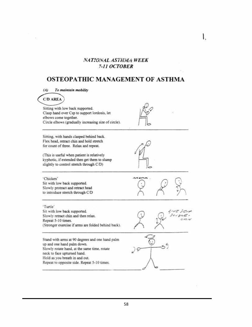
58

59

60

61

62

63
Appendix 3
Asthma Case Study One
Doctorate dissertation questionnaire Please answer the following questions as honestly as possible. Male/Female - female Current age – 36 Age at Diagnosis – 17 Please list the prescribed medications you were taking when you started having treatment with me – Salbutamol & Beclometasone diproprionate Please list the current prescribed medications you use – Salbutamol Please state your initial symptoms – Wheezing & tightness mostly during exercise and when cold, but also in relation to hay fever. Salbutamol inhaler used Please state your current symptoms – Less wheezing and tightness on exercise –most times use of inhaler is not required. When was the last time you received medical intervention for a relapse, attack or hypo/hyperglycaemic attack – Use of salbutamol inhaler is less frequent– usually only needed if compromising factors such as cold, chest infection, or hay fever symptoms. Preventive inhaler rarely used. How do you feel the treatment has benefitted yourself and how has it or does it affect your condition – asthma symptoms seem greatly improved in general, less medical intervention (inhalers) required. Are you taking any supplements or following any specific diet – Dairy, egg and wheat free due to allergies. Any other information – Emma rocks!
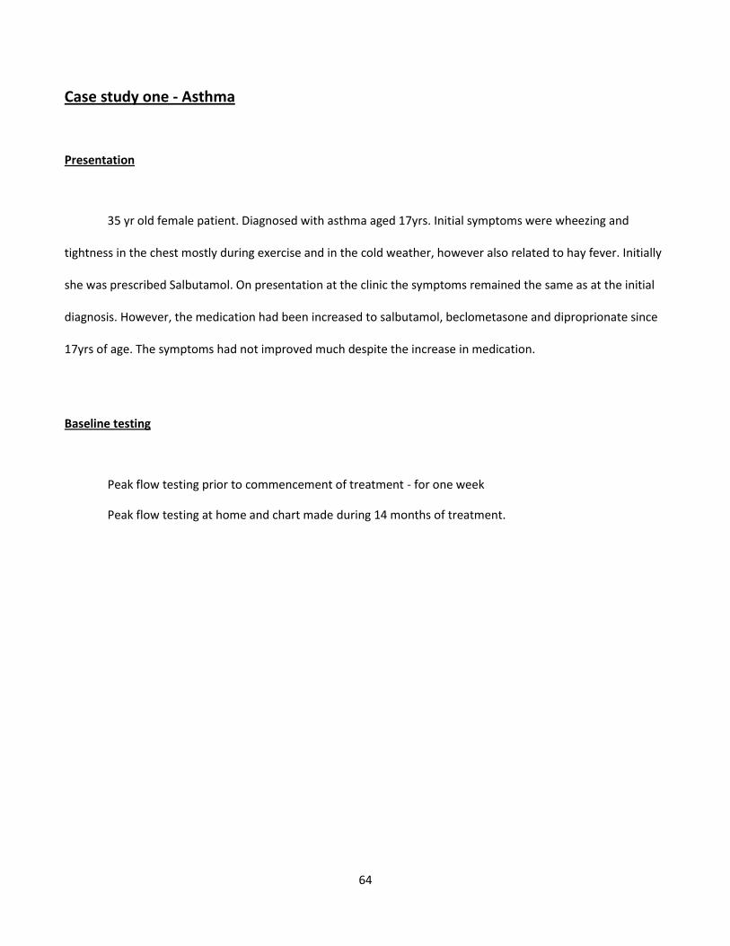
64
Case study one - Asthma
Presentation
35 yr old female patient. Diagnosed with asthma aged 17yrs. Initial symptoms were wheezing and
tightness in the chest mostly during exercise and in the cold weather, however also related to hay fever. Initially
she was prescribed Salbutamol. On presentation at the clinic the symptoms remained the same as at the initial
diagnosis. However, the medication had been increased to salbutamol, beclometasone and diproprionate since
17yrs of age. The symptoms had not improved much despite the increase in medication.
Baseline testing
Peak flow testing prior to commencement of treatment - for one week
Peak flow testing at home and chart made during 14 months of treatment.

65
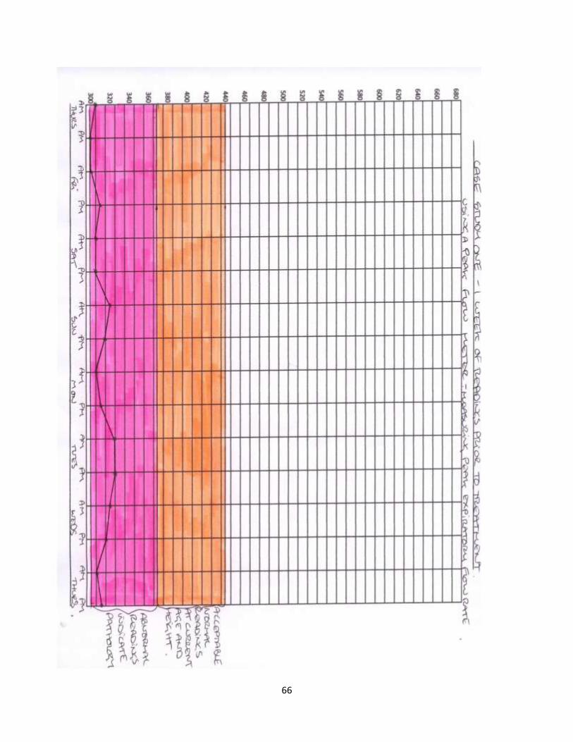
66
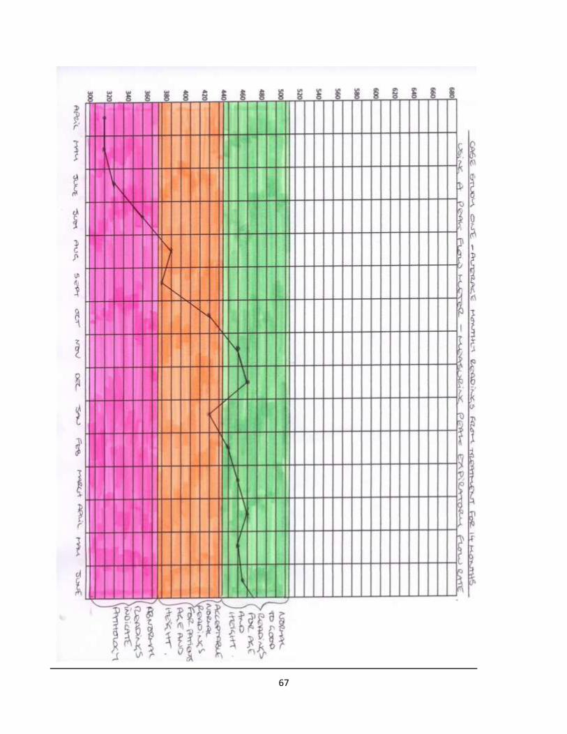
67

68
Initial observations and palpatory findings
My initial observations of this patient were as follows;
The patient presented with a scoliosis lumbar spine L5/S1 concavity left, to D10. D10 concavity right to
D2. D2 concavity left to C2. The dorsal spine anteriorised with posteriorisation of the C/D junction and the D/L.
The overall posture showed swayback tendencies. The movement within the rib cage is reduced, and restricted.
The pelvis is rotated right anterior, the upper shoulder girdle is rotated left anterior. Specific osseous lesions
were located at C5 rotated left, C/D rotated right side bent left, D4 rotated left, and the D/L rotated left side
bent right.
Treatment regime and management
This patient had already altered her diet, due to the expression of wheat, egg and cow dairy allergies
within the body. I had therefore discussed the diet, but the mucous producers within the diet had already been
reduced to a minimum, so we left the diet as it was. The treatment was focused on reducing the swayback
posture, and increasing the thoracic movement. Reducing the lateral curves and removing the specific lesions as
listed above. The general body adjustment was used to balance the body. And the side roll technique was used
to release the L5 restriction and help to reduce the lateral curve. The Dogs were done to the dorsal spine to
increase the A/P curve and reduce the anteriorisation of the dorsal spine. This technique was also used to
address the specific lesions within the dorsal spine. The prone C/D HVT technique was used to address the C/D
lesion. And a LVT manipulative technique was used to address the cervical spine specific lesions. I prescribed the
exercises as seen in appendix 2. I asked the patient to record her peak flow readings for my records.

69
Re assessment using initial assessment criteria including one week peak flow meter readings
The patient currently describes reduced wheezing and tightness on exercise, if, on the rare occasion this
does occur, she can usually control it without the use of the inhalers. She has reduced the inhaler use frequency
and is now rarely using any of them. Even the preventative medication is sporadic.

70

71
Treatment regimen adjustments
The treatment has now been reduced and is purely carried out as management for the condition, this
patient now visits on a 4-6 weekly basis and is usually reporting muscle aches and pains, which is a result of the
running that she has now taken up, she doesn’t report anything relating to the asthma condition. Therefore, the
treatment now consists of a general body adjustment, with focus on correction of any specific osseous lesions,
and checking the spinal areas indicated for asthma treatment, including the physiological centres. The D2-4 area
of the spine can be found to be rotated right and side bent left regularly. However, it is asymptomatic in its
presentation. We have discussed the possibility that her typing posture at work could be the habitual cause of
this lesion.

72
Appendix 4
Asthma Case Study Two
Doctorate dissertation questionnaire.
Please answer the following questions as honestly as possible.
Male/Female - Male
Current age – 29Yrs
Age at Diagnosis – 3Yrs
Please list the prescribed medications you were taking when you started having treatment with me – Ventalin,
becatide, occasionally prednisalone
Please list the current prescribed medications you use – occasional ventalin usage
Please state your initial symptoms – Difficult to remember due to my age however, my parents described
extreme attacks that resulted in several admissions to hospital, and use of a nebuliser at home.
Please state your current symptoms – Occasionally in cold weather whilst I’m playing rugby I struggle to get
my breath and have to use my ventalin. Also when there is the presence of tree pollen in the air it can
exacerbate my symptoms. The common cold usually spreads to my chest quickly but it is controllable.
When was the last time you received medical intervention for a relapse, attack or hypo/hyperglycaemic attack –
December 2000 had to use inhaler for 3 days consistently when I had contracted a chest infection.
How do you feel the treatment has benefitted yourself and how it has, or does it affect your condition – My
symptoms have been reduced and I haven’t had an attack since 6 months into the treatment. The treatment
has allowed me to pursue my career as a professional rugby league player. This is my fifth season with Leeds
Rhinos. The treatment has enabled me to play throughout the four previous seasons and feature in 3 back to
back grand finals which we were victorious in and are now super league champions for the third season in a
row.
Are you taking any supplements or following any specific diet – Beetroot juice which increases oxygen up take
within the body. Cherry extract, which increases recovery rates after a game. Jointease from a company called
natures range. Emu oil tablets which are a natural anti inflammatory (I only use these when I have an injury.).
I reduce the amount of wheat and cow dairy products which are both mucous producing within the body. The
beetroot juice and cherry extracts were a discovery of my own on the internet; the remaining supplements
were suggested by Emma. The diet alterations were also advised at the first consultation.
Any other information – None
73
Case Study two – Asthma
Presentation
This is a male patient aged 27yrs old, diagnosed asthmatic aged 3yrs old. His initial symptoms were
described by his parents as extreme attacks in nature which resulted in several admissions into hospital and the
use of a nebuliser at home. His birth history was uneventful however, shortly after birth he had to be aspirated
due to his breathing being laboured and difficult. He had fluid in his lungs and he was also diagnosed with
jaundice. His initial medications at diagnosis were ventalin, becatide and prednisalone.
Baseline testing
Peak flow testing prior to commencement of treatment - for one week
Peak flow testing at home and chart made during the treatment.

74

75

76

77
Initial observations and palpatory findings
Initial presentation on palpatory findings:-
This patient is very tall at 6ft 6in. He is very muscular and lean, he is a professional Rugby League player.
The patient has a flattened dorsal spine. He has an increased lumbar curve L5/S1 to D11/12. There is a minor
lateral curvature through the dorsal spine, concavity left, from D9 to D4. D4 to D2 are rotated right side bent
left. D1 to C6 are rotated left side bent right. C2 is rotated left.
Treatment regime and management
This patient has a very healthy diet as a requirement of his job as a professional rugby player. He has
paid an interest in the possibility of alternative therapies prior to coming to see myself. He has tried
acupuncture, herbalist treatment, and various supplements as part of his daily routine. The treatment was
focused on reducing the lateral curvature through the dorsal spine and encouraging the posture away from the
sway back presentation. General mobilisation of the rib cage was addressed, which was generally restricted. The
specific lesions were addressed with the use of HVT techniques. Side lying (side roll), Dogs, prone CDJ
adjustment and LVT neck correction. I gave supplement advice as listed in the questionnaire. The patient was
highly active and participated in rigorous cardio-vascular exercise every day, as part of his professional contract.
He struggled with the swimming, although he believed it would help with his breathing and lung capacity the
chlorine in the swimming pool exacerbated the mucous secretion and increased the fluid on his lungs, which he
then had to cough up. He had been using anti-histamines every time he had to train in the pool.

78
Re assessment using initial assessment criteria (including one weeks peak flow meter readings) and treatment
regimen adjustments.
The patient began treatment aged 27yrs old and upon reassessment for purposes of this study he was
aged 29yrs old. At reassessment the patient’s treatment schedule had been reduced to once or twice a month.
He is playing comfortably within the super league which is the highest national league within the UK. He only
uses any medication sporadically. He occasionally has to use his ventalin in cold weather games, although he is
trying to use essential oil peppermint drops instead, to open his airways as it is a bronchodilator.
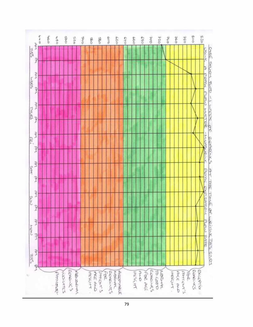
79

80
Asthma Case studies – evaluation.
In both cases the patient had already addressed their diet prior to attending my clinic. They had either
had prior advice, or intervention which suggested the same as my investigations. As a consequence I did not
address the diet with them. This could show that the diet, at least on its own is not sufficient to reduce the
symptoms. In case two the patient was extremely active and partook in a very high level of sports and a vigorous
exercise regimen. This could show that exercise combined with diet is not sufficient to reduce the symptoms.
However, as asthma can quite often be triggered by exercise this is perhaps not an accurate assessment criteria.
Both patients in the case studies have shown significant improvement of their symptoms, and I have produced
peak flow charts that clearly track these improvements. If I were to try to test the treatment rigorously I would
have to either withdraw the treatment to see if the patient regresses, or I would have to have a control group
that uses the same diet and exercise regimen without the treatment as performed by myself.

81
Appendix 5
Exercises for the MS Patient (training from MS
relapse); Darren Bristowe – Personal Trainer:
Total Fitness Huddersfield. 20th April 2010.
(Notes from a Dictation to myself)
Begin with basic balance work and gentle stretching of the major muscle groups. The stretching should
particularly focus on passive work to the hamstrings, quadriceps, gastrocnemius, pectorals and latissimus dorsi
bilaterally. The balance work should include walking on tip toes, walking on heels, walking pigeon steps (both
forward and backwards), and marching on the spot.
Then proceed to static work holding onto a ballet rail, this should involve the following movements; Hip
flexion, extension, abduction and circumduction.
Progress the exercises as the difficulty is overcome to involve Swiss ball squats and Swiss ball lunges.
This will continue the work on the hip movement, and will start to involve core stability therefore affecting
general balance. It will also start to build strength in the leg muscles and assist the neurological system to
interact with the muscles and assist with improving the neurological signal to aid control of movement.
The next stage in the progression is to isometric work involving the major muscle groups as listed above.
You should then vary the degree of hold, before moving onto negative reps for each exercise. This will improve
muscle conditioning and will involve recruitment of more muscle fibres with negative reps. These particular

82
exercises should include isometric leg extensions, laying leg curls, bench press, pectoral fly, lat pull down, bar
bell bicep curl.
When balance and posture is visibly improved work can begin on the Bosu balance disc. Use of a stress
ball is useful to improve grip strength before further weight work can begin.
Work on the Bosu disc should be carried out on the inflated side in the first instance. The basic exercises
to be performed are; marching on the inflated side of the disc, then standing on inflated side 30 seconds with
their eyes open, and 30 seconds with their eyes closed.
When the patient can perform the above exercises with reasonable ease, they should then stand on the
inflated side with a medicine ball in both hands - take ball overhead, take ball to shoulder height and rotate left
then rotate right. With a small medicine ball – pass overhead in an arc from one hand to the other. (Begin with
a sponge ball if their grip is poor or their proprioception is reduced.)
The patient should then advance to the Bosu disc reversed so that the base that feet are on is unstable
and the above exercises should be repeated. Change their stance from having their feet shoulder width apart, to
feet together to increase difficulty. (This should be done gradually as they improve.)
When their balance, grip and proprioception have all visibly improved, the next stage would be a single
leg stance with arm line activity i.e.; shoulder press, lateral raises, forward raises bicep curl, knee raise (hip
flexion), hip extension, abduction (the last three are performed with the leg that is not on the disc.).
Work can progress to a trampet to provide an unpredictably unstable base. When the patient is
comfortable with this progression to jogging on the spot varying the action between normal leg action jogging,
jogging with high knees, leg kicks, torso twists, star jumps. Use of a step box, small plyometric hurdles and floor
ladders can also be included to give variation and prevent the patient from getting bored.

83
Many things that would seem easy to most people can often be a great challenge, bunny hops and
hopping on one leg are good examples of this. Confidence is the main goal at this stage.
The final stage mirrors work that I would prescribe for a new client that has never done any gym training
before. Exercise on Swiss balls will advance the core stability work – this should include Swiss pectoral fly with
uneven weights, and Swiss bar bell press with odd weights on either side of the bar bell.
Fixed resistance machines require good grip strength; they are therefore useful for improving the
patients grip. The examples of these machines are; lat pull down, vertical seated row, high pulley row.
If the patient’s cardio vascular fitness is poor to begin with they should start with non weight bearing
exercises until their balance is improving e.g. bike, but it is extremely important for the patient to stretch after
each cardio vascular routine.
NB a Bosu disc can sometimes be described as a wobble board; however, it must be the type with a soft half ball
underneath and not hard.
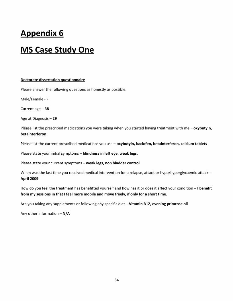
84
Appendix 6
MS Case Study One
Doctorate dissertation questionnaire
Please answer the following questions as honestly as possible.
Male/Female - F
Current age – 38
Age at Diagnosis – 29
Please list the prescribed medications you were taking when you started having treatment with me – oxybutyin,
betainterferon
Please list the current prescribed medications you use – oxybutyin, baclofen, betainterferon, calcium tablets
Please state your initial symptoms – blindness in left eye, weak legs,
Please state your current symptoms – weak legs, non bladder control
When was the last time you received medical intervention for a relapse, attack or hypo/hyperglycaemic attack –
April 2009
How do you feel the treatment has benefitted yourself and how has it or does it affect your condition – I benefit
from my sessions in that I feel more mobile and move freely, if only for a short time.
Are you taking any supplements or following any specific diet – Vitamin B12, evening primrose oil
Any other information – N/A

85
Case study one – MS
Presentation
This patient presented to my clinic aged 35yrs, she had been diagnosed with MS aged 29yrs. She initially
went to the GP with symptoms of weak legs with pins and needles and numbness in the left leg especially. She
suffered with this for several months with the GP prescribing diclofenac and co-codamol as he had diagnosed a
trapped nerve with lumbar spine dysfunction. She went to a chiropractor who gave her some treatment for a
while. However, he suggested to her to seek a second diagnosis as he couldn’t get any improvement from his
treatment and she had developed blindness in her left eye. She saw a neuro consultant who eventually
diagnosed her with MS. She was prescribed oxybutin and betainterferon.
When I first saw this patient she had been diagnosed with MS for 6 yrs and had been taking the
medications for 5 ½ yrs. She still had the blindness in her left eye and had bi lateral lower extremity symptoms of
pain, weakness, numbness and stiffness. She had difficulty walking into my clinic.
Baseline testing
1) Cerebellar dysarthria – is the speech slurred as if drunk, disjointed rhythm sometimes with scanning
speech.
- Observation of the patient showed that this was not present.
2) Rare structural deficit or more commonly focal deficit (demyelination) – diffuse or multifocal
abnormalities – observe – attention, memory, calculation, abstract thought, loss of special
appreciation, visual and body perception and apraxia.

86
- Observation and testing of the patient showed that no abnormalities were present.
3) Gait – Scissoring: indicates spastic paraparesis. Cerebellar ataxia: veers to side of lesion. Hemiplegic:
one leg swings out to the side – unilateral upper motor neurone lesion.
- Observation showed that the patient displayed a scissor gait and was extremely unsteady on her feet,
she refused to use any aids to assist with her mobility.
4) Cranial nerve abnormalities – multiple lesions affecting multiple cranial nerves possible. Intrinsic
brain stem lesions most commonly caused by MS in young people.
- Cranial nerve testing showed the following; Nothing abnormal was detected. (NAD)
- The following tests were performed. CN1 – Acuity, and visual field testing. CN2 – An opthalmascope was
used to look at the eye to see if anything abnormal was present. CN3,4&6 – Eye movements and
nystagmus. CN5&7 – sensory and motor functions. CN8 – Auditory testing. CN9,10&12 – Sticking tongue
out and pushing it against the cheek. Speech. CN11 – Shoulder shrug (Traps).
5) General motor system – Tone, reflexes, wasting, muscle weakness, fatigueable, spasticity,
fasiculation.
- Upper limb – C5 - Shoulder abduction, elbow flexion, biceps reflex. C6 – Elbow flexion, supinator reflex.
C7 – Finger and elbow extension, triceps reflex. C8 – Finger flexors, finger reflex. T1 – Small muscles of
the hand, no reflex.
- Lower limb – L1, L2 – Hip flexion, no reflex. L3, L4 – Knee extension, knee reflex. L5 – Extension of the
great toe, no reflex. S1 – Hip extension, knee flexion, plantarflexion, ankle reflex. Babinski test was
positive. The patient had loss of hip flexor power and knee extension, both on the left side. There was
increased reflex of the knee on the left also. There was increased lower limb tone with spasicity.
6) Sensation – Vibration test, Joint position sense, light touch, pin prick, temperature, 2 point
discrimination. MS usual patterning is complete cord transaction – all modalities affected or
thalamic and cortical loss distribution – right or left side of the body all modalities absent.

87
- The patient showed loss of all modalities on the left lower extremity.
Initial observations and palpatory findings
The patient has a sway back posture and cannot stand very long for the assessment. There are osseous
lesions at D3-4 with a rotation right side bent left. D10 – 12 is also rotated right side bent left. The lumbar spine
is scoliotic and impacted with a concavity on the right. L1-5 shows rotations at L1 rotation left and L5 rotated
right. D2/3 showed rotation right.
Treatment regime and management
The patient was directed to a personal trainer and dietary advice was given. The general body
adjustment was performed with the prescription of breathing exercises. Abdominal massage was used to help
with the constipation. Deep tissue work was carried out to the lower extremity specifically to the Achilles
tendon, outer calf muscle, hamstring, quadriceps and the plantar surface of the feet. Supplements were advised
in the form of vitamin b and d calcium and evening primrose oil. The specific lesions were addressed with the
use of HVT manipulative techniques.
Re assessment using initial assessment criteria
The patient is currently 38yrs old, she no longer has the blindness in the left eye, however, the patient
has lower extremity symptoms as first presented. She has developed a lack of bladder control and is now having
to use a catheter. She is also taking more medications than on first presentation to my clinic. She is taking

88
vitamin B12 and evening primrose oil as I suggested. The patient reports that she gets benefit from the
treatment but it is not long lived. She can tell if she hasn’t been to see me on her regular fortnightly
appointment as she struggles with her mobility more. Every treatment is just trying to give her some mobility
and maintain the current quality of life. The patient does not follow the diet that I gave her, and she has given
up on the exercise regimen at the gym and the personal training schedule that was put together for her.
Baseline testing
1) Cerebellar dysarthria – is the speech slurred as if drunk, disjointed rhythm sometimes with scanning
speech.
- Observation of the patient showed that this was present the patient is now struggling with speech
formation.
2) Rare structural deficit or more commonly focal deficit (demyelination) – diffuse or multifocal
abnormalities – observe – attention, memory, calculation, abstract thought, loss of special
appreciation, visual and body perception and apraxia.
- Observation and testing of the patient showed that no abnormalities were present.
3) Gait – Scissoring: indicates spastic paraparesis. Cerebellar ataxia: veers to side of lesion. Hemiplegic:
one leg swings out to the side – unilateral upper motor neurone lesion.
- Observation showed that the patient displayed a scissor gait and was extremely unsteady on her feet,
she is now using either walking sticks bilaterally, a walker or a wheel chair to assist with her mobility.
4) Cranial nerve abnormalities – multiple lesions affecting multiple cranial nerves possible. Intrinsic
brain stem lesions most commonly caused by MS in young people.
- Cranial nerve testing showed the following; Nothing abnormal was detected. (NAD) The following tests
were performed. CN1 – Acuity, and visual field testing. CN2 – An opthalmascope was used to look at the
eye to see if anything abnormal was present. CN3,4&6 – Eye movements and nystagmus. CN5&7 –

89
sensory and motor functions. CN8 – Auditory testing. CN9,10&12 – Sticking tongue out and pushing it
against the cheek. Speech. CN11 – Shoulder shrug (Traps).
- The patient has nystagmus in the left eye but her sight has returned. She has difficulty tracking a
pencil and controlling the eye movement.
5) General motor system – Tone, reflexes, wasting, muscle weakness, fatigueable, spasticity,
fasiculation.
- Upper limb – C5 - Shoulder abduction, elbow flexion, biceps reflex. C6 – Elbow flexion, supinator reflex.
C7 – Finger and elbow extension, triceps reflex. C8 – Finger flexors, finger reflex. T1 – Small muscles of
the hand, no reflex.
- Lower limb – L1, L2 – Hip flexion, no reflex. L3, L4 – Knee extension, knee reflex. L5 – Extension of the
great toe, no reflex. S1 – Hip extension, knee flexion, plantarflexion, ankle reflex. Babinski test.
- The patient fatigues easily and there is increased tone and hypereflexia bilaterally in the knee and
babinski was positive bilaterally. There was muscle power weakness in the hip flexor muscles and the
knee extensor muscles.
Upper motor neuron – increased tone, brisk reflexes, pyramidal pattern of weakness, extensor plantar response.
Positive babinski test.
6) Sensation – Vibration test, Joint position sense, light touch, pin prick, temperature, 2 point
discrimination. MS usual patterning is complete cord transaction – all modalities affected or
thalamic and cortical loss distribution – right or left side of the body all modalities absent.
- The patient has absent modalities on the left side in the lower extremity and reduced modalities in
the right side lower extremity. Showing cord transaction pattern.

90
Treatment regime adjustments
The treatment follows a similar pattern each fortnight as the initial lesions recur each time. The patient
does not fully participate in the programme set for her. She attends the fortnightly appointments and states that
she feels better afterwards, but also that this improvement doesn’t last very long. I think that the patient would
see a more sustained improvement in her symptoms if she would continue with the diet and exercise between
the appointments.

91
Appendix 7
MS Case Study Two
Doctorate dissertation questionnaire
Please answer the following questions as honestly as possible.
Male/Female - Female
Current age – 28
Age at Diagnosis – 26
Please list the prescribed medications you were taking when you started having treatment with me – Painkillers
that are available over the counter.
Please list the current prescribed medications you use – Exactly the same as above. Normally I use ibuprofen
and occasionally syndol as it has a slight muscle relaxant property.
Please state your initial symptoms – My symptoms began in my left leg with numbness, weakness and a feeling
of my leg being heavy. I went to my doctor and he diagnosed a trapped nerve and referred me to Emma. My
symptoms were worsening and Emma advised me to go back to my GP for a referral as she suspected that it
was more than purely a trapped nerve. Emma didn’t give me any treatment at that first consultation.
However, before I got to the GP my symptoms got worse and I ended up unable to walk and went to A&E.
They admitted me and I was diagnosed with MS by the specialist the next day. It was confirmed 2 weeks later.
I went back to see Emma and we came up with a plan of action.
Please state your current symptoms – Much better between Emma and Darren (a personal trainer that Emma
recommended) my balance has improved and I no longer need to use crutches. I feel stiff in the mornings in
both of my legs, and I still have numbness in my left foot. I have managed to keep my driving license which is
extremely important to me. I am not taking any medications although they have been suggested by the
specialist. I feel at this moment I am coping as I am and I don’t like the idea of taking medication long term. I
can always revert to this if I feel that I need it at a later date.
When was the last time you received medical intervention for a relapse, attack or hypo/hyperglycaemic attack –
When I was first diagnosed.
How do you feel the treatment has benefitted yourself and how it has or does it affect your condition –?
Greatly, the combination of the treatment with Emma and the training with Darren has got me back to work
and leading my life practically as normal.

92
Are you taking any supplements or following any specific diet -
Emma gave me a diet to follow; I am not sticking to it as strictly as I did in the beginning. I have found though
that if I do not follow it I sometimes have an increased amount of numbness in my leg or I feel stiffer in the
mornings.
Any other information – N/A

93
Case study two – MS
Presentation
The patient presented to my clinic aged 26yrs old. She was complaining of a heavy feeling in the lower
extremities on the left side particularly. She also felt weakness and numbness on the same side. She had been to
her GP who had said she had a trapped nerve. She was only taking over the counter painkillers as she did not
want to take any other medication. After performing the tests listed below I suggested that she should go back
to her GP and ask to have a referral to a neuro consultant. Before she could do this her symptoms worsened and
she was admitted into hospital. After a 4 night stay on the ward the neuro specialist diagnosed her with MS. She
refused the medications offered and returned to my clinic.
Baseline testing
1) Cerebellar dysarthria – is the speech slurred as if drunk, disjointed rhythm sometimes with scanning
speech.
- Observation of the patient showed that this was not present.
2) Rare structural deficit or more commonly focal deficit (demyelination) – diffuse or multifocal
abnormalities – observe – attention, memory, calculation, abstract thought, loss of special
appreciation, visual and body perception and apraxia.
- Observation and testing of the patient showed that no abnormalities were present.
3) Gait – Scissoring: indicates spastic paraparesis. Cerebellar ataxia: veers to side of lesion. Hemiplegic:
one leg swings out to the side – unilateral upper motor neurone lesion.
94
- Observation showed that the patient displayed a gait with one leg swinging out to the side as seen
with unilateral upper motor neuron lesion, and was extremely unsteady on her feet, I lent her a crutch
to assist with her mobility.
4) Cranial nerve abnormalities – multiple lesions affecting multiple cranial nerves possible. Intrinsic
brain stem lesions most commonly caused by MS in young people.
- Cranial nerve testing showed the following; Nothing abnormal was detected. (NAD)
- The following tests were performed. CN1 – Acuity, and visual field testing. CN2 – An opthalmascope was
used to look at the eye to see if anything abnormal was present. CN3,4&6 – Eye movements and
nystagmus. CN5&7 – sensory and motor functions. CN8 – Auditory testing. CN9,10&12 – Sticking tongue
out and pushing it against the cheek. Speech. CN11 – Shoulder shrug (Traps).
5) General motor system – Tone, reflexes, wasting, muscle weakness, fatigueable, spasticity,
fasiculation.
- Upper limb – C5 - Shoulder abduction, elbow flexion, biceps reflex. C6 – Elbow flexion, supinator reflex.
C7 – Finger and elbow extension, triceps reflex. C8 – Finger flexors, finger reflex. T1 – Small muscles of
the hand, no reflex.
- Lower limb – L1, L2 – Hip flexion, no reflex. L3, L4 – Knee extension, knee reflex. L5 – Extension of the
great toe, no reflex. S1 – Hip extension, knee flexion, plantarflexion, ankle reflex. Babinski test.
- The patient had hyper-reflexia in the left knee and spasticity of the lower extremity muscles on the
left side. The babinski test was positive on the left too. The muscle power was reduced in the hip
flexor and knee extension muscle in the left lower extremity.
Upper motor neuron – increased tone, brisk reflexes, pyramidal pattern of weakness, extensor plantar response.
Positive babinski test.

95
6) Sensation – Vibration test, Joint position sense, light touch, pin prick, temperature, 2 point
discrimination. MS usual patterning is complete cord transaction – all modalities affected or
thalamic and cortical loss distribution – right or left side of the body all modalities absent.
- The patient had retained all the modalities except light touch sensation which was reduced on the left
lower extremity throughout the entire limb.
Initial observations and palpatory findings
The patient presented with a hyperlordotic lumbar spine which was scoliotic with the concavity to the
left side. The lumbar spine was impacted and there was a specific lesion rotated right side bent left at L5/S1 the
defecation and circulation to the lower body. D10- L3 was posteriorised and rotated right side bent left. D4 also
showed rotation to left side bent right.
Treatment regime and management
We began by discussing the patient’s diet I gave her information as suggested in the MS part of this
study, and I referred her to my colleague for personal training and an exercise regime. I also gave her breathing
exercises. Each treatment was holistic using the general body adjustment. In each treatment we worked on the
lymphatic drainage of the head and neck with deep tissue work to the axilla and pelvis and lower extremity.
Cranial massage was used to increase the CSF flow throughout the cranium and spinal cord. Abdominal massage
was used as the patient had a history of chronic constipation. I performed deep tissue work to the quadriceps,
Achilles tendon, hamstrings and the plantar surface of the feet. She began to take the following supplements –

96
Vitamin d and b complex, Calcium and evening primrose oil. The specific osseous lesions were addressed using
HVT techniques.
Re assessment using initial assessment criteria
The patient has shown dramatic improvement, and from pure observations appears to be normal. Her
osseous lesion patterning has reduced and she no longer shows signs of rotations, scoliosis, impaction or
hyperlordosis. The testing below was performed to reassess. The patient is currently 28yrs old and has been
maintaining her diet and exercise regime with enthusiasm.
Baseline testing
1) Cerebellar dysarthria – is the speech slurred as if drunk, disjointed rhythm sometimes with scanning
speech.
- Observation of the patient showed that this was not present.
2) Rare structural deficit or more commonly focal deficit (demyelination) – diffuse or multifocal
abnormalities – observe – attention, memory, calculation, abstract thought, loss of special
appreciation, visual and body perception and apraxia.
- Observation and testing of the patient showed that no abnormalities were present.
3) Gait – Scissoring: indicates spastic paraparesis. Cerebellar ataxia: veers to side of lesion. Hemiplegic:
one leg swings out to the side – unilateral upper motor neurone lesion.

97
- The patient displays a normal gait and is not using any aids to assist her mobility.
4) Cranial nerve abnormalities – multiple lesions affecting multiple cranial nerves possible. Intrinsic
brain stem lesions most commonly caused by MS in young people.
- Cranial nerve testing showed the following; Nothing abnormal was detected. (NAD)
- The following tests were performed. CN1 – Acuity, and visual field testing. CN2 – An opthalmascope was
used to look at the eye to see if anything abnormal was present. CN3,4&6 – Eye movements and
nystagmus. CN5&7 – sensory and motor functions. CN8 – Auditory testing. CN9,10&12 – Sticking tongue
out and pushing it against the cheek. Speech. CN11 – Shoulder shrug (Traps).
5) General motor system – Tone, reflexes, wasting, muscle weakness, fatigueable, spasticity,
fasiculation.
- Upper limb – C5 - Shoulder abduction, elbow flexion, biceps reflex. C6 – Elbow flexion, supinator reflex.
C7 – Finger and elbow extension, triceps reflex. C8 – Finger flexors, finger reflex. T1 – Small muscles of
the hand, no reflex.
- Lower limb – L1, L2 – Hip flexion, no reflex. L3, L4 – Knee extension, knee reflex. L5 – Extension of the
great toe, no reflex. S1 – Hip extension, knee flexion, plantarflexion, ankle reflex. Babinski test.
- The patient does not display any abnormalities on any testing.
6) Sensation – Vibration test, Joint position sense, light touch, pin prick, temperature, 2 point
discrimination. MS usual patterning is complete cord transaction – all modalities affected or
thalamic and cortical loss distribution – right or left side of the body all modalities absent.
- The patient does not display any abnormalities on the above testing.

98
Treatment regimes adjustments
The patient now comes for realignment once every 4-6 weeks to maintain her current level of well being
and to keep the symptoms from recurring. She also feels that the treatment loosens her up and she feels less
stiff generally. Any specific osseous lesions are addressed weekly and if she notices specific aches or pains she
makes an appointment before her scheduled attendance. She refers to the treatment as her MOT.

99
MS Case Study evaluation
Although both of these patients have MS their attitudes to the condition are very different. Case study
one does not follow the suggested diet or follow the exercise regime. Case study two has followed the diet and
exercise regime rigorously and has also attended each appointment. The difference between the improvements
within both patients may be due to the medications, or perhaps the regimen. Case study one was already taking
betainterferon and painkillers prescribed by specialists prior to attending my clinic. Case study two has not taken
any medications other than some over the counter painkillers. Perhaps the use of the medication interferes with
the treatment progress.
The patient in case study one has not worsened in her symptom presentation as predicted by her
specialist. However, she has not improved to the extent of the patient in case study two who now does not
present any symptoms.

100
Appendix 8
Diabetes Diet
Sacred Heart Memorial Hospital
Used for overweight patients in need of rapid weight loss. The diet has also shown to have success with reducing
diabetic symptoms.
Fat burning soup.
Recipe – 3 Large onions, 1 head celery, 2 medium tins of chopped tomatoes, 1 large green pepper, ½ head of
cabbage, 1 pack of onion soup mix and 3 vegetable stock cubes.
Method – Cut all vegetables into small pieces and put into large pan. Add all other ingredients and cover well
with water. Season to taste and simmer until all vegetables are soft.
Serve liquidized or whole whichever you prefer.
Eat as much as you want whenever you want. It will not add calories and the more you eat the more weight you
lose.
DO NOT eat only the soup as it will cause malnutrition.
DAY 1
Eat as much fruit (except bananas) and soup as you can manage. For drinks have unsweetened tea, coffee,
cranberry juice or water.
DAY 2
Eat as much fresh vegetables, raw or cooked, but stay away from peas, dry beans and sweet corn. At dinner 1
jacket potato and butter, all the soup required no fruit.
DAY 3
Mix days 1 and 2, eat as much fruit, vegetables and soup as wanted but no jacket potato.
DAY 4
Eat up to 8 bananas, drink skimmed milk and eat as much soup as you want.
DAY 5
Eat 10 to 20oz of beef or fish with a large tin of tomatoes or 6 fresh tomatoes. Drink 6-8 glasses of water to wash
out toxins. Eat soup at least once today.

101
DAY 6
Eat as much beef or fish and fresh vegetables as you wish. You can have 2-3 steaks or fillets of fish with salad or
fresh veg. Eat soup at least once today.
DAY 7
Brown rice, fresh vegetables – eat as much as required. Eat soup at least once today. Drink unsweetened fruit
juice.
DEFINITE NO’S - No bread, alcohol, or fizzy drinks.
Stick to water, tea, coffee, fruit juice (unsweetened) and skimmed milk. You may have fish instead of beef but
not fried.
This diet will NOT affect medication and can be used for as long as you wish.
At the end of the 7th day if you have not cheated you will have lost 10-17lbs.
If you have lost more than 17lbs then stay off the diet for 2 days before resuming from DAY 1.
This diet, if used properly will clean your system of impurities and give you a feeling of well being. This diet is a
fat burning diet and the secret is that you burn more calories than you consume. This diet does not lend itself to
alcohol at anytime. Finish the diet for 24 hours before drinking alcohol. You can use the basic principles of this
diet once you have reached your suggested weight for your height. The balance of the diet and the reduction of
fats and processed sugars will help to keep diabetic symptoms at bay including the associated neurological
symptoms.
NB – If you do not stick to the diet exactly be prepared that your symptoms will return.

102
Appendix 9
Diabetes Case Study One

103

104
Case study one – Diabetes
Presentation
The patient is male and presented to my clinic aged 58yrs old. He had been diagnosed with diabetes
aged 42 yrs old. He had begun with classic diabetes symptoms of extreme thirst, increased Micturition, fatigue,
and bouts of dizziness. Prior to his diagnosis he had passed out at home and had been admitted into hospital. He
had been to his GP several times but had not had his blood glucose levels tested. In the hospital his blood
glucose was 32. Since his diagnosis he had not had any medical intervention and had managed the diabetes on
his own. He had been prescribed metformin initially, and then had progressed to injecting insulin. On
presentation to my clinic he had been prescribed insulin, ramipril, simvastatin, baclofen, and oxybutin, as he had
developed high blood pressure, high cholesterol and symptoms of neuropathy in his right hand and left lower
extremity.
Baseline testing
Blood glucose levels were taken for one week prior to the commencement of treatment. The blood
sugar levels were monitored during the treatment both before and after. As there were neurological symptoms,
testing was performed to monitor the extent and progress if any occurred.
The following neurological tests are suggested for diabetic patients;
- Observe for Argyll-Robertson pupil. - Negative in this patient.
105
- Check the patient’s acuity and eye movement – Both normal in this patient.
- Opthalomoscope should be used to check for changes in the eye – Normal presentation.
- Mononeuropathy is common – Normal presentation.
- Peripheral neuropathy is also common – The patient showed symptoms of reduced sensation and
motor power and function, in a glove and stockings distribution. The left lower extremity and right
upper extremity more prevalently. (Muscle power testing was performed for both the upper and
lower extremities.) (see Fuller, Geraint; Neurological Examination made Easy. 2nd edition. 1999)
- Single nerve sensory loss is common – Most commonly median, ulnar, perineal and lateral cutaneous
nerve to the thigh. – The patient presented with left lateral thigh reduced sensation and left lateral
calf reduced sensation. He also had right thumb and index finger reduced sensation. With some
weakness in the grip on this same side.

106

107
Initial observations and palpatory findings
The patient had a hyperlordotic posture with a posteriorised and impacted D/L, which was also rotated
right side bent left. D8-10 was rotated left side bent right. D5-9 was rotated right side bent left. The spine was
very immobile throughout.
Treatment regime and management
At the first treatment we discussed the patient’s diet. He had not been given any advice by any
dieticians, his GP, or a diabetic nurse specialist which surprised me greatly. We also talked about beginning a
regular exercise regime, and I warned him that he must monitor his blood glucose levels very carefully when
doing this as the exercise would be likely to reduce glucose levels. It is suggested that the patient should not
exercise within 1 hr of taking an injection of insulin. If they feel like they are having a hypo during an exercise
session they should have 10-15g or a carbohydrate snack and rest for 45mins. It is suggested that during
exercise the diabetic patient should have a carbohydrate snack every 30mins again it is suggested that this
should be around 10-15g. They should not exercise alone just in case, and it is especially important for diabetic
patients to warm up, and to cool down to help the circulation and to prevent muscle injuries. Diabetic patients
do not heal as quickly as other people. He agreed to follow the diet as shown in this study, he also began taking
green tea extract, and omega 3, 6 and 9.
I gave him a general body adjustment with the following focus; liver, pancreas, spleen and kidney pump
to stimulate function. Abdominal massage including the descending colon was performed to aid with the
congestion and chronic constipation. He had osseous lesions at D/L posterior and impacted in nature as
expected from the research. The lesion was also rotated right side bent left in nature. HVT techniques were used
to address the specific lesions.

108
Re assessment using initial assessment criteria
The patient’s blood glucose levels were tested regularly and his neurological testing was performed at
each treatment. The patient had normal blood glucose and had stopped his insulin and metformin. He is no
longer required to take ramipril or simvastatin, as his blood pressure and cholesterol have also returned to
normal. Although his neurological damage still remains in that he has some peripheral numbness and weakness
in the left lower extremity and the right hand. He is not taking any of the prescribed medication. He states that if
he remains on the diet and continues his exercise and regular treatment he does not need prescribed
medications. If he doesn’t follow this regimen then the symptoms do recur and his feeling of well being
diminishes, he also feels that his depression returns. He is also able to have a successful physical relationship
with his wife which he had not been able to do when he first presented to my clinic.
The following neurological tests are suggested for diabetic patients;
- Observe for Argyll-Robertson pupil. - Negative in this patient.
- Check the patient’s acuity and eye movement – Both normal in this patient.
- Opthalomoscope should be used to check for changes in the eye – Normal presentation.
- Mononeuropathy is common – Normal presentation.
- Peripheral neuropathy is also common – The patient showed symptoms of reduced sensation
(however the motor power and function had improved), in a glove and stockings distribution. The left
lower extremity and right upper extremity more prevalently. (Muscle power testing was performed
for both the upper and lower extremities.) (see Fuller, Geraint; Neurological Examination made Easy.
2nd edition. 1999)
- Single nerve sensory loss is common – Most commonly median, ulnar, perineal and lateral cutaneous
nerve to the thigh. – The patient presented with left lateral thigh reduced sensation and left lateral

109
calf reduced sensation. He also had right thumb and index finger reduced sensation. The grip strength
had returned to within normal parameters.

110

111
Treatment regimes adjustments
The patient has monthly appointments with me at my clinic, at which we address any lesions that have
occurred. I regularly check his pulses as diabetes commonly affects the circulation. I also perform regular
neurological testing to ensure that the symptoms are not progressing. He seems to be stable and in much better
spirits.

112
Appendix 10
Diabetes Case Study Two
Questionnaire for my Dissertation.
Please answer the following questions as honestly as possible.
Male/Female - Female
Current age – 48
Age at Diagnosis – 46
Please list the prescribed medications you were taking when you started having treatment with myself –
Metformin
Please list the current prescribed medications you use – Nothing
Please state your initial symptoms – I was fatigued and had a severe thirst. I was always drinking and would
also find I had to go to the toilet a lot more. I ached generally.
Please state your current symptoms – I have no symptoms currently.
When was the last time you received medical intervention for a relapse, attack or hypo/hyperglycaemic attack –
I haven’t had a hypo for a year now since I was 47yrs old 6 months after starting my treatment with Emma.
How do you feel the treatment has benefitted yourself and how has it or does it affect your condition – I
originally went to see Emma with lower back pain. I told her in the case history about my diabetes and we
decided to try to reduce my need for medication with her type of treatment.

113
Are you taking any supplements or following any specific diet – I am following the diet that Emma gave me and
have joined the gym, taking regular cardiovascular exercise.
Any other information – I hadn’t thought there was anything that could be done for my diabetes. I am glad
that I went to see Emma with my back pain as I did not enjoy having to take regular medication.

114
Case study two – Diabetes
Presentation
This female patient presented to my clinic aged 46 ½ having been diagnosed with diabetes aged 46yrs
old. She actually presented to my clinic with lower back pain. On presentation to my clinic she was taking
metformin, and had originally had symptoms of fatigue, extreme thirst, increased frequency of Micturition, and
general aching. She did not have any further medical conditions and was not taking any other medication.
Baseline testing
The patient’s blood glucose levels were tested for one week prior to commencement of treatment. She
monitored her blood glucose levels throughout the treatment both before and after.

115

116
Initial observations and palpatory findings
The patient was very rotund, she had an increased lordosis in the lumbar spine and a posteriorised D/L.
She had generally reduced mobility throughout the spine. The D/L was posteriorised, impacted, and rotated
right side bent left. The rest of the spine was generally scoliotic, with the concavity left L5 – D11, concavity right
D10 to D2. Concavity left D1 to C2. There were no other osseous lesions. The patient has only had the symptoms
of diabetes for a short time before attending my clinic, perhaps this is why there is so little of any consequence.
Treatment regime and management
We discussed use of the diet as listed within this study, and we discussed the importance of taking
regular exercise. However, I warned her about the effect that the exercise could have on her blood glucose
levels. Each treatment consisted of a general body adjustment with focus to stimulating the liver, spleen,
pancreas and kidneys. The excretory systems were stimulated also including abdominal massage to the
ascending and descending colon to ease the congestion and chronic constipation that the patient suffered from.
Side lying techniques were done to alleviate the posteriorisation at the D/L with the aid of traction. Any specific
osseous lesions that had been found were addressed by the use of HVT techniques.
Re assessment using initial assessment criteria
The patient does not take any medication currently, and has not suffered any symptoms of diabetes.
She is still following the diet as instructed and she is attending the gym at least 3 times a week for cardio

117
vascular exercise although she has now started to do some core stability classes and has even started to do
some light weights. She does not have the lower back pain either.

118

119
Treatment regimes adjustments
The patient is now seeing me every couple of months for a treatment. She is maintaining her current
regime very well and we just give her body a general check addressing any specific lesions that may have
become apparent. She had some difficulties with the general lack of mobility within her spine when she
commenced the core stability classes. We have done general manipulative treatments to improve the overall
mobility.

120
Diabetes case study evaluation
Both the patients in this study have late onset diabetes. Case study one was very much more
complicated and had had more medical intervention due to the existence of other medical conditions. However,
he has still managed to reduce all of his medication. Unlike the patient in the MS case study one, this patient
was fully participatory with the treatment and management regimen. Perhaps this is why we were able to
reduce his medications. Perhaps this is what is necessary to maintain the reduction of symptoms, complete
dedication to all the aspects of the treatment programme. The patient in case study two was uncomplicated, she
has reduced her medication to zero and is maintaining well. She had taken dietary advice prior to seeing myself
from the diabetic nurse specialist. Perhaps she had not taken it seriously enough. Perhaps it would never have
been sufficient without my intervention.

121
Appendix 11
ADHD Case Study One
Questionnaire for my doctorate dissertation.
Please answer the following questions as honestly as possible.
Due to the age of this patient the questionnaire should be filled in by the Mother.
Male/Female – Male
Current age – 10yrs old
Age at Diagnosis – 5yrs old
Please list the prescribed medications you were taking when you started having treatment with myself – My son
was not taking any medications. He had been assessed by the educational psychologist, and had been visited
by a behaviour specialist within the school. The educational psychologist suggested that there was nothing
that could be done as he could not be helped. The behaviour specialist had been in consultation with Emma
and put us in touch with her. I was willing to try anything.
Please list the current prescribed medications you use – My son does not take any medications.
Please state your initial symptoms – My son did not develop as my friend’s children did. He was unable to
communicate even with us, he had not developed any speech. His behaviour was poor and he had violent
outbursts. He became frustrated quickly and did not sit or pay attention to any tasks. He was extremely
hyperactive and constantly on the go. He was underachieving in all areas of school and his general
development.
Please state your current symptoms – My son has been able to remain in mainstream schooling which we
never imagined would be possible. The educational psychologist told us to remove him from the school, and
place him in a special school. He is still hyperactive and has a lot of energy, but he can control himself. He
always needs to run about at playtimes. He even knows when he needs to see Emma he asks to book an
appointment for a treatment especially after a growth spurt. He is achieving average grades in his SAT’s tests.

122
The teachers still find him energetic but he is not violent or difficult, they understand the importance for my
son to run off some energy and send him to do errands if they have to have a wet playtime.
When was the last time you received medical intervention for a relapse, attack or hypo/hyperglycaemic attack –
The only intervention we have had for our son is the treatment from Emma. He doesn’t have attacks as such.
How do you feel the treatment has benefitted yourself and how has it or does it affect your condition – Without
the treatment our son would’ve been put into a special school. He is developing at the normal rate expected
for his age. He behaves normally even though he may be a little energetic.
Are you taking any supplements or following any specific diet – As Emma suggested, he takes omega 3,6 and 9.
He does not have any caffeine, and we try to reduce any ‘E’ numbers and colourants. We try to reduce the
amount of wheat, cow dairy, and red meat.
Any other information – As a family we are very grateful for the help that Emma has given us.

123
Case study one – ADHD
Presentation
The patient presented at aged 5yrs to my clinic. He was unable to speak, and had severe difficulties
communicating with his parents, peers and teachers. He was unable to concentrate on any task for long. He
could become very violent and frustrated quickly. He had not been diagnosed with any specific condition by the
educational psychologist, he just said that he was somewhere on the autistic spectrum. The educational
psychologist had washed his hands of the patient and stated that there was no point trying to organise any help
for him as he would be unable to learn. His mother described that he had been a slow second stage of labour
and that they had had to use forceps to assist in the delivery.
Baseline testing
Before we began any treatment I asked his parents to fill out a behaviour questionnaire, and asked his
teachers to fill out an observational questionnaire with regards his behaviour in the classroom. The patient was
unable to fill out a questionnaire about his own behaviour as initially he was unable to read and write.

124

125

126

127

128
Initial observations and palpatory findings
My first assessment of the patient at my clinic was to allow him to become used to the environment and
also myself. His parents had been told by the educational psychologist that part of the reason for him not
recommending any assistance or one to one help, was that he had no imagination, couldn’t play and could not
therefore learn or retain information. As part of the first visit, I used a video to record the patient at play. With
his parents permission we taped for a ten minute duration. I gave him the middle of a couch roll to see what his
reaction would be. Within the ten minutes, the patient had pretended that the cardboard tube was a fishing rod,
a listening device, a telescope, a sword, a gun, and a trumpet. I contacted the educational psychologist by letter
and asked him to share his thoughts on the matter especially considering his initial comments with regards the
patient’s lack of imagination. I never received a reply. I was able to observe the patient’s posture as he played in
my clinic. He was willing to sit on my knee and allowed me to palpate his spine and the musculature either side.
He drew me pictures of pirates whilst we did this.
My initial observations of the patient saw that his posture was swayback with a flattened dorsal spine.
Having researched ADHD for my degree dissertation this presentation was not unexpected. On seated palpation
on my knee I found that C2, 3 and 7 were rotated right and side bent left. D2 to 10 was flattened and
anteriorised. D11 to L1 was posteriorised and rotated right side bent left. D4 was rotated left side bent right. S1
to L2 was scoliotic with the concavity left. L1 to D2 was scoliotic with the concavity right.
Treatment regime and management
The treatment was initially carried out with the patient sitting on my knee. I used gentle techniques to
relax the muscles that were contracted and tried to ease the rotations towards the normal presentation that we

129
should see. The patient was distracted by allowing him to draw pictures or colour in a colouring book. When he
was more comfortable with the setting we proceeded to him laying down on the couch so I could perform more
specific adjustments. Eventually I was able to do gentle manipulative corrections to derotate the osseous lesions
and move the dorsal spine into a normal A/P curve. We discussed the patients diet and decided that
supplements of omega 3,6 and 9 should be added, removing caffeine, ‘E’ numbers cow dairy, wheat and red
meat. He had a very unbalanced diet with a lot of processed foods, so we discussed the inclusion of more
homemade meals.
Re assessment using initial assessment criteria
I asked the parents and teachers to fill out the same questionnaires and assessments as they had done
prior to the treatment commencing. There were significant improvements. We repeated this process after 12
months and again after 24 months. The patient had been able to remain in normal mainstream schooling and
was able to read and write and communicate well, with his parents, peers and teachers. His behaviour had
improved and his violent outbursts were no longer an occurrence. He was able to retain information and
concentrate on classroom tasks set for him and his classmates. He participated with all regular school activities
and had been provided with an assistant within the classroom to help him to catch up, following a reassessment
by the educational psychologist.

130

131

132

133

134

135

136

137

138

139

140
Treatment regimes adjustments
We have reduced the treatment intervals to whenever the patient feels he requires them. He is able to
detect the tensions and dysfunctions within his own body now, and indicates to his parents when he feels that
this has occurred. He visits approximately once every 3 months or following a growth spurt. He is maintaining his
diet plan and the use of the omega 3,6 and 9 supplement. The osseous lesions found originally are not
maintained by the body treatment to treatment. I am able to sustain balance within the body with use of a
general body adjustment as a treatment. If an osseous lesion is located this is addressed within each treatment.
I have found that following a growth spurt the upper dorsal spine and in particular D4 seem to revert back to the
original lesion patterning. This will perhaps be the case until he has stopped growing, and we can maintain the
mature spine without the lesions.
ADHD case study evaluation.
The patient has maintained a combination of supplements and dietary control. He has returned for
treatment and has reduced the frequency of his visits. He recently had a few behaviour problems within the
classroom. However, his parents have recently separated. One may postulate that the improvements could have
been produced by his maturing and would have been a natural progression. We would be unable to test this
hypothesis in this particular case. We would need to have several more case study subjects and monitor the
progress with and without the treatment. The patient’s behaviour has now settled down and his teachers report

141
that he is calm within the classroom again. This external vector of his parents’ separation is beyond our control,
and children that have not been assessed as autistic or ADHD may well have suffered with behaviour problems
under similar circumstances. There are several books that cite parental separation, bereavement and other
changes in home life circumstances as possible reasons for a sudden change in a child’s behaviour.
The patient may have responded to myself as the positive vector as opposed to my actual treatment and
we should bear this in mind. If we were to continue this study I would need to have several practitioners
involved so as to rule myself out as the positive vector.

142
Appendix 12
Asthma – Physiological centres Diagram

143

144

145
Appendix 13
MS - Physiological centres Diagram

146

147

148
Appendix 14
Diabetes - Physiological centres Diagram

149

150
151
Appendix 15
ADHD - Physiological centres Diagram

152
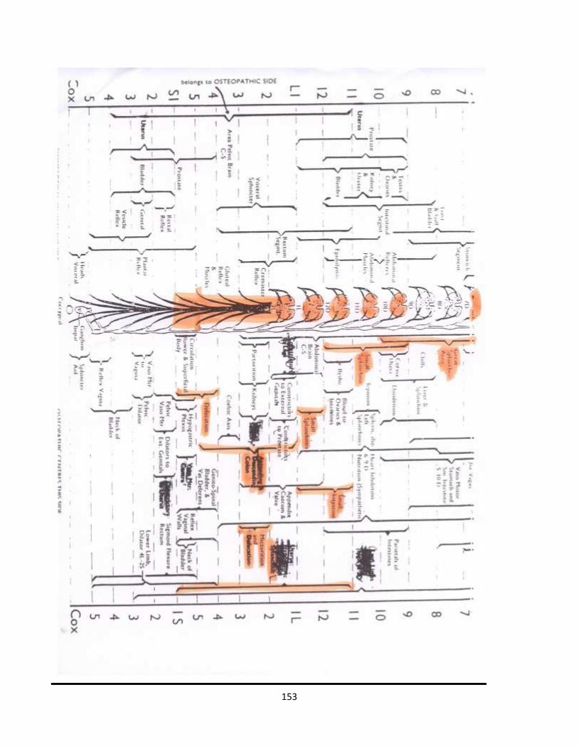
153

154
Bibliography
- Burns, L. – Pathogenesis – Gastrointestinal Changes Following Lesions – 226-233 unknown date
- Bowling, Allen.C – Complementary and Alternative Medicine and Multiple Sclerosis – 2nd Edn – 2007
Demos Medical Publishing
- Davis, F A. – Taber’s Cyclopedic Medical Dictionary – 18th Edn - 1993
- Digiovanna, Eileen L., Schiowitz, Stanley. - An Osteopathic Approach to Diagnosis and Treatment 2nd
Edition Lippincott Williams & Wilkins 1997
- Harruff, Richard C. – Pathology Facts – J B Lippincott Company - 1994
- Hazzard, Charles. – The Practice and Applied Therapeutics of Osteopathy – Chapter XIX – Constitutional
Diseases - 1905
- Kuchera, Michael L., Kuchera, William A. – Osteopathic Considerations in Systemic Dysfunction 2nd
Edition Greyden Press 1994
- Littlejohn, J.M – The Fundamentals of Osteopathic Technique, 1998, John Wernham
- McConnell, C P., Teall C C. – The Practice of Osteopathy – Journal printing company; Kirksville -3rd edn -
1906
- Parsons, Jon., Marcer, Nicholas. - Osteopathy – Models for Diagnosis, Treatment and Practice –Churchill
Livingstone 2006
- Patten J, - Neurological Differential Diagnosis; 2nd Edn Springer 1996
- Pottenger, Francis Marion. - Symptoms Of Visceral Disease – A Study Of The Vegetative Nervous System
In Its Relationship To Clinical Medicine – 2nd Edition St Louis C.V. Mosby Company 1922
- Rang HP, Dale MM, Ritter JM – Pharmacology; 3rd Edn Churchill Livingstone 1996
- Still, Andrew.T – Philosophy of Osteopathy, 1899, A.T.Still, Kirksville, MO

155
- Trowbridge, Carol – Andrew Taylor Still 1828 to 1917 – 1991 – The Thomas Jefferson University Press
Kirksville, Missouri 63501 USA
- Vander, Sherman, Luciano – Human Physiology; 6th Edn WBC McGraw-Hill; 1990
- Ward, Robert.C – Foundations for Osteopathic Medicine, American Osteopathic Association, Lippincott
Williams and Wilkins, 1997
- Wernham, John – Classical Osteopathy, Reprinted Lectures from the archives of the Osteopathic
Institute of Applied Technique, Printed and Published by The Maidstone College of Osteopathy, 1996
- Zubay GL – Biochemistry; 4th Edn WBC Wm. C. Brown publishers, 1998

156
References
- Balon J, Aker PD, Crowther ER et al; A comparison of active and stimulated chiropractic manipulation as
adjunctive treatment for childhood asthma. N Engl J Med; 339:1013-1020 (1998)
- Barkley R; ADHD and the nature of self control: New York: Guildford. 1997
- Baumgaertel A; Alternative and Controversial treatments for Attention-Deficit/Hyperactivity disorder;
Paediatric Clinics of North America. Vol 46, No 5, PP 3955/99. 1999
- Cayce Edgar; www.edgarcayce.org
- Dulgar G, Hill D, Sirucek A, Davis B; Evidence for a possible antihypertensive effect of basic technique
apex contact adjusting. ACA J Chiro: 7:231 – 36: 1980
- Geisen JM, Center DB, Leach RA; An evaluation of chiropractic manipulation as a treatment of
Hyperactivity in children; Journal of Manipulative and Physiological Therapeutics. Vol 12, No.5, pp 353-
363; 1989
- Hinshaw S; Attention Deficit and Hyperactivity in Children: London. New York. New Delhi: Sage: 1994
- Kimmel EH; Psychophysiological aspects of subluxation; In. Schwartz HS, ed Mental Health and
chiropractic. New York: Sessions: 1973
- Licciardone John C, Fulda Kimberly G, Stoll Scott T, Gamber Russell G, Cage Clifton; A case-control study
of osteopathic palpatory findings in type 2 diabetes mellitus: Published 8th February 2007: Osteopathic
Medicine and Primary care.
- LYON J, Multi-Modal management of Attention Deficit/Hyperactivity Disorder (AD/HD) International
Psychology Services pp 1-6 (1996)
- LYON J, The nature and identification of Attention Deficit/Hyperactivity Disorder (AD/HD) International
Psychological Services pp 1 – 14 (1996)

157
- (http://www.msrc.co.uk/index.cfm?fuseaction=show&pageid=1405)
- SATTERSFIELD J H, DAWSON M E, Electrodermal correlates hyperactivity in children. Psychophysiology
(1971)
- Shaw DO, Quentin; Spinal Development and Balance of Children. (No date available)
- TANNOCK R. ADHD: advances in cognitive, neurobiological and genetic research. Journal of child
Psychology and Psychiatry. 39 (1): 65 – 99 (1998)
- Valitutto Michael; Common crossroads in diabetes management: Published 15th February 2008:
Osteopathic Medicine and Primary Care.
- Vardy Terence; Enhancing Homeostasis using Osteopathic Techniques for Multiple Sclerosis: Clinical
Practice. No known Date.
- WHOOTON L W, (Date Unknown) Spinal lesion patterns at circa five years, and their correlation with
labour and birthing technique. Maidstone College of Osteopathy.
- WICKES D. Effects of thoracolumbar spinal manipulations on arterial flow in the lower extremity. J
Manipulative Physiol Ther 3:3 – 6 (1980)
- Yates Herbert A, Vardy Terence C, Kuchera Michael L, Ripley Bret D, Johnson Jane C; Effects of
osteopathic manipulative treatment and concentric and eccentric maximal-effort exercise on women
with multiple sclerosis: A pilot study: JAOA.Vol 102.No 5.May 2002. 267
- Zamboni et al; Chronic cerebrospinal venous insufficiency (CCSVI) and MS: Published in J Neurology
Neurosurgery Psychiatry. 2009 Apr; 80 (4): 392-9.Epub.2008 Dec 5.