ems-swissquality.com save cells · 2019-10-02 · ems-swissquality.com for more information >...
TRANSCRIPT


32010
i s sn 1868-3207 Vol. 11 • Issue 3/2010
| researchTiming of dental implant loading
| case reportPiezoelectric repositioning of theinferior alveolar nerve
| meetings1st Hvar International Dental Congress
implantsinternational magazine of oral implantology

Ems-swissqualitY.com
For more information > www.ems-swissquality.com
savE cEllsNEw Ems swiss iNstrumENts surgErY –saviNg tissuE with NEw iNNovatioNs iN implaNt dENtistrYThe inventor of the Original Piezon Method has won another battle against the destruction of tissue when dental implants are performed. The mag ic word is dual cooling –instrument cooling from the inside and outside together with simultane-ous debris evacuation and eff icientsurgical preparations in the maxilla.
cooliNg hEalsA unique spiral design and internal irrigation prevent the instrument’s temperature from rising during the surgical procedure. These features combine effectively to promote excel-lent regeneration of the bone tissue.
EMS Swiss Instruments Surgery MB4, MB5 and MB6 are diamond-coated cyl indr ica l instruments for secondary surgical preparation (MB4, MB5) and f inal osteotomy (MB6). A spiral design combined with innovative dual cooling makes these instruments unique in implant dentistry.
coNtrol savEsEffective instrument control fosters atraumatic implant preparation and minimizes any potential damage to the bone tissue.
prEcisioN rEassurEsSelective cutting represents virtually no risk of damage to soft tissue
(membranes, nerves, blood vessels, etc.). An optimum view of the operative site and minimal bleeding thanks to cavitation (hemostatic effect!) further enhance efficacy.
The new EMS Swiss Instruments Surgery stand for unequaled Swiss precision and innovation for the
benefit of dental practitioners and patients alike – the very philosophy embraced by EMS.
> EMS Swiss InstrumentSurgery MB6 with unique spiraldesign and internal instrumentirrigation for ultralowtemperature at the operative site

I 03
editorial _ implants I
implants3_2010
Dr med dent Roland Hille
_Nobody could have imagined in 1970 what a success story oral implantology would turn outto be, when seven dentists, headed by Prof Dr Hans L Grafelmann, a dentist from Bremen, foundedDGZI. In spite of various negative opinions, mainly from universities, this then-adventurous ther-apy was established in Germany against the mainstream, thanks to a great deal of perseveranceand conviction, an incredible drive and much operative skill. There were a growing number of col-leagues who were fascinated by the possibility of fixing dental prostheses on implanted new den-tal roots, a process which could give patients the feeling of no longer being handicapped.
40 years ago the obligatory tooth conserving methods were incomparable to current meansand methods. Thus many patients, especially those with edentulous jaws, could achieve a com-pletely new quality of life courtesy of intraosseous or subperiostal implants. Let us remember: Atthat time, we did not have any bone substitutes, membranes etc. at our disposal, all of which areconsidered absolutely standard today. For the past four decades oral implantology has greatly in-fluenced dental rehabilitation measures, and has without question become the most innovativediscipline in dento-maxillo-facial medicine in the last 25 years.
On September 24, 1982, the DGZMK (German Association for Dento-Maxillo-Facial Medicine)approved implantology as a new method for use. Oral implantology also became scientifically invogue, when universities intensified their research activities, and industrial companies sensed anew market with adequate financial resources.
In the beginning scientific journals referred to implantology as “the red light district in dentalmedicine”, but nowadays there is no doubt about the important role which this subdiscipline playsin dentistry. Patients actively request this therapy, and any colleagues who underestimate the im-portance of implantology for the success and future for their own dental practices will be left be-hind.
DGZI has achieved significant accomplishments in education and advanced training, as up un-til now university education has not attached that much value to implantology. A postgraduatestructured educational program has existed since 1998, which almost 1,500 colleagues have par-ticipated in and have learned from implantology specialists and university professors about thestate of the art in implantology. Patients increasingly ask for treatment by a “DGZI Specialist in Im-plantology”, because such specialists often have much more operative skills than those colleagueswho obtained a masters degree.
40 years of DGZI is truly a great milestone in Europe’s oldest scientific implantological associ-ation, an association which also enjoys an extraordinarily good reputation nationally and interna-tionally. The Consensus Conference for Implantology congratulates DGZI heartily and wishes all itsmembers much success and an exciting future in oral implantology.
I hope to see you on the occasion of our anniversary congress on October 1 and 2 in Berlin.
Dr med dent Roland HillePresident of the Consensus Conference for Implantology
Celebrating 40 Yearsof DGZI in Berlin

04 I implants3_2010
I content _ implants
I editorial
03 Celebrating 40 Years of DGZI in Berlin| Dr Roland Hille
I research
06 Timing of dental implant loading| Dr Marius Hary Silvasan
I report
18 Lateralization of the inferior aveolar nerve| Dr Bernd Quantius
I case report
22 Piezoelectric repositioning of the inferior alveolar nerve| Dr Burghard Peter
26 Success without using cement| Dr Christoph Thiemann, Friedrich Schotsch
I study
28 Comparing various implant designs and surfaces| Dr Roy Leshem, David Leshem
II meetings
34 International events 2010/2011
36 1st Hvar International Dental Congress
38 EAO Congress—Glasgow 2010
40 Osteology in Cannes| Dr Birgit Wenz
42 3rd International CAMLOG Congress
I news
44 Manufacturer news
48 Congratulations and Happy Birthdayto all DGZI-members around the world
I about the publisher
49 | submission guidelines
50 | imprint
report 18 case report 22 case report 26
Cover image courtesy: Nobel Biocare Holding AG;
NobelReplace Tapered Groovy RP with Snappy Abutment.
study 30 meetings 36 meetings 40

Built-in platform shifting.Dual-function prosthetic connection.
Bone-condensing property.
Adjustable implant orientation for optimal final placement.
High initial stability, even in compromised
bone situations.
NobelActive equally satisfies surgical and restorative clinical goals. NobelActive thread design progressively condenses bone with each turn during insertion, which is designed to enhance initial stability. The sharp apex and cutting blades allow surgical clinicians to adjust implant orienta-tion for optimal positioning of the
prosthetic connection. Restorative clinicians benefit by a versatile and secure internal conical prosthetic connection with built-in platform shifting upon which they can produce excellent esthetic results. Based on customer feedback and market demands for NobelActive, the product assortment has been expanded – dental professionals
will now enjoy even greater flexi -bility in prosthetic and implant selection. Nobel Biocare is the world leader in innovative and evidence-based dental solutions. For more information, visit our website
www.nobelbiocare.com
NobelActiveTM
A new direction for implants.
© N
ob
el B
ioca
re S
ervi
ces
AG
, 2
01
0.
All
rig
hts
res
erve
d.
No
bel
Bio
care
, th
e N
ob
el B
ioca
re lo
go
typ
e an
d a
ll o
ther
tra
dem
arks
are
, if
no
thin
g e
lse
is s
tate
d o
r is
evi
den
t fr
om
th
e co
nte
xt
in a
cer
tain
cas
e, t
rad
emar
ks o
f N
ob
el B
ioca
re
Disclaimer: Some products may not be regulatory cleared/released for sale in all markets. Please contact the local Nobel Biocare sales office for current product assortment and availability.
10 YEARS WITH
TIUNITE® SURFACE
New data confi rm
long-term stability.
NobelActive A4 TiU Implants rev.indd 1 10-08-10 09.50.23

06 I
I research _ dental implant loading
Fig. 1_Direct loading at placement
and delayed loading after bone
healing.
Fig. 2_Conventional implant protocol
without any loading performing the
prosthetic in part after bone healing
(e.g. Brånemark protocol).
implants3_2010
_Osseointegration is the process by which livingbone attaches to the artificial surface of an implant bythe formation of bony tissue without growth of fi-brous tissue at the bone-implant interface.
_Introduction
Osseointegration is a highly dynamic process, whichdoes not only address the formation of bone onto animplant surface after it has been placed, but it also ad-dresses the remodelling or maintenance of bone dur-ing the life of the implant.
The long term success of an implant treatment istheoretically determined by factors related to the pa-
tient, the implant components and the treating clini-cians.1 Before the introduction of the Prof. Brånemarkprotocol, dental implants were commonly loaded atplacement because immediate bone stimulation wasconsidered to avoid crestal bone loss (Fig.1).2 The cli-nician is often faced with the challenge of identifyingthe successful osseointegration of implant. Clinicalsuccess is often determined by a lack of mobility andability of the implant to resist functional loading.3
Radiographically, bone should appear to be closelyapposed to the implant surface. The current achiev-able resolution obtained in medical imaging, how-ever, is about 10 times less than what is required to ob-serve a soft tissue cell. Therefore, radiographic as-sessment alone is unsuitable to determine with cer-tainty if a soft tissue layer is present. When an implantis exposed to excessive micromotion at the bone-im-plant interface during healing, fibrous tissue encap-sulation of the implant rather than osseointegrationmay occur. Conventional implant protocols havebeen based on the achievement of primary stabilityand prolonged non-loaded healing periods (Fig. 2).4
That was achieved by a two stage technique andan unloaded healing period of three to six months.Delayed implant loading was empirically based on thebelief that the transfer of any micromotion to the im-plant surface during healing would result in fibrousencapsulation rather than osseointegration. A per-ceived psychological, economical and functional ad-vantage of shortened treatment periods has encour-aged clinicians to challenge this convention with im-mediate temporization (Fig. 3) and/or the early andimmediate loading of dental implants.
The relative merits of these shortened loading pro-tocols will be discussed with respect to their biologi-
Timing of dental implant loadingA Literature ReviewAuthor_Dr Marius Hary Silvasan, Romania
Fig. 1
Fig. 2

I 07
research _ dental implant loading I
implants3_2010
cal implication, the current evidence based literatureand the factors that might influence their outcomes.There is a growing body of published literature sup-porting reduced implant loading times. Abutmentconnection and placement of a restoration in occlu-sion with the opposing dentition of an implant at thetime of surgery or within 48 hours of placement is re-ferred to as “immediate loading” The functionalrestoration of an implant from 48 hours up to 3months after placement has been defined as “earlyloading”.5 Both the immediate and early functionalloading of implants before lamellar bone formationcarry an inherent biological risk. Shortened loadingprotocols may expose the healing bone to implant in-terface to mechanical overload as described in WollfsLaw and Frosts Mechanostat theory (Fig. 4).
Interfacial micromotion above the biologicalthreshold can result in the subsequent loss of implantstability. Rough titanium surfaces offer better im-plant anchorage in bone and more rapid bone depo-sition.6 The general applicability of these principleswill be considered as to their biological implications,the current evidence base and the factors that influ-ence their results.
_Materials and Methods
Clinical reports on dental implants found in majorscientific journals and through searching in PUBMED, QUINTESSENZ and MED-LINE, have served asthe basis for this review. The following search terms,alone or in combination, were used: implant loading,immediate loading, early loading, delayed loading.After screening the titles and abstracts for possiblerelevance, they were ordered in full text. We alsoscreened reference list of publications and relevantsystematic reviews. To minimise bias, only RCTs of os-seointegrated dental implants were considered. To beincluded, RCTs had to compare the same osseointe-grated implants loaded at different times for a periodof at least 12 months of loading.
For the purpose of this review immediate loadingwas defined as an implant put in function within 48hours after its placement; early loading as those im-plants put in function from 48 hours up to 3 monthsafter placement, and conventional loading as thoseimplants put in function between 3 to 6 months afterinsertion. Implant mobility and removal of stable im-plants dictated by progressive marginal bone loss orinfection have been assessed. Implant mobility of in-dividual implants could be assessed manually or withdevices such as Periotest® (Siemens, Munich, Ger-many) or Resonance frequency—Analysis—Osstell®(Integration diagnostics, Göteborg, Sweden). In oursearch we aimed at including randomized controlledtrials. Most clinical reports were on a few implant sys-
tems only and threaded commercially pure titaniumimplants ad modum Brånemark dominated the liter-ature. The quality assessment of the included trialswas undertaken independently. The following qualitycriteria were examined:
Allocation concealment was recorded as ade-quate ( A ), unclear ( B ), or inadequate (C), as describedelsewhere [Higgins, Green S. Handbook for systematicreviews of interventions].
Allocation concealment was considered adequateif it was centralized (e.g. Allocation by a central officeunaware of subject characteristics). If randomizationwas pharmacy controlled; if prenumbered or codedidentical containers were administered serially toparticipants.
A score of A was recorded if there was a clear ex-planation for a withdrawals or dropouts in each treat-ment group or if there were no dropouts. If clear ex-planation for any dropouts were given, the risk of biasof the assessment of reasons for dropping out wasevaluated. A “strong scientific basis” is required aswell. A score of B was recorded if clear explanationsfor any dropouts or withdrawals were not provided.Articles or authors that stated that allocation con-cealment procedures were implemented but did notprovide details on how this was accomplished werecoded as unclear. A score of C was recorded if there
Fig. 3_Immediate temporization and
delayed loading.
Fig.4_Loading zones acc. to
H. M. Frost
Fig. 3
Fig. 4

08 I
I research _ dental implant loading
implants3_2010
were “insufficient scientific basis” or any procedurethat was entirely transparent before allocation, suchas an open list of random numbers. Hence, after athorough reading of the studies included in this re-view, one of these scores has been qualified accord-ing to accuracy and the underlying scientific bases.
_Results
In 2002, a consensus meeting was convenedwithin the World Congress organized by the SpanishBoard of Implantology in Barcelona.5 There was anagreement on terminology for the timing of loading(immediate, early, delayed) and for the implant load-ing (occlusal loading and nonocclusal loading). Ac-cording to this consensus meeting the following ter-minology was described:
Immediate loadingThe prosthesis is attached to the implants the same
day the implants are placed
Early loadingThe prosthesis is attached at a second procedure,
earlier than the conventional healing period of 3 to 6months. The time of loading is started after somedays/weeks.
Delayed loadingThe prosthesis is attached at a second procedure
after a conventional healing period of 3 to 6 months.
Occlusal loadingThe crown/bridge is in contact with the opposing
dentition in centric occlusion.
Nonocclusal loadingThe crown/ bridge is not in contact in centric oc-
clusion with the opposing dentition in natural jaw po-sition.
The available literature demonstrates the possibil-ity of achieving good results with different protocols,especially with immediate loading protocol, at least ingood-quality bone, which supports the idea thatthese concepts may serve as a viable option in implantdentistry. However, the prerequisites for achievingand maintaining acceptable results and the limita-tions of immediate/early loading are not fully known.Moreover, the terminology used in these protocols isconfusing since the difference between different pro-tocols is not well defined, and publication titles cantherefore be very misleading. Of 26 potential studies,7 have been excluded because of insufficient patientselection data or prothesis loading longer than oneday (immediate loading), not corresponding to theBarcelona consensus, and 5 have been excluded sincethe follow up was shorter than 12 months. Fourteen
studies have been introduced in this review, the con-clusions having been discussed on their basis.
The majority of the studies considered in this re-view registered a relatively short follow up. In 6 stud-ies the follow up covered a period longer than 24months.
Daniel Sullivan, Giampaolo Vicenzi, Sylvan Feld-man performed a multicenter study: the performanceof Osseotite implants after an 1 stage surgery and ab-breviated healing period of 2 months in 10 privatepractice centers. 142 patients, partially or completelyedentulous, enrolled in this early loading study, re-ceived 526 implants, 65.4 % in mandible and 34.6 %in maxilla. Implants were loaded after a healing periodof about two months. The distribution of the prosthe-sis types included 118 single tooth restoration (118implants), 134 short-span prosthesis (327 implants)and 16 long-span restoration (81 implants).
Eight of the eleven implant failures occurred dur-ing nonsubmerged healing prior to prosthetic load-ing. Provisional restoration was placed at 2.1 ± 0.5months, at which time implants were evaluated formobility, gingival health and radiolucency. The cumu-lative success rate of these 526 implants was 97.9 %at 5 years.
These results suggest that success can be expectedwith Osseotite implants after a nonsubmerged re-duced healing period of two months in this patientpopulation.7
Par-Ölov Östman, Mats Hellman, Lars Sennerbyevaluated in a prospective clinical study the radi-ographic and clinical outcome of immediately load-ing implants in the partial edentulous mandible overa 4 year follow up period.
96 patients were evaluated and 77 patients whomet the inclusion criteria were included. A total of 111fixed partial dentures supported by 257 BrånemarkSystem implants (77 turned and 180 Ti Unite im-plants) was delivered. Four (1.16 %) of the 257 im-plants did not osseointegrate after 4 years. Threeturned implants (3.9 %) and one oxidized implant (0.6 %) failed after 4 to 13 months. Immediate load-ing of implants with firm primary stability in partiallyedentulous areas of the mandible appears to be a vi-able procedure with predictable outcome.8
Richard P. Kinsel, Mindy Liss evaluated in a retro-spective study the effects of implants dimensions,surface treatment, location in the dental arch, num-bers of supporting implant abutments, surgical tech-nique, and generally recognized risk factors on thesurvival of a series of single stage Straumann dental

connect your competenciesStraumann® CarES® GuidEd SurGEry – Global ExCEllEnCE mEEtS loCal ExpErtiSE
Local template production: surgical templates with verified fit just in time from your local dental lab
User friendly – open software system: offers you large flexibility and easy usage
Customized cost model: benefit from suitable price models for your personal business requirements
please contact us at 0041 (0)61 965 1111. more information on www.straumann.com

10 I
I research _ dental implant loading
implants3_2010
implants, placed into edentulous arches using an im-mediate loading protocol. Data were collected for 344single-stage implants placed into 56 edentulousarches (39 maxillae and 17 mandibles ) of 43 patientsand immediate loaded with a one piece provisionalfixed prosthesis.
Each patient received between 4 and 18 implantsin one or both dental arches. Periapical radiographswere obtained over a 2 to 10 year follow up period toevaluate crestal bone loss following insertion of thedefinitive metal-ceramic fixed prostheses. A total of16 implants failed to successfully integrate. Increasedrates of failure were associated with reduced implantlength, placement in the posterior region of the jaw,increased implant diameter and surface treatment.Implant length emerged as the sole significant pre-dictor of implant failure.
In this prospective analysis, in 56 consecutivelytreated edentulous arches with multiple single stagedental implants loaded immediately, reduced implantlength was the sole significant predictor of failure.9
George Romanos, Georg Hubertus Nentwig evalu-ated immediate loading of oral implants on heavysmokers. Nine patients (5 male and 4 female) with amean age of 52.4 ± 8.3 years who smoked more than2 packs a day for more than 10 years (heavy smokers)were included in this prospective clinical study. Sev-enty two implants, 6 implants in each jaw, 6 maxillaeand 6 mandibles, made from comercially pure tita-nium (grade 2), with a progressive thread design andsandblasted surface (Ankylos, Friadent) were used.Provisional fixed prostheses had centric occlusal con-tacts and group function in the lateral movements ofthe mandible (immediate occlusal loading). Clinicaland radiographic indices were evaluated at the startof loading and at 3 month intervals after loading. After a mean loading period of 33.7 ± 19.0 months(range 6 to 66 months) one implant was mobile. Allclinical indices had values in normal ranges. The Peri-otest values decreased with time, indicated increasedsecurity of implants in bone. Crestal bone loss wasstable, with only two sites presented minimal verticalbone loss and six presented minimal horizontal boneloss. This study showed that immediate loading of oralimplants may be successful in heavy smokers undersome circumstances.10 Gioacchino Cannizzaro,Michele Leone,Ugo Con Solo, Vittorio Ferri, Marco Es-posito compared the efficacy of immediae function-ally loaded implants placed with a flapless procedure(test group) versus implants placed after flap eleva-tion and conventional load-free healing (controlgroup) in partially edentulous patients. Forty patientswere randomized: 20 to the flapless immediateloaded group and 20 to the conventional group. Im-plants in the immediately loaded group were providedwith full acrylic resin temporary restoration in the
same day. Implants in the conventional group weresubmerged (anterior region) or left unsubmerged(posterior region) and left load-free for 3 months(mandibles) or 4 months (maxillae). 52 implants wereplaced in the in the flapless group and 56 in the con-ventionally group. After three years no dropouts orfailures occurred.
When comparing baseline data with those at theyears 1, 2, and 3 within each group, mean Osstell val-ues of the flapless group did not increase, whereasthere were statistically significant increases in the Periotest values.
Implants can be successfully placed flapless andloaded immediately without compromising successrates; the procedure decreases treatment time andpatient discomfort.11
Roberto Crespi, Paolo Cappare, Enricho Gherlone,George E. Romanos performed a study to report aclinical comparative assessement of crestal bone levelchange around single implants in fresh extractionsockets in the esthetic zone of the maxilla either im-mediately loaded or loaded after a delay. Forty pa-tients were included in a prospective, randomizedstudy. All patient required 1 tooth extraction. Im-plants were positioned immediately after tooth ex-traction and were loaded immediately in the testgroup (20 implants) and after 3 months in the controlgroup (20 implants). All implants were 13 mm long.Thirty implants had a diameter of 5 mm, and 10 had adiameter of 3.75 mm. Radiographic examination wasmade at baseline, at 6 months and at 24 months. Af-ter a 24-month follow up period, a cumulative sur-vival rate of 100 % was reported for all implants. Thesuccess rate and radiographic results of immediaterestorations of dental implants placed in fresh ex-traction sockets were comparable to those obtainedin delayed loading group.12 Two studies registered a18 month follow up. Joseph Nissan, George E. Ro-manos, Ofer Mardinger, Gavriel Chaushu assessed theclinical effectiveness of immediate nonfunctionalloading for single tooth implants placed in the ante-rior maxilla following augmentation with cancellousfreeze-dried block graft, with clinical outcomes up to18 months after placement. Implants were immedi-ately restored with unsplinted acrylic resin provi-sional crowns. Eleven patients received 12 implants inthe anterior maxilla, and intraorally radiographs wereobtained immediate after implant placement and at6, 12 and 18 months. Survival rate and radiographicmarginal bone loss were evaluated at 0, 6, 12 and 18months. Marginal bone loss did not extend beyondthe first thread up to a 18 month follow-up.
Within the limits of this study, immediate non-functional loading for single-tooth implants placed

I 11
research _ dental implant loading I
implants3_2010
in the anterior maxilla following augmentation withcancellous freeze-dried block graft seems a promis-ing treatment alternative.13
Roberto Crespi, Paolo Cappare, Enricho Gherlone,George E. Romanos evaluated the clinical and radi-ographic outcome of dental implants immediateplaced and loaded into fresh extraction sockets after18 months. Twenty-seven patients, 15 women and 12men, received a total of 160 implants. 150 were placedimmediately after extraction.The sockets in the study
had fully preserved walls, and 10 were placed inhealed sites. Immediately after surgical procedure, allpatients received the temporary prosthetic recon-struction in occlusion. Five months post surgery, de-finitive metal-ceramic restorations were cementedon abutments. Intraoral digital radiographic exami-nation were performed 3 and 18 months after implantplacement. Mean marginal bone loss 18 months afterimmediate loading was 0.65 ± 0.58 mm to the mesialside and 0.84 ± 0.69 to the distal side in the maxillaand 1.13 ± 0.51 mm mesially and 1.24 ± 0.60 distally
Load time Splinttime
Sit. Impl.type
Followup
No.of pac. No.of impl.
Succ.rate
Reference Lev. of evid
Immediateloading
1 Day Ed.mand.
NovumBrånemark
12 Months 10 pac.30 impl.
86.7 % Els De Smetet al.
B
Immediateloading
1 Day Max.esthetic zone
Sweden &Martina
24 Months 20 pac.20 impl.
100 % Roberto Crespiet al.
C
Immediateloading
< 1 Day Part. ed.mand.
Ti UniteBrånemark
48 Months 77 pac.257 impl.
98.4% Rar-OslovOstman et al.
B
Immediateloading
< 1 Day Ed. max.Ed. mandib.
AnkylosFriadent
12–60Months
9 pac.72 impl.
98.6 % George Romanos et al.
B
Immediateloading
1 Day Part.Edent.
ZimmerSwiss Plus
36 Months 20 pac.52 impl.
100 % GioacchinoCannizzaro et al.
B
Immediateloading
1 Day Ed. max. 39Ed. man. 17
Straumann 2–10Years
56 pac.344 impl.
95.6 % Richard P.Kinsel et al.
B
Immediateloading
1 Day Ant.maxila
3 I -9 impl.Zimmer-3 impl
18 Months 11 pac.12 impl.
100 % Joseph Nissanet al.
B
Immediateloading
< 1 Day All Edent.
Bicon 12 Months 209 pac.477 impl.
90.3 % Mohamed S.Erakat et al.
B
Immediateloading
< 1 Day Lat ed.mand.
Straumann 12 Months 20 pac.40 impl.
97.5 % Roberto Cornelini et al.
B
Immediateloading
< 1 Day Ed. max.part.
Sweden &Martina
18 Months 27 pac.160 impl.(150 after extr.)
100 % Roberto Crespiet al.
C
Immediateloading
< 1 Day Ed. max. ZimmerSwiss Plus
12 Months 33 pac.202 impl.
99 % GioacchinoCannizzaro et al.
B
Immediateloading
< 1 Day Ed. max. Various 60 Months 44 pac.338 impl.
99.1 % Degidi et al. B
Immediateloading
1 Day Ed. mandib. Straumann 24 Months 9 pac.36 impl.
100 % PedroTortamano et al.
C
Tab. 1_Summarized data from the
studies /approaches used in this re-
view with reference to immediate
loading.

12 I
I research _ dental implant loading
implants3_2010
in the mandible. Within the limits of this clinical study,the results indicate that immediate loading of im-plants placed in immediate extraction sites can becarried out successfully.14 Six studies covered a 12month follow up. Els de Smet, Joke Duyck, JosvanderSloten, Ignace Naert performed a clinical trial to re-port on the implant outcome of delayed, early and immediate loading of implants in the edentulousmandible. On a consecutive basis, the first ten patientsreceived an overdenture retained by 2 ball attach-ments four months after implant insertion (delayed),and the next 10 patients received an overdentureone week after implant surgery (early). The next tenpatients were treated with a fixed prosthesis on 3implants (Brånemark, Novum) either the day of orthe day after surgery (immediate). All patients werefollowed for one year, half were followed for twoyears. One patient in each OD group lost both im-plants.
The losses occurred six months after loading in thedelayed group and one month after loading in theearly group. In the immediate group, one patient lostboth distal implants five months after loading. In twoother patients, one distal implant failed after one yearof loading. Maximal bite forces increased over timefor all groups. Marginal bone loss was the highest forthe immediate group.
According to this prospective controlled clinicaltrial, the results achieved with early implant loadingwere comparable to those achieved with implantsloaded after a delay. Distal implants are at higher riskfor failure in the immediate loading protocol.15 PedroTortamano, Tadashi Carlos Orii, Julio Yamanochi, At-las Edson Moleros Nakame, Tatiana de CarvalhoGuarnieri presented a new method for fabricating ef-fective definitive prostheses to immediate load im-plants in edentulous patients.Nine patients receivedfour implants each, and resin metal prostheses wereinstalled less than 48 hours after implant placement.Clinical evaluation of soft peri implant tissues wasconducted monthly after the sutures were removed,and radiographs were obtained 6, 12 and 24 monthsafter the surgery. The periotest revealed statisticalvalues that were stable, with no mobility. No signs ofinflammation and/or bleeding were observed.The ra-diographs did not reveal any continuous areas of ra-diolucency beyond the first thread of the 36 implantsafter 24 months.
Under immediate load, osseointegration of im-plants is possible, and the method for the fabricationof resin-metal prostheses has been reliable and pre-dictable.16 Giuseppe Luongo, Rosario Di Raimondo,Paolo Filippini, Federico Gualini, Cesare Paoleschievaluated the concept of an immediate loading pro-tocol in the posterior maxilla and mandible through
analysis of implant survival at 1 year. Eighty two ITIsandblasted, acid-etched (SLA) implants in 40 pa-tients were loaded between 0 and 11 days after im-plant placement. The restorations consisted of either2 splinted crowns or a 3-unit fixed prosthesis. Allrestorations were put into full functional occlusion.Periapical radiographs were evaluated for changes increstal bone level from baseline to 1 year postloading.Three patients’ implants were not loaded because oflack of primary stability, and a fourth patient was ex-cluded from the study because of a protocol violation(more than 4 implants were used).The mean bone lossat 1 year 0.52 ± 0.98. The early results from this studyindicate that early and immediate loading of two im-plants in the posterior maxilla and mandible may besuitable in selected patients. On the basis of one yearobservation, the results appear similar to thoseachieved with a delayed procedure.17
Mohamed S Erakat, Sung-Kiang Chuang MeghanWeed, Thomas B. Dodson estimated the 1-year sur-vival rate of immediate vertical load splinted lockingtaper implants and identified the risk factors for im-plant failure. The study cohort was composed of 209patients who received 477 implants. The overall oneyear Kaplan Mayer survival estimate was 90.3 %. Af-ter controlling other variables, 3 variables-timing ofimplant placement relative to extraction (delayed im-plant placement after tooth extraction), coating ofimplant (uncoated), and increased number of pon-tics—were associated with an increased risk for im-plant failure. An overall 1-year survival estimate of90.3 % (95 % CL: 86.9 %, 93.7 %) was calculated forimmediately loaded splinted implants. After control-ling other variables, 3 variables-timing of implantplacement relative to extraction (delayed implantplacement after tooth extraction), coating of implant(uncoated), and increased number of pontics—wereassociated with an increased risk for implant failure.18
Roberto Cornelini, Filippo Cangini, Ugo Covani,Antonio Barone, Daniel Buser evaluated the succesrate at 12 months of titanium dental implants placedin the posterior mandible and immediately loadedwith 3-unit fixed partial dentures. Patients withmissing mandibular premolars and molars wereenrolled in this study. Forty implants with a sand-blasted, large grit, acid-etched (SLA) surface (Strau-mann) were placed in 20 patients. Implant stabilitywas measured with resonance frequency analysisusing the Osstell device. Implants were included inthe study when the stability quotient (ISQ) exceeded62. At 12 months, only one implant had been lost be-cause of an acute infection. The remaining 39 im-plants were successful, resulting in a 1-year successrate of 97.5 %. Neither peri-implant bone levels,measured radiographically, nor implant stabilitychanged significantly from baseline to the 12 monthfollow-up.

For occlusally screw-retained crown and bridge restorations. Proven CAMLOG handling.Improves safety, saves time thanks to special aligning tool. CAMLOG offers more.Further information: www.camlog.com
VARIO SR SCREW-RETAINED COMPONENTS FOR
MORE POSSIBILITIES

14 I
I research _ dental implant loading
Tab. 2_Summarized data from the
studies /approaches used in this
review with reference to early
loading.
Tab. 3_Summarized data from the
studies/approaches used in this
review with reference to delayed
loading.
implants3_2010
The findings from this clinical study showed thatthe placement of SLA transmucosal implants in themandibular area and their immediate loading with 3-unit fixed partial dentures may be a safe and successful procedure.19 Gioacchino Cannizzaro,Michele Leone, Marco Esposito have performed a oneyear follow-up of a single cohort study. Thirty threeconsecutively treated edentulous patients received202 implants in the maxila. In 10 patients, 53 implantswere immediately inserted in fresh extraction sockets.Three implants in two patients did not reach sufficientstability and were left to heal for 45 to 90 days. Allrestorations (21 fixed prostheses and 12 overden-tures) were delivered the same day of the surgery. Nomajor complication occurred. Five patients experi-enced biologic complication, e.g. peri-implantitis; tenexperienced prosthetic complication. Two implantsfailed in two patients but were successfully replacedthe same day they were removed. No prosthesisfailed. Implants placed in the edentulous maxilla witha flapless procedure can be successfully loaded thesame day of surgery.20 The activity around dental im-plants has been approached by Hiroto Sasaki et al.who performed a study to determine dynamicchanges in bone metabolism around osseointegrated
titanium implants under mechanical stress. After in-sertion of implants, the uptake ratio increased duringthe first week and then decreased gradually. It wassignificantly higher than baseline on days 4.7 and 10(p < 0.01 Friedman test) and during the second andthird week (p < 0.5 Steel test). However, it was not sig-nificantly higher at 4 weeks and 7 weeks (i.e. meta-bolic activity had returned to the baseline level). Theuptake ratio changed with the loading. With 2.0 and4.0-N loading, change of activities over the 7 weekexperimental period was almost the same in termsof magnitude and timing.The ratio reached a maxi-mum during the first week (more than twice thatwithout loading) and then decreased a little. Meta-bolic activity returned to the baseline level at about2 to 7 weeks after loading. The ratio from 3 days to6 weeks after loading was significantly higher thanwithout loading (Friedman and Steel test, P < 0.05).There was no significant difference 7 weeks afterloading. The results for the 0.5 and 1.0-N loadinggroups were similar but different from those for the2.0 and 4.0-N loading groups. With the smallerloadings, the uptake ratio gradually increased afterloading and returned to the baseline level at 7 days.It then decreased, reaching baseline level at 2 to 7
Load time Splinttime
Sit. Impl.type
Followup
No.of pac. No.of impl.
Succ.rate
Reference Lev. of evid
Earlyloading
2Months
Ed. max.Ed. mandib.
Osseotite 60 Months 142 pac.526 impl.
97.9 % Sullivan et al. B
Earlyloading
1-11Days
Ed. post.mandib.Ed. post.max.
ITIStraumann
12 Months 40 pac.82 impl.
98.8 % GiuseppeLuongo et al.
B
Earlyloading
7 Days Ed.mandib.
NovumBrånemark
12 Months 10 pac.20 impl.
90 % Els De Smetet al.
B
Load time Splinttime
Sit. Impl.type
Followup
No.of pac. no.ofimpl.
Succ.rate
Reference Lev. of evid
Delayedloading
4Months
Ed.mandib.
NovumBrånemark
1 YearHalf 2 Years
10 pac.20 impl.
90 % Els De Smetet al.
C
Delayedloading
3Months
Max. estheticzone
Sweden &Martina
24 Months 20 pac.20 impl.
100 % RobertoCrespi et al.
B
Delayedloading
4Months
Part.Edent.
ZimmerSwiss Plus
36 Months 20 pac.56 impl.
100 % GioacchinoCannizzaro et al.
B

I 15
research _ dental implant loading I
implants3_2010
weeks after loading. With 1.0-N loading, the uptakeratio did not differ among measurement points(Friedman and Steel tests, P > .05 ). The uptake ratioswith the 2.0 and 4.0 loads were significantly higherthan those with the 0.5 and 1.0-N loads (Tukey test,P < 0,5).21
_Discussion
Successfully osseointegrated dental implants areanchored directly to the bone. However, in the pres-ence of movement , a soft tissue interface may incap-sulate the implant causing its failure.To minimize therisk of soft tissue encapsulation, it has been recom-mended that implants should be kept load-free bysubmerging them during the healing period.24
Immediatly loaded or early loaded implants afterinsertion develop special and specific clinical implica-tions with an impact on the treatment time. If it canshortened to a very large extent it involves a signifi-cant fact to the benefit of the patients. The main pur-pose of these studies is actually the achievement of asuccessful final prothesis. Implant loss is a significantrisk factor in this respect.
This review has been intended for gathering dataand information available in reference literature inorder to achieve a clinical conclusion as to fixed or re-movable implant-supported prostheses based ontime of loading. Attempts to use standard systematicreview procedures (application of scientific strategiesin ways that limit bias to the assembly, critical ap-praisal and synthesis of all relevant studies that ad-dress a specific clinical question) have not been en-tirely possible because of report variability, and thislimits the ability to draw conclusive comments fromthe work.
Nowadays, immediate or early dental implantsloading with a careful patients’ selection is possible.The clinician’s experience is an obligatory prerequisitein reaching optimum results with immediate loading.One of the conditions or requirements influencing theprocedure success appears to be the high primary sta-bility of the implant at the insertion time. In future,additional and well structured studies are importantand necessary to complete a clear protocol for imme-diate and early loading. No statistic difference forprothesis and implants success rate or marginal boneloss with different time of implant loading has beenobserved. All known risk factors and contraindica-tions for osseointegration with a standard protocolwill be equally or even more important with immedi-ate or early loading protocols. It is thus implied thatsuccessful osseointegration with reduced loadingprotocols requires critical case selection and meticu-lous surgical and prosthetic management.
A surgical technique that minimizes heat genera-tion and pressure necrosis is of particular importancewith both early and immediate implant loading. It isalso dependent on the quality and quantity of exist-ing bone at the implant site and the ability to achieveand maintain adequate stability of the implant so thatmicromotion is kept below the biological threshold.The level of skill and experience of the surgeon play arole in treatment outcomes. The presence of infectionin the implant area will affect osseointegration. Un-treated periodontitis and periapical pathology mustbe addressed before implant placement, independentof the loading protocol.
Management of micromotion of the implant iscritical for osseointegration and many studies stressthe importance of minimizing functional loading inboth centric and lateral excursion. Non axial loadingis difficult to measure clinically and the ideal occlusalscheme has not been outlined. It is therefore impos-sible to state that parafunction is an implicit contra-indication to immediate or early loading but it is gen-erally considered to be a risk factor.
Relatively few data about the relationship be-tween soft tissue and immediate or early loading areavailable. Marginal recessions around the immedi-ately loaded implant were comparable to those con-ventionally loaded.22, 23
Smoking has been shown to have a negative im-pact on osseointegration 25, 26 and, as such, it must bealso considered a potential risk factor for immediateand early loading protocols even though some stud-ies showed that immediate loading of oral implantsmay be successful in heavy smokers under some cir-cumstances.10, 27, 28
It is fundamentally necessary for a treatment planto offer an advantage to the patient. Immediate andearly loading benefits reduce surgical steps by elimi-nating the second procedure, shorten treatment timeand provide a functional and psychologic advantageof prosthetic rehabilitation.
Immediate restauration or loading may be partic-ulary attractive to a patient as temporization with aremovable appliance is not required after implant fix-ture placement. The advantage must be carefully con-sidered against a potential increased risk of failure forimmediate or early loading times.
An increased success rate was generally stated inthe studies; however, two studies 15, 18 have revealed arelatively high failure rate. In one study15, one patientof each group lost both implants. The loss occurred sixmonths after loading in delayed group and one monthafter loading in early group. In the immediate group,

16 I
I research _ dental implant loading
implants3_2010
one patient lost both distal implants five months af-ter loading. In two other patients, distal implantsfailed after one year of loading. Marginal bone losswas the highest for the immediate group. In anotherstudy18, there has been reported a success rate of 90.3 %, i.e. 47 lost implants out of 477 inserted im-plants, respectively. It might be important to specifythat Bicon implants were used in the study. It is worthmentioning that, in general, the success rate was high(95.6 % – 100 %), a fact confirming immediate andearly loading of dental implants to be a viable treat-ment option.7,8,9,10,11,12,13,14,16,17,19,20 Marginal bone losswas observed to be higher with immediately loadedimplants.15 Furtheron, bone loss has not been ex-tended beyond the first implant thread.13,16 Both im-plant length reducing and diameter shortening in-crease the risk of failure.9 Another important aspectis that immediate loading can be achieved under cir-cumstances of a high primary stability.8,9,10,11,12,13,14,15,
16,17,18,19,20
_Conclusion and Clinical Relevance
Nowadays, immediate and early loading with out-comes comparable to conventional results is possible.However, a rigurously and thoroughly selected surgi-cal and prothetic management is of utmost impor-tance and necessity in achieving the goal. It is alsocompulsory for dental implants to show a very goodprimary stability and bone quantitaty and quality aswell as bruxism and parafunctional habits must becorrectly assessed. The risk of failure with immediateand early loading is extremely high in the lateral max-illary area due to poor bone quality as well as whenone tooth only is replaced. A high success rate hasbeen observed when optimum density bone existsand when the implants are splinted. Biological limitsin the immediate and early loading process of dentalimplants have not been entirely defined yet. Furtherresearches are required and important for a more ac-curate setting of limits between immediate, early anddelayed loading of dental implants.
_Summary
The scope of this review is to find an answer to thequestions “when” and “how” implants can be loadedin different time after insertion. For the purpose ofthis review, immediate loading was defined as an im-plant put in function within 48 hours after its place-ment; early loading as those implants put in functionfrom 48 hours up to 3 months after insertion, andconventional loading as those implants put in func-tion between 3 to 6 months after placement. The re-view has been accomplished on the basis of 14 stud-ies selected out of 26, with a minimum 12 month fol-low up. The concern for immediate or early loading af-ter insertion determines special and specific clinical
implications with an impact on the treatment timesince it is shortened to a very large extent, being thusa benefit to the patients.
The main purpose of the studies underlying this re-view is in fact the success of the final prothesis, sinceimplants loss engenders a great risk for protheses. Im-mediate or early loading of dental implants is nowa-days possible for carefully selected patients. Allknown risk factors and contraindications for osseoin-tegration with a standard protocol will be equally oreven more important with immediate or early loadingprotocols. It is thus implied that successful osseoin-tegration with reduced loading protocols requirescritical case selection and meticulous surgical andprosthetic management. A surgical technique thatminimizes heat generation and pressure necrosis is ofparticular importance with both early and immediateimplant loading. It is also dependent on the qualityand quantity of existing bone at the implant site andthe ability to achieve and maintain adequate stabilityof the implant so that micromotion is kept below thebiological threshold. The level of skill and experienceof the surgeon play a role in treatment outcomes. Bi-ological limits in the immediate and early loadingprocess of dental implants have not been entirely de-fined yet. Further researches are required and impor-tant for a more accurate setting of limits between im-mediate, early and delayed loading of dental implants.
For reviewing this article and the support I thank Dr Roland Hille, Dr Rolf Vollmer and Dr Mazen Tamimi.
Cited literature upon request.
Dr Marius Hary Silvasanstr. Romulus nr. 34A300238 Timisoara, RomaniaPhone: +40 722 367 490Fax: +40 256 294 085
_contact implants

The new kit for success.
www.geistlich-pharma.com LEADING REGENERATION
Geistlich Combi-Kit Collagen – the best kit for successful and predictable results in ridge preservation and minor augmentations.
combined in Geistlich
Combi-Kit Collagen
Geistlich
Bio-Oss® Collagen and
Geistlich Bio-Gide®
From May 2010

18 I
I report _ inferior alveolar nerve
Fig.1_Crestal route of the inferior
alveolar nerve.
Fig. 2_OPG before surgery.
Fig. 3_Evaluation with Med
3-D software.
implants3_2010
_Depending on the anatomical situation, thelateralization of the inferior alveolar nerve may beone, or perhaps the only, solution to manufacture afixed prosthesis for a patient with a free-end situa-tion. This article describes the surgical technique usedto minimize probable risks.
_Problems
If a patient with conservable residual dentition inthe anterior mandibular area with a free-end situa-tion requires an implant-supported restoration,problems may arise regarding the route of the inferioralveolar nerve. If the route of the nerve runs too far to-ward the crestal bone, or if there are already signs ofatrophy in the crestal part of the jaw, a restorationwith a common implant may be difficult, or even im-possible. Here are several solutions for this problem.
One solution is the use of short implants(< 10 mm). The minimum length of common implantsystems is 7–9 mm. Therefore, the bottom line for aconventional implant should be calculated with asafety margin of 2 mm, provided that there are ap-proximately 9–11 mm of crestal bone. As observed inthe mandible, the survival rates of 8 mm long implants
are similar to the survival rates of longer implants(Grant5 2009).
Another alternative is a vertical augmentationwith autologous bone or allogenic materials. Withrespect to resorption, the long-term prognosis iscontroversial. Schlegel13 states a resorption rate ofapproximately 30 % after five years. Moreover, thissolution must be excluded for those cases in whichatrophy of the jaw bone is not due to insufficient cre-stal bone, but to the crestal route of the inferior alve-olar nerve (Fig. 1). This method requires the usage ofpelvic bone, which implies a second surgery site.Probable rates of long-term complaints in this areaare partially stated as 11 % (Cricchio1 2003).
Another option is the osteodistraction in the lat-eral mandibular area. In order to place the distractorcranially to the nerve canal, a minimum of 8 mmresidual bone substance is necessary for the appli-cation of this technique. Here, the resorption rate is lower than in cases of vertical augmentation (Esposito2 2009).
Thus, the lateralization of the inferior alveolarnerve facilitates implantation in the lateral
Lateralization of the inferior alveolarnerve Author_Dr Bernd Quantius, Germany
Fig. 2 Fig. 3Fig. 1

I 19
report _ inferior alveolar nerve I
implants3_2010
mandibular tooth area. There are two operative ap-proaches cited in literature that suggest how tochange the route of the nerve, and how to make im-plantation possible. This article describes a tech-nique which minimizes risks thanks to exact plan-ning and by using Piezo surgery.
_Surgical techniques
In 1987, Jensen8 and Nock were the first to pub-lish this technique developed for the translocationof the mental foramen.
The technique shows the exit of the inferior alve-olar nerve at the mental foramen. Being observedand taking care of the nerve, the foramen is ex-tended into distal direction, thus the nerve’s exitfrom the jaw is further distal and in the buccal di-rection.
This allows implantation in position 5 and/or 6without damaging the nerve. Kan, Pelg and Ferrignodescribe another surgical technique for the lateral-ization of the nerve, distal to the mental foramen.With this technique the inferior alveolar nerve staysintact in the area of the mental foramen. The tech-nique is described in detail in this article. The fenes-tration of the compact bone was carried out distalto the foramen. The route of the nerve is visualizedand the nerve lateralized. The optically controlledimplant insertion is carried out leaving the nerveaside. After insertion the nerve will be put back intothe bony window.
_Risks and complications
This technique carries the important risk oftemporary or even permanent irritation of thenerve, which may lead to anesthesia, hypesthesia
Fig. 4_Clinical initial situation.
Figs. 5 & 6_Preparation of the buccal
bony window with the cogged part of
the Piezo device.
Tab. 1_A variety of studies con-
cerning the lateralization of nerves.Surgeries Technique Implants Sensoric disorders
Survical rate
Rosenquist12 1992 10 26 0 %12 M
96 %
Jensen7 1994 10 Displacementof the foramen
21 10 %12 M50 %
3 M
100 %
Kan6 1997 912
Displ.of foramentranslocation
2935
66,7 %10–67 M
33,3 %10–67 M
93,8 %
Peleg10 2002 10 Translocation 23 10 %6W
no permanentdis orders
100 %
Ferrigno3 2005 19 Translocation 46 10 %12 M
96 %
Fig. 4 Fig. 5 Fig. 6

20 I
I report _ inferior alveolar nerve
Fig. 7_Preparation of the buccal bony
window with the cogged part of the
Piezo device.
Fig. 8_Preparation of the nerve.
Fig. 9_The encircled nerve.
Fig. 10_Post-implantation status.
Fig. 11_Repositioning of the nerve.
Fig. 12_Covering with a collagen
membrane.
Fig. 13_Status following prosthetic
restoration.
Fig. 14_X-ray control.
implants3_2010
or paresthesia. Several studies have consideredthis risk.
In his 1992 study Rosenquist12 demonstratedthat 12 months later sensory disorders could notbe observed in all 10 patients (26 implantations).Peleg’s10 2002 study did not show any permanentdisorders either. Jensen7 quoted 10 % sensory dis-orders after 12 months. In 2005 Ferrigno3 reachedthe same results, and he also agreed with the fig-ure stated by Watzek 14. The interesting retrospec-tive study by Kan9 1997 is the only one that com-pares both surgical techniques, the “displacementof the foramen” and the “lateralization of the in-ferior alveolar nerve”. He analyzed 21 surgeries (64implantations) after 10 to 67 months. He foundout that sensory disorders occurred significantlymore often in cases of displacement of the fora-men (66.7 %) compared to the lateralization of thenerve (33.3 %).
These results show that in this regard, lateral-ization is less risky. The implant survival rate stated
in the above-mentioned studies is between 93.8 %and 100 %. Kan describes for example anotherprobable complication, i.e. a fracture of themandible at the operation site. The mandible isweakend by the removal of the buccal corticalis,and by the crestal implantation at the same time,and thus there is an increased risk of fracture.
We observed temporary irritations of the men-tal nerve appearing as paresthesia in 90 % of ourown patients. These irritations disappeared com-pletely within 8 weeks.
_Clinical procedures
DiagnosisThorough clinical and radiological examina-
tions are crucial preparations for this surgical procedure. In addition to the conventional OPG(panoramic radiography) (Fig. 2), a three-dimen-sional examination using CT (computer tomogra-phy) or DVT (digital volume tomography) images,and their evaluation with the appropriate soft-ware, is absolutely necessary. Therefore it is possi-ble beforehand to get a three-dimensional imageof the route of the inferior alveolar nerve in themandible. Figure 3 shows an evaluation usingMed-3-D software.
The positioning of the buccal bony windowshould be especially considered when planningthe surgery. After having prepared the buccal bonywindow and the implant cavity, it is of great im-portance to preserve enough bone in the buccalarea of the implant, in order to guarantee suffi-cient primary stability.
Fig. 11 Fig. 12
Fig. 8 Fig. 9
Fig. 10
Fig. 7
Fig. 14Fig. 13

I 21
report _ inferior alveolar nerve I
implants3_2010
Operative proceduresAfter carrying out an insertion of the jaw ridge
and the preparation of the mucoperiosteal flap, themental foramen can be shown. This is importantand enables orientation when positioning the lat-eral bone incision. The horizontal incision linestarts approximately 3–5 mm distal of the fora-men. The incision depth depends on the route ofthe inferior alveolar nerve distal from the foramen.Piezo surgery is recommended for the preparationof the bone incision and the latter preparation ofthe inferior alveolar nerve because it guaranteesmaximum safety for the soft tissue, while at thesame time the risk of nerve irritation can also be re-duced. After the removal of the buccal corticalisthe nerve can be prepared in the cancellous bone.Usage of the diamond-coated part of the Piezo de-vice is recommended for this procedure. Afterpreparation, the nerve will be encircled withethiloop silicone slinga.
The preparation of the nerve is followed by theinsertion of the implant. In order to obtain suffi-cient primary stability, there must still remainenough bone in the buccal area after the prepara-tion of the cavity. If there is not enough bone left,the buccal bone lamella may break during inser-tion, which might endanger the primary stability ofthe implant. The preparation of the counter corti-calis is also suggested, provided that the implant islong enough. A previously manufactured—bymeans of 3-D diagnosis—orientation template, canbe used for the bucco-lingual and mesio-distal po-sitioning of the implant.
The nerve can be repositioned directly on theimplant (in this case a CAMLOG Srewline, 4,3 x13 mm, was used, Fig. 10 and 11) without takingany further measures. Some authors (Rosenquist11,Friberg4) state that the contact with sharp threadedges often causes chronic irritation. Use of im-plants with a low incisive thread is therefore rec-ommended in order to avoid nerve irritation. Afterrepositioning the nerve the bone cavity will befilled with bone chips, which were obtained bygrinding the buccal compact bone. Afterwards, thecavity will be covered with the collagen mem-braneb, which will be fixed with membrane nailsc.The wound is carefully closed with successive sin-gle interrupted suturesd.After a waiting period ofthree months, the fixed prosthetic restoration can
be done. During this time the operative site shouldnot be irritated.
_Discussion
The lateralization of the inferior alveolar nerveoffers patients the possibility of obtaining a fixedprosthesis in the mandible, provided that they havea conservable anterior residual dentition and afree-end situation.
This is sometimes the only feasible procedure tohelp patients obtain a fixed prosthesis, especially inthose cases where there is only very little residualbone height depth left due to the route of the in-ferior alveolar nerve rather than atrophy. Other ad-vantages are the fixation in the pre-existing bone,and the one site surgery, which make augmenta-tive procedures unnecessary. This also avoids thedisadvantages of other procedures for example therisk of resorption. The evaluation values for im-plant survival rates are similar to those for stan-dard implantations. However, there are two rea-sons that might advise against a lateralization ofthe inferior alveolar nerve: (i) the complicated sur-gical technique requires a skilled surgeon and (ii)the risk of nerve irritation.
Patients have to consider 6–8 weeks of lastingparesthesia of the mental nerve, and the possibil-ity of a permanent paresthesia cannot be excluded.It is therefore of utmost importance to inform thepatient in detail beforehand. A rather rarely-oc-curring complication is a mandibular fracture inthe area of the bony window. In 10 of the 11 later-alization surgeries carried out in the authors clinic,the function of the mental nerve was completelyrecovered within 6–8 weeks. In one case, one pa-tient still suffers from permanent paresthesia,though it does not disturb much. However, eventhis patient would again decide upon this surgeryinstead of choosing a removable mandibular pros-thesis as alternative solution. No case of implantloss can be reported. In all cases, the fixed implant-supported prosthesis could be manufactured ac-cording to the previous planning._
Editorial note: The literature list can be requestedfrom the author.
Dr Bernd Quantius MScGiesenkirchener Str. 4041238 Mönchengladbach, GermanyE-mail: [email protected]
_contact implants
a Ethiloop—Ethiconb Bio-Gide—Geistlich Biomaterialsc Frios Membrannägel—DENTSPLY Friadentd Ethibond Excel 4-0—Ethicon

22 I
I case report _ piezosurgery
Fig. 1a_Radiographic initial
situation, first case.
Fig. 1b_Clinical initial situation,
first case.
Fig. 1c_Intraoperative situation
before explantation of implant 44,
first case.
implants3_2010
_Abstract
In cases of moderate to severe atrophy in eden-tulous posterior areas of the lower jaw, diminishedbone height between the alveolar crest and themandibular canal may preclude placement of eventhe shortest implants. Repositioning of the inferioralveolar nerve has proven to be an excellent alter-native to augmentation procedures. Especially inconjunction with piezosurgery the lateral nervetransposition provides a viable, reliable and rela-tively secure surgical procedure.
_Introduction
The first account about inferior alveolar nerverepositioning was published in 1977 by Alling1 inthe context of prosthetic rehabilitation of patientswith severe atrophy and emergence of the mentalnerve close to the alveolar crest. In 1987, Jensen andNock2 described the first inferior alveolar nervetransposition in conjunction with dental implantsurgery.
Up to now, the nerve transposition techniquehas developed to an excellent alternative to aug-
mentation procedures for placement of dental im-plants in the lateral tooth area of the lower jaw.
The lateralisation of the inferior alveolar nerveoffers the following main advantages:_Implants of greater length can be inserted simul-
taneously._No bone grafting is needed.
However, nerve repositioning is a complex pro-cedure, with a high risk of sensory disturbances.3
Since the introduction of an ultrasonic instru-mentation for bone cutting in 1975 by Horton et al.4
ultrasound-based piezoelectric devices have beenapplied increasingly often in head and neck recon-structive surgery, oncological cranio-maxillo-fa-cial surgery, dysgnathic surgery, dental surgery andeven in hand surgery.5,6
Subsequent to a publication of Vercellotti7 in theyear 2000 about piezoelectric oral surgery thismethod more and more has been used in dental im-plantology. The also as piezosurgery known tech-nique is used in oral surgery to section hard tissueswithout damaging adjacent soft tissues.
Piezoelectric repositioningof the inferior alveolar nerveReview and two case reports
Author_ Dr Burghard Peter, Austria
Fig. 1b Fig. 1cFig. 1a

I 23
case report _ piezosurgery I
implants3_2010
In this connection an in vitro comparison ofMetzger et al.8 verified that the degree of nerve in-jury after piezosurgical inferior alveolar nervetransposition is lower than after usage of conven-tional rotary burs.
_Piezosurgery technique
Piezosurgery employs a specific instrumentwhich transfers a significantly elevated level of ul-trasound energy upon the bone surfaces. Thus thisdevice is allowing osteotomy to be carried out evenwhen the bone is highly mineralized and thick.4
The ultrasonic technique is characterized by afunctional frequency of 25–29 kHz and the possi-bility of 30 Hz digital modulation. The system com-prises a series of inserts of different forms with alinear vibration ranging from 60 to 200 µm.6
In order to prevent an excessive increase in tem-perature the system is connected with a peristalticpump for irrigating physiological solution.
_Surgical Procedure
The repositioning of the inferior alveolar nervemay be accomplished with general anaesthetic orintravenous sedation, but also in local anaesthesiaalone. Independent of the used instrumentation wedistinguish basically two surgical techniques as de-scribed below.9,10
Lateralisation or anterior approach: An os-teotomy is performed around the mental foramencontinuing with posterior bone removal until thenerve can be retracted past the last implant site.
Fenestration or posterior approach: The mentalnerve and foramen are identified as before, but acortical window is performed posterior the mentalforamen at the planned fixture site. In conventionaltransposition procedures fine chisels are used fornerve exposition and mobilisation. Special piezo-surgical inserts instead facilitate comparativelygentle access and visualisation of the nerve.
After carefully freeing, the nerve is separatedusing elastic vessel loops for applying gentle trac-tion outwards as the implants are positioned.
The following two case reports explain a seldom(case 1) and a typical indication (case 2) for inferioralveolar nerve repositioning in the context of im-plant surgery.
_Case 1
In 2007, a 68-year-old male patient in good gen-eral health was referred by his dentist for explanta-tion of two implants regio 34, 44. Overload induced,each implant- and abutment-screw-fractures andadditional periimplantitis regio 44 had caused fail-ure of the implants and the two years old crown-and sleeve-coping denture (Figs. 1a, 1b & 1c). Si-multaneously and at most with a minimum of boneaugmentation four implants ought to be inserted.As soon as possible, the patient wanted to betreated with an implant-supported fixed bridge-work. Four implants should be placed regio 32, 42and in combination with an inferior alveolar nervetransposition regio 36, 46. Subsequent to a detailedconsultation, study casts and a CT scan the patientwas treated in local anaesthesia. After the extrac-tion of the implants 34, 44 again two implants wereinstalled interforaminal, regio 32, 42. Additionally,regio 36 and 46 implants were placed each in com-bination with a piezosurgery-assisted inferior alve-olar nerve transposition (Figs. 2a & 2b). In the upperjaw already four Ankylos® plus implants (DENTSPLYFriadent, Germany) had been fixed for a tooth andimplant supported removable denture. AccordinglyAnkylos® plus implants also were used in this pro-cedure. In combination with an uneventful healingprocess regular nerve function was assessed al-ready two weeks post-surgery.
_Case 2
In 2008, a 69-year-old female patient in slightlyreduced general health was referred by her dentist.In the upper and lower jaw all remaining teeth hadto be extracted and each six implants ought to be
Fig. 2a_Posterior piezosurgical
approach regio 46, first case.
Fig. 2b_Postoperative panoramic
X-ray, first case.
Fig. 3a_Radiographic initial
situation, second case.
Fig. 2a Fig. 2b Fig. 3a

24 I
I case report _ piezosurgery
Fig. 3b_Clinical initial situation,
second case.
Fig. 4a_Posterior piezosurgical
approach regio 46, second case.
Fig. 4b_Postoperative panoramic
X-ray, second case.
Fig. 5_Postprosthetic panoramic
X-ray, second case.
implants3_2010
fixed minimal-invasive with preferably less boneaugmentation effort. In as short a timeframe aspossible the osseointegrated implants should beready for screwed implant-supported bridges inboth jaws. After an extensive consultation, studycasts and a CT scan the patient was treated in localanaesthesia as follows: In the mandible tooth 43was extracted, four implants were inserted inter-foraminal and regio 36, 46 each one implant wasplaced post piezosurgical transposition of the in-ferior alveolar nerve. In the maxilla the teeth 12, 21,23, 25, 26 were extracted and again sic implantswere anchored (Figs. 3a, 3b, 4a & 4b). Each Anky-los® plus implants (DENTSPLY Friadent, Germany)were used. Comparison of the postoperative (Fig.4b) and the postprosthetic (Fig. 5) panoramic X-ray, 5 months later, impressively clarify the fastbony regeneration in both fenestration locations.An about eight months lasting, less than 1 cmmean diameter measuring aera of minor hypaes-thesia on the left chin side, did not impair patientsatisfaction with the final reconstruction out-come.
_Discussion
Severe resorption of the posterior mandibleposes one of the most difficult restorative chal-lenges in dental implantology. Bone augmentationprocedures (e.g. bone grafting or alveolar distrac-tion) may increase the amount of bone in deficientareas. But these treatment options are costly,time-consuming and involve an elevated risk of in-conveniences and complications.
Nerve repositioning has proved as an excellentalternative to augmentation procedures for place-ment of dental implants. The technique permits
implant therapy in atrophied lower jaws with in-sufficient vertical height superior to the mandibu-lar canal. Integration of fixed bridges instead of re-movable appliances is enabled with just one surgi-cal session even in instances, where—as describedin the first case—only 2 implants can be installedinterforaminal.
Safety and precision of the relocation of the in-ferior alveolar nerve have been further improvedby the use of a new approach, the ultrasonic os-teotomy. Piezoelectric surgery maintains blood-free sites and allows to perform precise linear andcurvilinear osteotomies without the risk of cuttingsoft tissues.
Bone drills and oscillating saws represent moreaggressive cutting instruments which are rela-tively difficult to control (e.g. due to the generationof macrovibrations) and which are more damagingto soft tissues.
Compared with these traditional cutting in-struments the main disadvantage of piezosurgeryconcerns the increase in the operating time.
Independent of the osteotomy technique, nervedamage can be the result of an overstretched mu-coperiosteal flap in the premolar area to achieveoptimal view in the operating field. Especially withpiezosurgery overstretching of the mental nervecan be reduced by creating smaller bone fenestra-tions.
Touching the inferior alveolar nerve with piezo-electric inserts results at most in roughening of theepineurium without harming deeper structures,8
as far as heat injuries are prevented by an appro-priate handling of the ultrasonic device.
Referring to the author’s experience it seems tobe favourable to place particulated bone aroundthe implants just to prevent a direct nerve-fixture-contact and in order to aid the subsequent os-seointegration. Additionally, or at the very leastalone, the bone window should be covered by a re-
Fig. 4bFig. 3b Fig. 4a
Fig. 5

sorbable membrane. Sensory changes after nerverepositioning seem to be less distinct after piezo-surgery compared to conventional drill and oscillat-ing devices. Nethertheless, at the first postoperativevisit, a clinical assessment of the nerve function ismandatory.
Considering ethical and forensic implications,patients should be explicitly informed preopera-tively that nerve injury might be expected to occur.In combination with implant placement, also thepotential for mandibular fracture must be discussedwith the patient.
The majority of our patients choose this treat-ment not in general anaesthetic or in intravenoussedation but just in local anaesthesia, which hasproved to be reliably employable. In comparison tothe application of conventional instruments pa-tients detect the usage of piezoelectric devices usu-ally less invasive. Accordingly, piezosurgery is welltolerated by the patients. Even in rare cases withpersistent neurosensory deficits the patients weresatisfied with the overall procedure.
Summarizing can be inferred, that especially incomplex situations with compromised bone bed,the inferior alveolar nerve transposition should betaken into account as a viable treatment modalityfor the attainment of individually optimized im-plant-supported reconstructions._
_References
1. Alling CC. Lateral repositioning of inferior alveolar neurovascu-lar bundle. J Oral Surg 1977; 35: 419.
2. Jensen O, Nock D. Inferior alveolar nerve repositioning in con-junction with placement of osseointegrated implants: a case re-port. Oral Surg Oral Med Oral Pathol 1987; 63: 263–268.
3. Nocini PF, De Santis D, Fracasso E, Zanetta G. Clinical and elec-trophysiological assessment of inferior alveolar nerve functionafter lateral nerve transposition. Clin Oral Implants Res 1999;10: 120–130.
Editorial note: The whole literature list can be re-quested from the author.
Dr Burghard PeterBerchtesgadner Str. 115020 Salzburg, AustriaTel.: +43 662 830808Fax: +43 662 830808-11E-mail: [email protected]: www.miramed.at
_contact implants
✂ ���������� �
I hereby agree to receive a free trail subscription of ����� ��������������������� ������������ (4 issues per year). I would like to subscribe to cosmetic dentistry for€ 44 including shipping and VAT for German customers, € 46 including shipping andVAT for customers outside Germany, unless a written cancellation is sent within 14 daysof the receipt of the trial subscription. The subscription will be renewed automaticallyevery year until a written cancellation is sent to OEMUS MEDIA AG, Holbeinstr. 29, 04229Leipzig, Germany, six weeks prior to the renewal date.
Reply per Fax +49 341 48474-290 to OEMUS MEDIA AG or per E-mail [email protected]
Last Name, First Name
Company
Street
ZIP/City/Country
E-mail Signature
Notice of revocation: I am able to revoke the subscription within 14 days after my order by sending a written cancellation to OEMUS MEDIA AG, Holbeinstr. 29, 04229 Leipzig, Germany.
OEMUS MEDIA AG Holbeinstraße 29, 04229 Leipzig, Germany
Tel.: +49 341 48474-0, Fax: +49 341 48474-290, E-Mail: [email protected]
Signature
IM 3
/10
YYou can also subscribe via www.oemus.com/abo
����������������
�������������������������� ������������
AD

26 I
I case report _ prosthetic restoration
Fig. 1_Drilling template with titanium
tube and lateral biteplates.
Fig. 2_OPG control of the position of
the implants before exposure.
Fig. 3_Individual impression taking
by means of bite registration.
Fig. 4_Fixed impression post in the
individual impression by means of
bite registration.
Fig. 5_Implant master cast with
a gum mask and skull-related
mounting of the models.
Fig. 6_Four titanium abutments, one
15° angulated abutment.
implants3_2010
_The laboratory manufacturing of single-partlaser corrected and implant-supported telescopicabutments—not a very difficult task to undertake. Inthe following we would like to explain the advantagesof this cement-free method, as well as the relatedmethod for bite registration-supported individual im-plant dental impression.
A 50-year-old male patient with pronounced car-diovascular disease and instable complete mandibulardenture was referred to us for prosthetic rehabilita-tion. After being informed of the different options forrestoration, the patient chose a telescopic restorationon four interforaminal placed implants. Using thebackward planning method and with the aid of the ex-isting complete mandibular denture, we carried outthe fixation of the bite, and took a functional impres-
sion by using a low viscosity A-silicon type material(e.g. Panasil Contact Plus, Fa. Kettenbach, Germany).On this basis, we manufactured a wax-up of a com-plete mandibular denture and a corresponding siliconmatrix. Implant planning was transferred to themounted complete mandibular denture model, and atemplate with a titanium drill guide and lateralbiteplate made of light-curing resin (e.g. Primotec)was manufactured (Fig. 1). After this, the implant wasinserted. Following anesthesia, a crestal cut was madefrom region 35 to 32 and 45 to 42. One mucoperiostealflap for both sides of the mental nerve and the surgi-cal site were prepared. The pilot hole preparations weremade by using a drilling template. The angulation ofthe implants was controlled by means of parallel indi-cators. Thereafter, in order to achieve good osseointe-gration, at least three months’ lasting healing time is
Success withoutusing cementProsthetic restoration of an edentulous mandible
Author_Dr Christoph Thiemann, Friedrich Schotsch, Germany
Fig. 5
Fig. 3Fig. 1 Fig. 2
Fig. 4 Fig. 6 Fig. 7

recommended, which is ensued by implant exposureand soft tissue conditioning (Fig. 2). The taking of im-pressions of four implants was carried out with an in-dividually manufactured, bite registration-supportedopen impression tray made of light-curing resin, giventhat the position of each implant is fixed by the drillingtemplate. The impression tray is additionally equippedwith biteplates that correspond to the fixed bite open-ing in vertical dimension (Figs. 3 and 4). The impressiontaking was done by using a low viscosity A-silicon typematerial, which is advantageous due to the four im-pression posts (PITT-EASY, Sybron Implant Solutions,Germany) being connected without force or strainwith self-hardening resin (GC, Pattern Resin), throughthe use of the individually manufactured impressiontray. Additionally, this special implant impression tak-ing technique enables a simultaneous fixation of thejaw relation, because the patient is brought into theright bite position at the time the impression materi-als are set using biteplates. This is followed by the man-ufacturing of an implant master cast with a gum maskand skull-related mounting of the models (Fig. 5). Fourtitanium abutments (three V.D.L. abutments AnatomicLine straight; one 15° angulated PITT-EASY abutment,Sybron Implant Solutions) (Fig. 6) were individualizedaccording to the soft tissue profile. They were also usedto determine the height of the titanium abutments ofthe previously manufactured silicon matrix of thecomplete mandibular denture. Parallel primarycrowns made of resin (GC, Pattern Resin) were mod-eled, milled, embedded, and cast on the abutments,observing the direction of insertion. These primarycrowns have an occlusally open design. They are fixedon the titanium implant abutments, and after that,welded with a laser (Fig. 7). The laser-welding of theprimary crowns and the titanium abutments requiresvery low power, as both alloys are extremely suitable
for laser-welding. The individual implant primarycrowns are milled in parallel (1°or 2°) according to theinitial situation. Both the amount and the height of theimplant primary crown are important with regard to thealloy used. The galvano secondary crowns were manu-factured afterwards. The manufacturing of the implantprimary crown was carried out according to electro-plating requirements, i.e. the use of copper-free noblemetal alloys (e.g. Stabilor NF IV, company Degu-Dent,Germany). The galvano caps are directly galvanized ontothe implant primary crown (Solaris, Degu-Dent). This isfollowed by the manufacturing of the transfer guideand the rotation lock with buccal labeling. The resintransfer guide is also used for control, especially beforebonding the tertiary construction and the galvano sec-ondary crown in the oral cavity (Figs. 8-11). The finalis-ing of the implant can be done after bonding (AGC Cem,Wieland, Germany) the galvano secondary crown withthe tertiary construction in the oral cavity. After the den-tal prosthesis had been incorporated, the patient wasstill under individual preventive medical supervision.The restoration did not show any radiological or clinicalabnormalities (Figs. 12 and 13).
_Conclusion
The advantage of individually manufactured noblemetal primary crowns, which are laser-welded to tita-nium abutments, is that they do not require cement forfixation. This construction can easily be removed evenafter its intraoral integration, and it thus guaranteesthe option for further extension of the prosthesis.Combining this with a telescopic galvano-superstruc-ture also offers ideal adhesion and avoids friction. An-other crucial advantage of this laboratory-basedmethod is the cost-saving compared to industriallymanufactured ready-made systems._
Fig. 7_Primary crown laser-welded
to the titanium abutments, open in
the occlusal direction.
Fig. 8_Integration of laboratory
manufactured primary crowns.
Fig. 9_Control and transfer guide.
Fig. 10_Galvano secondary crown.
Fig. 11_Intraoral bonding of the sec-
ondary and tertiary constructions.
Fig. 12_Incorporation of the manu-
factured prosthesis.
Fig. 13_The road to success: from
backward planning (above, left) to
the finished prosthesis (below, right).
I 27
case report _ prosthetic restoration I
implants3_2010
Dr Christoph ThiemannFrankfurter Straße 6959425 Unna, Germany
Friedrich SchotschDental-Technik-UnnaGmbHMPG geprüftes implanto -logisches FachlaborFrankfurter Straße 6759425 Unna, Germany
_contact implants
Fig. 8 Fig. 9 Fig. 10
Fig. 11 Fig. 12 Fig. 13

_Abstract
A clinical study of various types of Hi-Tec Im-plants (Herzlia, Israel)—uncoated titaniumthread implants & push-in cylinder implants,coated with either TPS or hydroxyapatite (HA)surfaces, used by a surgical team in various sur-gical procedures. The purpose of the study wasto find whether the design or coating of implantshas any effect on the success rate and integra-tion of the implant in different procedures. Thestudy did not indicate any statistical signifi-cance in the success rate of the different im-plants in the different types of procedures: sim-ple implantology, sinus lift procedures, boneaugmentation and immediate extraction sites.
_Introduction
Various implant designs and implant coatingsare in wide use and success rates are of the var-ious designs and surfaces are well documentedSuccess rate comparison between HA coatedand non coated threaded implants1, 2 as well as
comparison between HA and TPS coated cylinderimplants have been documented.3 Use of im-plants varies in different procedures, and com-parisons between the success rate in differentprocedures including placing implants immedi-ately in to fresh extraction sits is documented4, 5, 6
as well as success rate in different locations.7
The objective of the study was to present thesuccess rate of fixtures of different designs andsurfaces used in complex implant procedures,implants placed in internal sinus lift procedures(Figs. 1a–b), implants placed in lateral sinus liftprocedures (Figs. 2a–b) bone augmentations,implants placed including grafting of buccaldefect (Figs. 3a–d), and implants placed simulta-neously with teeth extractions (Figs. 4a–d), allperformed by one team.
The retrospective study was conducted on pa-tients treated at the Maxillary Facial Dept. of theMeir Hospital, Kfar Saba Israel, and comprised of144 implants consequently placed over a period of4 years in 44 patients with partial or complete eden-
28 I
I study _ design and surfaces
Fig. 1a_Alveolar bone height 1 mm to
sinus, before extracting second
premolar.
Fig. 1b_13 mm 4.2 thread implant
placed with closed technique sinus
augmentation.
Fig. 2a_Alveolar bone height 3 mm
to sinus.
Fig. 2b_11.5 mm 5.0 thread im-
plants placed with closed technique
sinus augmentation.
implants3_2010
Comparing various implantdesigns and surfacesA clinical study
Authors_Dr Roy Leshem1, David Leshem2, Israel
Fig. 1bFig. 1a Fig. 2bFig. 2a

Tollfree Infoline: 00800 4030 4030www.implantdirect.eu
Europe’s No. 1 Online Provider for Dental Implants
In times of financial constraints - look for innovation with the best value!
Implant Direct sets new standards with high-quality implants with low prices and value added All-in-One™ Packaging for only 115 Euro per implant, including the corresponding prosthetic components. Besides the unique Spectra System, we offer compatible implant systems to Nobel BiocareTM, Straumann and Zimmer© Dental. Make a decision today and choose a path of smart solutions and considerably more profit.
*Registered trademarks of Straumann, Zimmer© Dental and Nobel BiocareTM
Spectra-SystemSix application-specific implants
All-in-One Package: € 115
It’s Time for a ReThink: High Quality at factory-direct Prices
Zimmer © Dental*
Implant: from €100
Hexagon Tri-Lobe Octagon
Nobel BiocareTM*RePlant LineRePlus Line
ReActive LineAll-in-One Package: from € 115
All-in-One Package: includes implant, abutment, cover screw, healing collar, comfort cap and transfer
Straumann*SX-Plant Line
All-in-One Package: € 145
Unbenannt-2 1 24.09.2010 10:35:53 Uhr

30 I
I study _ design and surfaces
implants3_2010
tulous. Five implants failed (3.47 %), two in the max-illa (2.81 %) and three in the mandible (4.10 %).
_Materials and Methods
The study consists of 144 consequently placed3.50 mm hydroxyapatite-coated cylinder shape
implants (Smooth-Fit, Hi-Tec Implants, Herzlia,Israel).
Titanium Plasma Spray Coated cylinder shapeImplants (Smooth-Fit, Hi-Tec Implants, Herzlia,Israel). 3.75 and 5.00 uncoated Self-TappingThread Titanium Implants (Self Thread, Hi-TecImplants, Herzlia, Israel). The coated fixtures weremade of surgical titanium alloy, coated with 50 mi-crons layer of hydroxyapatite or titanium plasmaspray and have a 1 mm polished (uncoated) collarfrom the neck of the implant. The uncoated threadimplants were made of surgical titanium alloywith acid etched surface. Various length implantswere used.
The patient underwent routine medical, den-tal and radiographic assessment (includingpano ramic radiography) and was evaluated todetermine whether the procedure was feasibleand if positive, the treatment procedure wasplanned. Each patient was counseled concern-ing the nature of the treatment, and a compre-hensive consent form was signed.
Surgical placement of the implants was basedon the following procedure: The patient wasplaced under local or general anesthesia. De-pending upon the site of the intended procedur,a mid-crestal, incision was made, and a flap waslifted exposing the under lying bone. An os-teotomy was performed with internal irrigateddrills using sterile physiological water. The im-plant was inserted into the prepared site andthe flaps were closed by sutures. During this fouryear period surgeries were performed on 44 pa-tients: 26 women and 18 men.
Stage II was performed under local anesthetic3–6 months after Stage I.
This entailed opening a flap, exposing the coverscrew and replacing it with a Titanium 3 mm or5 mm Healing Cap (Hi-Tec Implants, Herzlia, Israel).
Fig. 3a_Buccal defect connecting
to the socket of first premolar with
2.0 mm remaining of buccal wall.
Fig. 3b_Placing 4.2 mm Tapered
Self Thread Implant.
Fig. 3c_Complete closure after
augmentation of buccal defect
and socket.
Fig. 3d_Final restoration with
Zirconium Abutment.
Distribution of implants regarding type of endulism & jaw
Jaw Completeedentulous
Multiplemissingteeth
Singlemissingteeth
Total
Maxilla 20 55 5 80
Mandible 30 30 4 64
Total 50 85 9 144
Distribution of implants by types of anesthesia
Anesthetic General Local Total
Maxilla 33 47 80
Mandible 10 54 64
Fig. 3a Fig. 3b Fig. 3c Fig. 3d
Tab. 2
Tab. 3
Distribution of implants regarding sex and jaw
Jaw Female Male Total
Maxilla 44 36 80
Mandible 29 35 64
Total 73 71 144Tab. 1

I 31
study _ design and surfaces I
implants3_2010
Fig. 4a_Extraction of central incisor
and canine.
Fig. 4b_Paralleling tools in extraction
sits.
Fig. 4c_Two 3.75 mm Tapered Self
Thread Implant placed in the fresh
sockets.
Fig. 4d_Abutments placed on im-
plants for immediate provisional
restoration.
_Results
There were no dropouts of patients during thefollow-up stages. Prior to performing the prosthe-sis, the implant site was evaluated to determineosseointegration. Five implants 3.47 % wererecorded as failures during the follow-up period.Two of the failed implants, 2.81 %, were in maxillaand three of the implants, 4.10 %, in the mandible.Failed implants were present in five patients. Thedistribution of the failed implants regarding sex,jaw type, presented in the following table.
Two of the failing implants were identified andremoved during Surgical Stage II and one was lostduring preparation of temporary restoration. Twoof the lost implants, posterior maxilla and poste-rior mandible, were 10 mm long. Three of the lostimplants (anterior maxilla and anterior mandible,were 13 mm long.
One of the failed implants (anterior maxilla)was placed in the site of bone augmentation andassociated with a jaw splitting procedure duringStage I surgery, followed up by using a temporaryfull denture and commented in protocol at thetime of implant placement.
Failures were related to:One 10 mm implant located in poor bone qual-
ity of posterior maxilla in an extraction site. One10 mm implant located in posterior mandible. One
13 mm implant located in anterior maxilla wasplaced in a resorbed narrow ridge (2 mm). One13 mm implant, placed in the anterior mandible,immediately after the extraction of a contami-nated fractured tooth. No specific pattern regard-ing fixture size could be observed.
All types of implants used in the sinus lift proce-dures presented a 100 % success rate. Four Implantswere lost in immediate extraction sites, in resorbedbone sites, and poor bone quality, all lost implantswere threaded non-coated implants, statistical sig-nificance was not substantiated. Using Pearson’sChi Square test a statistically significant associationwas found between the three types (p = 0.04)
_Discussion
The results of the study present 3.47 % failurerate (five implants). This is a most favorable resulttaking into consideration that many of the implantswere placed in most unfavorable sites includingthose with bone defects, unhealed bone extractionsites, sinus lift procedures, bone grafting sites, ridgeaugmentation and implants placed in extremelynarrow ridges.4,5,6 Implants lost were correlated tothe posterior zone due to poorer bone quality,7nar-row ridge and other unfavorable conditions. Poste-rior maxilla and mandible bone structure is less con-densed and therefore the ability of firm osseointe-gration of the implant is reduced.
Fig. 4cFig. 4a Fig. 4b
Fig. 4d
Distribution of implant length
8 mm 10 mm 13 mm 16 mm Total
HA Smooth Fit 0 0 3 4 17
TPS Smooth Fit 0 12 33 18 63
Self Thread 0 22 42 0 64
Tab. 4

32 I
I study _ design and surfaces
implants3_2010
Placing implants in poor quality bone in poste-rior areas and sites with complications increasethe risk failure rate. It is even more crucial when thebone is not able to provide initial stability for im-plants or if the preparation has a fractured wall onone side or more. No considerable difference wasnoticed in the success rate when the implantplacement was combined with bone grafting orbone grafting with sinus lifting.
Implant sites must therefore be evaluated priorto surgery and high risk sites should be bonegrafted prior to inserting the implant in order to re-duce the occurrence of early and late failures. Nat-urally should the necessity arise, the surgeon mustbe skilled in all the different procedures. One fixturethat was considered successful during Stage II wasfound to be mobile during abutment connection.This raises the the theory that in poor bone quality,opening and tightening of the healing screw candamage newly formed bone which will conse-quently resorb and lead to implant mobility.
The study did not find any statistical correlationbetween the success rates of different proceduresto the types of implants used._
_References
1. A comparison of hydroxyapatite (HA) – Coated threaded,HA – coated cylindric, and titanium threaded endosseousdental implantsd. Jeffcoat MK, et al. Int J Oral MaxillofacImplants. 2003 May-Jun;18(3):406–10.
2. A comparison of hydroxyapatite-coated Micro-Vent andpure titanium Swede-Vent implants. Evian CI. Int J OralMaxillofac Implants. 1996 Sep-Oct;11(5):639–44.
3. A 5-year comparison of hydroxyapatite-coated titaniumplasma-sprayed and titanium plasma-sprayed cylinderdental implants. Jones JD, et al.Oral Surg Oral Med OralPathol Oral Radiol Endod. 1999 Jun;87(6):649–52.
4. Immediate or early placement of implants following toothextraction: review of biologic basis, clinical procedures,and outcomes. Chen ST, Wilson TG Jr, Hämmerle CH. Int JOral Maxillofac Implants. 2004;19 Suppl:12–25.
5. Survival of immediately provisionalized dental implantsplaced immediately into fresh extraction sockets.Schwartz-Arad D, Laviv A, Levin L. J Periodontol. 2007Feb;78(2):219–23.
6. Placment of hydroxyapatite-coated implants into fresh orrecent extraction sites. Raymond AY. Dental Clinics of NorthAmerica. 1992; Jan;36(1):97–115.
7. Implant zones of the jaws: implant location and related suc-cess rate. Tolstunov L. J Oral Implantol. 2007;33(4): 211–20.
Distribution of implant placement combined with bone grafting or sinus lift
Self Thread TPS SmoothFit HA Smooth Fit Total
Bone graft 26 9 1 46
Sinus lift 6 22 7 35
Total 32 31 18 81
Distribution of implants placed in immediate extraction site
Self Thread TPS Smooth Fit HA Smooth Fit
16 4 0
Distribution of failed implant with regard to jaw, sex & location
Jaw Type Female Male Posterior Anterior
Maxilla 2 0 2 0
Mandible 2 1 1 2
Total 4 1 3 2
Distribution of lengths of failed implants
10 mm 12 mm 16 mm Total
TPS Smooth Fit 3.5 0 0 0 0
HA Smooth Fit 3.5 0 0 0 0
Self Thread 3.75 1 3 0 4
Self Thread 5.00 1 0 0 1
Total 2 3 0 5
Dr Roy Leshem DMD1 MaskitstreetHerzlia, IsraelE-mail: [email protected]
1 Privite practice, Herzlia, Israel2 Head of Pediatric & Craniofacial Plastic Surgery, The Tel Aviv Sourasky Medical Center, Israel.
_contact implants
Tab. 6
Tab. 7
Tab. 8
Tab. 5

ULTRADENT Dental-Medizinische Geräte GmbH & Co. KGGermany - 85649 Brunnthal • Eugen-Sänger-Ring 10
Fon: +49 89/420 992-70 • Fax: +49 89/420 992-50
www.ultradent.de
PREMIUMCLASS
U 5000 F
SPECIALCLASS
UD 500
Newstandards.
Newgoals.
Youdeserve
INNOVATION.
Youdeserve
PERFECTION.
Moresafety.More
comfort.
DENTAL QUALITY. MADE IN GERMANY.

34 I
I meetings _ events
implants3_2010
201040th International Congress of DGZIWhere: Berlin, GermanyDate: 01–02 October 2010Website: www.dgzi.de
19th Annual Scientific Meeting of EAOWhere: Glasgow, ScotlandDate: 06–09 October 2010Website: www.eao.org
AAID 59th Annual MeetingWhere: Boston, MA, USADate: 20–23 October 2010Website: www.aaid.com
ITI Education WeekWhere: Toronto, CanadaDate: 27–30 OctoberWebsite: www.iti.org/educationweek
96th Annual Meeting of AAPWhere: Honolulu, USADate: 30 October–2 November 2010Website: www.perio.org
17th AIDC 2010Where: Alexandria, EgyptDate: 2–5 November 2010Website: ww.aidc-egypt.org
2nd Future Trends in ImplantologyWhere: Florence, ItalyDate: 11–13 November 2010Website: www.ftidental.com
Annual Meeting of SGIWhere: Zurich, SwitzerlandDate: 12–13 November 2010Website: www.sgi-ssio.ch
ITI Education WeekWhere: London, UKDate: 22–27 November 2010Website: www.iti.org/educationweek
Greater New York Dental MeetingWhere: New York, NY, USADate: 26 November–01 December 2010Website: www.gnydm.org
201134th International Dental ShowWhere: Cologne, GermanyDate: 22–26 March 2011E-Mail: [email protected]: www.ids-cologne.de
International Osteology SymposiumWhere: Cannes, FranceDate: 14–17 April 2011Website: www.osteology-cannes.org
International events

www.dentistryme.com
9 - 11 November 2010 • Abu Dhabi National Exhibition Centre, UAE
Member OfOrganised BySupported By
CME
Accreditation
Media Partners
+971 4 336 7334 +971 4 336 4021 [email protected]
The conference will feature the most up-to-date information on the diagnosis and treatments available from international experts including:
Renowned Dental PersonalityProf Nasser Barghi,Professor and Head of Division – Esthetic Dentistry, University of Texas, San Antonio, USA
Award Winner: Excellence in Dental EducationProf Patricia Reynolds,Director of Flexible Learning – Dental Institute,King’s College, UK
Diplomate of the American Board of Oral MedicineProf Juan Yepes, Associate Professor and Director of Radiology, University of Kentucky, USA
Register your delegate place todaywww.dentistryme.com
Unbenannt-1 1 20.09.2010 9:31:56 Uhr

36 I
I meetings _ Hvar International Dental Congress
implants3_2010
_From 10th to 12th of June 2010, the HSK (Croa-tian Dental Chamber) in cooperation with Oral Dentcompany, supported by international associationslike DGZI (Deutsche Gesellschaft für ZahnärztlicheImplantologie e.V.), organized an extraordinary as-sembly of dental experts from all over the world onthe island of Hvar, Croatia.
More than 40 lecturers from Europe, the UnitedStates of America and Middle East gave lectures onthe island of Hvar. Well organized programs for den-tists, dental technicians, many workshops, a LaserSymposium and a big international exhibition alongwith the educational program were provided. Thewhole event took place in the Grand Hotel Amfora, inthe town of Hvar. It is important to mention that theCroatian Dental Chamber rated this congress withmaximal 12 points. So the ranking of this congresswas amongst the best events in Croatia in 2010. Theorganizers agreed that this dental highlight will be re-peated in 2011. The date for the 2nd Hvar InternationalDental Congress will be published soon.
More information you will find on the website:www.hvarkongres.hr
1st Hvar International Dental Congress
International renowned lecturers participating the Hvar congress
Dr David L Hoexter USA
Dr Rainer Valentin GER
Dr Thomas Schindler AUT
Prof Dr Bilal Al-Nawas GER
Dr Istvan Urban HUN
Dr Suheil Michael Boutros USA
Dr Francesco Mintrone ITA
Dr Darko Slovsa CRO
Prof Dr Claus Udo Fritzemeier GER
Dr Mazen Tamimi JOR
Dr Nadim Abou Jaoude LBN
Prof Dr Nabil Jean Barakat LBN
Doz Dr Jiri Holahovski CZE
Dr Rolf Vollmer GER
Dr Ulf Kruger Janson GER
Dr Wolfgang Richter AUT
Dr Luca Lorenzo Dalloca & MDT Roberto Iafrate ITA
Dr Stefano Ardu CHE
Dr Dusko Gedosev GER
Dr Gregory Brambilla ITA
Prof Dr Ivica Anic CRO
Prof Dr Ivana Miletic CRO
MDT Harald Hoehr AUT
Dr Vanja Coric CRO
Dr Fay Goldstep CAN
Dr Ross W. Nash USA
Dr Elliot Mechanic CAN
Dr George Freedman CAN
Prof Dr Edward Lynch GBR
Dr Orsolya Rigo HUN
Prof Dr Hrvoje Juric CRO
Prof Dr Bozidar Pavelic CRO
Dr Zeljka Cabunac SRB


38 I
I meetings _ EAO
implants3_2010
_The European Association for Osseointegra-tion (EAO)will award its first Certi ficates in Implant-based Therapy at the EAO annual Congress which willbe held this year in Glasgow from 6–9 October. As theonly Europe-wide standardised assessment of im-plant-based therapy, the certi ficate will provide abenchmark for assessing knowledge and skills. Thefirst candidates to participate in the new certi ficationscheme will undergo their final examinations in Glas-gow just prior to the awards ceremony. They will bequestioned about six case studies they have submit-ted and will be required to demonstrate knowledge oftheoretical and clinical implant based therapy. Thisincludes basic knowledge of anatomy, pathology,biomechanics, physiology, histology, applied dentalmaterials, applied pharmacology, radiology and bio-statistics.
Candidates who successfully complete the certi -fication programme will be able to demonstrate toboth patients and regulatory authorities that theyare competent to perform straightforward implanttreatments. The certification process is both rigor-ous and time-consuming so the EAO will only ini-tially be able to certify a limited number of candi-dates each year. However, it is expected this will in-crease as more resources are made available.
Details of the 2011 certification programme will appear later this year on the EAO website: www.eao.org
The 2010 EAO Congress takes place at the ScottishExhibition and Conference Centre (SECC) which isScotland’s premier national conference and concertvenue. More than 3,000 dentists, academics and spe-cialists from around the world are expected to attendto discuss some of the most important issues anddilemmas they face in daily practice. A world-classfaculty of more than 40 speakers and chairpersonswill be participating. There will also be a wide rangeof other attractions including multiple parallel ses-sions, master classes, short oral communications, aposter presentation, EAO Research Prize Competi-tions in Clinical and Basic Research, and a series ofpre-congress ‘step-by-step’ courses. A large tradeexhibition runs throughout the conference andthere will be further industry satellite symposia pre-sented by the main sponsors.
„We have nearly 500 abstracts submitted for theClinical and Research competitions making this oneof the leading forums for implant dentistry in theworld,“ said EAO Scientific Chairman, and EAO Presi-dent Elect Dr Paul Stone. There will be simultaneoustranslation to French, German, Italian and Spanish.
Advance bookings for the trade exhibition alreadyindicate that it is likely to be one of the biggest everorganised by the EAO._
More details and registration information www.eao.org
EAO Congress—Glasgow 2010First EAO certificates to be awarded


I meetings _ Osteology
implants3_2010
_In Cannes, the city of the rich and beautifulpeople, aesthetics has always played a major role, es-
pecially at the international film festival. InApril 2011, the next international OsteologySymposium will illuminate aesthetics froma different side and put the spotlight on re-generative therapies with their currentpossibilities and limits.
In recent years, regenerative thera-pies in implantology and periodontol-ogy have changed markedly, from purehard tissue augmentation to compre-hensive management and augmenta-tion of bone and soft tissue. Both tis-sues are essential for a stable, func-tional and aesthetic outcome. Thescientific programme of Osteologyin Cannes reflects this fact: besidesrecent results on bone regenera-tion and the handling of compli-
cations, an important focus will be
on new treatments and products for soft tissue aug-mentation.
From 14–16 April 2011, experienced researchersand well-known practitioners will discuss the latestresearch results and current treatment concepts. Thepreliminary congress on Thursday will be entirelypractice-oriented: in workshops the participants willbe able to discuss treatment methods and train inthem practically. The main scientific programme onFriday and Saturday will show which therapies are to-day’s state of the art and underpinned by clinical evi-dence, how risk factors are assessed and how compli-cations are treated. Talks and presentations on newstudies and with specific treatment tips, and also theclinical forum with a podium discussion of clinicalcases, will provide answers to the questions: what aretoday’s possibilities and limits for regenerative ther-apy and in what direction will they develop?
The international Osteology symposia have be-come established in the last few years as the most im-
Osteology in CannesBeauty, function and their challenges
Author_Dr Birgit Wenz, Switzerland
40 I

I 41
meetings _ Osteology I
implants3_2010
portant series of congresses on the topic of regener-ation. Osteology in Cannes will again combine high-quality science with specific clinic and dental practicedemands, on one of Europe’s most beautiful coast-lines. And while “more illusion than reality“ is pre-sented at the famous Cannes Film Festival, Osteologywill show how clinicians can today achieve not only“reality“ in regeneration that will also be “long-stay“.
Osteology in Cannes—the topics: _GBR in implant patients—a critical overview_Clinical evidence for improvement of the long-term
prognosis of teeth through GTR_Regenerative treatment of peri-implantitis
_Sinus floor augmentation_Treatment of complex cases_Soft tissue aesthetics around teeth and implants_New treatment methods in soft tissue augmenta-
tion.
Osteology FoundationLandenbergstrasse 356002 Lucerne, SwitzerlandE-mail: [email protected]: www.osteology-cannes.org
_contact implants
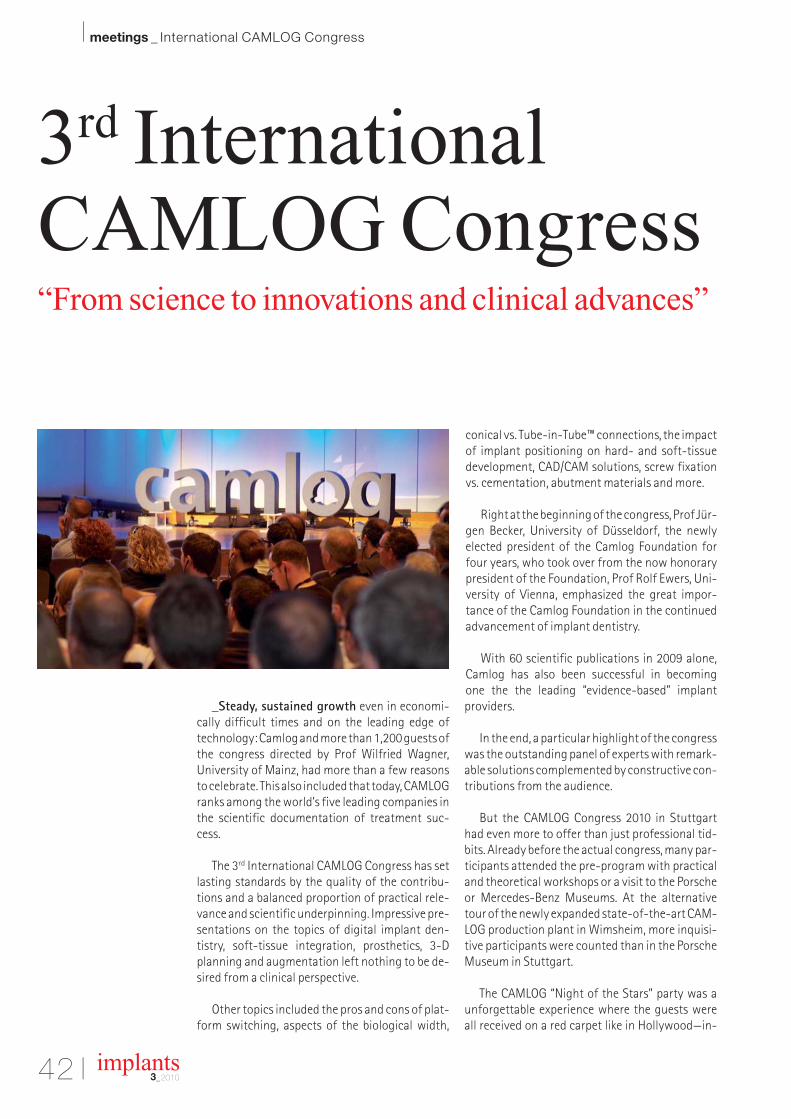
42 I
I meetings _ International CAMLOG Congress
implants3_2010
_Steady, sustained growth even in economi-cally difficult times and on the leading edge oftechnology: Camlog and more than 1,200 guests ofthe congress directed by Prof Wilfried Wagner,University of Mainz, had more than a few reasonsto celebrate. This also included that today, CAMLOGranks among the world’s five leading companies inthe scientific documentation of treatment suc-cess.
The 3rd International CAMLOG Congress has setlasting standards by the quality of the contribu-tions and a balanced proportion of practical rele-vance and scientific underpinning. Impressive pre-sentations on the topics of digital implant den-tistry, soft-tissue integration, prosthetics, 3-Dplanning and augmentation left nothing to be de-sired from a clinical perspective.
Other topics included the pros and cons of plat-form switching, aspects of the biological width,
conical vs. Tube-in-Tube™ connections, the impactof implant positioning on hard- and soft-tissuedevelopment, CAD/CAM solutions, screw fixationvs. cementation, abutment materials and more.
Right at the beginning of the congress, Prof Jür-gen Becker, University of Düsseldorf, the newlyelected president of the Camlog Foundation forfour years, who took over from the now honorarypresident of the Foundation, Prof Rolf Ewers, Uni-versity of Vienna, emphasized the great impor-tance of the Camlog Foundation in the continuedadvancement of implant dentistry.
With 60 scientific publications in 2009 alone,Camlog has also been successful in becoming one the the leading “evidence-based” implantproviders.
In the end, a particular highlight of the congresswas the outstanding panel of experts with remark-able solutions complemented by constructive con-tributions from the audience.
But the CAMLOG Congress 2010 in Stuttgarthad even more to offer than just professional tid-bits. Already before the actual congress, many par-ticipants attended the pre-program with practicaland theoretical workshops or a visit to the Porscheor Mercedes-Benz Museums. At the alternativetour of the newly expanded state-of-the-art CAM-LOG production plant in Wimsheim, more inquisi-tive participants were counted than in the PorscheMuseum in Stuttgart.
The CAMLOG “Night of the Stars” party was aunforgettable experience where the guests wereall received on a red carpet like in Hollywood—in-
3rd InternationalCAMLOG Congress“From science to innovations and clinical advances”

meetings _ International CAMLOG Congress I
cluding screaming fans and a glittering sparklingwine reception. Encouraged by the rousing TinaTurner impressionist Dana Smith and an equallyconvincing Robbie Williams show, the interna-tional CAMLOG community celebrated into thenext morning.
In his closing words, CAMLOG’s CEO Dr MichaelPeetz described the 3rd International CAMLOGCongress in Stuttgart as an outstanding and well-used opportunity to maintain networks and to fur-ther develop the team concept.
And it is Dr. Peetz’s firm conviction that theCAMLOG Group is well on its way to becoming aninternational leader in implant dentistry with itsuser-friendly, high-precision and thoroughly doc-umented dental implant system.
The Camlog Foundation is a foundation estab-lished by scientists under Swiss law. It engages intargeted supporting of gifted young scientists,promotion of basic and applied research, and
continuing training and education to promoteprogress in implant dentistry and related fields toserve the patient. As part of its scientific mission,the Camlog Foundation has assumed patronage ofthe International CAMLOG Congresses, which takeplace every two years._
���������������� � �������� ����
✂ ��� ���������I hereby agree to receive a free trail subscription of �����������������(4 issues per year).I would like to subscribe to ���������������� for € 44 including shipping and VAT for German customers, € 46 including shipping and VAT for customers outside of Germany, unless a written cancellation is sent within 14 days of the receipt of the trial subscription. The subscription will be renewed automatically every year until a written cancellation is sent to OEMUS MEDIA AG, Holbeinstr. 29, 04229 Leipzig, Germany, six weeks prior to the renewal date.
Reply via Fax +49 341 48474-290 to OEMUS MEDIA AG or per E-mail to [email protected]
Last Name, First Name
Company
Street
ZIP/City/Country
E-mail Signature
Notice of revocation: I am able to revoke the subscription within 14 days after my order by sending a written cancellation to OEMUS MEDIA AG, Holbeinstr. 29, 04229 Leipzig, Germany.
OEMUS MEDIA AG Holbeinstraße 29, 04229 Leipzig, GermanyTel.: +49 341 48474-0, Fax: +49 341 48474-290, E-Mail: [email protected]
Signature
IM 3
/10
������������������ ������
You can also subscribe via www.oemus.com/abo
AD

44 I
I manufacturer _ news
implants3_2010
Manufacturer news
Candulor
“Removable or Fixed”—KunstZahnWerk 2011
Candulor has again joined up with CAMLOG to arrange the thrilling upcomingKunstZahnWerk (art of prosthetics) competition at the IDS 2011 in Cologne. Asalways, the latest patient case will be another fresh challenge to participants too.
Modern, innovative, yet in line with a dental technician's daily routine—these arethe requirements for the new KunstZahnWerk competition. International partici-pants from all over Europe, the USA and Canada show their proficiency at each IDS.The latest challenge is to make a total reconstruction combined with a completeprosthesis supported by the mucous membrane and to fit a denture held by an im-plant. All work has to be done using the Gerber set-up technique and the teeth andimplant parts supplied free of charge by Candulor and CAMLOG. Candulor will pro-vide you with the full patient case. Detailed information will aid you in solving thistask. You will of course also get the appropriate plaster models, prosthetic teeth(Candulor Composite NFC) and implant parts. The finished prostheses may only besubmitted in the Candulor Articulator or Condylator. There are further prizes for those
participants providing additional documentation to their work and prizes for the bestdocumentation. A jury made up of prosthetics specialists and practitioners willjudge every individual project. Each of the winners will be awarded their prizes atthe Candulor press conference at the IDS in Cologne on Friday, 25 March, 2011. Allthe finished projects will be shown on the Candulor stand at the IDS 2011 in Cologne.The documentation we receive will be published in various professional journals.
Prices1st prize: Cheque for ⇔ 1.500,–2nd prize: Cheque for ⇔ 1.000,–3rd prize: Cheque for ⇔ 500.–Further prizes for the best documentation along with many material prizes!
Register for participation by no later than 29 October, 2010. You can get the registration form on the internet from www.candulor.com or by phone on +49 7731 79783-0.
Candulor Dental GmbH
Am Riederngraben 6, 78239 Rielasingen- Worblingen, Germany
E-Mail: [email protected], Website: www.candulor.de
CAMLOG
Prosthetic compo-nents for occlusallyscrew-retainedrestorations
With the introduction of the new Vario SR prostheticcomponents, users are now able to choose bet-ween cement-retained or screw-retained crownand bridge restorations on CAMLOG® implants.Vario SR abutments are available in straight and in20° and 30° angled versions for implant diameters3.8/4.3/ 5.0/6.0 mm.
All advantages and indications at a glance• Occlusally screw-retained crown, bridge and bar
constructions• Broadening the area of use of the CAMLOG® Im-
plant System to include screw-retained crownsand bridges
• Up to 30° angled Vario SR abutments make bridg-ing large implant axes divergences in splintedstructures possible
• Special Vario SR components for impression-taking and cast fabrication
• Impressions can be taken using Vario SR abut-ment shoulders or implant shoulders
• Standardized fabrication of the prosthetic restora-tion with prefabricated components
• Sterile packaged and color-coded Vario SR abut-ments
• Temporary restoration with Vario SR protectivecaps or Vario SR titanium copings possible
• Proven CAMLOG handling
• Precise, mechanically sturdy and rotationallystable connection due to the patented Tube-in-Tube™ implant/abutment connection.
Prefabricated Vario SR prosthetic componentsBurn-out plastic copings can be used to fabricatecrown, bridge and bar constructions. The titaniumcaps have a retention surface on the outside and aredesigned for temporary or final bridge restorationsmade of plastic. Titanium bar caps are available forlaser-welded bar constructions. For bridge and barconstructions, the impression can be taken usingVario SR impression caps, open or closed tray, directlyover the Vario SR abutment already in its final positionin the implant. The retention screw of the impressioncap, open tray, can be shortened by 3 mm extra-orallyif space limitations are encountered. For crownrestoration, the impression can be taken directly overthe implant shoulder using CAMLOG® impressionposts, open or closed tray.
CAMLOG Biotechnologies AG
Margarethenstrasse 38
4053 Basel, Switzerland
E-mail: [email protected]
Website: www.camlog.com

Nobel Biocare
Versatility, easy of use, predictability,and pleasing esthetic results
Nobel Biocare stands for 40 years of experience in restorative dentistry as well as fourdecades of scientifically-documented and successful implant systems. One of the inno-vations of the company is NobelReplaceTM, the time-proven and most frequently insertedimplant system throughout the world that stands for versatility, ease of use, and pre-dictability.1 NobelReplace is very well-suited for a comprehensive range of applicationsranging from the rehabilitation of single teeth to the restoration in completely edentu-lous patients. NobelReplace is an implant system with impressive benefits and is therefore consideredto be the implant system of first choice both in surgery and for the options of prostheticmanagement. NobelReplace is a universally applicable, two-part implant system forsuccessful use in one- or two-stage surgical procedures both in soft and dense bone.
NobelReplace offers experienced and advanced users alike a comprehensive implant system that sup-ports the treatment in virtually all indications of implantology. The root-like shape, grooves and TiUnitesurface favor optimal primary stability and thus allow the implant to be used even in challenging indi-cations, such as between diverging roots of neighboring teeth, in front of the mesial wall of the maxil-lary sinus or for insertion right after extraction. Considering the large number of treatment options thatare available to date, an implant system like NobelReplace is a major benefit for the user and providesa high degree of flexibility. The NobelReplace implant system is universally applicable and offers manyoptions for surgical treatment and downstream prosthetic management.
1Source: Millennium Research Group 2008
Nobel Biocare AG
P.O. Box 8058 Zürich – Airport, Switzerland
E-mail: [email protected], Website: www.nobelbiocare.com
bio
nic
s
ticky
gra
nule
s
easy
-gra
ft®C
RY
STA
L
Degradable Solutions AGWagistrasse 23CH-8952 SchlierenPhone: +41 43 433 62 [email protected]
Ingenious: Simple handling and accelerated osteocon-duction for long-term volume preservation.
Order your free test sample over the internet!
AD
EMS
Piezon Master Surgery with threenew instrument systems
Since it was introduced, Piezon Master Surgery—based on Piezon technol-ogy—has had a remarkable track record in many practices. Today, EMS hasexpanded the clinical scope of application of the Piezon Master Surgery prod-uct range. With an enhanced product offering—and special instruments such
as Sinus System and Implant System—practitioners have access to technolo-gies allowing them to work even more efficiently. With Piezon Master Surgery, additional application-specific instruments arenow available: a total of four perio instruments especially designed for resec-
tive and regenerative periodontal surgery, five advanced surgical instrumentsfor gentle and uniform sinus lifts, as well as six special fully diamondcoated instruments for implantapplications with dual cooling system and extraefficient debris evacuation. These instruments are seen as particularly suitable for four clinical applications: implant site prepa-ration following extraction, implant site preparation following splitting of the alveolar ridge, implantsite preparation in the posterior tooth area, and implant site preparation in compromised areas, suchas a narrow alveolar ridge. In principle, instruments can be used at low OP temperature of no morethan 33 degrees centigrade. They provide drilling efficiency and precision in the maxillary area.
EMS Electro Medical Systems S.A.
Chemin de la Vuarpillière 31, 1260 Nyon, Switzerland
E-mail: [email protected], Website: www.ems-dent.com

46 I
I manufacturer _ news
implants3_2010
ULTRADENT
The future has begun at ULTRADENT
A big celebration was held to mark the openingof ULTRADENT's new, modern company build-ing at an event attended by the mayor, the me-dia, the architects, prominent members of thedental profession and three generations of theOstner family. Under the management of thecurrent owner, Ludwig Ostner, ULTRADENT has,over the last 12 years, become one of the mostfamiliar brands of modern treatment units in allareas of dentistry. The future of ULTRADENTalso lies in the hands of the family, as Ludwig Jo-hann Ostner, the son of the current head of thecompany, is joint managing director with his father and has already assumed re-sponsibility for product development. Manufacturing many components in-house,the family enterprise develops and produces dental equipment that is character-ized by excellent quality, superb reliability and practical design.
Good prospects. Dental quality made in GermanyThe success of the Munich-based dental specialist is proof that their concept iscorrect and the new head office at the Brunnthal industrial park in Munich will
provide extensive logistical opportunities. Here an even larger exhibition areawill be available, where our customers can experience the latest products putto practical use. Countless innovative product ideas, the company's own
patents and registered designs represent acompetitive technological edge, exclusivity,maximum product reliability and long-termprovision of spare parts.Moreover, thanks to investments in develop-ment, the latest production technology andongoing training for employees, ULTRADENTwill be able to continue making its ideas andvisions a reality in the future.
Modern jobs in modern buildingsEverything has been redesigned and reor-ganized, from the company’s own paint shop, showroom and development depart-
ment right through to the warehouse and administrative area. This has createda light workplace fit for the future and the pleasure this gives the employees isclear to see. We can look forward to the new products from ULTRADENT.
ULTRADENT
Dental-Medizinische Geräte GmbH & Co. KG
Eugen-Sänger-Ring 10, 85649 Brunnthal, Germany
E-mail: [email protected], Website: www.ultradent.de
Dentaurum Implants
Cleaning made easy
Time-consuming work for staff,variable cleaning results and the as-sociated expenses—that was standardpractice for the preparation of drills and ac-cessories after implantology procedures inthe past. After every surgical procedure all in-struments had to be manually cleaned one by oneand then sorted into the right implant-surgerytray. Cleaning and disinfection of the completelyfilled implant-surgery tray was formerly not pos-sible, because the instruments did not sufficientlycome into contact with water and cleaningagents. Dentaurum Implants GmbH and MieleProfessional have now worked together to de-velop an innovative system solution for efficientand reproducible machine preparation. The heartof this development is the tioLogic© easyCleansurgery tray, which now enables consistent, out-standing machine cleaning and disinfection re-sults in both dental practices and in centralizedpreparation centres in hospitals. This not only offers huge savings in time and costs but also significantly increased safety for users with thereproducible machine preparation results. Thecombination of the innovative grid structure with
specialretaining clips
fixes all rotary instrumentsand accessory components to
hold them in position and to ensure that the in-struments are completely cleaned with water andcleaning agent. All drills and accessory compo-nents can be replaced in the correct position in thetioLogic© easyClean tray as they are used in theimplant procedure to remain in the correct orderat all times throughout the operation. SMP GmbHof Tübingen, an independent institute specialisingamong other things in the testing and validation ofmedical devices, was commissioned to test andvalidate the cleaning results. The tests were animpressive confirmation of the preparation re-sults of the instruments and accessory compo-nents in the tioLogic© easyClean.
Dentaurum Implants GmbH
Turnstr. 31, 75228 Ispringen, Germany
E-mail: [email protected]
Website: www.dentaurum-implants.de
®
www.omniaspa.eu
OMNIA S.p.A.Via F. Delnevo, 190 - 43036 Fidenza (PR) ItalyTel. +39 0524 527453 - Fax +39 0524 525230
VAT. IT 01711860344 - R.E.A. PR 173685 Company capital € 200.000,00
Since our beginnings, we have always been focused on quality and innovation
toward the battle againstcross - contamination and infections.
In the last 20 years, we have ensured safety and protection to you and your patients, with advanced and reliable products. Tools
that represent the ideal solution for who is operating in dentistry, implantology/oral surgery and general surgery.
With Omnia sure to be safe.
AD

Geistlich Biomaterials
The successful duo now also avail-able as Combi-KitCollagen
The new Geistlich Combi-Kit Collagen com-bines two established and reliable products ina single package: Geistlich Bio-Oss® Collagen100 mg and the native collagen membraneGeistlich Bio-Gide® in a new size of 16 x 22 mm.The Geistlich Combi-Kit Collagen® offers theperfect solution for Ridge Preservation, i.e.for the treatment of alveolar bone defectsfollowing tooth extraction, as well as for minoraugmentations. Today the combined appli-cation of bone substitute materials and re-sorbable membranes to treat bone defects
has already become clinical routine and shows predictable and effective results (Aghaloo 2007;Sammartino 2009). Scientific evidence and a wealth of practical experience reported have also shown that the insertion ofGeistlich Bio-Oss® Collagen in an extraction socket may be the ideal way to preserve the alveolar di-mensions (Ackermann 2009), and the inevitable loss of the bundle bone – and therefore of the buccalbone lamellae—following tooth extraction is compensated (Araujo 2008; Araujo 2009).
Geistlich Biomaterials
Bahnhofstr. 40, 6110 Wolhusen, Switzerland
E-mail: [email protected], Website: www.geistlich.ch
AD
Straumann
Medical Device Technology of theYear Award for Straumann’s RoxolidStraumann, a global leader in regenerative,restorative and replacement dentistry, has beenpresented with the 2009 ‘Medical Device Tech-nology of the Year Award’ for Roxolid®, the com-pany’s innovative high performance materialfor dental implants.
The Award recognizes excellence in technological innovation and is one of the Best Practices Awardsbestowed by Frost & Sullivan. Engineered and developed by Straumann, Roxolid is an alloy of titaniumand zirconium which has been designed to increase reliability and confidence with small diameter im-plants. Roxolid can accommodate the sophisticated microstructuring processes required for Strau-mann’s SLActive® surface technology, which enhances osseointegration. Roxolid has been undergo-ing a broad program of clinical trials in nine countries. Involving 60 centers and more than 300 patients,this is one of the largest clinical research programs ever undertaken by a dental implant company priorto market launch. Apart from the clinical program, Roxolid was made available to 450 selected spe-cialists in a controlled release program, in which more than 11,000 implants were distributed.
Institut Straumann AG
Peter Merian-Weg 12, 4052 Basel, Switzerland
E-Mail: [email protected], Website: www.straumann.com
Susanne Breuer, Stephen Booth and Sandro De Gruttolarepresented Straumann at the Award Ceremony.

48 I
I DGZI _ social news
implants3_2010
Congratulations and Happy Birthday to all DGZI-members around the world
75th BirthdayZA Tor Wahl (07.07.)
70th BirthdayDr Dr Marius Rimbasiu (07.07.)
65th BirthdayDr Horst Beck (06.07.)
ZÄ Renate Bauer-Küchle (08.07.)
60th BirthdayDr Hans Florack (01.07.)
Dr Heinrich Meis (03.07.)
Dr Felix Schirner (04.07.)
Dr Eckhard Benninghoff (13.07.)
Dr Gerhard Martin Cube (14.07.)
Dr Dov Berger (17.07.)
ZA Jürgen Conrad (18.07.)
55th BirthdayDr Alfred Plötz (03.07.)
Dr Hubert Stratz (11.07.)
Alabtah Zoher (25.07.)
Achim Kneuertz (26.07.)
ZT Stefan Reif (26.07.)
Dr Ralf August (29.07.)
50th BirthdayDr Heike Gründler (01.07.)
Dr Wolfram Arndt (04.07.)
Dr Peter Pötschke (04.07.)
Dr Serge Bailly-Thuir (14.07.)
Dr Dr. Hans van der Elst (17.07.)
Dr Anton Öttl (25.07.)
Dr Gundula Flottmann (27.07.)
Dr Marton Yakubovich (28.07.)
45th BirthdayDr Marwan Shahin (05.07.)
Dr Ralph Heel (14.07.)
Dr Michael Lenz (18.07.)
Dr Bernd Krebs (25.07.)
Dr Frank Seidel (30.07.)
40th BirthdayDr Frederik Friese (07.07.)
70th BirthdayDr Jürgen Oberbeckmann (12.08.)
65th BirthdayDr Hans Konrad Hühnlein (01.08.)
Dr Bernd Ulrich (28.08.)
60th BirthdayDr Volker Hellwich (13.08.)
Dr J.P. Flieller (14.08.)
Dr Klaus M. Linke (24.08.)
55th BirthdayDr Adrian Wetz (01.08.)
Dr Sören Atrup Nielsen (23.08.)
Hans-Bodo Ronsheimer (23.08.)
Dr Salah Al-Tawil (30.08.)
50th BirthdayDr Bassel Al Sibai (01.08.)
Jürgen Holzwarth (08.08.)
Dr Martina Frantzen (11.08.)
Dr Mattias Tamke (11.08.)
Jochen Graf (12.08.)
Dr Martin Mrowka (14.08.)
ZA Mario Ohlinger (15.08.)
Dr Mircea Teodor Parau (19.08.)
Dr Zeev Ormianer (23.08.)
Rainer Franz Latzko (29.08.)
45th BirthdayHaddad Jawad (02.08.)
Dr Andreas Meyer (03.08.)
Katrin Mielke (03.08.)
Dr Alexander Martin (13.08.)
Dr Sohayb Arrasat Al-Hindia (18.08.)
Silke Gudrun Bauer (19.08.)
Dr Janine Affeldt (20.08.)
Dr Martin Bauer (30.08.)
40th BirthdayMohammad Khaled (17.08.)
JULY 2010
70th BirthdayDr Dr Hans Hebbinghaus (07.09.)
65th BirthdayDr Gerhard Brückmann (19.09.)
60th BirthdayDr Heinz-Werber Heller (01.09.)
Dr Kaffan Murwan (05.09.)
Peter Quadfuß (16.09.)
John Giblin (17.09.)
Dr Roland Sireborn (23.09.)
Dr Bernd Neuschulz (29.09.)
55th BirthdayDr Harald Wilkat (05.09.)
Dr Thomas Wiff (19.09.)
Dr Peter Aaen (19.09.)
Dr Wolfgang Ercken (22.09.)
Mouhamad Sameer Wahbeh (22.09.)
Dr Barbara Langen (23.09.)
50th BirthdayDr Hubert Litter (02.09.)
Dr Jochen Heibach (04.09.)
Dr Christoph Falk (06.09.)
ZA Michael Quitzke (07.09.)
Dr Ralf Grießke (10.09.)
Dr Lutz Schneider (20.09.)
Dr Bernd Quantius (21.09.)
Torben Arlt (24.09.)
45th BirthdayDrs Jan-Guido Kisters (03.09.)
Dr Alexander Scholz (03.09.)
Dr Ulf Berkenkamp (06.09.)
Dr Thorsten Zickuhr (06.09.)
Dr Tom Wilken (13.09.)
Holger Heß (21.09.)
Dr Robert Bungartz (29.09.)
40th BirthdayDr Uwe Held (05.09.)
SEPTEMBER 2010
AUGUST 2010

I 49
about the publisher _ submission guidelines I
implants3_2010
submission guidelines:Please note that all the textual components of your submissionmust be combined into one MS Word document. Please do not submit multiple files for each of these items:
_the complete article;_all the image (tables, charts, photographs, etc.) captions;_the complete list of sources consulted; and_the author or contact information (biographical sketch, mailingaddress, e-mail address, etc.).
In addition, images must not be embedded into the MS Word document. All images must be submitted separately, and detailsabout such submission follow below under image requirements.
Text lengthArticle lengths can vary greatly—from 1,500 to 5,500 words— depending on the subject matter. Our approach is that if you need more or less words to do the topic justice, then please makethe article as long or as short as necessary.
We can run an unusually long article in multiple parts, but thisusually entails a topic for which each part can stand alone be-cause it contains so much information.
In short, we do not want to limit you in terms of article length, so please use the word count above as a general guideline and ifyou have specific questions, please do not hesitate to contact us.
Text formattingWe also ask that you forego any special formatting beyond theuse of italics and boldface. If you would like to emphasise certainwords within the text, please only use italics (do not use underli-ning or a larger font size). Boldface is reserved for article headers.Please do not use underlining.
Please use single spacing and make sure that the text is left jus -tified. Please do not centre text on the page. Do not indent para-graphs, rather place a blank line between paragraphs. Please donot add tab stops.
Should you require a special layout, please let the word processingprogramme you are using help you do this formatting automati-cally. Similarly, should you need to make a list, or add footnotes or endnotes, please let the word processing programme do it foryou automatically. There are menus in every programme that willenable you to do so. The fact is that no matter how carefully done,errors can creep in when you try to number footnotes yourself.
Any formatting contrary to stated above will require us to removesuch formatting before layout, which is very time-consuming.Please consider this when formatting your document.
Image requirementsPlease number images consecutively throughout the article by using a new number for each image. If it is imperative that certain images are grouped together, then use lowercase lettersto designate these in a group (for example, 2a, 2b, 2c).
Please place image references in your article wherever they are appropriate, whether in the middle or at the end of a sentence.If you do not directly refer to the image, place the reference at the end of the sentence to which it relates enclosed withinbrackets and before the period.
In addition, please note:
_We require images in TIF or JPEG format._These images must be no smaller than 6 x 6 cm in size at 300 DPI._These image files must be no smaller than 80 KB in size (or theywill print the size of a postage stamp!).
Larger image files are always better, and those approximately the size of 1 MB are best. Thus, do not size large image files downto meet our requirements but send us the largest files available.(The larger the starting image is in terms of bytes, the more lee-way the designer has for resizing the image in order to fill up morespace should there be room available).
Also, please remember that images must not be embedded intothe body of the article submitted. Images must be submitted separately to the textual submission.
You may submit images via e-mail, via our FTP server or post a CD containing your images directly to us (please contact us for the mailing address, as this will depend upon the country fromwhich you will be mailing).
Please also send us a head shot of yourself that is in accordancewith the requirements stated above so that it can be printed withyour article.
AbstractsAn abstract of your article is not required.
Author or contact informationThe author’s contact information and a head shot of the authorare included at the end of every article. Please note the exact information you would like to appear in this section and for-mat it according to the requirements stated above. A short biographical sketch may precede the contact information if you provide us with the necessary information (60 words or less).
Questions?Kristin Urban (Managing Editor)[email protected]

50 I
I about the publisher _ imprint
implants3_2010
Copyright Regulations _implants international magazine of oral implantology is published by Oemus Media AG and will appear in 2010 with one issue every quarter. The
magazine and all articles and illustrations therein are protected by copyright. Any utilization without the prior consent of editor and publisher is inad-mi ssible and liable to prosecution. This applies in particular to duplicate copies, translations, microfilms, and storage and processing in electronic systems.
Reproductions, including extracts, may only be made with the permission of the publisher. Given no statement to the contrary, any submissions to theeditorial department are understood to be in agreement with a full or partial publishing of said submission. The editorial department reserves the right tocheck all submitted articles for formal errors and factual authority, and to make amendments if necessary. No responsibility shall be taken for unsolicitedbooks and manuscripts. Articles bearing symbols other than that of the editorial department, or which are distinguished by the name of the author, representthe opinion of the afore-mentioned, and do not have to comply with the views of Oemus Media AG. Responsibility for such articles shall be borne by the author.Responsibility for advertisements and other specially labeled items shall not be borne by the editorial department. Likewise, no responsibility shall be assumedfor information published about associations, companies and commercial markets. All cases of consequential liability arising from inaccurate or faulty representation are excluded. General terms and conditions apply, legal venue is Leipzig, Germany.
PublisherTorsten R. Oemus [email protected]
CEOIngolf Dö[email protected]
Members of the BoardJürgen [email protected]
Lutz V. [email protected]
Chief Editorial ManagerDr Torsten Hartmann (V. i. S. d. P.)[email protected]
Editorial CouncilDr Friedhelm [email protected]
Dr Roland [email protected]
Prof Dr Dr Kurt [email protected]
Dr Torsten [email protected]
Dr Suheil [email protected]
Editorial OfficeKristin [email protected]
Executive ProducerGernot [email protected]
Designer Sarah [email protected]
Customer ServiceMarius [email protected]
Published byOemus Media AGHolbeinstraße 2904229 Leipzig, GermanyTel.: +49 341 48474-0Fax: +49 341 [email protected]
Printed byMessedruck Leipzig GmbHAn der Hebemärchte 604316 Leipzig, Germany
implantsinternational magazine of oral implantologyis published in cooperation with the GermanAssociation of Dental Implantology (DGZI).
DGZI PresidentDr Friedhelm HeinemannDGZI Central Office Feldstraße 80, 40479 Düsseldorf, GermanyTel.: +49 211 16970-77Fax: +49 211 [email protected]
www.dgzi.dewww.oemus.com
implantsinternational magazine of oral implantology

implantsinternational magazine of oral implantology
One issue free of charge!I hereby agree to receive a free trial subscription of implantsinternational magazine of oral implantology (4 issuesper year).
I would like to subscribe to implants for € 44 includingshipping and VAT for German customers, € 46 including ship-ping and VAT for customers outside of Germany, unless a writ-ten cancellation is sent within 14 days of the receipt of the trialsubscription. The subscription will be renewed automaticallyevery year until a written cancellation is sent to OEMUS MEDIAAG, Holbeinstr. 29, 04229 Leipzig, Germany, six weeks prior tothe renewal date.
Last Name, First Name
Company
Street
ZIP/City/County
E-mail Signature
Signature
OEMUS MEDIA AGHolbeinstraße 29, 04229 Leipzig, Germany, Tel.: +49 341 48474-0, Fax: +49 341 48474-290, E-Mail: [email protected]
Reply via Fax +49 341 48474-290 to OEMUS MEDIA AG or per E-mail to [email protected]
Notice of revocation: I am able to revoke the subscription within 14 days after my order by sending a writtencancellation to OEMUS MEDIA AG, Holbeinstr. 29, 04229 Leipzig, Germany.
IM 3
/10
✂ Subscribe now!
You can also subscribe via www.oemus.com/abo
Implants_Abo_A4_Implants_Abo_A4 22.09.10 10:35 Seite 1

ww
w.a
dwor
k.de
BIONIC ENGINEERING DESIGNED IMPLANTS
BEGO Semados® patented Implants embody:
Indication-optimised contour design
Function-optimised implant-abutment-connection
High purity and ultra-homogenous-surface
Polished rim for an infl ammation-free gingiva-attachment
100 % German design – 100 % German manufacturing
Value for money
www.bego-implantology.com
GO FOR GOLD.
BEGO Semados® Mini-Implant
Bionic Engineering Design transfer of optimal living nature solutions
to technical productsBEGO Semados®
S-ImplantBEGO Semados®
RI-Implant
Official care provider to the German Olympic teams since 2002
Are YouInterested?
BES-09049_Bionik_GB Teamwork Media 210x297.indd 1BES-09049_Bionik_GB Teamwork Media 210x297.indd 1 14.04.10 13:5014.04.10 13:50
