en plaque tuberculoma: a case report - imri · en plaque tuberculoma: a case report ... of the...
TRANSCRIPT

www.i-mri.org200
En Plaque Tuberculoma: a Case Report
INTRODUCTION
In Korea, tuberculosis (TB) remains a serious public health problem and reported TB cases have not significantly decreased over the last decade (1). Tuberculosis involving the central nervous system (CNS) is the most significant type of extrapulmonary tuberculosis because of its high mortality rate and possible morbidity of neurologic sequelae. CNS tuberculosis can manifest in a variable forms, including tuberculous meningitis, tuberculous cerebritis, tuberculoma, tuberculous abscess, and miliary tuberculosis (2). Although intra-axial tuberculomas are the more common type of CNS tuberculosis, extra-axial lesions are rarely encountered (3). Here, we report a rare case of an en plaque tuberculoma.
CASE REPORT
A 73-year-old woman visited the emergency department with mental change and vomiting. She had a past medical history of hypertension. Two days prior, she presented with a headache, dizziness and drowsy mental status which were slowly progressing. She complained of febrile sensation, and body temperature was 36.9°C. A cerebrospinal fluid (CSF) analysis revealed WBC count of 520/µℓ (normal range, 0-4/µℓ) with lymphocyte dominance (90%), an adenosine deaminase (ADA) level of 24.3 IU/L (normal range, 0-8 IU/L), a total protein of 204.2 mg/dL (normal range, 15-45 mg/dL) and a glucose of 44 mg/dL (normal range, 40-75 mg/dL).
A brain computed tomography (CT) scan showed multifocal low and suspicious isodensity lesions in the right medial frontal and left temporal lobes and a suspicious iso to high density lesion in the left temporal extra-axial space (Fig. 1a).
On diffusion weighted imaging (DWI) of the brain (Magnetom Avanto 1.5T, Siemens,
This is an Open Access article distributed under the terms of the Creative Commons Attribution Non-Commercial License (http://creativecommons.org/licenses/by-nc/3.0/) which permits unrestricted non-commercial use, distribution, and reproduction in any medium, provided the original work is properly cited.
Received: August 13, 2016Revised: September 13, 2016Accepted: September 13, 2016
Correspondence to: Donghoon Lee, M.D.Department of Radiology, Seoul Medical Center, 156 Sinnae-ro, Jungnang-gu, Seoul 02053, Korea.Tel. +82-2-2276-7000Fax. +82-2-2276-7093Email: [email protected]
Copyright © 2016 Korean Society of Magnetic Resonance in Medicine (KSMRM)
iMRI 2016;20:200-205 http://dx.doi.org/10.13104/imri.2016.20.3.200
Case Report
In Korea, tuberculosis is still common disease. Central nervous system tuberculosis can manifest in a variety of forms, including tuberculous meningitis, tuberculous cerebritis, tuberculoma, tuberculous abscess, and miliary tuberculosis. Although intra-axial tuberculomas are the more common type of CNS tuberculosis, extra-axial lesions are rarely encountered. En plaque tuberculoma is an extremely rare presentation of intracranial tuberculosis with mimicking primary or secondary meningeal neoplasia. We describe a rare case of an en plaque tuberculoma accompanied by tuberculous meningitis and tuberculomas.
Keywords: En plaque tuberculoma; CNS tuberculosis; Magnetic resonance imaging
pISSN 2384-1095eISSN 2384-1109
Young-eun Kim, Donghoon Lee, Hokyeong Hwang, Minji KimDepartment of Radiology, Seoul Medical Center, Seoul, Korea

201www.i-mri.org
http://dx.doi.org/10.13104/imri.2016.20.3.200
Erlangen, Germany), a lesion with iso to low signal intensity was observed in the left temporal convexity, as compared with brain parenchyma (Fig. 1b).
Magnetic resonance imaging (MRI) obtained on a 3.0T system (Achieva, Philips Healthcare, Best, The Netherlands) by hospital day 1. A diffuse dural-based enhancing lesion of about 4 cm in length was located mainly in the left temporal convexity on contrast enhanced T1-weighted images (T1WI) (Fig. 1e, f). The lesion showed iso signal intensity to the brain parenchyma on T1WI and T2-weighted images (T2WI) with marked perilesional T2 high
signal intensity (Fig. 1c, d). In addition, multiple ring and nodular enhancing lesions were observed in the cortico-sulcal area of the right medial frontoparietal lobe and the left paramedian cerebellum. Some of these showed dark signal intensity on susceptibility weighted images (SWI) (Fig. 2).
By hospital day 1, the patient presented with a fever (38.0°C) and anti-tuberculosis therapy was initiated with HERZ (isoniazid, rifampin, ethambutol, and pyrazinamide) regimens, in combination with tapering doses of steroids (dexamethasone) for 4 weeks. Although there was
Fig. 1. A 73-year-old woman with an en plaque tuberculoma. (a) CT scan showed iso to hyperattenuating lesion at left temporal extra-axial space with low density area in the Lt. temporal lobe. (b) DWI showed iso to low signal intensity lesion at left temporal extra-axial space. (c) T2-weighted axial MRI showed iso-signal intensity lesion with perilesional high signal intensity. (d-f) T1-weighted axial (d) MRI showed iso-signal intensity lesion with contrast-enhanced T1-weighted axial (e) and coronal (f) MRI showing homogeneous enhancing dural-based lesion in the left temporal convexity (arrows).
a b c
d e f

www.i-mri.org202
En Plaque Tuberculoma | Young-eun Kim, et al.
no evidence of pulmonary tuberculosis on her chest radiography and CT scan, the result of a sputum acid-fast bacilli (AFB) culture confirmed the presence of tuberculosis on hospital day 13.
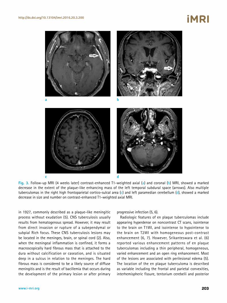
By hospital day 28, a follow-up MRI demonstrated a marked decrease in size of the plaque-like enhancing mass lesion in the left temporal convexity (Fig. 3a, b). Also, previously noted multiple tuberculomas in the right high frontoparietal cortico-sulcal area and left paramedian cerebellum, showed a marked decrease in size and number, as compared to the last MRI (Fig. 3c, d). The patient showed an improvement in clinical status at the time of discharge after hospital day 28.
DISCUSSION
According to the 2015 TB notification data, there were 32,181 reported cases of TB (63.2 cases per 100,000 people) in Korea. Of these, 6631 cases were extrapulmonary TB (13.0 cases per 100,000) which comprised 20.6% of all cases of TB. Among extrapulmonary TB, CNS TB was reported in 385 cases (4). CNS tuberculosis can manifest in a variable forms, including tuberculous meningitis, tuberculous cerebritis, tuberculoma, tuberculous abscess, and miliary tuberculosis. Although intra-axial tuberculomas are the more common type of CNS tuberculosis, extra-axial lesions are rarely encountered (3).
An en plaque tuberculoma is a rare entity, first reported
Fig. 2. Contrast-enhanced T1-weighted axial (a) and coronal (b) MRI showed multiple ring and nodular enhancing lesions in the cortico-sulcal area of the right medial frontoparietal lobe and left paramedian cerebellum (c). Some of these showed dark signal intensity on SWI (d).
a b
c d
203www.i-mri.org
http://dx.doi.org/10.13104/imri.2016.20.3.200
in 1927, commonly described as a plaque-like meningitic process without exudation (5). CNS tuberculosis usually results from hematogenous spread. However, it may result from direct invasion or rupture of a subependymal or subpial Rich focus. These CNS tuberculosis lesions may be located in the meninges, brain, or spinal cord (2). Also, when the meningeal inflammation is confined, it forms a macroscopically hard fibrous mass that is attached to the dura without calcification or caseation, and is situated deep in a sulcus in relation to the meninges. The hard fibrous mass is considered to be a likely source of diffuse meningitis and is the result of bacillemia that occurs during the development of the primary lesion or after primary
progressive infection (5, 6). Radiologic features of en plaque tuberculomas include
appearing hyperdense on noncontrast CT scans, isointense to the brain on T1WI, and isointense to hypointense to the brain on T2WI with homogeneous post-contrast enhancement (6, 7). However, Srikanteswara et al. (6) reported various enhancement patterns of en plaque tuberculomas including a thin peripheral, homogeneous, varied enhancement and an open ring enhancement. Most of the lesions are associated with perilesional edema (5). The location of the en plaque tuberculoma is described as variable including the frontal and parietal convexities, interhemispheric fissure, tentorium cerebelli and posterior
Fig. 3. Follow-up MRI (4 weeks later) contrast-enhanced T1-weighted axial (a) and coronal (b) MRI, showed a marked decrease in the extent of the plaque-like enhancing mass of the left temporal subdural space (arrows). Also multiple tuberculomas in the right high frontoparietal cortico-sulcal area (c) and left paramedian cerebellum (d), showed a marked decrease in size and number on contrast-enhanced T1-weighted axial MRI.
a b
c d

www.i-mri.org204
En Plaque Tuberculoma | Young-eun Kim, et al.
fossa (6). However, similar imaging findings can be seen in various cases of inflammatory and non-inflammatory conditions, showing dural-based lesions, the most common being meningiomas (5, 7). Khanna et al. (3) reported a case of MR spectroscopy findings of giant extra-axial tuberculoma. In this case, the extra-axial tuberculoma showed high lipid-lactate peaks and a decrease in N-acetyl aspartate (NAA)/creatinine (Cr) and NAA/choline ratios which differ from meningioma with an elevation of alanine (Ala). The differing MR spectroscopy pattern and presence of perilesional edema are among the few points that help in differentiating it from meningiomas (5). Srikanteswara et al. (6) reported that a follow-up MRI study performed 6 months after initiation of anti-tuberculosis therapy in 5 patients showed a disappearance of the lesion as well as of the meningeal enhancement.
Alkan et al. (8) reported a case of the en plaque tuberculomas of tentorium in a pregnant woman. She had en plaque tuberculoma in the right tentorium with multiple intra-axial ring-like enhanced tuberculomas on contrast enhanced T1WI. The rest of pregnancy was without problem and a healthy child was born by vaginal delivery. After 8 months of anti-tuberculosis therapy, follow-up MRI showed disappearance and improvement of the tuberculomas.
Patients with extracranial tuberculosis usually present with focal neurologic deficits and seizures in the absence of increased intracranial pressure (5). Clinical manifestations of the en plaque tuberculomas are variable and nonspecific, including fever, headache, visual blurring, vomiting, neck tilt, focal seizures and limb weakness. These features depend on the location (6).
The detection of TB cases is based primarily on the microscopic examination of sputum for AFB. The patient with AFB-positive sputum is diagnosed to be "bacteriologically confirmed TB case" (9). Concomitant extracranial tuberculous infection is present in 30% to 50% of cases, with about 33-50% having no abnormalities indicated by chest radiograph (10). Moreover, an ADA cut-off value of 8 IU/L showed a sensitivity of 59% and a specificity of 96% in the diagnosis of TB meningitis (9).
On imaging, en plaque tuberculomas mimic other dural-based lesions. The differential diagnoses are wide variety of neoplastic and non-neoplastic lesions such as meningioma, metastasis, lymphoma and sarcoidosis (5). In our case, CSF analysis suggested with TB meningitis, positive sputum AFB culture confirmed this diagnosis and multiple intracranial tuberculomas were detected on brain MRI with a dural-based plaque-like lesion. Also, other lab findings such as
complete blood cell count were within normal limit. And intracranial and extra-axial lesions were improved on a follow up MRI study after administering anti-tuberculosis therapy. The en plaque tuberculoma responds well to anti-tuberculosis therapy (7). These findings help to establish the diagnosis of CNS tuberculosis without biopsy and pathologic confirmation.
Anti-tuberculosis therapy constitutes the mainstay of treatment for CNS tuberculosis. The regimen is usually continued for 12-30 months (5, 10). Early treatment with steroids is recommended in patients with TB meningitis in a dose of 0.4 mg/kg/day for 1-2 weeks and tapered over the next few weeks with monitoring regardless of severity in order to significantly decrease the risk of mortality and reduce neurological sequelae (5, 9).
In conclusion, we report a rare case of an en plaque tuberculoma that presented as hyperdense on nonenhanced CT scans, isointense to the brain on T1WI, and isointense to hypointense to the brain on T2WI with homogeneous post-contrast enhancement. Awareness of these characteristics will allow early diagnosis and optimal management of en plaque tuberculomas.
REFERENCES 1. Kwon YS, Koh WJ. Diagnosis of pulmonary tuberculosis
and nontuberculous mycobacterial lung disease in Korea. Tuberc Respir Dis (Seoul) 2014;77:1-5
2. Burrill J, Williams CJ, Bain G, Conder G, Hine AL, Misra RR. Tuberculosis: a radiologic review. Radiographics 2007;27:1255-1273
3. Khanna PC, Godinho S, Patkar DP, Pungavkar SA, Lawande MA. MR spectroscopy-aided differentiation: "giant" extra-axial tuberculoma masquerading as meningioma. AJNR Am J Neuroradiol 2006;27:1438-1440
4. Korea Centers for Disease Control and Prevention. Annual report on the notified tuberculosis in Korea. Cheongju (South Korea): The Centers; 2015
5. Aggarwal A, Patra DP, Gupta K, Sodhi HB. Dural tuberculoma mimicking meningioma: a clinicoradiologic review of dural en-plaque lesions. World Neurosurg 2016;88:686 e681-687
6. Srikanteswara PK, Pampapati PK, Yelsangikar KR. En-plaque central nervous system tuberculoma - an uncommon entity: clinico-radiological profile in a Cohort from a tertiary referral centre. J Clin Diagn Res 2016;10:OC11-14
7. Patkar D, Narang J, Yanamandala R, Lawande M, Shah GV. Central nervous system tuberculosis: pathophysiology and imaging findings. Neuroimaging Clin N Am 2012;22:677-
205www.i-mri.org
http://dx.doi.org/10.13104/imri.2016.20.3.200
705 8. Alkan A, Parlak M, Baysal T, Sigirci A, Kutlu R, Altinok T. En-
plaque tuberculomas of tentorium in a pregnant woman: follow-up with MRI(2003:2b). Eur Radiol 2003;13:1190-1193
9. Lee JY. Diagnosis and treatment of extrapulmonary tuberculosis. Tuberc Respir Dis (Seoul) 2015;78:47-55
10. Adachi K, Yoshida K, Tomita H, Niimi M, Kawase T. Tuberculoma mimicking falx meningioma--case report. Neurol Med Chir (Tokyo) 2004;44:489-492