endocytosis and cancer - cshl pcshperspectives.cshlp.org/content/5/12/a016949.full.pdf ·...
TRANSCRIPT
Endocytosis and Cancer
Ira Mellman1 and Yosef Yarden2
1Genentech Inc., South San Francisco, California 940802Department of Biological Regulation, Weizmann Institute of Science, Rehovot 76100, Israel
Correspondence: [email protected]
Endocytosis entails selective packaging of cell-surface proteins, such as receptors for cyto-kines and adhesion components, in cytoplasmic vesicles (endosomes). The series of sortingevents that determines the fate of internalized proteins, either degradation in lysosomes orrecycling back to the plasma membrane, relies on intrinsic sequence motifs, posttranslation-al modifications (e.g., phosphorylation and ubiquitination), and transient assemblies of bothRab GTPases and phosphoinositide-binding proteins. This multicomponent process is en-hanced and skewed in cancer cells; we review mechanisms enabling both major drivers ofcancer, p53 and Ras, to bias recycling of integrins and receptor tyrosine kinases (RTKs).Likewise, cadherins and other junctional proteins of cancer cells are constantly removedfrom the cell surface, thereby disrupting tissue polarity and instigating motile phenotypes.Mutant forms of RTKs able to evade Cbl-mediated ubiquitination, along with overexpressionof the wild-type forms and a variety of defective feedback regulatory loops, are frequentlydetected in tumors. Finally, we describe pharmacological attempts to harness the peculiarendocytic system of cancer, in favor of effective patient treatment.
Cancer cells are fundamentallysimilar to theirnormal counterparts. Their differences lie
in a series of relatively subtle modifications ofnormal physiological processes that, when com-bined, can create markedly altered phenotypesand behaviors. It has long been suspected thatendocytosis is one such physiological processthat is modified in cancer. Not only do cancercells show alterations in the overall appearanceand dynamics of the plasma membrane, but alsothe common inability of cancer cells to properlyregulate the function of several types of recep-tors, including many RTKs, strongly suggestsan inability to internalize, recycle, or degradethese key cancer drivers. In recent years, there hasbeen considerable progress made toward under-
standing the breadth and mechanisms of alter-ations to the endocytic pathway that occur dur-ing cancer. Although our knowledge remainsincomplete and the pathophysiological contri-butions of these alterations may not be whollyunderstood, this review considers just how pro-foundly the pathways of endocytosis can bemodified in cancer and what this reveals aboutdisease mechanisms and normal processes.
Organization of the Endocytic Pathway
Although the basic features of the endocyticpathway in animal cells were established morethan two decades ago (Mellman 1996a), subse-quent years have witnessed the accumulation of
Editors: Sandra L. Schmid, Alexander Sorkin, and Marino Zerial
Additional Perspectives on Endocytosis available at www.cshperspectives.org
Copyright # 2013 Cold Spring Harbor Laboratory Press; all rights reserved; doi: 10.1101/cshperspect.a016949
Cite this article as Cold Spring Harb Perspect Biol 2013;5:a016949
1
on August 12, 2020 - Published by Cold Spring Harbor Laboratory Press http://cshperspectives.cshlp.org/Downloaded from

a vast array of new information that has not onlyfilled out mechanistic details but has also pro-vided some important new concepts regardingthe role of endosomes and lysosomes in regulat-ing cell physiology (for a recent review, see Huo-tari and Helenius 2011). Here, we provide only abrief introduction to endocytosis by way of con-text for a consideration of its role in cancer.
By definition, endocytosis is initiated by theinvagination of a segment of plasma membrane.Typically, this involves the concerted action ofthe coat protein clathrin together with its asso-ciated subunits and regulatory proteins (Brod-sky 2012), yielding a “clathrin-coated vesicle”(CCV) of �0.2 mm in diameter. CCVs are im-portant in cancer because they have the abilityto select receptors intended for entry, the firststep in the process of receptor down-regulation.CCVs perform this task by decoding specificrecognition sequences found on the cytoplas-mic domains of many receptors or interactingwith posttranslational modifications such asubiquitination, acetylation (Goh et al. 2010),or lysine methylation of the internalizing recep-tor (Hsu et al. 2011). Other types of endocyticvesicles can also form notably small vesicles thatlack clathrin coats, some of which are derivedfrom plasma membrane “caveolae” that containdefined-lipid microdomains that are involvedin a variety of signal transduction events (e.g.,GPI-anchored proteins and some G-protein-coupled receptors) (see Mayor et al. 2014). Larg-er vesicles, called “macropinosomes,” can alsoform in many cell types with macropinocytosisoccurring either constitutively in some exam-ples or by induced receptor stimulation or bac-terial entry in others; typically, macropinocyto-sis reflects local activation of Cdc42 (Garrettet al. 2000).
In general, endocytic vesicles fuse with apopulation of small vesicles and tubules referredto as early endosomes (EEs). These structuresare mildly acidic (pH 6.0–6.8) and facilitate thedissociation of many ligands from their recep-tors. The newly freed ligands accumulate in theEE lumen and are transferred to late endosomesand finally to lysosomes for degradation. Recep-tors show two fates. First, they can be recycledback to the plasma membrane by either return-
ing directly from EEs or passing through a pop-ulation of pericentriolar organelles termed “re-cycling endosomes.” The kinetics of recyclingvary between these two routes, with the directreturn being manifold faster (1–2 min vs 15–20 min). One result of passage through recy-cling endosomes is the formation of a substan-tial intracellular pool of recycling receptors.
Alternatively, especially in the case of activat-ed RTKs, recycling can be rendered relatively in-efficient with the receptors (with or withoutbound ligand) being transferred to late endo-somes and lysosomes for degradation (“down-regulation”). This pathway is triggered by recep-tor ubiquitination, with ubiquitin monomers oroligomers being recognized by a second cytosol-ic coat termed the endosomal sorting complexrequired for sorting (ESCRT) (Henne et al. 2013;Piper et al. 2014). First described in yeast (Katz-mann et al. 2001), four such subcomplexesexist (ESCRT0–3) with proteins Hrs, STAM(ESCRT0), and Tsg101 (ESCRT1) responsiblefor the ubiquitin recognition event. In concertwith ESCRT2–3, already beginning at the levelof EEs, these complexes help to drive the invag-ination of small segments of endosomal mem-brane (�0.1 mm) to form the “intralumenalvesicles” (ILVs) characteristic of late endosomesand lysosomes, therefore often called “multive-sicular bodies” (MVBs). RTKs selected for entryinto forming ILVs are therefore sequestered, pre-vented from recycling, and degraded as the pro-teolytic environment within the endosome de-velops, whereas EEs mature to late endosomesand lysosomes. The ESCRTs, therefore, act asagents of cargo selection and vesicle formation.At least under some conditions, Tsg101 may actas a tumor suppressor and is dysregulated incancer (Li and Cohen 1996), although redun-dancy within the ESCRT system has preventeda clear assessment of its role (Raiborg and Sten-mark 2009). As discussed below, c-Cbl is the E3ligase likely responsible for adding the ubiqui-tins that are required for sequestration of inter-nalized RTKs in MVBs (Levkowitz et al. 1998).
Endocytic organelles, therefore, are func-tionally organized to permit the sorting of re-cycling receptors from receptors and ligandsdestined for degradation. Most of this sorting
I. Mellman and Y. Yarden
2 Cite this article as Cold Spring Harb Perspect Biol 2013;5:a016949
on August 12, 2020 - Published by Cold Spring Harbor Laboratory Press http://cshperspectives.cshlp.org/Downloaded from

occurs at the level of EEs. It must be empha-sized, however, that the entire system is incred-ibly dynamic, handling a bidirectional flux ofmembrane components that dwarf each hourthe aggregate surface area of the organelles in-volved. Therefore, it is most useful to think ofthe endocytic pathway as a series of functionallyand biochemically defined organelles that areintimately interconnected, with a continuousbut highly regulated maturation process me-diated by the selective insertion and removalof individual membrane components (Huotariand Helenius 2011).
Overview of Endocytic OrganelleSignaling Functions
Although it is commonly assumed that mostsignaling events occur at the plasma membrane,there is increasing reason to believe that endo-cytic organelles may have important roles insignal transduction, beyond simply supplyinga mechanism to down-regulate RTKs. It is clearthat various lipid kinases together with Ras-re-lated Rab family GTPases have key roles in reg-ulating the formation and maturation of endo-cytic organelles. Beyond generating signals toregulate their own behavior, however, endo-cytic organelles may also serve as signaling plat-forms for the mitogen-activated protein kinase(MAPK) pathway and possibly transforminggrowth factorb (TGF-b) receptor signaling (seebelow). In addition, recent evidence has impli-cated lysosomes—typically only thought of asend-stage degradative organelles—as key play-ers in signaling via mTOR. Cellular ATP andamino acid levels regulate the V-ATPase-medi-ated assembly of the “Ragulator” complex, com-prising the RAG family GTPases together withRHEB GTPase to recruit mTORC1, thereby ac-tivating an mTOR kinase (Zoncu et al. 2011).Similarly, the fusion of autophagosomes withlysosomes is also regulated at least in part bymTOR activity, further emphasizing a broaderfunction for lysosomes in cellular homeostasisrelevant to generating or suppressing the cellu-lar oncogenic phenotype (for reviews, see La-plante and Sabatini 2012; Jewell et al. 2013).Inhibiting mTORC1 function has proved to
be an effective therapy in hormone-dependentbreast cancer (Baselga et al. 2012a), emphasiz-ing a likely relationship between endocytic or-ganelle function and cancer.
We now turn to a consideration of how theendocytic pathway is altered or otherwise co-opted in cancer cells and how these altera-tions contribute to pathophysiology. The mainthemes are vesicular trafficking-mediated for-mation of specialized cell-surface extensions aswell as cellular processes leading to acquisitionof rapid proliferation or invasive growth (Fig. 1)
BOTH MAJOR DRIVERS OF HUMANCANCER, p53 AND RAS, HARNESSENDOCYTOSIS
Mutant Forms of p53
Wild-type p53 acts as a transcription factor thatinduces cell-cycle arrest, apoptosis, or senes-cence after stress (Oren and Rotter 2010). In�50% of human tumors, p53 is either lost orinactivated by point mutations. Mutant formsof p53 may act in cancer as trans-dominant in-hibitors of p53 (Blagosklonny 2000), and studiesperformed by Vousden and Norman identifiedan endocytosis-related gain of function of p53mutant that is independent of loss of wild-typep53 (Muller et al. 2010). Accordingly, certainmutants drive random migration of cancercells by accelerating recycling of b1-integrin.This function may at least in part depend onthe Rab-coupling protein (RCP), an effectorand binding partner of the Rab11 family of smallGTPases dedicated to the control of vesicle recy-cling (Mills et al. 2009). In addition to integrins,two RTKs, EGFR and c-MET, are influencedby mutant forms of p53; enhanced recycling ofthese receptors signals downstream from theproinvasive kinase Akt, which results in disor-dered lamellipodia, cell scattering, reduced di-rectional cell migration, and increased inva-siveness (Muller et al. 2012). Interestingly, byrecruiting RCP and accelerating receptor recy-cling, mutants of p53 overcome the metastasis-inhibitory function of p63, a kin of p53 (Fig. 2).
Coincident with the discovery of a link be-tween mutant p53 and RCP, a surveyof recurrent
Endocytosis and Cancer
Cite this article as Cold Spring Harb Perspect Biol 2013;5:a016949 3
on August 12, 2020 - Published by Cold Spring Harbor Laboratory Press http://cshperspectives.cshlp.org/Downloaded from

genomic amplicons and their impact on patientsurvival identified RCP as a human breast-can-cer-promoting gene (Zhang et al. 2009). Thecorresponding gene localizes to a region ofchromosome 8 (8p11–12) that is frequently am-plified in breast cancer. Importantly, amplifica-tion of 8p11–12 has been observed in 10%–25%of breast tumor cases and is correlated with poorpatient survival (Letessier et al. 2006). In vitrostudies support the ability of an overexpressedRCP to confer aggressiveness to mammary tu-mor, andhave also found that this associateswithactivation of the Ras-Erk MAPK pathway. As de-scribed below, the plot implicating RCP in ma-
lignant transformation has thickened even fur-ther by the finding that RCP partners, such asRab25, are overexpressed in breast and ovariantumors (Cheng et al. 2004), and the RCP-Rab25complex can promote invasive migration inthree dimensions (Caswell et al. 2007). Thus,multiple mechanisms maybe used bycancercellsto achieve increased receptor recycling, therebyenhancing invasiveness.
Mutant Forms of Ras
Like p53, the three Ras family members H-Ras,K-Ras, and N-Ras acquire oncogenic properties
PNRC
Lysosome MVB EE
CCVCCP Lamellipodium
Adherensjunctions
Filopodia
Dorsalruffle
Invadopodia
Focaladhesion
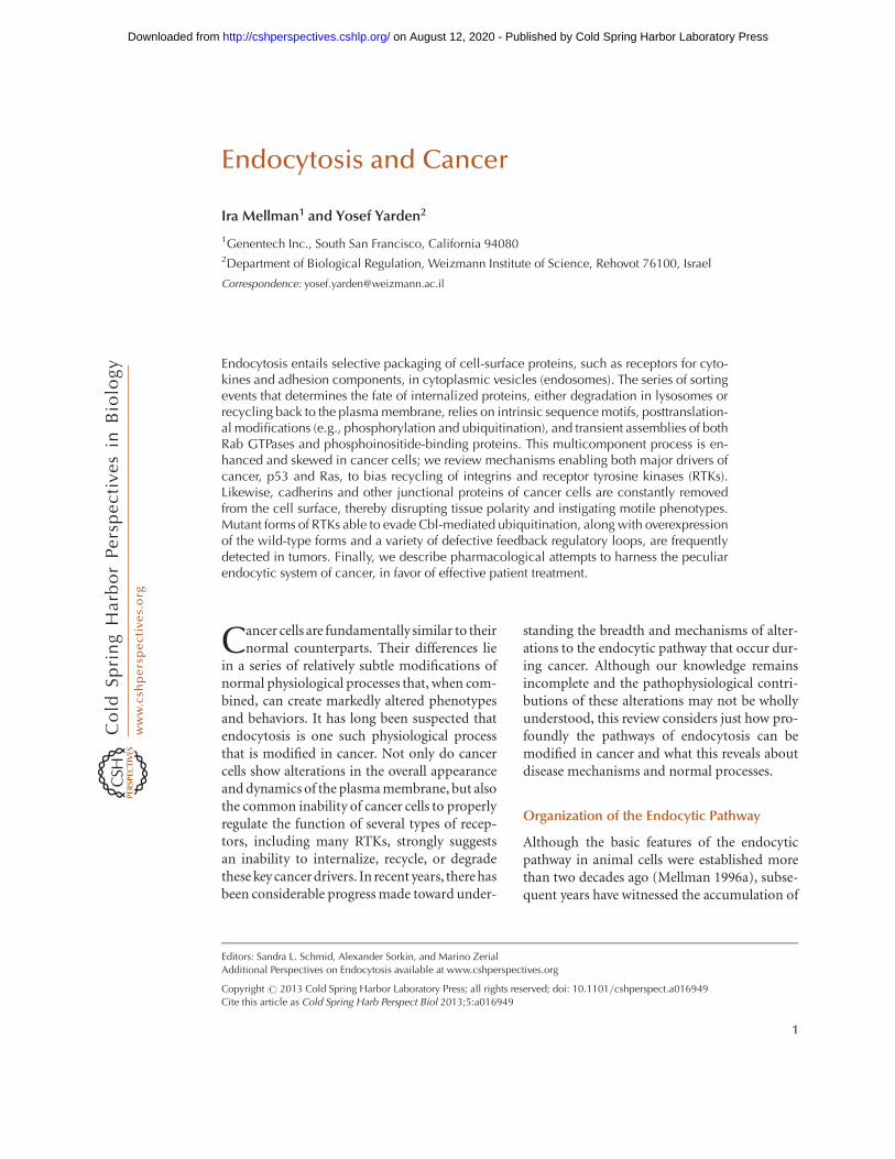
Figure 1. Cell-surface structures regulated in tumors by vesicular trafficking. A schematic view of an epithelialcell, which is in close contact with both a highly polarized neighboring cell, via adherens junctions, and theunderlying extracellular matrix (via focal adhesions). Several actin-filled projections of the plasma membraneare presented (e.g., a lamellipodium and several filopodia). In addition, ventrally located invadopodia are shownas actin-filled fingers that perforate the underlying extracellular matrix. Turnover of all presented surfacestructures is regulated by vesicular trafficking, which is outlined as a route starting at the clathrin-coated pit(CCP), leading to EEs, late endosomes, or multivesicular bodies (MVBs), and eventually reaches lysosomes.Both adhesion molecules, such as integrins and signaling receptors (e.g., EGFR), are transported to lysosomesthrough this pathway, but an alternative route recycles receptors back to the cell surface. In the case of integrins,this latter route involves the perinuclear recycling compartment (PNRC). Note that Figures 2–5 highlightportions of the general view shown in this scheme.
I. Mellman and Y. Yarden
4 Cite this article as Cold Spring Harb Perspect Biol 2013;5:a016949
on August 12, 2020 - Published by Cold Spring Harbor Laboratory Press http://cshperspectives.cshlp.org/Downloaded from

by single missense mutations, usually at codon12 or codon 61 (Pylayeva-Gupta et al. 2011).Whereas several lines of evidence indicate thatmutants of Ras exploit the endocytic machinery,the functional significance of these events re-mains poorly understood. For example, it wasnoted early on that microinjection of the H-Rasprotein into fibroblasts increased both surfaceruffles and fluid phase macropinocytosis within30–60 minutes (Bar-Sagi and Feramisco 1986).However, although it is clear that Ras can remod-el the actin cytoskeleton to promote ruffling,
the interaction with vesicle-forming machiner-ies, along with the contribution of macropino-cytosis to the transformed phenotype, are lessunderstood. Another isoform of Ras, K-Ras,transcriptionally elevates caveolin-1 through amechanism involving Akt, and this enhances tu-mor cell migration (Basu Roy et al. 2012).
Along with macropinocytosis, Ras activa-tion strongly induces clathrin-dependent en-docytosis at least in part by regulating the mo-nomeric GTPase Rab5. Rab5 is known as aregulator of fusion between EEs. Key players
Lamellipodium
Recyclingendosome
Earlyendosome
RCP
Rab 11/25p63
Mutant p53
EGFRα5β1
Lysosome
Figure 2. Involvement of mutant p53 in lamellipodium dynamics and cell migration. To safeguard sustainedforward movement of the leading edge, integrin and RTKs constantly internalize and directionally recycle bymeans of vesicular trafficking. A critical player is the RCP, a Rab11 effector, that physically binds with both RTKsand integrins such as the fibronectin receptor a5b1. This enables cotrafficking of adhesion and signalingmolecules to the forefront of the leading edge. Importantly, the endosomal protein RCP, like its partner,Rab25, is overexpressed in some tumors and indirectly down-regulated by p63. The latter transcription factoris a member of a tumor suppressor family that includes also p73 and the wild-type form of p53. Notably,oncogenic mutant forms of p53, such as R175H and R273H, transcriptionally repress p63. Thus, in cancer cellsexpressing p53 mutants, both RTKs and certain integrins evade degradation in lysosomes, thereby enhancingcell migration and downstream signaling, primarily to the Akt pathway.
Endocytosis and Cancer
Cite this article as Cold Spring Harb Perspect Biol 2013;5:a016949 5
on August 12, 2020 - Published by Cold Spring Harbor Laboratory Press http://cshperspectives.cshlp.org/Downloaded from

in these events are the Ras-interaction/interfer-ence (Rin) proteins, Ras effectors that connectsignaling to the control of receptor endocytosisand actin remodeling (Tall et al. 2001). Via itsRas association (RA) domain, Rin1 competeswith binding of another Ras effector, Raf, where-as the Vps9 domain is necessary for binding toRab5 and for GDP/GTP exchange activity. Inaddition, Rin1 directly binds active EGFRs viaits SH2 domain and uses its own phosphotyro-sines to bind Abl tyrosine kinases that regulateactin remodeling. Thus, Rin1 functions as a hublinking potent proto-oncogenes, such as Rafand Abl, to the endocytic machinery. BecauseRas potentiates the Rab5 nucleotide exchangeactivity of Rin1, this interaction augmentsRab5A-dependent endosome fusion and EGFR-mediated endocytosis. In contrast, Rin1-Ablsignaling stabilizes EGFR and inhibits macro-pinocytosis (Balaji et al. 2012).
The potential functional significance ofRin1’s interaction with Ras, EGFR, and Abl issupported by evidence showing that overexpres-sion of Rin1 suppresses apoptosis in vitro andcorrelates with poor prognosis of melanomapatients (Fang et al. 2012). A similar prognosticcorrelation was reported in non-small-cell lungcancer and in gastric tumors (Wang et al. 2012).In colorectal tumors, Rin1 expression correlatednot only with poor prognosis but also with ve-nous invasion (Senda et al. 2007). Congruentwith growth-factor-driven invasiveness and me-tastasis, studies performed on lung cancer celllines suggested that Rin1 regulates cell prolifer-ation through EGFR (Tomshine et al. 2009).These observations suggest that the interactionsbetween Rin1 and Abl are favored in tumorsmore so than the alternative actions of Rin1on Rab5 and Raf.
Yet more complexity has been introduced byreports on ubiquitination of Ras and Rho fam-ily members and potential relevance to tumor-igenesis (de la Vega et al. 2011). The group ofBar-Sagi reported that H-Ras is subject to ubiq-uitin conjugation (Jura and Bar-Sagi 2006). In-terestingly, ubiquitin attachment to H-Ras ismediated by another Rab5 GEF—an E3 ligaseand an ubiquitin-binding protein called Rabex-5. Ubiquitination stabilizes the association of
H-Ras with endosomes an inhibits its abilityto activate Raf (Xu et al. 2010). Moreover, Rin1is required for Rabex-5-dependent H-Ras ubiq-uitination, suggesting a feedback mechanism bywhich H-Ras activates Rin1 and the latter re-cruits Rabex-5 to ubiquitinate, thereby inacti-vating H-Ras. A different model was proposedby a study of K-Ras ubiquitination. This mod-ification enhanced, rather than weakened, cou-pling to downstream effectors such as Erk andPI3K (Sasaki et al. 2011), raising the possibilitythat endocytosis of K-Ras activates signaling,but internalization of H-Ras inhibits down-stream signals. It is worthwhile to note thatthat earlier observations concluded that H-Rassignaling and K-Ras signaling are differentiallydependent on endocytosis (Roy et al. 2002).Moreover, EGFR signaling results in the recruit-ment of K-Ras to late endosomes and lyso-somes, an event that does not occur in thecase of H-Ras or N-Ras (Lu et al. 2009).
ONCOGENIC MUTANTS OF GROWTHFACTOR RECEPTORS ARE ENDOCYTOSISIMPAIRED
The canonical pathway that clears from thecell-surface-activated forms of RTKs, alongwith their bound growth factor molecules (Sor-kin and von Zastrow 2009), is often avoided bygrowth factor receptors of cancer cells, eitherbecause they carry oncogenic mutations or theyare otherwise aberrantly expressed (Mosessonet al. 2008; Parachoniak and Park 2012). Recep-tor internalization is mediated by clathrin-de-pendent and -independent, routes (Sigismundet al. 2005), eventually delivering cargoes to EEs.Within endosomes, activated RTKs are eithertransferred to late endosomes and lysosomesfor degradation, or they are recycled, providingsustained signaling (Parachoniak et al. 2011).Multiple factors regulate sorting at the endo-some and they include receptor autophosphor-ylation, ligand affinity and its sensitivity to pH,ubiquitination by Cbl and other ubiquitin ligas-es, and several adaptor proteins such as Grb2and ubiquitin binders. Oncogenic tricks thatmanipulate RTK sorting are reviewed below byfocusing on two subfamilies of RTKs (Fig. 3).
I. Mellman and Y. Yarden
6 Cite this article as Cold Spring Harb Perspect Biol 2013;5:a016949
on August 12, 2020 - Published by Cold Spring Harbor Laboratory Press http://cshperspectives.cshlp.org/Downloaded from
The EGFR/ErbB Family
Four transmembrane receptor RTKs comprisethe ErbB family, of which EGFR is the proto-type. The 11 mammalian EGF-like ligands dis-play wide variation in terms of receptor specif-icity, binding affinities, and dependence on pH.
Likewise, multiple EGF-like ligands are encoded
by smallpox viruses and, like the mammalian
growth factors, they differ in their rate of clear-
ance by means of receptor-mediated endocyto-
sis (Tzahar et al. 1998). Ligands that are either
sensitive to the low pH of endocytic compart-
RTK endocytosis
EGFR
EGF
CCP
EE
P P
P
Cbl Dep1Cezanne1SrcCool1AIP4AlixTula
RaltLrig 1Grb 2Cin85Numb
Clathrin-independentendocytosis
MVB
Rab5
Degradation Recycling
Rab4Rab11Rab7
Hip1
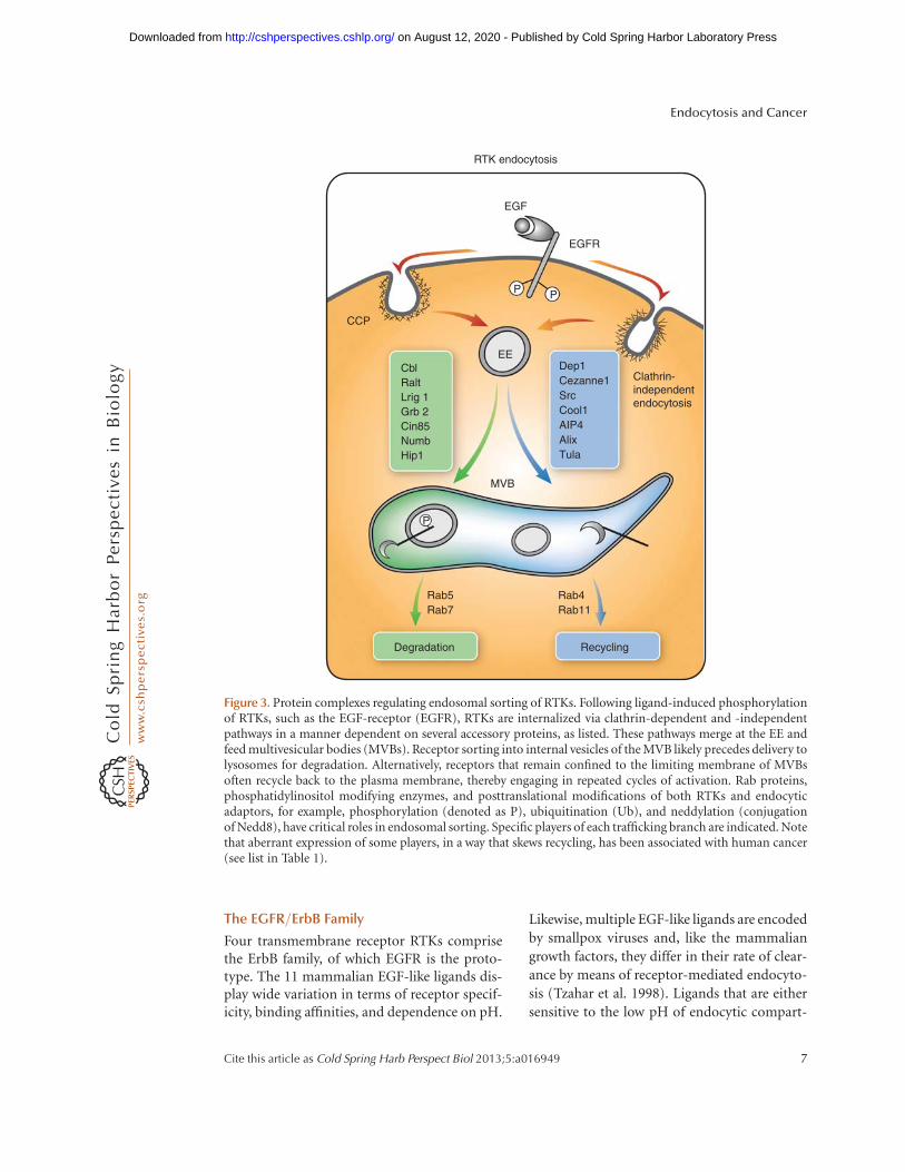
Figure 3. Protein complexes regulating endosomal sorting of RTKs. Following ligand-induced phosphorylationof RTKs, such as the EGF-receptor (EGFR), RTKs are internalized via clathrin-dependent and -independentpathways in a manner dependent on several accessory proteins, as listed. These pathways merge at the EE andfeed multivesicular bodies (MVBs). Receptor sorting into internal vesicles of the MVB likely precedes delivery tolysosomes for degradation. Alternatively, receptors that remain confined to the limiting membrane of MVBsoften recycle back to the plasma membrane, thereby engaging in repeated cycles of activation. Rab proteins,phosphatidylinositol modifying enzymes, and posttranslational modifications of both RTKs and endocyticadaptors, for example, phosphorylation (denoted as P), ubiquitination (Ub), and neddylation (conjugationof Nedd8), have critical roles in endosomal sorting. Specific players of each trafficking branch are indicated. Notethat aberrant expression of some players, in a way that skews recycling, has been associated with human cancer(see list in Table 1).
Endocytosis and Cancer
Cite this article as Cold Spring Harb Perspect Biol 2013;5:a016949 7
on August 12, 2020 - Published by Cold Spring Harbor Laboratory Press http://cshperspectives.cshlp.org/Downloaded from

ments (e.g., transforming growth factor a) orthat bind EGFR with low affinity (e.g., epiregu-lin) are generally more sustained stimulants,and they often associate with human malignan-cies (Normanno et al. 2001). Conceivably, theirability to dissociate in EE favors receptor recy-cling, as opposed to transport to late endocyticcompartments. Such factors are often autocrinein nature (Sporn and Todaro 1980), and provideone common mechanism that underlays con-stitutive proliferation by providing a sustainedsource of ligands that do not promote RTKdown-regulation. Several additional mecha-nisms, which are briefly described below, mayhelp cancer cells to evade regulation by RTKendocytosis.
Receptor Overexpression
Head and neck, brain, and other tumors oftenoverexpress ErbB family members as a result ofgene amplification or other mechanisms (Hynesand MacDonald 2009). This represents anothermode that prolongs signaling; owing to thelimited internalization capacity of the clath-rin-coated pit and unbalanced ratio of cargoesversus endocytic adaptors, internalization ofoverexpressed EGFRs is either inhibited or pref-erentially followed by recycling (Wiley 1988;French et al. 1994). At the plasma membrane,overexpression also promotes the collision fre-quency of EGFR monomers, yielding the forma-tion of dimers that are primed for ligand bindingand signaling (Chung et al. 2010). Importantly,the four ErbB proteins form both homodimersand heterodimers, and each receptor com-bination follows different routing caused bythe receptor’s intrinsic internalization signals,association with ubiquitin ligases, and sensitiv-ity of the ligand-receptor complex to low pH.The closest kin of EGFR, an oncogenic corecep-tor called HER2 or ErbB-2, is frequently overex-pressed in some tumors, including 15%–20% ofbreast and gastric tumors. HER2 effectively es-capes the endocytic pathway, because it binds noknown ligand, lacks characteristic internaliza-tion signals, or HER2 molecules associate withheparin sulfate proteoglycans or plasma mem-brane microdomains that retain HER2 (Baulida
et al. 1996; Pinkas-Kramarski et al. 1996). WhenHER2 is internalized, however, it rapidly recycles(Austin et al. 2004). In addition, when overex-pressed, HER2 forms heterodimers with EGFR,which enhances recycling of both receptors(Lenferink et al. 1998; Worthylake et al. 1999),in part by reducing receptor ubiquitination(Levkowitz et al. 1996; Muthuswamyet al. 1999).
Deletion Mutants of EGFR
Approximately 50% of glial tumors harborEGFR gene amplification (Wong et al. 1987),and a large fraction of these also present EGFR-vIII (Jeuken et al. 2009), a deletion mutant lack-ing exons 2–7, including a portion of the ligand-binding cleft. EGFRvIII molecules are basallydimerized and activated in the absence of ligandbinding, thus conferring high tumorigenic po-tential (Nagane et al. 1996). In contrast to EGFR,EGFRvIII is inefficiently internalized and de-graded, and after internalization it is recycledrather than delivered to lysosomes (Grandalet al. 2007). EGFRvIII binds the ubiquitin ligasec-Cbl via Grb2, whereas binding via phosphor-ylated tyrosine residue 1045, the direct bindingsite of Cbl, is limited, probably because EGFR-vIII’s tyrosine 1045 is hypophosphorylated(Han et al. 2006). Carboxy-terminal deletionmutants, collectively termed EGFRvIV, were alsoidentified in brain tumors. Interestingly, the on-cogenic function of both EGFRvIII and EGFRsharboring carboxy-terminal deletions dependson the chaperoning function of heat shock pro-tein 90 (HSP90) (Lavictoire et al. 2003; Pineset al. 2010), and this feature is shared by anotherendocytosis-defective receptor, namely, HER2(Citri et al. 2002; Neckers and Ivy 2003).
Kinase Domain Mutants of EGFR
Approximately 10% to 30% of tumors frompatients with non-small-cell lung cancer (NS-CLC) harbor somatic activating mutations inthe gene encoding EGFR (Lynch et al. 2004;Paez et al. 2004; Pao et al. 2004). All mutationsare restricted to the tyrosine kinase domain ofEGFR. The most frequent point mutation is asubstitution of an arginine for leucine at posi-tion 858 (L858R). Yet other mutants carry in-
I. Mellman and Y. Yarden
8 Cite this article as Cold Spring Harb Perspect Biol 2013;5:a016949
on August 12, 2020 - Published by Cold Spring Harbor Laboratory Press http://cshperspectives.cshlp.org/Downloaded from

frame short deletions. Differential patterns ofautophosphorylation resulting in enhancedAkt and Stat signaling have been associatedwith EGFR mutants (Sordella et al. 2004). Alongwith pathway-selective activation by EGFR mu-tants, their ligand-induced autophosphoryla-tion decays relatively slowly (Lynch et al. 2004),raising the possibility that the mutant recep-tors evade degradation. Analyses of several mu-tants, including a double-mutant L858R/T790-M, which is resistant to tyrosine kinase inhib-itors such as gefitinib and erlotinib, uncovered adefect in Cbl-mediated ubiquitination and deg-radation of EGFR (Shtiegman et al. 2007). Thedefect was attributed to a propensity of the mu-tants to heterodimerize with HER2, therebyevading c-Cbl-mediated ubiquitination. Con-sistent with this model, chromosomal analysisof lung tumors revealed that HER2 was ampli-fied in 12% of tumors with acquired resistanceto kinase inhibitors, versus only 1% of untreat-ed patients with lung adenocarcinomas (Take-zawa et al. 2012). Thus, activating mutants ofEGFR in lung cancer exploit endocytosis-relat-ed mechanisms to reduce rapid inactivation byinternalization and MVB sorting, further en-hancing their oncogenic properties.
Evasion of EGFR Feedback Regulatorsby Cancer Cells
A major source of information on negative ErbBsignals arises from developmental genetics ofinvertebrate organisms such as Caenorhabditiselegans and Drosophila. In C. elegans, loss ofSli-1, the ortholog of Cbl in mammals, leadsto excessive vulva formation, and naturally oc-curring aberrant forms of c-Cbl are oncogenicin mammals (Thien and Langdon 2001; Kaleset al. 2010). For example, DNA sequencing ofleukemic bone marrow revealed a case with ac-Cbl point mutation (Cbl-R420Q) that inhibitsinternalization and ubiquitination of the Flt re-ceptor (Sargin et al. 2007). In addition, muta-tions of Cbl-binding tyrosine in the cytoplasmicdomain of the human-colony-stimulating fac-tor-1 (CSF-1) receptor were found in childrenwith secondary myelodysplasia and secondaryacute myeloid leukemia (Ridge et al. 1990).
Similar to Cbl proteins, a group of negativeEGFR regulators in insects undergo transcrip-tional up-regulation following activation ofEGFR. For example, kekkon-1, which encodesa transmembrane protein, physically binds toand inhibits EGFR molecules (Ghiglione et al.1999). Although the multiple Kekkon proteinsof insects have no clear orthologs in mammals,the three mammalian LRIG proteins share do-main organization with Kekkons. Moreover,LRIG1 physically associates with all four ErbBproteins of mammals and its up-regulation isfollowed by enhanced ubiquitination and deg-radation of EGFR. The underlying mechanisminvolves recruitment of c-Cbl, which simultane-ously ubiquitinates EGFR and LRIG1, and sortsthem for degradation (Gur et al. 2004; Laeder-ich et al. 2004). In line with growth suppression,LRIG1 expression correlates with good progno-sis of breast, cutaneous squamous cell carcino-ma (SCC), and other types of cancer (Tanemuraet al. 2005; Hedman and Henriksson 2007; Kriget al. 2011).
Like LRIG1, Mig6/RALT interacts with allErbB members, along with additional RTKs,and blocks downstream signaling (Anastasiet al. 2003). Crystal structures of complexes be-tween the EGFR kinase domain and a fragmentof Mig6 showed binding to the distal surface ofthe C-lobe of the kinase domain (Zhang et al.2007). Although the kinase region within suchcomplexes is inactive, Mig6 nevertheless targetsEGFR to endocytosis and degradation by re-cruiting a set of endocytic adaptors (e.g., AP-2and intersectins) (Frosi et al. 2010). Presumably,Mig6 evolved as a suppressorof the inactive formof EGFR as a result of the scaffold, kinase-inde-pendent functions of EGFR and other RTKs.Interestingly, high-resolution genomic profilesof glial tumors identified a highly recurrent fo-cal 1p36 deletion encompassing Mig6 (Yinget al. 2010), and high Mig6 expression in papil-lary thyroid cancer is associated with favor-able outcomes (Ruan et al. 2008). In summary,the integration of in vitro lines of evidence anddata from cancer patients attributes growth-suppressive functions to RTK endocytosis,but diverse and multiple aberrations weakenthis mechanism in tumors. More examples of
Endocytosis and Cancer
Cite this article as Cold Spring Harb Perspect Biol 2013;5:a016949 9
on August 12, 2020 - Published by Cold Spring Harbor Laboratory Press http://cshperspectives.cshlp.org/Downloaded from

endocytosis-related proteins, which are aber-rantly expressed in human tumors, are shownin Table 1.
c-Met (Hepatocyte Growth Factor Receptor)
In analogy to EGFR, c-Met is implicated in thegrowth, survival, and spread of various humancancers (Blumenschein et al. 2012). Overexpres-sion of c-Met and autocrine HGF signaling areconsidered to be the two major aberrations ofthe HGF-Met axis in cancer, although rare mu-tations affecting several domains of the receptorhave been documented that affect its behaviorduring endocytosis. For example, overexpres-sion was observed in NSCLC, breast, renal,and ovarian cancer, and this has been associatedwith poor prognosis. Similarly, mutations in c-Met were found in lymph-node metastases ofhead and neck squamous cell carcinomas, im-plying that the mutations are selected duringmetastatic spread (Di Renzo et al. 2000). Impor-tantly, the juxtamembrane domain regulates li-gand-dependentc-Met internalizationby meansof tyrosine-1003 phosphorylation in responseto HGF binding, leading to c-Met ubiquitina-tion and degradation (Abella et al. 2005). Thus,when an exon 14 deletion occurs, such as inlung and gastric cancer, the loss of Y1003 resultsin c-Met accumulation at the cell surface andpersistent HGF stimulation, leading to tumor-igenesis. Cbl’s binding tyrosine is missing inanother oncogenic mutant of c-Met, namely,the Tpr-Met fusion protein generated followinga carcinogen-induced chromosomal rearrange-ment. As expected, this mutant is stable andcytoplasmic, undergoes no ubiquitination, andshows strong tumorigenesis (Mak et al. 2007).Differential ubiquitination and recruitment ofCbl proteins might underlie relatively strongsignaling and transforming potential of otherRTKs and their derivatives, such as Ret (Scottet al. 2005) and Flt-1 (Kobayashi et al. 2004).
REDOX-REGULATED TRAFFICKING OF RTKS
Aggressive cancer cells have high oxidative stressas a result of their acidic environment, whichenhances the formation of reactive oxygen
species (ROS) (Riemann et al. 2011). Anothersource of ROS is intrinsic; excessive activity ofRTKs is known to produce free radicals (Lan-der 1997). Early observations found that hydro-gen peroxide enhances EGFR phosphorylationon tyrosines (Gamou and Shimizu 1995) by amechanism distinct from ligand-induced stim-ulation, in that no kinase activation is neededand phosphorylation is insensitive to EGFRkinase inhibitors (Filosto et al. 2011). Impor-tantly, ROS-activated receptors undergo noubiquitination or degradation. Instead, theytranslocate to a perinuclear location (most like-ly recycling endosomes) that permits sustainedsignaling (Khan et al. 2007). Evasion of receptordegradation, through mechanisms that likelyinvolve Src activation and degradation of Cbl(Bao et al. 2003), seems to be common in tu-mors. Furthermore, it was shown that the gasphase of cigarette smoke contains hydrogenperoxide at doses sufficient to affect RTK func-tion (Khan et al. 2007). Interestingly, underhypoxia, RTK signaling is commonly enhancedthrough transcription- or translation-mediatedmechanisms. For instance, hypoxic microenvi-ronments and activation of hypoxia-induciblefactor (HIF) 2a in the core of solid tumors leadto overexpression of EGFR by increasing trans-lation of the respective mRNA (Franovic et al.2007). In addition, hypoxia prolongs the acti-vation of EGFR because of lengthened receptorhalf-life and retention in the endocytic pathway.This is caused by the attenuation of Rab5-me-diated EE fusion, via HIF-dependent down-reg-ulation of a critical Rab5 effector, rabaptin-5,at the level of transcription (Wang et al. 2009).In conclusion, severe environmental conditionsenhance RTK signaling by shunting receptor en-docytosis, thereby enhancing signaling, cell sur-vival, and tumorigenesis.
THE SIGNALING ENDOSOME HYPOTHESIS
Several lines of evidence support the possibilitythat RTKs and other receptors might generatesignals while en route. Early studies identifiedactive EGFRs and specific components of theRas pathway in endosomes isolated from EGF-stimulated livers (Di Guglielmo et al. 1994).
I. Mellman and Y. Yarden
10 Cite this article as Cold Spring Harb Perspect Biol 2013;5:a016949
on August 12, 2020 - Published by Cold Spring Harbor Laboratory Press http://cshperspectives.cshlp.org/Downloaded from

Table 1. Endocytosis-regulating proteins aberrantly acting in human tumors
Protein Normal function Defective function Aberrations in human tumors References
Ack1 Binds clathrin, ubiquitin, and Cdc42;promotes EGFR degradation.
The S985N mutant enhances proliferation andstabilizes EGFR. Overexpression increasesmetastasis.
Gene amplification and overexpression inlung ovarian and prostate cancers. Severalsomatic and germline mutations.
van der Horst et al.2005; Chuaet al. 2010
Caveolin 1 Major coat protein of caveolae.Invaginates lipid raft domains. Binds
with and enhances action of tumorsuppressor DLC1.
Normally, caveolin inhibits several RTKs; deletionof the fragile 7q31.1 locus of caveolin 1 or gene
ablation enhances cell proliferation.
Down-regulation and sporadic mutations(e.g., P132 L) in breast, ovarian, and liver
cancer; up-regulated in kidney andesophageal cancers.
Goetz et al. 2008;Du et al. 2012
Cbl An E3 ubiquitin ligase of several RTKs. Causes mutations in RING and linker domains,deletions and insertion that inhibit receptor
ubiquitination. Aviral short form is oncogenic.
Mutant forms in myeloid neoplasias. Sargin et al. 2007;Kales et al. 2010
Clathrin Major component of the coat ofmembrane invaginations that mediate
endocytosis.
Constitutive activation of fusion partners such asthe Alk kinase.
A fusion protein found in inflammatorymyofibroblastic tumors.
Bridge et al. 2001
Cortactin Links endocytosis to the actincytoskeleton by binding with actin anddynamin and activating Arp2/3.
When overexpressed, inhibits EGFRubiquitination and endocytosis.
Chromosomal amplification of 11q13 leadsto overexpression in some tumors.Overexpression in breast, head and neck,
colorectal, liver, kidney, and brain tumors.
Cai et al. 2010
Disabled 2(Dab2)
Cargo-selective clathrin adaptor thatbinds Eps15 and intersectin; recruitsmyosin VI to clathrin-coated structure.Enables endocytosis of b integrins.
Acts as a tumor suppressor by dictating tumor cellTGF-b responses. Regulates EMT and recyclingof TGF-b receptors.
Down-regulated in ovarian, bladder, prostate,colorectal, and breast cancers. DAB2promoter hypermethylation found innasopharyngeal tumors.
Karam et al. 2007;Hannigan et al.2010
Dynamin A large GTPase involved in endocytosisand cell migration.
Dynamin 2 might be involved in preventingmetastasis in carcinoma of the cervix, but inpancreatic cells, dynamin 2 potentiatesmetastasis.
Increased expression in tumors of thepancreas, but low expression correlateswith lymph-node metastasis of cervixcancer.
Lee et al. 2010;Eppinga et al.2012
Endophilin Induces membrane curvature duringsynaptic vesicle formation. Bindsdynamin and synpatojanin.
The fusion protein containing a portion ofendophilin, MLL-EEN, is nuclear rather thancytoplasmic; may interact with the Ras pathway.
Chromosomal translocation positionsendophilin next to MLL (mixed lineageleukemia). The fusion protein is found in
acute myeloid leukemia.
So et al. 1997
Eps15 Endocytic adaptor for clathrin; promotesendocytosis.
The coiled coil domain of Eps15 mediatesoligomerization of a histone methyltransferasecalled MLL and enhances self-renewal ofhematopoietic progenitors.
A fusion protein, Eps15-MLL is present inchildhood leukemia.
Rogaia et al. 1997
Continued
Cite
this
articleas
Cold
Sprin
gH
arbPersp
ectB
iol2013;5
:a016949
11
on August 12, 2020 - P
ublished by Cold S
pring Harbor Laboratory P
ress http://cshperspectives.cshlp.org/
Dow
nloaded from

Table 1. Continued
Protein Normal function Defective function Aberrations in human tumors References
GEP100 A guanine-nucleotide exchange factor ofArf6; binds active EGFRs.
Mediates metastasis in animal models. Overexpressed in invasive ductal carcinomasof breast origins. Co-overexpression with
HER2 predicts nodal metastasis in primarylung adenocarcinomas.
Morishige et al.2008; Menju
et al. 2011
HAX1 A multifunctional protein regulatingcalcium homeostasis, cell migration,and apoptosis. Binds with the tail of
avb6.
In carcinoma cells, HAX1 enables cell invasion byregulating endocytosis of integrin avb6.
Up-regulation in melanoma, lung cancer, andbreast cancer. Mutations causeneutropenia.
Ramsay et al. 2007;Trebinska et al.2010
HIP1 Coordinates actin remodeling duringformation of clathrin-coated vesicles.Binds phosphoinositides, AP-2, andclathrin.
Induces cytokine-independent growth. Up-regulates several RTKs.
Overexpressed in gliomas and carcinomas ofthe breast and prostate. A fusion protein,HIP1-PDGFBR is found in CMML.
Rao et al. 2003
Mdm2 A proto-oncogene that regulates p53stability and ubiquitinates b arrestins,mediators of internalization of G-protein-coupled receptors.
Likely regulates endocytosis of chemokine andother seven transmembrane receptors, alongwith negative feedback of p53.
Acquires oncogenicity through increasedexpression in a range of common tumors(e.g., bladder cancer).
Shenoy et al. 2001
NDRG1 A Rab4a effector protein that localizes to
sorting vesicles by binding tophophatidylinositol 4-phosphate.Involved in recycling of E-cadherin.
Acts as a metastasis suppressor in animal models.
Down-regulated by N-Myc.
Down-regulated in prostate, breast, and
pancreatic cancers.
Bandyopadhyay
et al. 2003
Numb A multifunctional regulator of signalingby Notch, Hedgehog, and p53.
Involved in endocytosis,determination of polarity, andmigration.
Normally, Numb acts as an antagonist of Notchand a stabilizer of p53. In its absence, these
pathways are altered and cells acquire malignantfeatures.
Down-regulated in breast and lung cancers. Pece et al. 2004
Rab25 A rab11 family member that physically
interacts with b1 integrin and rescueslysosomally targeted integrins.
Forced overexpression of Rab25 increases
aggressiveness of cancer cells by activating theAkt pathway.
Amplification of 1q22, centered on rab25,
increases expression of Rab25 in breast(and ovarian) cancer and correlates withlymph-node metastasis.
Cheng et al. 2004;
Yin et al. 2012
RCP A Rab11 effector protein that associateswith a5b1 integrin and guides
recycling of integrins and EGFR.
Recycling of EGFR and integrin a5b1 is normallyinhibited by transcriptionally active TAp63, but
this is relieved when mutant p53 is expressed.
Overexpressed in breast, ovarian, and head/neck tumors. The respective 8p11–12 locus
is frequently amplified in breast cancer.
Zhang et al. 2009
12C
iteth
isarticle
asC
old
Sprin
gH
arbPersp
ectB
iol2013;5
:a016949
on August 12, 2020 - P
ublished by Cold S
pring Harbor Laboratory P
ress http://cshperspectives.cshlp.org/
Dow
nloaded from

This implied that receptor endocytosis fulfillsroles other than desensitization, in line withthe decrease in Erk activation observed in cellsexpressing a dominant-negative mutant of dy-namin (Vieira et al. 1996) as well as a loss ofactive Mek, the Erk-specific kinase, in cells lack-ing expression of an endosomal scaffold com-plex (MP1-p14) (Teis et al. 2002). Yet, cells ex-pressing a mutant of dynamin showed enhancedDNA synthesis (Vieira et al. 1996), but a low-molecular-weight inhibitor of dynamin inhib-ited, rather than enhanced, in vivo tumorigen-esis of cells expressing a mutant form of c-Metthar led to the proposition that endosomal sig-naling enhances oncogenicity of c-Met (Joffreet al. 2011). Another inhibitory compound,of EGFR’s kinase, was used to arrest EGFR inendosomes and derive evidence in support ofendosomal signaling (Wang et al. 2002). Where-as it is widely accepted that signaling endosomescontaining neurotrophins and their receptorsare retrogradely transported along microtu-bules back to neuronal cell bodies, where theycontrol transcriptional events (Howe and Mob-ley 2005), endosome signaling by EGFR andother receptors has received diverse interpre-tations. For example, spatially restricted phos-phatases might regulate intracellular signaling.Accordingly, internalization of EGFR and sig-naling at sites in close proximity to the endo-plasmic reticulum enable PTP1B-catalyzed de-phosphorylation (Haj et al. 2002), whereasDEP-1, a transmembrane tyrosine phosphataseencoded by a tumor suppressor gene (Ruiven-kamp et al. 2002), can prevent EGFR signaling atthe plasma membrane, yet cannot inhibit en-dosomal signals (Tarcic et al. 2009). Accordingto an alternative interpretation, EGFRs inter-nalized via clathrin-mediated endocytosis arerecycled to the cell surface, thus enabling EGF-dependent DNA synthesis, but clathrin-inde-pendent internalization preferentially commitsthe receptor to degradation (Sigismund et al.2008). Conceivably, better understanding of dif-ferential routings, along with heterogeneity atthe level of EEs and selective engagement of en-dosomal scaffold proteins would throw light onintracellular signaling and its currently unclearcontribution to tumor progression.
ABERRANT INTEGRIN TRAFFICKINGIN TUMORS
Efficient cell migration requires constant endo-cytosis and recycling of integrins rather thantheir degradation. Accordingly, it was estimatedthat the plasma membrane pool of integrins isrecycled through the endosomal system once in30 min (Roberts et al. 2001), and this flux ofintegrins correlates with both migration speed(Teckchandani et al. 2009) and the overall dy-namics of the plasma membrane (Mellman1996a). Because integrin recycling is also im-portant for cell division (cytokinesis) (Caswellet al. 2009), and integrins have major roles intumor-stroma interactions, aberrant recyclingof the 25 different integrin heterodimers is in-volved in tumor growth, invasion, metastasis,and evasion of apoptosis (Mosesson et al. 2008).One critical aspect of integrins’ “outside-in sig-naling” is their ability to influence the mannerin which RTKs respond to their ligands. Forexample, recycling of EGFR and integrin a5b1is coordinated and this promotes cell migrationin 3D matrices (Caswell et al. 2008).
The cycle of integrin endocytosis–exocyto-sis may initiate on inducible polymerization oftubulin, a well-known target for cancer thera-pies (Fig. 4). The growing tips of microtubulesdisintegrate focal adhesions and instigate dyna-min-dependent endocytosis of some integrinheterodimers, in a mechanism that also requiresthe adaptor Dab2 and the kinase Fak (Ezrattyet al. 2005). Internalized integrins follow threealternative routes: a short, Rab4-dependent re-cycling loop, a longer loop that depends onRab11 family members and translocates cargoesto the perinuclear recycling endosomes, and apathway leading to degradation in lysosomes.Although the details of the sorting process thattakes place in dedicated vesicles located just be-hind the leading lamella (Pierini et al. 2000) areincompletely understood, it is clear that theseroutes are linked to tumor progression. For ex-ample, growth factors can shunt integrin avb3to the short recycling loop (Roberts et al. 2001)through a mechanism that requires PKD-medi-ated phosphorylation of rabaptin, a bindingpartner of both Rab4 and Rab5 (Woods et al.
Endocytosis and Cancer
Cite this article as Cold Spring Harb Perspect Biol 2013;5:a016949 13
on August 12, 2020 - Published by Cold Spring Harbor Laboratory Press http://cshperspectives.cshlp.org/Downloaded from

2004; Christoforides et al. 2012). Likewise, an-other kinase, Akt/PKB, controls recycling of thea5b1 integrin through several downstream tar-gets, including an Arf6-specific GTPase activat-ing protein (GAP), ACAP1, which is needed forclathrin coat assembly (Li et al. 2005; 2007).
As already discussed, accelerated recyclingof certain integrins characterizes cancer cells ex-
pressing mutant forms of p53, and this involvesthe Rab-coupling protein (RCP) (Muller et al.2012). Another driver of both integrin recy-cling and cancer is the epithelial member ofthe Rab11 family, Rab25. Early observations re-vealed overexpression of the respective genein hepatocellular cancer (He et al. 2002) andlater studies by the laboratory of Gordon Mills
Focal adhesions
MTOC
PNRC(Rab11)
Lysosome
EE(Rab5)
F-actin
Recycling(Rab4)
Microtubule
DAB2
FAK
FAK
Dynamin
α1/2/3β1
Figure 4. Endosomal sorting regulates integrin-based focal adhesions. Focal adhesions containing active con-formers of integrin b1 establish strong, yet dynamic, contacts between the ventral aspect of migrating cells andthe underlying extracellular matrix. Both actin filaments and microtubules projecting from the microtubule-organizing center (MTOC) regulate cell adhesion. The latter polymers dissolve focal adhesions once theyapproach the plasma membrane. This requires protein kinases, such as FAK, and recruitment of both dynamin2 and disabled 2 (Dab2), a clathrin adaptor, which directly binds with the cytoplasmic tails of integrin b-subunits. Once internalized by activated dynamin, the formed clathrin-coated vesicles mature to integrin-loaded EEs. These organelles are transported backward along microtubules to reach the PNRC and the recyclingpathway, or these EEs deliver their cargo to lysosomes for degradation. These alternative itineraries are regulatedby specific Rab proteins. Note that the flux of integrins through the endocytic pathway, rather than their surfacelevels, dictates migration speed. Accordingly, the levels of both Dab2 and dynamin display broad variation inseveral types of carcinomas (see Table 1).
I. Mellman and Y. Yarden
14 Cite this article as Cold Spring Harb Perspect Biol 2013;5:a016949
on August 12, 2020 - Published by Cold Spring Harbor Laboratory Press http://cshperspectives.cshlp.org/Downloaded from
showed that amplification of 1q22, centered onrab25, increases expression of the GTPase in�50% of ovarian and breast cancers (Chenget al. 2004). Furthermore, forced expression ofRab25 markedly increased anchorage-inde-pendent cell proliferation and enhanced aggres-siveness of cancer cells in vivo. Other studiesconfirmed these observations (Brusegard et al.2012) and also linked Rab25 to the group ofandrogen-responsive ovarian tumors (Sheachet al. 2009). These observations are consistentwith the reported ability of Rab25 to physicallyinteract with b1 integrin (Caswell et al. 2007)and rescue lysosomally targeted integrins (Do-zynkiewicz et al. 2012). Interestingly, the latterfunction involves a chloride channel, CLIC3,which is a marker for invasive, poor prognosistumors of the pancreas. Thus, the wealth of cur-rently available lines of information identifiesvesicular transport of integrins as an importantdeterminant of tumor progression.
EMT AND ENDOCYTOSIS-MEDIATEDDISRUPTION OF EPITHELIAL POLARITYIN TUMORS
Epithelial-mesenchymal transition (EMT) rep-resents the loss of cell–cell adhesions and api-cal-basal polarity, along with concomitant de-velopment of a motile phenotype (Thiery et al.2009). Epithelial sheets acquire such pheno-types in the context of normal physiology,such as in embryogenesis and during tissue re-pair, as well as under pathological conditionsthat include organ fibrosis and metastasis for-mation (Kalluri and Weinberg 2009). To sustainEMT, cells apply a myriad of switches, bothtranscription-independent events such as vesic-ular trafficking as well as transcriptional switch-es involving newly synthesized macromolecules.The inducers of the switches are often solublepolypeptides, namely, chemokines and growthfactors. The latter include HGF, Wnt, PDGF(platelet-derived growth factor), Notch ligands,and transforming growth factors. Importantly,both tight junctions and adherens junctions arelost during EMT. This is mediated by the sup-pression of junctional complexes (e.g., E-cad-herin, ZO-1, occludins, and specific claudins)
and components of polarity complexes, suchas LGL2 (Fig. 5). The crucial part played byjunctional complexes in cancer progression isexemplified by the frequent occurrence of ge-netic alterations in the epithelial cadherin (E-cadherin) in breast, gastric, colon, and othertypes of cancer (Paredes et al. 2012). Vesiculartrafficking critically regulates junctional com-plexes by means of its polarizing function. Forexample, clathrin knockdown experimentsperformed with epithelial cells resulted in mis-localization of basolateral proteins, whereasthe apical surface remained unaltered (Debordeet al. 2008). Polarity-maintaining trafficking iscritically regulated by Cdc42 and the Par (par-titioning-defective) group of proteins altered incancer. For example, EGF was shown to regulatetight junction assembly by phosphorylation ofPar3 (Wang et al. 2006), whereas HER2 disruptsepithelial polarity by binding to Par6 (Arandaet al. 2006).
The best understood components of adhe-rens junctions are E-cadherins and their part-ners, the catenins. The exocytic pathway con-trols delivery of newly synthesized E-cadherinin complex with b-catenin, and this is regulatedby sorting nexin 1 (SNX1) and Rab11 (Lock andStow 2005). Additionally, the endocytic path-way controls E-cadherin, and this is regulatedby Arf6, tyrosine kinases, and p120-catenin, themost potent inhibitor of E-cadherin endocyto-sis. The importance of E-cadherin trafficking isdual: At the surface, E-cadherin mediates cell-to-cell homophilic interactions, but it also trapsb-catenin. Loss of this interaction enables b-catenin to translocate to the nucleus, where itacts as an activator of the Wnt pathway. Hence,it comes as no surprise that multiple mecha-nisms regulate endocytosis of E-cadherin, andthis bears clinical implications in oncology. Theactive form of Arf6, Arf6-GTP, recruits Nm23-H1, a nucleoside diphosphate kinase that en-hances fission of coated vesicles and causesinternalization of E-cadherin (Palacios et al.2002). In the same vein, EGFR interacts specif-ically with GEP100/BRAG2, an Arf6 guaninenucleotide exchange factor, to promote Arf6activation and consequent E-cadherin inter-nalization (Morishige et al. 2008). Interestingly,
Endocytosis and Cancer
Cite this article as Cold Spring Harb Perspect Biol 2013;5:a016949 15
on August 12, 2020 - Published by Cold Spring Harbor Laboratory Press http://cshperspectives.cshlp.org/Downloaded from

co-overexpression of HER2 and GEP100 in lungtumors predicts metastasis in patients (Menjuet al. 2011). Tyrosine kinases, such as Src orRTKs, also regulate trafficking of E-cadherin.Ubiquitination and trafficking to lysosomes ofphosphorylated E-cadherin are mediated byHakai, a Cbl-like E3 ubiquitin ligase. Hakai in-duces monoubiquitination of E-cadherin in re-sponse to Src activation (Fujita et al. 2002). Ad-ditionally, the viral form of Src stimulates Rab5and Rab7, which target E-cadherin to lysosomes(Palacios et al. 2005). Along with phosphoryla-
tion and regulation by growth factors, E-cad-herin stability is strongly regulated by p120-catenin (p120-ctn), which binds to the cyto-plasmic portion of cadherins and inhibits theirendocytosis. This is mediated by a dual-func-tion motif consisting of three highly conservedacidic residues that serve as a p120-binding in-terface and an endocytic signal (Nanes et al.2012).
Predictably, similarly detailed understand-ing of the endocytosis of other cell-to-cell ad-hesion molecules will not only deepen the way
Occludin
Claudin
E-cadherin
p120-Catenin
Lysosome
αβ
Adherens junctions
F-actin
Src (P)
Hakai (Ub)
PUb
Figure 5. Endocytosis-mediated control of cell-to-cell junctions. Intact tight junctions and adherens junctionsare essential for the integrity and polarity of epithelial sheets; their turnover is regulated by means of vesiculartrafficking. Shown are two major components of tight junctions, occludin and claudin, and the epithelialcadherin, E-cadherin, the major component of adherens junctions. E-cadherin maintains calcium-dependentadhesion by means of homophilic extracellular interactions as well as association of the cytoplasmic tails withthe actin cytoskeleton. This involves several types of catenins, including p120-catenin, an inhibitor of E-cadherinendocytosis. Tyrosine kinases such as Src have major roles in the disruption of adherens junctions; one mech-anism involves inactivation of p120-catenin. Alternatively, tyrosine phosphorylation (denoted as P) of E-cad-herin enables recognition by a Cbl-like ubiquitin ligase called Hakai that ubiquitinates (denoted as Ub) and sortsE-cadherin molecules to lysosomal degradation. Low expression of E-cadherin, attributable to genetic and otherreasons, characterizes a broad spectrum of advanced tumors.
I. Mellman and Y. Yarden
16 Cite this article as Cold Spring Harb Perspect Biol 2013;5:a016949
on August 12, 2020 - Published by Cold Spring Harbor Laboratory Press http://cshperspectives.cshlp.org/Downloaded from

we conceive EMT but unravel molecules bearingprognostic value. It seems increasingly likely thatmaintenance of cell polarity may serve as one ofthe final brakes that suppress tumorigenesis.
IMPLICATIONS OF DERAILEDENDOCYTOSIS TO CANCER THERAPY
In theory, forced removal of endocytosis-defec-tive oncogenic proteins, such as aberrant formsof certain receptors for growth factors, mightinhibit tumor progression. This paradigm hasbeen addressed using inhibitors of the heatshock protein 90 (HSP90), which stabilizes someoncogenic receptors at the cell surface, andone inhibitor, 17-allylamino-17-demethoxygel-danamycin (17-AAG, tanespimycin) has shownclinical activity in HER2-positive breast cancer(Neckers and Workman 2012). Similarly, kinaseinhibitors that can specifically and covalentlybind with HER2 and disassemble the HER2-HSP90 complex, are able to target HER2 todegradation by the 26S proteasome, therebycurtailing the transformed phenotype (Citri etal. 2002, 2004).
In contrast, monoclonal antibodies, by vir-tue of their intrinsic bivalence, might acceleratesorting of surface proteins to degradation inlysosomes, rather than in proteasomes. It isnotable that recombinant antibodies targetingspecific surface antigens of cancer cells are rap-idly becoming the mainstay drugs in specificdiseases, such as lymphoma and breast cancer(Ben-Kasus et al. 2007). Therapeutic antibodiesalmost invariably induce endocytosis of theirantigens, and this attribute is already harnessedas a strategy to deliver cytotoxic payloads intocancer cells. For example, trastuzumab emtan-sine (T-DM1) is an antibody-drug conjugateincorporating the HER2-targeted antitumorproperties of trastuzumab with the cytotoxicactivity of the microtubule-inhibitory agentDM1. This drug significantly prolonged pro-gression-free and overall survival of patientswith HER2-positive advanced breast cancer(Verma et al. 2012). Importantly, the drug isdesigned to dissociate within the acidic endo-somal compartment; hence, it acts as a Trojanhorse that delivers chemotherapy.
Interestingly, combinations of monoclonalantibodies, each engaging a distinct epitope ofthe same antigen, synergistically induce re-ceptor degradation and correspondingly col-laborate in tumor inhibition. The underlyingmechanism might comprise enhanced tumorcytotoxicity mediated by NK cells or macro-phages that are engaged by the Fc tail of theantibodies (Spiridon et al. 2002). Alternatively,synergy might be the result of accelerated deg-radation of the antigen in lysosomes, a commonconsequence of receptor cross-linking (Mell-man 1996b) as shown for HER2 (Friedman et al.2005; Ben-Kasus et al. 2009) and EGFR (Peder-sen et al. 2010; Koefoed et al. 2011). A studyperformed with combinations of antibodiesto EGFR concluded that antibody pairs shuntinternalized receptors from recycling to thedegradative pathway (Spangler et al. 2010). Yetanother study concluded that the endocyticpathway, although involving receptor ubiquiti-nation, differs from the canonical ligand-in-duced route of RTK degradation (Ferraro et al.2013). Notably, a combination of chemothera-py along with trastuzumab and another an-tibody to HER2 (pertuzumab), which bindswith a nonoverlapping epitope, significantlyprolonged progression-free survival of HER2-positive breast cancer patients (Baselga et al.2012b). Whether or not combinations of epi-tope-distinct antibodies to other surface anti-gens required for survival of cancer cells (e.g.,c-Met and the receptor for insulin-like growthfactor 1) will show similar synergy is an intrigu-ing question that relates to the yet unclear en-docytic route taken by immunocomplexes aswell as the immunological differences betweenmonoclonal and oligoclonal antibodies.
CONCLUDING REMARKS ANDFUTURE CHALLENGES
It seems highly likely that derailed endocytosiscan make major contributions to several hall-marks of cancer (Hanahan and Weinberg 2011).These include not only sustained proliferationof cancer cells, but also enhanced invasivenessand avoidance of apoptosis. In this vein, theeffects of ROS on receptor endocytosis might
Endocytosis and Cancer
Cite this article as Cold Spring Harb Perspect Biol 2013;5:a016949 17
on August 12, 2020 - Published by Cold Spring Harbor Laboratory Press http://cshperspectives.cshlp.org/Downloaded from
translate to survival advantages while tumorsundergo radiotherapy, chemotherapy, or angio-genesis therapy, but only a few studies have ad-dressed these issues so far. This, along with theemerging multiplicity of cancer-related alter-ations of intracellular trafficking, underscorethe absence of a universal model able to inte-grate the known sorting alterations with thewealth of currently available information on thestepwise accumulation of genetic aberrationsduring tumor progression. A systems biologyapproach to vesicular trafficking might delin-eate the required overarching model (Zwangand Yarden 2009). Accordingly, a hubcentricnetwork controls cargo endocytosis. The typi-cal hub contains a membrane-anchoring phos-phoinositide-binding domain, a Rab protein, aubiquitin-binding module that recruits ubiqui-tinated cargo, and machinery enabling homo-assembly. Scheduled hub transitions, as well asmembrane bending machineries, define pointsof commitment to vesicle budding and to uni-directional trafficking. Thus, to derail traffick-ing in a balanced way, oncogenic mechanismsmight target Rab proteins as well as ubiquitinligases and the overall homeostasis of phospho-lipids. Especially attractive are 30-phosphatidy-linositol lipids, because several enzymes in-volved in phosphatidylinositol metabolism aremutationally altered in cancer. For example,PI3K and PTEN, which respectively increaseand decrease intracellular levels of 30 phosphoi-nositides, display oncogenic aberrations in abroad spectrum of tumors (Wong et al. 2010).
We propose that mutational effects on phos-phoinositide lipids alter GTP loading of Rhoand Rab proteins, and thereby finely bias endo-cytosis of RTKs and integrins in favor of recy-cling, rather than sorting for intracellular deg-radation. This is exemplified by the mechanismthat activates Rac1:Rab5-mediated endocytosispositions Rac1 in endosomes, where it meetsthe GTP-exchange factor Tiam1 and later recy-cles to the plasma membrane in its active, GTP-loaded form (Palamidessi et al. 2008). A similartrafficking mechanism that involves Arf6 local-izes active Cdc42 at the cell leading edge, afterGTP loading occurs within endosomes by theGEF called bPIX (Osmani et al. 2010). Thus,
future studies will likely describe endosomesas a nexus that permits oncogenic signals totune endocytosis and recycle cargoes to specificdomains of the cell surface. Endosomes alreadyemerge as important signaling platforms, capa-ble of controlling the amplitude and durationof oncogenic signals. Moreover, according to arecently proposed theory, by assembling alter-native complexes of endocytic adaptors (e.g.,Appl1 and SARA), endosomes might allowcross talk between distinct signaling pathways,such as the EGFR and the TGF-b pathways (Os-mani et al. 2010). Endosomal signaling andcross talk, along with trafficking-dependent ac-tivation of small GTPases and the putative piv-otal roles of phosphoinositides, await in-depthunderstanding. High-resolution knowledge ofthe endosomal sorting-cancer interface mayyet yield trafficking nodes amenable for thera-peutic interception, but even if not, enhancedunderstanding will reveal how even subtlechanges to the endocytic pathway can contrib-ute disproportionately to the pathophysiologyof cancer cells.
REFERENCES
Abella JV, Peschard P, Naujokas MA, Lin T, Saucier C, UrbeS, Park M. 2005. Met/Hepatocyte growth factor recep-tor ubiquitination suppresses transformation and is re-quired for Hrs phosphorylation. Mol Cell Biol 25: 9632–9645.
Anastasi S, Fiorentino L, Fiorini M, Fraioli R, Sala G, Cas-tellani L, Alema S, Alimandi M, Segatto O. 2003. Feed-back inhibition by RALT controls signal output by theErbB network. Oncogene 22: 4221–4234.
Aranda V, Haire T, Nolan ME, Calarco JP, Rosenberg AZ,Fawcett JP, Pawson T, Muthuswamy SK. 2006. Par6-aPKCuncouples ErbB2 induced disruption of polarized epithe-lial organization from proliferation control. Nat Cell Biol8: 1235–1245.
Austin CD, De Maziere AM, Pisacane PI, van Dijk SM,Eigenbrot C, Sliwkowski MX, Klumperman J, SchellerRH. 2004. Endocytosis and sorting of ErbB2 and thesite of action of cancer therapeutics trastuzumab andgeldanamycin. Mol Biol Cell 15: 5268–5282.
Balaji K, Mooser C, Janson CM, Bliss JM, Hojjat H, ColicelliJ. 2012. RIN1 orchestrates the activation of RAB5GTPases and ABL tyrosine kinases to determine the fateof EGFR. J Cell Sci 125: 5887–5896.
Bandyopadhyay S, Pai SK, Gross SC, Hirota S, Hosobe S,Miura K, Saito K, Commes T, Hayashi S, Watabe M, et al.2003. The Drg-1 gene suppresses tumor metastasis inprostate cancer. Cancer Res 63: 1731–1736.
I. Mellman and Y. Yarden
18 Cite this article as Cold Spring Harb Perspect Biol 2013;5:a016949
on August 12, 2020 - Published by Cold Spring Harbor Laboratory Press http://cshperspectives.cshlp.org/Downloaded from
Bao J, Gur G, Yarden Y. 2003. Src promotes destruction of c-Cbl: Implications for oncogenic synergy between Src andgrowth factor receptors. Proc Natl Acad Sci 100: 2438–2443.
Bar-Sagi D, Feramisco JR. 1986. Induction of membraneruffling and fluid-phase pinocytosis in quiescent fibro-blasts by ras proteins. Science 233: 1061–1068.
Baselga J, Campone M, Piccart M, Burris HA 3rd, Rugo HS,Sahmoud T, Noguchi S, Gnant M, Pritchard KI, Lebrun F,et al. 2012a. Everolimus in postmenopausal hormone-receptor-positive advanced breast cancer. N Engl J Med366: 520–529.
Baselga J, Cortes J, Kim SB, Im SA, Hegg R, Im YH, RomanL, Pedrini JL, Pienkowski T, Knott A, et al. 2012b. Pertu-zumab plus trastuzumab plus docetaxel for metastaticbreast cancer. N Engl J Med 366: 109–119.
Basu Roy UK, Henkhaus RS, Loupakis F, Cremolini C,Gerner EW, Ignatenko NA. 2012. Caveolin-1 is a novelregulator of K-RAS-dependent migration in colon carci-nogenesis. Int J Cancer 133: 43–57.
Baulida J, Kraus MH, Alimandi M, Di Fiore PP, Carpenter G.1996. All ErbB receptors other than the epidermal growthfactor receptor are endocytosis impaired. J Biol Chem271: 5251–5257.
Ben-Kasus T, Schechter B, Sela M, Yarden Y. 2007. Cancertherapeutic antibodies come of age: Targeting minimalresidual disease. Mol Oncol 1: 42–54.
Ben-Kasus T, Schechter B, Lavi S, Yarden Y, Sela M. 2009.Persistent elimination of ErbB-2/HER2-overexpressingtumors using combinations of monoclonal antibodies:Relevance of receptor endocytosis. Proc Natl Acad Sci106: 3294–3299.
Blagosklonny MV. 2000. p53 from complexity to simplicity:Mutant p53 stabilization, gain-of-function, and domi-nant-negative effect. FASEB J 14: 1901–1907.
Blumenschein GR Jr, Mills GB, Gonzalez-Angulo AM. 2012.Targeting the hepatocyte growth factor-cMETaxis in can-cer therapy. J Clin Oncol 30: 3287–3296.
Bridge JA, Kanamori M, Ma Z, Pickering D, Hill DA, LydiattW, Lui MY, Colleoni GW, Antonescu CR, Ladanyi M,et al. 2001. Fusion of the ALK gene to the clathrin heavychain gene, CLTC, in inflammatory myofibroblastic tu-mor. Am J Pathol 159: 411–415.
Brodsky FM. 2012. Diversity of clathrin function: New tricksfor an old protein. Annu Rev Cell Dev Biol 28: 309–336.
Brusegard K, Stavnes HT, Nymoen DA, Flatmark K, TropeCG, Davidson B. 2012. Rab25 is overexpressed in Mul-lerian serous carcinoma compared to malignant meso-thelioma. Virchows Arch 460: 193–202.
Cai JH, Zhao R, Zhu JW, Jin XL, Wan FJ, Liu K, Ji XP, ZhuYB, Zhu ZG. 2010. Expression of cortactin correlates witha poor prognosis in patients with stages II-III colorectaladenocarcinoma. J Gastrointest Surg 14: 1248–1257.
Caswell PT, Spence HJ, Parsons M, White DP, Clark K,Cheng KW, Mills GB, Humphries MJ, Messent AJ, An-derson KI, et al. 2007. Rab25 associates with a5b1 integ-rin to promote invasive migration in 3D microenviron-ments. Dev Cell 13: 496–510.
Caswell PT, Chan M, Lindsay AJ, McCaffrey MW, BoettigerD, Norman JC. 2008. Rab-coupling protein coordinatesrecycling of a5b1 integrin and EGFR1 to promote cell
migration in 3D microenvironments. J Cell Biol 183:143–155.
Caswell PT, Vadrevu S, Norman JC. 2009. Integrins: Mastersand slaves of endocytic transport. Nat Rev Mol Cell Biol10: 843–853.
Cheng KW, Lahad JP, Kuo WL, Lapuk A, Yamada K, Auer-sperg N, Liu J, Smith-McCune K, Lu KH, Fishman D,et al. 2004. The RAB25 small GTPase determines aggres-siveness of ovarian and breast cancers. Nat Med 10:1251–1256.
Christoforides C, Rainero E, Brown KK, Norman JC, TokerA. 2012. PKD controlsavb3 integrin recycling and tumorcell invasive migration through its substrate Rabaptin-5.Dev Cell 23: 560–572.
Chua BT, Lim SJ, Tham SC, Poh WJ, Ullrich A. 2010. So-matic mutation in the ACK1 ubiquitin association do-main enhances oncogenic signaling through EGFR reg-ulation in renal cancer derived cells. Mol Oncol 4: 323–334.
Chung I, Akita R, Vandlen R, Toomre D, Schlessinger J,Mellman I. 2010. Spatial control of EGF receptor activa-tion by reversible dimerization on living cells. Nature464: 783–787.
Citri A, Alroy I, Lavi S, Rubin C, Xu W, Grammatikakis N,Patterson C, Neckers L, Fry DW, Yarden Y. 2002. Drug-induced ubiquitylation and degradation of ErbB receptortyrosine kinases: Implications for cancer therapy. EMBOJ 21: 2407–2417.
Citri A, Gan J, Mosesson Y, Vereb G, Szollosi J, Yarden Y.2004. Hsp90 restrains ErbB-2/HER2 signalling by limit-ing heterodimer formation. EMBO Rep 5: 1165–1170.
Deborde S, Perret E, Gravotta D, Deora A, Salvarezza S,Schreiner R, Rodriguez-Boulan E. 2008. Clathrin is akey regulator of basolateral polarity. Nature 452: 719–723.
de la Vega M, Burrows JF, Johnston JA. 2011. Ubiquitina-tion: Added complexity in Ras and Rho family GTPasefunction. Small GTPases 2: 192–201.
Di Guglielmo GM, Baass PC, Ou WJ, Posner BI, Bergeron JJ.1994. Compartmentalization of SHC, GRB2 and mSOS,and hyperphosphorylation of Raf-1 by EGF but not in-sulin in liver parenchyma. EMBO J 13: 4269–4277.
Di Renzo MF, Olivero M, Martone T, Maffe A, Maggiora P,Stefani AD, Valente G, Giordano S, Cortesina G, Como-glio PM. 2000. Somatic mutations of the METoncogeneare selected during metastatic spread of human HNSCcarcinomas. Oncogene 19: 1547–1555.
Dozynkiewicz MA, Jamieson NB, Macpherson I, Grindlay J,van den Berghe PV, von Thun A, Morton JP, Gourley C,Timpson P, Nixon C, et al. 2012. Rab25 and CLIC3 col-laborate to promote integrin recycling from late endo-somes/lysosomes and drive cancer progression. Dev Cell22: 131–145.
Du X, Qian X, Papageorge A, Schetter AJ, Vass WC, Liu X,Braverman R, Robles AI, Lowy DR. 2012. Functional in-teraction of tumor suppressor DLC1 and caveolin-1 incancer cells. Cancer Res 72: 4405–4416.
Eppinga RD, Krueger EW, Weller SG, Zhang L, Cao H,McNiven MA. 2012. Increased expression of the largeGTPase dynamin 2 potentiates metastatic migrationand invasion of pancreatic ductal carcinoma. Oncogene31: 1228–1241.
Endocytosis and Cancer
Cite this article as Cold Spring Harb Perspect Biol 2013;5:a016949 19
on August 12, 2020 - Published by Cold Spring Harbor Laboratory Press http://cshperspectives.cshlp.org/Downloaded from

Ezratty EJ, Partridge MA, Gundersen GG. 2005. Microtu-bule-induced focal adhesion disassembly is mediated bydynamin and focal adhesion kinase. Nat Cell Biol 7: 581–590.
Fang P, Zhao Z, Tian H, Zhang X. 2012. RIN1 exhibitsoncogenic property to suppress apoptosis and its aber-rant accumulation associates with poor prognosis in mel-anoma. Tumour Biol 33: 1511–1518.
Ferraro DA, Gaborit N, Maron R, Cohen-Dvashi H, Porat Z,Pareja F, Lavi S, Lindzen M, Ben-Chetrit N, Sela M, et al.2013. Inhibition of triple-negative breast cancer modelsby combinations of antibodies to EGFR. Proc Natl AcadSci 110: 1815–1820.
Filosto S, Khan EM, Tognon E, Becker C, Ashfaq M, Ravid T,Goldkorn T. 2011. EGF receptor exposed to oxidativestress acquires abnormal phosphorylation and aberrantactivated conformation that impairs canonical dimeriza-tion. PloS ONE 6: e23240.
Franovic A, Gunaratnam L, Smith K, Robert I, Patten D, LeeS. 2007. Translational up-regulation of the EGFR by tu-mor hypoxia provides a nonmutational explanation forits overexpression in human cancer. Proc Natl Acad Sci104: 13092–13097.
French AR, Sudlow GP, Wiley HS, Lauffenburger DA. 1994.Postendocytic trafficking of epidermal growth factor-re-ceptor complexes is mediated through saturable and spe-cific endosomal interactions. J Biol Chem 269: 15749–15755.
Friedman LM, Rinon A, Schechter B, Lyass L, Lavi S, BacusSS, Sela M, Yarden Y. 2005. Synergistic down-regulationof receptor tyrosine kinases by combinations of mAbs:Implications for cancer immunotherapy. Proc Natl AcadSci 102: 1915–1920.
Frosi Y, Anastasi S, Ballaro C, Varsano G, Castellani L, Mas-pero E, Polo S, Alema S, Segatto O. 2010. A two-tieredmechanism of EGFR inhibition by RALT/MIG6 via ki-nase suppression and receptor degradation. J Cell Biol189: 557–571.
Fujita Y, Krause G, Scheffner M, Zechner D, Leddy HE,Behrens J, Sommer T, Birchmeier W. 2002. Hakai, a c-Cbl-like protein, ubiquitinates and induces endocytosisof the E-cadherin complex. Nat Cell Biol 4: 222–231.
Gamou S, Shimizu N. 1995. Hydrogen peroxide preferen-tially enhances the tyrosine phosphorylation of epider-mal growth factor receptor. FEBS Lett 357: 161–164.
Garrett WS, Chen LM, Kroschewski R, Ebersold M, Turley S,Trombetta S, Galan JE, Mellman I. 2000. Developmentalcontrol of endocytosis in dendritic cells by Cdc42. Cell102: 325–334.
Ghiglione C, Carraway KL 3rd, Amundadottir LT, BoswellRE, Perrimon N, Duffy JB. 1999. The transmembranemolecule kekkon 1 acts in a feedback loop to negativelyregulate the activity of the Drosophila EGF receptor dur-ing oogenesis. Cell 96: 847–856.
Goetz JG, Lajoie P, Wiseman SM, Nabi IR. 2008. Caveolin-1in tumor progression: The good, the bad and the ugly.Cancer Metastasis Rev 27: 715–735.
Goh LK, Huang F, Kim W, Gygi S, Sorkin A. 2010. Multiplemechanisms collectively regulate clathrin-mediated en-docytosis of the epidermal growth factor receptor. J CellBiol 189: 871–883.
Grandal MV, Zandi R, Pedersen MW, Willumsen BM, vanDeurs B, Poulsen HS. 2007. EGFRvIII escapes down-reg-ulation due to impaired internalization and sorting tolysosomes. Carcinogenesis 28: 1408–1417.
Gur G, Rubin C, Katz M, Amit I, Citri A, Nilsson J, Amar-iglio N, Henriksson R, Rechavi G, Hedman H, et al. 2004.LRIG1 restricts growth factor signaling by enhancingreceptor ubiquitylation and degradation. EMBO J 23:3270–3281.
Haj FG, Verveer PJ, Squire A, Neel BG, Bastiaens PI. 2002.Imaging sites of receptor dephosphorylation by PTP1Bon the surface of the endoplasmic reticulum. Science295: 1708–1711.
Han W, Zhang T, Yu H, Foulke JG, Tang CK. 2006. Hypo-phosphorylation of residue Y1045 leads to defec-tive downregulation of EGFRvIII. Cancer Biol Ther 5:1361– 1368.
Hanahan D, Weinberg RA. 2011. Hallmarks of cancer: Thenext generation. Cell 144: 646–674.
Hannigan A, Smith P, Kalna G, Lo Nigro C, Orange C,O’Brien DI, Shah R, Syed N, Spender LC, Herrera B,et al. 2010. Epigenetic downregulation of human disabledhomolog 2 switches TGF-b from a tumor suppressor to atumor promoter. J Clin Invest 120: 2842–2857.
He H, Dai F, Yu L, She X, Zhao Y, Jiang J, Chen X, Zhao S.2002. Identification and characterization of nine novelhuman small GTPases showing variable expressions inliver cancer tissues. Gene Expr 10: 231–242.
Hedman H, Henriksson R. 2007. LRIG inhibitors of growthfactor signalling—Double-edged swords in human can-cer? Eur J Cancer 43: 676–682.
Howe CL, Mobley WC. 2005. Long-distance retrograde neu-rotrophic signaling. Curr Opin Neurobiol 15: 40–48.
Hsu JM, Chen CT, Chou CK, Kuo HP, Li LY, Lin CY, Lee HJ,Wang YN, Liu M, Liao HW, et al. 2011. Crosstalk betweenArg 1175 methylation and Tyr 1173 phosphorylationnegatively modulates EGFR-mediated ERK activation.Nature Cell Biol 13: 174–181.
Huotari J, Helenius A. 2011. Endosome maturation. EMBOJ 30: 3481–3500.
Hynes NE, MacDonald G. 2009. ErbB receptors and signal-ing pathways in cancer. Curr Opin Cell Biol 21: 177–184.
Jeuken J, Sijben A, Alenda C, Rijntjes J, Dekkers M, Boots-Sprenger S, McLendon R, Wesseling P. 2009. Robust de-tection of EGFR copy number changes and EGFR variantIII: Technical aspects and relevance for glioma diagnos-tics. Brain Pathol 19: 661–671.
Jewell JL, Russell RC, Guan KL. 2013. Amino acid signallingupstream of mTOR. Nat Rev Mol Cell Biol 14: 133–139.
Joffre C, Barrow R, Menard L, Calleja V, Hart IR, Kermor-gant S. 2011. A direct role for Met endocytosis in tumor-igenesis. Nat Cell Biol 13: 827–837.
Jura N, Bar-Sagi D. 2006. Mapping cellular routes of Ras: Aubiquitin trail. Cell Cycle 5: 2744–2747.
Kales SC, Ryan PE, Nau MM, Lipkowitz S. 2010. Cbl andhuman myeloid neoplasms: The Cbl oncogene comes ofage. Cancer Res 70: 4789–4794.
Kalluri R, Weinberg RA. 2009. The basics of epithelial-mes-enchymal transition. J Clin Invest 119: 1420–1428.
Karam JA, Shariat SF, Huang HY, Pong RC, Ashfaq R, Sha-piro E, Lotan Y, Sagalowsky AI, Wu XR, Hsieh JT. 2007.
I. Mellman and Y. Yarden
20 Cite this article as Cold Spring Harb Perspect Biol 2013;5:a016949
on August 12, 2020 - Published by Cold Spring Harbor Laboratory Press http://cshperspectives.cshlp.org/Downloaded from
Decreased DOC-2/DAB2 expression in urothelial carci-noma of the bladder. Clin Cancer Res 13: 4400–4406.
Katzmann DJ, Babst M, Emr SD. 2001. Ubiquitin-depen-dent sorting into the multivesicular body pathway re-quires the function of a conserved endosomal proteinsorting complex, ESCRT-I. Cell 106: 145–155.
Khan EM, Lanir R, Danielson AR, Goldkorn T. 2007. Epi-dermal growth factor receptor exposed to cigarette smokeis aberrantly activated and undergoes perinuclear traf-ficking. FASEB J 22: 910–917.
Kobayashi S, Sawano A, Nojima Y, Shibuya M, Maru Y. 2004.The c-Cbl/CD2AP complex regulates VEGF-induced en-docytosis and degradation of Flt-1 (VEGFR-1). FASEBJ 18: 929–931.
Koefoed K, Steinaa L, Soderberg JN, Kjaer I, Jacobsen HJ,Meijer PJ, Haurum JS, Jensen A, Kragh M, Andersen PS,et al. 2011. Rational identification of an optimal antibodymixture for targeting the epidermal growth factor recep-tor. MAbs 3: 584–595.
Krig SR, Frietze S, Simion C, Miller JK, Fry WH, Rafidi H,Kotelawala L, Qi L, Griffith OL, Gray JW, et al. 2011. Lrig1is an estrogen-regulated growth suppressor and correlateswith longer relapse-free survival in ERa-positive breastcancer. Mol Cancer Res 9: 1406–1417.
Laederich MB, Funes-Duran M, Yen L, Ingalla E, Wu X,Carraway KL III, Sweeney C. 2004. The leucine-rich re-peat protein LRIG1 is a negative regulator of ErbB familyreceptor tyrosine kinases. J Biol Chem 279: 47050–47056.
Lander HM. 1997. An essential role for free radicals andderived species in signal transduction. FASEB J 11: 118–124.
Laplante M, Sabatini DM. 2012. mTOR signaling in growthcontrol and disease. Cell 149: 274–293.
Lavictoire SJ, Parolin DA, Klimowicz AC, Kelly JF, LorimerIA. 2003. Interaction of Hsp90 with the nascent form ofthe mutant epidermal growth factor receptor EGFRvIII.J Biol Chem 278: 5292–5299.
Lee YY, Do IG, Park YA, Choi JJ, Song SY, Kim CJ, Kim MK,Song TJ, Park HS, Choi CH, et al. 2010. Low dynamin 2expression is associated with tumor invasion and metas-tasis in invasive squamous cell carcinoma of cervix. Can-cer Biol Ther 10: 329–335.
Lenferink AE, Pinkas-Kramarski R, van de Poll ML, vanVugt MJ, Klapper LN, Tzahar E, Waterman H, Sela M,van Zoelen EJ, Yarden Y. 1998. Differential endocyticrouting of homo- and hetero-dimeric ErbB tyrosine ki-nases confers signaling superiority to receptor hetero-dimers. EMBO J 17: 3385–3397.
Letessier A, Sircoulomb F, Ginestier C, Cervera N, MonvilleF, Gelsi-Boyer V, Esterni B, Geneix J, Finetti P, ZemmourC, et al. 2006. Frequency, prognostic impact, and subtypeassociation of 8p12, 8q24, 11q13, 12p13, 17q12, and20q13 amplifications in breast cancers. BMC Cancer 6:245.
Levkowitz G, Klapper LN, Tzahar E, Freywald A, Sela M,Yarden Y. 1996. Coupling of the c-Cbl protooncogeneproduct to ErbB-1/EGF-receptor but not to other ErbBproteins. Oncogene 12: 1117–1125.
Levkowitz G, Waterman H, Zamir E, Kam Z, Oved S, Lang-don WY, Beguinot L, Geiger B, Yarden Y. 1998. c-Cbl/Sli-1 regulates endocytic sorting and ubiquitination of the
epidermal growth factor receptor. Genes Dev 12: 3663–3674.
Li L, Cohen SN. 1996. Tsg101: A novel tumor susceptibilitygene isolated by controlled homozygous functionalknockout of allelic loci in mammalian cells. Cell 85:319–329.
Li J, Ballif BA, Powelka AM, Dai J, Gygi SP, Hsu VW. 2005.Phosphorylation of ACAP1 by Akt regulates the stimula-tion-dependent recycling of integrin b1 to control cellmigration. Dev Cell 9: 663–673.
Li J, Peters PJ, Bai M, Dai J, Bos E, Kirchhausen T, KandrorKV, Hsu VW. 2007. An ACAP1-containing clathrin coatcomplex for endocytic recycling. J Cell Biol 178: 453–464.
Lock JG, Stow JL. 2005. Rab11 in recycling endosomes reg-ulates the sorting and basolateral transport of E-cad-herin. Mol Biol Cell 16: 1744–1755.
Lu A, Tebar F, Alvarez-Moya B, Lopez-Alcala C, Calvo M,Enrich C, Agell N, Nakamura T, Matsuda M, Bachs O.2009. A clathrin-dependent pathway leads to KRas sig-naling on late endosomes en route to lysosomes. J CellBiol 184: 863–879.
Lynch TJ, Bell DW, Sordella R, Gurubhagavatula S, OkimotoRA, Brannigan BW, Harris PL, Haserlat SM, Supko JG,Haluska FG, et al. 2004. Activating mutations in the epi-dermal growth factor receptor underlying responsivenessof non-small-cell lung cancer to gefitinib. N Engl J Med350: 2129–2139.
Mak HH, Peschard P, Lin T, Naujokas MA, Zuo D, Park M.2007. Oncogenic activation of the Met receptor tyrosinekinase fusion protein, Tpr-Met, involves exclusion fromthe endocytic degradative pathway. Oncogene 26: 7213–7221.
Mayor S, Parton RG, Donaldson JG. 2014. Clathrin-inde-pendent pathways of endocytosis. Cold Spring Harb Per-spect Biol doi: 10.1101/cshperspect.a016758.
Mellman I. 1996a. Endocytosis and molecular sorting. AnnuRev Cell Dev Biol 12: 575–625.
Mellman I. 1996b. Membranes and sorting. Curr Opin CellBiol 8: 497–498.
Menju T, Hashimoto S, Hashimoto A, Otsuka Y, Handa H,Ogawa E, Toda Y, Wada H, Date H, Sabe H. 2011. En-gagement of overexpressed Her2 with GEP100 inducesautonomous invasive activities and provides a biomarkerfor metastases of lung adenocarcinoma. PloS ONE 6:e25301.
Mills GB, Jurisica I, Yarden Y, Norman JC. 2009. Genomicamplicons target vesicle recycling in breast cancer. J ClinInvest 119: 2123–2127.
Morishige M, Hashimoto S, Ogawa E, Toda Y, Kotani H,Hirose M, Wei S, Hashimoto A, Yamada A, Yano H,et al. 2008. GEP100 links epidermal growth factor recep-tor signalling to Arf6 activation to induce breast cancerinvasion. Nat Cell Biol 10: 85–92.
Mosesson Y, Mills GB, Yarden Y. 2008. Derailed endocytosis:An emerging feature of cancer. Nat Rev Cancer 8: 835–850.
Muller PAJ, Caswell PT, Doyle B, Iwanicki MP, Tan EH,Karim S, Lukashchuk N, Gillespie DA, Ludwig RL, Gos-seline P, et al. 2010. Mutant p53 drives invasion by pro-moting integrin recycling. Cell 139: 1327–1341.
Endocytosis and Cancer
Cite this article as Cold Spring Harb Perspect Biol 2013;5:a016949 21
on August 12, 2020 - Published by Cold Spring Harbor Laboratory Press http://cshperspectives.cshlp.org/Downloaded from
Muller PA, Trinidad AG, Timpson P, Morton JP, Zanivan S,van den Berghe PV, Nixon C, Karim SA, Caswell PT, NollJE, et al. 2012. Mutant p53 enhances MET trafficking andsignalling to drive cell scattering and invasion. Oncogene32: 1252–1265.
Muthuswamy SK, Gilman M, Brugge JS. 1999. Controlleddimerization of ErbB receptors provides evidence for dif-ferential signaling by homo- and heterodimers. Mol CellBiol 19: 6845–6857.
Nagane M, Coufal F, Lin H, Bogler O, Cavenee WK, HuangHJ. 1996. A common mutant epidermal growth factorreceptor confers enhanced tumorigenicity on humanglioblastoma cells by increasing proliferation and reduc-ing apoptosis. Cancer Res 56: 5079–5086.
Nanes BA, Chiasson-MacKenzie C, Lowery AM, IshiyamaN, Faundez V, Ikura M, Vincent PA, Kowalczyk AP. 2012.p120-catenin binding masks an endocytic signal con-served in classical cadherins. J Cell Biol 199: 365–380.
Neckers L, Ivy SP. 2003. Heat shock protein 90. Curr OpinOncol 15: 419–424.
Neckers L, Workman P. 2012. Hsp90 molecular chaperoneinhibitors: Are we there yet? Clin Cancer Res 18: 64–76.
Normanno N, Bianco C, De Luca A, Salomon DS. 2001. Therole of EGF-related peptides in tumor growth. Front Bio-sci 6: D685–D707.
Oren M, Rotter V. 2010. Mutant p53 gain-of-function incancer. Cold Spring Harb Perspect Biol 2: a001107.
Osmani N, Peglion F, Chavrier P, Etienne-Manneville S.2010. Cdc42 localization and cell polarity depend onmembrane traffic. J Cell Biol 191: 1261–1269.
Paez JG, Janne PA, Lee JC, Tracy S, Greulich H, Gabriel S,Herman P, Kaye FJ, Lindeman N, Boggon TJ, et al. 2004.EGFR mutations in lung cancer: Correlation with clinicalresponse to gefitinib therapy. Science 304: 1497–1500.
Palacios F, Schweitzer JK, Boshans RL, D’Souza-Schorey C.2002. ARF6-GTP recruits Nm23-H1 to facilitate dyna-min-mediated endocytosis during adherens junctionsdisassembly. Nat Cell Biol 4: 929–936.
Palacios F, Tushir JS, Fujita Y, D’Souza-Schorey C. 2005.Lysosomal targeting of E-cadherin: A unique mechanismfor the down-regulation of cell-cell adhesion duringepithelial to mesenchymal transitions. Mol Cell Biol 25:389–402.
Palamidessi A, Frittoli E, Garre M, Faretta M, Mione M,Testa I, Diaspro A, Lanzetti L, Scita G, Di Fiore PP.2008. Endocytic trafficking of Rac is required for thespatial restriction of signaling in cell migration. Cell134: 135–147.
Pao W, Miller V, Zakowski M, Doherty J, Politi K, Sarkaria I,Singh B, Heelan R, Rusch V, Fulton L, et al. 2004. EGFreceptor gene mutations are common in lung cancersfrom “never smokers” and are associated with sensitivityof tumors to gefitinib and erlotinib. Proc Natl Acad Sci101: 13306–13311.
Parachoniak CA, Park M. 2012. Dynamics of receptor traf-ficking in tumorigenicity. Trends Cell Biol 22: 231–240.
Parachoniak CA, Luo Y, Abella JV, Keen JH, Park M. 2011.GGA3 functions as a switch to promote Met receptorrecycling, essential for sustained ERK and cell migration.Dev Cell 20: 751–763.
Paredes J, Figueiredo J, Albergaria A, Oliveira P, Carvalho J,Ribeiro AS, Caldeira J, Costa AM, Simoes-Correia J, Oli-veira MJ, et al. 2012. Epithelial E- and P-cadherins: Roleand clinical significance in cancer. Biochim Biophys Acta1826: 297–311.
Pece S, Serresi M, Santolini E, Capra M, Hulleman E, Ga-limberti V, Zurrida S, Maisonneuve P, Viale G, Di FiorePP. 2004. Loss of negative regulation by Numb over Notchis relevant to human breast carcinogenesis. J Cell Biol 167:215–221.
Pedersen MW, Jacobsen HJ, Koefoed K, Hey A, Pyke C,Haurum JS, Kragh M. 2010. Sym004: A novel synergisticanti-epidermal growth factor receptor antibody mixturewith superior anticancer efficacy. Cancer Res 70: 588–597.
Pierini LM, Lawson MA, Eddy RJ, Hendey B, Maxfield FR.2000. Oriented endocytic recycling of a5b1 in motileneutrophils. Blood 95: 2471–2480.
Pines G, Huang PH, Zwang Y, White FM, Yarden Y. 2010.EGFRvIV: A previously uncharacterized oncogenic mu-tant reveals a kinase autoinhibitory mechanism. Onco-gene 29: 5850–5860.
Pinkas-Kramarski R, Soussan L, Waterman H, Levkowitz G,Alroy I, Klapper L, Lavi S, Seger R, Ratzkin BJ, Sela M,et al. 1996. Diversification of Neu differentiation factorand epidermal growth factor signaling by combinatorialreceptor interactions. EMBO J 15: 2452–2467.
Piper RC, Dikic I, Lukacs G. 2014. Ubiquitin-dependentsorting in endocytosis. Cold Spring Harb Perspect Bioldoi: 10.1101/cshperspect.a016808.
Pylayeva-Gupta Y, Grabocka E, Bar-Sagi D. 2011. RAS on-cogenes: Weaving a tumorigenic web. Nat Rev Cancer 11:761–774.
Raiborg C, Stenmark H. 2009. The ESCRT machinery inendosomal sorting of ubiquitylated membrane proteins.Nature 458: 445–452.
Ramsay AG, Keppler MD, Jazayeri M, Thomas GJ, ParsonsM, Violette S, Weinreb P, Hart IR, Marshall JF. 2007. HS1-associated protein X-1 regulates carcinoma cell migrationand invasion via clathrin-mediated endocytosis of integ-rin avb6. Cancer Res 67: 5275–5284.
Rao DS, Bradley SV, Kumar PD, Hyun TS, Saint-Dic D,Oravecz-Wilson K, Kleer CG, Ross TS. 2003. Altered re-ceptor trafficking in Huntingtin Interacting Protein 1-transformed cells. Cancer Cell 3: 471–482.
Ridge SA, Worwood M, Oscier D, Jacobs A, Padua RA. 1990.FMS mutations in myelodysplastic, leukemic, and nor-mal subjects. Proc Natl Acad Sci 87: 1377–1380.
Riemann A, Schneider B, Ihling A, Nowak M, Sauvant C,Thews O, Gekle M. 2011. Acidic environment leads toROS-induced MAPK signaling in cancer cells. PloS ONE6: e22445.
Roberts M, Barry S, Woods A, van der Sluijs P, Norman J.2001. PDGF-regulated rab4-dependent recycling ofavb3integrin from early endosomes is necessary for cell adhe-sion and spreading. Curr Biol 11: 1392–1402.
Rogaia D, Grignani F, Carbone R, Riganelli D, LoCoco F,Nakamura T, Croce CM, Di Fiore PP, Pelicci PG. 1997.The localization of the HRX/ALL1 protein to specificnuclear subdomains is altered by fusion with its eps15translocation partner. Cancer Res 57: 799–802.
I. Mellman and Y. Yarden
22 Cite this article as Cold Spring Harb Perspect Biol 2013;5:a016949
on August 12, 2020 - Published by Cold Spring Harbor Laboratory Press http://cshperspectives.cshlp.org/Downloaded from
Roy S, Wyse B, Hancock JF. 2002. H-Ras signaling and K-Rassignaling are differentially dependent on endocytosis.Mol Cell Biol 22: 5128–5140.
Ruan DT, Warren RS, Moalem J, Chung KW, Griffin AC,Shen W, Duh QY, Nakakura E, Donner DB, KhanafsharE, et al. 2008. Mitogen-inducible gene-6 expression cor-relates with survival and is an independent predictor ofrecurrence in BRAFV600E positive papillary thyroid can-cers. Surgery 144: 908–913; discussion 913–904.
Ruivenkamp CA, van Wezel T, Zanon C, Stassen AP, Vlcek C,Csikos T, Klous AM, Tripodis N, Perrakis A, Boerrigter L,et al. 2002. Ptprj is a candidate for the mouse colon-cancer susceptibility locus Scc1 and is frequently deletedin human cancers. Nat Genet 31: 295–300.
Sargin B, Choudhary C, Crosetto N, Schmidt MH, GrundlerR, Rensinghoff M, Thiessen C, Tickenbrock L, Schwable J,Brandts C, et al. 2007. Flt3-dependent transformation byinactivating c-Cbl mutations in AML. Blood 110: 1004–1012.
Sasaki AT, Carracedo A, Locasale JW, Anastasiou D, Take-uchi K, Kahoud ER, Haviv S, Asara JM, Pandolfi PP,Cantley LC. 2011. Ubiquitination of K-Ras enhances ac-tivation and facilitates binding to select downstream ef-fectors. Sci Sig 4: ra13.
Scott RP, Eketjall S, Aineskog H, Ibanez CF. 2005. Distinctturnover of alternatively spliced isoforms of the RET ki-nase receptor mediated by differential recruitment of theCbl ubiquitin ligase. J Biol Chem 280: 13442–13449.
Senda K, Goi T, Hirono Y, Katayama K, Yamaguchi A. 2007.Analysis of RIN1 gene expression in colorectal cancer.Oncol Rep 17: 1171–1175.
Sheach LA, Adeney EM, Kucukmetin A, Wilkinson SJ, Fish-er AD, Elattar A, Robson CN, Edmondson RJ. 2009. An-drogen-related expression of G-proteins in ovarian can-cer. Br J Cancer 101: 498–503.
Shenoy SK, McDonald PH, Kohout TA, Lefkowitz RJ. 2001.Regulation of receptor fate by ubiquitination of activat-ed b2-adrenergic receptor and b-arrestin. Science 294:1307–1313.
Shtiegman K, Kochupurakkal BS, Zwang Y, Pines G, StarrA, Vexler A, Citri A, Katz M, Lavi S, Ben-Basat Y, et al.2007. Defective ubiquitinylation of EGFR mutants oflung cancer confers prolonged signaling. Oncogene 26:6968–6978.
Sigismund S, Woelk T, Puri C, Maspero E, Tacchetti C, Tran-sidico P, Di Fiore PP, Polo S. 2005. Clathrin-independentendocytosis of ubiquitinated cargos. Proc Natl Acad Sci102: 2760–2765.
Sigismund S, Argenzio E, Tosoni D, Cavallaro E, Polo S, DiFiore PP. 2008. Clathrin-mediated internalization is es-sential for sustained EGFR signaling but dispensable fordegradation. Dev Cell 15: 209–219.
So CW, Caldas C, Liu MM, Chen SJ, Huang QH, Gu LJ,Sham MH, Wiedemann LM, Chan LC. 1997. EEN en-codes for a member of a new family of proteins contain-ing an Src homology 3 domain and is the third genelocated on chromosome 19p13 that fuses to MLL in hu-man leukemia. Proc Natl Acad Sci 94: 2563–2568.
Sordella R, Bell DW, Haber DA, Settleman J. 2004. Gefitinib-sensitizing EGFR mutations in lung cancer activate anti-apoptotic pathways. Science 305: 1163–1167.
Sorkin A, von Zastrow M. 2009. Endocytosis and signalling:Intertwining molecular networks. Nat Rev Mol Cell Biol10: 609–622.
Spangler JB, Neil JR, Abramovitch S, Yarden Y, White FM,Lauffenburger DA, Wittrup KD. 2010. Combination an-tibody treatment down-regulates epidermal growth fac-tor receptor by inhibiting endosomal recycling. Proc NatlAcad Sci 107: 13252–13257.
Spiridon CI, Ghetie MA, Uhr J, Marches R, Li JL, Shen GL,Vitetta ES. 2002. Targeting multiple Her-2 epitopes withmonoclonal antibodies results in improved antigrowthactivity of a human breast cancer cell line in vitro andin vivo. Clin Cancer Res 8: 1720–1730.
Sporn MB, Todaro GJ. 1980. Autocrine secretion and ma-lignant transformation of cells. N Engl J Med 303: 878–880.
Takezawa K, Pirazzoli V, Arcila ME, Nebhan CA, Song X, deStanchina E, Ohashi K, Janjigian YY, Spitzler PJ, MelnickMA, et al. 2012. HER2 amplification: A potential mech-anism of acquired resistance to EGFR inhibition inEGFR-mutant lung cancers that lack the second-siteEGFRT790M mutation. Cancer Discov 2: 922–933.
Tall GG, Barbieri MA, Stahl PD, Horazdovsky BF. 2001. Ras-activated endocytosis is mediated by the Rab5 guaninenucleotide exchange activity of RIN1. Dev Cell 1: 73–82.
Tanemura A, Nagasawa T, Inui S, Itami S. 2005. LRIG-1provides a novel prognostic predictor in squamous cellcarcinoma of the skin: Immunohistochemical analysisfor 38 cases. Dermatol Surg 31: 423–430.
Tarcic G, Boguslavsky SK, Wakim J, Kiuchi T, Liu A, ReinitzF, Nathanson D, Takahashi T, Mischel PS, Ng T, et al.2009. An unbiased screen identifies DEP-1 tumor sup-pressor as a phosphatase controlling EGFR endocytosis.Curr Biol 19: 1788–1798.
Teckchandani A, Toida N, Goodchild J, Henderson C, WattsJ, Wollscheid B, Cooper JA. 2009. Quantitative proteo-mics identifies a Dab2/integrin module regulating cellmigration. J Cell Biol 186: 99–111.
Teis D, Wunderlich W, Huber LA. 2002. Localization of theMP1-MAPK scaffold complex to endosomes is mediatedby p14 and required for signal transduction. Dev Cell 3:803–814.
Thien CB, Langdon WY. 2001. Cbl: Many adaptations toregulate protein tyrosine kinases. Nat Rev Mol Cell Biol2: 294–307.
Thiery JP, Acloque H, Huang RY, Nieto MA. 2009. Epithe-lial-mesenchymal transitions in development and dis-ease. Cell 139: 871–890.
Tomshine JC, Severson SR, Wigle DA, Sun Z, Beleford DA,Shridhar V, Horazdovsky BF. 2009. Cell proliferation andepidermal growth factor signaling in non-small cell lungadenocarcinoma cell lines are dependent on Rin1. J BiolChem 284: 26331–26339.
Trebinska A, Rembiszewska A, Ciosek K, Ptaszynski K, Ro-winski S, Kupryjanczyk J, Siedlecki JA, Grzybowska EA.2010. HAX-1 overexpression, splicing and cellular local-ization in tumors. BMC Cancer 10: 76.
Tzahar E, Moyer JD, Waterman H, Barbacci EG, Bao J, Lev-kowitz G, Shelly M, Strano S, Pinkas-Kramarski R, PierceJH, et al. 1998. Pathogenic poxviruses reveal viral strate-gies to exploit the ErbB signaling network. EMBO J 17:5948–5963.
Endocytosis and Cancer
Cite this article as Cold Spring Harb Perspect Biol 2013;5:a016949 23
on August 12, 2020 - Published by Cold Spring Harbor Laboratory Press http://cshperspectives.cshlp.org/Downloaded from
van der Horst EH, Degenhardt YY, Strelow A, Slavin A,Chinn L, Orf J, Rong M, Li S, See LH, Nguyen KQ,et al. 2005. Metastatic properties and genomic amplifica-tion of the tyrosine kinase gene ACK1. Proc Natl Acad Sci102: 15901–15906.
Verma S, Miles D, Gianni L, Krop IE, Welslau M, Baselga J,Pegram M, Oh DY, Dieras V, Guardino E, et al. 2012.Trastuzumab emtansine for HER2-positive advancedbreast cancer. N Engl J Med 367: 1783–1791.
Vieira AV, Lamaze C, Schmid SL. 1996. Control of EGFreceptor signaling by clathrin-mediated endocytosis. Sci-ence 274: 2086–2089.
Wang Y, Pennock S, Chen X, Wang Z. 2002. Endosomalsignaling of epidermal growth factor receptor stimulatessignal transduction pathways leading to cell survival. MolCell Biol 22: 7279–7290.
Wang Y, Du D, Fang L, Yang G, Zhang C, Zeng R, Ullrich A,Lottspeich F, Chen Z. 2006. Tyrosine phosphorylatedPar3 regulates epithelial tight junction assembly promot-ed by EGFR signaling. EMBO J 25: 5058–5070.
Wang Y, Roche O, Yan MS, Finak G, Evans AJ, Metcalf JL,Hast BE, Hanna SC, Wondergem B, Furge KA, et al. 2009.Regulation of endocytosis via the oxygen-sensing path-way. Nat Med 15: 319–324.
Wang Q, Gao Y, Tang Y, Ma L, Zhao M, Wang X. 2012. Prog-nostic significance of RIN1 gene expression in humannon-small cell lung cancer. Acta Histochem 114: 463–468.
Wiley HS. 1988. Anomalous binding of epidermal growthfactor to A431 cells is due to the effect of high receptordensities and a saturable endocytic system. J Cell Biol 107:801–810.
Wong AJ, Bigner SH, Bigner DD, Kinzler KW, Hamilton SR,Vogelstein B. 1987. Increased expression of the epidermalgrowth factor receptor gene in malignant gliomas is in-variably associated with gene amplification. Proc NatlAcad Sci 84: 6899–6903.
Wong KK, Engelman JA, Cantley LC. 2010. Targeting thePI3K signaling pathway in cancer. Curr Opin Genet Dev20: 87–90.
Woods AJ, White DP, Caswell PT, Norman JC. 2004. PKD1/PKCm promotes avb3 integrin recycling and delivery tonascent focal adhesions. EMBO J 23: 2531–2543.
Worthylake R, Opresko LK, Wiley HS. 1999. ErbB-2 ampli-fication inhibits down-regulation and induces constitu-tive activation of both ErbB-2 and epidermal growth fac-tor receptors. J Biol Chem 274: 8865–8874.
Xu L, Lubkov V, Taylor LJ, Bar-Sagi D. 2010. Feedback reg-ulation of Ras signaling by Rabex-5-mediated ubiquiti-nation. Curr Biol 20: 1372–1377.
Yin YX, Shen F, Pei H, Ding Y, Zhao H, Zhao M, Chen Q.2012. Increased expression of Rab25 in breast cancer cor-relates with lymphatic metastasis. Tumour Biol 33: 1581–1587.
Ying H, Zheng H, Scott K, Wiedemeyer R, Yan H, Lim C,Huang J, Dhakal S, Ivanova E, Xiao Y, et al. 2010. Mig-6controls EGFR trafficking and suppresses gliomagenesis.Proc Natl Acad Sci 107: 6912–6917.
Zhang X, Pickin KA, Bose R, Jura N, Cole PA, Kuriyan J.2007. Inhibition of the EGF receptor by binding of MIG6to an activating kinase domain interface. Nature 450:741–744.
Zhang J, Liu X, Datta A, Govindarajan K, Tam WL, Han J,George J, Wong C, Ramnarayanan K, Phua TY, et al.2009. RCP is a human breast cancer-promoting genewith Ras-activating function. J Clin Invest 119: 2171–2183.
Zoncu R, Bar-Peled L, Efeyan A, Wang S, Sancak Y, Saba-tini DM. 2011. mTORC1 senses lysosomal amino acidsthrough an inside-out mechanism that requires the vac-uolar Hþ-ATPase. Science 334: 678–683.
Zwang Y, Yarden Y. 2009. Systems biology of growth factor-induced receptor endocytosis. Traffic 10: 349–363.
I. Mellman and Y. Yarden
24 Cite this article as Cold Spring Harb Perspect Biol 2013;5:a016949
on August 12, 2020 - Published by Cold Spring Harbor Laboratory Press http://cshperspectives.cshlp.org/Downloaded from

2013; doi: 10.1101/cshperspect.a016949Cold Spring Harb Perspect Biol Ira Mellman and Yosef Yarden Endocytosis and Cancer
Subject Collection Endocytosis
Endocytosis: Past, Present, and Future
ZerialSandra L. Schmid, Alexander Sorkin and Marino Clathrin-Mediated Endocytosis
Imaging and Modeling the Dynamics of
Marcel Mettlen and Gaudenz Danuser
Endosomal SystemRab Proteins and the Compartmentalization of the
Angela Wandinger-Ness and Marino ZerialClathrin-Mediated EndocytosisEndocytic Accessory Factors and Regulation of
Christien J. Merrifield and Marko Kaksonen
Regulator of Cell Polarity and Tissue DynamicsCargo Sorting in the Endocytic Pathway: A Key
Suzanne Eaton and Fernando Martin-BelmonteSystemThe Complex Ultrastructure of the Endolysosomal
Judith Klumperman and Graça Raposo
Links to Human DiseaseCytoskeleton, Cell Cycle, Nucleus, and Beyond:and Other Endocytic Regulators in the Unconventional Functions for Clathrin, ESCRTs,
et al.Frances M. Brodsky, R. Thomas Sosa, Joel A. Ybe,
Lysosome-Related OrganellesThe Biogenesis of Lysosomes and
Dieckmann, et al.J. Paul Luzio, Yvonne Hackmann, Nele M.G.
Endocytosis of Viruses and BacteriaPascale Cossart and Ari Helenius
Endocytosis, Signaling, and BeyondPier Paolo Di Fiore and Mark von Zastrow
Responds to External CuesLysosomal Adaptation: How the Lysosome
Carmine Settembre and Andrea Ballabio
Clathrin-Independent Pathways of Endocytosis
DonaldsonSatyajit Mayor, Robert G. Parton and Julie G.
MetabolismReciprocal Regulation of Endocytosis and
Amira KlipCostin N. Antonescu, Timothy E. McGraw and
SignalingThe Role of Endocytosis during Morphogenetic
Marcos Gonzalez-Gaitan and Frank Jülicher
Cooperation?Endocytosis and Autophagy: Exploitation or
Sharon A. Tooze, Adi Abada and Zvulun ElazarDiseaseRole of Endosomes and Lysosomes in Human
Frederick R. Maxfield
http://cshperspectives.cshlp.org/cgi/collection/ For additional articles in this collection, see
Copyright © 2013 Cold Spring Harbor Laboratory Press; all rights reserved
on August 12, 2020 - Published by Cold Spring Harbor Laboratory Press http://cshperspectives.cshlp.org/Downloaded from