endogenously produced peroxynitrite induces the oxidation of mitochondrial and nuclear proteins in...
TRANSCRIPT
FEBS 18611 FEBS Letters 409 (1997) 147-150
Endogenously produced peroxynitrite induces the oxidation of mitochondrial and nuclear proteins in immuno stimulated macrophages
Csaba Szabó*, Michael O'Connor, Andrew L. Salzman Children's Hospital Medical Center, Division of Critical Care, 3333 Burnet Avenue, Cincinnati, OH 45229, USA
Received 1 April 1997
Abstract Here we investigated the role of endogenous nitric oxide (NO) and peroxynitrite in the process of protein oxidation (as measured by the detection of 2,4-dinitrophenylhydrazine-reactive carbonyls) in imiminostimulated macrophages. Immu-nostimulation of the macrophages by bacterial lipopolysaccha-ride and gamma-interferon (LPS/IFNy) resulted in a marked increase in the oxidation of a large number of mitochondrial and nuclear proteins. The inhibitor of NO synthase, NG-methyl-L-arginine (3 mM), and the cell-permeable Superoxide dismutase mimetic Mn(III)tetrakis(4-benzoic acid)porphyrin (300 μΜ) both reduced the extent of protein oxidation in response to LPS/IFNy. These results support the view that endogenously produced peroxynitrite induces protein oxidation in the mito-chondria and nucleus of immunostimulated macrophages.
© 1997 Federation of European Biochemical Societies.
Key words: Inflammation; Superoxide; Nitric oxide; Shock; Oxidation; Protein; Macrophage; Peroxynitrite
1. Introduction
The oxidation of cellular proteins in response to free-radical attack results from exposure to exogenous insults, such as ionizing radiation and environmental pollutants, and endoge-nous toxins, produced during various physiologic processes [1,2]. In both instances, metalloenzymes catalyze the produc-tion of highly toxic species which contribute to tissue injury and dysfunction. Oxidation of cellular proteins has been shown to be associated with aging and with various forms of inflammation [1,2].
Hydroxyl radical, produced from the metal-catalyzed reac-tion of hydrogen peroxide and Superoxide is generally consid-ered a major factor responsible for protein oxidation [1-4]. The chemistry of the hydroxyl radical-induced protein oxida-tion has been extensively investigated [3,4].
Recent work has demonstrated that peroxynitrite, a reactive oxidant produced from the reaction of the free-radical nitric oxide (NO) with Superoxide anión, can initiate a variety of oxidative reactions [5-7], including the modification of nucleic acids, lipids, and proteins [5-7]. The production and reactions of peroxynitrite have generated much interest, because many pathophysiological conditions, including ischemia/reperfusion, arthritis, inflammatory bowel disease, and various forms of
*Corresponding author. Fax: (513) 559-4892
Abbreviations: ΙΕΝγ, interferon-gamma; LPS, bacterial lipopolysac-charide; NO, nitric oxide; NOS, nitric oxide synthase; iNOS, inducible NOS; L-NMA, NG-methyl-L-arginine; MnTBAP, Mn(III)tetrakis(4-benzoic acid)porphyrin; OD, optical density
circulatory shock are associated with the production of this oxidant [5-7]. Peroxynitrite has been proposed to be an im-portant pathophysiological effector of tissue injury, mediating many of the cytotoxic effects previously attributed to NO.
Many immunostimulated cells express an inducible isoform of NO synthase (iNOS). Large amounts of NO produced by iNOS have been suggested to mediate the cytotoxic/cytostatic effects of immunostimulated macrophages and other immune cells [8]. These conclusions were mainly based on studies in which NO production was inhibited by specific inhibitors of NOS, and, as a result, reduced cytotoxicity was observed [8,9]. However, more recent data suggest that the cytotoxic effects of immunostimulated macrophages are, at least in part, due to the production of peroxynitrite, which is produced from iNOS-derived NO and Superoxide in a variety of cell types including immunostimulated macrophages [10,11]. In addition to the cytotoxic/cytostatic effects towards extracellular and intracellular targets, immuno stimulation of macrophages, smooth muscle cells, and other cell types also results in a suppression of their own mitochondrial respiration, which can be blocked by NOS inhibitors [8-11]. Recent evidence supports the view that this autocrine cytotoxic effect is medi-ated, at least in part, by endogenously produced peroxynitrite, rather than NO per se [12-15].
Exposure of various proteins to peroxynitrite can cause protein oxidation, as measured by an increase in the 2,4-dini-trophenylhydrazine-reactive carbonyls in the proteins [16,17]. In the present study, we examined whether immunostimula-tion of cultured macrophages also produces protein oxidation and have investigated the subcellular sites of protein oxidation in response to immunostimulation. Furthermore, by using NG-methyl-L-arginine (L-NMA), an inhibitor of NOS [8] and Mn(III)tetrakis(4-benzoic acid)porphyrin (MnTBAP), a cell-permeable Superoxide dismutase mimetic [18] and perox-ynitrite scavenger [14] compound, we have attempted to char-acterize the relative contribution of NO, Superoxide, and per-oxynitrite to the protein oxidation in immunostimulated cells.
2. Materials and methods
2.1. Materials L-NMA monoacetate salt and MnTBAP were purchased from Cal-
biochem (La Jolla, CA). Sucrose was purchased from Boehringer-Mannheim (Indianapolis, IN). Percoll was purchased from Pharmacia Fine Chemicals (Piscataway, NJ). SDS-PAGE gels and equipment were obtained from NOVEX (San Diego, CA). Chemiluminescent reagents were from Amersham (Arlington Heights, IL). Oxidized pro-tein detection kit was purchased from Oncor (Gaithersburg, MD). Murine interferon-gamma (IFNy) was from Genzyme (Cambridge, MA). Tissue culture medium and fetal calf serum were from Gibco (Grand Island, NY). Bacterial lipopolysaccharide (E. coli, serotype No. Olli:B4) and all other reagents were obtained from Sigma (St. Louis, MO).
0014-5793/97/S17.00 © 1997 Federation of European Biochemical Societies. All rights reserved. P / /S0014-5793(97)00466-3

148 C. Szabó et al.lFEBS Letters 409 (1997) 147-150
2.2. Cell culture and treatment conditions J774.2 mouse macrophages were cultured in DMEM medium with
high glucose and supplemented with 10% fetal bovine serum. Cells were used at 80% confluence. For cultures involving stimulation with cytokines, heat-inactivated serum was used. Cells were treated with bacterial lipopolysaccharide (LPS, 10 μg/ml) and gamma-interferon (IFNy, 50 U/ml) in the presence or absence of L-NMA (3 mM) or MnTBAP (300 μΜ) for 6-24 h.
2.3. Preparation of nuclear extracts Mininuclear extracts were prepared as described [19]. Briefly, cells
were scraped and pellets resuspended in 400 μΐ of cold Buffer A (10 mM HEPES (pH 7.9), 10 mM KC1, 0.1 mM EDTA, 0.1 mM EGTA, 1 mM DTT, 0.5 mM PMSF, 1 μ^ιηΐ pepstatin A, 10 \i^Jm\ leupeptin] on ice for 15 min, followed by the addition of 25 μΐ of 1% NP-40. Then, samples were vortexed, centrifuged for 1 min at lOOOOXg, and the pellet resuspended with 100 μΐ of Buffer B (20 mM HEPES (pH 7.9), 400 mM NaCl, 1 mM EDTA, 1 mM EGTA, 1 mM DTT, 0.5 mM PMSF, 1 μ^παΐ pepstatin A, and 10 μg/ml leupeptin). After shaking on a rocker platform for 15 min 4°C, samples were centrifuged for 15 min at lOOOOXg at 4°C.
2.4. Preparation of mitochondria! fractions After 24 h in culture, cells were washed once with cold PBS, scraped
in cold PBS, and centrifuged at lOOOOXg for 1 min in order to pellet the cells. PBS was removed by aspiration and the pellets were resus-pended in 4 ml of 0.25 M sucrose, 1 mM EDTA, with 10 μg/ml leupeptin and 10 μg/ml pepstatin A. To enhance cell disruption, digi-tonin in DMSO was added to the cell suspensions to a final concen-tration of 0.10% and the cells were incubated on ice for 15 min, according to the method described by Moreadith and Fiskum [20]. The cell suspensions were then homogenized briefly in a hand-held glass homogenizer and centrifuged at 1300Xg for 5 min. The pellets were washed twice with 0.25 M sucrose, 1 mM EDTA and the washes added to the original supernatant. The post-nuclear supernatant was layered over a 'hybrid' Percoll: metrizamide discontinuous gradient as described by Madden and Storrie [21]. These gradients were prepared in polycarbonate tubes and centrifuged in a Beckman J2-HS centri-fuge at 30000Xg for 30 min and the mitochondrial fraction isolated at the Percoll/17% metrizamide and the 17%/35% metrizamide inter-faces and resuspended in PBS.
2.5. Oxidized protein detection by SDS-PAGE and Western blotting For detection of oxidized proteins in whole cells, cells were scraped
in PBS into microfuge tubes and centrifuged 14000Xg for 20 s. Supernatant was removed and 400 μΐ of RIPA lysis buffer with pep-statin A (10 μΜ) and PMSF (0.5 mM) was used to resuspend the cells. RIPA buffer contains 1% Tergitol (Nonidet P-40), 0.5% sodium deoxycholate and 0.1% sodium dodecyl sulfate (SDS). DNA was sheared by repeated passage through a 22-gauge needle and the lysates incubated on ice 30 min. Cell lysates were then centrifuged in an Eppendorf 5402 centrifuge at 14000Xg for 30 min at 4°C. For the detection of oxidized proteins in nuclear fractions and mitochondrial fractions, fractions were separated as described above.
The OxyBlot oxidized protein detection kit (Oncor, Gaithersburg, MD) was used to detect the carbonyl groups of protein side-chains caused by oxidation. Protein content was first determined by Bradford method (BIO-RAD, Melville, NY). Then, 5 μg of each sample was made to 5 μΐ with water and an equal volume of 12% SDS was added. 10 μΐ of 2,4-dinitrophenylhydrazine was added and the reaction al-lowed to proceed at room temperature for 15 min. Neutralization solution (7.5 μΐ, 0.74 M 2-mercaptoethanol) was added to stop the reaction. Cell lysates or cellular fractions which received no 2,4-dini-trophenylhydrazine served as a negative control.
The derivatized samples and standards were loaded onto pre-cast 8-16% Tris/glycine mini gels and electrophoresed at 120 V for 2 h. The proteins were then transferred to nitrocellulose using the semi-dry method in isotachophoretic buffers at 15 V for 1 h. Membranes were then blocked with 1% bovine serum albumin (BSA) in PBS-T (0.05% Tween-20) for 1 h on an orbital shaker. The blocked membranes were then incubated with rabbit anti-DNP 1:150 in 1% BSA/PBS-T for 1 h, washed according to kit protocol, and incubated with goat anti-rabbit IgG-HRP 1:300 in 1% BSA/PBS-T for 1 h. The blots were washed as above and ECL chemiluminescent reagent added to the membrane for 1 min. The blots were then exposed to X-ray film. When the results
are presented as representative gels, results identical to the ones shown were obtained in experiments performed on at least three experimental days.
3. Results
3.1. Protein oxidation in response to immunostimulation: time-course
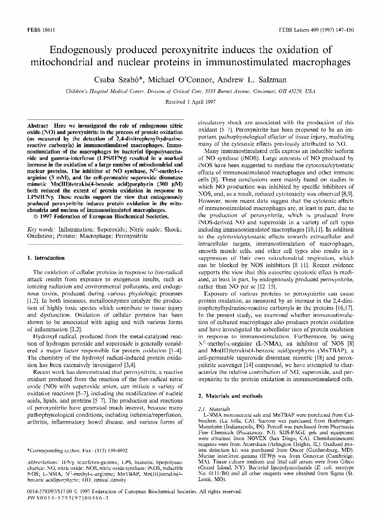
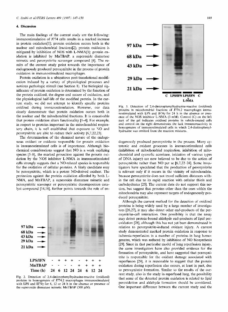
There was a significant increase in the quantity of oxidized proteins at 24 h after exposure to the combination of LPS and IFNy. Some increase in the oxidations of proteins of approx-imately 70 and 80 k D a were already detected at earlier time points (Figs. 1 and 2). However, at 24 h, a massive protein oxidation occurred. A significant port ion of the oxidized pro-teins was found in the regions corresponding to molecular masses greater than 97 k D a (Figs. 1 and 2).
3.2. Subcellular localization of oxidized proteins in immunostimulated cells
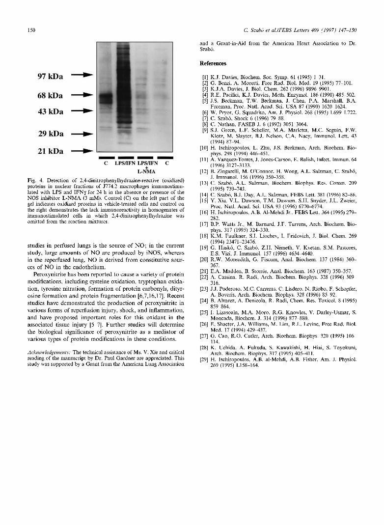
Both in the mitochondrial fraction (Fig. 3) and in the nu-clear fraction (Fig. 4) of immunostimulated cells, we have detected a marked increase in the oxidation of multiple pro-teins after exposure of LPS and I F N . In control (unstimu-lated) cells, there was also some protein oxidation of proteins of 50-60 k D a (Figs. 3 and 4).
3.3. An inhibitor of NO synthase and a Superoxide dismutase mimetic inhibit protein oxidation in immunostimulated cells
Inhibition of N O biosynthesis with the isoform-nonselective inhibitor L - N M A (3 m M ) significantly reduced the degree of protein oxidation in whole-cell homogenates (Fig. 1) and in mitochondrial and nuclear fractions (Figs. 3 and 4). Similar to the effect of L-NMA, the cell-permeable Superoxide dismutase mimetic M n T B A P (300 μΜ) reduced the extent of protein oxidation in immunostimulated cells, although its eifects, at this concentration, appeared to be somewhat less pronounced, in comparison to L - N M A (Figs. 1 and 2).
Fig. 1. Detection of 2,4-dinitrophenylhydrazine-reactive (oxidized) proteins in homogenates of J774.2 macrophages immunostimulated with LPS and IFNy for 6, 12 or 24 h in the absence or presence of the NOS inhibitor L-NMA (3 mM).
C. Szabó et al.lFEBS Letters 409 (1997) 147-150 149
4. Discussion
The main findings of the current study are the following: immunostimulation of J774 cells results in a marked increase in protein oxidation[l]; protein oxidation occurs both in the nuclear and mitochondrial fractions[2]; protein oxidation is mitigated by inhibition of NOS with L-NMA[3]; protein ox-idation is inhibited by MnTBAP, a Superoxide dismutase mimetic and peroxynitrite scavenger compound [4]. The re-sults of the current study point towards the importance of endogenously produced peroxynitrite in the process of protein oxidation in immunostimulated macrophages.
Protein oxidation is a ubiquitous post-translational modifi-cation induced by a variety of physiological processes and noxious pathologic stimuli (see Section 1). The biological sig-nificance of protein oxidation is determined by the function of the protein oxidized, the degree and nature of oxidation, and the physiological half-life of the modified protein. In the cur-rent study, we did not attempt to identify specific proteins oxidized during immunostimulation. However, our data clearly demonstrate that protein oxidation occurs both in the nuclear and the mitochondrial fractions. It is conceivable that protein oxidation alters functionality [1-4]. For example, in respect to proteins important in the mitochondrial respira-tory chain, it is well established that exposure to NO and peroxynitrite are able to reduce their activity [6,7,22,23].
The determination of the chemical nature of the endoge-nous oxidant or oxidants responsible for protein oxidation in immunostimulated cells is of importance. Although bio-chemical considerations suggest that NO is a weak oxidizing species [5-8], the marked protection against the protein oxi-dation by the NOS inhibitor L-NMA in immunostimulated cells strongly suggests that a NO-related species is responsible for the oxidation of cellular proteins. A likely candidate may be peroxynitrite, which is a potent NO-derived oxidant. The protection against the protein oxidation afforded by both L-NMA, and MnTBAP, a Superoxide dismutase mimetic and peroxynitrite scavenger or peroxynitrite decomposition cata-lyst compound [14,18], further points towards the role of en-
Fig. 2. Detection of 2,4-dinitrophenylhydrazine-reactive (oxidized) proteins in homogenates of J774.2 macrophages immunostimulated with LPS and IFNy for 6, 12 or 24 h in the absence or presence of the Superoxide dismutase mimetic MnTBAP (300 μΜ).
Fig. 3. Detection of 2,4-dinitrophenylhydrazine-reactive (oxidized) proteins in mitochondrial fractions of J774.2 macrophages immu-nostimulated with LPS and IFNy for 24 h in the absence or pres-ence of the NOS inhibitor L-NMA (3 mM). Control (C) on the left part of the gel indicates oxidized proteins in vehicle-treated cells and control on the right demonstrates the lack immunoreactivity in homogenates of immunostimulated cells in which 2,4-dinitrophenyl-hydrazine was omitted from the reaction mixtures.
dogenously produced peroxynitrite in the process. Many cy-totoxic and oxidant processes in immunostimulated cells (inhibition of mitochondrial respiration, inhibition of mito-chondrial and cytosolic aconitase, initiation of various types of DNA injury) are now believed to be due to the action of peroxynitrite rather than NO per se [6,7,22-24]. Some inves-tigators have speculated that the production of peroxynitrite is relevant only if it occurs in the vicinity of mitochondria, because peroxynitrite does not travel sufficient distances with-in the cell due to its rapid reaction with cellular thiols and carbohydrates [25]. The current data do not support this no-tion, but suggest that proteins other than the ones within the mitochondria may also represent targets of endogenously pro-duced peroxynitrite.
Although the current method for the detection of oxidized proteins is being widely used by a large number of investiga-tors [26,27], it may also detect other end-products of the per-oxynitrite-cell interaction. One possibility is that the assay may detect protein-bound aldehyde end-products of lipid per-oxidation [28], although this has not yet been demonstrated in relation to peroxynitrite-induced oxidant injury. A current study demonstrated marked protein oxidation in response to ischemia-reperfusion in a number of proteins in lung homo-genates, which was reduced by inhibition of NO biosynthesis [29]. Since in that particular model of lung reperfusion injury, the same investigators have also provided evidence for the formation of peroxynitrite, and have suggested that peroxyni-trite is responsible for the oxidant damage associated with reperfusion [29], it is reasonable to suggest that the protein oxidation during reperfusion also occurs, at least in part, due to peroxynitrite formation. Similar to the results of the cur-rent study, also in the study in reperfused lung, the possibility that some of the detected protein oxidation is related to lipid peroxidation and aldehyde formation should be considered. One important difference between the current study and the
150 C. Szabó et al.lFEBS Letters 409 (1997) 147-150
and a Grant-in-Aid from the American Heart Association to Dr. Szabó.
References
Fig. 4. Detection of 2,4-dinitrophenylhydrazine-reactive (oxidized) proteins in nuclear fractions of J774.2 macrophages immunostimu-lated with LPS and IFNy for 24 h in the absence or presence of the NOS inhibitor L-NMA (3 mM). Control (C) on the left part of the gel indicates oxidized proteins in vehicle-treated cells and control on the right demonstrates the lack immunoreactivity in homogenates of immunostimulated cells in which 2,4-dinitrophenylhydrazine was omitted from the reaction mixtures.
studies in perfused lungs is the source of N O ; in the current study, large amounts of N O are produced by iNOS, whereas in the reperfused lung, N O is derived from constitutive sour-ces of N O in the endothelium.
Peroxynitrite has been reported to cause a variety of protein modifications, including cysteine oxidation, t ryptophan oxida-tion, tyrosine nitration, formation of protein carbonyls, dityr-osine formation and protein fragmentation [6,7,16,17]. Recent studies have demonstrated the production of peroxynitrite in various forms of reperfusion injury, shock, and inflammation, and have proposed important roles for this oxidant in the associated tissue injury [5-7]. Fur ther studies will determine the biological significance of peroxynitrite as a mediator of various types of protein modifications in these conditions.
Acknowledgements: The technical assistance of Ms. V. Xie and critical reading of the manuscript by Dr. Paul Gardner are appreciated. This study was supported by a Grant from the American Lung Association
[10[
[11
[is:
[i3:
[14]
[is:
tie: [17
[is:
[i9:
per
[21
[22
[23:
[24]
[25
[26
[27
[28:
[29:
K.J. Davies, Biochem. Soc. Symp. 61 (1995) 1-31. G. Benzi, A. Moretti, Free Rad. Biol. Med. 19 (1995) 77-101. K.J.A. Davies, J. Biol. Chem. 262 (1996) 9896-9901. R.E. Pacifici, K.J. Davies, Meth. Enzymol. 186 (1990) 485-502. J.S. Beckman, T.W. Beckman, J. Chen, P.A. Marshall, B.A. Freeman, Proc. Nati. Acad. Sei. USA 87 (1990) 1620-1624. W. Pryor, G. Squadrito, Am. J. Physiol. 268 (1995) L699-L722. C. Szabó, Shock 6 (1996) 79-88. C. Nathan, FASEB J. 6 (1992) 3051-3064. S.J. Green, L.F. Scheller, M.A. Marietta, M.C. Seguin, F.W. Klotz, M. Slayter, B.J. Nelson, C.A. Nacy, Immunol. Lett. 43 (1994) 87-94. H. Ischiropoulos, L. Zhu, J.S. Beckman, Arch. Biochem. Bio-phys. 298 (1994) 446^151. A. Vazquez-Torres, J. Jones-Carson, E. Balish, Infect. Immun. 64 (1996) 3127-3133. B. Zingarelli, M. O'Connor, H. Wong, A.L. Salzman, C. Szabó, J. Immunol. 156 (1996) 350-358. C. Szabó, A.L. Salzman, Biochem. Biophys. Res. Comm. 209 (1995) 739-743. C. Szabó, B.J. Day, A.L. Salzman, FEBS Lett. 381 (1996) 82-86. Y. Xia, V.L. Dawson, T.M. Dawson, S.H. Snyder, J.L. Zweier, Proc. Nati. Acad. Sei. USA 93 (1996) 6770-6774. H. Ischiropoulos, A.B. Al-Mehdi Jr., FEBS Lett. 364 (1995) 279-282. B.P. Watts Jr., M. Barnard, J.F. Turrens, Arch. Biochem. Bio-phys. 317 (1995) 324-330. K.M. Faulkner, S.I. Liochev, I. Fridovich, J. Biol. Chem. 269 (1994) 23471-23476. G. Haskó, C. Szabó, Z.H. Németh, V. Kvetan, S.M. Pastores, E.S. Vizi, J. Immunol. 157 (1996) 4634-^1640. R.W. Moreadith, G. Fiscum, Anal. Biochem. 137 (1984) 360-367. E.A. Madden, B. Storrie, Anal. Biochem. 163 (1987) 350-357. A. Cassina, R. Radi, Arch. Biochem. Biophys. 328 (1996) 309-316. J.J. Poderoso, M.C. Carreras, C. Lisdero, N. Riobo, F. Schopfer, A. Boveris, Arch. Biochem. Biophys. 328 (1996) 85-92. B. Alvarez, A. Denicola, R. Radi, Chem. Res. Toxicol. 8 (1995) 859-864. I. Lizasoain, M.A. Moro, R.G. Knowles, V. Darley-Usmar, S. Moneada, Biochem. J. 314 (1996) 877-880. E. Shacter, J.A. Williams, M. Lim, R.L. Levine, Free Rad. Biol. Med. 17 (1994) 429-437. G. Cao, R.G. Cutler, Arch. Biochem. Biophys. 320 (1995) 106-114. K. Uchida, A. Fukuda, S. Kawakishi, H. Hiai, S. Toyokuni, Arch. Biochem. Biophys. 317 (1995) 405^111. H. Ischiropoulos, A.B. al-Mehdi, A.B. Fisher, Am. J. Physiol. 269 (1995) L158-164.