endovascular ascending repair: is this the next frontier? · michael d. dake, m.d. thelma and henry...
TRANSCRIPT

Department of Cardiothoracic Surgery, Stanford University School of Medicine
Endovascular Ascending Repair: Is This the Next Frontier?
Michael D. Dake, M.D. Thelma and Henry Doelger Professor
Department of Cardiothoracic Surgery Stanford University School of Medicine
Falk Cardiovascular Research Center
The 11th Houston Aortic Symposium Session II: Ascending Aorta/Aortic Dissection (Proximal)
February 15, 2018

Department of Cardiothoracic Surgery, Stanford University School of Medicine
Michael Dake, MD
• Research/Research Grants, Clinical Trial Support
– W. L. Gore
– Cook Medical
– Bard
– Novate
• Consulting Fees/Honoraria
– W. L. Gore
– Cook Medical
• Equity Interests/Stock Options
– Endoluminal Sciences
– Graftwork
– Intact
• Officer, Director, Board Member or other Fiduciary Role
– VIVA Physicians Group
• Speaker’s Bureau
– None
Within the past 12 months, the presenter or
their spouse/partner have had a financial interest/arrangement
or affiliation with the organization listed below.

Department of Cardiothoracic Surgery, Stanford University School of Medicine
Many ways to consider a response

Department of Cardiothoracic Surgery, Stanford University School of Medicine
Many ways to consider a response …but I only have 8 minutes

Department of Cardiothoracic Surgery, Stanford University School of Medicine
Control
Zone Of Complexity
Chaos
Agreement
Certainty + -
+
-

Department of Cardiothoracic Surgery, Stanford University School of Medicine

Department of Cardiothoracic Surgery, Stanford University School of Medicine
ASCENDING AORTIC ENDOGRAFTS Initial Examples (n=41)
• Type A IMH/Dissection: 10 (3 <14days)
• Trauma: 4
• Fistula to PA (iatrogenic): 1
• Mycotic Aneurysm: 10; fungal: 4 (transplant: single lung - 1, heart/lung - 2, and double lung - 1)
• Anastomotic Pseudoaneurysm: 16 (heart transplant: 3 )

Department of Cardiothoracic Surgery, Stanford University School of Medicine
CHALLENGES POSED BY THE ASCENDING AORTA
Anatomy • Stent parking place (shorter)
• Curved geometry
• Non-cylindrical shapes
• Large diameter
• Neck mismatch frequent (taper, reverse taper)
• Branches (coronaries, innominate, bypass grafts)

Department of Cardiothoracic Surgery, Stanford University School of Medicine
CHALLENGES POSED BY THE ASCENDING AORTA
Physiology • Increased flow
• Enhanced compliance--dynamic aortic deformation
• Proximal aortic movement
• Cardiac dynamics
• Respiratory motion

Department of Cardiothoracic Surgery, Stanford University School of Medicine
86-year-old woman with acute type A dissection

Department of Cardiothoracic Surgery, Stanford University School of Medicine
LCA
RBCA
LCA
Acute Type A Dissection
CTA post stent-graft

Department of Cardiothoracic Surgery, Stanford University School of Medicine
Follow-up at 6 weeks

Department of Cardiothoracic Surgery, Stanford University School of Medicine
Now, @ 90-years-old, woman who underwent stent-graft
placement in ascending aorta three years ago to manage
type A dissection

Department of Cardiothoracic Surgery, Stanford University School of Medicine
Axial scans @ 3 years
Peri-graft opacification at root

Department of Cardiothoracic Surgery, Stanford University School of Medicine
3-year CT follow-up

Department of Cardiothoracic Surgery, Stanford University School of Medicine
CT @ 3 years s/p endograft

Department of Cardiothoracic Surgery, Stanford University School of Medicine
Proximal “bird beak”

Department of Cardiothoracic Surgery, Stanford University School of Medicine
The left coronary’s friend

Department of Cardiothoracic Surgery, Stanford University School of Medicine
Proximal “bird beak”

Department of Cardiothoracic Surgery, Stanford University School of Medicine

Department of Cardiothoracic Surgery, Stanford University School of Medicine
Distal “bird beak”

Department of Cardiothoracic Surgery, Stanford University School of Medicine
67-year-old woman, who is a devoted Jehovah’s Witness, with a history of hypertension presents to outside facility
with acute onset of chest pain. CT scan diagnostic for focal type A aortic dissection with extensive IMH.
Upon transfer to Stanford, she has an episode of confusion,
expressive aphasia and right facial droop when blood pressure pharmacologically lowered to SBP <110mmHg. Neurologic exam
improves to baselinewith SBP >120. CT/CTA/CTP shows short segment severe stenosis in a superior division left M2 branch associated with perfusion delay, but no stroke core. Neuro IR
study and catheter-directed tPA, but no PTA or stent.

Department of Cardiothoracic Surgery, Stanford University School of Medicine

Department of Cardiothoracic Surgery, Stanford University School of Medicine

Department of Cardiothoracic Surgery, Stanford University School of Medicine

Department of Cardiothoracic Surgery, Stanford University School of Medicine

Department of Cardiothoracic Surgery, Stanford University School of Medicine

Department of Cardiothoracic Surgery, Stanford University School of Medicine

Department of Cardiothoracic Surgery, Stanford University School of Medicine

Department of Cardiothoracic Surgery, Stanford University School of Medicine

Department of Cardiothoracic Surgery, Stanford University School of Medicine

Department of Cardiothoracic Surgery, Stanford University School of Medicine

Department of Cardiothoracic Surgery, Stanford University School of Medicine

Department of Cardiothoracic Surgery, Stanford University School of Medicine

Department of Cardiothoracic Surgery, Stanford University School of Medicine

Department of Cardiothoracic Surgery, Stanford University School of Medicine
48 hours post-procedure, Hgb 4.5. Upper GI endoscopy performed 2 days later. Exam significant
for duodenal ulcer with active bleeding – 5 clips placed. Patient becomes progressively somnolent and hypotensive –
expires 18 hours later.

Department of Cardiothoracic Surgery, Stanford University School of Medicine
89-year-old man in relatively good health; lives independently, s/p bio-prosthetic aortic valve in 2012. At that time, surgeon described small focal dissection
at cannula site that was ”tacked down” during operation. Now with follow-up CT 4 years later.

Department of Cardiothoracic Surgery, Stanford University School of Medicine

Department of Cardiothoracic Surgery, Stanford University School of Medicine
Small lung nodule noted and follow-up CT scan performed 10 months later. He is asymptomatic.

Department of Cardiothoracic Surgery, Stanford University School of Medicine

Department of Cardiothoracic Surgery, Stanford University School of Medicine
CT follow-up 8 months later. He remains asymptomatic

Department of Cardiothoracic Surgery, Stanford University School of Medicine

Department of Cardiothoracic Surgery, Stanford University School of Medicine

Department of Cardiothoracic Surgery, Stanford University School of Medicine
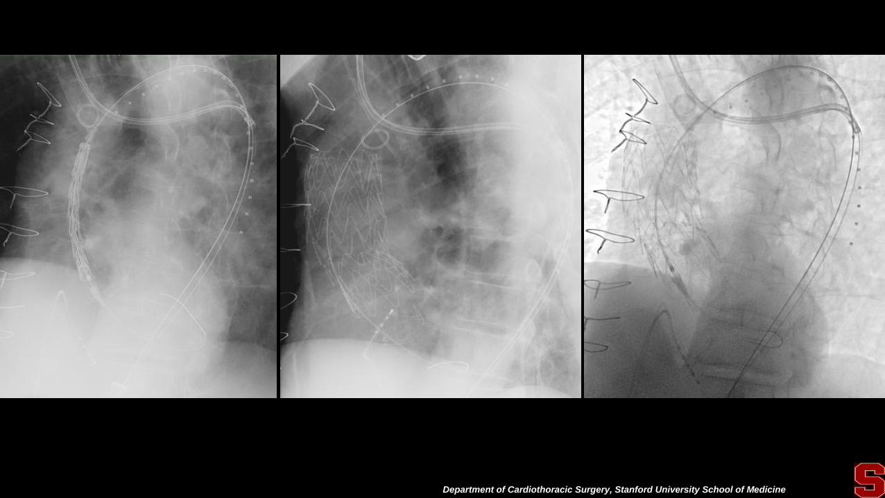
Referred for possible endovascular management. Repeat CT scan 8 weeks later prior to consultation

Department of Cardiothoracic Surgery, Stanford University School of Medicine

Department of Cardiothoracic Surgery, Stanford University School of Medicine

Department of Cardiothoracic Surgery, Stanford University School of Medicine

Department of Cardiothoracic Surgery, Stanford University School of Medicine

Department of Cardiothoracic Surgery, Stanford University School of Medicine

Department of Cardiothoracic Surgery, Stanford University School of Medicine

Department of Cardiothoracic Surgery, Stanford University School of Medicine
Department of Cardiothoracic Surgery, Stanford University School of Medicine

Department of Cardiothoracic Surgery, Stanford University School of Medicine

Department of Cardiothoracic Surgery, Stanford University School of Medicine

Department of Cardiothoracic Surgery, Stanford University School of Medicine

Department of Cardiothoracic Surgery, Stanford University School of Medicine

Department of Cardiothoracic Surgery, Stanford University School of Medicine

Department of Cardiothoracic Surgery, Stanford University School of Medicine

Department of Cardiothoracic Surgery, Stanford University School of Medicine
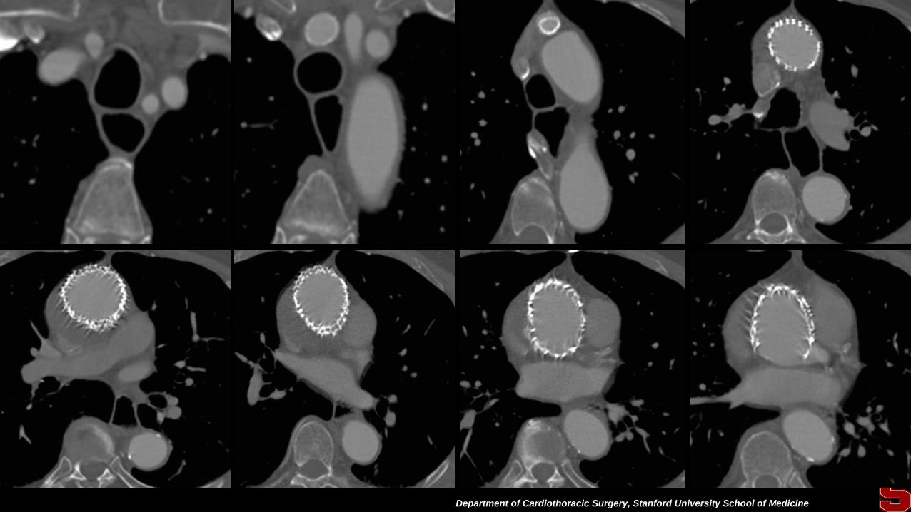
CT scan 1 month after stent-graft placement

Department of Cardiothoracic Surgery, Stanford University School of Medicine

Department of Cardiothoracic Surgery, Stanford University School of Medicine

Department of Cardiothoracic Surgery, Stanford University School of Medicine

Department of Cardiothoracic Surgery, Stanford University School of Medicine

Department of Cardiothoracic Surgery, Stanford University School of Medicine

Department of Cardiothoracic Surgery, Stanford University School of Medicine
Department of Cardiothoracic Surgery, Stanford University School of Medicine
2 months later

Department of Cardiothoracic Surgery, Stanford University School of Medicine
Department of Cardiothoracic Surgery, Stanford University School of Medicine

Department of Cardiothoracic Surgery, Stanford University School of Medicine

Department of Cardiothoracic Surgery, Stanford University School of Medicine

Department of Cardiothoracic Surgery, Stanford University School of Medicine

Department of Cardiothoracic Surgery, Stanford University School of Medicine

Department of Cardiothoracic Surgery, Stanford University School of Medicine
ASCENDING AORTIC ENDOGRAFTS Consideration of Ideal Device Attributes
• Graft prosthesis – Extremely conformable (especially proximally)
– Variety of lengths, diameters, tapers, reverse tapers, etc.
– Potential for pre-curved or pre-shaped devices
– Migration resistant
– Capacity for branch fenestrations (proximal, distal, both) and/or branch opportunities (distally for arch branches)
• Delivery system – Short, well-transitioned tip
– Very flexible shaft with centering capabilty pre-deployment
– Short and long catheter lengths (trans-apical, arch or femoral introduction)
– Final release after partial graft expansion
– Graft re-constraining/re-positioning capability

Department of Cardiothoracic Surgery, Stanford University School of Medicine
My idiosyncratic view of what is missing from current devices and underappreciated as an important
consideration to better address both the potent physiological and anatomical challenges we face in
the ascending aorta…

Department of Cardiothoracic Surgery, Stanford University School of Medicine
My idiosyncratic view of what is missing from current devices and underappreciated as an important
consideration to better address both the potent physiological and anatomical challenges we face in
the ascending aorta…
Enhanced Conformability

Department of Cardiothoracic Surgery, Stanford University School of Medicine
Enhanced Conformability
(think, truck bed liner)

Department of Cardiothoracic Surgery, Stanford University School of Medicine
Enhanced Conformability (think, truck bed liner)
and…

Department of Cardiothoracic Surgery, Stanford University School of Medicine
Enhanced Conformability (think, truck bed liner)
and… true orthogonal deployment at the STJ

Department of Cardiothoracic Surgery, Stanford University School of Medicine

Department of Cardiothoracic Surgery, Stanford University School of Medicine
Ascending Device
Type A Dissection Low profile 16-20 Fr Controlled deployment Cover entry tear Promote false lumen thrombosis Promote aortic remodeling
*Picture courtesy of Matt Thompson- London

Department of Cardiothoracic Surgery, Stanford University School of Medicine

Department of Cardiothoracic Surgery, Stanford University School of Medicine
Curing Aortic Dissection: Where to Next?

Department of Cardiothoracic Surgery, Stanford University School of Medicine
Gore Type A Dissection Early Feasibility Study
• The Gore 14-02 Type A EFS is the first industry sponsored FDA approved trial for endovascular repair in the ascending aorta.
• Focused goal of this EFS is to assess the feasibility of endovascular repair of Type A dissections. • Early clinical learnings will inform
the evolution of the clinical treatment and design of devices for ascending dissections.

Department of Cardiothoracic Surgery, Stanford University School of Medicine
Gore Type A EFS Overview
• Study Population: DeBakey Type I/II Dissection • Approved for up to 10 patients
• 3 sites enrolling
• Primary treatment suing the TBE Aortic Extenders to stabilize the length of the dissected ascending aorta
Site Principal
Investigator
Houston Methodist
Hospital
Jean Bismuth, MD
Memorial Hermann Heart
& Vascular Institute
Anthony Estrera,
MD
St. Luke’s Health Baylor Joseph Coselli, MD
National Principal Investigator:
Michael Reardon, MD
Houston Methodist
Gore Investigational Device

Department of Cardiothoracic Surgery, Stanford University School of Medicine
Evaluation of the GORE® TAG® Thoracic Branch Endoprosthesis in the Treatment of DeBakey Type I/II Aortic Dissection
• The complete GORE® TAG® TBE system is available for further distal repair of the Type A dissection as needed
Gore Investigational Device

Department of Cardiothoracic Surgery, Stanford University School of Medicine

Department of Cardiothoracic Surgery, Stanford University School of Medicine

Department of Cardiothoracic Surgery, Stanford University School of Medicine
Conclusion • Our appreciation of the variety and complexity
posed by thoracic aortic pathologies has out paced our technical sophistication and ability to develop endovascular solutions.
• This is especially true in the ascending aorta and aortic arch where unique anatomical and physiological factors present special challenges.
• Now, we are poised to tackle the next frontier with purpose built endovascular grafts designed to address these challenges

Thank you

