engineered commensal bacteria reprogram...
TRANSCRIPT

Franklin F. Duan, Joy H. Liu, and John C. March
Engineered CommensalBacteria Reprogram IntestinalCells Into Glucose-ResponsiveInsulin-Secreting Cells for theTreatment of DiabetesDOI: 10.2337/db14-0635
The inactive full-length form of GLP-1(1–37) stimulatesconversion of both rat and human intestinal epithelial cellsinto insulin-secreting cells. We investigated whether oraladministration of human commensal bacteria engineeredto secrete GLP-1(1–37) could ameliorate hyperglycemia ina rat model of diabetes by reprogramming intestinal cellsinto glucose-responsive insulin-secreting cells. Diabeticrats were fed daily with human lactobacilli engineered tosecrete GLP-1(1–37). Diabetic rats fed GLP-1-secretingbacteria showed significant increases in insulin levelsand, additionally, were significantly more glucose tolerantthan those fed the parent bacterial strain. These rats de-veloped insulin-producing cells within the upper intestinein numbers sufficient to replace ∼25–33% of the insulincapacity of nondiabetic healthy rats. Intestinal tissues inrats with reprogrammed cells expressed MafA, PDX-1,and FoxA2. HNF-6 expression was observed only in cryptepithelia expressing insulin and not in epithelia locatedhigher on the villous axis. Staining for other cell markersin rats treated with GLP-1(1–37)-secreting bacteria sug-gested that normal function was not inhibited by the closephysical proximity of reprogrammed cells. These resultsprovide evidence of the potential for a safe and effectivenonabsorbed oral treatment for diabetes and support theconcept of engineered commensal bacterial signaling tomediate enteric cell function in vivo.
Reprogramming non-b-cells into b-cells or cells with insulin-secreting potential has been the subject of several studies
over the past decade (1–9). Research has focused on a num-ber of areas, including in vitro generation of b-cells frompancreatic (e.g., acinar cells) and liver cell lineages for trans-plantation as well as causing either pancreatic or othertissue-specific cells to convert to b-cells in vivo (10). Thepotential of this latter approach became evident with thediscovery by Suzuki et al. (11) that the full-length formof GLP-1(1–37), previously thought to be inactive, couldstimulate rat intestinal epithelial cells to become glucose-responsive insulin-secreting cells, ostensibly through theNotch signaling pathway. The Suzuki et al. (11) results sug-gested that undifferentiated intestinal epithelia in rats (dif-ferentiation occurring after E15) can develop into b-likecells. The study also demonstrated the reversal of strepto-zotocin (STZ)-induced type 1 diabetes in adult rats aftersurgical implantation with embryonic jejunum (E14.5) incu-bated with GLP-1(1–37) in vitro. The authors concludedthat adult enterocyte differentiation, which occurs fromthe intestinal crypts, would not give rise to significant num-bers of insulin-producing cells and that the proliferating andpseudostratified cells of the developing fetus (pre-E17)would likely be required for significant differentiation intocells with b-like functionality.
While the Suzuki et al. (11) reported positive resultswith GLP-1(1–37) as an agent to reprogram intestinalcells, their study also highlighted the difficulty in deliver-ing this bioactive compound by injection and surgery. Thecirculating active form of GLP-1 is GLP-1(7–37), whichhas a very short biological half-life of the order of just
Department of Biological and Environmental Engineering, Cornell University,Ithaca, NY
Corresponding author: John C. March, [email protected].
Received 22 April 2014 and accepted 10 December 2014.
This article contains Supplementary Data online at http://diabetes.diabetesjournals.org/lookup/suppl/doi:10.2337/db14-0635/-/DC1.
© 2015 by the American Diabetes Association. Readers may use this article aslong as the work is properly cited, the use is educational and not for profit, andthe work is not altered.
Diabetes 1
PHARMACOLOGYAND
THERAPEUTIC
S
Diabetes Publish Ahead of Print, published online January 27, 2015
a few minutes in blood (12). This short half-life may bea reason for the lower reprogramming rates with GLP-1(1–37) observed in adult rats, as it would be necessary forGLP-1(1–37) to be present in systemic circulation fora longer period of time in order to reach the intestinalcrypts.
Other means of delivering bioactive compounds to theluminal (villous) side of the upper intestine, avoidingthe potential pitfalls of surgery or degradation in thebloodstream, have been published using intestinal com-mensal bacteria that populate the gut with the ability tosecrete specific signals (13–24). In this approach, signals(small molecules, peptides) can be delivered directly tothe luminal side of the intestine by bacteria that alreadyhave an established line of communication with intestinalepithelia.
In a previous in vitro study, we demonstrated thatengineered commensal bacteria can deliver GLP-1(1–37)to human intestinal carcinomas and stimulate glucose-responsive insulin secretion (23). In that work, Escherichiacoli Nissle 1917 was transformed to secrete GLP-1(1–37)from a plasmid in response to an exogenous inducer. Wealso confirmed that GLP-1(1–37) and not the active form(GLP-1[7–37]) reprograms enterocytes as part of the workbeing reported here (Supplementary Fig. 1).
Further, for this investigation, we tested the hypoth-esis that a chromosomally modified human gram-positivebacterial strain that constitutively secretes GLP-1(1–37)could reduce hyperglycemia in a rat model of diabetes.Our goal was to reprogram rat intestinal cells intoglucose-responsive insulin-secreting cells through dailyoral administration of GLP-1(1–37)-secreting bacteria. Wemeasured coexpression of b-cell and enteroendocrinemarkers to determine the extent and possible mechanismof reprogramming.
RESEARCH DESIGN AND METHODS
Strain ConstructionTo transform Lactobacillus gasseri ATCC 33323 (L) intoa strain that secretes GLP-1(1–37), constitutively stan-dard techniques were used. Details are in the Supplemen-tary Data. We called the positive integrants LG.
Rat ExperimentsAll rats used in this study were purchased from JacksonLaboratory and housed at the East Campus ResearchFacility at Cornell University. Studies were conducted inaccordance with protocols approved by the CornellUniversity Institutional Animal Care and Use Committee.All rat experiments were repeated twice with six rats pertreatment group.
STZ ModelRats were fasted for 6 h before being injected with STZ ata dose of 70 mg/kg of body weight (BW; in cold 0.1 mol/Lsodium citrate buffer pH 4.2) via intraperitoneal route.Blood glucose levels were monitored every 3 days until
diabetic glucose levels (.350 mg/dL) were reached. Oncerats had sustained blood glucose levels over 350 mg/dL,they were enrolled in the study.
Bacterial FeedingAfter enrollment in the study, rats were given ampicillin-treated (1 g/L) water for 18 h. Lactobacillus strains LG(2 mg/mL erythromycin) or L were grown in MRS media.The resulting pellet was redissolved in sterile MRS with1% sucrose. The rats were fed 1.6 mL/kg BW MRS with1% sucrose containing 1010 CFU/mL of MRS-grownLactobacillus strains separately (L or LG). All bacterial-strain-fed rats were fed 23 per day with bacteria for 90 days.
Glucose Tolerance Test and ELISAAfter bacterial feeding for 51 days, STZ-treated rats werefasted 10 h and weighed, and a blood sample was collectedfrom the tail vein using heparinized Micro-HematocritCapillary Tubes (Fisher, PA). They were then orally ad-ministered 1 g glucose/kg BW, and blood samples weretaken at 0.5, 1, 1.5, and 2 h. Plasma glucose was measuredusing the Breeze 2 blood glucose monitoring system.Plasma insulin was measured using Rat/Mouse InsulinELISA Kit (Millipore, MA) according to the manufacturer’sinstructions.
Tissue Homogenization of Pancreas and UpperIntestine, Blood Sampling, and ELISAAfter 90 days of bacterial feeding, rats were killed andtheir pancreata and intestines were removed and frozenat 280°C until analysis. For each rat, the pancreas andupper intestine were weighed and independently homog-enized using a chilled mortar with 13 PBS. Insulin wasmeasured for both pancreata and upper intestines usingRat/Mouse Insulin ELISA Kit (Millipore, MA) according tothe manufacturer’s instructions.
Bacterial Colonization CountsAfter feeding for 90 days, rats were transferred to newcages for 3 days. Feces were collected from the new cages,and rats were killed. Three rats from each treatment weredissected, and their upper GI (gastrointestinal) tractswere removed. The upper GI tracts were each weighed andhomogenized in 2 mL of fresh MRS medium. Homoge-nized tissue was plated onto MRS with erythromycin(2 mg/mL) by serial dilution. Plates were incubated over-night at 37°C and their colonies counted.
ImmunofluorescenceUpper intestinal, pancreatic, and liver tissues of rats killedafter feeding with L or LG for 90 days and treated IEC-6cells were fixed, and tissues were dissected. After depar-affinization, fixed tissue slides were steamed in 0.01 mol/Lcitrate buffer, pH 6.0. After washing in PBS, 10% normalblocking serum (Santa Cruz Biotechnology, CA) wasapplied for 1 h at room temperature. To treated IEC-6cells in six-well plates were added 200 mL 4% formalde-hyde diluted in warm PBS for 15 min in a fume hood. The
2 Engineered Probiotics for Treating Diabetes Diabetes
cells were washed three times in 13 PBS for 5 min each,and 10% normal blocking serum (Santa Cruz Biotechnology,CA) was applied for 1 h at room temperature. For alltissues and IEC-6 cells, 1:500 diluted goat anti-PDX-1,1:50 diluted genuine pig anti-insulin, 1:50 mouse anti-GLP-1 (ab23447; Abcam, Cambridge, MA), 1:50 dilutedrabbit anti-insulin, 1:500 rabbit anti-PDX-1, 1:50 rabbitanti-HNF 6, 1:50 goat anti-chromogranin A (ChrA), 1:50goat antilysozyme, 1:50 goat anti-SOX-9, 1:50 goat anti-sucrase isomaltase (anti-SI), 1:50 goat antiglucagon, or1:50 goat anti-FoxA2 (Santa Cruz Biotechnology, CA)was applied to blocked samples that were then incubatedovernight at 4°C. After 43 washing in PBS, a fluoro-chrome-conjugated secondary antibody Alexa Fluor 488donkey anti-rabbit IgG and Alexa Fluor 555 donkeyanti-goat IgG or Alexa Fluor 488 donkey anti-goat IgGor Alexa Fluor 488 goat anti-genuine pig IgG and AlexaFluor 555 donkey anti-rabbit IgG or Alexa Fluor 488 don-key anti-mouse IgG (Invitrogen) diluted 1:200 in PBS wasapplied to samples for 1.5 h at room temperature in a hu-mid chamber. After 33 washing in PBS, samples werethen mounted with Vectshield mounting medium withDAPI (Vector, CA). Specimens were examined immedi-ately using the appropriate excitation wavelength foreach fluorophore. Images were taken with a Zeiss 710Confocal Microscope (Zeiss, Jena, Germany).
RT-PCRTotal RNA was isolated from the intestines of rats fedwith LG or L and also from the pancreases and liversof healthy control rats using the RNAqueous Kit (LifeTechnologies, NY). cDNA was synthesized by SuperScriptIII First-Strand Kit (Invitrogen, CA). The primers used foramplifying different genes are listed in the SupplementaryData.
IEC6 Cell Culture With GLP-1(1–37) and SmallInterfering RNA and ELISASixty percent confluent monolayers of IEC-6 cells in four-chamber culture slides were covered with 1 mL DMEMwith 10% FBS and 10 mg/mL insulin incubated at 37°Cwith 5% CO2. Small interfering RNA (siRNA) transfectionwere processed according to manufacturer’s manual. After24 h, 200 nmol/L, 400 nmol/L, or 2 mmol/L GLP-1(1–37)(Bachem, King of Prussia, PA) or 13 PBS was added sep-arately into different wells. Following a 16-h incubation,cells were washed with DMEM with 10% FBS three times,and 1 mL DMEM with 10% FBS plus 200 nmol/L, 400nmol/L, or 2 mmol/L GLP-1(1–37) or GLP-1(7–37) wasadded to the cells, supplemented with 0.4% glucose foran additional 2 h. The media was removed from the cells,supplemented with leupeptin (10 ng/mL), 0.2 mmol/Lphenylmethylsulfonyl fluoride, and aprotinin (10 ng/mL);centrifuged (12,0003 rpm; Effendorf 5804R, Westbury,NY); and kept briefly at 4°C prior to ELISA analysis forinsulin expression. Insulin was measured using Rat/MouseELISA Kit (Millipore, MA) according to the manufacturer’s
instructions. The treated IEC-6 cells were used for RT-PCRor immunofluorescence (see immunofluorescence and RT-PCR in the RESEARCH DESIGN AND METHODS section) for specificgene expression.
Statistical AnalysisOral glucose tolerance test (OGTT) and nonfastingglucose data were compared using area under the curve(AUC) estimates over the course of the time periodspecified. Specifically, AUC data for rats under differentconditions were grouped (LG or L) and compared withcontrol rats using one-way ANOVA, with Dunnet testused to determine significant differences. Differenceswere considered significant at a , 0.05 (n = 6).
RESULTS
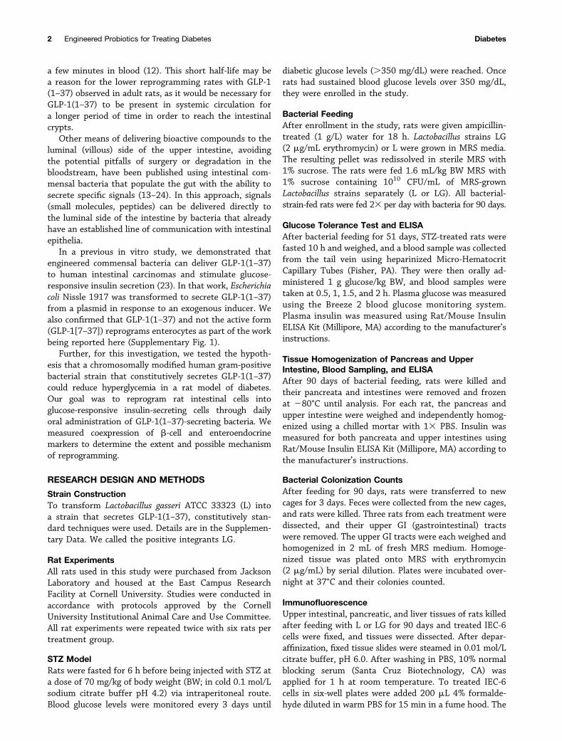
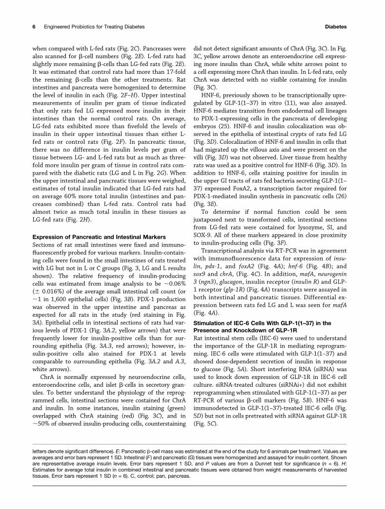
Bacterial Secretion of GLP-1(1–37) in Rat UpperIntestinal TractsLactobacillus gasseri (L) was engineered to secrete GLP-1(1–37) using the SlpA promoter and USP45-LEISS se-cretion tag (SEC) (19) (LG) (Fig. 1A). A polyhistidine(HIS) tag was added to the N-terminus (for Westernblotting) separated by an enterokinase site so that theprotein would be separated from the tag once secretedin the intestine. Secretion of GLP-1(1–37) was verifiedin culture (Fig. 1B). The secreted fraction of GLP-1(1–37) was recovered from the culture medium (medium)and compared with that which was still in the cell pellet(cells).
Rats were fed 23 per day with L, LG, or sterile mediafor 90 days. At the end of the experiment, individual ratintestines were homogenized and bacterial counts wereperformed in order to establish the level of colonizationin the upper GI tract and to compare that to counts inthe feces. From these data, it is clear that bacterialstrains colonized the intestines (Fig. 1C). There was nodifference between L and LG in colonizing the feces (Fig.1C.2). It should be noted that L did not carry antibioticresistance, hence the counts are much higher than LGdue to the lack of selective pressure. To count totalamounts of Lactobacillus present in the rat upper intes-tines, homogenates were plated on MRS plates whichselect (without antibiotics) predominately for Lactobacil-lus strains. For rats fed LG or L, the counts were almostidentical, indicating that both strains did little to changethe total number of lactobacilli in the homogenates. Toselect only for LG, erythromycin was used as a selectablemarker (Fig. 1C). The lower counts on erythromycinplates, as compared with the MRS plates, indicated thedifference in colonization between culturable native ratlactobacilli and LG.
Diabetic rats that were fed LG stained positively forGLP-1(1–37) in their upper intestines as revealed by im-munofluorescence using antibodies that react specificallywith the first six amino acids of GLP-1 (Fig. 1D, LG resultsare shown), while rats fed either L or buffer did not
diabetes.diabetesjournals.org Duan, Liu, and March 3
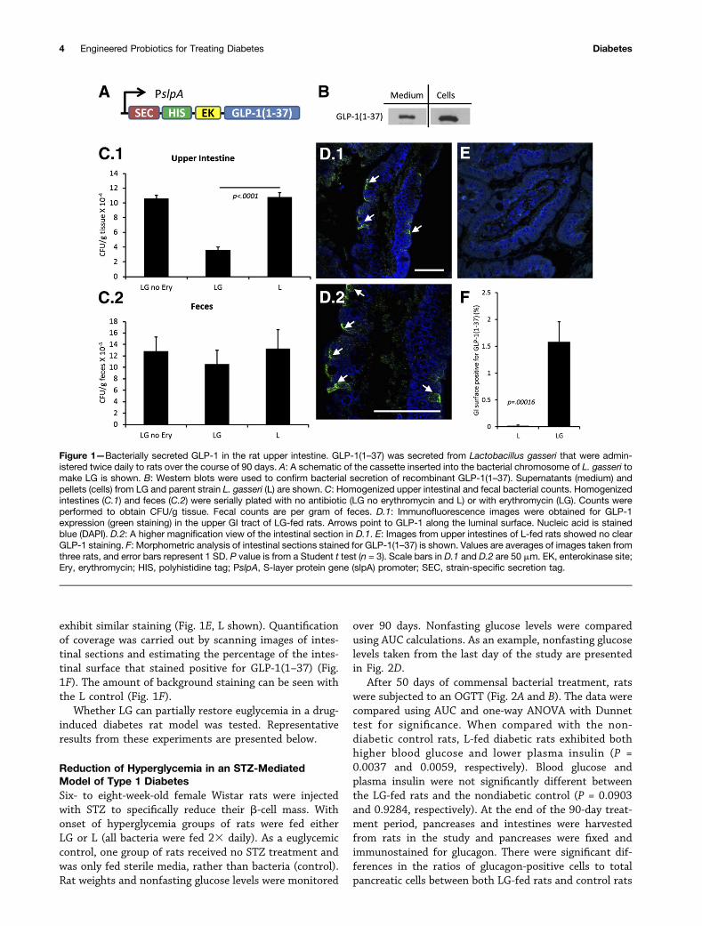
exhibit similar staining (Fig. 1E, L shown). Quantificationof coverage was carried out by scanning images of intes-tinal sections and estimating the percentage of the intes-tinal surface that stained positive for GLP-1(1–37) (Fig.1F). The amount of background staining can be seen withthe L control (Fig. 1F).
Whether LG can partially restore euglycemia in a drug-induced diabetes rat model was tested. Representativeresults from these experiments are presented below.
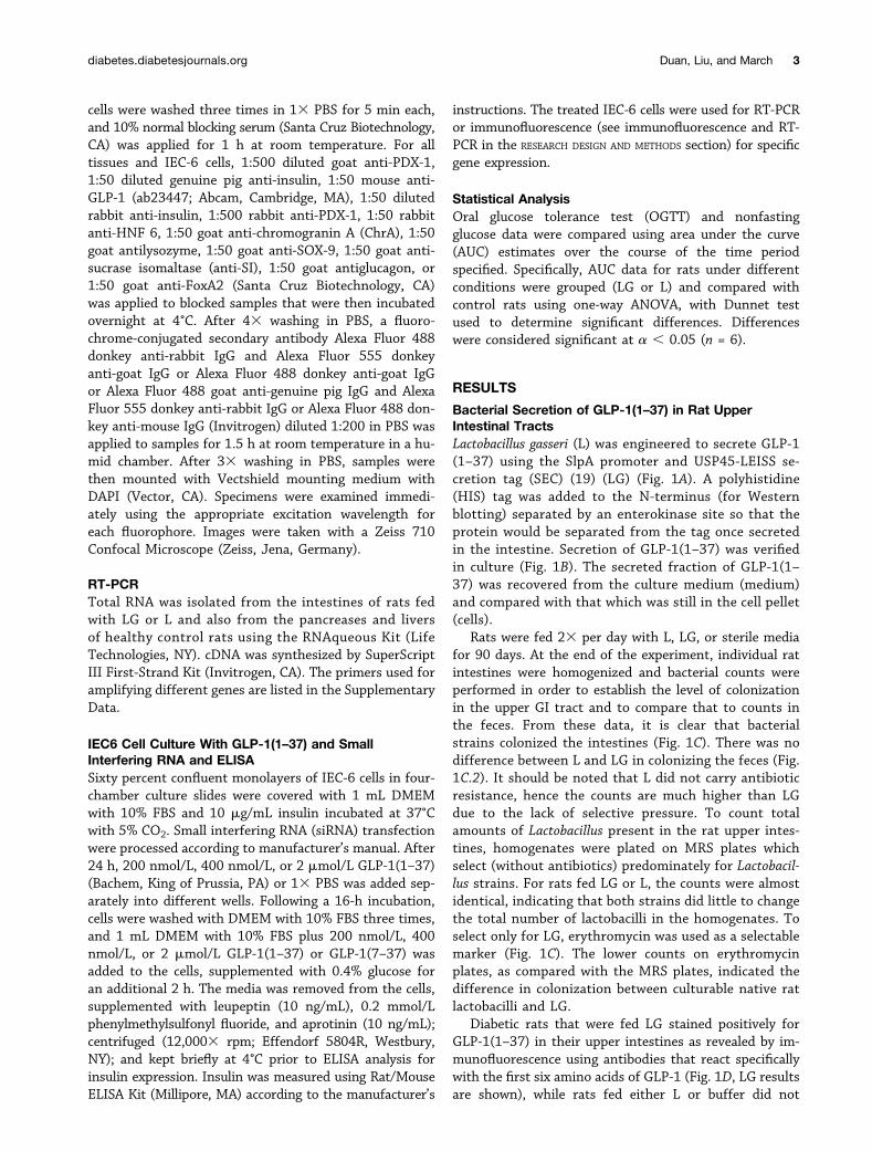
Reduction of Hyperglycemia in an STZ-MediatedModel of Type 1 DiabetesSix- to eight-week-old female Wistar rats were injectedwith STZ to specifically reduce their b-cell mass. Withonset of hyperglycemia groups of rats were fed eitherLG or L (all bacteria were fed 23 daily). As a euglycemiccontrol, one group of rats received no STZ treatment andwas only fed sterile media, rather than bacteria (control).Rat weights and nonfasting glucose levels were monitored
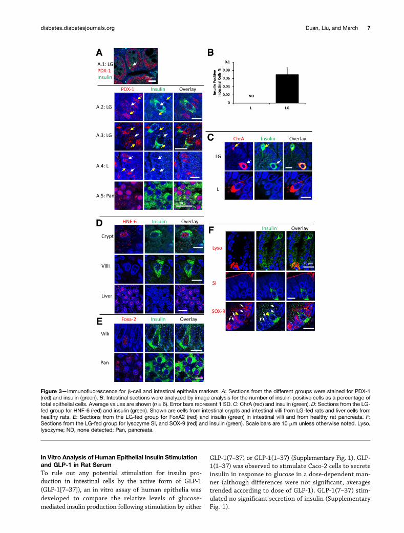
over 90 days. Nonfasting glucose levels were comparedusing AUC calculations. As an example, nonfasting glucoselevels taken from the last day of the study are presentedin Fig. 2D.
After 50 days of commensal bacterial treatment, ratswere subjected to an OGTT (Fig. 2A and B). The data werecompared using AUC and one-way ANOVA with Dunnettest for significance. When compared with the non-diabetic control rats, L-fed diabetic rats exhibited bothhigher blood glucose and lower plasma insulin (P =0.0037 and 0.0059, respectively). Blood glucose andplasma insulin were not significantly different betweenthe LG-fed rats and the nondiabetic control (P = 0.0903and 0.9284, respectively). At the end of the 90-day treat-ment period, pancreases and intestines were harvestedfrom rats in the study and pancreases were fixed andimmunostained for glucagon. There were significant dif-ferences in the ratios of glucagon-positive cells to totalpancreatic cells between both LG-fed rats and control rats
Figure 1—Bacterially secreted GLP-1 in the rat upper intestine. GLP-1(1–37) was secreted from Lactobacillus gasseri that were admin-istered twice daily to rats over the course of 90 days. A: A schematic of the cassette inserted into the bacterial chromosome of L. gasseri tomake LG is shown. B: Western blots were used to confirm bacterial secretion of recombinant GLP-1(1–37). Supernatants (medium) andpellets (cells) from LG and parent strain L. gasseri (L) are shown. C: Homogenized upper intestinal and fecal bacterial counts. Homogenizedintestines (C.1) and feces (C.2) were serially plated with no antibiotic (LG no erythromycin and L) or with erythromycin (LG). Counts wereperformed to obtain CFU/g tissue. Fecal counts are per gram of feces. D.1: Immunofluorescence images were obtained for GLP-1expression (green staining) in the upper GI tract of LG-fed rats. Arrows point to GLP-1 along the luminal surface. Nucleic acid is stainedblue (DAPI). D.2: A higher magnification view of the intestinal section in D.1. E: Images from upper intestines of L-fed rats showed no clearGLP-1 staining. F: Morphometric analysis of intestinal sections stained for GLP-1(1–37) is shown. Values are averages of images taken fromthree rats, and error bars represent 1 SD. P value is from a Student t test (n = 3). Scale bars in D.1 and D.2 are 50 mm. EK, enterokinase site;Ery, erythromycin; HIS, polyhistidine tag; PslpA, S-layer protein gene (slpA) promoter; SEC, strain-specific secretion tag.
4 Engineered Probiotics for Treating Diabetes Diabetes
Figure 2—Reducing hyperglycemia in diabetic rats. Hyperglycemia was induced in Wistar rats via STZ depletion of pancreatic b-cells. Aftertreatment with L. gasseri (L) and L. gasseri-secreting GLP-1(1–37) (LG) rats were subjected to an OGTT. One group of rats was not treatedwith STZ and fed sterile media rather than a bacterial suspension (control). A: Blood glucose and (B) insulin levels were measured for theOGTT. Presented are averages for each treatment group in a single experiment (n = 6). Statistical analysis was performed using one-wayANOVA for AUC measurements. and results are discussed in the text. C: Pancreatic sections from each treatment group were morpho-metrically analyzed for the ratio of glucagon-positive cells to total pancreatic cells. D: Random (nonfasting) blood glucose levels wereobtained for all rats in the study 23 per week. Shown are representative average blood glucose levels from one day of an experiment foreach group (n = 6). Error bars represent 1 SD and significant differences (a = 0.05) are denoted by letters over each value (i.e., different
diabetes.diabetesjournals.org Duan, Liu, and March 5
when compared with L-fed rats (Fig. 2C). Pancreases werealso scanned for b-cell numbers (Fig. 2E). L-fed rats hadslightly more remaining b-cells than LG-fed rats (Fig. 2E).It was estimated that control rats had more than 17-foldthe remaining b-cells than the other treatments. Ratintestines and pancreata were homogenized to determinethe level of insulin in each (Fig. 2F–H). Upper intestinalmeasurements of insulin per gram of tissue indicatedthat only rats fed LG expressed more insulin in theirintestines than the normal control rats. On average,LG-fed rats exhibited more than fivefold the levels ofinsulin in their upper intestinal tissues than either L-fed rats or control rats (Fig. 2F). In pancreatic tissue,there was no difference in insulin levels per gram oftissue between LG- and L-fed rats but as much as three-fold more insulin per gram of tissue in control rats com-pared with the diabetic rats (LG and L in Fig. 2G). Whenthe upper intestinal and pancreatic tissues were weighed,estimates of total insulin indicated that LG-fed rats hadon average 60% more total insulin (intestines and pan-creases combined) than L-fed rats. Control rats hadalmost twice as much total insulin in these tissues asLG-fed rats (Fig. 2H).
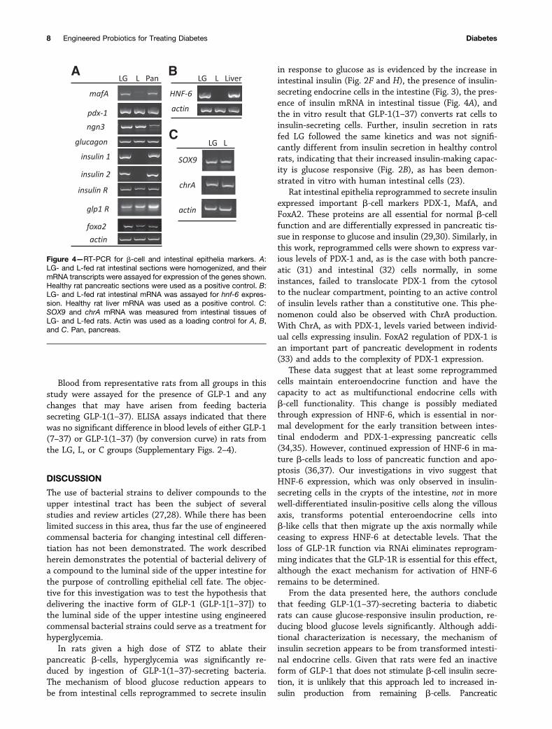
Expression of Pancreatic and Intestinal MarkersSections of rat small intestines were fixed and immuno-fluorescently probed for various markers. Insulin-contain-ing cells were found in the small intestines of rats treatedwith LG but not in L or C groups (Fig. 3, LG and L resultsshown). The relative frequency of insulin-producingcells was estimated from image analysis to be ;0.06%(6 0.016%) of the average small intestinal cell count (or;1 in 1,600 epithelial cells) (Fig. 3B). PDX-1 productionwas observed in the upper intestine and pancreas asexpected for all rats in the study (red staining in Fig.3A). Epithelial cells in intestinal sections of rats had var-ious levels of PDX-1 (Fig. 3A.2, yellow arrows) that werefrequently lower for insulin-positive cells than for sur-rounding epithelia (Fig. 3A.3, red arrows); however, in-sulin-positive cells also stained for PDX-1 at levelscomparable to surrounding epithelia (Fig. 3A.2 and A.3,white arrows).
ChrA is normally expressed by neuroendocrine cells,enteroendocrine cells, and islet b-cells in secretory gran-ules. To better understand the physiology of the reprog-rammed cells, intestinal sections were costained for ChrAand insulin. In some instances, insulin staining (green)overlapped with ChrA staining (red) (Fig. 3C), and in;50% of observed insulin-producing cells, counterstaining
did not detect significant amounts of ChrA (Fig. 3C). In Fig.3C, yellow arrows denote an enteroendocrine cell express-ing more insulin than ChrA, while white arrows point toa cell expressing more ChrA than insulin. In L-fed rats, onlyChrA was detected with no visible costaining for insulin(Fig. 3C).
HNF-6, previously shown to be transcriptionally upre-gulated by GLP-1(1–37) in vitro (11), was also assayed.HNF-6 mediates transition from endodermal cell lineagesto PDX-1-expressing cells in the pancreata of developingembryos (25). HNF-6 and insulin colocalization was ob-served in the epithelia of intestinal crypts of rats fed LG(Fig. 3D). Colocalization of HNF-6 and insulin in cells thathad migrated up the villous axis and were present on thevilli (Fig. 3D) was not observed. Liver tissue from healthyrats was used as a positive control for HNF-6 (Fig. 3D). Inaddition to HNF-6, cells staining positive for insulin inthe upper GI tracts of rats fed bacteria secreting GLP-1(1–37) expressed FoxA2, a transcription factor required forPDX-1-mediated insulin synthesis in pancreatic cells (26)(Fig. 3E).
To determine if normal function could be seenjuxtaposed next to transformed cells, intestinal sectionsfrom LG-fed rats were costained for lysozyme, SI, andSOX-9. All of these markers appeared in close proximityto insulin-producing cells (Fig. 3F).
Transcriptional analysis via RT-PCR was in agreementwith immunofluorescence data for expression of insu-lin, pdx-1, and foxA2 (Fig. 4A); hnf-6 (Fig. 4B); andsox9 and chrA, (Fig. 4C). In addition, mafA, neurogenin3 (ngn3), glucagon, insulin receptor (insulin R) and GLP-1 receptor (glp-1R) (Fig. 4A) transcripts were assayed inboth intestinal and pancreatic tissues. Differential ex-pression between rats fed LG and L was seen for mafA(Fig. 4A).
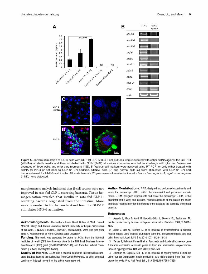
Stimulation of IEC-6 Cells With GLP-1(1–37) in thePresence and Knockdown of GLP-1RRat intestinal stem cells (IEC-6) were used to understandthe importance of the GLP-1R in mediating reprogram-ming. IEC-6 cells were stimulated with GLP-1(1–37) andshowed dose-dependent secretion of insulin in responseto glucose (Fig. 5A). Short interfering RNA (siRNA) wasused to knock down expression of GLP-1R in IEC-6 cellculture. siRNA-treated cultures (siRNAi+) did not exhibitreprogramming when stimulated with GLP-1(1–37) as perRT-PCR of various b-cell markers (Fig. 5B). HNF-6 wasimmunodetected in GLP-1(1–37)-treated IEC-6 cells (Fig.5D) but not in cells pretreated with siRNA against GLP-1R(Fig. 5C).
letters denote significant difference). E: Pancreatic b-cell mass was estimated at the end of the study for 6 animals per treatment. Values areaverages and error bars represent 1 SD. Intestinal (F ) and pancreatic (G) tissues were homogenized and assayed for insulin content. Shownare representative average insulin levels. Error bars represent 1 SD, and P values are from a Dunnet test for significance (n = 6). H:Estimates for average total insulin in combined intestinal and pancreatic tissues were obtained from weight measurements of harvestedtissues. Error bars represent 1 SD (n = 6). C, control; pan, pancreas.
6 Engineered Probiotics for Treating Diabetes Diabetes
In Vitro Analysis of Human Epithelial Insulin Stimulationand GLP-1 in Rat SerumTo rule out any potential stimulation for insulin pro-duction in intestinal cells by the active form of GLP-1(GLP-1[7–37]), an in vitro assay of human epithelia wasdeveloped to compare the relative levels of glucose-mediated insulin production following stimulation by either
GLP-1(7–37) or GLP-1(1–37) (Supplementary Fig. 1). GLP-1(1–37) was observed to stimulate Caco-2 cells to secreteinsulin in response to glucose in a dose-dependent man-ner (although differences were not significant, averagestrended according to dose of GLP-1). GLP-1(7–37) stim-ulated no significant secretion of insulin (SupplementaryFig. 1).
Figure 3—Immunofluorescence for b-cell and intestinal epithelia markers. A: Sections from the different groups were stained for PDX-1(red) and insulin (green). B: Intestinal sections were analyzed by image analysis for the number of insulin-positive cells as a percentage oftotal epithelial cells. Average values are shown (n = 6). Error bars represent 1 SD. C: ChrA (red) and insulin (green). D: Sections from the LG-fed group for HNF-6 (red) and insulin (green). Shown are cells from intestinal crypts and intestinal villi from LG-fed rats and liver cells fromhealthy rats. E: Sections from the LG-fed group for FoxA2 (red) and insulin (green) in intestinal villi and from healthy rat pancreata. F:Sections from the LG-fed group for lysozyme SI, and SOX-9 (red) and insulin (green). Scale bars are 10 mm unless otherwise noted. Lyso,lysozyme; ND, none detected; Pan, pancreata.
diabetes.diabetesjournals.org Duan, Liu, and March 7
Blood from representative rats from all groups in thisstudy were assayed for the presence of GLP-1 and anychanges that may have arisen from feeding bacteriasecreting GLP-1(1–37). ELISA assays indicated that therewas no significant difference in blood levels of either GLP-1(7–37) or GLP-1(1–37) (by conversion curve) in rats fromthe LG, L, or C groups (Supplementary Figs. 2–4).
DISCUSSION
The use of bacterial strains to deliver compounds to theupper intestinal tract has been the subject of severalstudies and review articles (27,28). While there has beenlimited success in this area, thus far the use of engineeredcommensal bacteria for changing intestinal cell differen-tiation has not been demonstrated. The work describedherein demonstrates the potential of bacterial delivery ofa compound to the luminal side of the upper intestine forthe purpose of controlling epithelial cell fate. The objec-tive for this investigation was to test the hypothesis thatdelivering the inactive form of GLP-1 (GLP-1[1–37]) tothe luminal side of the upper intestine using engineeredcommensal bacterial strains could serve as a treatment forhyperglycemia.
In rats given a high dose of STZ to ablate theirpancreatic b-cells, hyperglycemia was significantly re-duced by ingestion of GLP-1(1–37)-secreting bacteria.The mechanism of blood glucose reduction appears tobe from intestinal cells reprogrammed to secrete insulin
in response to glucose as is evidenced by the increase inintestinal insulin (Fig. 2F and H), the presence of insulin-secreting endocrine cells in the intestine (Fig. 3), the pres-ence of insulin mRNA in intestinal tissue (Fig. 4A), andthe in vitro result that GLP-1(1–37) converts rat cells toinsulin-secreting cells. Further, insulin secretion in ratsfed LG followed the same kinetics and was not signifi-cantly different from insulin secretion in healthy controlrats, indicating that their increased insulin-making capac-ity is glucose responsive (Fig. 2B), as has been demon-strated in vitro with human intestinal cells (23).
Rat intestinal epithelia reprogrammed to secrete insulinexpressed important b-cell markers PDX-1, MafA, andFoxA2. These proteins are all essential for normal b-cellfunction and are differentially expressed in pancreatic tis-sue in response to glucose and insulin (29,30). Similarly, inthis work, reprogrammed cells were shown to express var-ious levels of PDX-1 and, as is the case with both pancre-atic (31) and intestinal (32) cells normally, in someinstances, failed to translocate PDX-1 from the cytosolto the nuclear compartment, pointing to an active controlof insulin levels rather than a constitutive one. This phe-nomenon could also be observed with ChrA production.With ChrA, as with PDX-1, levels varied between individ-ual cells expressing insulin. FoxA2 regulation of PDX-1 isan important part of pancreatic development in rodents(33) and adds to the complexity of PDX-1 expression.
These data suggest that at least some reprogrammedcells maintain enteroendocrine function and have thecapacity to act as multifunctional endocrine cells withb-cell functionality. This change is possibly mediatedthrough expression of HNF-6, which is essential in nor-mal development for the early transition between intes-tinal endoderm and PDX-1-expressing pancreatic cells(34,35). However, continued expression of HNF-6 in ma-ture b-cells leads to loss of pancreatic function and apo-ptosis (36,37). Our investigations in vivo suggest thatHNF-6 expression, which was only observed in insulin-secreting cells in the crypts of the intestine, not in morewell-differentiated insulin-positive cells along the villousaxis, transforms potential enteroendocrine cells intob-like cells that then migrate up the axis normally whileceasing to express HNF-6 at detectable levels. That theloss of GLP-1R function via RNAi eliminates reprogram-ming indicates that the GLP-1R is essential for this effect,although the exact mechanism for activation of HNF-6remains to be determined.
From the data presented here, the authors concludethat feeding GLP-1(1–37)-secreting bacteria to diabeticrats can cause glucose-responsive insulin production, re-ducing blood glucose levels significantly. Although addi-tional characterization is necessary, the mechanism ofinsulin secretion appears to be from transformed intesti-nal endocrine cells. Given that rats were fed an inactiveform of GLP-1 that does not stimulate b-cell insulin secre-tion, it is unlikely that this approach led to increased in-sulin production from remaining b-cells. Pancreatic
Figure 4—RT-PCR for b-cell and intestinal epithelia markers. A:LG- and L-fed rat intestinal sections were homogenized, and theirmRNA transcripts were assayed for expression of the genes shown.Healthy rat pancreatic sections were used as a positive control. B:LG- and L-fed rat intestinal mRNA was assayed for hnf-6 expres-sion. Healthy rat liver mRNA was used as a positive control. C:SOX9 and chrA mRNA was measured from intestinal tissues ofLG- and L-fed rats. Actin was used as a loading control for A, B,and C. Pan, pancreas.
8 Engineered Probiotics for Treating Diabetes Diabetes
morphometric analysis indicated that b-cell counts were notimproved in rats fed GLP-1-secreting bacteria. Tissue ho-mogenization revealed that insulin in rats fed GLP-1-secreting bacteria originated from the intestine. Morework is needed to further understand how the GLP-1Rstimulates HNF-6 activation.
Acknowledgments. The authors thank David Brillon of Weill CornellMedical College and Andrea Quaroni of Cornell University for helpful discussionsof this work. L, NCK334, EC1000, NCK1391, and NCK1609 were kind gifts fromTodd R. Klaenhammer at North Carolina State University.Funding. This work was supported by grants to J.C.M. from the NationalInstitutes of Health (DP2 New Innovator Award), the NIH Small Business Innova-tion Research (SBIR) grant (1R41DK094638-01A1), and from the Hartwell Foun-dation (Hartwell Investigator Award).Duality of Interest. J.C.M. has a financial conflict of interest with a com-pany that has licensed this technology from Cornell University. No other potentialconflicts of interest relevant to this article were reported.
Author Contributions. F.F.D. designed and performed experiments andwrote the manuscript. J.H.L. edited the manuscript and performed experi-ments. J.C.M. designed experiments and wrote the manuscript. J.C.M. is theguarantor of this work and, as such, had full access to all the data in the studyand takes responsibility for the integrity of the data and the accuracy of the dataanalysis.
References1. Assady S, Maor G, Amit M, Itskovitz-Eldor J, Skorecki KL, Tzukerman M.Insulin production by human embryonic stem cells. Diabetes 2001;50:1691–16972. Alipio Z, Liao W, Roemer EJ, et al. Reversal of hyperglycemia in diabeticmouse models using induced-pluripotent stem (iPS)-derived pancreatic beta-likecells. Proc Natl Acad Sci U S A 2010;107:13426–134313. Ferber S, Halkin A, Cohen H, et al. Pancreatic and duodenal homeobox gene1 induces expression of insulin genes in liver and ameliorates streptozotocin-induced hyperglycemia. Nat Med 2000;6:568–5724. Zalzman M, Gupta S, Giri RK, et al. Reversal of hyperglycemia in mice byusing human expandable insulin-producing cells differentiated from fetal liverprogenitor cells. Proc Natl Acad Sci U S A 2003;100:7253–7258
Figure 5—In vitro stimulation of IEC-6 cells with GLP-1(1–37). A: IEC-6 cell cultures were incubated with either siRNA against the GLP-1R(siRNA+) or sterile media and then incubated with GLP-1(1–37) at various concentrations before challenge with glucose. Values areaverages of three wells, and error bars represent 1 SD. B: Various cell markers were assayed using RT-PCR for cells either treated withsiRNA (siRNA+) or not prior to GLP-1(1–37) addition. siRNA+ cells (C) and normal cells (D) were stimulated with GLP-1(1–37) andimmunostained for HNF-6 and insulin. All scale bars are 20 mm unless otherwise indicated. chra = chromogranin A; ngn3 = neurogenin3; ND, none detected.
diabetes.diabetesjournals.org Duan, Liu, and March 9

5. Blyszczuk P, Czyz J, Kania G, et al. Expression of Pax4 in embryonic stemcells promotes differentiation of nestin-positive progenitor and insulin-producingcells. Proc Natl Acad Sci U S A 2003;100:998–10036. Kania G, Blyszczuk P, Czyz J, Navarrete-Santos A, Wobus AM. Differenti-ation of mouse embryonic stem cells into pancreatic and hepatic cells. MethodsEnzymol 2003;365:287–3037. Hori Y, Rulifson IC, Tsai BC, Heit JJ, Cahoy JD, Kim SK. Growth inhibitorspromote differentiation of insulin-producing tissue from embryonic stem cells.Proc Natl Acad Sci U S A 2002;99:16105–161108. Lumelsky N, Blondel O, Laeng P, Velasco I, Ravin R, McKay R. Differenti-ation of embryonic stem cells to insulin-secreting structures similar to pancreaticislets. Science 2001;292:1389–13949. Soria B, Roche E, Berná G, León-Quinto T, Reig JA, Martín F. Insulin-se-creting cells derived from embryonic stem cells normalize glycemia in strepto-zotocin-induced diabetic mice. Diabetes 2000;49:157–16210. Noguchi H. Stem cells for the treatment of diabetes. Endocr J 2007;54:7–1611. Suzuki A, Nakauchi H, Taniguchi H. Glucagon-like peptide 1 (1-37) convertsintestinal epithelial cells into insulin-producing cells. Proc Natl Acad Sci U S A2003;100:5034–503912. Baggio LL, Drucker DJ. Biology of incretins: GLP-1 and GIP. Gastroenter-ology 2007;132:2131–215713. Farrar MD, Whitehead TR, Lan J, et al. Engineering of the gut commensalbacterium Bacteroides ovatus to produce and secrete biologically activemurine interleukin-2 in response to xylan. J Appl Microbiol 2005;98:1191–119714. Ahmed FE. Genetically modified probiotics in foods. Trends Biotechnol2003;21:491–49715. Westendorf AM, Gunzer F, Deppenmeier S, et al. Intestinal immunity ofEscherichia coli NISSLE 1917: a safe carrier for therapeutic molecules. FEMSImmunol Med Microbiol 2005;43:373–38416. Duan F, March JC. Interrupting Vibrio cholerae infection of human epithelialcells with engineered commensal bacterial signaling. Biotechnol Bioeng 2008;101:128–13417. Corthésy B, Gaskins HR, Mercenier A. Cross-talk between probiotic bacteriaand the host immune system. J Nutr 2007;137(Suppl. 2):781S–790S18. Sansonetti PJ. War and peace at mucosal surfaces. Nat Rev Immunol 2004;4:953–96419. Hazebrouck S, Pothelune L, Azevedo V, Corthier G, Wal JM, Langella P.Efficient production and secretion of bovine beta-lactoglobulin by Lactobacilluscasei. Microb Cell Fact 2007;6:1220. Daniel C, Repa A, Wild C, et al. Modulation of allergic immune responses bymucosal application of recombinant lactic acid bacteria producing the major birchpollen allergen Bet v 1. Allergy 2006;61:812–81921. Kuehn MJ. Genetically engineered probiotic competition. Gastroenterology2006;130:1915–1916
22. Ait-Belgnaoui A, Han W, Lamine F, et al. Lactobacillus farciminis treatmentsuppresses stress induced visceral hypersensitivity: a possible action throughinteraction with epithelial cell cytoskeleton contraction. Gut 2006;55:1090–109423. Duan F, Curtis KL, March JC. Secretion of insulinotropic proteins by com-mensal bacteria: rewiring the gut to treat diabetes. Appl Environ Microbiol 2008;74:7437–743824. Quigley EM, Flourie B. Probiotics and irritable bowel syndrome: a rationalefor their use and an assessment of the evidence to date. Neurogastroenterol Motil2007;19:166–17225. Kumar M, Jordan N, Melton D, Grapin-Botton A. Signals from lateral platemesoderm instruct endoderm toward a pancreatic fate. Dev Biol 2003;259:109–12226. Lee CS, Sund NJ, Vatamaniuk MZ, Matschinsky FM, Stoffers DA, KaestnerKH. Foxa2 controls Pdx1 gene expression in pancreatic beta-cells in vivo. Di-abetes 2002;51:2546–255127. Aurand TC, Russell MS, March JC. Synthetic signaling networks for ther-apeutic applications. Curr Opin Biotechnol 2012;23:773–77928. Goh YL, He H, March JC. Engineering commensal bacteria for prophylaxisagainst infection. Curr Opin Biotechnol 2012;23:924–93029. Szabat M, Luciani DS, Piret JM, Johnson JD. Maturation of adult beta-cellsrevealed using a Pdx1/insulin dual-reporter lentivirus. Endocrinology 2009;150:1627–163530. Zhou Q, Brown J, Kanarek A, Rajagopal J, Melton DA. In vivo reprogram-ming of adult pancreatic exocrine cells to beta-cells. Nature 2008;455:627–63231. Kebede M, Ferdaoussi M, Mancini A, et al. Glucose activates free fatty acidreceptor 1 gene transcription via phosphatidylinositol-3-kinase-dependent O-GlcNAcylation of pancreas-duodenum homeobox-1. Proc Natl Acad Sci U S A2012;109:2376–238132. Fang R, Olds LC, Sibley E. Spatio-temporal patterns of intestine-specifictranscription factor expression during postnatal mouse gut development. GeneExpr Patterns 2006;6:426–43233. Gao N, LeLay J, Vatamaniuk MZ, Rieck S, Friedman JR, Kaestner KH.Dynamic regulation of Pdx1 enhancers by Foxa1 and Foxa2 is essential forpancreas development. Genes Dev 2008;22:3435–344834. Jacquemin P, Lemaigre FP, Rousseau GG. The Onecut transcription factorHNF-6 (OC-1) is required for timely specification of the pancreas and acts up-stream of Pdx-1 in the specification cascade. Dev Biol 2003;258:105–11635. Poll AV, Pierreux CE, Lokmane L, et al. A vHNF1/TCF2-HNF6 cascaderegulates the transcription factor network that controls generation of pancreaticprecursor cells. Diabetes 2006;55:61–6936. Gannon M, Ray MK, Van Zee K, Rausa F, Costa RH, Wright CV. Persistentexpression of HNF6 in islet endocrine cells causes disrupted islet architectureand loss of beta cell function. Development 2000;127:2883–289537. Hara M, Shen J, Pugh W, Polonsky KS, Le Beau MM, Bell GI. Sustainedexpression of hepatocyte nuclear factor-6 leads to loss of pancreatic beta-cellsby apoptosis. Exp Clin Endocrinol Diabetes 2007;115:654–661
10 Engineered Probiotics for Treating Diabetes Diabetes