engineering and production of quality viral proteins in ... · cowpox vaccines produced by jenner...
TRANSCRIPT

Engineering and Production of Quality Viral Proteins in
Prokaryotic and Eukaryotic Systems
Tesi doctoral
Mónica Martínez Alonso
2010
Departament de Genètica i de Microbiologia
Institut de Biotecnologia i de Biomedicina
Facultat de Biociències
Universitat Autònoma de Barcelona


PhD Programme in Biotechnology
Engineering and Production of Quality Viral Proteins in Prokaryotic and Eukaryotic Systems
Report presented by Mónica Martínez Alonso in order to complete the requirements to be granted the degree of Doctor of Philosophy in Biotechnology by the Autonomous University of Barcelona.
Mónica Martínez Alonso
Approval of the thesis directors,
Antonio Villaverde Corrales Neus Ferrer Miralles Rob Noad


A mis padres,


Contents


3
Contents
Contents
1. Introduction ..................................................................................................................... 7
1.1. Overview of the currently available protein production systems ....................... 11
1.2. Escherichia coli for recombinant protein production .......................................... 14
1.2.1. Protein folding ........................................................................................... 18
1.2.2. Quality control in the bacterial cytoplasm ................................................ 21
1.2.2.1. Chaperones ...................................................................................... 21
1.2.2.2. Proteases ......................................................................................... 26
1.2.3. Inclusion bodies ......................................................................................... 28
1.2.3.1. Morphology, composition and structure ........................................ 29
1.2.3.2. Minimising inclusion body formation .............................................. 32
1.2.3.3. Conformational quality of inclusion body proteins ......................... 33
1.3. The baculovirus-insect cell expression system .................................................... 35
1.3.1. Overview of baculovirus biology ............................................................... 36
1.3.1.1. Baculovirus structure ....................................................................... 36
1.3.1.2. Infection progress ............................................................................ 37
1.3.2. Expression vectors ..................................................................................... 39
1.3.2.1. Transfer plasmids ............................................................................. 40
1.3.2.2. Parental genomes ............................................................................ 42
1.3.3. Insect hosts ................................................................................................ 47
1.3.3.1. Cell lines ........................................................................................... 47
1.3.3.2. Insect larvae ..................................................................................... 48
1.4. Model proteins .................................................................................................... 50
1.4.1. Green Fluorescent Protein ......................................................................... 50
1.4.2. Foot-and-Mouth Disease Virus VP1 and VP2 capsid proteins ................... 53
1.4.3. Human α-Galactosidase ............................................................................. 54
1.5. Previous work ...................................................................................................... 55
2. Objectives ....................................................................................................................... 57

4
Contents
3. Results ............................................................................................................................ 61
3.1. Article 1 ................................................................................................................ 63
3.2. Article 2 ................................................................................................................ 71
3.3. Article 3 ................................................................................................................ 77
3.4. Article 4 ................................................................................................................ 85
3.5. Article 5 ................................................................................................................ 93
4. Discussion ..................................................................................................................... 103
4.1. Independent control of protein yield and quality ............................................. 106
4.2. Functional status of soluble protein .................................................................. 108
4.3. Bacterial folding modulators for eukaryotic systems ........................................ 110
5. Conclusions .................................................................................................................. 117
6. Annex I ......................................................................................................................... 121
7. Annex II ........................................................................................................................ 133
8. References ................................................................................................................... 147
9. Acknowledgements ...................................................................................................... 181


Introduction
1.

9
Introduction
Biotechnology is defined as the use of living organisms or biological substances to
perform specific industrial or manufacturing processes, and as such, it has been known
to mankind for a long time. Over 10,000 years ago, long before the term ‘biotechnology’
was even coined microorganisms were already used in fermentation processes to
produce wine, beer or bread. Early farmers, even if unaware, also relied on
biotechnology for crop improvement through careful seed selection to obtain higher
yields or better taste.
More recently, the end of the 19th century experienced a significant improvement in
health conditions in over-crowded industrial cities when large-scale sewage purification
systems based on microbial activity were first introduced [1]. That was also the time
when fermentation industry was born, as industrial processes were developed for the
manufacture of chemicals such as acetone or butanol using bacteria [1]. Moreover,
cowpox vaccines produced by Jenner in 1796 and the discovery of penicillin by
Alexander Fleming in 1927 [2] and its further development in the 1940s are two
examples of the early impact of biotechnology in the medical arena.
However, the development of biotechnology as we know it today would still need
some major breakthroughs. The first came with the discovery of the double helix
structure of DNA in 1953 by Watson and Crick [3], followed by the cracking of the
genetic code by Marshall Nirenberg and Heinrich J. Matthaei in 1961 [4]. Soon after, in
the early 1970s the discovery of new restriction enzymes by Paul Berg [5], combined
with Herbert Boyer and Stanley Cohen’s first genetic engineering of living organisms [6]
gave way to recombinant DNA technology. The modern biotechnology era had just
started.
Today, biotechnology is present in nearly all sectors of industry, with applications in
major areas such as medicine, agriculture and crop production, and environment.
Products derived from biotechnology have steadily increased over the years, and those
commercially available today include antibiotics, antibodies, biofuels, fermented foods
and beverages and recombinant proteins [1].
Proteins are the building blocks of life. No matter their origin, all proteins are
assembled from a set of 20 amino acids linked together to form the linear chain that
defines their primary structure. Being the most abundant macromolecules in living cells,
they are also highly versatile. Their biological importance lies in the fact that proteins are
the molecular tools required to carry out the functions encoded in the genome, with
almost every event that takes place in a cell requiring action from one or several

10
Introduction
proteins. Thus, they have important roles in cellular processes such as cell signalling,
immune responses, cell adhesion or the cell cycle. Proteins also have structural roles and
act as catalysts in many cell reactions [7]. This versatility translates in recombinant
proteins (i.e. those derived from recombinant DNA) having applications in a wide variety
of sectors, ranging from biopharmaceutical to enzyme and agricultural industries. Also,
because they enter both industrial and therapeutic markets, recombinant proteins have
a prominent position in the economical arena.
Although insulin was the first pharmaceutical produced as early as 1922 [8], the
difficulty to obtain proteins from their natural sources in sufficient amounts for their
study, characterisation and further use still represented a major roadblock. The
availability of new restriction enzymes and recombinant DNA techniques, together with
the parallel development of heterologous systems for recombinant protein production
has resulted in an increasing number of commercially available biotechnological
products, which has in turn boosted the biotechnological industry.
Some examples of the already marketed recombinant proteins include human insulin
(which became the first E. coli produced biopharmaceutical approved by the FDA in 1982
[9]), growth hormone, Factor VIII or gamma interferon [10]. Enzymes are also marketed
either for industrial use (amidase for the production of 6-aminopenicillanic acid, nitrile
hydratase to produce acrylamide, amylases, proteases...) or to be used as therapeutic
agents in the treatment of diseases like thromboses, cystic fibrosis, metabolic diseases
or even cancer [1]. The production system must be carefully chosen to successfully
obtain each of these proteins, as protein features and the processing abilities of the
recombinant host will ultimately determine whether a protein can be obtained in a
functional form.

11
Introduction
1.1. Overview of the currently available protein production systems
The product to be obtained is the key element to be considered when choosing a
production system. Depending on protein features such as size, origin or need for post-
translational modifications, the available options will be narrowed down to the most
convenient expression system. Production costs, time constraints and the yield and
quality of the product must also be taken into account.
Prokaryotes are usually the first choice for protein production because of their fast
growth and availability of easy-to-handle procedures. The many advantages of
Escherichia coli make it the most widely used and best characterised microorganism.
Cultivation is easy and essentially inexpensive. Recombinant gene expression is fast and
high protein yields can be obtained in a cost-effective manner. Although recombinant
proteins can be engineered for secretion to the periplasmic space, E. coli is often used
for the production of cytoplasmic proteins. Despite the many advantages of this host,
recombinant protein production in Escherichia coli has some drawbacks too. The two
main obstacles encountered are proteolytic digestion by cell proteases [11] and
accumulation of the protein in insoluble deposits, known as inclusion bodies (IBs)
[12;13]. Both events are the result of the recombinant protein not being able to reach its
native conformation. Although many strategies have been devised along the years to
reduce inclusion body formation and promote the synthesis of soluble protein, protein
deposition in inclusion bodies still represents a major bottleneck for protein production
in this system. Moreover, eukaryotic proteins are often obtained as insoluble or inactive,
due to the inability of the system to carry out complex post-translational modifications.
However, the N-glycosylation system of Campylobacter jejuni has successfully been
transferred to E. coli, rendering a strain capable of glycosylation [14].
Other bacteria can also be used as cell factories. Bacillus systems provide the
advantage of stronger secretion compared to E. coli. Also, they have GRAS (Generally
Recognised as Safe) status, which will eventually facilitate FDA approval of recombinant
proteins obtained in this system. Bacillus megaterium, B. subtilis, B. licheniformis and B.
brevis are often used for expression [1]. However, the production of many extracellular
proteases by B. subtilis represents an important drawback.
Among the eukaryotic organisms, single-celled yeasts represent the simplest system.
In common with E. coli, yeasts are also fast and cost-effective for protein production,
offering high yields of the recombinant product and with the added advantage of being
able to perform post-translational modifications. For this reason, many proteins which

12
Introduction
fail to fold properly in E. coli or require post-translational modifications are produced in
yeast. However, glycosylation patterns are different from higher eukaryotes [15]. The
genetics of the system are well characterised, with the most common hosts being
Saccharomyces cerevisiae and Pichia pastoris. Although approved biopharmaceuticals
produced in yeast are derived exclusively from S. cerevisiae [15], P. pastoris is currently
the most widely used yeast for heterologous protein expression due to its superior
secretion characteristics [10].
Filamentous fungi provide complex post-translational modifications, which are then
more similar to the mammalian version [10]. However, the system is not well
characterised both genetically and physiologically, secretion yields are not competitive
and proteases can hamper protein production [1;10].
Insect cells can perform post-translational modifications which are even more
complex than those carried out in fungi. Being animal cells, cultivation is more difficult
and expensive but they are still more resistant and easy to handle than mammalian
systems. Their folding machinery is better suited for mammalian proteins, and thus
soluble proteins of mammalian origin can be obtained [16]. Protein production is
accomplished by infection of the insect cell host with a recombinant baculovirus
encoding the target protein. Other advantages of this system include proper disulfide
bond formation and high expression levels. The system is safe as baculovirus vectors
have a restricted host range, infecting only insects but not vertebrates. Cells can be
adapted to suspension cultures and chemically defined, serum-free media. Large
proteins and also multi-protein complexes have been obtained, and simultaneous
expression of multiple genes is also possible [17;18]. However, some shortcomings are
also present. Proteins can sometimes be seen as intracellular aggregates [19;20],
protease activity is high [10;21;22] and glycosylation patterns provided by insects still
differ from mammalians, limiting protein half-life when administered to humans [23].
Mammalian cell lines are sometimes the only choice for expression of difficult
proteins, especially heavily glycosylated ones. Expressed proteins are often soluble and
active, and high yields are obtained. However, the system is expensive and process
duration is long. Nevertheless, most of the approved therapeutic proteins have been
obtained in hamster-derived cell lines, namely CHO (Chinese Hamster Ovary) and BHK
(Baby Hamster Kidney) [15]. These cell lines can also be adapted to suspension cultures
and defined serum-free media, which increases the biosafety of the recombinant

13
Introduction
products. Although they are both recognised as safe regarding infectious and pathogenic
agents [10], lack of contamination by viruses and DNA still needs to be proven [1].
Transgenic animals are also used to produce recombinant proteins in milk, egg
white, blood, urine, seminal plasma and silk worm cocoons [1]. So far, milk has given the
best results. Although production in milk is more cost-effective than in mammalian cell
culture [1], safety concerns represent a great challenge because of possible transmission
of infectious diseases (both viral and prion infections) and immunogenic responses [15].
Transgenic plants have also been used for production of recombinant proteins. The
system presents many advantages, such as being cheap, highly productive, easy to scale
up, and safe as it lacks human pathogens. Eukaryotic post-translational modifications are
also available. However, disadvantages of transgenic plants include possible
contamination with pesticides, herbicides and toxic plant metabolites [24], and the need
to deal with the uncontrolled spread of the transgenic gene. Also, negative public
perception of transgenic plants does not encourage their use as a promising system.
Besides recombinant protein production in prokaryotic or eukaryotic hosts, protein
synthesis is also possible in cell-free expression systems, where transcription and
translation reactions are carried out in vitro. This system is fast and simple, and an
excellent alternative for proteins which are toxic for the host when produced in vivo
[25].
Because they have been the two expression systems used in this study, both E. coli
and the Baculovirus Expression System will be discussed in further detail.

14
Introduction
1.2. Escherichia coli for recombinant protein production
Escherichia coli is the most widely used prokaryotic organism for expression of
recombinant proteins [26]. Being one of the most studied microorganisms since early
times, its genetics and physiology are well-known and this has facilitated the
development of the wide set of molecular tools available today [15].
The use of E. coli as a host for protein production is relatively simple and inexpensive
[27]. Added advantages include its short duplication time, growth to high cell densities,
ease of cultivation and high yields of the recombinant product, which can accumulate up
to around 30% of the total protein content of the cell [10;27;28]. Thus, it is not
surprising that almost 30% of the recombinant proteins that are currently on the market
are obtained in E. coli [15].
The basic requirement for protein production in E. coli is a strain that provides a
suitable genetic background and harbours a compatible plasmid encoding the gene to be
expressed [27]. The deep knowledge of the system provides flexibility and allows a
better control of protein production. However, the choice of both strain and expression
plasmid has to be carefully considered, as there are some key elements that need to be
taken into account:
Host strain
The most important feature to consider is the ability of the host strain to stably
maintain the expression plasmid. Moreover, for some expression systems the host strain
will also be required to provide relevant genetic elements (e.g., DE3 in the pET system).
Expression strains deficient in the main proteases have been developed with the
aim of attaining a more efficient recovery of intact protein [29-31]. In this regard, there
are currently many strains commercially available. BL21 is a non-pathogenic E. coli B
strain deficient in ompT and Lon proteases. Novagen BLR is a recA- BL21 derivative, used
to improve stability of plasmids with repetitive sequences. However, proteases are an
important element of the protein quality control system, surveying conformational
quality in cooperation with other folding assistants (see section 1.2.2.2). Therefore,
although proteolysis is minimised in protease deficient mutants, this leads to the
accumulation of the misfolded polypeptides in the form of inclusion bodies [32-34].

15
Introduction
Strains for improved disulfide bond formation are also available. The genes for
thioredoxin and glutathione reductases are disrupted in Novagen Origami (trxB/gor) and
AD494 (trxB) strains, thus allowing disulfide bond formation in the cytoplasm of E. coli.
Other mutants can enhance soluble expression of difficult proteins (Avidis C41(DE3)
and C43(DE3) strains) or allow for adjustable levels of protein expression (Novagen
Tuner series). Rosetta and Rosetta-gami strains are also useful to alleviate use of codon
bias (see below). A summary of E. coli strains commonly used for protein production is
presented in Table 1.
Table 1. E. coli strains for recombinant protein production.
E. coli strain Derived Relevant features AD494 K-12 Cytoplasmic disulfide bond formation enabled (trxB mutant) BL21 B834 Deficient in lon and ompT proteases BL21 trxB BL21 Cytoplasmic disulfide bond formation enabled (trxB mutant)
Deficient in lon and ompT proteases BL21 CodonPlus-RIL
BL21 Deficient in lon and ompT proteases Overcome bias in codon usage (supplies AGG, AGA, AUA and CUA codons)
BL21 CodonPlus-RP
BL21 Deficient in lon and ompT proteases. Overcome bias in codon usage (supplies AGG, AGA and CCC codons)
BLR BL21 Stabilizes tandem repeats (recA mutant) Deficient in lon and ompT proteases
B834 B strain Met auxotroph; 35S-met labeling C41 BL21 Mutant for expression of membrane proteins C43 BL21 Double mutant for expression of membrane proteins HMS174 K-12 Stabilizes tandem repeats (recA mutant)
Rifampicin resistance JM 83 K-12 Protein secretion to periplasm Origami K-12 Enhanced cytoplasmic disulfide bond formation (trxB/gor mutant) Origami B BL21 Enhanced cytoplasmic disulfide bond formation (trxB/gor mutant) Deficient
in Ion and ompT proteases Rosetta BL21 Deficient in lon and ompT proteases
Overcome bias in codon usage (supplies AUA, AGG, AGA, CGG, CUA, CCC, and GGA codons)
Rosetta-gami BL21 Enhanced cytoplasmic disulfide bond formation (trxB/gor mutant) Deficient in Ion and ompT proteases Overcome bias in codon usage (supplies AUA, AGG, AGA, CGG, CUA, CCC, and GGA codons)
All strains are commercial, and most are also available as DE3 and DE3 pLysS strains.
Adapted from Appl Microbiol Biotechnol. 2006 Sep;72(2):211-22.

16
Introduction
Plasmids for gene expression
Plasmids are double-stranded circular DNA molecules that replicate independently
of the host’s chromosome.
Expression plasmids contain several genetic elements:
o The replicon
o
, which contains the origin of replication that will in turn determine
the plasmid copy number [35]. For multi-copy expression plasmids, ColE1 and p15A are
the most common. Also, plasmid incompatibility groups must be taken into account
when gene products are to be co-expressed from different plasmids. In that case,
different replicon incompatibility groups will be required for plasmids to be compatible.
In that regard, plasmids containing ColE1 and p15A are compatible, and thus are
frequently combined for co-expression.
Resistance markers
o
, which confer a genetic trait that allows for artificial
selection. Common resistance markers include ampicillin, kanamycin, chloramphenicol
or tetracycline. Ampicillin resistance is obtained by expression of β-lactamase from the
bla gene encoded in the plasmid. When secreted to the periplasm, the enzyme
hydrolises the β-lactam ring. Kanamycin, chloramphenicol and tetracycline bind to the
ribosomes, interfering with protein synthesis. Aminoglycoside phosphotransferases
inactivate kanamycin in the periplasm, and resistance to chloramphenicol is provided by
chloramphenicol acetyl transferase. Resistance to tetracycline can be conferred by
several genes. However, tetA genes encoding a tetracycline efflux system or tetM and
tetQ, encoding a protein that protects ribosomes from the inhibiting effects of
tetracycline, are often used in molecular biology.
Transcriptional promoters
o
, which enable control of the gene expression levels in
inducible systems. Ideally, promoters should be strong to provide high yields of the
recombinant protein. It is also convenient that the inducer is cheap in order to minimise
production costs. Promoter induction can be either thermal or chemical. Thermal
induction will usually require a temperature upshift, whereas for chemical induction
isopropyl-beta-D-thiogalactopyranoside (IPTG) is the most common molecule [36].
Minimising basal transcription is important, especially when the expression of target
genes poses a cellular stress. This is achieved by the presence of a suitable repressor
that will bind the promoter in absence of inducer.
Translation initiation regions, which are necessary for ribosome binding to
messenger RNA. Thus, these will include a ribosomal binding site (RBS) containing the
17
Introduction
Shine-Dalgarno sequence located 7±2 nucleotides upstream the canonical AUG
translation initiation codon used in efficient recombinant systems [37;38].
o Transcriptional terminators
o
, which prevent transcription starting from irrelevant
promoters or through the origin of replication. They are placed downstream of the
sequence encoding the gene and stabilise mRNA by forming a stem loop at the three
prime end [39].
Translational terminators
Stability of messenger RNA
, which mediate translation termination usually by the
stop codon UAA in E. coli. Efficiency can be increased by placing several stop codons
together [40].
Gene expression levels mainly depend on four factors: efficiency of transcription,
mRNA stability, frequency of translation and protein stability. Although transcription and
translation have been thoroughly optimised in recombinant expression systems, mRNA
stability is not often addressed. Therefore, gene expression is controlled by mRNA
decay. Because of this, mRNA stability is an important factor in controlling gene
expression levels as the expression rate depends directly on its stability, with the
average half-life of mRNA in E. coli ranging from seconds to 20 minutes [41;42].
Messenger RNA is susceptible to degradation by cellular RNases, and protection
depends on its folding, protection of ribosomes and polyadenylation, which in bacteria
influences mRNA stability by promoting its decay. Thus, in poly(A)-deficient strains,
mRNA is stabilised [43;44]. Moreover, commercially available mutant strains for the
RNaseE gene (Invitrogen BL21 star) provide enhanced mRNA stability [45].
Bias in codon usage
Because the genetic code is degenerate, most amino acids can be determined by
more than one codon. Also, the preferred codons for each amino acid vary in different
organisms and this can become a problem in recombinant expression systems.
Heterologous genes from viral origin, eukaryotes or archaeabacteria often contain
high frequencies of codons which are rare in E. coli [46]. Because of the low availability
of the tRNAs corresponding to rare codons, ribosomes are likely to stop at those
18
Introduction
positions [47]. This leads to translational errors that can include amino acid
substitutions, frameshifting or premature termination [48;49].
To overcome this bias, the recombinant gene sequence can be engineered so that
rare codons are substituted by those which are optimal for the host system. Although
this strategy can result in enhanced expression levels and reduced translational errors
[50;51] it is also time-consuming, especially when considering biotechnological high-
throughput applications. A faster alternative consists in co-transforming the host with a
plasmid encoding the tRNAs corresponding to the problematic codons.
Complementation plasmids and already transformed strains, such as Novagen Rosetta
and Rosetta-gami, are commercially available for this purpose.
1.2.1. Protein folding
Protein folding is the process by which an unfolded polypeptide adopts its
characteristic three-dimensional and functional structure. According to the fundamental
principle of protein folding stated by Anfinsen in 1973, the folding of a protein is
determined by its amino acid sequence, which contains all the information required for
the protein to reach its native conformation [52]. The native conformation of a protein is
usually the most thermodynamically stable, having the lowest Gibbs free energy.
However, even if this means that thermodynamics is the driving force that guides
protein folding it does not explain how most proteins reach their native conformation in
a matter of seconds, as randomly exploring the billions of possible spatial conformations
would take astronomical amounts of time. This view, which is known as the Levinthal
paradox [53], assumes that the folding of every residue is independent from the rest.
However, since folding is a cooperative process [54;55] every residue does not have to
search for random conformation states, as their conformational freedom will be
narrowed down by the folding of previous residues [56].
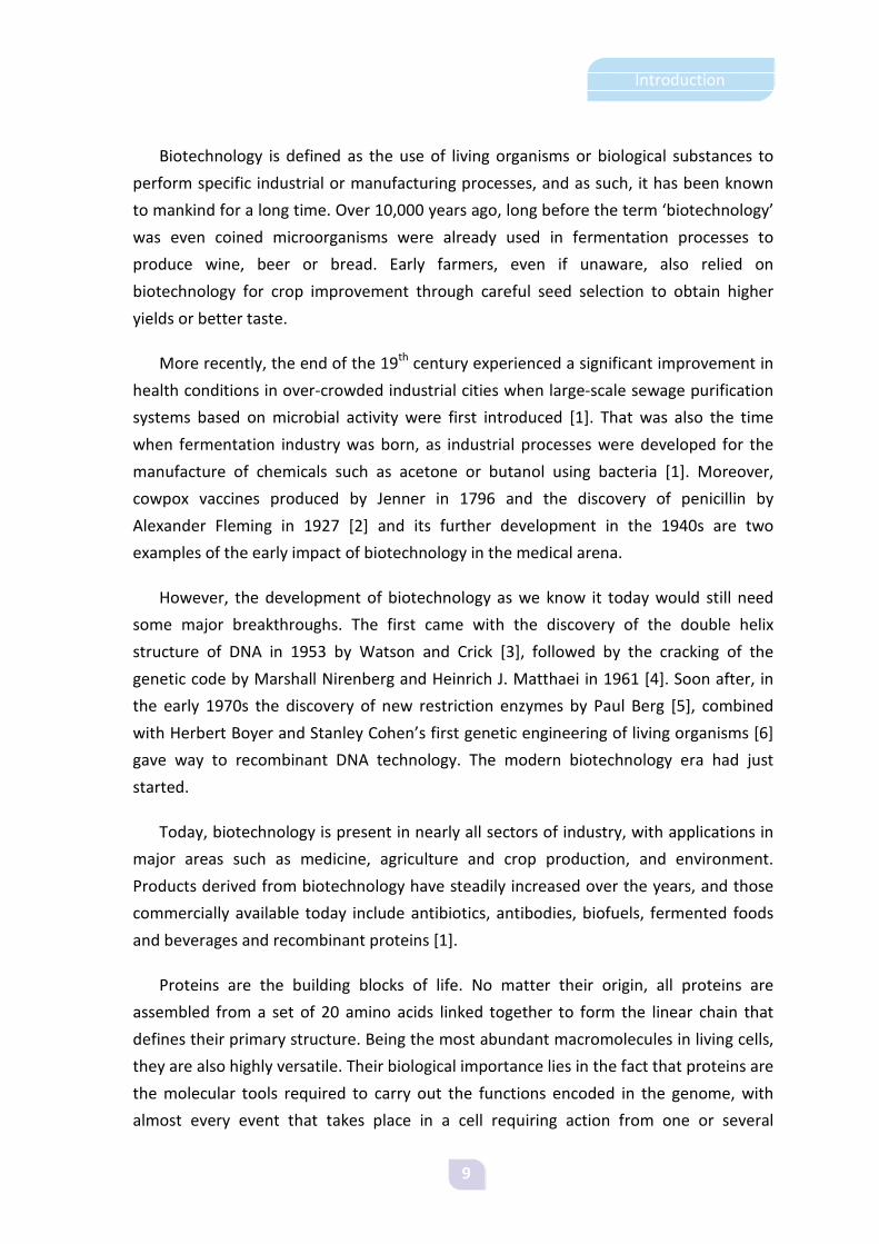
Figure 1. Model of the energy landscape for a polypeptide folding, according to Levinthal. Every residue folds independently from each other, so the time required for the protein to reach the native conformation is extremely large.
Adapted from Nat Struct Biol. 1997 Jan; 4(1):10-19.

19
Introduction
Levinthal also suggested that the stable conformation could have a higher energy if
the lowest Gibbs energy was not kinetically accessible. Thus, different kinetic models
have been proposed to solve the paradox.
The hydrophobic collapse model describes the initial stages of protein folding.
Hydrophobic forces, which drive the collapse, arise from the repulsion between
hydrophobic side chains of the protein and the hydrophilic water molecules of the
environment. The collapse results in the protein being in the “molten globule” state,
with hydrophobic side chains in the interior while the hydrophilic residues are on the
surface. With a volume slightly larger than the native structure of the protein, the
molten globule contains secondary structures but lacks a definite tertiary structure [57].
The nucleation theory proposes the existence of folding nuclei in the protein
structure during the early stages of folding. The most recent view of this theory [58]
proposes a mechanism in which weak nuclei are stabilised by long distance interactions.
Currently, the “new-view” in protein folding is illustrated by the folding funnel model
proposed by Wolynes and co-workers [59]. This model, which requires a high
cooperativity and is therefore very fast [57], describes both the thermodynamic and
kinetic behaviour that unfolded polypeptides undergo to reach their native state, and is
represented in terms of energy landscapes. Multiple pathways exist, and every single
polypeptide can follow its own route. The number of possible conformations decreases
towards the bottom of the funnel, and the folding is faster as the slope grows steeper
[60]. For a protein which can only have two states, unfolded and native, a smooth
mechanism is the simplest way of folding. A two-state folding reflects the existence of an
energy barrier between unfolded and native states. When there is no energy barrier, this
is called smooth folding [56] (Figure 2A), which is often seen when the viscosity of the
solvent is the only limitation for protein folding [61]. Moreover, a protein can sometimes
either fold by a two-state mechanism or adopt an intermediate conformation where
unfolded and folded states coexist, which presents a kinetic trap (Figure 2B).
When cooperativity is not so high, distinct intermediates occur in the folding
process, with local structures that can be different to those observed in the native
structure for the same residues [57]. These structures may be locally favorable but
unfavorable for the whole structure, which leads to kinetic traps determined by the
presence of local energy barriers. This is represented by a rugged energy landscape
(Figure 2C) which is often useful to picture the nucleation model, where local folding
nuclei are formed prior to the molecule adopting its native conformation.
20
Introduction
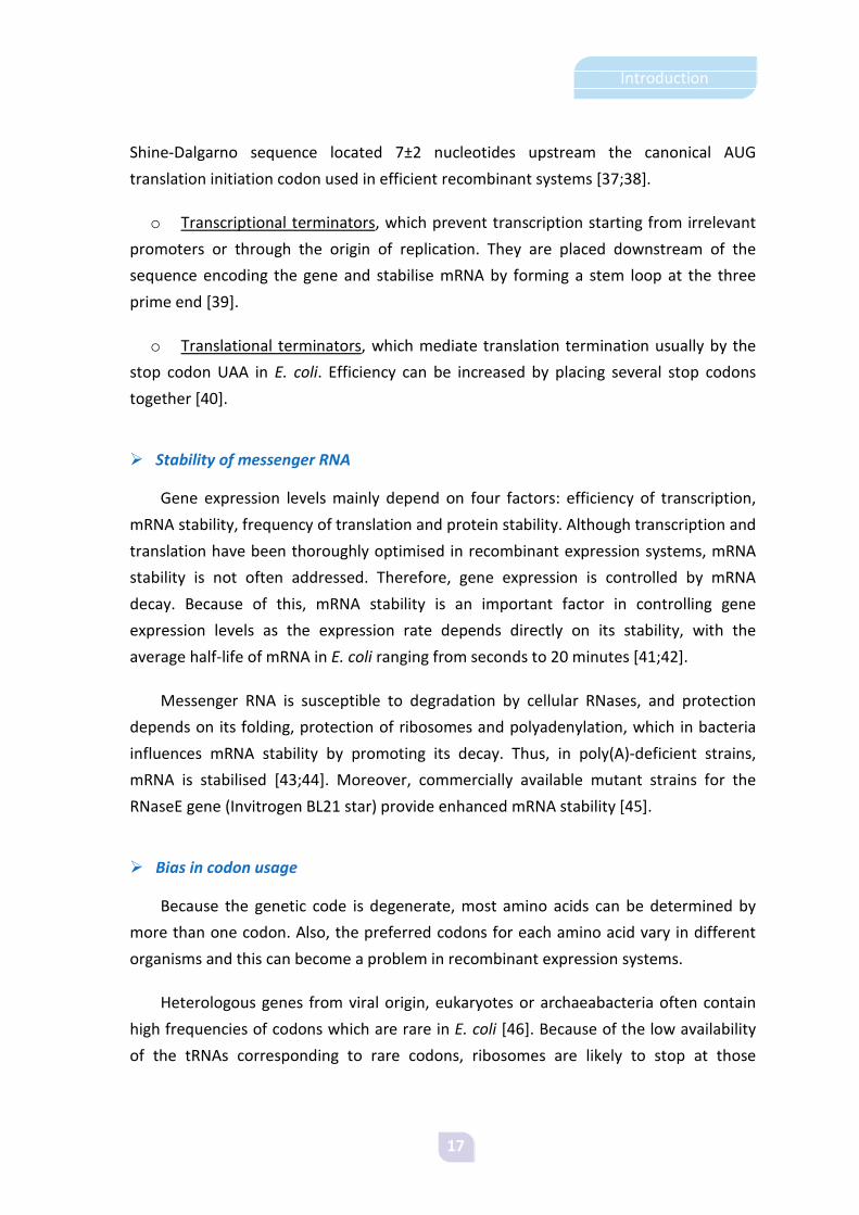
Sometimes polypeptides can fall into kinetic traps with a global free energy similar to
that of the folded state. In this case, the deep kinetic trap results in the two conformers
not being able to interconvert in a reasonable time scale, which may lead to misfolding
and aggregation of the protein. The rough energy landscape corresponding to this
scenario is depicted in Figure 2D.
Figure 2. A) Smooth funnel for a protein following a two-step folding. B) Fast-folding process, in parallel with a slow-folding process involving a kinetic trap. C) Rugged energy landscape with kinetic traps and energy barriers for a multi-state folding protein. D) Rough energy landscape depicting a deep kinetic trap (*) easily accessible from unfolded conformations. Access to the global energy minimum will be very slow for trapped intermediates.
Adapted from Nat Struct Biol. 1997 Jan; 4(1):10-19 (panels A-C) and Proteins. 1998 Jan; 30(1):2-33 (panel D).
Folding in the cellular environment presents an extra challenge. In the very crowded
E. coli cytoplasm, transcription and translation are tightly coupled. With proteins being
released from the ribosomes at a rate of one every 35 seconds [62], the cytoplasm
becomes a very crowded space where macromolecule concentrations can reach 300-400
mg/mL [63]. Because of this, many proteins need assistance of folding modulators to
reach their native conformation. This requirement is dramatically increased in the
context of recombinant protein production, when the cell has an additional input of de
novo synthesis. In fact, folding modulators are considered to be limiting in these
conditions.
During folding, proteins can establish persistent non-native interactions that
significantly affect their structure and biological functions. This is known as “misfolding”
[64]. Misfolded and incompletely folded polypeptides expose hydrophobic stretches that
would be hidden in the native conformation, which makes them prone to aggregation
[65]. Failure of proteins to fold correctly, or to remain properly folded, gives rise to
malfunctioning of living systems [66-68]. In humans, diseases related to incorrect
protein folding, which prevents their normal function, include cystic fibrosis [66] and

21
Introduction
some types of cancer [69]. Proteins with high tendency to misfold can form aggregates
within cells or in the extracellular space, which can also be deposited in tissues such as
brain, heart or spleen [67;68;70;71]. Disorders involving aggregate deposition in tissues
include Alzheimer’s and Parkinson’s diseases, the spongiform encephalopathies and type
II diabetes. Thus, living organisms have cellular factors responsible for avoiding
aggregation by assisting in protein folding, such as molecular chaperones and folding
catalysts [72;73]. In addition, proteases assist in protein quality control by degrading
irreversibly damaged polypeptides which cannot be rescued by the action of
chaperones.
1.2.2. Quality control in the bacterial cytoplasm
Surveillance of protein quality is accomplished by the coordinated action of
chaperones and proteases, which act together to assist protein folding, prevent
accumulation of misfolded polypeptides, remove protein from aggregates and degrade
folding-reluctant species [74]. Thus, the system promotes solubility by minimising
aggregation. Solubility, expressed as the relative amount of recombinant protein in the
soluble cell fraction, is the parameter commonly used to evaluate the success of
biotechnological processes regarding protein quality [75;76]. Although in E. coli quality
control takes place both in the cytoplasm and the periplasm, this section will focus on
the cytosolic branch of the quality control system.
1.2.2.1. Chaperones
The term “chaperone” was first used by Ron Laskey in 1978 to describe an activity
associated to nucleoplasmin in Xenopus oocytes, which allowed the correct assembly of
histones into nucleosomes [77]. Currently, the term chaperone includes a much wider
set of more than 20 protein families which have a major role in the quality control of the
proteome [74;78;79]. Although chaperones are constitutively expressed in physiological
conditions, they become upregulated under stress situations. As thermal stress
promotes an increase of chaperone levels in the cell, they have traditionally been named
as heat shock proteins (Hsp) [80]. However, not all heat shock proteins are chaperones
and vice versa. In E. coli this stress response is positively regulated at the transcriptional
level by the product of the rpoH gene, the factor σ32, which binds as an alternative σ

22
Introduction
subunit to the RNA polymerase and targets it to the promoters of the heat shock genes
[81;82].
Molecular chaperones constitute one of the better characterised groups of folding
modulators, highly conserved in all kingdoms of life. These ubiquitous proteins play a
central role in the conformational control of the proteome by helping other
polypeptides reach their native conformation without affecting their folding rates or
becoming part of their final structure. Chaperones bind hydrophobic patches of amino
acids that would normally be buried within the core of the substrate protein, but have
become exposed to the solvent because of their incorrect folding. The transient
formation of chaperone-substrate complexes shields misfolded polypeptides from
interacting with each other [83]. Chaperones normally target short unstructured
stretches of hydrophobic amino acids which lack acidic residues and are flanked by basic
ones. These motifs are extremely common, which explains why chaperones are so
promiscuous [84].
Based on their mechanism of action, molecular chaperones can be divided into three
functional subclasses:
o Folding chaperones
o
, which drive the net refolding/unfolding of their bound
substrates through ATP-mediated conformational changes. These chaperones promote
the yield of correctly folded proteins without affecting their folding rates. Folding
chaperones in the E. coli cytoplasm are the trigger factor (TF) [85] and the DnaK-DnaJ-
GrpE and GroELS systems [86].
Holding chaperones
o
, which bind to partially folded proteins and stabilise them
until folding chaperones become available, thus preventing them from aggregation [87-
89]. In E. coli, the best characterised holding chaperones are IbpA and IbpB, which
belong to the group of small Hsp family [90] and are commonly found within inclusion
bodies [91]. Hsp31 is another cytoplasmic modulator in this group, which binds early
unfolding intermediates under severe stress conditions and therefore prevents
overloading of the DnaK-DnaJ-GrpE system [92]. Another holdase is Hsp33, a redox-
regulated chaperone that deals with oxidative protein misfolding [93].
Disaggregating chaperones, which promote protein removal from inclusion
bodies and other aggregates formed under prolonged or severe stress conditions
[84;94]. Solubilisation of protein aggregates occurs through ATP-driven conformational
changes, and polypeptides are transferred to folding chaperones for refolding [83]. ClpB

23
Introduction
is the best characterised disaggregase, and works together with DnaK and IbpAB
chaperones assisting refolding and promoting the solubilisation of protein aggregates
[95-97].
I. Trigger factor
Trigger factor is a three-domain cytosolic chaperone which associates to the large
subunit of the ribosomes, close to the exit site, where it binds to nascent polypeptidic
chains and thus stabilises them [84]. This chaperone also exhibits peptidyl-prolyl
cis/trans isomerase activity (PPIase), although the presence of proline residues in its
substrates is not required [98]. Unlike other chaperones, trigger factor is not an ATPase
[99]. In addition, trigger factor is not a heat shock protein either. Indeed, it is induced
upon cold shock and thus enhances cell viability at low temperatures [100].
Therefore, trigger factor aids in de novo protein folding by stabilising nascent chains
or targeting them to other chaperones, like the DnaK-DnaJ-GrpE system with which it
has been shown to cooperate [101].
II. The Hsp70 system: DnaK, DnaJ and GrpE
After being released from trigger factor, a newly synthesised polypeptide can either
fold into its native conformation without any further help or require assistance of other
chaperone sets. In this early stage of folding, polypeptides will expose unfolded
segments. The major cytosolic chaperones involved in the recognition of this set of
substrates are the Hsp70 system [102], that being highly conserved is present in all
kingdoms of life. The bacterial member of the Hsp70 family is the chaperone DnaK,
which acts together with its cofactor DnaJ (the Hsp40 homologue) [103] and a
nucleotide exchange factor named GrpE [104;105]. Although all the three proteins are
induced by heat shock, only DnaK has ATPase activity.
DnaK has a wide set of roles in the multichaperone network, such as folding newly
synthesised polypeptides [73;106], mediating ATP-dependent unfolding, preventing
aggregation, stabilising substrates for refolding by GroELS [107-113], solubilising protein
aggregates in cooperation with ClpB and Ibps [88;107;114-118], participating in
proteolysis [119;120] and protecting proteins against oxidative damages [121;122].
Moreover, it is also a negative regulator of the heat shock response acting in

24
Introduction
cooperation with DnaJ, which binds the σ32 subunit of the RNA polymerase and targets it
for degradation by the inner-membrane associated Ftsh protease [82].
DnaK is a monomeric protein with an N-terminal ATPase domain, a substrate binding
site formed by two β-sheets and a C-terminal domain that interacts with partner
proteins to modulate their function [123;124]. DnaK has two functional states depending
on the phosphorylation state of the bound nucleotide. Affinity for substrates is low
when DnaK is bound to ATP and high when bound to ADP [125-128]. DnaJ is a modular
dimeric protein with at least four distinct domains. The J domain is a highly conserved
motif which stimulates the ATPase activity of DnaK, converting it to the high affinity
ADP-DnaK state [129]. DnaJ has chaperone activity itself and the C-terminal region
seems to be the substrate binding site [99]. GrpE is a homodimer that binds to DnaK in a
ratio of 2:1 [130;131]. It binds to the ATPase domain of DnaK causing the dissociation of
ADP which determines the transition to the low affinity state. This results in release of
the substrate from the chaperone [132;133].
During the functional cycle of the Hsp70 system the target polypeptide is first bound
by DnaJ, which recognises hydrophobic stretches in its structure. The DnaJ-bound
polypeptide is then transferred to DnaK, which is bound to ATP and thus in a low affinity
state. Both DnaJ and the substrate stimulate the ATPase activity of DnaK, which
hydrolyses ATP switching to the high affinity ADP-bound state. Thus, a stable ADP-DnaK
substrate complex is formed. GrpE binding to DnaK stimulates nucleotide exchange and
therefore ADP is dissociated, destabilising the interaction between DnaK and its
substrate, which is then released. After completion of this cycle, the released
polypeptide can fold to its native conformation, require more cycles in this system or be
transferred to the GroELS chaperones. Proteins which have unfolded as a result of stress
conditions can also be refolded by this system [84].
Figure 3. Functional cycle of the bacterial Hsp70 system.
Adapted from Mol Microbiol. 2007 Nov;66(4):840-57.

25
Introduction
III. ClpB
Clp ATPases are members of the AAA family of proteins (ATPases Associated with a
variety of cellular Activities) [134]. The highly conserved AAA module is the key feature
of this family. Structurally, they are formed by subunits arranged in ring-shaped
complexes [135-138].
ClpB is one of the main Clp ATPases in E. coli. This chaperone is a member of the
Hsp100 family and is also induced upon heat shock [99]. ClpB acts by forming a ring-
shaped hexameric structure and translocating its substrate protein through an axial
channel [139]. It works as a “disaggregase” in cooperation with DnaK-DnaJ-GrpE,
reverting aggregation [95;109;115;140]. It has an important role in quality control by
removing protein from aggregates in cooperation with DnaK, reducing aggregate size
and exposing hydrophobic surfaces [107;114]. Disaggregation is facilitated by the
presence of small heat shock proteins within the aggregates [95], but complete
renaturation of the partially unfolded substrates requires transfer from ClpB to DnaK
[107;118].
IV. The Hsp60 system: GroEL and GroES
The GroEL-GroES system handles around 10% of newly synthesised proteins [141].
This is the only chaperone system of the E. coli cytoplasm essential for life under all
growth conditions [142]. GroEL is a bacterial chaperonin of around 60 kDa which belongs
to the Hsp60 family. Structurally, GroEL forms a large oligomer of approximately 800 kDa
organised as two stacked homoheptameric rings, with its cochaperone GroES (a member
of the Hsp10 family) always bound to one of the rings [73]. GroEL substrates are
structured but non-native proteins up to 60 kDa in size [143]. The mechanism of this
chaperone complex is well established in vitro [144-147]. In the substrate acceptor state
of GroEL, GroES and seven ADP molecules are bound to the same ring. During the folding
process, substrates are bound by the GroEL free ring. Then, ATP binding to the newly
occupied ring mediates a conformational change [148] that renders GroEL able to bind
GroES [73]. A second conformational change results in displacement of the substrate to
a chamber defined by the GroEL ring and the GroES cap. This also results in GroES and
ADP release from the opposite ring, as well as any previously encapsulated polypeptide.
By this mechanism, partially folded polypeptides are allowed to fold at infinite dilution
inside the GroEL cavity. Usually, more than one cycle of binding and release will be

26
Introduction
required for a protein to fold into its native state [99]. Equally to the Hsp70 system,
GroEL-GroES can also refold polypeptides which have become unfolded under stress
conditions [84].
Figure 4. Functional cycle of the bacterial Hsp60 system.
Adapted from Curr Biol. 2005 Sep 6;15(17):R661-3.
V. Small heat shock proteins
Small heat shock proteins are ubiquitous and conserved proteins belonging to the
group of the holding chaperones [90]. In E. coli, the best characterised are the Inclusion
Body Proteins (Ibp) which receive their name because of their frequent association with
inclusion bodies [91] and are usually found forming large oligomeric structures (80, 129).
Bacterial IbpA and IbpB are two homologous proteins of 14 and 16 kDa respectively,
encoded on a single operon [91]. Although IbpB is mainly soluble, it comigrates to the
insoluble fraction when produced with the insoluble IbpA [149]. Their function is not
well understood, but they seem to bind hydrophobic stretches of thermally unfolded
polypepdides protecting them from aggregation until the stress disappears. Then, Ibp-
bound polypeptides are transferred to DnaK or GroEL for refolding [149-152]. Recently,
IbpA and IbpB have been shown to assist in the disaggregating and refolding activity of
ClpB [95].
1.2.2.2. Proteases
Proteases have an important role in the control of protein quality, because by
degrading misfolded polypeptides they guarantee that abnormal species do not
accumulate in the cell, which in turn allows for amino acid recycling. Targets for

27
Introduction
degradation include truncated polypeptides, kinetically trapped folding intermediates
which are sensitive to proteolysis and partially folded protein species that after many
folding attempts have still failed to reach their native conformation [84]. In the E. coli
cytoplasm, Lon and ClpP are the two main proteases [30;153;154].
I.Lon
The homotetrameric serine protease Lon is formed by 87 kDa subunits with three
functional domains. Substrate recognition and binding are associated to its N-terminus,
while central and C-terminus domains are linked to ATPase and proteolytic activities,
respectively [84]. Lon is responsible for bulk protein degradation[155;156], and it also
has a regulatory function associated to proteolysis of proteins which are designed to be
unstable (e.g. SulA).
II. ClpP
Together with Lon, the protease ClpP is believed to be responsible for the
degradation of abnormal proteins [155]. Although it also intervenes in bulk degradation
of folded and misfolded polypeptides, ClpP is specifically in charge of truncated proteins
which have been tagged for degradation [157]. ClpP is structured as two stacked
heptamers of 23 kDa subunits, and forms a complex with ClpA and ClpX, two members
of the Hsp100 family of ATPases [158-160]. Only when complexed to ClpA and ClpX is
the degrading system fully-competent, as ClpP alone can digest small peptides but not
large ones or proteins [99]. ClpA and ClpX flank the rings of ClpP and act as molecular
chaperones, unfolding proteins in an ATP-dependent manner and translocating them
into ClpP central channel [161].

28
Introduction
Figure 5. Conventional model of protein folding, aggregation and proteolysis in the cytoplasm of E. coli. Newly synthesised polypeptides can fold to their native state, aggregate or be proteolysed, in a process that is tightly regulated by the quality control system.
Adapted from Nat Biotechnol. 2004 Nov;22(11):1399-408.
1.2.3. Inclusion bodies
In 1975, Prouty and co-workers described for the first time the formation of
amorphous proteinaceous granules in E. coli cells growing in the presence of canavanine
[162]. These deposits contained abnormal cell proteins and were not surrounded by
membranes. Although this was first thought to be an irrelevant cell response in non-
physiological conditions, it turned out to be a common feature in recombinant cells used
as factories for protein production [13] and protein deposition in the form of insoluble
deposits, known as inclusion bodies, is still today a major roadblock in the recovery of
soluble and functional recombinant proteins.

29
Introduction
Under the non-physiological conditions induced by overexpression of recombinant
proteins, the amount of available chaperones in producing cells becomes a limiting
factor [62;163;164]. Intermolecular contacts of exposed hydrophobic stretches in the
unfolded polypeptides are then favoured because of the high yields of recombinant
protein and the limited availability of folding modulators. This situation results in
deposition of folding intermediates [165], especially if they are resistant to proteolysis
[166], leading to aggregation. Bacteria are well prepared genetically to respond to
adverse natural conditions, such as mild protein denaturation under high temperatures
[88;167]. However, despite the many cell responses triggered during recombinant
protein production, no natural mechanism which favours protein folding has been found
[168-175]. Even though, some heat-shock genes including chaperones and proteases are
upregulated in response to recombinant stress [91;176-179], but still this response is not
enough to prevent inclusion body formation.
From a biotechnological point of view, inclusion bodies have been regarded as a
parameter to control in bacterial cell factories [180]. Because aggregation as IBs is not
associated to particular protein sequences [181] predicting yield or solubility for a new
protein production process becomes an obstacle. Therefore, recombinant protein
production in bacteria remains a trial-and-error process.
1.2.3.1. Morphology, composition and structure
Inclusion bodies are insoluble protein deposits observed as cylindrical or ovoid
refractile particles of up to 2 µm3 under an optical microscope [182] and as electron-
dense aggregates lacking a defined structure by transmission electron microscopy
[183;184]. Usually, one or two inclusion bodies are formed per cell [185] and generally
localise in the bacterial cytoplasm, although secreted proteins can also aggregate in the
periplasmic space [186]. The surface topology of inclusion bodies can vary from rough to
smooth [183], and they present a porous architecture [187] and high level of hydration
which are in agreement with density data [188].

30
Introduction
Figure 6. A) Transmission electron microscopy micrograph of an Escherichia coli strain producing inclusion bodies. B) and C) Scanning electron microscopy micrographs of purified inclusion bodies. (García-Fruitós et al, not published).
Generally, the major component of inclusion bodies is the target recombinant
protein itself, which can account for 50 to 90% of the insoluble protein [189]. However,
other cell components can be found associated to inclusion bodies, either adsorbed or
entrapped in their structure. For instance, lipids, nucleic acids, lipopolysaccharides and
outer membrane proteins can coprecipitate with inclusion bodies during sedimentation
by centrifugation [183], although they are not integral components. Membrane proteins
can be removed from inclusion bodies by detergent washing and other procedures that
do not unfold proteins but solubilise membrane proteins [190;191]. Detergents, EDTA,
and enzymes to degrade DNA or the bacterial cell wall are also used in washing
procedures [13;192-194]. Truncated versions of the target protein and other plasmid-
derived proteins (e.g. those conferring antibiotic resistance) can also be found within
inclusion bodies [163;179;191;195-197].
Heat-shock proteins have also been found associated to inclusion bodies. DnaK is
localised in the surface of inclusion bodies [184], and can be recovered during sucrose
density centrifugation together with ClpB [198]. GroEL is also found in small amounts
inside the aggregates, but absent from their surface [184]. In addition, inclusion body
proteins IbpA and IbpB received their names after being described as IB components of
unknown function [91].
Aggregation has long been regarded as an unspecific process driven by random
interaction of exposed hydrophobic patches, resulting in aggregates with no specific
internal molecular architecture. However, there is now an increasing body of evidence
against this view [199-205], which pictures inclusion bodies as highly ordered structures.
Fourier-Transform Infra-Red (FTIR) analysis reveals a characteristic formation of new β-

31
Introduction
sheet structures [32;200;206;207] at expenses of α-helices [65;204], even in rich-β-sheet
native proteins [203;208]. This newly formed β-sheet is non-native, creating a tightly
packed extended intermolecular β-sheet conformation [65].
Remarkably, this enrichment in β-sheet structures is one of the features that
inclusion bodies share with amyloid fibril formation [32;200;204;209] together with
structural homogeneity [32;65;200;201;208], amyloid-tropic dye binding [200] and
cytotoxicity linked to amyloid-like structures [206]. Moreover, for amyloid fibrils
sequence determinants act as “hot spots” for aggregation, modulating the specific
nucleation of amyloid proteins [210-213]. In the case of inclusion bodies, several
observations support the high specifity of their formation process. Besides being
essentially composed of the recombinant protein [182;209;214], their presence in
reduced numbers [182] suggests their formation could be driven by the growth of a
small number of founder aggregates acting as nucleation cores. This is supported by
several observations. First, in vitro refolding studies of proteins in complex mixtures
have shown specificity in polypeptide association during aggregation [215]. Second,
folding intermediates of different IB-forming proteins tend to self-associate in vitro
instead of coaggregating [199]. Third, coexpression of two proteins encoded in the same
gene leads to the formation of two types of cytoplasmic aggregates, showing the
selectivity of the process [191]. Furthermore, preformed inclusion bodies can act as
seeding nuclei for aggregation of their soluble counterparts, but not of unrelated
proteins, in a dose-dependent manner [200].
The increase in non-native β-sheet structures does not necessarily involve the full
unfolding of the IB-embedded protein. Actually, native-like structure of soluble and
inclusion body versions of several proteins has been shown to be highly similar. These
include IL-2 [203], β-lactamase [216], Pseudomonas fragi lipase [201], human growth
hormone and interferon-alpha-2b [202], recombinant E. coli β-galactosidase [209], and
fluorescent proteins [208;217]. The presence of native-like structure in inclusion bodies
seems to facilitate solubilisation of the embedded proteins. In this line, human
granulocyte-colony stimulating factor (hGCSF) produced in E. coli at low temperatures
forms “non classical” inclusion bodies which contain high amounts of correctly folded
protein, enabling protein extraction from these IBs using non denaturing conditions and
low concentrations of polar solvents [218].

32
Introduction
1.2.3.2. Minimising inclusion body formation
Inclusion body formation has affected the development of biotechnology, because
even when inclusion bodies are a rich source of protein, the refolding processes required
to recover the protein in a native form are complex and expensive [219]. For this reason,
much effort has been made to minimise or prevent inclusion body formation, aiming to
improve the yield of soluble protein.
Because recombinant protein can account up to around 30% of the total cell protein
and this produces an enormous metabolic load on the E. coli expression machinery [28],
many strategies have been devised to minimise aggregation, either based on a tight
control of the cellular milieu or in favouring protein folding.
Besides the use of genetically engineered strains that favour production of soluble
protein, (which has already been discussed in section 1.2) other factors can be
considered to increase protein solubility. For instance, the composition of growth media
affects the levels of soluble protein, and by optimising media composition it has been
possible to reduce expression times, increase soluble fraction yield and enhance
biological activity of human PDE-3A, PDE-5A and p38-α Map kinase enzymes [28;220].
Moreover, certain proteins can require the presence of specific cofactors in the growth
media to fold properly, which can include metal ions (e.g., iron-sulphur) or polypeptide-
cofactors (e.g., flavin-mononucleotide). Thus, addition of these factors to the growth
media can improve both protein solubility and folding rates [221;222].
Another common strategy consists of lowering the growth temperature of the
culture. Protein expression at temperatures below the optimal of 37 °C for E. coli growth
usually leads to increased stability and correct folding because the hydrophobic
interactions that determine inclusion body formation are temperature dependent
[223;224]. This has resulted in a number of proteins being successfully expressed in a
soluble form in E. coli [208;225;226]. Moreover, a number of chaperones show increased
expression at low temperatures, which results in better protein quality under these
conditions [227]. In addition, reduced degradation of recombinant protein has been
observed within a temperature range of 15-23 °C due to poor activity of some of the
heat shock proteases [228;229]. However, reduced yields and poor turnover of the
recombinant protein are frequent disadvantages when using this strategy because low
temperatures result in reduced transcription and translation rates.

33
Introduction
Coproduction of folding modulators has been a widely used strategy aimed to
overcome limited chaperone availability during recombinant protein expression, but the
obtained results are controversial and inconsistent [83;230;231]. Some of the positive
reports required coproduction of the major cytosolic chaperone systems (DnaK-DnaJ-
GrpE or GroELS) to observe any increase in solubility [113;232-237] or even
combinations of them, the most successful being KJE, ClpB and ELS [75]. Although the
best results have been obtained when coexpressing several sets of folding modulators,
determining the best set of chaperones for a certain target protein is still a trial and
error process.
Another common approach consists of metabolic engineering through fusion protein
technology, which usually leads to soluble expression [28]. “Tags” consist of proteins or
peptides which are fused to the target protein and help to the proper folding of their
fusion partners, thereby leading to enhanced solubility [238]. Tags are also convenient
for affinity purification, and they can also be expression reporters or provide added
advantages, such as protection from proteolysis. The successful use of small peptides
(<30 amino acids) called SET tags [239] is promising because their small size may lead to
less folding interference making the protein suitable for structural studies without
needing to remove the tag, which sometimes results in loss of solubility. Nevertheless, if
tags need to be removed, linking the target protein to its fusion partner through a
protease-specific recognition sequence will provide an easy separation method by
cleavage with the specific protease. For this purpose, TEV protease from tobacco etch
virus is often used because of its high specificity and ease of production [240;241].
1.2.3.3. Conformational quality of inclusion body proteins
Ever since recombinant DNA technology was implemented, biotechnological
processes have focused on maximising protein solubility [84] often disregarding
conformational quality or assuming it to be linked to solubility [242]. However, an
increasing number of studies report the existence of different conformational states of
proteins trapped in inclusion bodies, many of them being at least partially active.
Back in 1989, Worrall and Goss reported specific activity in inclusion bodies formed
by E. coli β-galactosidase [243]. Soon after, Tokatlidis and co-workers showed highly
active inclusion bodies formed by Clostridium thermocellum endoglucanase D [244].

34
Introduction
Later on, structural data presented by Oberg and co-workers described the existence of
native-like secondary structure present in inclusion bodies [203].
More recently, data from our group showed that biological activity is also retained in
fluorescent proteins, which remain highly fluorescent even when trapped in inclusion
bodies [217]. Moreover, active inclusion bodies have also been found in the periplasm
[245].
The presence of active polypeptides as structural components of inclusion bodies
suggests that solubility and functionality are not necessarily linked. In fact, the presence
of aggregates has also been reported in the soluble cell fraction [198]. On this
background, we decided to further explore the scenario of recombinant protein
production and test the coincidence of solubility and activity as indicators of
conformational quality.

35
Introduction
1.3. The baculovirus-insect cell expression system
Baculovirus-mediated expression of foreign genes emerged in the early 1980s as a
promising system which seemed capable of providing both the high yields obtained in
bacteria and the eukaryotic post-translational modifications provided by mammalian
systems. Although these expectations turned out to be not completely realistic,
important technological advances over the past 20 years have overcome the main
drawbacks of the system, which is increasingly popular for recombinant protein
production.
Baculoviruses are a large group of dsDNA viruses that infect arthropods, mainly
insects. Their host range is very limited, and often restricted to just one species.
However, Autographa californica multicapsid nucleopolyhedrosis virus (AcMNPV) has a
broader host range, being able to infect around 25 lepidopteran insects [246]. AcMNPV
is the most studied and exploited member of the Baculoviridae family, and was used to
develop the first expression vectors [247;248]. Indeed, the backbone of most of the
vectors available today is still based on its genome.
A key feature of baculoviruses enabled their development as vectors for
recombinant protein production. Late in the infection cycle, progeny virions are coated
with a protective matrix formed of a virus-encoded protein called polyhedrin, which is
produced in very large amounts reaching up to 30-50% of the total cellular protein at the
end of the baculovirus life cycle [246;249]. However, polyhedrin is not essential in cell
culture, as it is not required for virus replication in cultured insect cells [250]. For this
reason, it can be replaced by the gene of interest to obtain very high levels of the target
protein. Indeed, this is one of the main advantages of the baculovirus system, with yields
as high as ≥ 100 mg of the target protein per litre of infected cells [246]. Moreover, in
contrast to bacterial systems, the formation of inclusion bodies is rarely observed.
Eukaryotic protein processing capabilities are another important advantage of the
baculovirus system. However, these pathways are not identical to those of higher
eukaryotes, and also baculovirus infection can have an unfavourable effect on the
processing functions of the infected host [251;252].
The baculovirus system is also a powerful tool to obtain multiprotein subunit
complexes [253]. Production of virus-like particles which can be used as immunogens
[254] is a clear example of its important applications.
36
Introduction
Besides the baculovirus, the system has another essential component which is of
course the host. Lepidopteran cell lines are the most frequent hosts, although
alternatively an insect host can be used. In both cases, Spodoptera frugiperda and
Trichoplusia ni are the most common hosts [249].
1.3.1. Overview of baculovirus biology
This section will focus on the main features of the virus structure and life cycle that
will provide the basis for comprehending the principles of the baculovirus expression
system.
1.3.1.1. Baculovirus structure
Baculoviridae is a diverse group of double-stranded circular DNA genomes [255],
between 80-200 kbp long [256]. These viruses get their name from their rod-shape
morphology (baculum meaning “stick” in Latin). Virus capsids are usually 40-50 nm in
diameter and 200-400 nm in length [257]. For viruses carrying larger DNA genomes, as
can be the case with recombinant viruses, the capsid length can extend to accommodate
the insert [258]. Also, virions have polarity because the ends of the capsids are
structurally different [258]. The two commonly used baculoviruses for expression
vectors, Autographa californica multicapsid nucleopolyhedrovirus (AcMNPV) and
Bombyx mori nucleopolyhedrovirus (BmNPV), both have genomes of approximately 130
kpb.
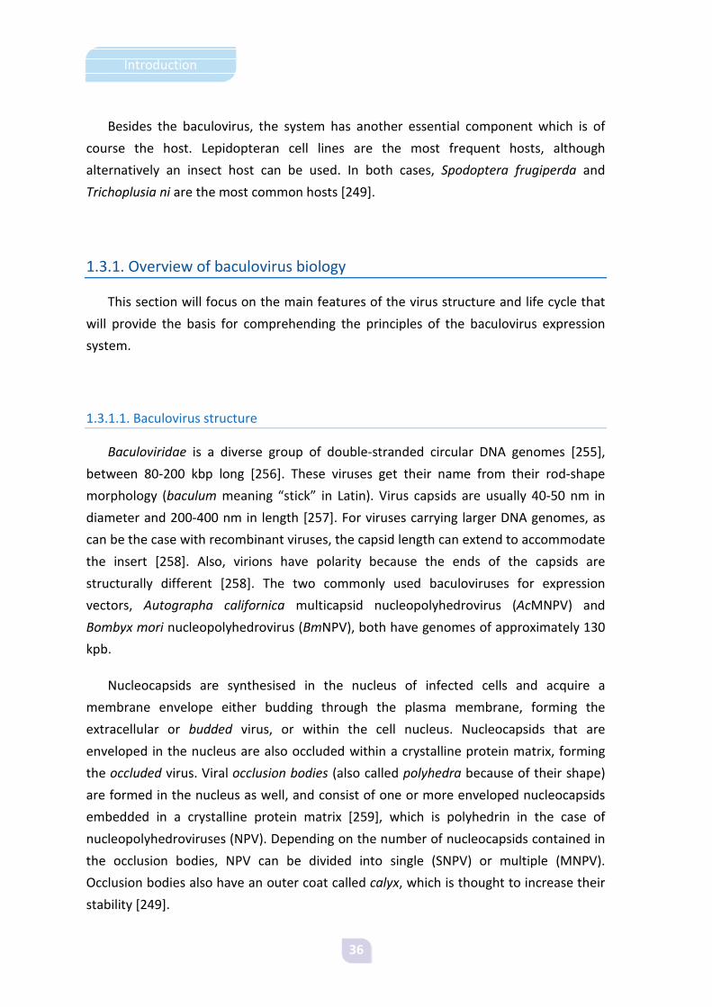
Nucleocapsids are synthesised in the nucleus of infected cells and acquire a
membrane envelope either budding through the plasma membrane, forming the
extracellular or budded virus, or within the cell nucleus. Nucleocapsids that are
enveloped in the nucleus are also occluded within a crystalline protein matrix, forming
the occluded virus. Viral occlusion bodies (also called polyhedra because of their shape)
are formed in the nucleus as well, and consist of one or more enveloped nucleocapsids
embedded in a crystalline protein matrix [259], which is polyhedrin in the case of
nucleopolyhedroviruses (NPV). Depending on the number of nucleocapsids contained in
the occlusion bodies, NPV can be divided into single (SNPV) or multiple (MNPV).
Occlusion bodies also have an outer coat called calyx, which is thought to increase their
stability [249].

37
Introduction
Figure 7. Structure of the different forms of multinucleopolyhedroviruses
throughout their life cycle.
Adapted from Wikipedia.
Although nucleocapsids are thought to be identical in both budded and occluded
viruses, their membranes are biochemically different. Budded viruses have projections in
one end of their structure, called peplomers, that contain the glycoprotein gp64 which is
absent in occluded viruses. Protein gp64 is involved in virus entry into cells by
endocytosis during secondary infection [260], while enveloped viruses liberated from
occlusion bodies enter cells by a different route [261]. Also, the O-glycosylated protein
gp41 and protein p74 are present in occluded virus but not in the budded form.
A second type of occluded baculovirus exists in the baculovirus family. These are
called the granulosis viruses (GV), and in contrast to NPV they have only a single virion
embedded in a very small occlusion body. In this case, the matrix protein is granulin.
Moreover, some baculoviruses do not synthesise an occluded form, and are
consequently name nonoccluded baculoviruses.
1.3.1.2. Infection progress
Infection in the insect has two distinct phases. Primary infection is caused when
larvae ingest polyhedra as contaminants of their food. Upon arrival to the insect midgut,
polyhedra are dissolved in the alkaline environment and release the embedded virions
[262], which enter midgut cells after fusing to the membrane of the microvilli [263]. This
takes places during the early phase of infection, when cells are reprogrammed for virus
replication.

38
Introduction
Figure 8. Baculovirus infection of an insect host.
Adapted from http://www.microbiologybytes.com /virology/kalmakoff/baculo/baculo.html
Nucleocapsids can then be transported to the nucleus, where they replicate, or to
the basal side of the cells for rapid budding [264]. During the secondary phase of the
infection both budded viruses and polyhedra are produced. The late phase of infection is
characterised by extensive DNA replication and release of budded virus [249]. Released
virions reach the hemocoel and are transported via the hemolymph to other tissues,
causing a systemic infection [249].
Figure 9. Phases of baculovirus infection.
Adapted from http://www.microbiologybytes.com /virology/kalmakoff/baculo/baculo.html
The very late phase of infection is characterised by hyperexpression of polyhedrin
and P10 [263]. During this phase polyhedra accumulate in the nucleus and the
production of budded virus is greatly reduced, if not terminated [265]. By the end of the
infection larvae liquefy due to extensive cell lysis, in which P10 protein is involved
[266;267]. The insect literally melts, becoming a sac of milky fluid containing polyhedra
which are released to the environment upon cuticle breakage. Because polyhedra are
relatively stable in the environment, they can reinitiate the infection cycle when
consumed by a new host.

39
Introduction
1.3.2. Expression vectors
The classic baculovirus expression vector consists of a recombinant baculovirus
genome which contains a foreign nucleic acid sequence encoding the target protein
under the control of a polyhedrin promoter. The heterologous gene is generally placed
in the polyhedrin locus of the viral genome, replacing the wild-type polyhedrin. This
recombinant baculovirus can be used to infect cultured insect cells or larvae, yielding
high transcription levels during the very late phase of infection, which is usually
translated to high levels of recombinant protein production.
Because baculovirus genomes are large, they usually contain one or more
recognition sites for restriction endonucleases. By the time that these first baculovirus
vectors were being developed no known restriction enzymes that lacked recognition
sites in the genome had been described, so homologous recombination was the chosen
method to insert the foreign genes into the baculovirus genome [247;248]. This method
involved the construction of a “transfer” plasmid containing the heterologous gene
flanked by baculoviral sequences homologous to the polyhedrin locus, which would then
be cotransfected into cultured cells together with purified genomic DNA of wild-type
AcMNPV. However, the process was highly inefficient because a double crossover
recombination was necessary to knock out the polyhedrin gene while knocking-in the
gene encoding the target protein, so only about 0.1% recombinants were obtained
[250]. Plaque assays were required to isolate the small amount of recombinant
baculoviruses from the large parental background, and then visual screening for the
polyhedron-negative phenotype allowed for identification of recombinant virus.
However, this was a critical step constraining the use of the system, as identifying the
recombinants could be a difficult task.
Figure 10. Baculovirus expression vector obtained by homologous recombination.
Adapted from Methods Enzymol. 2009;463:191-222.

40
Introduction
To overcome these technical limitations, and also to improve the system in other
ways, many modifications have been developed over the years, involving both the
parental genomes and the transfer plasmids.
1.3.2.1. Transfer plasmids
Transfer plasmids are used to transfer the foreign gene into the viral genome by
means of homologous recombination [249]. A typical transfer plasmid contains the gene
of interest under control of a baculovirus promoter (which is often polyhedrin) and
flanked by sufficient amount of viral DNA to allow recombination.
Figure 11. Baculovirus transfer plasmid. Adapted from Methods Enzymol. 2009;463:191-222.
Several factors must be considered when cloning the gene of interest into the
transfer plasmid. First, it is important to use genes without introns because although low
levels of splicing have been reported [268] strong protein expression has not been
observed from spliced mRNAs. Also, the AUG context is important for initiation of
translation [249]. The AUG contexts for several promoters are shown in Table 2.
Adapted from Baculovirus Expression Vectors. A Laboratory Manual. Oxford University Press, 1994.
Table 2. AUG contexts of highly expressed AcMNPV proteins.
Gene Transcription AUG context Reference polh Very late CCUAUAAAUAUGCCGG [269] p10 Very late UUUACAAUCAUGUCAA [270] p6.9 Late AAUUUAAACAUGGUUU [271] vp39 Late GGCAACAAUAUGGCGC [272]Consensus A YAUG Y

41
Introduction
The choice of promoter is also important, as it will determine the production levels
of the target protein. Polyhedrin is a common strong promoter which is equally efficient
in either orientation with respect to the AcMNPV genome [273]. Moreover, two
polyhedrin promoters or two very late promoters (e.g., polh and p10) work at almost
optimal efficiency when placed back-to-back to drive expression of two different genes
[274-276]. The p10 promoter has a similar strength to polh. Both are very late promoters
with essential TAAG sequences at their transcriptional start point, and the region from
around the initiation point to the ATG is sufficient to promote high transcription levels
[249;275]. Although these promoters are very effective, a decline in the level of post-
translational modifications at very late times post infection has been reported [277;278].
Thus, the use of late promoters such as vp39 or p6.9 may present an advantage when
uniformity of post-translational modifications is important because the proteins will
have additional hours to move through the endoplasmic reticulum and Golgi apparatus,
in spite of the lower yields that will be obtained with the use of such promoters [249].
Early promoters such as ie1 have also been used, and although these promoters drive
lower levels of transcription they seem to promote higher quality products, being
especially useful for secreted proteins [279-284].
Polyadenylation signals are also required for viral transcripts to be processed at their
3’ end [249]. The polyadenylation signal for polyhedrin is located in the downstream
orf1629 [285], and because this is an essential gene polh deletions in polyhedrin-based
transfer plasmids do not extend into this orf. Therefore, the polh polyadenylation site is
maintained in the viral genome, so it will not be necessary to include a polyadenylation
signal in the transfer plasmid. For transfer plasmids supplying back-to-back promoters in
the polyhedrin region, transcripts extending in opposite direction to the wild-type
polyhedrin gene transcription are expected to terminate at a polyadenylation signal at
the 3’ end of the flanking orf603 [286;287]. For p10-based vectors, a polyadenylation
signal is located downstream the stop codon, and this is usually included in the available
transfer plasmids [249].
A bias in codon usage has been described for highly expressed baculovirus genes,
such as polyhedrin or p10. However, heterologous genes with rare codons can be well
expressed in the baculovirus system, as reported for E. coli β-galactosidase [247].
Nonetheless, the UAA codon is preferred for termination, as it is used for most AcMNPV
genes [249].

42
Introduction
The main objective of modifying transfer plasmids was to facilitate identification of
recombinant baculovirus plaques by visual screening. For that purpose, marker genes
such as E. coli β-galactosidase were introduced under the control of baculovirus
promoters [288]. However, this could be a trap because the presence of the marker
gene could indicate a single crossover homologous recombination, which produces
recombinant baculoviruses containing the entire transfer plasmid and thus being
genetically unstable. For this reason, further screening would be required to map the
position of the foreign gene in the baculovirus genome and confirm that a double
crossover recombination event had taken place.
A second modification of transfer plasmids was aimed at facilitating expression and
purification of the recombinant protein. This included addition of sequences such as
secretory signal peptides or purification tags, as well as replacing the polyhedrin
promoter with alternate baculovirus promoters or multiple promoter elements that
would allow coexpression of multiple recombinant proteins in the same cell during
infection [289], as discussed.
1.3.2.2. Parental genomes
Modifications in the parental genomes have been addressed to solve technical
problems related to isolation of recombinant viruses and to enhance the production of
the target protein.
I. Enhancing recombination efficiency
The first important step towards increasing the efficiency of recombination was
made with the development of a baculovirus with a linearisable DNA genome [290]. This
was achieved by introducing a unique Bsu36I restriction site in the polyhedrin locus.
Linearising the parental DNA prevented its replication, which reduced the number of
parental virus after recombination. Homologous recombination was still possible, and
indeed restored the ability of the baculovirus vector to replicate. This approach
increased the efficiency of baculovirus vector production up to 10-20%.
The next improvement was the development of BakPAK6TM, a recombinant
baculovirus that could be gapped with Bsu36I deleting a portion of orf1629, which

43
Introduction
encodes an essential phosphoprotein of the viral nucleocapsid [291], and that also
included an E. coli lacZ gene which allowed for easy detection of recombinants as the
white plaques on a blue background [292]. In this case, recombinant baculovirus
production increased to about 95%. This was commercialised by ClonTech.
Figure 12. Baculovirus expression vector obtained by homologous recombination with a linearised/gapped parental viral genome.
Adapted from Methods Enzymol. 2009;463:191-222.
In parallel to linearisable genomes, another approach based on genetic transposition
was developed [293]. Key to this method was the creation of a new E. coli strain that
contained an autonomously replicating bacmid which included a copy of the entire
baculovirus genome and a helper plasmid encoding a transposase. The bacmid
contained an E. coli lacZ gene and a “mini-Att Tn7” site, an attachment site used during
transposition. The transfer plamid contained the target gene flanked by the ends of Tn7,
and thus could be transposed to the polyhedrin locus of the bacmid when transformed
into the bacteria. The lacZ gene would be knocked out of the bacmid upon transposition,
and the recombinants could be selected by standard blue-white screening. This system is

44
Introduction
commercialised as Bac-to-BacTM by Invitrogen, and provides a 100% efficiency of
recombinant baculovirus production. However, recombinant viruses are genetically
unstable upon passage in insect cells, seemingly because they retain the bacterial
replicon [294].
Figure 13. Baculovirus expression vector obtained by transposition.
Adapted from Methods Enzymol. 2009;463:191-222.
Recently, a new method consisting of cross-hybridising the linearisable baculoviral
DNA and bacmid strategies has been developed [295]. This approach relies on a bacmid
that contains a recombinant baculoviral genome with a bacterial replicon in the

45
Introduction
polyhedrin locus and a deletion in the orf1629 gene. The bacmid can replicate in E. coli
but not in insect cells, so it can easily be produced in E. coli and used to cotransfect
insect cells together with the transfer plasmid. Homologous recombination restores the
orf1629 deletion, knocking-in the gene of interest and at the same time knocking out the
bacterial replicon. This is marketed under the name of flashBACTM by Oxford Expression
Techonologies, and yields very high levels of recombinant baculovirus production.
However, despite the bacmid not being able to replicate in insect cells, progeny derived
from the defective parental viral genome can be obtained by genetic complementation
when the orf1629 product is provided in trans by the recombinant virus. For this reason,
plaque assay is still recommended to purify the recombinant virus.
Figure 14. Baculovirus expression vector obtained using the flashBAC method.
Adapted from Methods Enzymol. 2009;463:191-222.
Baculovirus vectors can also be produced in vitro by site-specific recombination
[246]. A prelinearised virus genome contains an E. coli lacZ gene and a herpes simplex
virus thymidine kinase gene flanked by site-specific recombination sites from
bacteriophage lambda (attR1 and attR2) replacing the polyhedrin coding sequence. The
target gene is encoded in an entry plasmid, flanked by recombination sites attL1 and
attL2. Genome and plasmid are mixed in vitro in presence of a purified recombinase to
obtain the recombinant baculovirus. The mixture is transfected into insect cells, which
are cultured in presence of gancyclovir to select against replication of parental viral DNA.
This is commercialised by Invitrogen as BaculoDirectTM.

46
Introduction
Figure 15. Baculovirus expression vector obtained using BaculoDirect.
Adapted from Methods Enzymol. 2009;463:191-222.
II. Improving protein production
The general approach used to improve protein production in the baculovirus system
consists of deleting nonessential genes that are thought to interfere with heterologous
protein production or to degrade the target protein. However, addition of new
heterologous genes to the baculovirus genome has also been reported.
Chitinase [296] and cathepsin-like protease [297] have been deleted in several
commercial vectors, and although the impact of these deletions is not totally clear less
degradation of foreign glycoproteins has been shown [298]. Chitinase is a resident
endoplasmic reticulum protein [299] thought to interfere with protein secretion by
saturation of the host translocation machinery [298]. Thus, deletion of the chitinase
gene is expected to increase the yields of secreted proteins.
Parental baculovirus DNA lacking a functional p10 gene is also available commercially
under the name of DiamondBacTM. As p10 is involved in cell lysis [266], it is expected
that infected cells will retain higher viabilities throughout the course of infection.
Moreover, in DiamondBac the p10 gene has been replaced by a protein disulfide

47
Introduction
isomerase (PDI), a chaperone that drives disulfide bridge formation, thus increasing
solubility and secretion of the target protein [300].
Other baculovirus vectors encoding heterologous protein processing enzymes have
also been described. Polydnavirus vankyrin gene under control of the p10 promoter
[301] has been found to prolong the viability of Sf9 cells infected with baculovirus, which
can thereby enhance the production of the target protein. In this line, heterologous
glycosyltransferases [302;303] or enzymes involved in CMP-sialic acid biosynthesis [304]
under the control of baculovirus ie1 promoters have been used to expand the processing
capabilities of the baculovirus system.
Production of multi-subunit complexes has also been addressed. Although transfer
plasmids allow coproduction of several proteins, the number of genes that can be
inserted in the plasmid is limited by its size. Moreover, the use of repeated sequences
such as promoters or terminators can result in recombination events [294;305]. As a
solution, a new system allowing each protein in the complex to be expressed from
different loci has been developed [18]. Because single gene insertions are distributed
along the genome, these problems are overcome. Furthermore, the system is based on
lambda red recombination [306], which allows fast generation of recombinants in E. coli.
1.3.3. Insect hosts
Insect hosts constitute the second half of the baculovirus system. Lepidopteran
insects are hosts for many viruses from the Baculoviridae family, including AcMNPV.
Although cell lines are the most frequent choice at laboratory scale, insect larvae
provide an interesting alternative to cell culture scale-up for producing large amounts of
recombinant protein, with the added advantage of reduced production costs.
1.3.3.1. Cell lines
The first established lepidopteran cell lines were described by Grace in 1962 [307],
and so far over 250 insect cell lines have been described [308]. Two of the most common
cells used with AcMNPV vectors are Sf9 and Sf21 cell lines, both originated from IPLB-SF-
21 cells derived from pupal ovarian tissue from the fall armyworm Spodoptera
frugiperda [309]. The other common cell line originated from adult ovarian cells of the

48
Introduction
cabbage looper Trichoplusia ni, which was originally described as BTI Tn 5B-1 [310;311]
and is now marketed by Invitrogen as High FiveTM.
These cell lines can grow in adherent and suspension cultures, and thus can be easily
scaled-up in shake flasks, spinner flasks, or bioreactors to obtain large amounts of
recombinant proteins [312;313]. Moreover, Sf9 and Sf21 cells are also routinely used to
plaque purify and quantify recombinant baculovirus vectors.
Insect cell cultures grow at an optimal temperature of 28 °C. Since the cells are
loosely adherent neither trypsin nor EDTA is required for subculture. Also, CO2
incubators are not necessary because insect cell culture media are buffered with
phosphate instead of carbonate. Moreover, cells can grow both in media supplemented
with serum or in serum-free media, both of which are commercially available.
Currently, transgenic insect cell lines are already in the market. One of the most
important modifications has been the introduction of constitutively expressed
mammalian genes involved in post-translational processing, with the aim of obtaining
partially humanised glycosylations [314-318]. A Sf9-derived insect cell line with an
extended N-glycosylation pathway is commercialised by Invitrogen under the name of
MIMICTM [317].
Another transgenic Sf9 cell line derivative contains a polydnavirus vankyrin gene
expressed constitutively under the control of an immediate-early baculovirus promoter,
which enhances the life span of the cells [301]. Three vankyrin-enhanced Sf9 derivatives
are already marketed by ParaTechs.
1.3.3.2. Insect larvae
Although proteins produced in cell culture are easier to purify and usually have more
uniform post-translational modifications than those obtained in insect larvae as a result
of only one cell type being involved in protein synthesis, the main drawback of scaling-
up protein production in cell culture is the cost. As culture media are expensive, the use
of large volumes may become prohibitive. Moreover, bioreactors will often be required
to handle large culture volumes, which will add to the cost of the production process
[249].
Larvae offer the advantage of being cheaper to maintain because they do not require
growth media or sterile conditions [319]. However, protein production in larvae will

49
Introduction
require feeding and handling living insects. Moreover, protein purification may become
more difficult due to the presence of insect parts as contaminating products.
Nevertheless, although yields of recombinant proteins produced in insect larvae can be
reduced due to protein aggregation [320], larvae can still be regarded as natural
bioreactors for recombinant protein production.
In addition to Spodoptera frugiperda and Trichoplusia ni larvae being used as hosts,
Bombyx mori larvae are also commonly used, mainly due to the inability of growing large
culture volumes of Bombyx mori cells [249].

50
Introduction
1.4. Model proteins
Several proteins have been used in this study as reporters of protein aggregation and
conformational quality. Our main model protein has been a chimeric fusion protein
between the aggregation-prone VP1 capsid protein of foot-and-mouth disease virus
(FMDV) and the green fluorescent protein (GFP). Nonetheless, for some specific
experiments VP1 has also been used without the fluorescence reporter, along with
FMDV VP2 and human α-galactosidase.
1.4.1. Green Fluorescent Protein
In 1962, Shimomura reported the existence of a green fluorescent protein in the
jellyfish Aequorea victoria [321]. In nature, this protein absorbs the blue
bioluminescence of its partner protein, the calcium activated aequorin, and converts it
to the greenish glow observed in living animals [322]. Although green fluorescent
proteins exist in other organisms [323] and recently GFPs from Renilla mullerei, Renilla
reniformis and Ptilosarcus gurneyi have been cloned and patented [324], the Aequorea
GFP gene was the first to be cloned [325] and expressed in heterologous systems
[326;327] and thus is the most widely used today. GFP is still fluorescent without the
need of jellyfish-specific enzymes or cofactors; therefore, the gene contains all the
necessary information for the correct formation of the chromophore.
Wild-type GFP consists of a single chain of 238 amino acid residues which is highly
stable and resistant to proteolysis and has two absorption maxima at about 395 and 475
nm, with excitation at the major peak of 395 nm yielding an emission maximum at 508
nm [328]. The structure of GFP is an 11-stranded β-barrel wrapped around a single α-
helix where the chromophore attaches, remaining buried in the centre of the cylinder,
which has been called a β-can [328;329]. The barrel forms an almost perfect cylinder
which is 42 Å long and 24 Å in diameter [328].
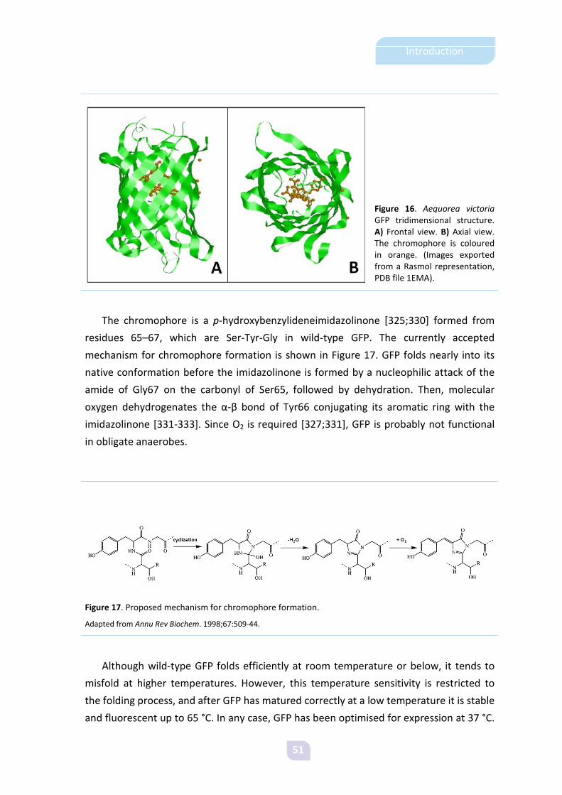
51
Introduction
Figure 16. Aequorea victoria GFP tridimensional structure. A) Frontal view. B) Axial view. The chromophore is coloured in orange. (Images exported from a Rasmol representation, PDB file 1EMA).
The chromophore is a p-hydroxybenzylideneimidazolinone [325;330] formed from
residues 65–67, which are Ser-Tyr-Gly in wild-type GFP. The currently accepted
mechanism for chromophore formation is shown in Figure 17. GFP folds nearly into its
native conformation before the imidazolinone is formed by a nucleophilic attack of the
amide of Gly67 on the carbonyl of Ser65, followed by dehydration. Then, molecular
oxygen dehydrogenates the α-β bond of Tyr66 conjugating its aromatic ring with the
imidazolinone [331-333]. Since O2 is required [327;331], GFP is probably not functional
in obligate anaerobes.
Figure 17. Proposed mechanism for chromophore formation.
Adapted from Annu Rev Biochem. 1998;67:509-44.
Although wild-type GFP folds efficiently at room temperature or below, it tends to
misfold at higher temperatures. However, this temperature sensitivity is restricted to
the folding process, and after GFP has matured correctly at a low temperature it is stable
and fluorescent up to 65 °C. In any case, GFP has been optimised for expression at 37 °C.
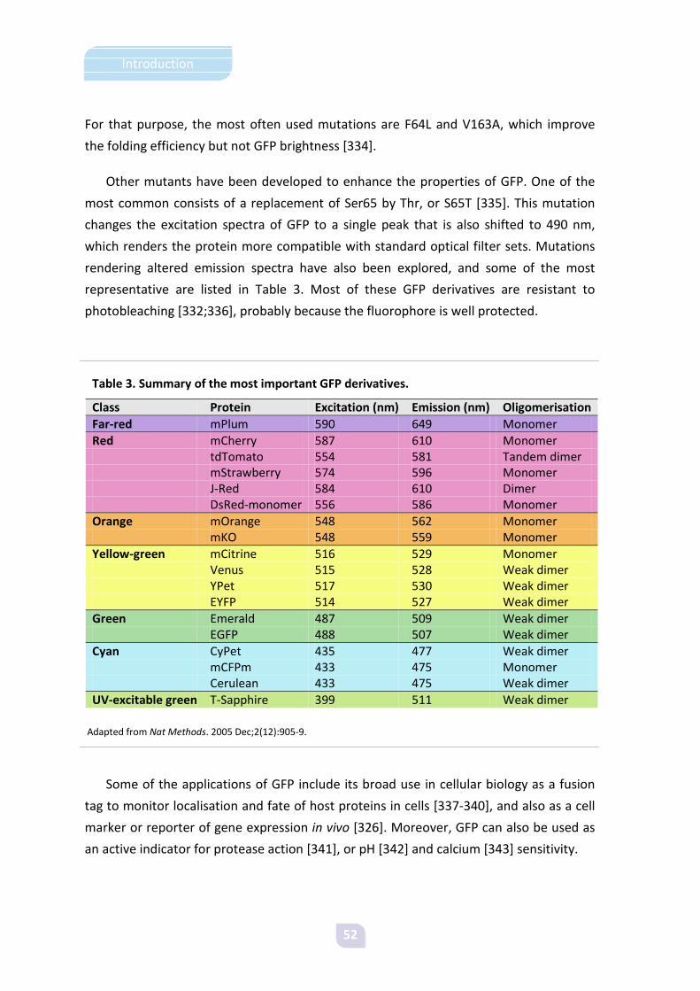
52
Introduction
For that purpose, the most often used mutations are F64L and V163A, which improve
the folding efficiency but not GFP brightness [334].
Other mutants have been developed to enhance the properties of GFP. One of the
most common consists of a replacement of Ser65 by Thr, or S65T [335]. This mutation
changes the excitation spectra of GFP to a single peak that is also shifted to 490 nm,
which renders the protein more compatible with standard optical filter sets. Mutations
rendering altered emission spectra have also been explored, and some of the most
representative are listed in Table 3. Most of these GFP derivatives are resistant to
photobleaching [332;336], probably because the fluorophore is well protected.
Table 3. Summary of the most important GFP derivatives.
Class Protein Excitation (nm) Emission (nm) Oligomerisation Far-red mPlum 590 649 Monomer Red mCherry 587 610 Monomer tdTomato 554 581 Tandem dimer mStrawberry 574 596 Monomer J-Red 584 610 Dimer DsRed-monomer 556 586 Monomer Orange mOrange 548 562 Monomer mKO 548 559 Monomer Yellow-green mCitrine 516 529 Monomer Venus 515 528 Weak dimer YPet 517 530 Weak dimer EYFP 514 527 Weak dimer Green Emerald 487 509 Weak dimer EGFP 488 507 Weak dimer Cyan CyPet 435 477 Weak dimer mCFPm 433 475 Monomer Cerulean 433 475 Weak dimer UV-excitable green T-Sapphire 399 511 Weak dimer
Adapted from Nat Methods. 2005 Dec;2(12):905-9.
Some of the applications of GFP include its broad use in cellular biology as a fusion
tag to monitor localisation and fate of host proteins in cells [337-340], and also as a cell
marker or reporter of gene expression in vivo [326]. Moreover, GFP can also be used as
an active indicator for protease action [341], or pH [342] and calcium [343] sensitivity.

53
Introduction
1.4.2. Foot-and-Mouth Disease Virus VP1 and VP2 capsid proteins
The virion of foot-and-mouth disease virus contains 60 copies each of the four
structural proteins forming the capsid. Three of these proteins, VP1, VP2 and VP3, are
wedge-shaped, eight-stranded β-sandwiches partially exposed to the surface while VP4
and the N termini of VP1 and VP3 are located at the capsid interior [344].
The surface of the particle is fairly smooth with a major protruding element in VP1,
which is called the G-H loop. This loop is highly flexible and comprises about 20 residues
around positions 140-160 [345]. The G-H loop also contains a highly conserved Arg-Gly-
Asp (RGD) triplet that interacts with integrin receptors in the cell surface, and the major
antigenic site [346-348]. For serotype C, different overlapping epitopes have been
mapped in the G-H loop [348]. The highly exposed C terminus of VP1 has also been
related to both the antigenic and receptor binding properties of the virus [349;350].
Both VP1 and VP2 proteins used in this study are from serotype C (isolate C-S8c1) of
the virus.
Figure 18. A) FMDV virion structure. The capsid is composed of 12 pentamers with 5 protomers each (adapted from illustration by David S. Goodsell of The Scripps Research Institute) and B) Ribbon representation of the capsid proteins forming a protomer. (Image exported from a Rasmol representation, PDB file 1FMD).

54
Introduction
1.4.3. Human α-Galactosidase
The enzyme α-galactosidase is responsible of removing galactose from glycosylated
macromolecules in the lysosomes [351]. The absence of functional α-galactosidase in
humans results in a condition known as Fabry disease, a lysosomal storage disease
caused by the accumulation of the enzyme’s substrates in the tissues. In addition, some
α-galactosidase mutations have been associated to enzyme aggregation in Fabry
condition [352].
Human α-galactosidase is a homodimeric glycoprotein with each monomer
consisting of two domains, a (β/α)8 barrel that contains the active site and a C-terminal
domain formed of eight antiparallel β strands on two sheets in a β sandwich [353].
Figure 19. Human α-galactosidase tridimensional structure. (Image exported from a Rasmol representation, PDB file 1R46).

55
Introduction
1.5. Previous work
The objectives of this thesis were defined after a first study that involved
coproduction of folding modulators with the aim of determining their impact on both
protein solubility and conformational quality (see Annex I).
Background to that study was the controversy of results obtained after coproduction
of folding modulators, which although expected to increase solubility often resulted in
inconsistent results that were attributed to particular requirements for folding
modulators or to specific features of particular proteins. Moreover, reports of active
inclusion bodies were becoming increasingly frequent. Thus, we decided to explore
whether solubility was a good indicator of protein quality, or instead biological activity
would be a more suitable parameter.
For that purpose, we coproduced a recombinant GFP together with the DnaKJ
chaperone pair and analysed the impact of the folding modulators both on the
fractioning and conformational quality of the reporter protein. The experimental
approach consisted of determining protein and fluorescence levels in both soluble and
insoluble protein fractions that had been produced with or without the chaperone pair
and under different gene expression conditions modulated by the inducer
concentration. Our results indicated a different impact of the chaperones on protein
solubility and quality, as while solubility of the protein was only affected by its own yield,
DnaK promoted a quality enhancement at low production levels, which was impaired by
a chaperone excess that also resulted in proteolysis of the recombinant protein.
Moreover, soluble and insoluble protein populations displayed a coincident quality
profile, and the variability observed for the soluble fraction was associated to the
existence of oligomers in that population.
Thus, the results of this work prompted us to disregard solubility as a good indicator
of protein quality, since these parameters were divergently controlled by DnaK.


Objectives
2.


59
Objectives
The aim of the study was to explore the patent divergence in the control of protein
solubility and conformational quality observed in bacterial cells actively producing
recombinant proteins and test whether this principle is also true for eukaryotic systems.
For that purpose, we set the following objectives:
1. Explore conditions that can enhance simultaneously solubility and
conformational quality of recombinant proteins.
2. Study the conformational quality of soluble recombinant proteins by
determining:
a. Biological activity
b. Extent of native-like structure
3. Construct vectors that allow production of our model proteins in a eukaryotic
system.
4. Confirm whether conditions enhancing solubility and conformational quality in
bacterial systems are also valid in a eukaryotic system.
5. Determine the effect of the major bacterial folding modulator, the chaperone
DnaK, on protein quality and solubility in an environment that does not support
its associated proteolytic activity.


Results
3.


63
Results
3.1. Article 1
Yield, solubility and conformational quality of soluble proteins are not
simultaneously favored in recombinant Escherichia coli.
Mónica Martínez-Alonso, Elena García-Fruitós, Antonio Villaverde.
Biotechnology and Bioengineering, Vol. 101, No 6, 1353-8 (December 2008).
In this work we pursued the first objective of the study. Since solubility and quality
are divergently controlled we explored whether it was possible to engineer them
independently to simultaneously enhance both parameters.
The experimental design consisted of a two-step approach that combined genetic
and process engineering. Since our purpose was to improve solubility of recombinant
proteins, we chose a genetic background where our model protein was obtained mainly
as highly functional inclusion bodies. Solubility was enhanced by appropriately adjusting
growth temperature and gene expression rate. However, conditions promoting high
protein yields resulted in poor conformational quality of the recombinant protein. Thus,
since high yields of soluble and active protein cannot be gained simultaneously, the
requirement of either solubility or functionality for a determined protein must be clearly
established before designing its production process.


65
Results
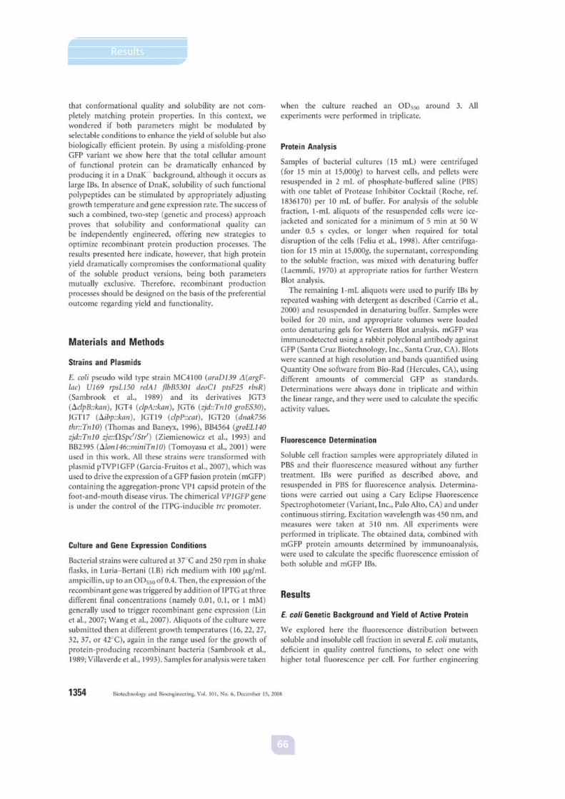
66
Results
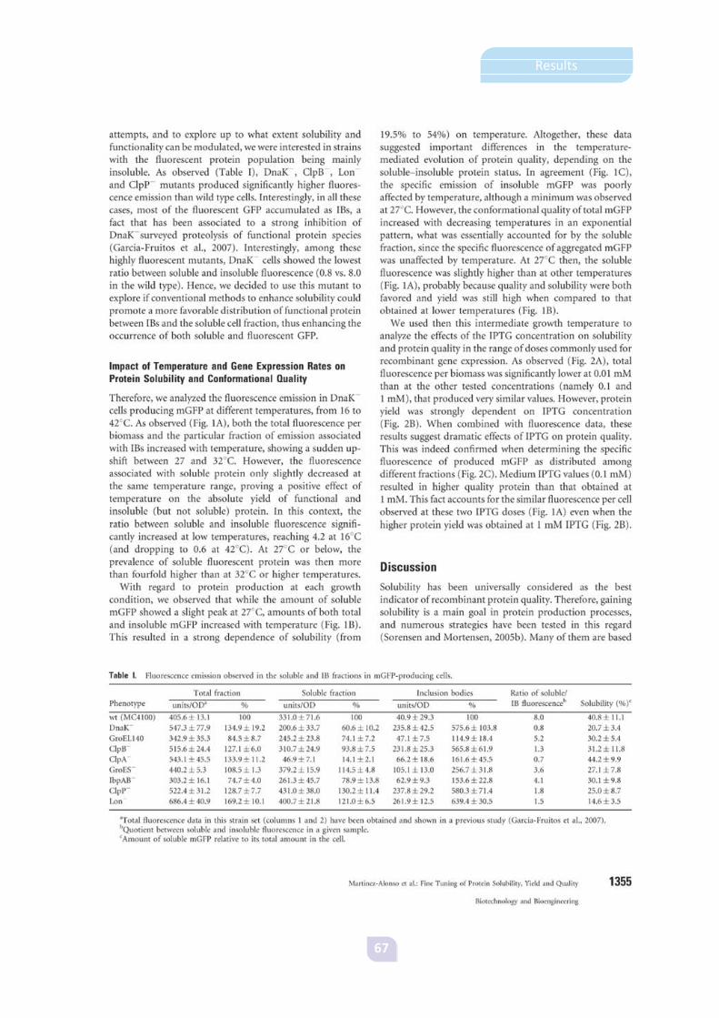
67
Results

68
Results

69
Results

70
Results

71
Results
3.2. Article 2
The functional quality of soluble recombinant polypeptides produced in
Escherichia coli is defined by a wide conformational spectrum.
Mónica Martínez-Alonso, Nuria González-Montalbán, Elena García-Fruitós and
Antonio Villaverde.
Applied and Environmental Microbiology, Vol. 74, No 23, 7431-3 (December 2008).
The finding of oligomeric versions in the soluble fraction of our recombinant protein
prompted us to further characterise this protein population in terms of functional
quality and molecular organisation.
To that end, we analysed the distribution of the soluble fraction of an aggregation-
prone recombinant GFP along a sucrose density gradient. The protein widely dispersed
along the gradient, indicating the presence of differently sized species within the soluble
population. Furthermore, the fluorescence profile did not match the protein
distribution, indicative of a variable functional status in the soluble fraction. Further
purification of one of the protein species observed in the gradient still resulted in a
heterogeneous population of microaggregates, as observed by transmission electron
microscopy. These soluble aggregates were also heterogeneous regarding their
secondary structure, as evidenced by the presence of both non native and native-like
conformations. However, the prevalence of native-like structures accounted for the
higher functionality of the soluble protein compared to inclusion bodies. Being
structurally more homogeneous than their soluble counterparts, IBs can be regarded as
a narrow subpopulation among the total recombinant protein species. Therefore, the
observed protein quality can be regarded as a statistical average of all the existing
protein species.


73
Results

74
Results

75
Results


77
Results
3.3. Article 3
Learning about protein solubility from bacterial inclusion bodies.
Mónica Martínez-Alonso, Nuria González-Montalbán, Elena García-Fruitós and
Antonio Villaverde.
Microbial Cell Factories, Vol. 8, 4-8 (January 2009).
In this work we summarised all the recent findings that support a new concept of
protein quality, which can no longer be linked to solubility.
Although formerly believed to be insoluble deposits of inactive protein, inclusion
bodies are actually rich in functional protein species with native secondary structure.
This opens avenues for a straightforward application of enzymatic inclusion bodies as
catalysers for industrial bioprocesses. Moreover, easy extraction of active polypeptides
has been achieved without the need of complex refolding procedures after adequate
engineering of IB protein quality, which may translate in enhanced in vitro release of
functional protein.
Soluble protein can no longer be thought of as a homogeneous population of protein
species either, since the existence of soluble aggregates with variable functional
conformations prompts to consider recombinant proteins as a “continuum of forms”
rather than the classic soluble and insoluble cell fractions.
In addition, protein production should be targeted to yield, quality or solubility of
the recombinant product, as these parameters are under a divergent control and cannot
be enhanced at the same time.


79
Results

80
Results

81
Results

82
Results

83
Results


85
Results
3.4. Article 4
Rehosting of bacterial chaperones for high-quality protein production.
Mónica Martínez-Alonso, Verónica Toledo-Rubio, Rob Noad, Ugutz Unzueta,
Neus Ferrer-Miralles, Polly Roy, Antonio Villaverde.
Applied and Environmental Microbiology, Vol. 75, No 24, 7850-4 (December 2009).
Although coproduction of folding modulators has been a widely tested strategy to
improve soluble protein production in bacteria, the results obtained have been
controversial. Promising sets of folding modulators usually include the chaperone pair
DnaKJ. However, besides their folding activity DnaKJ also act as proteolytic enhancers in
cooperation with bacterial proteases, which may account, at least partially, for some of
the negative results obtained when coproducing folding modulators in an attempt to
increase protein solubility. Since the DnaKJ pair has been widely conserved in evolution,
we envisaged rehosting of this chaperone set as a way to uncouple their valuable
foldase activity from the associated proteolysis observed in bacterial systems. Using
again our recombinant GFP as a model protein, we constructed baculovirus vectors that
allowed its production either alone or together with the chaperone pair upon infection
of insect cells. Deposition of the target protein in insoluble but fluorescent clusters was
in agreement with solubility and quality not being coincident parameters. When we
coproduced the chaperone pair and evaluated solubility and conformational quality of
the reporter protein, we observed enhanced yield and biological activity. Also, stability
was increased compared to when the protein was produced in E. coli, indicative of no
DnaK-mediated proteolysis. However, in agreement with our observations for bacterial
systems, yield and quality of the recombinant protein could not be increased in parallel
in this eukaryotic system either. Positive effects of this set of bacterial folding
modulators were also observed for the production of three other different proteins.


87
Results

88
Results

89
Results

90
Results

91
Results
