engineering nanomaterial surfaces for biomedical...
TRANSCRIPT

`
1
NE 489 Nano-Biomaterials
Term Project Paper Winter 2010
Engineering Nanomaterial Surfaces for Biomedical Applications
Rajesh Swaminathan
#20194189
1 Overview
Owing to their unique optical, electronic, magnetic, mechanical and chemical properties, an
extensive amount of research has been done on nanomaterials. These advantageous
properties can be attributed solely to their special size and shape. Furthermore,
nanomaterials can be very easily functionalized on the surface with synthetic ligands to
effect significant change in properties depending on the application at hand. In this respect,
nanomaterials are very versatile and simple to use.
Nanomaterials have proven themselves to have a significant range of application in a
variety of aspects of biology and medicine. Applications involving nanomaterials include
(but not limited to) bio-molecular sensing, biological imaging, drug delivery vehicles in the
form of micelles and vesicles, disease therapy, and as scaffolds in tissue engineering
applications.
The most critical part of nanomaterials that makes them special is their surface. It is the
surface that makes the nanomaterials significantly more useful than conventional non-
nano materials. As the size of the material decreases, its surface-to-volume ratio increases.
`
2
This presents considerable advantage to modify properties of nanomaterials through
surface functionalization techniques.
There are several different categories of nanomaterials. Not surprisingly, the field of
nanomaterials is quite vast, diverse and evolving rapidly as tremendous amounts of
research is being conducted in this burgeoning area. In this paper, we will focus exclusively
on classes of nanomaterials that are biomedically important. These materials are metals,
semiconductors and nanotubes. The four important biomedically important classes of
materials are:
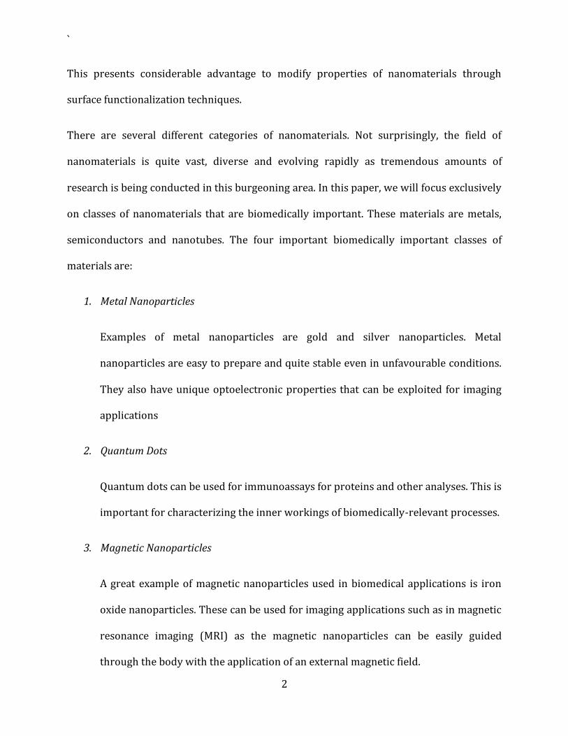
1. Metal Nanoparticles
Examples of metal nanoparticles are gold and silver nanoparticles. Metal
nanoparticles are easy to prepare and quite stable even in unfavourable conditions.
They also have unique optoelectronic properties that can be exploited for imaging
applications
2. Quantum Dots
Quantum dots can be used for immunoassays for proteins and other analyses. This is
important for characterizing the inner workings of biomedically-relevant processes.
3. Magnetic Nanoparticles
A great example of magnetic nanoparticles used in biomedical applications is iron
oxide nanoparticles. These can be used for imaging applications such as in magnetic
resonance imaging (MRI) as the magnetic nanoparticles can be easily guided
through the body with the application of an external magnetic field.
`
3
4. Carbon Nanotubes
Carbon nanotubes (CNTs) such as single-walled carbon nanotubes and mutil-walled
carbon nanotubes have excellent electrical and thermal conductivity. They are also
extremely strong mechanically, and very stable chemically. To top this, they are also
organic and therefore bio-compatible. They can therefore be used in a variety of
biomedical applications as carriers or conductors.
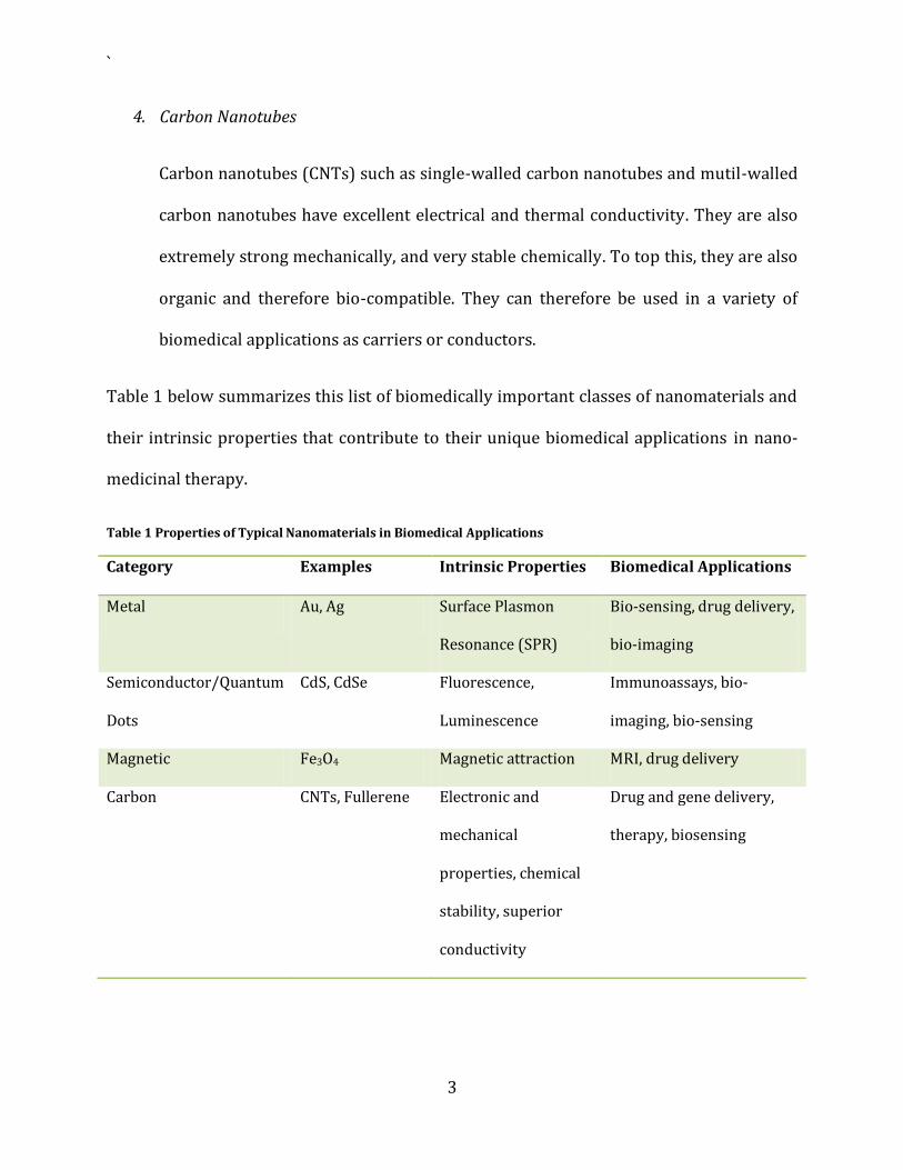
Table 1 below summarizes this list of biomedically important classes of nanomaterials and
their intrinsic properties that contribute to their unique biomedical applications in nano-
medicinal therapy.
Table 1 Properties of Typical Nanomaterials in Biomedical Applications
Category Examples Intrinsic Properties Biomedical Applications
Metal Au, Ag Surface Plasmon
Resonance (SPR)
Bio-sensing, drug delivery,
bio-imaging
Semiconductor/Quantum
Dots
CdS, CdSe Fluorescence,
Luminescence
Immunoassays, bio-
imaging, bio-sensing
Magnetic Fe3O4 Magnetic attraction MRI, drug delivery
Carbon CNTs, Fullerene Electronic and
mechanical
properties, chemical
stability, superior
conductivity
Drug and gene delivery,
therapy, biosensing

`
4
2 Choice of Ligands
The key advantage that nanomaterials bring to the table is their enlarged surface area.
Thus surface modification is a key aspect in the use of nanomaterials in biomedical
applications. Careful and selective surface treatment can meld the nanomaterial of choice
for the right application, depending on whether the nanomaterial is to be used for analysis,
sensing, imaging, or diagnostics.
As such, the choice of ligands that are to be attached to the nanomaterial surface are of
utmost importance. A variety of different ligands may be attached to a nanomaterial
depending on what effect is to be achieved. For example, ligands containing bulky
hydrophobic groups may be attached to nanomaterial surfaces to prevent agglomeration.
Conversely, two different ligands may be chosen to force nanomaterials to interact forming
weak bonds between nanoparticles. This is known as ligand coupling.
Ligands may also be attached to act like “tags” for molecular recognition properties that
can be exploited in drug targeting and in bio-imaging application as a marker. Ligands may
also be attached to nanomaterial surface to define the properties of the nanomaterial itself.
The smaller the nanomaterial, the larger the impact of the ligand on it. Multiple ligands may
be coupled on to a nanomaterial too. Finally, ligands can be used to fix the polarity and
consequently the solubility of the nanomaterial. If we want to precipitate a nanomaterial
out of a hydrophilic solution, we simply functionalize it with a hydrophobic ligand and the
nanomaterial will instantaneously precipitate out of the solution forming a cloud on the
top.
`
5
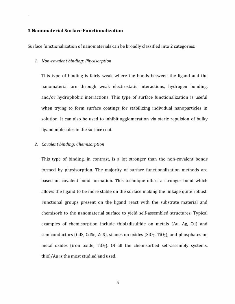
3 Nanomaterial Surface Functionalization
Surface functionalization of nanomaterials can be broadly classified into 2 categories:
1. Non-covalent binding: Physisorption
This type of binding is fairly weak where the bonds between the ligand and the
nanomaterial are through weak electrostatic interactions, hydrogen bonding,
and/or hydrophobic interactions. This type of surface functionalization is useful
when trying to form surface coatings for stabilizing individual nanoparticles in
solution. It can also be used to inhibit agglomeration via steric repulsion of bulky
ligand molecules in the surface coat.
2. Covalent binding: Chemisorption
This type of binding, in contrast, is a lot stronger than the non-covalent bonds
formed by physisorption. The majority of surface functionalization methods are
based on covalent bond formation. This technique offers a stronger bond which
allows the ligand to be more stable on the surface making the linkage quite robust.
Functional groups present on the ligand react with the substrate material and
chemisorb to the nanomaterial surface to yield self-assembled structures. Typical
examples of chemisorption include thiol/disulfide on metals (Au, Ag, Cu) and
semiconductors (CdS, CdSe, ZnS), silanes on oxides (SiO2, TiO2), and phosphates on
metal oxides (iron oxide, TiO2). Of all the chemisorbed self-assembly systems,
thiol/Au is the most studied and used.

`
6
In the case of chemisorption, the nanomaterial is already pre-derivatized with a
functional group. This group then reacts with the ligand that possesses
complementary functional group through a ligand-exchange technique. In this case,
the ligand should have at least equal or higher affinity than the capping molecule
towards the nanomaterial. The other alternative is the two functional groups react
together and form a derivatized final molecule as a result of the chemical synthesis.
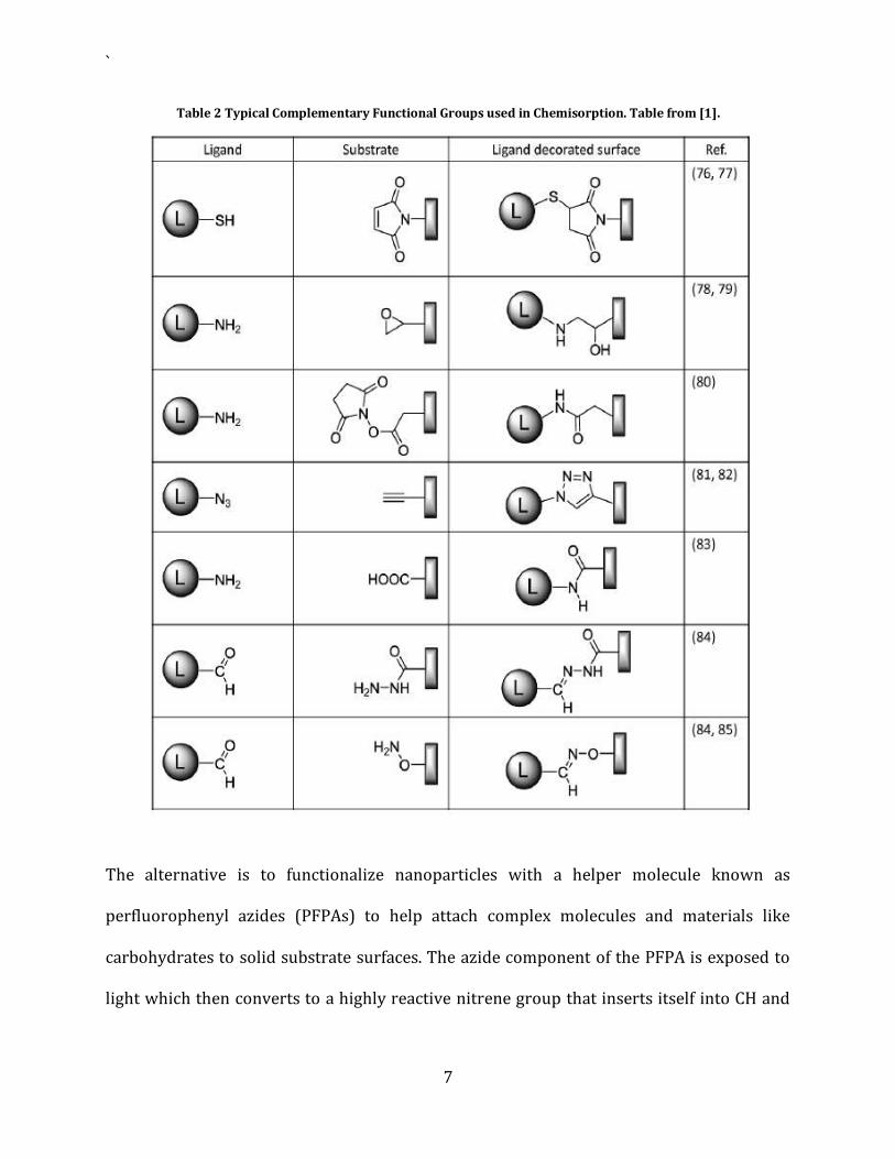
Table 2 shows a list of common complementary functional groups used for coupling
ligands to nanomaterials and the resulting groups from the chemisorption.
Figure 1 diagrammatically shows the two broad types of surface functionalization
techniques that can be applied to nanomaterials. The non-covalent (physical) bond is weak
and therefore temporary, while the covalent (chemical) bond is strong and therefore more
permanent.
4 Photo-Initiated Coupling Chemistry
Another method of surface modification for binding complex molecules like carbohydrates
is known as photo-initiated coupling chemistry. Since carbohydrates are complex in
structure, they are hard to chemically derivatize by combining one or more functional
groups. But carbohydrates are very important and useful biomolecules to bind to
nanomaterials for the formation of biomedical sensors. Glycoproteins and glycolipids are
naturally occurring carbohydrates and are present at the surface of almost all living cells.
Thus there is no easy and direct way to attach carbohydrates to a nanoparticle.
`
7
Table 2 Typical Complementary Functional Groups used in Chemisorption. Table from [1].
The alternative is to functionalize nanoparticles with a helper molecule known as
perfluorophenyl azides (PFPAs) to help attach complex molecules and materials like
carbohydrates to solid substrate surfaces. The azide component of the PFPA is exposed to
light which then converts to a highly reactive nitrene group that inserts itself into CH and

`
8
NH bonds creating very good covalent bonds. This chemistry is known photo-initiated
coupling chemistry.
Figure 1 Surface modification of nanomaterials using non-covalent and covalent approaches. Figure from [1].
PFPA photocoupling chemistry is well-understood and well-established and can be used to
attach carbohydrates to nanoparticles. This works by preparing PFPA-functionalized
nanoparticles that can be subsequently used to covalently couple carbohydrate structures
thanks to the highly reactive photo-activated nitrene species from the azide moiety of the
PFPA. This coupling reaction is quite fast and often takes place in a few minutes.
Nanoparticles can be functionalized with the photocoupling agent (PFPA) by chemisorption
via a simple solution incubation process. Gold nanoparticles can be easily functionalized
Monosaccharides and oligosaccharides can be attached on to gold and iron oxide particles

`
9
in this manner. A similar technique can also be used for attaching polymers too on to silica
nanoparticles. The coated nanoparticles are stable in solution and are readily dispersed in
water to give homogenous solutions.
Figure 2 below shows a schematic illustration of a gold nanoparticle that was successfully
prepared using the PFPA photo-coupling chemistry outlined above. Monosaccharides were
chosen since these are the smallest carbohydrate structures. They are also the most
challenging for the photo-coupling chemistry detailed above since they have the least
amount of affinity as a ligand compared to oligosaccharides. This is because the probability
of attaching the ligand by a simple C-H insertion reaction increases as the size of the
carbohydrate structure, i.e. the number of C-H bonds, increases.
Figure 2 Illustration of Gold Nanoparticles with Con A and Formation of gold nanoparticle aggregates. Figure from [1]
5 Synthesis of Surface Functionalized Gold Nanoparticles
A single process to both prepare gold nanoparticles and functionalize them with PFPAs was
developed in [1]. These gold nanoparticles were 20nm in diameter. Once these surface-
functionalized carbohydrates were formulated, a covalent attachment of carbohydrate
ligand was performed by activating the azide group in PFPA using UV light. The surface

`
10
coverage, measured using thermal gravimetric analysis (TGA), was around 80% which is
pretty substantial.
These coated gold nanoparticles were then developed into calorimetric biosensor for
probing carbohydrate-protein interactions. For example, when these gold nanoparticles
were treated with the Con A target protein, the absorbance spectrum of the nanoparticles
would broaden and be red-shifted which could be easily detected with modern computer
software. The gold nanoparticles were originally wine-red in color as can be seen through
surface plasmon resonance (SPR) with an absorbance peak at 520nm. Upon surface
functionalization, the nanoparticles form cross-linked aggregates as can be seen in Figure 3
where the Con A protein acts as the cross-linker.
Figure 3 TEM Images of resulting NP aggregation after treating carbohydrate-functionalized Au NPs with Con A. Figure from [1].
This formation of aggregates is manifested as a red-shift in their SPR absorption and a
broadening of the peak as can be seen in Figure 4, resulting in a change in colour of the
solution. Of course, the extent to which this colour change is detectable depends on the
surface coverage of carbohydrate as well as the extent of nanoparticle agglomeration.

`
11
Figure 4 UV-Vis absorption spectra of (a) Pristine Au NPs with surface-coupled D-mannose monosaccharide and (b) after the Au NPs are treating with Con A protein. Figure from [1].
The specificity of this biosensor was testing by treating the same monosaccharide-
functionalized Au NPs with other proteins, but the colour of the resulting solutions remain
unchanged and no notable red-shifts of the SPR peak were observed. This thus
demonstrates the high selectivity of this advanced glyconanoparticle-based sensing system.
6 Summary
Since surfaces are at the boundary of every material, they form as the interface between the
nanomaterial and the physical phase surrounding the nanomaterial. The surface also plays
a vital role in the properties and functions of nanomaterials. Also, the smaller the material,
the more critical the role played by the surface as compared to the bulk. Nanomaterials,
owing to their size, therefore have high surface energy and as such any ligands present on
the surface can serve to act as points of communication with external receptors and
detectors. The ligands must therefore be carefully chosen since they play a variety of roles

`
12
including determining surface and nanomaterial physical properties, inhibiting or
encouraging agglomeration, and helping to translate various molecular events into
electrical signals that can be reliably detected externally.
Effective surface coupling chemistry is of utmost importance. Consequently, novel
techniques of attaching complex molecules on to nanomaterials are always in demand.
These techniques need to be able to accommodate differences in ligands and their
bioaffinities and at the same time also be able to result in surfaces that are not only
bioactive but also stable.

`
13
References
[1] Xin Wang, Li-Hong Liu, Olof Ramstrom, and Mingdi Yan, “Engineering Nanomaterial
Surfaces for Biomedical Applications”, Society of Experimental Biology and Medicine, 234 pp.
1128-1139, 2009.
[2] Jelinek R, Kolusheva S. Carbohydrate biosensors. Chem Rev 104, pp. 5987–6016, 2004.
[3] Park S, Shin I. “Carbohydrate microarrays for assaying galactosyltransferase activity”,
Org Letters 9, pp. 1675–1678, 2007.
[4] Latham AH, Williams ME. “Controlling transport and chemical functionality of magnetic
nanoparticles”, Acc Chem Res 41 pp. 411–420, 2008.
[5] Hacliipanayis CG, Bonder MJ, Balakrishanan S, Wang X, Mao H, Hadjipanayis GC.
“Metallic iron nanoparticles for MRI contrast enhancement and local hyperthermia”. Small
4 pp. 1925–1929, 2008.