engineering new bacterial dye-decolourising peroxidases...
TRANSCRIPT

Universidade de Lisboa
Faculdade de Ciências
Departamento de Biologia Vegetal
Engineering new bacterial dye-decolourising
peroxidases for lignin degradation
Diogo Alexandre Martins Aires Tavares
Dissertação
MESTRADO EM MICROBIOLOGIA APLICADA
2014

Universidade de Lisboa
Faculdade de Ciências
Departamento de Biologia Vegetal
Engineering new bacterial dye-decolourising
peroxidases for lignin degradation
Diogo Alexandre Martins Aires Tavares
Dissertação
MESTRADO EM MICROBIOLOGIA APLICADA
Orientadores:
Professora Lígia Oliveira Martins, Instituto de Tecnologia Química e Biológica, UNL
Professor Rogério Tenreiro, Orientador Interno designado pela Faculdade de Ciências, UL
2014

Engineering new bacterial dye-decolourising
peroxidases for lignin degradation
Diogo Alexandre Martins Aires Tavares
2014
The work presented in this thesis was conducted at the Instituto de Tecnologia Química e
Biológica, in the Microbial and Enzymatic Technology (MET) Lab under the supervision of
Professor Lígia Oliveira Martins and co-supervision of Dr. Vânia Brissos.

i
Table of contents
Acknowledgments ............................................................................................................................................... iii
Abstract...................................................................................................................................................................... iv
Resumo....................................................................................................................................................................... v
Abbreviations........................................................................................................................................................ viii
1. Introduction .................................................................................................................................................... 1
1.1 Peroxidases ............................................................................................................................................ 2
1.2 Directed evolution ................................................................................................................................. 4
1.3 Directed evolution of PpDyP ............................................................................................................. 6
2. Material and Methods ................................................................................................................................ 7
2.1 Bacterial strains, plasmids and media ........................................................................................... 8
2.2 Preparation of electrocompetent cells ........................................................................................... 8
2.3 Selection of the appropriate enzyme substrate for activity screenings ............................. 8
2.4 Random mutagenesis by error-prone PCR (ep-PCR) ............................................................ 9
2.5 Recombination by DNA Shuffling and mutant library construction ................................... 10
2.6 Transformation of E. coli cells......................................................................................................... 11
2.7 Overexpression of ppDyP variants in 96-well plates.............................................................. 11
2.8 Cell disruption in 96-well plates...................................................................................................... 11
2.9 High-throughput screening for activity and stability ................................................................ 12
2.10 Production and purification of recombinant PpDyP variants............................................... 12
2.11 Spectroscopic analysis ..................................................................................................................... 13
2.12 Kinetic analysis .................................................................................................................................... 13
2.13 Enzyme kinetic stability ..................................................................................................................... 14
3. Results and Discussion ......................................................................................................................... 15
3.1 Directed Evolution of PpDyP .......................................................................................................... 16
3.1.1 Validation of high-throughput screenings ........................................................................ 16
3.1.2 Selection of appropriate recombinant strains for enzymatic activity assays ....... 16
3.1.3 Cell growth, cell disruption and activity assays in 96-well plates ............................. 17
3.1.4 Library construction by epPCR.............................................................................................. 18
3.1.5 Directed evolution of PpDyP ................................................................................................. 19
3.2 Spectroscopic and Biochemical Characterization of PpDyP Wild-type and Variants 27
3.2.1 Spectroscopic characterization............................................................................................. 27

ii
3.2.2 Kinetic properties ...................................................................................................................... 29
3.2.3 Kinetic Stability of PpDyP variants ....................................................................................... 31
3.3 Structural analysis of mutations ..................................................................................................... 32
4. Conclusions ................................................................................................................................................. 34
5. References.................................................................................................................................................... 35

iii
Acknowledgments
Quero agradecer a todas as pessoas que tornaram possível a elaboração desta
dissertação de mestrado:
À minha orientadora, Professora Lígia Martins, por me ter aceite no seu laboratório, e
me ter feito sentir como se estivesse em casa. Quero agradecer também as suas
‘’dicas’’ e, acima de tudo, a transmissão de conhecimento, e pela minha formação
como investigador e pessoa.
À Dra. Vânia Brissos, por todo o acompanhamento ao longo do trabalho, pelas horas
‘’perdidas’’, pela determinação e encorajamento que teve sempre para comigo. E claro
pelos ‘’puxões de orelhas’’ que me fizeram melhorar e aprender. O meu muito
obrigado por toda a dedicação.
Ao Professor Rogério Tenreiro, por ter aceite ser o meu Orientador Interno, e pela
disponibilidade demonstrada.
Aos membros do MET Lab: Sónia Mendes, que sempre me apoiou e ajudou no que
podia, estando sempre disponível, obrigado também pela companhia no sempre
relaxante chocolate a meio da tarde. Ao Joaquim Madeira (a.k.a. Jakas), pela
amizade, companheirismo, encorajamento, passes para golo no futebol, e claro por me
ouvir e compreender. À Lúcia Sabala (a.k.a Turbinada), pelas parvoíces e distracções
quando eu ‘’estava em stress’’. A todos os outros que passaram pelo laboratório neste
último ano, Marcelo, Bruna, Kamil, Marcin e Antonieta, o meu muito obrigado.
Aos meus colegas de mestrado, ‘’os fixes’’, Joana Carrilho, Margarida Duarte, Hélder
Ribeiro, Diogo Dias, Bruno Arez e Inês Basto, o meu muito obrigado pela companhia,
pelos jantares, pelas saídas, pelas distracções, e acima de tudo pela grande amizade.
À Joana tenho de fazer um agradecimento extra, pela paciência e carinho
incondicional.
Finalizar agradecendo à minha família, Mãe, Pai, Mano, Avó Fernanda, Tio Nuno, Tia
Elsa e Prima Inês, fico-lhes agradecido por todo o apoio e coragem que me deram.
Tenho a certeza que sem eles, não seria possível estar a escrever estes
agradecimentos, porque simplesmente não teria uma tese onde os escrever.

iv
Abstract
Dye-decolourising peroxidases (DyPs) are a novel family of heme-containing peroxidases
showing a high efficiency for a wide number of substrates, including synthetic dyes, lignin units
and metals, and are thus very attractive biocatalysts for application in the environmental and
industrial biotechnology fields.
In this work high-throughput protocols were optimized and validated for the application of
directed evolution approaches targeting PpDyP from Pseudomonas putida MET94 at the level
of cell growth, lysis and enzymatic assays. Libraries of variants were created by epPCR and
DNA shuffling techniques and explored for activity using high-throughput screenings activity
assays. Four libraries of variants in a total of 8163 clones were explored in three rounds () of
molecular evolution rounds to improve the efficiency for substrates and stability of PpDyP. The
best variants were overproduced and purified and their structural and catalytic properties were
characterized by spectroscopic and kinetic approaches. One variant, 21G11, was achieved
showing 25- and 6-fold higher specificities (kcat/Km) for ABTS oxidation and H2O2 reduction,
respectively. Importantly, variant 6E10, showing a 250-fold enhanced specificity for phenolics
as compared with the wild-type was selected with a pHopt = 5.4, 1.1 units up-shifted in relation
to the wild-type enzyme.
Noteworthy a 2-fold increase in the protein production levels in relation to the wild-type was
observed in both hit variants most probably related with the replacement of residue His125, for
either a Arginine or Threonine, suggesting that this residue is a hot-spot in the enzyme
structure to improve the protein production. Based in the model structure of PpDyP we have
rationalized the structural molecular basis for increased activity for ABTS or DMP and
improved kinetic stability and production yields of the PpDyP enzyme.
. .
Keywords: Dye-decolourising peroxidases, Pseudomonas putida, directed evolution, error-
prone PCR, high-throughput screening, kinetic and stability analysis.

v
Resumo
As ‘’ dye decolourising peroxidases’’ (DyPs) são uma nova família de peroxidases hémicas
que utilizam H2O2 e apresentam uma especificidade alargada para diferentes substratos,
sendo de destacar o seu enorme potencial oxidativo para compostos aromáticos, tais como
corantes sintéticos e unidades de lenhina fenólicas e não fenólicas.
Devido à especificidade alargada, as DyPs podem ser utilizadas como biocatalizadores em
diferentes aplicações biotecnológicas. Todos os anos 800 mil toneladas de corantes sintéticos
são produzidos e ~ 15% são libertados para os efluentes. Os tratamentos convencionais para
a remoção destes corantes dos efluentes são atualmente pouco eficazes e muito
dispendiosos. Devido à grande capacidade oxidativa das DyPs, estas enzimas têm um
grande potencial para serem utilizadas no tratamento destes efluentes. Noutro contexto, a
biorefinaria de lignocelulose poderá vir a ser uma solução para reduzir a utilização de matérias
primas fósseis, não renováveis, com implicações no aquecimento global, para a produção de
biocombustíveis, materiais e produtos de elevado valor acrescentado. Novamente, as DyPs
ao mostarem capacidade oxidativa de unidades fenólicas e não-fenólicas de lenhina, poderão
ter um papel importante, como enzimas ligninolíticas na degradação de material
lignocelulósico muito resistente à degradação química e biológica.
As enzimas apresentam várias vantagens como catalizadores, são eficientes, específicas,
seguras e amigas do ambiente, no entanto a sua eficiência e estabilidade por vezes ficam
aquém das necessidades para a sua aplicação a nível industrial.
As proteínas podem ser melhoradas usando técnicas de engenharia racional no entanto esta
abordagem requer um conhecimento extenso e muitas vezes inantingível da relação entre
estrutura e função das proteínas. Para ultrapassar esta limitação surgiu uma tecnologia chave
na área da engenharia molecular, denominada evolução dirigida, tendo mostrado resultados
impressionantes no melhoramento de várias características dos biocatalisadores.
A evolução dirigida envolve 4 passos essenciais: (i) selecção do gene de interesse, (ii)
construção da biblioteca de mutantes, (iii) selecção de proteínas mutantes com funções
melhoradas, (iv) repetir o processo até o objectivo ser atingido. Os métodos mais comuns de
mutagénese são a mutagénese aleatória, mutagenése por saturação e o ‘’DNA shuffling’’.
A mutagénese aleatória consiste em pequenas modificações dos protocolos de PCR,
modificações essas, que aumentam a normal taxa de erro da enzima Taq polimerase.
Normalmente estes protocolos contêm maiores concentrações de MgCl2 em relação aos

vi
tradicionais com o objectivo de estabilizar os pares não-complementares. A taxa de mutação
também pode ser aumentada através da adição de MnCl2 ou da variação do rácio de
nucleótidos.
A mutagénese por saturação permite a criação de uma biblioteca de mutantes, que contém
todas as mutações possíveis numa posição alvo da sequência da proteína. Desta forma um
amino ácido pode ser substituído por todos os 20 amino ácidos introduzindo todos os codões
possíveis nessa posição.
Por fim, o ‘’DNA shuffling’’ é uma técnica in vitro de recombinação aleatória dos mutantes
seleccionados . O PCR reagrupa os genes a partir de fragmentos de DNA que são gerados
por digestão aleatória prévia dos diferentes genes parentais.
Recentemente no Laboratório de Tecnologia Microbiana e Enzimática o gene que codifica
para uma DyP de Pseudomonas putida MET94 (PpDyP) foi clonado, expresso, e a proteína
foi super-produzida e purificada. Esta enzima mostrou ser eficiente na degradação de
corantes azo e antraquinónicos e na oxidação de compostos fenólicos e não-fenólicos e iões
metálicos como o manganês e o ferro. Esta especificidade alargada para diferentes substratos
torna esta enzima muito atractiva para ser utilizada como biocatalizador nas áreas da
biotecnologia ambiental e industrial.
O objectivo deste estudo foi estabelecer protocolos apropriados para a evolução dirigida da
enzima PpDyP de form a então criar biocatalisadores de elevada performance para serem
utilizados nas futuras biorefinarias. Nesse sentido, técnicas de evolução dirigida foram
aplicadas à PpDyP. Quatro bibliotecas de mutantes, em três ciclos de evolução, foram
construídas por técnicas de mutagénese aleatória e “DNA shuffling” e cerca de 8000 clones
foram rastreados.
Os melhores mutantes de cada geração foram produzidos e purificados e as suas
propriedades espetroscópicas e catalíticas foram estudadas. Espectroscópicamente todos os
mutantes revelaram ser semelhantes à estirpe selvagem, o que indica que as mutações
introduzidas não levaram a nenhuma alteração estrutural significativa da enzima. Durante o
processo de evolução foram seleccionados variantes com especificidades distintas. Os
mutantes 9F6 e 21G11 revelaram uma melhoria de 6 e 25 vezes na especificidade (kcat/Km) (1
ordem de grandeza) para os substratos ABTS e H2O2, respectivamente, quando comparados
com a estirpe selvagem. A especificidade dos mutantes 17F11 e 6E10 aumentou cerca de
250 vezes (3 ordens de grandeza) para o substrato DMP quando comparados com a estirpe
selvagem. Os mutantes 21G11 e 6E10 apresentaram um rendimento de produção proteica
duas vezes superior relativamente à enzima nativa. Foi interessante verificar que ambos

vii
adquiriram uma mutação na histidina125, sugerindo que este resíduo é um local importante
para melhorar a produção da proteína.
Foi verificado também ao longo deste trabalho que é muito difícil aumentar simultaneamente a
actividade catalítica e a estabilidade da enzima. Os mutantes 9F6 e 17F11 são bons
exemplos, uma vez que o aumento da actividade catalítica resultou numa menor estabilidade.
Isto deve-se provavelmente ao compromisso que existe entre a actividade e a estabilidade
proteica. Inúmeras vezes para continuar a melhorar a actividade catalítica de um variante
têmde ser introduzidas previamente mutações neutras importantes na estabilização da
estrutura. Introduzindo posteriormente novas mutações que melhorem a actividade catalítica
da enzima.
Neste trabalho a PpDyP foi evoluída com sucesso por técnicas de evolução dirigida, para
maior capacidade oxidoreductiva para o ABTS, H2O2 ou para os compostos fenólicos e níveis
de rendimento de produção melhorados.
Palavras-Chave: ‘’Dye-decolourising’’ peroxidases, Pseudomonas putida, evolução dirigida,
mutagénese aleatória, ‘’high-throughput screening’’, análise cinética e de estabilidade

viii
Abbreviations
ABTS – 2,2’-azino bis (3- ethylbenzthiazoline-6-sulfonic acid)
BSA – Bovine Serum Albumin
DMP – 2,6-Dimethoxyphenol
DyP – Dye decolourising peroxidase
epPCR – Error prone PCR
IPTG – isopropyl β-D-1-thiogalactopyranoside
LB – Luria-Bertani
MB9 - Mordant Black 9
OD600nm – Optical density at 600 nm
RB5 – Reactive Blue 5
SDS-PAGE – Sodium dodecyl sulfate polyacrylamide gel electrophoresis
SOB – Super Optimal Broth
TB - Terrific Broth
UV – Ultra-violet
Vis – Visible
Wt – Wild-type

1
1. Introduction

2
1.1 Peroxidases
Heme peroxidases catalyze the H2O2-dependent oxidation of a variety of small organic
substrates, playing multiple physiological roles in a wide range of living organisms. These
enzymes were originally classified into two superfamilies: the plant peroxidases and the animal
peroxidases (Welinder et al., 1992). The fungal and bacterial peroxidase were lately included in
the plant peroxidase superfamily and further classified into classes , , and , based on
primary structural homology. Class is composed by intracellular prokaryotic peroxidases,
including yeast cytochrome c peroxidase and chloroplast ascorbate peroxidase. Class
comprises extracellular fungal peroxidases, like lignin peroxidases, manganese peroxidases,
and versatile peroxidases. Class represents extracellular algae and plant peroxidases, such
as horseradish peroxidases (Hofrichter et al., 2010). Mammalian peroxidases have been
recognized to constitute a separate peroxidase superfamily, the peroxidase-cyclooxygenase
superfamily. They arose independently from the plant, fungal and (archae)bacterial superfamily
as is inferred by substantial differences in sequence, overall structure and the nature of their
prosthetic group, in which the ferri-protoporphyrin IX derivative is covalently linked to the protein
(Zederbauer et al., 2007). The main clades are represented by myeloperoxidases, eosinophil
peroxidases and lactoperoxidase. They figure prominently in human biology and contribute to
host defense against infection, hormone synthesis, and pathogenesis.
Recently a new family of peroxidases, known as the dye-decolourising peroxidases (DyPs)
family, was proposed (Kim and Shoda, 1999). DyPs were first discovered in fungi, and later in
a wide range of bacterial strains. DyP-type peroxidases are not considered members of plant
or animal peroxidases because of their unique reaction characteristics and specific primary and
tertiary structures (Sugano, 2009). These enzymes are sub-classified into the phylogenetically
distinct classes A, B, C, and D (Colpa et al., 2014). Class A contain a Tat-dependent signal
sequence, which suggests that they function outside of cytoplasm or even extracellularly
(Jongbloed et al., 2004). Classes B and C are cytoplasmic enzymes, probably involved in
intracellular metabolism. Class D contains primarily fungal enzymes. DyP-type peroxidases
bind the heme cofactor non-covalently, as plant peroxidases.
All DyP peroxidases contain the so-called GXXDG motif in their primary sequence, which is
part of the heme-binding region. The crystal structures of DyPs shows two domains with each
domain consisting of a four-stranded antiparallel -sheet and peripheral -helices with the
heme cofactor clefted in between (Figure 1A) distinct from the classic peroxidases that are
primarily α-helical proteins (Zubieta et al., 2007a; Zubieta et al., 2007b; Liu et al., 2011; Yoshida
et al., 2011; Singh et al., 2012; Strittmatter et al., 2013). A detail of the amino acid residues in
the DyPs heme environment is shown in Figure 1B. DyPs lack the distal His, which is highly

3
conserved among classical peroxidases and is proposed to act as an acid/base catalyst in the
catalytic mechanism. Instead, they house an Asp together with an Asn and an Arg. On the
proximal side, the heme iron is coordinated by a His hydrogen bonded to an Asp residue,
forming a Fe-His-Asp triad.
Figure 1 – (A) Crystal structure of TyrA (PDB 2iiz). The α helices are shown in red and the β sheets in
yellow. (B) Representation of the catalytically distal and proximal residues important for the peroxidase activity. DyPs houses a distal aspartate (Asp), asparagine (Asn) and an arginine (Arg) and a proximal histidine (His) and an aspartate (Asp) (Santos et al., 2014).
Considering their broad specificity, DyPs can be potentially used as biocatalysts in many
different biotechnological applications (Colpa et al., 2014). Every year 800,000 t of synthetic
dyes are produced and ~ 15% are released into effluents. The removal of these dyes from
effluents by conventional wastewater treatments is very expensive and technically challenge.
Dye-containing effluents cause deterioration of water quality and become a health threat
because of their mutagenic or carcinogenic properties (Rodriguez-Couto, 2009). DyPs shows
potential for the set-up of enzymatic treatment of dye-containing wastewaters. Recently, the
lignocelluloses biorefinery concept is receiving considerable attention as a source of a
renewable chemicals, materials, and flues for future sustainable development. The
lignocelluloses biorefinery should contribute to reduce the effects of global warming, caused by
the consumption of crude oils, providing the production of biofuels (Martinez et al., 2009). DyPs
have the ability to act as ligninolytic enzymes, with potential to exert a crucial role in the
oxidation of phenolic and non-phenolic lignin units since the lignocellulosic material is highly
resistant to (bio)chemical degradation (Bugg et al., 2011). Moreover, it was also shown that
DyPs can be important in the degradation of β-carotene, with direct implications in food
industry, enabling the enzymatic whitening of whey-containing foods and beverages
(Scheibner et al., 2008).
AsnArg
Asp
His
Asp
A B

4
1.2 Directed evolution
Enzymes are biocatalysts, capable of carrying out a tremendous range of biochemical
functions, however their efficiency, stability and costs often do not correspond to the needs of
industry (Bloom et al., 2005). One approach to engineering enzymes towards improved
properties is to use rational design however, this approach requires an extensive and
frequently unattainable knowledge of the relationship between structure and function of
proteins (Kuchner and Arnold, 1997). An evolutionary engineering, named ‘’directed evolution’’,
has emerged as a key technology for biomolecular engineering, generating impressive results
(Reetz et al., 2008).
Direct evolution involves four keys steps: (i) select a starting gene sequence, (ii) create a library
of variants, (iii) select variants by high-throughput screening with improved function, (iv) repeat
the process until the improvement or function is achieved (Romero and Arnold, 2009). The
most common mutagenesis method are error-prone PCR (epPCR), saturation mutagenesis
and DNA shuffling (Tracewell and Arnold, 2009).
Error-prone PCR protocols consist in small modifications of the standard PCR methods in
order to enhance the natural error rate of Taq polymerase. The epPCR protocols usually
contain high concentrations of MgCl2, in order to stabilize non-complementary pairs. Other
ways to increase the mutation rate includes the variation of the nucleotides ratios or the
addition of MnCl2 to the reaction (Cirino et al., 2003). Due to its simplicity and versatility, this
epPCR has emerged as the most common technique and can result in mutation frequencies
as high as 2% per nucleotide position (Brakmann, 2001).
Saturation mutagenesis enables the creation of a library of mutants containing all possible
mutations at one or more pre-determined target positions in a gene sequence (Tracewell and
Arnold, 2009). This is achieved by introducing all possible base triplets at a given codon,
thereby resulting in the insertion of all 20 amino acids at this position of the protein. This
method restricts the random mutations to predicted sites in the enzyme creating therefore what
is called focused libraries, but requires structural information in order for the correct
mutagenesis sites to be chosen (Reetz et al., 2008). Since random point mutagenesis can not
access a large fraction of the enzyme sequence, this technique is normally used to increase
the number of amino acid substitutions accessible by random mutagenesis (Georgescu et al.,
2003).
DNA shuffling performs random in vitro recombination of gene variants created by random
mutagenesis. It employs the PCR reassembly of whole genes from a pool of short overlapping
DNA sequences that are generated by random enzymatic fragmentation of different parental
genes (Brakmann, 2001).

5
The directed evolution method is highly versatile (Dougherty and Arnold, 2009), and has being
successfully applied to improve several enzymatic properties, e.g. gene expression levels of a
horseradish peroxidase (Lin et al., 1999; Morawski et al., 2000), protein stability of an
azoreductase from Pseudomonas putida and a versatile peroxidase from Pleurotus eryngii
(Garcia-Ruiz et al., 2012; Brissos et al., 2014) and enzymatic activity of a peroxygenase
(Molina-Espeja et al., 2014).

6
1.3 Directed evolution of PpDyP
Recently in Microbial and Enzymatic Technology (MET) Lab the gene coding for DyP from
Pseudomonas putida MET94 (PpDyP) was cloned and expressed and the protein
overproduced and purified showing a wide substrate specificity, including high redox potential
aromatic compounds such as synthetic dyes, phenolic and nonphenolic lignin units and metal
ions such as manganese and ferrous (Santos et al., 2014). This enzyme has an optimal pH in
the acidic range (pH 4-5) and showed higher activity between 10 and 30⁰C. Considering the
broad specificity, this enzyme has a considerable potential in the fields of environmental and
industrial biotechnology.
However, peroxidases are not well-suited for industrial uses that generally requires particular
substrate specificities and application conditions (pH and temperature) in addition to high
expression levels (Martinez et al., 2009). PpDyP is not an exception, and protein engineering
techniques need to be applied to obtain highly gene expression levels, and to increase the
protein stability and enzymatic activity.
Therefore, the major aim of this thesis was to create potential useful biocatalysts in the
biorefinery field, evolving PpDyP towards higher activity for phenolic compounds by using
directed evolution approaches. Libraries of variants were created by epPCR and DNA shuffling
techniques and explored for activity using high-throughput screenings. The best variants were
overproduced and purified and their structural and catalytic properties were addressed by
spectroscopic and kinetic approaches.

7
2. Material and Methods

8
2.1 Bacterial strains, plasmids and media
Escherichia coli strain DH5α (Novagen) was used for routine propagation and amplification of
plasmid constructs. E. coli Tuner (DE3, Novagen), KRX (Promega) and BL21 star (DE3,
Novagen) strains were used to express the ppDyP wild-type (Wt) and variant genes cloned in
pET21-21a (+) plasmid (Novagen). In the Tuner and BL21 star strains the target genes are
under the control of T7 promoter, induced by isopropyl β-D-1-thiogalactopyranoside (IPTG)
and in the KRX strain, the genes are under the control of rhaPBAD promoter, induced by
rhamnose. Luria-Bertani medium (LB) and Terrific Broth medium (TB) were used for the
maintenance and growth of E. coli strains, supplemented with 100 µg/mL ampicillin. LB
medium contains (per liter): 10 g of tryptone, 5 g of yeast extract and 10 g of NaCl2. TB
medium contains the following components (per liter): 12 g of tryptone, 24 g of yeast extract,
3.4 g of glycerol, 2.3 g of KH2PO4 and 12.5 g of K2HPO4. Super Optimal Growth medium
(SOB) was used for the growth of competent cells. SOB medium contains (per liter): 20 g of
tryptone, 5 g of yeast extract, 0.584 g of NaCl2 and 0.186 g of KCl2. All culture media were
sterilized in an autoclave and stored at room temperature.
2.2 Preparation of electrocompetent cells
A LB agar plate was streaked out with E. coli KRX cells and incubated overnight at 37⁰C. A
single colony was picked, used to inoculate 20 mL of SOB medium and incubated overnight at
37⁰C at 120 rpm. Growth at a scale of 500 mL in SOB medium was started with an optical
density at 600 nm (OD600nm) of 0.05 and incubated at 37⁰C, with 120 rpm. After 3.5 h (OD600nm ~
0.8), cells were transferred to ice-cold centrifuge bottles and spin down at 5,000 rpm for 15 min
at 4⁰C. The supernatant was discarded, and the cell pellet was washed with 500 mL of a sterile
ice-cold 10% glycerol solution. The cells were centrifuged, re-washed in 500 mL of the same
solution, centrifuged for a second time and re-suspended in the remained solution ( 2.5 mL).
Aliquots of 100 µL were frozen on nitrogen and stored at - 80⁰C.
2.3 Selection of the appropriate enzyme substrate for activity screenings
Growth of recombinant strains and ppDyP overexpression
The plasmid pRC-1, containing the ppDyP gene, was transformed into E. coli Tuner (DE3,
Novagen), KRX (Promega) and BL21 star (DE3, Novagen) strains (Santos et al., 2014). Single
colonies were used to inoculate 10 mL of LB medium supplemented with 100 µg/mL ampicillin,
grown overnight at 37⁰C, 160 rpm. Fresh cultures were transferred to 100 mL of LB medium
supplemented with 100 µg/mL ampicillin, in order to start the growth with an OD600nm = 0.05.
Cultures were incubated at 37⁰C, 160 rpm and when OD600nm ~ 0.6, 100 µM IPTG was added
to recombinant E. coli Tuner and BL21 star strains, and 0.1 % rhamnose to recombinant E. coli

9
KRX. After 24 h of cultivation, cells were collected by centrifugation (8,000 rpm, 10 min at 4⁰C).
The cell pellets were resuspended in 1 mL of 20 mM Tris-HCl buffer (pH 7.6), containing 5 mM
MgCl2, 1 U/mL of DNase I, and 2 µL/mL of a mixture of protease inhibitors, antipain and
leupeptin. After cells were disrupted by French Press (Thermo EFC) and then centrifuged at
15,000 for 2 h at 4⁰C. The supernatants (cell crude extracts) were collected and used for
enzymatic assays. The protein concentration was determined using the Bradford assay with
bovine serum albumin (BSA) as standard. SDS-PAGE electrophoresis was used to visualize
protein overproduction in crude extracts.
Enzymatic assays
Enzymatic activities of PpDyP were monitored using a Nicolet Evolution 300 spectrophometer
(Thermo Industries). The activities were measured by monitoring the oxidation of different
substrates at their maximum absorption wavelengths in the presence of 0.2 mM H2O2
Enzymatic assays were performed at 30⁰C, in 20 mM acetate buffer at pH 4.3 using 1 mM of
2,2’-azino bis (3-ethylbenzthiazoline-6-sulfonic acid) (ABTS), 1 mM guaiacol (Ɛ470nm=26,600 M-1
cm-1) or 1 mM 2,6-Dimethoxyphenol (DMP) (Ɛ468nm=49,600 M-1 cm-1), and at pH 5 using 0.1
mM of reactive blue 5 (RB5) (Ɛ610nm=7,690 M-1 cm-1) or 0.1 mM mordant black 9 (MB9)
(Ɛ550nm=15,641 M-1 cm-1).
2.4 Random mutagenesis by error-prone PCR (ep-PCR)
Mutations in the ppDyP gene were generated by using error-prone PCR (ep-PCR). Primers 5’-
GGATTAGCCTCA’TATGCCGTTCCAGCAAGG-3’ (PpDyP Forward) and 5’-
GTGTTTCTGTATCTG’GATCCTTAGAGATCAGGCCCGC-3’ (PpDyP Reverse) flanking the
gene beyond the NdeI and BamHI sites were used for amplification. ep-PCR was carried out in
50 µL reaction volume containing 3 ng of DNA template, 0.5 µM of primers, 200 µM of dNTPs,
7 mM MgCl2, Taq polymerase buffer and 5 U of Taq polymerase (Fermentas). The effect of
MnCl2 concentration (0.01-0.25 mM) in the mutation rate was assessed. After an initial
denaturation period of 10 min at 94⁰C, the following steps were repeated for 28 cycles in a
thermal cycler (MyCyclerTM thermocycler, Biorad): 1 min at 94⁰C, 1 min at 57⁰C and 1.5 min at
72⁰C and at the end 10 min at 72⁰C. The amplified product (5 µL) was visualized by agarose
(1%) electrophoresis, purified using the GFX PCR DNA and the Gel Band Purification kit (GE
Healthcare) and eluted with mili-Q H20. Ten microliters of purified PCR product and 10 µL of
plasmid pET-21a (+) (Novagen) were digested with 10 U of NdeI (Fermentas) at 37⁰C for 2 h.
The mixture was placed at 65⁰C for 20 min and then dialyzed against milli-Q H20 for 30 min
using MFTM filters (Milipore). A second digestion was performed with 10 U of BamHI
(Fermentas) for 2 h at 37⁰C, and pET-21a (+) was simultaneously dephosphorylated with 1 U
Alkaline Phosphatase (FastAP, Fermentas), to prevent plasmid self-ligation. The products

10
were purified using GFX PCR DNA and the Gel Band Purification kit (GE Healthcare). The
ligations were performed with 0.5 U of T4 DNA ligase (Fermentas) using a ratio of 1:8 of vector
to insert. This mixture was incubated overnight, at room temperature then incubated at 65⁰C
for 20 min. The preparation was then dialyzed against mili-Q H20 for 30 min using MFTM filters
(Milipore). After that 5 µL of this mixture was used to transform electrocompetent E. coli KRX
cells.
2.5 Recombination by DNA Shuffling and mutant library construction
DNA shuffling was performed using PpDyP variants, 21G11, 5B9, 22B3, 9G8, 27B3. The
mutant ppDyP genes were amplified by PCR using primers pET21F (5’-
CTTCCCCATCGGTGATGTCGGCGATATAG-3’) and pET21R (5 -
CCAAGGGGTTATGCTAGTTATTGCTCAG-3’). A mixture containing 200 ng of each parental
gene was digested with 0.05 U/ µL of DNase I in a 200 mM Tris-HCl buffer, pH 7 with 80 mM
MnCl2 for 7 min at 15⁰C in a thermocycler (MyCyclerTM thermocycler, Biorad). Digestion was
stopped by adding 6 µL of 50 mM EDTA. The PCR reassembly was carried out in a 20 µL
reaction volume containing 3 µL of DNA fragments, 200 µM of dNTPs, NZYProof polymerase
buffer and 2.5 U of NZYProof Polymerase (NZYTech). After an initial denaturation period of 3
min at 96⁰C, the following steps were repeated for 45 cycles in a thermal cyler (MyCyclerTM
thermocycler, Biorad): 1 min at 94⁰C, 90 s at 59⁰C, 90 s at 56⁰C, 90 s at 53⁰C, 90 s at 50⁰C, 90
s at 47⁰C, 90 s at 44⁰C, 90 s at 41⁰C, and 1 min + 5 s/ cycle at 72⁰C followed by a final 10 min
period at 72⁰C. The PCR reassembly products were amplified by PCR using primers 5’-
GGATTAGCCTCA’TATGCCGTTCCAGCAAGG-3’ (PpDyP Forward) and 5’-
GTGTTTCTGTATCTG’GATCCTTAGAGATCAGGCCCGC-3’ (PpDyP Reverse). PCR was
carried out in a 50 µL reaction volume containing 1 µL of PCR reassembly products, 1 µM of
primers, 200 µM of dNTPs, 0.01 mM of MnCl2, NZYProof polymerase buffer and 2.5 U of
NZYProof Polymerase (NZYTech). After an initial denaturation period of 3 min at 94⁰C, the
following steps were repeated for 20 cycles in a thermal cyler (MyCyclerTM thermocycler,
Biorad): 30 s at 94⁰C, 1 min at 55⁰C, 90 s at 72⁰C followed by a final 10 min period at 72⁰C.
The amplified products were purified using GFX PCR DNA and the Gel Band Purification Kits
(GE Healthcare). The final PCR products were digested with NdeI/BamHI (Fermentas), and
cloned into pET-21a (+) (Novagen) as described above. Ligation reaction mixtures were used
to transform electrocompetent E. coli KRX cells.

11
2.6 Transformation of E. coli cells
One or five microliters of purified plasmids or ligation mixtures, respectively, were added to an
aliquot of 100 µL electrocompetent cells (previously thawed on ice) mixed and placed on ice for
5 min. This mixture was transferred to a sterile and ice-cold electroporation cuvette, which was
placed in the Xcell ShockPod chamber (Gene Pulser XcellTM
, Biorad) and pulsed using set
conditions C = 25 µF, PC = 200 Ω, V = 2.5 kV. One mL of LB medium was immediately added
and the suspension was transferred to an Eppendorf, and incubated at 37⁰C for 1 h at 200
rpm. The cells were then centrifuged at 5,000 rpm for 5 min; 0.9 mL of supernatant was
discarded and the cells were re-suspended in the remaining medium ( 0.1 mL). The cells
were spread on a LB agar plate, supplemented with 100 µg/mL of ampicillin and incubated
overnight at 37⁰C.
2.7 Overexpression of ppDyP variants in 96-well plates
From a fresh agar plate, individual colonies were randomly picked and transferred to a 96-well
plate (Greiner Bio-One) containing 200 µL of LB medium supplemented with 100 µg/mL
ampicillin. In order to avoid evaporation only the interior wells were used while peripheral wells
were filled with sterilized water and the plates were sealed with a foil. Four wells in each plate
were used to inoculate the parent of each generation (in which WT was the parent of the 1st
generation). Cultures were grown for 24 h at 30⁰C, 750 rpm in a Titramax 1000 shaker
(Heidolph). Twenty microliters aliquots were used to inoculate 180 µL of TB medium
supplemented with 100 µg/mL ampicillin. The 96-well plate were sealed with parafilm and
incubated at 30⁰C, 750 rpm. After 4 h of incubation, 0.2% rhamnose and 15 µM hemin were
added to induce gene expression, and the growth proceeded for a 24 h period. The cells were
harvested by centrifugation at 4,000 rpm, for 30 min at 4⁰C.
2.8 Cell disruption in 96-well plates
Three different methods were tested for cell disruption. First in the chemical cell disruption
method, 100 µL of a 40% Bacterial Protein Extraction Reagent (B-PER®) lysis solution
(Thermo Scientific) was used to re-suspend the cell pellets. In the physical method, 96-well
plates were submerged in liquid nitrogen and then thawed at room temperature for 5 min. After
3 cycles of freeze and thaw, cell pellets were re-suspended in 100 µL of 20 mM Tris-HCl, pH
7.6. The third method combined both a physical and enzymatic approach; 96-well plates
were frozen at - 80⁰C for 15 min and then thawed at 30⁰C for 5 min. After 3 cycles of freeze
and thaw, cell pellets were re-suspended in 100 µL of 20 mM Tris-HCl, pH 7.6, and lyzozyme
(2 mg/mL) was added. After cell disruption, plates were centrifuged at 4,000 rpm, for 30 min, at
4⁰C and the supernatant collected (crude extracts) and used for enzymatic activity
measurements.

12
2.9 High-throughput screening for activity and stability
Cell crude extracts (20 µL) were transferred to a new 96-well plate using a Liquid Handling
High-Throughput robot (Hamilton), and initial activity (Ai) was measured by adding 180 µL of 20
mM acetate buffer at pH 4.3 containing 0.2 mM H2O2 and 1 mM ABTS or 1 mM DMP. The
oxidation was followed on a Synergy 2 (BioTek) micro plate reader. After 2.5 h a new reaction
was performed using the crude extracts in order to test the residual activity of variants (A2.5h).
Between the two measurements the crude extracts were stored at 4⁰C.
The activity of each variant in relation to the parent enzyme was calculated using the ratio of
the initial activity of the variant (v) to the parent (p) (Aiv/Aip). The stability values were calculated
using the ratio of the residual activity after 2.5 h to the initial activity of the variant, normalized to
the parent type (A2.5h/Ai)v/(A2.5h/Ai)p. The variants exhibiting the highest activity or stability were
selected and re-screened, to rule out false positives. The selected hit variants from each
generation were grown overnight in 10 mL LB medium supplement with 100 µg/mL of
ampicillin, at 37⁰C, 120 rpm. The plasmidic DNA was extracted by using GeneJET Plasmid
Miniprep Kit (Thermo Scientific) and mutations identified by DNA sequencing analysis. To
identify the codons exchanges and amino acids substitutions, the Basic Local Alignment
Search Tool (BLAST) was used. In each generation, the parent for the next generation was
chosen among the variants showing the highest initial activity and/or stability.
2.10 Production and purification of recombinant PpDyP variants
The production of PpDyP variants at a larger scale was performed as described above. Briefly,
single colonies were used to inoculate 100 mL of LB medium supplemented with 100 µg/mL
ampicillin, grown overnight at 37⁰C, 150 rpm. Fresh cultures were transferred to 1 L of LB
medium in 5 L-Erlenmeyer flasks, supplemented with 100 µg/mL ampicillin, in order to start the
growth with an OD600nm = 0.05. Cultures were incubated at 37⁰C at 150 rpm and when OD600nm
~ 0.6, 100 µM IPTG and 15 µM hemin were added to the growth. After 24h of cultivation, cells
were collected by centrifugation (8,000 rpm, 15 min at 4⁰C) and were disrupted in a French
press. Cell debris was removed by centrifugation 15,000 rpm (3h, 4⁰C) and crude extracts
were used for protein purification in an AKTA purifier (GE Healthcare, BioSciences, Uppsala,
Sweden) at room temperature (Santos et al., 2014). The crude extract was loaded onto a Q-
Sepharose column equilibrated with 20 mM Tris-HCl buffer, pH 7.6. Elution was carried out
with 1 M NaCl in buffer. The active fractions were pooled and concentrated before applying on
a Superdex 200 HR 16/60 column (GE Healthcare, BioSciences) equilibrated with 20 mM Tris-
HCl buffer, pH 7.6 with 0.2 M NaCl. The purified protein was stored at -20⁰C until it was used.

13
2.11 Spectroscopic analysis
The UV-visible absorption spectra of purified enzymes were recorded on a Nicolet Evolution
300 spectrophotometer from Thermo Industries (Madison, USA) at room temperature. The
heme content was determined by the pyridine ferrohemochrome method using an extinction
coefficient of ƐR-O 556nm (28.32 mM-1 cm
-1) (Berry and Trumpower, 1987). In short, this method
consisted in adding a solution of 500 µl pure protein, a stock solution of 0.5 mL solution
containing 200 mM NaOH, 40% pyridine and 3 µl 0.1 M K3Fe(CN)6. This mixture was stirred
and a spectrum was traced, corresponding to the oxidized protein. Thereafter sodium dithionite
was added, the mixture stirred and a spectrum was traced again, corresponding to the reduced
protein. The heme content was determined by the difference between the absorbance values
at 556 nm of the oxidized and reduced protein spectra using the molar extinction coefficient of
oxidized ƐR-O 556nm at 28.32 mM-1 cm-1.
2.12 Kinetic analysis
The enzymatic activity of PpDyP wild-type and variants was monitored using a Synergy2
microplate reader (BioTek, Vermont, USA). All enzymatic assays were performed at least in
triplicate. The activity dependence on pH was measured by monitoring the oxidation of different
substrates (ABTS, DMP, guaiacol, FeSO4 and MnCl2) at their maximal absorption wavelengths
in the presence of 0.2 mM H2O2 at 25⁰C using Britton-Robinson buffer (100 mM phosphoric
acid, 100 mM boric acid, and 100 mM acetic acid mixed with NaOH to the desired pH in the
range 2-11).
Apparent steady-state kinetic constants (kcat and Km) were measured. Reactions with MnCl2
(0.1-3 mM) were performed in 50 mM sodium lactate at pH 5. The Mn3+ lactate formation was
monitored at 270 nm (Ɛ270 nm= 3,500 M-1 cm-1)(Kuan et al., 1993). Fe3+ formation was monitored
at 300 nm (Ɛ300 nm= 2,200 M-1 cm-1) upon reaction with FeSO4 (0.05-1.5 mM), at optimal pH (pH
5 for PpDyP Wt, 9F6 and 21G11, and pH 6 for 17F11 and 6E10). The kinetic constants for
H2O2 (0.005-5 mM) were measured in the presence of the 1 mM ABTS, at optimal pH (pH 4.3
for Wt, 9F6 and 21G11 and pH 5.4 for 17F11 and 6E10). The apparent steady-state kinetic
constants for ABTS (0.1-5 mM) were performed at the optimum pH for each variant (pH 4.3 for
Wt, 9F6 and 21G11 and pH 5.4 for 17F11 and 6E10) and also for the phenolic substrates
guaiacol (0.001-3 mM) and DMP (0.001-1 mM), at their optimal pH (pH 4.3 for Wt, 9F6 and
21G11, and pH 8.4 for 17F11 and 6E10). Kinetic data were fitted directly into the Michaelis-
Menten equation (Origin-Lab software, Northampton, MA, USA).

14
2.13 Enzyme kinetic stability
The enzymes were incubated at 40⁰C in 20 mM Tris-HCl buffer, pH 7.6, and at fixed time
intervals, samples were withdraw and tested for activity following the ABTS oxidation at 25°C.
The assays were performed on a Nicolet Evolution 300 spectrophotometer from Thermo
Industries (Madison, USA).

15
3. Results and Discussion

16
3.1 Directed Evolution of PpDyP
3.1.1 Validation of high-throughput screenings
The potential involvement of DyPs in the degradation or conversion of phenolics and
recalcitrant methoxylated aromatics in lignin renders these enzymes very attractive for
lignocellulosics related-applications. PpDyP is able to oxidise not only phenolic but also non-
phenolic lignin units, (Santos et al., 2014). The main goal of this work is to improve the PpDyP
activity for phenolics compounds. These compounds can also be used as redox mediators for
the oxidation of non-phenolic lignin units. In directed evolution experiments, a high number of
information is generated in each generation. The precision of the high-throughput methods
used is of an utmost importance in assessing the evolvability characteristics of the parental
enzyme of each generation and the mutant libraries quality. These have a direct impact in the
achievement of improved hits for the characteristic of choice (Salazar and Sun, 2003). The
implementation of efficient screening protocols, in which a reduced risk of selecting false
positives is achieved, is therefore essential and requires an optimization of each of several
steps throughout the process. The variability among assays must be minimized during: (i) cell
growth, (ii) cell lysis, and (iii) enzyme activity assays.
3.1.2 Selection of appropriate recombinant strains for enzymatic activity assays
The enzymatic activity in crude extracts of three recombinant E. coli strains overexpressing
ppDyP, BL21 star, KRX and Tuner, was tested using as substrates, ABTS, the phenolic
compounds, guaiacol and DMP, the azo dye, mordant black 9 (MB9), and the anthraquinonic
dye, reactive blue 5 (RB5) (Table 1).
Table 1 – Specific activity of PpDyP towards different substrates using crude extracts of BL21 star, Tuner and KRX expressing the ppDyP gene. Enzymatic assays were performed at 30ºC, in 20
mM acetate buffer at pH 4.3 for ABTS, guaiacol and DMP (1 mM) and pH 5 for RB5 and MB9 (0.1 mM) in the presence of 0.2 mM H202.
ND not determined
Specific Activity (nmol/min.mg)
ABTS Guaiacol DMP RB5 MB9
BL21 star Induced 160 ± 30 14 ± 3 ND 0.33 ± 0.17 3.8 ± 0.5
Non-Induced 102 ± 7 9 ± 1.6 ND 0.05 ± 0.02 0.38 ± 0.06
Tuner Induced 1421 ± 118 3 ± 1.8 20 ± 3 2 ± 0.52 12 ± 4
Non-Induced 64 ± 6 1.2 ± 1.7 4.5 ± 1.6 0.28 ± 0.09 2.6 ± 4.5
KRX Induced 3673 ± 837 ND 36 ± 7 ND 48 ± 15
Non-Induced 778 ± 768 ND 20 ± 8 ND 25 ± 5

17
The results show that the enzymatic activities in crude extracts of recombinant strain BL21 star
are in general lower than those measured using other E. coli strains. Moreover, no major
differences are detected in the oxidation rates for ABTS and guaiacol in cells induced and non-
induced (Table 1). The activities in crude extracts of induced recombinant strain Tuner are 3 to
22 times higher when compared to non-induced conditions for all the tested substrates. The
recombinant strain KRX shows the highest activity for all the tested substrates and a
reasonable variation between induced vs. non-induced conditions. Therefore KRX was
selected for further studies. Moreover, KRX is a cloning as well as an expressing strain, which
allows avoiding additional steps of plasmid transference during the laboratory evolution
process.
3.1.3 Cell growth, cell disruption and activity assays in 96-well plates
E. coli KRX overexpressing the ppDyP gene was cultivated in LB and TB media in 96-well
plates. The final OD600nm was 1.0 ± 0.25 and 1.2 ± 0.1 for cell cultivation in LB and TB,
respectively. The coefficient of variation (CV = standard deviation/ mean x 100%) was
significantly lower in TB medium, ~ 8%, than in LB medium, ~ 30%. Cells grown in TB medium
were therefore harvested by centrifugation and cell disruption procedures applied.
The B-PER® lysis solution, an easy and fast chemical lysis method, containing a mild and non
ionic detergent, was first used. The protein concentration and enzymatic activities for ABTS as
substrate were determined in crude extracts after cell lysis and very high coefficients of
variation, 40 and 333%, for the protein content and enzyme activity, respectively, were
measured ruling out the utilization of this method for cell disruption in subsequent experiments
(Table 2).
Table 2 - Protein content and specific activity in crude extracts. Cells were disrupted using chemical
(B-PER® lysis solution), physical (freeze/thaw using Liquid Nitrogen (LN)) or Physical + enzymatic
(Lysozyme and Liquid Nitrogen or incubation at -80⁰C) methods.
Disruption Method Protein Specific Activity
(mg/mL) CV (%) (nmol/min.mg) CV (%)
Chemical B-PER 0.5 ± 0.2 40 18 ± 60 333
Physical Freeze/Thaw (LN) 0.3 ± 0.2 67 110 ± 266 266
Physical + Enzymatic
Freeze/Thaw (LN) + Lysozyme
0.4 ± 0.2 50 55 ± 115 209
Freeze/Thaw (-80˚C) + Lysozyme
0.6 ± 0.1 17 117 ± 24 21

18
A physical method was then tested, in which the cell pellets were subjected to cycles of
freezing and thawing. The 96-well plates were submerged in liquid nitrogen and thawed at
room temperature, and the process was repeated 3 times. Using this disruption method the
protein concentration in crude extracts was ~ 2-fold lower than those observed when chemical
lysis was used (Table 2). Eventually the lower protein yields are due to limitations of the
membrane pores size that are induced by freeze and thaw, that could likely retain proteins with
large molecular masses (Johnson and Hecht, 1994). Additionally, the application of this
method also resulted in high and unacceptable coefficients of variation, 67% for protein
concentration and 266% for enzyme activity (Table 2). Interestingly by applying this
methodology the specific activity is ~ 6 times higher when compared to the chemical method
which could indicate that the detergent used in the latter method could result in PpDyP
inactivation (Table 2).
In an attempt to improve this latter protocol cell pellets were re-suspended in a solution
containing lysozyme (2 mg/mL), that hydrolyses the 1,4-β linkages connecting N-
acetylmuramic acid and N-acetylglucosamine in peptidoglycan of bacterial cell walls, however
the results achieved were similar (Table 2). Finally, instead of using liquid nitrogen we
incubated the 96 well-plates at - 80°C for 15 min followed by thaw at 30°C for 5 min and the
process was repeated 3 times. The cell pellets were then re-suspended with a solution
containing lysozyme (2 mg/mL) for 5 min. This sequence resulted in highest protein
concentrations and specific activities in cell extracts with the lowest coefficients of variation, 17
and 21%, respectively, as compared with the other methods and therefore was selected for
further studies (Table 2).
3.1.4 Library construction by epPCR
Libraries of mutants were constructed by epPCR using Taq DNA polymerase and MnCl2
concentration from 0.01 to 0.2 mM (Cirino et al., 2003) to attain 1-3 amino acid changes (1-5
nucleotide substitution/gene) which corresponds roughly to 30 to 45% of the total number of
clones with less than 10% of activity of the Wt (Salazar and Sun, 2003). .
Enzymatic assays were performed in three libraries constructed in the presence of 0.1 mM,
0.15 mM and 0.2 mM MnCl2, however the number of inactive clones was ~ 80% in all of them.
Therefore the concentration of MnCl2 was decreased ~ 10 times and two other libraries with
0.01 mM and 0.02 mM MnCl2 (Figure 2) were constructed. The percentages of clones with
less than 10% of Wt activity was 30% and 52%, for the libraries with 0.01 mM and 0.02 mM
MnCl2, respectively (Figure 2). Therefore, 0.01 mM MnCl2 was selected to create the PpDyP
variant libraries.

19
Figure 2 - Landscape for five variant libraries using different MnCl2 concentration: 0.01 mM (∆), 0.02 mM (), 0.1 mM (), 0.15 mM (), and 0.2 mM (). Activity of clones relative to the Wt is plotted in descending order.
3.1.5 Directed evolution of PpDyP
3.1.5.1 Towards improved catalytic efficiency for phenolics
The specific activity for DMP in purified wild-type PpDyP is 100 times lower than that for ABTS
(Table 1) (Santos et al., 2014). Using crude extracts in 96-well plates no activity for DMP could
be detected in wild-type and for this reason the first libraries were screened using ABTS as
substrate. Additionally, it was observed a loss of enzymatic activity after leaving the crude
extracts at 4°C for 2.5 h which might hinder the high-throughput screening protocols. Therefore
a simultaneous screening for activity and stability was set-up using an incubation time of 2.5 h
which resulted in a residual activity of about one-third of the initial activity (Garcia-Ruiz et al.,
2010).
In the 1st generation, a total of 1874 clones were screened (Figure 3A) and 16 variants were
identified with improvements in initial activity and/or in stability and re-screened to rule out false
positives (Figure 3B).
0
0.4
0.8
1.2
1.6
2
0 10 20 30 40 50 60
Activity R
ela
tive
to W
T
Number of clones

20
Figure 3 – Directed evolution landscape for the first generation mutant library. A – Initial activity vs stability of 1874 clones screened relative to the wild type. B – Re-screening of the best variants identified in the first generation. Stability is measured by the ratio of residual activity following incubation at 4°C for
2.5 h to initial activity. Six variants were selected as hits: two variants, named 5B9 and 31F3, exhibited an
improvement in the initial activity of around 3-fold when compared to the wild-type, while
maintaining a similar stability (Table 3, Figure 3B). Another two variants, named 9G8 and
27B3, showed an improvement in initial activity of around 13-fold, and 6-fold, respectively,
when compared to the wild-type however the stability, especially of the variant 9G8, was lower.
The 22B3 variant exhibited a similar initial activity as compared with the wild-type, while the
stability was improved about 1.6 times. The 9F6 variant showed an improvement in the initial
activity and stability of around 3.5-fold and 1.5-fold, respectively, when compared to the wild-
type. The best hits were also re-screened using DMP as substrate and only a residual activity
could be detected in variant 9F6.
The DNA sequence information of the variants showed that the mutation rate was one
substitution per gene, except in 9G8 variant, where two substitutions were found. The variants
5B9, 9F6, 22B3 27B3 and 31F3 revealed the following mutations, Q165R, E188K, S78G,
H121Y and H125R, respectively, while variant 9G8 showed , H121Y and E209G mutations
(Table 3).
The 9F6 variant was selected as parent for the 2nd generation of directed evolution, considering
its best compromise between the activity and stability properties (Figure 4 and Table 3).
0
1
2
3
0 4 8 12 16
Re
lative
sta
bility
Relative initial activity ABTS
A
0
1
2
3
0 4 8 12 16
Relative initial activity ABTS
B
9F6
9G8
27B3
22B3
5B9
31F3

21
Table 3 – Summary of libraries amino acids substitutions accumulated in PpDyP variants and initial activity relative to their parents, using ABTS and DMP
as substrates. Parents for the next generation are in bold for libraries constructed by epPCR and in italic for libraries constructed by DNA shuffling.
Generation Variants Mutations Initial activity relative to parent1) Stability relative to parent2)
ABTS DMP (4°C, 2.5 h)
1st 9F6 E188K 3.7 ± 0.1 r 1.5 ± 0.2
5B9 Q165R 2.8 ± 0.1 nd 1.2 ± 0.2
9G8 E209G, H121Y 13.2 ± 0.3 nd 0.6 ± 0.1
22B3 S78G 1.1 ± 0.2 nd 1.6 ± 0.1
27B3 H121Y 6.3 ± 0.2 nd 1.0 ± 0.1
31F3 H125R 3.4 ± 0.2 nd 1.2 ± 0.2
2nd 17F11 E188K, A142V 2.0 ± 0.05 3.0 ± 0.5 1.0 ± 0.05
21G11 E188K, H125R 6.3 ± 0.3 1.5 ± 0.6 1.0 ± 0.1
3rd 6E10 E188K, A142V, H125T 2.0 ± 0.1 3.5 ± 0.2 3)
3rd 2F3 V12L, L23P, R65S, E188K, I254V 3.1 ± 0.4 1.2 ± 0.2 ND
(DNA shuffling)4)
3E10 H121Y, V241E 4.5 ± 0.5 nd ND
5E11 H121Y, E188K, E209G, W270R 3.5 ± 0.4 1.4 ± 0.2 ND
nd not detected; ND not determined; r residual activity; 1) Ratio of the initial activity of the variant to the parent type (Aiv/Aip).
2) Ratio of the relative activity to the initial activity of the variant (v), normalized to the parent type (p) - (Ar/Ai)v/(Ar/Ai)p.
3) 100% of activity after 3.5 h of incubation at 4⁰C;
4) Initial activity and stability relative to variant 21G11
22
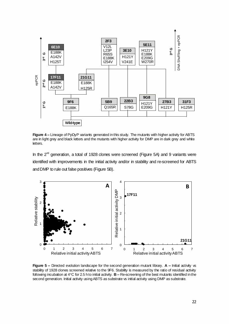
Figure 4 – Lineage of PpDyP variants generated in this study. The mutants with higher activity for ABTS are in light grey and black letters and the mutants with higher activity for DMP are in dark grey and white letters. In the 2nd generation, a total of 1928 clones were screened (Figure 5A) and 9 variants were
identified with improvements in the initial activity and/or in stability and re-screened for ABTS
and DMP to rule out false positives (Figure 5B).
Figure 5 – Directed evolution landscape for the second generation mutant library. A – Initial activity vs
stability of 1928 clones screened relative to the 9F6. Stability is measured by the ratio of residual activity following incubation at 4°C for 2.5 h to initial activity. B – Re-screening of the best mutants identified in the second generation. Initial activity using ABTS as substrate vs initial activity using DMP as substrate.
Wild-typeWild-type-type-
B1G6 K7E3B1G6B1G6 K7E3K7E3
21G11
B1G6B1G6
E188K
9F6 K7E3
Q165R
5B9
E188K
H125R
K7E3K7E3K7E3K7E327B3H121Y
H121Y
9G8
E209G
31F3
H125R
17F11
E188K
A142V
6E10
E188K
A142V
H125T
5E11
H121YE188KE209GW270R
2n
dG
1s
tG
3rd
G 3rd
G3E10
H121Y
V241E
2F3
V12LL23P
E188KR65S
I254V
DN
A S
huff
ling
+ e
pP
CR
ep
PC
R
K7E3K7E3K7E3K7E3K7E3
S78G
22B3
0
1
2
3
0 1 2 3 4 5 6 7
Re
lative
sta
bility
Relative initial activity ABTS
A
0
1
2
3
4
0 1 2 3 4 5 6 7
Re
lative
initia
l activity D
MP
Relative initial activity ABTS
B17F11
21G11

23
Two variants were selected: the 21G11 variant showing a 6-fold higher activity for ABTS, while
maintaining a similar stability when compared with the parent (9F6) (Figure 5B and Table 3)
and 17F11 variant showing 3-fold higher activity relative to the parent (9F6) using DMP as
substrate (Table 3). Both variants shared the mutation E188K from the parent and while the
17F11 acquired the additional mutation A142V, 21G11 acquired mutation H125R.
Variant 17F11 was therefore chosen as parent for the 3rd generation with the aim of improving
activity for the phenolic DMP. First we have validated the screening using this DMP was
performed; 17F11 variant was overproduced in 96-well plates, cells were disrupted and crude
extracts were used for activity measurements. A CV ~ 45% was obtained for the 17F11
variant. This high CV was only observed for the enzymatic activity while the OD600nm of cell
cultures and protein concentration of crude extracts show CVs below 20%, suggesting that
mutation A142V introduces most likely a considerable variation in the expression/transcription
of the gene or translation of RNA to proteins. After several attempts of optimization and despite
the risks of having a very high CV, it was decided to move forward using this variant as the
parent for the next round of directed evolution.
In the 3rd generation of epPCR using 17F11 as parent, a total of 1793 clones were screened
(Figure 6A) and 5 variants were identified with improvements in initial activity and/or in stability
in relation to the parental enzyme. Their re-screening allowed identifying a positive hit, the
6E10 variant with the following mutations: E188K, A142V, H125T (Figure 6B).
Figure 6 – Directed evolution landscape for the third generation mutant library. A – Initial activity of 1793 clones screened relative to 17F11. B – Re-screening of the best variants identified. Initial activity using ABTS as substrate vs initial activity using DMP as substrate.
0
1
2
3
4
0 400 800 1200 1600
Re
lative
activity D
MP
Mutants
A
0
1
2
3
4
0 1 2 3 4
Re
lative
activity D
MP
Relative activity ABTS
B6E10

24
This variant showed 3.5- and 2-fold higher activity for DMP and ABTS, respectively, when
compared to the parental enzyme (17F11) and was selected as parent for the 4th generation
(Figure 4, Table 3). Moreover, it seems to be more stable than the parent since it did not lose
activity after 3.5 h at 4ºC.
To further validate the screening, the 6E10 variant was overproduced in 96-well plates, cells
were disrupted and crude extracts were used for activity measurement using DMP as
substrate. The CV for the enzymatic activity was 28%, showing a drop of ~ 20% when
compared to the one obtained for the parental enzyme (17F11).
In the 4th generation using 6E10 as parent, a total of 1920 clones were screened. However, a
high number of inactive variants (~ 70%) were detected and none of the variants screened
showed higher activity than the parental enzyme. In fact, the DNA sequence analysis of
several variants revealed that they all had the same genotype of the parental variant 6E10.
Therefore, new libraries were created by epPCR with a higher concentration of MnCl2, 0.02
mM, since it is more likely to find variants with improved or novel functions with a higher error-
rate (Drummond et al., 2005). As expected, these conditions resulted in a higher number of
inactive variants ( 85%) however it was not possible to detect any improved variants. Thus, it
was decided to screen the libraries for thermostable hits since more robust enzymes will
accept with easiness the introduction of new mutations that tend to be destabilizing due to the
well-known trade-off between high activity and high stability (Jaenicke and Bohm, 1998). For
the set-up of the thermostability assays a time of incubation of 45 min at 50°C was selected
using the 6E10 variant based on conditions that resulted in a residual activity of about one-third
of the initial activity. Therefore, in a near future libraries of 3rd and 4th generation will be re-
screened in an attempt to find more thermostable variants that will allow continuing the
evolution of PpDyP towards an improved activity for phenolic substrates.
3.1.5.2 Towards improved catalytic efficiency for ABTS
As a result of the screening of the 2nd generation variant 21G11 was selected showing 6-fold
higher activity for ABTS (Table 3, Figure 5B). The mutation acquired by this variant, H125R,
was already present in variant 31F3 from the 1st generation (Figure 4). The observation of this
synergistic effect between mutation E188K and H125R led us to follow a
recombination/epPCR approach using variant 21G11 and variants 5B9, 9G8, 22B3 and 27B3
identified in the 1st generation (Figure 4); genes from these variants were amplified by epPCR
in the presence of 0.01 mM MnCl2, pooled and digested with DNase I during a period of time
that lead to a smear of DNA fragments with sizes below 250 bp in agarose gels. These
fragments were submitted to a “progressive hybridization” PCR program involving seven
hybridization steps to force low homology recombination. A large smear of high molecular

25
weight DNA was formed. A second PCR step, involving primers located on the cDNA flanking
regions, was performed and amplification of a well-defined band ~ 1200 bp was observed
(Figure 7).
Figure 7 - Steps of DNA shuffling performed in the 3rd generation. 1 – DNA amplification of the gene by
epPCR. 2 - Digestion with DNase I. 3 - Reassembly of DNA fragments. 4 – Amplification of full-lenght sequence.
This PCR product was cloned and used to transform E. coli KRX cells and a total of 648
variants were screened. Nine variants were identified with improvements in initial activity using
ABTS as substrate (Figure 8A) and re-screened to rule out false positives (Figure 8B).
Figure 8 – Directed evolution landscape for the third generation mutant library, constructed by DNA shuffling and epPCR. A – Initial activity of 648 clones screened relative to 21G11. B – Re-screening of the best variants identified. Initial activity using DMP as substrate vs initial activity using ABTS as
substrate.
0
1
2
3
4
0 100 200 300 400 500 600
Re
lative
Activity A
BT
S
Variants
A B
3E10
5E11
2F3
0
0.5
1
1.5
0 1 2 3 4 5
Re
lative
activity D
MP
Relative activity ABTS

26
Three hit variants were selected, 2F3, 3E10 and 5E11, showing an improvement in initial
activity of around 3- to 4.5-fold when compared to the 21G11 variant (Table 3).
The DNA sequence analysis of these variants revealed the mutations V12L, L23P, R65S,
E188K, I254V in the variant 2F3, mutations H121Y, V241E in the variant 3E10 and mutations
H121Y, E188K, E209G, W270R in the variant 5E11. These results show: (1) that variant 5E11
resulted from a simple recombination between the genes from 21G11 and 9G8 variants while
(2) 2F3 and 3E10 variants acquired new mutations during the epPCR step. Interestingly,
variant 3E10 acquired four mutations in contrast to the average of one amino acid substitution
observed during the PpDyP evolution (Figure 4, Table 3). Future work may include screening
of more variants from this library or construction of a new library using DNA shuffling approach
without the introduction of new mutations by epPCR.

27
3.2 Spectroscopic and Biochemical Characterization of PpDyP Wild-type
and Variants
3.2.1 Spectroscopic characterization
The ppDyP genes from Wt and variants 9F6, 21G11, 17G11 and 6E10 were cloned into the
expressing vector pET-21a(+) and subsequently transformed into E. coli Tuner strain. SDS-
PAGE analysis of crude extracts from the recombinant cells revealed that the addition of IPTG
to the culture medium resulted in the accumulation of an additional band at ~ 32 kDa. The
recombinant enzymes were purified from crude extracts using two chromatographic steps to
apparent homogeneity as determined by SDS-PAGE analysis (Figure 9), with purification
yields of 46, 73, 47, 48 and 30% for PpDyP Wt and variants 9F6, 21G11, 17F11 and 6E10,
respectively. Variants 21G11 (2nd G) and 6E10 (3rd G) exhibited a 2.6- and 2.0-fold increase in
the enzyme production (14.5 and 11.5 mg/L, respectively) when compared with the Wt
enzyme (5.5 mg/L). Interestingly, both variants acquired a mutation in the residue His125,
suggesting that this position is a hot-spot to enhance the PpDyP production.
MM
(kDa)
66
48
40
32
26
MM
(kDa)
66
48
40
32
26
1 2 3 1 2 3 1 2 3
1 2 3 1 2 3
A B C
D E
Figure 9 –SDS-PAGE of
purif ied PpDyP Wt (A), variant
9F6 (B), variant 21G11 (C),
variant 17F11 (D), variant
6E10 (E). (1) Cude extracts,
(2) Q-sepharose, (3) Superdex
200.

28
The overall electronic absorption features of PpDyp variants as assessed by UV-visible spectra
reveal the characteristic Soret band at ~ 400 nm, Q bands at ~ 502 and 545 nm, and a charge
transfer band at ~ 630 nm (Figure 10), as observed in Wt enzyme (Santos et al., 2014).
Figure 10 - UV-vis spectra of PpDyP variants (A) 9F6, (B) 21G11, (C) 17F11 and (D) 6E10. Resting state (black solid line), immediately after the addition of 1 equivalent of H2O2 (gray solid line) and after 1
hour (dashed line).
Heme b content was estimated by the pyridine ferrohemochrome method. Spectra of PpDyP
Wt and variants showed absorption peaks corresponding to the Soret Band at ~ 419 nm and to
the Q band at ~ 522 nm, characteristics of iron protoporphyrin IX. Heme b content for 9F6,
21G11, 17F11 and 6E10 variants was estimated to be 0.9 ± 0.1, 0.8 ± 0.1, 0.9 ± 0.1 and 0.9 ±
0.08 mol per mole of protein, respectively, similarly to the Wt (1 ± 0.07 mol per mole of protein).
All heme peroxidases require hydrogen peroxide or other hydroperoxides to activate the heme
cofactor yielding the so-called compound I intermediate in a common catalytic cycle (Hiner et
al., 2002). Compound I contains a reactive Fe4+ oxo complex with a cation radical at the
porphyrin ring, formed by two-electron oxidation of the Fe3+-containing heme of the resting
0
10
20
30
40
50
60
300 350 400 450 500 550 600 650 700
Ɛ (m
M-1
cm
-1)
450 500 550 600 650 700
0
10
20
30
40
50
60
300 350 400 450 500 550 600 650 700
450 500 550 600 650 700
0
10
20
30
40
50
60
300 350 400 450 500 550 600 650 700
Ɛ (m
M-1
cm
-1)
Wavelength (nm)
450 500 550 600 650 700
0
10
20
30
40
50
60
300 350 400 450 500 550 600 650 700
Wavelength (nm)
450 500 550 600 650 700
A B
C D
400
402
401 400
403405
409
411
502503
502 500
544545
547 541
640 631
632
630

29
enzyme. The formation of an intermediate with spectral features of Compound I (decrease in
intensity of a Soret band, disappearance of Q bands and appearance of a band ~ 550-600 nm)
was observed upon addition of 1 equivalents of hydrogen peroxide to all variants (Figure 10).
After 1 h variants returned to its ferric initial resting state, similarly to the Wt (Santos et al.,
2014).
3.2.2 Kinetic properties
The overall pH activity profile of PpDyP Wt and variants was determined using DMP and ABTS
as substrates in the presence of H2O2 (Figure 11A and B). Variants 17F11 (2nd G) and 6E10
(3rd G) revealed different pH optima values when compared to Wt (pHopt = 4.3). Both variants
increase the pH optima by 4.1 and 1.1 units to pH 8.4 and 5.4 for DMP and ABTS,
respectively. The mutation A142V, introduced in the 17F11 gene (2nd G), seems to be the
responsible for this up-shift in the optimal pH. Recently, the function of the distal residues
D132, R214 and N136 were studied by combining site-directed mutagenesis and
spectroscopic, kinetic and electrochemical studies. It was observed that the N136L mutant
shows maximal activity for ABTS at around pH 5.6. The mutation A142V is located 7.5 Å from
the catalytic residue N136 and probably introduces some structural changes, inducing a
variation in the pH profile that is presently unclear (Mendes et al., submitted, 2014).
Figure 11 - pH activity profile using (A) DMP and (B) ABTS as substrates of PpDyP wild-type () and
variants, 9F6 (), 21G11 (),17F11 (◊) and 6E10 ().
3.2.2.1 Towards improved catalytic efficiency for DMP
The catalytic properties of PpDyP Wt and 9F6, 17F11 and 6E10 variants were probed by
stead-state kinetics for reduction of H2O2 and oxidation of several substrates including ABTS,
phenolics compounds (DMP and guaiacol), and the metal ions Mn2+ and Fe2+ (Table 4).
2 4 6 8 10 120
50
100
150
200
250
pHpH
A B
2 4 6 8 10 120
2
4
6
8
10
12
Sp
ecif
ic A
ctivity (U
/mg
)

30
Table 4 – Steady-state apparent constants of purified PpDyP and 9F6, 17F11 and 6E10 variants.
Km (µM)
Wt 9F6 17F11 6E10
H2O2 (ABTS) 79 ± 5 60 ± 16 100 ± 14 34 ± 7
ABTS 2494 ± 276 250 ± 56 788 ± 90 720 ± 88
DMP 120 ± 21 78 ± 13 42 ± 4 58 ± 6
Guaiacol 32 ± 6 ND 61 ± 9 36 ± 0.4
Mn2+ 272 ± 54 152 ± 36 334 ± 66 284 ± 43
Fe2+ - 131 ± 51 102 ± 15 300 ± 63
Vmax (U/mg)
Wt 9F6 17F11 6E10
H2O2 (ABTS) 27 ± 1.2 131 ± 10 16 ± 2 27 ± 1.1
ABTS 80 ± 2.5 204 ± 13 65 ± 6 83 ± 9
DMP 0.15 ± 0.01 0.17 ± 0.01 7 ± 0.2 12 ± 3
Guaiacol 0.1 ± 0.003 0.07 ± 0.01 0.7 ± 0.03 1.4 ± 0.03
Mn2+ 8 ± 0.9 11 ± 0.9 7 ± 0.6 8 ± 1.3
Fe2+ 0.5 ±0.05 1.2 ± 0.1 1 ± 0.05 1.2 ± 0.1
Kcat/Km (M-1s-1)
Wt 9F6 17F11 6E10
H2O2 (ABTS) 1.8 x 10⁵ 1.1 x 10
6 0.8 x 10
5 4 x 10
5
ABTS 1.6 x 10⁴ 4.2 x 10
5 4.3 x 10
4 6 x 10
4
DMP 4 x 10² 10 x 10
2 8 x 10
4 10 x 10
4
Guaiacol 1.6 x 10³ ND 0.6 x 10
4 2 x 10
4
Mn2+ 15 x 103 38 x 10
3 11 x 10
3 15 x 10
3
Fe2+ - 4.8 x 10³ 5.1 x 10
3 2.1 x 10
3
The analysis of the kinetic data of PpDyP Wt and variants (Table 4) reveals that 9F6 variant
(from the first generation (1st G)) shows an improvement of 1 order of magnitude for the
specificity (kcat/Km) of ABTS when compared to the Wt as a result of a ~ 10 times lower Km and
2.5 times higher Vmax (204 U/mg vs 80 U/mg). Moreover, an improvement of 1 order of
magnitude in the specificity for hydrogen peroxide as a result of ~ 5 times higher Vmax was also
observed. This variant also revealed an improvement of 2.4-fold in the rate of Fe2+ oxidation.
The inhibitory effect of high concentrations of Fe2+ over the enzyme activity was similar to the
one observed in the Wt enzyme (Santos et al., 2014). This variant, as expected, does not show
major differences in the catalytic constants for the oxidation of phenolic compounds when
compared to the Wt enzyme.
Variants 17F11 (2nd G) and 6E10 (3rd G) show an improvement of ~ 2 orders of magnitude for
DMP specificity as a result of a ~ 3- and 2-fold lower Km and ~ 45- and 80-fold higher Vmax,
respectively when compared to the PpDyP Wt.. For the other phenolic compound tested,
guaiacol, the improvement was not so pronounced however, it was also observed a 1 order of

31
magnitude increase in the specificity, resulting in a ~ 10 times higher Vmax. The analysis of the
kinetic data for H2O2 and ABTS shows that introduction of A142V mutation results in a recede
for ABTS oxidation since variant 17F11 shows similar specificities to those of the Wt.
The mutations introduced during the molecular evolution did not altered the inhibitory effect of
hydrogen peroxide over the PpDyP activity and all variant enzymes showed a Ki of ~ 0.4 mM,
as previously observed for the Wt enzyme (Santos et al., 2014).
3.2.2.2 Towards improved catalytic efficiency for ABTS
The kinetic properties of the variant 21G11 (2nd G) selected using ABTS as substrate are very
similar to the parental enzyme (9F6) showing however an improvement of 1 order of
magnitude for the specificity (kcat/Km) of ABTS when compared to the Wt as a result of a ~ 10
times lower Km (Table 4 and 5). These results suggest that mutation H125R acquired in this
generation led to an increase of the total amount of active enzyme, rather than a more active
variant, as suggested previously.
Table 5 – Steady-state apparent constants of purified 21G11 variant.
Km (µM) Vmax (U/mg) Kcat/Km (M-1.s-1)
H2O2 (ABTS) 52 ± 13 154 ± 13 1.5 x 10⁶
ABTS 228 ± 57 82 ± 5 1.8 x 10⁵
DMP 34 ± 10 0.04 ± 0.003 6 x 10²
Guaiacol 32 ± 6 0.08 ± 0.003 1.2 x 10³
Mn2+ 161 ± 29 12.3 ± 1.6 40 x 103
Fe2+ 136 ± 37 1.3 ± 0.08 4.9 x 10³
3.2.3 Kinetic Stability of PpDyP variants
Considering the known potential of DyPs for applications in the areas of transformation of
lignin-related substrates, we have studied the kinetic stability of the enzymes (Figure 12) that
quantifies the amount of enzyme that loses activity irreversibly during incubation at a certain
temperature.

32
Figure 12 – Kinetic stability of PpDyP (♦), and variants 9F6 (), 21G11 (), 17F11 () and 6E10 () at
40⁰C at pH 7.6.
The kinetic thermal denaturation profiles indicate that the most active variant from the 1st
generation (9F6) showed a lower half-life inactivation at 40⁰ C and pH 7.6 (52 ± 8 min-1), when
compared with the Wt (110 ± 17 min-1), revealing a trade-off between high activity and high
stability (Jaenicke and Bohm, 1998). Interestingly, both variants that acquired a mutation in the
position His125 (the 21G11 from the 2nd generation and the 6E10 from the 3rd generation)
showed an increase in kinetic stability when compared with the 9F6 variant and half-lives
inactivation of 71 ± 11 min-1
and 114 ± 12 min-1, similar to that of the Wt. Thus, substitution of
His125 induced an increase not only in the protein production but also in protein stabilization.
3.3 Structural analysis of mutations
PpDyP is a homotetramer and its model structure was reasonably modeled using template
sequences from the bacterial structure from crystal structures of BtDyP (Zubieta et al., 2007b)
and TryA (Zubieta et al., 2007a). The overall fold shows the typical ferrodoxin-like fold with the
heme cofactor sandwiched between distal and proximal sides (Santos et al., 2014). The distal
side is composed by an aspartate, an asparagine and an arginine and the proximal side
contains a histidine and an aspartate residues.

33
From the three modified positions during the evolution process, two (E188 and H125) are
located in loops on the surface of the enzyme and one (A142) in an -helix on the interface of
the enzyme. The three positions are situated in the second shell of the active site residues
(Figure 13).
Figure 13 – Structural mapping of key amino acids for protein activity and stability/expression in PpDyP model structure. A - 21G11 and B - 6E10 variants. Mutation from 1
st generation is in red, mutations from
2nd
generation are in yellow and the mutation from 3rd generation is in dark blue. The catalytic residues
are in dark gray.
The variant with the best performance for ABTS oxidation and H2O2 reduction, 21G11 (Figure
8A), share the mutation E188K introduced in the 1st generation with the variant 6E10 (Figure
8B). The negatively charged residue Glu188 is located around 12 Å from the catalytic distal
residues. The introduction of a positively charged residue in this position, Lys seems to
facilitate the binding of the negatively charged oxidizing substrate ABTS and also induces
conformational changes in the active site, which increases the rate for ABTS and metals
oxidation and H2O2 reduction. Both variants, 21G11 and 6E10, also acquired other mutation in
the same position, His125, located 5 Å from the catalytic residue Asp132 and as discussed
above the substitution for either an arginine or a threonine enhances the stability of the
enzyme, improving the protein production and kinetic stability. The variant 6E10 accumulated
one more mutation, A142V, located 8 Å from the catalytic residue Asn136 and to a large extent
responsible for the increased of phenolic compounds oxidation rate.
A B
E188K E188K
H125R H125T
A142V
N136 N136
R214 R214D132 D132

34
4. Conclusions Directed evolution, using epPCR approach has proven to be an important tool for improving
the catalytic properties and protein production of PpDyP, by allowing the accumulation of one
amino acid change in each generation. The mutation rate of only one substitution per gene in
each generation allowed us to better understand the role of each mutation.
After 3 rounds of evolution using epPCR and DNA shuffling we identified 12 hits with improved
catalytic activity. Through the analysis of DNA sequences and kinetic studies the mutation
E188K seemed to be responsible an improvement of 25- and 6-fold for ABTS oxidation and
H2O2 reduction specificity, respectively. The mutation A142V revealed to be responsible for a
250-fold enhancement of the specificity for the phenolic compound DMP. It was also observed
that position His125 is a hot-spot to enhance the stability of the enzyme, improving both the
protein production and kinetic stability.
It was also observed that it is difficult to improve simultaneously the activity and the stability of
the enzyme due to the trade-off between high activity and high stability.
In the future it would be interesting to perform a screening for stability in the libraries of the 3rd
and 4th generation, in an attempt to find a more stable variant, and then continuing the
screening for variants with higher activity for phenolic compounds. It would be also important to
perform a new library, with variant 21G11 and the variants from 1st generation, using DNA
shuffling approach to understand if the mutations identified in the first generation have a
synergistic effect.

35
5. References Berry, E.A., Trumpower, B.L., 1987. Simultaneous determination of hemes a, b, and c from
pyridine hemochrome spectra. Anal Biochem 161, 1-15.
Bloom, J.D., Meyer, M.M., Meinhold, P., Otey, C.R., MacMillan, D., Arnold, F.H., 2005.
Evolving strategies for enzyme engineering. Curr Opin Struct Biol 15, 447-452.
Brakmann, S., 2001. Discovery of superior enzymes by directed molecular evolution.
Chembiochem 2, 865-871.
Brissos, V., Goncalves, N., Melo, E.P., Martins, L.O., 2014. Improving kinetic or
thermodynamic stability of an azoreductase by directed evolution. PLoS One 9, e87209.
Bugg, T.D., Ahmad, M., Hardiman, E.M., Singh, R., 2011. The emerging role for bacteria in
lignin degradation and bio-product formation. Curr Opin Biotechnol 22, 394-400.
Cirino, P.C., Mayer, K.M., Umeno, D., 2003. Generating mutant libraries using error-prone
PCR. Methods Mol Biol 231, 3-9.
Colpa, D.I., Fraaije, M.W., van Bloois, E., 2014. DyP-type peroxidases: a promising and
versatile class of enzymes. J Ind Microbiol Biotechnol 41, 1-7.
Dougherty, M.J., Arnold, F.H., 2009. Directed evolution: new parts and optimized function. Curr
Opin Biotechnol 20, 486-491.
Drummond, D.A., Iverson, B.L., Georgiou, G., Arnold, F.H., 2005. Why high-error-rate random
mutagenesis libraries are enriched in functional and improved proteins. J Mol Biol 350, 806-
816.
Garcia-Ruiz, E., Gonzalez-Perez, D., Ruiz-Duenas, F.J., Martinez, A.T., Alcalde, M., 2012.
Directed evolution of a temperature-, peroxide- and alkaline pH-tolerant versatile peroxidase.
Biochem J 441, 487-498.
Garcia-Ruiz, E., Mate, D., Ballesteros, A., Martinez, A.T., Alcalde, M., 2010. Evolving
thermostability in mutant libraries of ligninolytic oxidoreductases expressed in yeast. Microb
Cell Fact 9, 17.
Georgescu, R., Bandara, G., Sun, L., 2003. Saturation mutagenesis. Methods Mol Biol 231,
75-83.
Hiner, A.N., Raven, E.L., Thorneley, R.N., Garcia-Canovas, F., Rodriguez-Lopez, J.N., 2002.
Mechanisms of compound I formation in heme peroxidases. J Inorg Biochem 91, 27-34.
Hofrichter, M., Ullrich, R., Pecyna, M.J., Liers, C., Lundell, T., 2010. New and classic families of
secreted fungal heme peroxidases. Appl Microbiol Biotechnol 87, 871-897.
Jaenicke, R., Bohm, G., 1998. The stability of proteins in extreme environments. Curr Opin
Struct Biol 8, 738-748.
Johnson, B.H., Hecht, M.H., 1994. Recombinant proteins can be isolated from E. coli cells by
repeated cycles of freezing and thawing. Biotechnology (N Y) 12, 1357-1360.
Jongbloed, J.D., Grieger, U., Antelmann, H., Hecker, M., Nijland, R., Bron, S., van Dijl, J.M.,
2004. Two minimal Tat translocases in Bacillus. Mol Microbiol 54, 1319-1325.

36
Kim, S.J., Shoda, M., 1999. Purification and characterization of a novel peroxidase from
Geotrichum candidum dec 1 involved in decolorization of dyes. Appl Environ Microbiol 65,
1029-1035.
Kuan, I.C., Johnson, K.A., Tien, M., 1993. Kinetic analysis of manganese peroxidase. The
reaction with manganese complexes. J Biol Chem 268, 20064-20070.
Kuchner, O., Arnold, F.H., 1997. Directed evolution of enzyme catalysts. Trends Biotechnol 15,
523-530.
Lin, Z., Thorsen, T., Arnold, F.H., 1999. Functional expression of horseradish peroxidase in E.
coli by directed evolution. Biotechnol Prog 15, 467-471.
Liu, X., Du, Q., Wang, Z., Zhu, D., Huang, Y., Li, N., Wei, T., Xu, S., Gu, L., 2011. Crystal
structure and biochemical features of EfeB/YcdB from Escherichia coli O157: ASP235 plays
divergent roles in different enzyme-catalyzed processes. J Biol Chem 286, 14922-14931.
Martinez, A.T., Ruiz-Duenas, F.J., Martinez, M.J., Del Rio, J.C., Gutierrez, A., 2009. Enzymatic
delignification of plant cell wall: from nature to mill. Curr Opin Biotechnol 20, 348-357.
Mendes, S., Brissos, V., Gabriel, A., Catarino, T., Todorovic, S., Martins, L.O., submitted, 2014.
An integrated view of redox and catalytic properties of B-type 1 PpDyP from Pseudomonas
putida MET94 and its distal variants. Arch. Biochem. Biophys.
Molina-Espeja, P., Garcia-Ruiz, E., Gonzalez-Perez, D., Ullrich, R., Hofrichter, M., Alcalde, M.,
2014. Directed evolution of unspecific peroxygenase from Agrocybe aegerita. Appl Environ
Microbiol 80, 3496-3507.
Morawski, B., Lin, Z., Cirino, P., Joo, H., Bandara, G., Arnold, F.H., 2000. Functional
expression of horseradish peroxidase in Saccharomyces cerevisiae and Pichia pastoris.
Protein Eng 13, 377-384.
Reetz, M.T., Kahakeaw, D., Lohmer, R., 2008. Addressing the numbers problem in directed
evolution. Chembiochem 9, 1797-1804.
Rodriguez-Couto, S., 2009. Enzymatic biotransformation of synthetic dyes. Curr Drug Metab
10, 1048-1054.
Romero, P.A., Arnold, F.H., 2009. Exploring protein fitness landscapes by directed evolution.
Nat Rev Mol Cell Biol 10, 866-876.
Salazar, O., Sun, L., 2003. Evaluating a screen and analysis of mutant libraries. Methods Mol
Biol 230, 85-97.
Santos, A., Mendes, S., Brissos, V., Martins, L.O., 2014. New dye-decolorizing peroxidases
from Bacillus subtilis and Pseudomonas putida MET94: towards biotechnological applications.
Appl Microbiol Biotechnol 98, 2053-2065.
Scheibner, M., Hulsdau, B., Zelena, K., Nimtz, M., de Boer, L., Berger, R.G., Zorn, H., 2008.
Novel peroxidases of Marasmius scorodonius degrade beta-carotene. Appl Microbiol
Biotechnol 77, 1241-1250.
Singh, R., Grigg, J.C., Armstrong, Z., Murphy, M.E., Eltis, L.D., 2012. Distal heme pocket
residues of B-type dye-decolorizing peroxidase: arginine but not aspartate is essential for
peroxidase activity. J Biol Chem 287, 10623-10630.

37
Strittmatter, E., Liers, C., Ullrich, R., Wachter, S., Hofrichter, M., Plattner, D.A., Piontek, K.,
2013. First crystal structure of a fungal high-redox potential dye-decolorizing peroxidase:
substrate interaction sites and long-range electron transfer. J Biol Chem 288, 4095-4102.
Sugano, Y., 2009. DyP-type peroxidases comprise a novel heme peroxidase family. Cell Mol
Life Sci 66, 1387-1403.
Tracewell, C.A., Arnold, F.H., 2009. Directed enzyme evolution: climbing fitness peaks one
amino acid at a time. Curr Opin Chem Biol 13, 3-9.
Welinder, K.G., Mauro, J.M., Norskov-Lauritsen, L., 1992. Structure of plant and fungal
peroxidases. Biochem Soc Trans 20, 337-340.
Yoshida, T., Tsuge, H., Konno, H., Hisabori, T., Sugano, Y., 2011. The catalytic mechanism of
dye-decolorizing peroxidase DyP may require the swinging movement of an aspartic acid
residue. FEBS J 278, 2387-2394.
Zederbauer, M., Furtmuller, P.G., Brogioni, S., Jakopitsch, C., Smulevich, G., Obinger, C.,
2007. Heme to protein linkages in mammalian peroxidases: impact on spectroscopic, redox
and catalytic properties. Nat Prod Rep 24, 571-584.
Zubieta, C., Joseph, R., Krishna, S.S., McMullan, D., Kapoor, M., Axelrod, H.L., Miller, M.D.,
Abdubek, P., Acosta, C., Astakhova, T., Carlton, D., Chiu, H.J., Clayton, T., Deller, M.C., Duan,
L., Elias, Y., Elsliger, M.A., Feuerhelm, J., Grzechnik, S.K., Hale, J., Han, G.W., Jaroszewski,
L., Jin, K.K., Klock, H.E., Knuth, M.W., Kozbial, P., Kumar, A., Marciano, D., Morse, A.T.,
Murphy, K.D., Nigoghossian, E., Okach, L., Oommachen, S., Reyes, R., Rife, C.L., Schimmel,
P., Trout, C.V., van den Bedem, H., Weekes, D., White, A., Xu, Q., Hodgson, K.O., Wooley, J.,
Deacon, A.M., Godzik, A., Lesley, S.A., Wilson, I.A., 2007a. Identification and structural
characterization of heme binding in a novel dye-decolorizing peroxidase, TyrA. Proteins 69,
234-243.
Zubieta, C., Krishna, S.S., Kapoor, M., Kozbial, P., McMullan, D., Axelrod, H.L., Miller, M.D.,
Abdubek, P., Ambing, E., Astakhova, T., Carlton, D., Chiu, H.J., Clayton, T., Deller, M.C.,
Duan, L., Elsliger, M.A., Feuerhelm, J., Grzechnik, S.K., Hale, J., Hampton, E., Han, G.W.,
Jaroszewski, L., Jin, K.K., Klock, H.E., Knuth, M.W., Kumar, A., Marciano, D., Morse, A.T.,
Nigoghossian, E., Okach, L., Oommachen, S., Reyes, R., Rife, C.L., Schimmel, P., van den
Bedem, H., Weekes, D., White, A., Xu, Q., Hodgson, K.O., Wooley, J., Deacon, A.M., Godzik,
A., Lesley, S.A., Wilson, I.A., 2007b. Crystal structures of two novel dye-decolorizing
peroxidases reveal a beta-barrel fold with a conserved heme-binding motif. Proteins 69, 223-
233.