engineering the mechanical and physical properties of organic–inorganic composite...
TRANSCRIPT

Engineering the mechanical and physical propertiesof organic–inorganic composite microcapsulesLong, Yue; Song, Kai; York, David; Zhang, Zhibing; Preece, Jon
DOI:10.1016/j.colsurfa.2013.04.055
License:Creative Commons: Attribution (CC BY)
Document VersionPublisher's PDF, also known as Version of record
Citation for published version (Harvard):Long, Y, Song, K, York, D, Zhang, Z & Preece, JA 2013, 'Engineering the mechanical and physical properties oforganic–inorganic composite microcapsules' Colloids and Surfaces A: Physicochemical and EngineeringAspects, vol 433, pp. 30-36. DOI: 10.1016/j.colsurfa.2013.04.055
Link to publication on Research at Birmingham portal
Publisher Rights Statement:Eligibility for repository : checked 12/09/2014
General rightsUnless a licence is specified above, all rights (including copyright and moral rights) in this document are retained by the authors and/or thecopyright holders. The express permission of the copyright holder must be obtained for any use of this material other than for purposespermitted by law.
•Users may freely distribute the URL that is used to identify this publication.•Users may download and/or print one copy of the publication from the University of Birmingham research portal for the purpose of privatestudy or non-commercial research.•User may use extracts from the document in line with the concept of ‘fair dealing’ under the Copyright, Designs and Patents Act 1988 (?)•Users may not further distribute the material nor use it for the purposes of commercial gain.
Where a licence is displayed above, please note the terms and conditions of the licence govern your use of this document.
When citing, please reference the published version.
Take down policyWhile the University of Birmingham exercises care and attention in making items available there are rare occasions when an item has beenuploaded in error or has been deemed to be commercially or otherwise sensitive.
If you believe that this is the case for this document, please contact [email protected] providing details and we will remove access tothe work immediately and investigate.
Download date: 01. Jun. 2018

Colloids and Surfaces A: Physicochem. Eng. Aspects 433 (2013) 30– 36
Contents lists available at SciVerse ScienceDirect
Colloids and Surfaces A: Physicochemical andEngineering Aspects
jo ur nal ho me p ag e: www.elsev ier .com/ locate /co lsur fa
Engineering the mechanical and physical properties oforganic–inorganic composite microcapsules
Yue Longa,b,∗,1, Kai Songb, David Yorkc, Zhibing Zhangd, Jon A. Preecea,∗
a School of Chemistry, University of Birmingham, Edgbaston, Birmingham, B15 2TT, UKb Beijing National Laboratory for Molecular Sciences, Institute of Chemistry, Chinese Academy of Sciences, Zhongguancun North First Street 2, Beijing,100190, Chinac Institute of Particle Science & Engineering, School of Process, Environmental and Materials Engineering, University of Leeds, Leeds, LS2 9J, UKd School of Chemical Engineering, University of Birmingham, Edgbaston, Birmingham, B15 2TT, UK
h i g h l i g h t s
• Double-shell composite microcap-sules were synthesized.
• Mechanical property and leakinessof the microcapsules were character-ized.
• SEM, TEM, GC and micromanipula-tion techniques were employed.
• The strength and leakiness of themicrocapsules could be engineeredseparately.
g r a p h i c a l a b s t r a c t
a r t i c l e i n f o
Article history:Received 4 March 2013Received in revised form 19 April 2013Accepted 28 April 2013Available online 6 May 2013
Keywords:MicrocapsulePickering emulsionCompositeDouble-shellControlled delivery
a b s t r a c t
Double-shell composite microcapsule with a ripened CaCO3 nanoparticle outer shell and melamineformaldehyde (MF)/copolymer inner shell shows advantages in adjustable permeability and mechanicalstrength, comparing with single shell microcapsules. Here, we have systematically studied the effectsof certain formulation parameters on the properties of the double-shell composite microcapsules, i.e.the MF cross-linking time and the concentration of the aqueous CaCl2 and Na2CO3 used for the ripen-ing process of CaCO3 nanoparticles. The properties of the microcapsules such as average diameter, wallthickness, degree of wall formation formed by the ripened CaCO3 nanoparticles, nominal rupture stressand leakiness were characterized.
© 2013 Elsevier B.V. All rights reserved.
1. Introduction
Calcium carbonate is a natural shell material. It is non-toxic,stable, biocompatible and strong [1]. Therefore, in nature it is able
∗ Corresponding authors at: School of Chemistry, University of Birmingham, Edg-baston, Birmingham, B15 2TT, UK. Tel.: +44 0121 414 3528.
E-mail addresses: [email protected] (Y. Long),[email protected] (J.A. Preece).
1 Tel.: +86 10 82617303.
to act as a physical barrier in shells to protect organisms fromthe environment, while allowing the gas and nutrient exchangethrough the shell. Biomimetic core–shell structured microcap-sules have been developed to encapsulate active ingredients toprotect them from an external environment [2–4]. Recently, cal-cium carbonate has attracted considerable attention for makingwalls of microcapsules due to its biocompatibility and pH triggeredrelease mechanism [5,6]. Thomas et al. [7] have utilised a mem-brane technique to prepare CaCO3 microcapsules, creating a pseudowater-in-oil-in-water emulsion system to precipitate calcium car-bonate at the oil–water interface. The preparation of hollow CaCO3
0927-7757/$ – see front matter © 2013 Elsevier B.V. All rights reserved.http://dx.doi.org/10.1016/j.colsurfa.2013.04.055

Y. Long et al. / Colloids and Surfaces A: Physicochem. Eng. Aspects 433 (2013) 30– 36 31
Table 1Microcapsules batches 1–6.
Batch no. Reaction time (h) Concentration of CaCl2 (aq)and Na2CO3 (aq) (M)
1 4 1.52 8 1.53 4 04 24 1.05 24 1.56 24 2.0
microspheres is also an active field [8–10]. Such CaCO3 microcap-sules and microspheres have been used in a variety of industrialproducts, especially in biological applications [11–13]. However,the calcium carbonate wall possesses greater porosity when com-pared to organic wall materials [14,15], which limits its applicationsin encapsulation of small molecules.
We have previously introduced a methodology of form-ing double-shell composite microcapsule with a ripenedCaCO3 nanoparticle outer shell and melamine formaldehyde(MF)/copolymer inner shell to reduce the permeability [16]. As themechanical properties and leakiness are two essential parametersof microcapsules in many applications. For different uses themechanical strength and leakiness of microcapsules are alwaysneeded to be increased or decreased in order to meet the specificdemands. In this paper, we made a systematic study of the effectof CaCO3 nanoparticulate ripening process and MF cross-linkingreaction time on the following physical properties of the micro-capsules: wall thickness and completeness, nominal rupture stressand leakage of core oil, in order to manipulate the mechanicalstrength and leakiness of the microcapsules accordingly.
2. Materials and methods
2.1. Materials
The core oil is a typical organic blend of various fragrantcomponents which has a relatively low solubility in water andis used in consumer products. MF precondensate (70% (aq),w/w, formaldehyde to melamine molar ratio 0.20) was suppliedby British Industrial Plastics Ltd., Birmingham, UK, formalde-hyde (37% (aq), w/w) was supplied by Sigma–Aldrich, UK.Poly(acrylamide–acrylic acid, sodium salt) was supplied by Poly-mersciences, Inc., US and CaCO3 nanoparticles were supplied byOmya, UK. All chemicals were used without further purifica-tion.
2.2. Preparation of ripened double shell composite microcapsules
An aqueous solution of MF precondensate (2.50 g) and copoly-mer (0.58 g, poly(acrylamide–acrylic acid, sodium salt) in water(70 mL) was stirred (400 rpm) with a Rushton turbine (31 mm, in avessel with standard configuration) for 105 min at pH 4 (adjustedby acetic acid (1 M), monitored by a pH meter) to form pre-crosslinked MF precondensate and copolymer. The core oil (9.3 g)was added to an aqueous CaCO3 nanoparticle dispersion (40 mL,10% wt) and stirred using a homogenizer (Silverson MachinesLtd.) at a speed of 2500 rpm for 3 min to form an o/w emulsion.To the resulting emulsion, aqueous CaCl2 (10 mL) and Na2CO3(10 mL) (1.0, 1.5 and 2.0 M for batches 4, 5 and 6 (Table 1), respec-tively) was added via a pump system (Model 101U, Water Marlow,UK) over 15 min, and stirred for another 10 min at a speed of400 rpm. To the resulting aqueous dispersion, the pre-crosslinkedMF precondensate and copolymer solution was then added via
a pump system (Model 101U, Water Marlow, UK) over 10 min,and the pH was raised to 6.0 with the addition of aqueous NaOH(1 M, monitored by a pH meter). The resulting dispersion wasstirred at a speed of 400 rpm at 65 ◦C for 4, 8, and 24 hours forbatches 1 and 3, 2, and 4–6, respectively, and was allowed tocool to room temperature. The exact conditions for producing thedouble shell composite microcapsules batches 1–6 are listed inTable 1.
2.3. Environmental scanning electron microscopy (ESEM)
Environmental-SEM (FEI/Philips XL30 ESEM-FEG, Philips, UK)with an operating voltage shown in images was used to study themorphology of the microcapsules. Samples were operated in highvacuum mode.
2.4. Transmission electron microscopy (TEM)
Transmission Electron Microscopy (JEOL 1200EX, Jeol Ltd., UK)operating at 80 eV was used to examine the structure and thick-ness of the microcapsule wall. Microcapsules were embeddedin LR white hard grade acrylic resin, and the ultra thin sec-tions (90–150 nm) were obtained by using an ultracut microtome(Reichert-Jung), see below.
2.4.1. Sampling by ultra-microtomeThe microcapsule dispersions (2 mL) were centrifuged at a speed
of 500 rpm for 3 min, and the supernatant liquid was separated.Gluteraldehyde (2 mL) was added and the microcapsules werere-dispersed by gently shaking the vial manually. The resultingmicrocapsule dispersions were stored for 1 h to harden the outershell of the microcapsules by cross-linking the microcapsule wallwith gluteraldehyde, and centrifuged for 3 min at 500 rpm. Thesupernatant liquid (excess gluteraldehyde) was separated, and asolution of ethanol and water was added (1:1 volume ratio, 2 mL intotal), and shaken by hand. The resulting mixture was centrifugedagain at 500 rpm for 15 min, and the supernatant ethanol/watermixture was separated. The ethanol/water process was repeatedfour times, but with ethanol/water of 70:30, 90:10, 96:4 (vol-ume), and finally the microcapsules were suspended in absoluteethanol. The resulting microcapsules dispersion was centrifugedagain at 500 rpm for 15 min, and the supernatant liquid (excessabsolute ethanol) was decanted. To the microcapsule slurry wasadded absolute ethanol and LR white resin (1:1, 2 mL), and themicrocapsules were re-dispersed by gently shaking the vial man-ually. The resulting mixture was put on a rotator at a speed of4 rpm. After ∼3 min the microcapsules were displaced to the bot-tom of the vial, and rotating continued for a total of 3 h. Thesupernatant ethanol/resin solution was separated. 100% of LR whiteresin (2 mL) was added to the resulting microcapsule slurry, beforethey were rotated again at 4 rpm for 12 h, and the microcap-sules were separated from the supernatant. Two beam capsules(15 mm × 5 mm) were filled to the top with the air free LR whiteresin (2 mL). The separated microcapsules were added to eachresin filled beam capsule. The microcapsules settled on the bot-tom of the beam capsules. The mixture was heated (60 ◦C) undervacuum for 30 min, then left to cure for 48 h at 60 ◦C, affordinga polymerized resin-microcapsule block. The resulting block wassecured into a REICHERT-JUNG ultramicrotome apparatus and themicrocapsule end was trimmed to sections of thickness 90–150 nm.The gold coloured sections were placed on carbon coated grids(G2500 C, 2 mm × 1 mm slot, copper, 3.05 mm) ready to be exam-ined by TEM. The wall thicknesses of microcapsules were measuredfrom the analysis of their TEM images of the ultra microtome sec-tions.

32 Y. Long et al. / Colloids and Surfaces A: Physicochem. Eng. Aspects 433 (2013) 30– 36
Scheme 1. Schematic diagram of the formation of the double-shell composite microcapsules. (a) o/w Emulsion stabilized by CaCO3 nanoparticles was formed by emulsifyingcore oil and aqueous dispersion of CaCO3 nanoparticles. (b) Addition of aqueous CaCl2 and Na2CO3 (1.0/1.5/2.0 M) into (a) to form the ripened o/w emulsion stabilized byCaCO3 nanoparticles. (c) Ripened double-shell composite microcapsules were formed by the addition of pre-crosslinked MF and copolymer into (b), and heated at 65 ◦C for4/8/24 h.
2.5. Calculation of the wall thickness and degree of wallcompleteness of the ripened CaCO3 nanoparticles from TEMimages
The wall thickness for each microcapsule was obtained by mea-suring 30 different parts of the microcapsule wall from the TEMimage, and taking the average value. For each batch, 30 microcap-sules were measured. The degree of completeness of the ripenedCaCO3 nanoparticles of the double-shell composite microcapsuleswas calculated from an image analysis software (Image J, NationalInstitutes of Health, USA) on the ultra microtome TEM images, byusing the sum of the smallest perpendicular distance between eachdisconnected piece of the wall, subtracting this value from the totalwall perimeter, and dividing by the total wall perimeter. For eachbatch, 30 microcapsules were measured.
2.6. Leakiness measurement
The microcapsules were filtered from the original aqueousdispersion, and re-dispersed in deionized water (50 mL). To theresulting aqueous dispersion (batches 1–6), hexane (30 mL) wasadded and stirred for 10 min and then stopped. A hexane aliquot(1 �L) was removed and analysed by gas chromatography (GC).Further aliquots (1 �L) were removed at various time intervalsbetween 1 and 24 h, prior to which the dispersion was stirred for10 min.
2.7. Size analysis
Mean particle size and size distribution of the microcapsulesin aqueous dispersions were evaluated by a laser diffractiontechnique (Mastersizer 2000, Malvern Instruments Ltd., UK). Themeasurements were carried out using a Helium–Neon laser with ameasurement range of 0.05–900 �m connected to a sample disper-sion unit. Experiments were performed at 25 ◦C. The mean diameterand size distribution of microcapsules were the average value offive measurements.
2.8. Micromanipulation
The mechanical properties of the microcapsules were deter-mined by micromanipulation. A glass probe with a diameter of50 �m mounted on a force transducer (Model 405A, Aurora Scien-tific Inc., Canada) was positioned perpendicular to the glass slide.The microcapsules in aqueous dispersions were dried on the glassslide, and observed through side and bottom-view cameras. A sin-gle microcapsule was compressed by the glass probe travellingat 2 �m s−1. The voltage output generated by the transducer dueto compression of the microcapsule was recorded through a dataacquisition card in a personal computer. From the sensitivity of thetransducer, the voltage was converted to force; hence the ruptureforce of the microcapsule was determined. Details of this techniqueare described elsewhere [17].
3. Results and discussion
3.1. Synthesis
The double-shell composite microcapsules were prepared usinga method based on in situ polymerization of MF with a copoly-mer migrating though the interstice of the ripened CaCO3 shell, andpolymerizing at the oil–water interface inside the ripened CaCO3nanoparticulate shell. The synthesis is illustrated in Scheme 1.Core oil was mixed with the aqueous CaCO3 nanoparticulate dis-persion to form the o/w emulsion followed by the addition ofequivalent amount of aqueous CaCl2 and Na2CO3 (1.0, 1.5 and2.0 M, respectively) to ripen the CaCO3 nanoparticles. Finally, thepre-crosslinked MF precondensate and copolymer formed by mix-ing the MF precondensate and copolymer (poly(acrylamide–acrylicacid, sodium salt)) in water for 105 min at pH 4.3 were added tothe resulting ripened CaCO3 nanoparticulate aqueous dispersion,which was heated at 65 ◦C for 4, 8 and 24 h, respectively. Batches1–6 were prepared as detailed in Table 1.
3.2. Wall thickness and degree of wall completeness
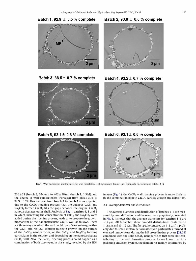
The wall thickness and degree of wall completeness formed bythe CaCO3 nanoparticles for batches 1–6 microcapsules were eval-uated and studied from the TEM images of the ultra-microtomesections, as shown in Fig. 1. As one might expect neither theouter CaCO3 wall thickness of the double-shell composite micro-capsules (402 ± 36, 406 ± 41 and 410 ± 47 nm for batches 1, 2 and5, respectively (Fig. 2)) nor the degree of wall formation formedby the ripened CaCO3 nanoparticles (92.9 ± 0.5%, 93.0 ± 0.5% and93.3 ± 0.4% for batches 1, 2 and 5, respectively) is significantlyaffected by the MF cross-linking reaction time. However, duringthe ultra-microtome sampling, some parts of the MF polymer innerwall were detached from the outer CaCO3 nanoparticluate wall.The detachment allowed us to measure the MF wall thickness asa function of reaction time. The wall thickness of the MF polymerincreased from 129 ± 2.5 nm (batch 1, 4 h) to 160 ± 3.1 nm (batch5, 24 h) when the MF cross-linking time increased from 4 to 24 h.Previous studies on the kinetics of the MF polymerization reactionshowed that the polymerization mainly took place during the first2–3 h of the reaction [18,19], which is in agreement with our resultthat the wall thickness does not increase much as the cross-linkingtime increased from 4 to 24 h. Prolonged reaction time producesmore cross-linked network, and consequently results smaller poreson the wall [20].
A significant increase in the CaCO3 wall thickness from360 ± 41 nm (batch 4) to 580 ± 48 nm (batch 6) was observedwhen the concentration of aqueous CaCl2 and Na2CO3 increasedfrom 1.0 to 2.0 M (Fig. 2). However, the degree of wall formationformed by the ripened CaCO3 nanoparticles remained unchanged(batch 4–6, ∼93%), so did the inner MF/copolymer wall thick-ness (batch 4–6, ∼160 nm). In addition, by ripening the CaCO3nanoparticulate wall, the CaCO3 wall thickness increased from
Y. Long et al. / Colloids and Surfaces A: Physicochem. Eng. Aspects 433 (2013) 30– 36 33
Fig. 1. Wall thicknesses and the degree of wall completeness of the ripened double-shell composite microcapsules batches 1–6.
210 ± 21 (batch 3, 0 M) nm to 402 ± 36 nm (batch 1, 1.5 M), andthe degree of wall completeness increased from 88.5 ± 0.7% to92.9 ± 0.5%. This increase from batch 3 to batch 1 is as expecteddue to the CaCO3 ripening process, that the aqueous CaCl2 andNa2CO3 formed CaCO3 fills the gaps between the original CaCO3nanoparticulates outer shell. Analysis of Fig. 1 batches 4, 5 and 6in which increasing the concentration of CaCl2 and Na2CO3 wereadded during the ripening process, leads us to propose the growthmechanism of the nanoparticulate CaCO3 wall as follows. Thereare three ways in which the wall could ripen. We can imagine thatthe CaCl2 and Na2CO3 solution nucleate growth on the surfaceof the CaCO3 nanoparticles, or the CaCl2 and Na2CO3 formingparticulates in the solution and depositing on the nanoparticulateCaCO3 wall. Also, the CaCO3 ripening process could happen as acombination of both two types. In this study, revealed by the TEM
images (Fig. 1), the CaCO3 wall ripening process is more likely tobe the combination of both CaCO3 particle growth and deposition.
3.3. Average diameter and distribution
The average diameter and distribution of batches 1–6 are mea-sured by laser diffraction and the results are graphically presentedin Fig. 3. It shows that the average diameters for batches 1–6 are∼14 �m. All 6 batches show bimodal distributions centered on1–2 �m and 13–15 �m. The first peak (centered on 1–2 �m) is prob-ably due to small melamine formaldehyde particulates formed atelevated temperature during the MF cross-linking process [21,22]combined with the solid CaCO3 nanoparticles that were not con-tributing to the wall formation process. As we know that in apickering emulsion system, the diameter is mainly determined by

34 Y. Long et al. / Colloids and Surfaces A: Physicochem. Eng. Aspects 433 (2013) 30– 36
Fig. 2. CaCO3 Wall thickness and nominal rupture stress for the ripened double-shellcomposite microcapsules batches 1–6.
the emulsifying speed, oil/water ratio, and the amount of particlesused in the emulsification stage. This is in agreement with our resultthat neither the MF cross-linking time nor the CaCO3 wall thicknessaffects the stability of the initial droplet formed during the emulsi-fication step. Hence, the average diameter and size distribution arenot changed.
3.4. Mechanical properties
The mechanical properties of batches 1–6 are evaluated usingthe micromanipulation technique. A typical relationship of theforce imposed on a single microcapsule as a function of the probemoving distance is shown in Fig. 4 for a batch 1 microcapsule. Afterpoint I the probe touches the microcapsule, and the force imposedon the microcapsule increases until point II at which the micro-capsule ruptures. As a result, the force drops to point III. The probecontinues to compress the debris of the microcapsule (III–IV) untilit touches the slide at point IV, after which the force increases as theprobe pushes onto the slide. From this curve, the rupture force ofthe microcapsule can be determined (point II). The displacement atrupture (ıR) is denoted as the distance that the probe travels fromcontact with microcapsule until rupture. The deformation at rup-ture is calculated using Eq. (1) (d = initial microcapsule diameter),and the nominal rupture stress is calculated using Eq. (2) as shownbelow, where FR is the rupture force (point II).
Deformation at rupture (%) = ıR
d× 100 (1)
Fig. 3. Average diameter and size distribution of the ripened double shell compositemicrocapsules batches 1–6.
Fig. 4. Typical force vs. probe moving distance curve obtained by compressing oneof the microcapsules in batch 1; I: probe/microcapsule contact, I–II: microcapsulecompression, II: microcapsule ruptured, III-IV: probe compressing the broken debris,IV: probe touching the glass slide.
Norminal rupture stress = FR
�(d/2)2(2)
The nominal rupture stress and wall thickness for all the 6batches are presented in Fig. 2. The deformation at rupture forall 6 batches are indeterminate of altering the CaCO3 nanopar-ticulate ripening process or the MF cross-linking reaction time,as all microcapsules deformed by ∼7.5% at rupture. However, thenominal rupture stress increased from 0.22 ± 0.05 (Batch 4) to0.91 ± 0.07 MPa (Batch 6, Fig. 2) when the concentration of CaCl2and Na2CO3 increased in the CaCO3 nanoparticulate ripening pro-cess (line 1). Also, we can see that the increase in nominal rupturestress follows closely the increase in wall thickness of the CaCO3outer wall. There is a slight increase of nominal rupture stress from0.49 ± 0.06 to 0.65 ± 0.09 when MF cross-linking time increased(line 2). However, when taking errors into consideration, we can-not safely conclude that prolonged cross-linking of MF enhancedthe nominal rupture stress of the microcapsules. Hence, we proposethat the rupture stress is dominantly contributed by the inorganicCaCO3 outer shell [16]. The displacement at rupture, rupture forceand nominal stress at rupture as a function of diameter for batch 6microcapsules are plotted, and are shown in Fig. 5a–c, respectively.It was observed that for the same type of microcapsules, the dis-placement at rupture (Fig. 5a) and rupture force (Fig. 5b) increase asthe diameter of the microcapsules increases. In contrast, the nom-inal stress at rupture (Fig. 5c) decreases as the particle diameterincreases. The stress at rupture of the microcapsules may be directlyrelated to their rupture behaviour in processing equipment. Largermicrocapsules were found to rupture more easily than smaller onesin the previous studies [23]. Similar trends are also seen in micro-capsules 1–6 (Please find the micromanipulation data for batches1–6 in the supporting information S1, S2 and S3).
3.5. Leakiness
The leakiness of the 1–6 microcapsules was monitored over aperiod of 24 h and the results are plotted in Fig. 6. It can be seenthat the unripened double-shell microcapsules (Batch 3) has amuch larger leakiness (0.15 ± 0.011%) after 24 h in comparison withthe ripened double-shell microcapsules (0.047 ± 0.002%) (Batch 1).However, when the concentration of the aqueous CaCl2 and Na2CO3in the ripening process increased from 1.0 to 2.0 M, the leakinessof the microcapsules only decreased from 0.052 ± 0.001% (batch4) to 0.045 ± 0.001% (batch 6). We found this leakiness result cor-relates well with the wall structure, that the higher leakiness ofthe unripened microcapsules might be resulted from their lower

Y. Long et al. / Colloids and Surfaces A: Physicochem. Eng. Aspects 433 (2013) 30– 36 35
Fig. 5. Displacement at rupture vs. diameter, rupture force vs. diameter, andnominal stress at rupture vs. diameter for the ripened double shell composite micro-capsules batch 6.
Fig. 6. Leakiness of the ripened double shell composite microcapsules batches 1–6.
degree of completerness of the wall compare to the ripened ones.The less reduction in leakiness between the ripened microcap-sules batch 4–6 might be due to their very similar completenessof wall (∼93%). Nevertheless, when the MF cross-linking reac-tion time is increased from 4 (batch 1) to 24 (batch 5) hours,the leakage of the ripened double-shell composite microcapsulesdecreased (from 0.095 ± 0.002% to 0.047 ± 0.001%). This decreasein leakage is probably due to the formation of a thicker and morecompact MF polymer wall (Fig. 1, batch 1 and 5) resulting from thelonger cross-linking reaction time. The leakage behaviour indicatesthat increasing the organic MF/copolymer inner wall thickness andcompactness is a more effective way for preventing the oil leakagethan further increasing the thickness of CaCO3 outer shell.
4. Conclusion
We have successfully prepared ripened double-shell compos-ite microcapsules with an organic MF/copolymer inner shell andan inorganic ripened CaCO3 nanoparticulate outer shell. It wasfound that by ripening the CaCO3 outer shell, the completenessof the microcapsules wall increased, the nominal rupture stressincreased, and consequently the leakiness of the microcapsules wasreduced. Hence, ripening process is a significant step for improvingthe mechanical and physical stabilities of the double-shell com-posite microcapsules. As we further adjust the concentration ofthe aqueous CaCl2 and Na2CO3 in the ripening step to modulatethe wall thickness of the CaCO3 outer shell. We found that whenthe CaCO3 wall thickness increased, the nominal rupture stressincreased, the leakiness remained unchanged. In contrast, the con-centration of the aqueous CaCl2 and Na2CO3 increased leadingto the CaCO3 nanoparticulate ripening process, both the ripenedCaCO3 wall thickness of the microcapsules and the nominal rup-ture stress of the microcapsules increased, and the leakiness of themicrocapsules decreased. When increasing the MF polymerizationtime, the MF/copolymer inner wall thickness increased, resultingin the decrease of the leakiness of the microcapsules. Therefore, weconclude that the mechanical properties of the double-shell com-posite microcapsules were dominated by the CaCO3 outer shell,whereas the leakage of the microcapsules was primarily governedby the MF inner shell. Thus, we now have a process whereby wecan engineer the strength of the microcapsules independently ofthe leakiness.
Acknowledgements
Yue Long was supported by the School of Chemistry, Univer-sity of Birmingham through Overseas Research Student AwardsScheme funding, Procter and Gamble and Advantage West Mid-land (AWM) and the European Research Development Fund (ERDF)through Science City, Advanced Materials Project 2 (InnovativeUses for Advanced Materials in the Modern World), and the EPSRC(EP/F068395/1).
Appendix A. Supplementary data
Supplementary data associated with this article can befound, in the online version, at http://dx.doi.org/10.1016/j.colsurfa.2013.04.055.
References
[1] G.J. Vermeij, A Natural History of Shells, Princeton University Press, New Jersey,1993.
[2] Q. He, Y. Cui, J. Li, Molecular assembly and application of biomimetic microcap-sules, Chem. Soc. Rev. 38 (2009) 2292.
[3] T. Boudou, K. Ren, G. Blin, C. Picart, Multiple functionalities of polyelectrolytemultilayer films: new biomedical applications, Adv. Mater. 22 (2010) 441.

36 Y. Long et al. / Colloids and Surfaces A: Physicochem. Eng. Aspects 433 (2013) 30– 36
[4] M.F. Haase, D.O. Grigoriev, H. Möhwald, D.G. Shchukin, Development ofnanoparticle stabilized polymer nanocontainers with high content of theencapsulated active agent and their application in water-borne anticorrosivecoatings, Adv. Mater. 24 (2012) 2429.
[5] X. Wang, W. Zhou, J. Cao, W. Liu, S. Zhu, Preparation of core–shell CaCO3 cap-sules via Pickering emulsion templates, J. Colloid Interface Sci. 372 (2012) 24.
[6] M. Fujiwara, K. Shiokawa, K. Morigaki, Y. Zhu, Y. Nakahara, Calcium carbonatemicrocapsules encapsulating biomacromolecules, Chem. Eng. J. 137 (2008) 14.
[7] J.A. Thomas, L. Seton, R.J. Davey, C.E. DeWolf, Using a liquid emulsion mem-brane system for the encapsulation of organic and inorganic substrates withininorganic microcapsules, Chem. Commun. 10 (2002) 1072.
[8] G. Hadiko, Y.S. Han, M. Fuji, M. Takahashi, Synthesis of hollow calcium carbon-ate particles by the bubble templating method, Mater. Lett. 59 (2005) 2519.
[9] N. Loges, K. Graf, L. Nasdala, W. Tremel, Probing cooperative interactions oftailor-made nucleation surfaces and macromolecules: a bioinspired route tohollow micrometer-sized calcium carbonate particles, Langmuir 22 (2006)3073.
[10] D. Walsh, B. Lebeau, S. Mann, Morphosynthesis of calcium carbonate (vaterite)microsponges, Adv. Mater. 11 (1999) 324.
[11] Y.S. Han, G. Hadiko, M. Fuji, M. Takahashi, A novel approach to synthesizehollow calcium carbonate particles, Chem. Lett. 34 (2005) 152.
[12] F.C. Meldrum, Calcium carbonate in biomineralisation and biomimetic chem-istry, Int. Mater. Rev. 48 (2003) 187.
[13] D.V. Volodkin, N.I. Larionova, G.B. Sukhorukov, Protein encapsulation viaporous CaCO3 microparticles templating, Biomacromolecules 5 (2004) 1962.
[14] M. Kitamura, Crystallization and transformation mechanism of calcium car-bonate polymorphs and the effect of magnesium ion, J. Colloid Interface Sci.236 (2001) 318.
[15] X.G. Cheng, P.L. Varona, M.J. Olszta, L.B. Gower, Biomimetic synthesis of calcitefilms by a polymer-induced liquid-precursor (PILP) process 1. Influence andincorporation of magnesium, J. Cryst. Growth 307 (2007) 395.
[16] Y. Long, B. Vincent, D. York, Z.B. Zhang, J.A. Preece, Organic–inorganic doubleshell composite microcapsules, Chem. Commun. 46 (2010) 1718.
[17] Z. Zhang, R. Saunders, C.R. Thomas, Mechanical strength of single microcapsulesdetermined by a novel micromanipulation technique, J. Microencapsulation 16(1999) 117.
[18] A. Kumar, V. Katiyar, Modeling and experimental investigation of melamine-formaldehyde polymerization, Macromolecules 23 (1990) 3729.
[19] I.W. Cheong, J.S. Shin, J.H. Kim, Preparation of monodispersed melamine-formalehyde microspheres via dispersed polycondensation, Macromol. Res. 12(2004) 225.
[20] M. Okano, Y. Ogata, Kinetics of the condensation of melamine with formalde-hyde, J. Am. Chem. Soc. 74 (1952) 5728.
[21] Y. Long, D. York, Z. Zhang, J.A. Preece, Microcapsules with low content offormaldehyde: preparation and characterization, J. Mater. Chem. 19 (2009)6882.
[22] S. Jahromi, Storage stability of melamine-formaldehyde resin solutions, 1 – themechanism of instability, Macromol. Chem. Phys. 200 (1999) 2230.
[23] G. Sun, Z. Zhang, Mechanical properties of melamine-formaldehyde microcap-sules, J. Microencapsulation 18 (2001) 593.