enhanced responsiveness to photodynamic therapy-induced apoptosis after mitochondrial dna depletion
TRANSCRIPT
Photochemistry and Photobiology, 1999, 70(6): 937-940
Enhanced Responsiveness to Photodynamic Therapy-Induced Apoptosis after Mitochondrial DNA Depletion
David Kessel* and Hai Hao Sun Departments of Pharmacology and Medicine, Wayne State University School of Medicine, Detroit, MI, USA
Received 30 June 1999; accepted 17 September 1999
ABSTRACT
Several lines of evidence indicate that mitochondria are an especially sensitive target for photodamage. Reports of cross resistance between photodynamic therapy (PDT) and the drug cisplatin, along with evidence that depletion of mitochondrial DNA (mtDNA) sensitized cells to cis- platin suggested a study of the photodynamic responsive- ness of murine leukemia control L1210 cells versus cells depleted of mtDNA. Loss of mtDNA led to an increased sensitivity to mitochondrial photodamage, while the cy- totoxic effects of lysosomal photodamage were not af- fected. Cells depleted of mtDNA showed an enhanced ap- optotic response to PDT involving a mitochondrial target, compared with control cells.
INTRODUCTION
Several studies (1.2) have implicated the mitochondrion as a sensitive target for photodynamic therapy (PDT).? A ra- tionale for these findings was provided when it was shown that when apoptotic cell death was induced by PDT (3-6); this was associated with mitochondrial photodamage (7,8). We have reported that the PDT-induced apoptosis involves release of cytochrome c into the cytoplasm (9), an effect known to trigger the apoptotic program (10). A subline of the RIF-1 murine tumor selected for PDT resistance exhib- ited a denser mitochondrial structure (ll), perhaps more re- sistant to the disruptive effects of photodamage. The PDT- resistant cell line described in Sharkey et al. (11) was cross- resistant to the alkylating agent cisplatin (12), a finding con- sistent with reports that mitochondria are important targets of cisplatin. A human ovarian carcinoma cell line selected
*To whom correspondence should be addressed at: Department of Pharmacology, Wayne State University School of Medicine, De- troit, MI 48201, USA. Fax: 313-577-6739; e-mail: [email protected]
TAbbreviutions: CCD, charge-coupled device; CPO, 9-capronyloxy- tetrakis(methyoxyethy1)porphycene; DEVD, Asp-Glu-Val-Asp; DTT, dithiothreitol; EB, ethidium bromide; Fmc, fluoromethyl coumarin; HO 342. Hochst dye H033342 (bis-benzamide); L1210/EB, L1210 cells depleted in mtDNA by treatment with EB; LCP, a chlorin e6 analog; mtDNA, mitochondrial DNA; MTO, MitoTracker Orange; MTT, 3-(4, 5-dimethylthiazol-2-y 1)-2,5-di- phenyltetrazolium bromide; PDT, photodynamic therapy; AY,,,, mitochondria1 membrane potential.
0 1999 American Society for Photobiology 003 1-8655/99 $5.00+0.00
for cisplatin resistance exhibited an increased mitochondrial membrane potential (A*,,,) and altered mitochondrial mor- phology (13). Cell lines depleted of mitochondrial DNA (mtDNA) by chronic exposure to low-dose ethidium bro- mide (EB) were found to exhibit an enhanced responsiveness to several alkylating agents including cisplatin (14,15).
In this study, we examined the effects of mtDNA deple- tion on PDT phototoxicity and initiation of apoptotic cell death in murine leukemia L1210 cells. The results indicate that mtDNA depletion can sensitize these cells to the apo- ptotic death initiated by mitochondrial but not lysosomal photodamage.
MATERIALS AND METHODS Cell lines. The L1210 murine leukemia cell line was maintained in suspension culture using Fischer’s medium supplemented with 10% horse serum, 1 mM glutamine, 1 mM mercaptoethanol and genta- micin. Depletion of mtDNA was carried out by growing cells for at least 10 doublings in 100 ng/mL of EB, using medium supplemented with 50 bg/mL of uridine + 1 mM pyruvate (14).
Photosensitizing agents, The preparation and characterization of the porphycene, 9-capronyloxytetrakis(methoxyethyI)porphycene (CPO), have been reported (16). We have reported that this agent selectively mediates mitochondrial photodamage in L1210 (9). A chlorin e6 analog designated LCP ( 17) selectively targets lysosomes for photodamage (18).
Other chemicals. The Asp-Glu-Val-Asp (DEVD)-fmc was pur- chased from Enzyme Systems Products, Livermore, CA; EB was obtained from Sigma Chemical Co., St Louis, MO. MitoTracker Orange (MTO), a probe for the mitochondrial membrane potential (AY,) and Htichst dye 33342 (bis-benzamide, HO 342) were pur- chased from Molecular Probes, Eugene, OR. The procedures are outlined in detail by Kessel and coworkers (8.9).
Mitochondrial DNA depletion. The DNA versus protein content of L1210 mitochondria were determined on fractions purified as de- scribed by Higuchi and Linn (19). This involves gentle homogeni- zation of cells in hypotonic buffer, differential centrifugation to sep- arate mitochondria from cell debris and cytosol and treatment with DNase I to eliminate nuclear DNA. The mitochondrial fraction was purified by layering on a discontinuous gradient of 1.5 M and 1 M sucrose in a TLN-100 near-vertical rotor (Beckman). After centri- fugation at 45 000 g for 25 min at 4°C. mitochondria at the interface were analyzed for protein by the Biuret procedure (Sigma) and for DNA using Hochst dye HO 33258 (20). The DNA : protein ratio was determined for each cell line.
Sensitizer loading. rransporr and photoactivation. Cells (7 mgl mL) were incubated with 2 phf CPO or 5 phf LCP for 30 min at 37°C in growth medium with 20 mM HEPES replacing Na,CO, (18). The drug distribution ratio (concentration in cells vs medium) was determined by measuring the fluorescence of the medium containing the sensitizer and of washed cell pellets, each dispersed in 10 mM Triton X-100 detergent. For these studies, the excitation wavelength was 400 nm and the emission optimum was measured using a
937
938 David Kessel and Hai Hao Sun
Table 1. Levels of mtDNA in L1210 and L1210/EB*
Cell line pg mtDNA/mg protein
L1210 L 12 1OEB
13.6 2 2.2 0.85 2 0.36
* Mitochondria were isolated as described by Christensen et al. (23) and dispersed in 10 mM Triton X-100. Protein and DNA levels were determined as described in the text. These results represent the average 2 SD for three determinations.
charge-coupled device (CCD) detector and multichannel analyzer (21).
Cells incubated as described above were washed and irradiated at 665 nm (LCP) or 620 nm (CPO) using graded light doses. Viability, numbers of apoptotic cells and caspase-3 activation were then de- termined as outlined below. Apoptotic cells were assessed by HO 342 labeling following a 60 min incubation at 37°C after irradiation. The DEVDase activity, a measure of activation of caspase 3, was determined after a 20 min incubation following irradiation.
Viability. Cell survival was assessed by a clonogenic assay using a dilution of Fischer’s medium in soft agar. The cloning efficiency of L1210 cells is -80%.
DEVDase activify. Activation of caspase-3, a precursor to the apoptotic program, was quantitated by a method previously de- scribed (9). Cell pellets (2 X lo6 cells) were collected at intervals after incubation at 37°C following irradiation and lysed in 50 mM Tris buffer pH 7.5 containing 0.03% Nonidet and 1 mM dithiothre- itol (D’IT). Nuclei were removed by low-speed centrifugation (800 g, 5 min) and the cytosol fraction was incubated with 40 )wM DEVD- fmc, 10 mM HEPES, pH 7.5, 50 mM NaCl and 2.5 mM DTT in a total volume of 200 pL for 120 min at 37°C. Coumarin fluorescence, released by caspase activity. was measured using 360 nm excitation. A CCD device (Instaspec IV, Oriel, Stratford, CT) fitted with a monochromator was used to measure the fluorescence emission spectrum; the intensity at the optimum (-450 nm) was averaged over 5 s. Units of enzyme activity are defined in terms of pmol substrate hydrolyzedmg proteid60 min. In all cases, blank deter- minations were made (no cells, or cells without PDT). The system was calibrated using known levels of fluoromethylcoumarin.
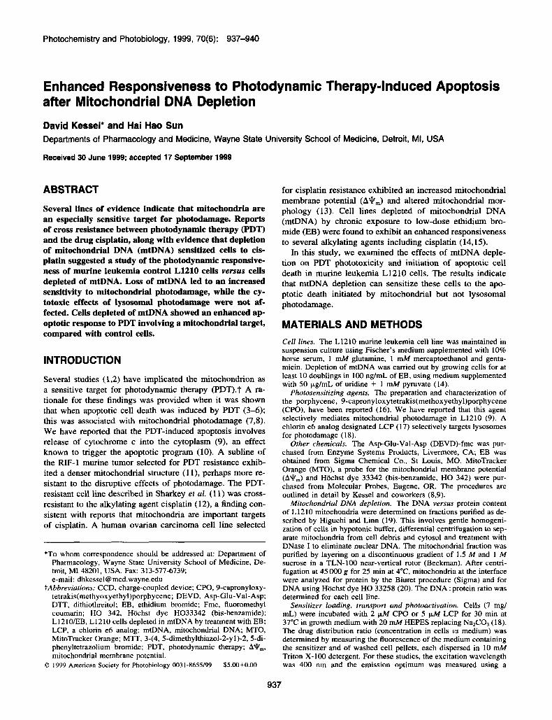
RESULTS AND DISCUSSION Chronic exposure of L1210 cells to EB led to a significant loss of mtDNA (Table 1). This was accompanied by a grad- ual loss of viability unless the growth medium was supple- mented with 50 pg/mL uridine and 1 mM pyruvate. In a previous study (13), a 30 ng/mL concentration of EB was employed, but we found that a 100 ng/mL drug was required to develop an L1210 subline whose survival depended on supplementation of the medium with pyruvate + uridine. When the L1210EB subline was transferred to Fischer’s medium without the supplement, the mitochondrial mem- brane potential, assessed by MTO labeling, decreased rap- idly (Fig. 1). In supplemented medium, there was no ob- servable difference between L1210 and L121OEB with re- gard to MTO labeling (not shown); this differs from the result reported with another cell line (13).
The distribution ratio of CPO in L1210 cells was 2.58 2 0.39. This was not significantly different from the result ob- tained with L121O/EB (2.43 ? 0.46). The distribution ratio indicates the relative concentration of a drug in intracellular space versus extracellular medium. We interpret these results to indicate that the intracellular drug concentration was sim- ilar in the two cell lines. This does not establish that mito- chondrial levels were similar, but because this drug shows a
Figure 1. A: Mitochondria1 membrane potential (A*,) of L1210/ EB cells as assessed by MTO during culture in Fischer’s medium supplemented with pyruvate + uridine. B: Effect of a 4 h incubation of L1210EB cells in Fischer’s medium without added nutrients. White bar = 10 pm.
marked specificity for mitochondria (9). we consider it likely that the intracellular drug concentration reflects mitochon- drial binding.
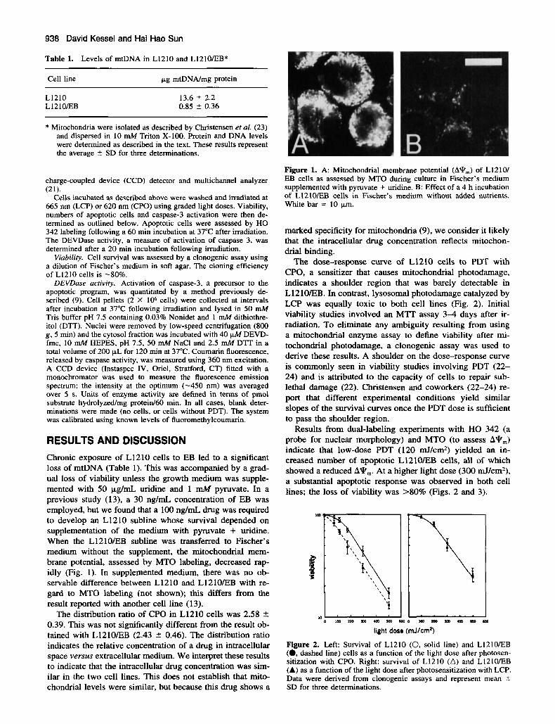
The dose-response curve of L1210 cells to PDT with CPO, a sensitizer that causes mitochondrial photodamage, indicates a shoulder region that was barely detectable in L121OEB. In contrast, lysosomal photodamage catalyzed by LCP was equally toxic to both cell lines (Fig. 2). Initial viability studies involved an MTT assay 3 4 days after ir- radiation. To eliminate any ambiguity resulting from using a mitochondrial enzyme assay to define viability after mi- tochondrial photodamage, a clonogenic assay was used to derive these results. A shoulder on the dose-response curve is commonly seen in viability studies involving PDT (22- 24) and is attributed to the capacity of cells to repair sub- lethal damage (22). Christensen and coworkers (22-24) re- port that different experimental conditions yield similar slopes of the survival curves once the PDT dose is sufficient to pass the shoulder region.
Results from dual-labeling experiments with HO 342 (a probe for nuclear morphology) and MTO (to assess A*,,,) indicate that low-dose PDT (120 mJ/cm2) yielded an in- creased number of apoptotic L121OEB cells, all of which showed a reduced A*,,,. At a higher light dose (300 mJ/cm2), a substantial apoptotic response was observed in both cell lines; the loss of viability was >80% (Figs. 2 and 3).
Im
P p ‘5
I6 rf 0 I r n r m r n a o a o
light dose (rnJ/cm2)
Figure 2. Left: Survival of L1210 (0. solid line) and L1210EB (0, dashed line) cells as a function of the light dose after photosen- sitization with CPO. Right: survival of L1210 (A) and L1210/EB (A) as a function of the light dose after photosensitization with LCP. Data were derived from clonogenic assays and represent mean t SD for three determinations.

Photochemistry and Photobiology, 1999, 70(6) 939
Con tro I 120 mJ/cm2 300 mJ/cm2 L1210 /EB L1210 /EB L1210 /EB
Figure 3. Effect of PDT on nuclear morphology and AY,,, in L1210 and LI2IOEB cells. Specified light doses represent irradiation at 630 ? 20 nm. White bar = 10 km.
An early step in initiation of the apoptotic program is the activation of the enzyme caspase-3 (25). This activation can be quantitated by measuring the cleavage of a fluorogenic substrate for the peptide sequence recognized by caspase-3: DEVD, coupled to a fluorogenic group. Appearance of fluo- rescence provides an indication of caspase-3 activation (9). The intrinsic level of DEVDase activity was greater in L1210/EB than in L1210. At a low PDT dose, we observed a greater level of DEVDase activity in L121O/EB cells. The difference was not significant when a higher PDT dose was used (Table 2). These values were measured 20 min after irradiation. In preliminary studies, we determined that this time interval provided the maximum level of DEVDase ac- tivity for both cell lines, using light doses specified in Table 2.
The mitochondrion is now emerging as a central element of an apoptotic mechanism, with effects leading to the loss of cytochrome c into the cytosol acting as a trigger for cas- pase activation and apoptotic cell death (10,26). Consider- able direct and indirect evidence supports the hypothesis that both cisplatin and PDT target mitochondrial structures, lead- ing to the initiation of apoptosis. Studies described in Kessel and Luo (18) were carried out with the human lymphoma U937 cell line. Marchetti’s group reported that the depletion of U937 mtDNA did not lead to a loss of AV,,, (27), consis- tent with results reported here. In U937, the apoptotic re- sponse to tumor necrosis factor + cycloheximide was not
Table 2. The DEVDase activity in control versus EB-treated cells*
Light dose (mJ/cm2)
Cell line 0 120 300
L1210 4 t 0.7 33.5 2 4.2 63 2 10.5 L 12 1OEB 9 t 1.4 73 t 10 58 2 9.5
* Units = pmol DEVD-fmc hydrolyzed per mg protein per hour (mean ? SD for three determinations). The DEVDase was as- sayed 20 min after irradiation. Results represent the mean 2 SD for three determinations.
affected by mtDNA depletion (28). The latter result suggests that the loss of mtDNA does not necessarily affect the ap- optotic response to cytotoxic agents.
Because both PDT resistance (1 1) and cisplatin resistance (13) were associated with mitochondrial alterations, we in- terpret these results, together with data reported here, to sug- gest that the mitochondrion is a common target for both modalities. The observed cross-resistance between PDT and cisplatin (12) is considered to reflect the result of alterations in a common target. In this regard, a variety of recent reports have now implicated mitochondria as a major target of cis- platin (28-30).
Using a different photosensitizing agent, human B lym- phoma cells (Namalwa) depleted of mtDNA were found to be unresponsive to PDT catalyzed by a boronated porphyrin (31). These results are difficult to interpret in the present context, because they involve a different cell line and a dif- ferent drug. These results imply that mitochondrial photo- damage will not affect cell survival in media supplemented with uridine + pyruvate. The extent of cytochrome c release into the cytosol after PDT was not reported in Munday et al. (31), nor was the death mode characterized. Namalwa cells appear to show a relatively slow apoptotic response to many chemotherapeutic agents (32), and the mode of cell death (apoptosis vs necrosis) was not reported.
We recently reported (33) that mitochondrial photodam- age can result in inactivation of the antiapoptotic mitochon- drial protein bcl-2. It is not yet known whether mtDNA de- pletion will enhance the sensitivity of bcl-2 to photodamage; such an effect could promote apoptotic responsiveness to PDT in L1210/EB cells.
Acknowledgements-This work was supported by grants CA 6556 1 and 23378 from the National Cancer Institute. We thank Ann Marie Santiago for excellent technical assistance.
REFERENCES 1. Gibson, S. L. and R. Hilf (1983) Photosensitization of mito-
chondrial cytochrorne c oxidase by hernatoporphyrin derivative and related porphyrins in vitro and in vivo. Cancer Rex 43, 4 19 1 4 197.
940 David Kessel and Hai Hao Sun
2.
3.
4.
5.
6.
7.
8.
9.
10.
I 1
12.
13.
14.
15.
16.
17.
18.
19.
Salet, C. and G. Moreno (1995) Photodynamic action increases leakage of the mitochondrial electron transport chain. Int. J . Radiat. Biol. 67, 477480. Agarwal, M. L., M. E. Clay, E. J. Harvey, H. H. Evans, A. R. Antunez and N. L. Oleinick (1991) Photodynamic therapy in- duces rapid cell death by apoptosis in L5178Y mouse lymphoma cells. Cancer Res. 51, 5993-5996. Luo, Y., C. K. Chang and D. Kessel (1996) Rapid initiation of apoptosis by photodynamic therapy. Phorochem. Photobiol. 63,
Granville, D. J., J. G. Levy and D. W. C. Hunt (1998) Photo- dynamic treatment with benzoporphyrin derivative monoacid ring A produces protein tyrosine events and DNA fragmentation in murine P815 cells. Photochem. Photobiol. 67, 358-362. He, X.-Y., R. Sikes, S . Thomsen, L. W. K. Chung and S. L. Jacques (1994) Photodynamic therapy with photofrin I1 induces programmed cell death in carcinoma cell lines. Phorochem. Pho- tobiol. 59, 468473. Kessel, D. and Y. Luo (1998) Mitochondrial photodamage and PDT-induced apoptosis. J. Photochem. Phorobiol. B Biol. 42,
Kessel, D., Y. Luo, Y. Deng and C. K. Chang (1997) The role of sub-cellular localization in initiation of apoptosis by photo- dynamic therapy. Photochem. Photobiol. 65, 422426. Kessel, D. and Y. Luo (1999) Photodynamic therapy: a mito- chondrial inducer of apoptosis. Cell Death Differ. 6, 28-35. Liu, X., C. N. Kim, R. Jemmerson and X. Wang (1996) Induc- tion of apoptotic program in cell-free extracts: requirement for dATP and cytochrome c. Cell 86, 147-157. Sharkey, S. M., B. C. Wilson, R. Moorehead and G. Singh ( 1993) Mitochondrial alterations in photodynamic therapy-resis- tant cells. Cancer Res. 53, 49944999. Moorehead, R. A., S. G. Armstrong, B. C. Wilson and G. Singh (1994) Cross-resistance to cisplatin in cells resistant to photof- rin-mediated photodynamic therapy. Cancer Res. 54, 2556- 2559. Andrews, P. A. and K. D. Albright (1992) Mitochondrial defects in cis-diamminedichloroplatinum(I1)-resistant human ovarian carcinoma cells. Cancer Res. 52, 1895-1901. Liang, B. C. and E. Ullyatt (1998) Increased sensitivity to cis- diamminedichloroplatinum induced apoptosis with mitochon- drial DNA depletion. Cell Death Differ. 5 , 694-701. Cavalli, L. R., M. Varella-Garcia and B. C. Liang (1997) Di- minished tumorigenic phenotype after depletion of mitochon- drial DNA. Cell Growth Differ. 11, 1189-1 198. Toledano, H., R. Edrai and S . Kimel(l998) Photodynamic dam- age by liposome-bound porphycenes: comparison between in vitro and in vivo models. J. Photochem. Photobiol. B Biol. 42, 2G27. Kessel. D., K. Woodburn, C. J. Comer, N. Jagerovic and K. M. Smith (1995) Photosensitization with derivatives of chlorin P6. J. Photochem. Photobiol. B Biol. 28, 13-18. Kessel, D. and Y. Luo (1998) Mitochondrial photodamage and PDT-induced apoptosis J. Photochem. Photobiol. B Biol. 42,
Higuchi, Y. and S . Linn (1995) Purification of all forms of HeLa
528-534.
89-95.
89-95.
cell mitochondrial DNA and assessment of damage to it caused by hydrogen peroxide treatment of mitochondria or cells. J. Biol. Chem. 270, 7950-7956.
20. Brunk, C. F., K. C. Jones and T. W. James (1979) Assay for nanogram quantities of DNA in cellular homogenates. Anal. Biochem. 92, 497-500.
21. Kessel, D. (1994) A CCD-based spectral analyzer. The Spectrum
22. Christensen, T. and J. Moan (1979) Photodynamic inactivation of synchronized human cells in vitro in the presence of hema- toporphyrin. Cancer Res. 39, 3735-3737.
23. Christensen. T., T. Sandquist, K. Feren, H. Waksvik and J. Moan (1983) Retention and photodynamic effects of haemato- porphyrin derivative in cells after prolonged cultivation in the presence of porphyrin. Br. J . Cancer 48, 3 5 4 3 .
24. Comer, C. J. (1980) DNA damage and repair in CHO cells following hematoporphyrin photoradiation. Cancer Lett. 11,
25. Porter. A. G. and R. U. Janicke (1999) Emerging roles of cas- pase-3 in apoptosis. Cell Death Differ. 6, 99-104.
26. Slee, E. A.. M. T. Harte, R. M. Kluck, B. B. Wolf, C. A. Cas- iano, D. D. Newmeyer, H. G. Wang, J. C. Reed, D. W. Nich- olson, E. S. Alnemri, D. R. Green and S. J. Martin (1999) Or- dering the cytochrome c-initiated caspase cascade: hierarchical activation of caspases-2, -3, -6, -7, -8, and -10 in a caspase-9- dependent Manner. J. Cell Biol. 144, 281-292.
27. Marchetti, P., S. A. Susin, D. Decaudin, S. Gamen, M. Castedo, T. Hirsch, N. Zamzami, J. Naval, A. Senik and G. Kroemer (1996) Apoptosis-associated derangement of mitochondrial function in cells lacking mitochondrial DNA. Cancer Res. 56.
28. Evans, R. M. and H. Simpkins (1998) Cisplatin induced inter- mediate filament reorganization and altered mitochondrial func- tion in 3T3 cells and drug-sensitive and -resistant Walker 256 cells. Exp. Cell Res. 245, 69-78.
29. Olivero, 0. A., P. K. Chang, D. M. Lopez-Larraza, M. C. Sem- ino-Mora and M. C. Poirier (1997) Preferential formation and decreased removal of cisplatin-DNA adducts in Chinese hamster ovary cell mitochondrial DNA as compared to nuclear DNA. Murat. Res. 391, 79-86.
30. Kojima, H., K. Endo, H. Moriyama, Y. Tanaka, E. S. Alnemri, C. A. Slapak, B. Teicher. D. Kufe and R. Datta (1998) Abro- gation of mitochondrial cytochrome c release and caspase-3 ac- tivation in acquired multidrug resistance. J. Biol. Chem. 273,
31. Munday, A. D., A. Sriratana, J. S. Hill, S . B. Kahl and P. Mag- ley (1996) Mitochondria are the functional intracellular target for a photosensitizing boronated porphyrin. Biochim. Biophys. Acra 1311, 1-4.
32. Schmitt, E.. A. Steyaert, G. Cimoli and R. Bertrand (1998) Bax- a promotes apoptosis induced by cancer chemotherapy and ac- celerates the activation of caspase 3-like cysteine proteases in p53 double mutant B lymphoma Namalwa cells. Cell Death Differ. 5 , 505-516.
33. Kim, H. R. C., Y. Luo, G. Li and D. Kessel (1999) Enhanced apoptotic response to photodynamic therapy after Bcl-2 trans- fection. Cancer Res. 59, 3429-3432.
7, 8-9.
16 1-1 67.
2033-2038.
16647-1 6650.