enteroaggregative escherichia strains to adhere in to...
TRANSCRIPT
Vol. 60, No. 5INFECrION AND IMMUNITY, May 1992, p. 2083-20910019-9567/92/052083-09$02.00/0
Ability of Enteroaggregative Escherichia coli Strains To AdhereIn Vitro to Human Intestinal Mucosa
STUART KNUTTON,1* ROBERT K. SHAW,' MAHARA K. BHAN,2 HENRY R. SMITH,3MOYRA M. McCONNELL,3 TOM CHEASTY,3 PETER H. WILLIAMS,4
AND TOM J. BALDWIN4Institute of Child Health, University ofBirmingham, Birmingham B16 8ET,' Division of Enteric Pathogens,
Central Public Health Laboratory, Colindale,3 and Department of Genetics, University of Leicester,Leicester LE1 7RH,4 United Kingdom, and Department of Paediatrics,
All India Institute ofMedical Sciences, New Delhi 110029, India2
Received 15 November 1991/Accepted 24 February 1992
A collection of 44 enteroaggregative Escherichia coli (EAggEC) strains isolated from infants with diarrhea inIndia and the United Kingdom were examined for their ability to adhere in vitro to human intestinal mucosaand by electron microscopy for production of putative adherence factors. None of the strains adhered to humanduodenal mucosa, and six strains tested did not adhere to ileal mucosa; all 44 strains, however, adhered tohuman colonic mucosa in localized aggregates. Electron microscopy of infected colonic mucosa indicatedfimbrially mediated adhesion of the EAggEC strains. Four morphologically distinct kinds of fimbriae, includinga new morphological type ofE. coli fimbriae consisting of bundles of fine filaments, were identified among theEAggEC strains; this new type of fimbria was observed in 43 of the 44 EAggEC strains. Forty-three of the 44EAggEC strains were positive with a DNA probe developed to identify EAggEC, and most of the strainsbelonged to serotypes unrelated to the other major classes of diarrheic E. coli. These results suggest thatEAggEC may be a large-bowel pathogen and colonize the colon by a fimbrially mediated adhesion mechanism.
Four major classes of Escherichia coli are recognized ascauses of diarrheal disease: enteropathogenic (EPEC), en-terotoxigenic (ETEC), enteroinvasive (EIEC), and entero-hemorrhagic (EHEC) (16). The use of tissue culture (HeLaand HEp-2) cell adhesion assays has led to the identificationof further putative classes of diarrheic E. coli. Three distinctpatterns of adhesion, termed localized, diffuse, and aggrega-tive, have been described previously (19). Localized, dif-fuse, and aggregative adherence patterns are characterizedby localized microcolonies of adherent bacteria, by diffuseadherence of individual bacteria and by characteristic aggre-gates of bacteria frequently giving a "stacked-brick" appear-ance, respectively (19). Localized adhesion is characteristicof attaching and effacing EPEC strains (10, 13); E. colistrains exhibiting the diffuse (diffuse adhering E. coli) andaggregative (enteroaggregative E. coli) adherence patternsare two new classes of adherent E. coli which have beenassociated with diarrhea (7, 25). Enteroaggregative E. coli(EAggEC) strains may be especially important because oftheir epidemiological association with persistent diarrhea ininfants in developing countries (3, 5).
Characterization of a collection of EAggEC strains (orig-inally called enteroadherent-aggregative E. coli) from Chileshowed them to be negative in tests for ETEC, EPEC,EIEC, and EHEC, nor did they fit these categories byserotyping. Many of the strains produced mannose-resistantfimbriae, and preliminary data suggested fimbrially mediatedadhesion of EAggEC to rabbit mucosa. EAggEC causedcharacteristic lesions in rabbit and rat ileal loops, suggestingtoxin involvement (25). A putative heat-stable enterotoxinproduced by EAggEC has since been described (22). A 1-kbfragment from a plasmid of one EAggEC strain (17-2) was
* Corresponding author.
found to be a highly specific DNA probe for identifyingEAggEC (2).There is little information about human enteroadherence
properties of EAggEC. Enteroadherence of an EAggECstrain (0127a:H2) isolated from a child with diarrhea inThailand has been examined in native and formalin-fixedmucosa. This one strain showed little adhesion to small-bowel mucosa but good adhesion to formalin-fixed large-bowel mucosa (26). In this paper, we report human enteroad-herence properties of a collection of 44 EAggEC strainsisolated from infants with diarrhea in India and the UnitedKingdom.
MATERIALS AND METHODS
Bacterial strains. Forty-four E. coli strains isolated frominfants with acute and persistent diarrhea in India (3) and inthe United Kingdom (15) which showed aggregative adhe-sion to HEp-2 cells were examined. Also included in thestudy were strain 221(092:H33), which displays aggregativeadhesion and has been shown to cause diarrhea in hu-man volunteers (18), and strain 17-2(03:H2), from which theEAggEC DNA probe was prepared (2). For adhesion andelectron microscopic studies, stock cultures of the strainswere subcultured into Mueller-Hinton broth and incubatedaerobically for 18 h at 37°C.
Serotyping. 0 and H antigens were determined by stan-dard agglutination methods (8).EAggEC probe tests. The EAggEC probe was that de-
scribed by Baudry et al. (2). Strains were tested by colonyhybridization as described by Maniatis et al. (17).
Hemagglutination tests. The strains were tested for agglu-tination of bovine, rat, and human type 0 erythrocytes in thepresence of 0.5% mannose and for agglutination of guineapig erythrocytes in the presence and absence of mannose.For the tests, the strains were subcultured twice in Mueller-
2083
on June 19, 2018 by guesthttp://iai.asm
.org/D
ownloaded from
2084 KNUTTON ET AL.
-,T, s , w {W ~iPv 0. !.
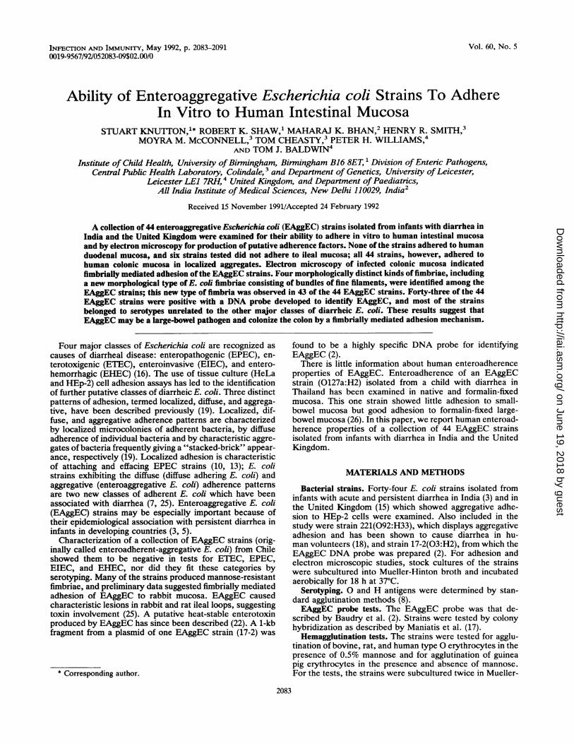
FIG. 1. HEp-2 cell adhesion assay preparations showing aggregative adhesion of bacteria to cells and background (A) and to cells only (B).Magnification, x500.
Hinton or nutrient broth (Difco) and incubated staticallyeach time for 48 h (23).
Tissue culture cell adhesion. Adhesion to HEp-2 cells wastested according to the method of Cravioto et al. (4) by using3- and 6-h incubations of cells and bacteria. An identicalprotocol was used to assess Caco-2 cell adhesion although,in this case, adhesion was assessed by scanning electronmicroscopy.Adhesion to human intestinal mucosa. Normal duodenal,
ileal, and colonic mucosal biopsies obtained with informedconsent from adults were maintained in organ culture andinfected with EAggEC for 8 h as previously described (13).Mucosal adhesion was assessed by scanning electron mi-croscopy (13).
Electron microscopy. Strains were monitored for fimbriaproduction by negative-staining electron microscopy (11).Biopsies with adherent bacteria were processed for trans-mission electron microscopy by standard methods as previ-ously described (11).
RESULTS
Aggregative adhesion. The 44 strains examined in the studywere selected because they displayed the characteristicaggregative pattern of HEp-2-cell adherence as defined byNataro et al. (19). Nineteen of the 44 strains showed aggre-gation of bacteria and the formation of a characteristicstacked-brick pattern both on the surface of and betweenHEp-2 cells (Fig. 1A); the remaining 25 strains showedaggregative adhesion to HEp-2 cells only (Fig. 1B). Ingeneral, aggregative adhesion of strains to cells only or tocells plus background was serotype specific (Table 1). Sev-eral strains were rejected from the study because theydisplayed aggregative adhesion to the background betweencells but showed no cell adhesion. Seven of the Indianstrains (F246A, F51OA, F278A, F23A, F54A, F55B, andH505B) were to varying degrees cytotoxic for HEp-2 cells ina 3-h incubation assay; i.e., they caused rounding and deathof cells.
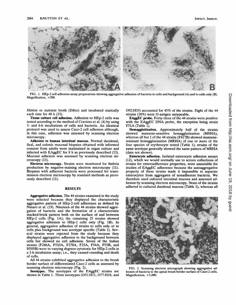
All 44 strains exhibited aggregative adhesion to the brushborder surface of differentiated Caco-2 cells as assessed byscanning electron microscopy (Fig. 2).
Serotypes. The serotypes of the EAggEC strains areshown in Table 1. Three serotypes (051:H11, 077:H18, and
092:H33) accounted for 45% of the strains. Eight of the 44strains (18%) were 0-antigen untypeable.EAggEC probe. Forty-three of the 44 strains were positive
with the EAggEC DNA probe, the exception being strainF51A (Table 1).
Hemagglutination. Approximately half of the strainsshowed mannose-sensitive hemagglutination (MSHA),whereas all but 1 of the 44 strains (F417B) showed mannose-resistant hemagglutination (MRHA) of one or more of thefour species of erythrocyte tested (Table 1); strains of thesame serotype generally showed the same pattern of MRHA(data not shown).
Enterocyte adhesion. Isolated enterocyte adhesion assays(12), which we would normally use to screen collections ofstrains for enteroadherence properties, were unsuitable forstudies of EAggEC adherence because the autoaggregativeproperty of these strains made it impossible to separateenterocytes from aggregates of nonadherent bacteria. Wetherefore used cultured intestinal mucosa and assessed ad-hesion by scanning electron microscopy. None of the strainsadhered to cultured duodenal mucosa (Table 1), whereas all
FIG. 2. Scanning electron micrograph showing aggregative ad-hesion of bacteria to the apical brush border surface of Caco-2 cells.Magnification, x5,000.
INFECT. IMMUN.
on June 19, 2018 by guesthttp://iai.asm
.org/D
ownloaded from
ADHERENCE OF ENTEROAGGREGATIVE E. COLI 2085
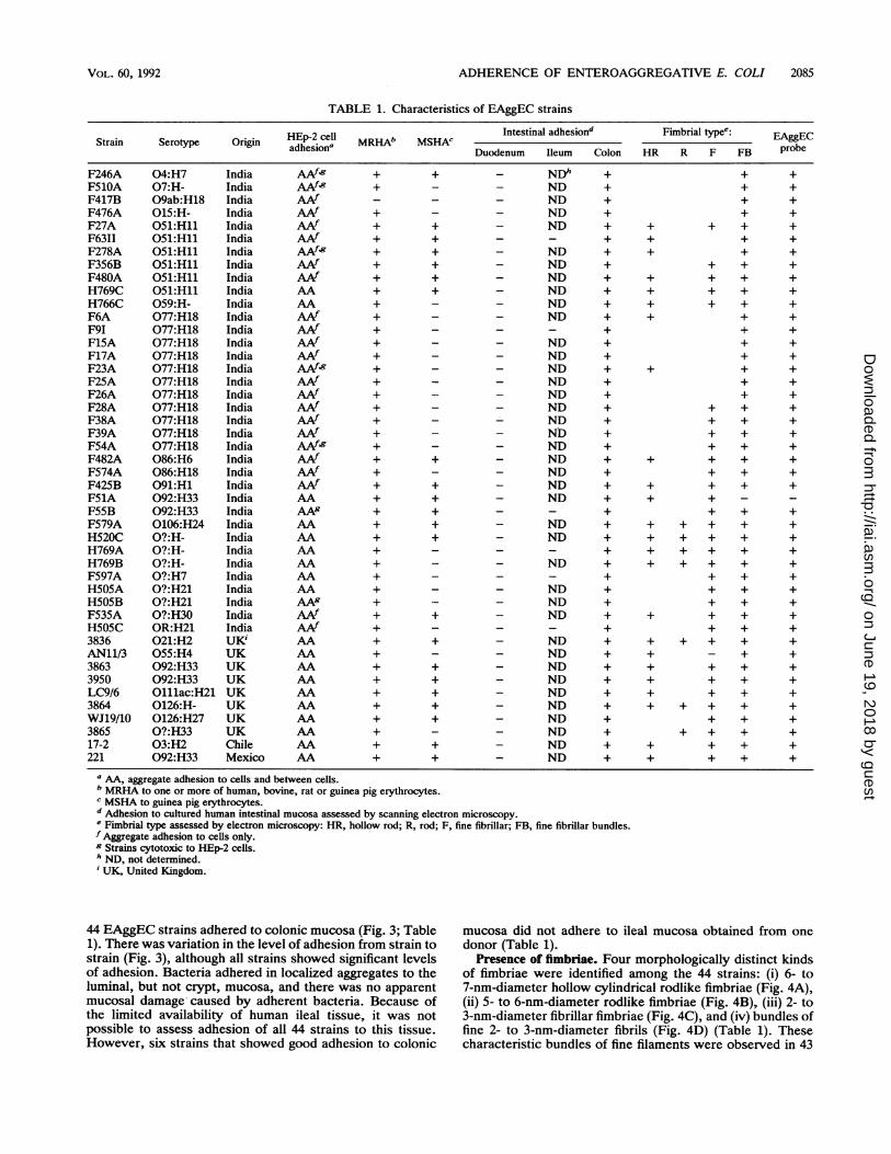
TABLE 1. Characteristics of EAggEC strains
HEp-2 cell Intestinal adhesiond Fimbrial typee: EAgECStrain Serotype Origin aPdh eso MRHADMSH uEAggEadhesion' Duodenum Ileum Colon HR R F FB probe
+ + - NDh+ - ND- - - ND+ - - ND+ + - ND+ +_ _
+ + - ND+ + - ND+ + - ND+ + - ND+ - - ND+ - - ND
+ - - ND+ _ - ND+ - - ND+ _ - ND+ - - ND+ _ - ND+ - - ND
+ - - ND
+ - - ND+ + - ND+ - - ND+ + - ND+ + - ND+ +_ _
+ + - ND+ + - ND
+ - - ND
+ - - ND+ _ - ND+ + - ND
+ + _ ND+ _ - ND+ + - ND+ + - ND+ + - ND+ + - ND+ + - ND+ - - ND
+ + - ND
+ + - ND
+ +
+ +
+ +
+ +
+ +
+ +
+ +
+ +
+ +
+ +
+ +
+ +
+ +
+ +
+ +
+ +
+ +
+ +
+ +
+ +
+ +
+ +
+ +
+ +
+ +
+ +
+ + +
+ + + + +
+ + + + +
+ + + + +
+ + + + +
+ + +
+ + +
+ + +
+ + + +
+ + +
+ + + + +
+ - + +
+ + + +
+ + + +
+ + + +
+ + + + +
+ + +
+ + + +
+ + + +
+ + + +
a AA, aggregate adhesion to cells and between cells.b MRHA to one or more of human, bovine, rat or guinea pig erythrocytes.c MSHA to guinea pig erythrocytes.d Adhesion to cultured human intestinal mucosa assessed by scanning electron microscopy.e Fimbrial type assessed by electron microscopy: HR, hollow rod; R, rod; F, fine fibrillar; FB, fine fibrillar bundles.f Aggregate adhesion to cells only.g Strains cytotoxic to HEp-2 cells.h ND, not determined.iUK, United Kingdom.
44 EAggEC strains adhered to colonic mucosa (Fig. 3; Table1). There was variation in the level of adhesion from strain tostrain (Fig. 3), although all strains showed significant levelsof adhesion. Bacteria adhered in localized aggregates to theluminal, but not crypt, mucosa, and there was no apparentmucosal damage caused by adherent bacteria. Because ofthe limited availability of human ileal tissue, it was notpossible to assess adhesion of all 44 strains to this tissue.However, six strains that showed good adhesion to colonic
mucosa did not adhere to ileal mucosa obtained from onedonor (Table 1).
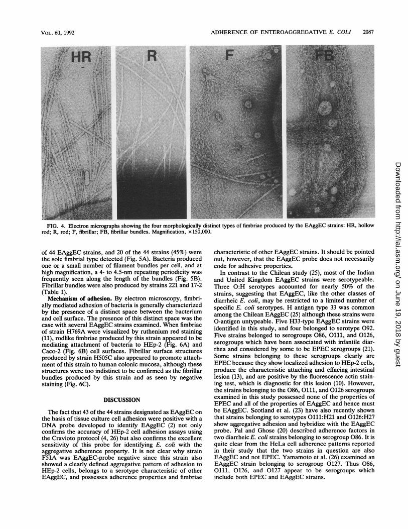
Presence of fimbriae. Four morphologically distinct kindsof fimbriae were identified among the 44 strains: (i) 6- to7-nm-diameter hollow cylindrical rodlike fimbriae (Fig. 4A),(ii) 5- to 6-nm-diameter rodlike fimbriae (Fig. 4B), (iii) 2- to3-nm-diameter fibrillar fimbriae (Fig. 4C), and (iv) bundles offine 2- to 3-nm-diameter fibrils (Fig. 4D) (Table 1). Thesecharacteristic bundles of fine filaments were observed in 43
F246AF510AF417BF476AF27AF63IIF278AF356BF480AH769CH766CF6AF91F15AF17AF23AF25AF26AF28AF38AF39AF54AF482AF574AF425BF51AF55BF579AH520CH769AH769BF597AH505AH505BF535AH505C3836AN11/338633950LC9/63864WJ19/10386517-2221
04:H707:H-O9ab:H18015:H-051:Hll051:Hll051:Hll051:Hll051:Hll051:Hll059:H-077:H18077:H18077:H18077:H18077:H18077:H18077:H18077:H18077:H18077:H18077:H18086:H6086:H18091:H1092:H33092:H330106:H24O?:H-O?:H-O?:H-0?:H70?:H210?:H21O?:H30OR:H21021:H2055:H4092:H33092:H33Olllac:H210126:H-0126:H27O?:H3303:H2092:H33
IndiaIndiaIndiaIndiaIndiaIndiaIndiaIndiaIndiaIndiaIndiaIndiaIndiaIndiaIndiaIndiaIndiaIndiaIndiaIndiaIndiaIndiaIndiaIndiaIndiaIndiaIndiaIndiaIndiaIndiaIndiaIndiaIndiaIndiaIndiaIndiaUK1UKUKUKUKUKUKUKChileMexico
AAf&9AAf-9AAfAAfAAfAAfAAf,gAAfAAfAAAAAAfAAfAAfAAfAAfM9AAfAAfAAfAAfAAfAAf,gAAfAAfAAfAAAA9AAAAAAAAAAAAAA9AAfAAfAAAAAAAAAAAAAAAAAAAA
VOL. 60, 1992
on June 19, 2018 by guesthttp://iai.asm
.org/D
ownloaded from

FIG. 3. Scanning electron micrographs showing typical adhesion of EAggEC strains to cultured human colonic mucosa. Bacteria adheredin localized aggregates to luminal but not crypt mucosa (A). After 8 h, some strains showed virtually confluent colonization of the mucosalsurface except for distinct areas around the crypts (B). Magnifications, x 1,000 (A) and x500 (B).
2086
on June 19, 2018 by guesthttp://iai.asm
.org/D
ownloaded from
ADHERENCE OF ENTEROAGGREGATIVE E. COLI 2087
R
- _:
FIG. 4. Electron micrographs showing the four morphologically distinct types of fimbriae produced by the EAggEC strains: HR, hollowrod; R, rod; F, fibrillar; FB, fibrillar bundles. Magnification, x 150,000.
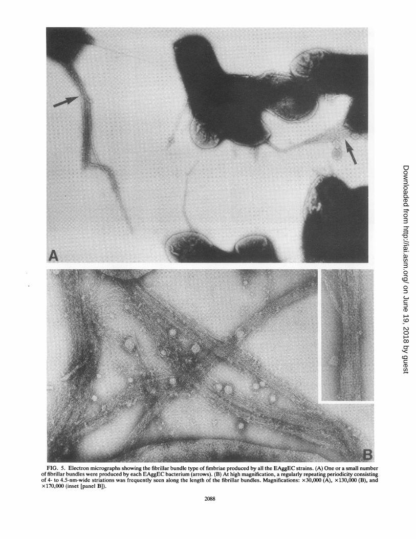
of 44 EAggEC strains, and 20 of the 44 strains (45%) werethe sole fimbrial type detected (Fig. 5A). Bacteria producedone or a small number of filament bundles per cell, and athigh magnification, a 4- to 4.5-nm repeating periodicity wasfrequently seen along the length of the bundles (Fig. SB).Fibrillar bundles were also produced by strains 221 and 17-2(Table 1).Mechanism of adhesion. By electron microscopy, fimbri-
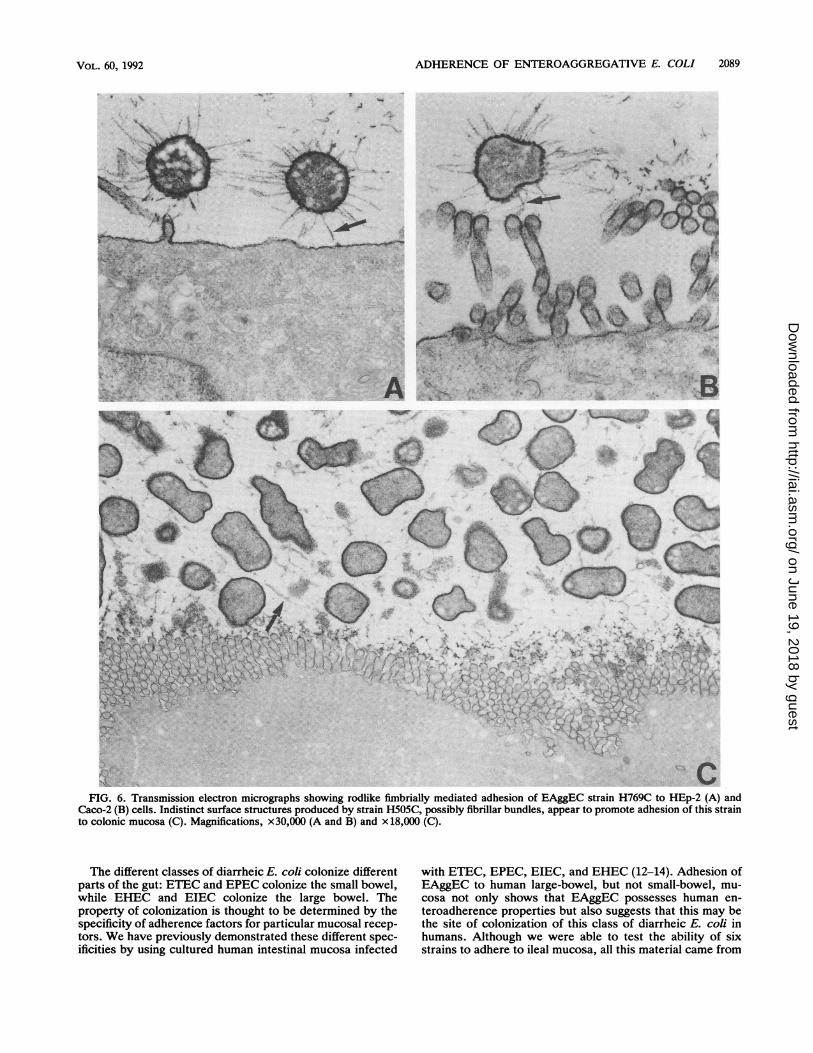
ally mediated adhesion of bacteria is generally characterizedby the presence of a distinct space between the bacteriumand cell surface. The presence of this distinct space was thecase with several EAggEC strains examined. When fimbriaeof strain H769A were visualized by ruthenium red staining(11), rodlike fimbriae produced by this strain appeared to bemediating attachment of bacteria to HEp-2 (Fig. 6A) andCaco-2 (Fig. 6B) cell surfaces. Fibrillar surface structuresproduced by strain H505C also appeared to promote attach-ment of this strain to human colonic mucosa, although thesestructures were too indistinct to be confirmed as the fibrillarbundles produced by this strain and as seen by negativestaining (Fig. 6C).
DISCUSSION
The fact that 43 of the 44 strains designated as EAggEC onthe basis of tissue culture cell adhesion were positive with aDNA probe developed to identify EAggEC (2) not onlyconfirms the accuracy of HEp-2 cell adhesion assays usingthe Cravioto protocol (4, 26) but also confirms the excellentsensitivity of this probe for identifying E. coli with theaggregative adherence property. It is not clear why strainF51A was EAggEC-probe negative since this strain alsoshowed a clearly defined aggregative pattern of adhesion toHEp-2 cells, belongs to a serotype characteristic of otherEAggEC, and possesses adherence properties and fimbriae
characteristic of other EAggEC strains. It should be pointedout, however, that the EAggEC probe does not necessarilycode for adhesive properties.
In contrast to the Chilean study (25), most of the Indianand United Kingdom EAggEC strains were serotypeable.Three 0:H serotypes accounted for nearly 50% of thestrains, suggesting that EAggEC, like the other classes ofdiarrheic E. coli, may be restricted to a limited number ofspecific E. coli serotypes. H antigen type 33 was commonamong the Chilean EAggEC (25) although these strains were0-antigen untypeable. Five H33-type EAggEC strains wereidentified in this study, and four belonged to serotype 092.Five strains belonged to serogroups 086, 0111, and 0126,serogroups which have been associated with infantile diar-rhea and considered by some to be EPEC serogroups (21).Some strains belonging to these serogroups clearly areEPEC because they show localized adhesion to HEp-2 cells,produce the characteristic attaching and effacing intestinallesion (13), and are positive by the fluorescence actin stain-ing test, which is diagnostic for this lesion (10). However,the strains belonging to the 086, 0111, and 0126 serogroupsexamined in this study possessed none of the properties ofEPEC and all of the properties of EAggEC and hence mustbe EAggEC. Scotland et al. (23) have also recently shownthat strains belonging to serotypes 0111:H21 and 0126:H27show aggregative adhesion and hybridize with the EAggECprobe. Pal and Ghose (20) described adherence factors intwo diarrheic E. coli strains belonging to serogroup 086. It isquite clear from the HeLa cell adherence patterns reportedin their study that the two strains in question are alsoEAggEC and not EPEC. Yamamoto et al. (26) examined anEAggEC strain belonging to serogroup 0127. Thus 086,0111, 0126, and 0127 appear to be serogroups whichinclude both EPEC and EAggEC strains.
VOL. 60, 1992
.1 .1.
i. .-. ..
kv, -':
on June 19, 2018 by guesthttp://iai.asm
.org/D
ownloaded from
A
FIG. 5. Electron micrographs showing the fibrillar bundle type of fimbriae produced by all the EAggEC strains. (A) One or a small numberof fibrillar bundles were produced by each EAggEC bacterium (arrows). (B) At high magnification, a regularly repeating periodicity consistingof 4- to 4.5-nm-wide striations was frequently seen along the length of the fibrillar bundles. Magnifications: x30,000 (A), x 130,000 (B), andx 170,000 (inset [panel B]).
2088
on June 19, 2018 by guesthttp://iai.asm
.org/D
ownloaded from
ADHERENCE OF ENTEROAGGREGATIVE E. COLI
iF0
A
0,0- .
'.
4 141 ,.
(14 .tU/C' .U
1- A
A .2'S"i\L It z,..
7,.:":{ ".:
FIG. 6. Transmission electron micrographs showing rodlike fimbrially mediated adhesion of EAggEC strain H769C to HEp-2 (A) andCaco-2 (B) cells. Indistinct surface structures produced by strain H505C, possibly fibrillar bundles, appear to promote adhesion of this strainto colonic mucosa (C). Magnifications, x30,000 (A and B) and x 18,000 (C).
The different classes of diarrheic E. coli colonize differentparts of the gut: ETEC and EPEC colonize the small bowel,while EHEC and EIEC colonize the large bowel. Theproperty of colonization is thought to be determined by thespecificity of adherence factors for particular mucosal recep-tors. We have previously demonstrated these different spec-ificities by using cultured human intestinal mucosa infected
with ETEC, EPEC, EIEC, and EHEC (12-14). Adhesion ofEAggEC to human large-bowel, but not small-bowel, mu-cosa not only shows that EAggEC possesses human en-teroadherence properties but also suggests that this may bethe site of colonization of this class of diarrheic E. coli inhumans. Although we were able to test the ability of sixstrains to adhere to ileal mucosa, all this material came from
2089VOL. 60, 1992
Al ;:.i4
1-A
%-w",oft-''lw
.49
--ll
on June 19, 2018 by guesthttp://iai.asm
.org/D
ownloaded from
2090 KNUTTON ET AL.
a single donor. Lack of adhesion to ileal mucosa could bedue to a lack of receptors for EAggEC in this one individual.We cannot rule out, therefore, the possibility that EAggECmay also adhere to distal small-bowel mucosa. Our obser-vations are consistent with those of Yamamoto et al. (26),who showed that an 0127:H2 EAggEC strain exhibited lowlevels of adhesion to native jejunal and ileal mucosa and highlevels of adhesion to formalin-fixed colonic mucosa. It isunfortunate that data were not available for adhesion tonative colonic mucosa, because this study also showed thatthere could be marked differences in adhesion to native andfixed tissue (26).Four morphologically distinct kinds of fimbriae were iden-
tified among the EAggEC isolates. Rigid rodlike fimbriaemay be either type 1 fimbriae responsible for MSHA orputative adhesion fimbriae responsible for MRHA. Theabsence of such fimbriae in four strains which showedMSHA probably reflects the different culture conditionsused for the hemagglutination and negative-staining studies.Of particular interest is the fibrillar type that exists as tightlypacked bundles, not only because of their distinctive struc-ture but also because this fimbrial type was observed in 43 ofthe 44 EAggEC strains examined, as well as in strains 221and 17-2. In contrast, the rodlike fimbriae produced by strain17-2 were found in only 17% of the strains in the study ofVial et al. (25). If these fibrillar bundles are adhesion fimbriaewhich promote mucosal adhesion of EAggEC, then thisantigen could be important as a potential vaccine candidate.Fibrillar bundles were the only type of fimbriae detected insome strains, although they often occurred along with one ormore of the other types of fimbriae. The regular periodicityfrequently seen with the fibrillar bundles presumably reflectsthe regular alignment of the fibrils within the bundles, inwhich case the periodicity should represent the subunitrepeat along a single fibril.The fibrillar bundle fimbriae are even more intriguing in
the light of the recent description of similar bundle-formingpili by EPEC (6) and their homology with the TcpA fimbriaeof Vibrio cholerae which also exist as bundles (9). Bundle-forming pili appear to participate in the formation of EPECcolonies by forming bundles that link bacteria together.Given that EAggEC also forms colonies, albeit of a differentpattern in tissue culture cell adhesion assays, the fibrillarbundles reported in this study could serve the same functionand participate in the formation of EAggEC colonies.The results of preliminary ultrastructural studies of HEp-2
and Caco-2 cells with adherent EAggEC strains are inagreement with the results of the studies by Vial et al. (25)and suggest that rodlike EAggEC fimbriae are important inadhesion. However, since EAggEC produces other types offimbriae, which are not necessarily stained with rutheniumred, it is premature to conclude that adhesion is definitelybeing promoted by the rodlike fimbriae. However, given thatEAggEC which adheres to human intestinal mucosa doesproduce several different morphological types of fimbriae, itseems likely that EAggEC strains of different serotypes, likeETEC, will produce more than one type of fimbrial adhesin.EAggEC is associated with persistent diarrhea (3, 5).
Unlike EPEC, which causes a brush border membranelesion that could lead to persistent diarrhea, EAggEC did notappear to cause specific brush border damage, although itshould be pointed out that scanning electron microscopywould not necessarily reveal damage beneath attached bac-teria; preliminary transmission electron microscopy did notreveal mucosal damage. This would suggest a toxin-medi-ated mechanism of disease. A hemorrhagic lesion in the
rabbit ligated ileal loop produced by EAggEC (25) and aheat-stable enterotoxin produced by EAggEC have beenreported previously (22). In an accompanying paper (1), wehave shown that EAggEC also produces a heat-labile hemol-ysinlike toxin. The ability of EAggEC to survive long term inthe large bowel in close association with the mucosal surfaceas a result of specific fimbrially mediated adhesion andproduce heat-labile and/or heat-stable enterotoxins couldexplain the persistence of the diarrhea in infected infants.
ACKNOWLEDGMENTS
We are grateful to Professor Levine for providing the EAggECprobe and EAggEC strains 221 and 17-2.
This study was supported by the Wellcome Trust (S.K., P.H.W.,and T.J.B.).
REFERENCES1. Baldwin, T. J., S. Knutton, L. Sellers, H. A. Manjarrez Hernan-
dez, A. Aitken, and P. H. Williams. 1992. EnteroaggregativeEscherichia coli strains secrete a heat-labile toxin antigenicallyrelated to E. coli hemolysin. Infect. Immun. 60:2092-2095.
2. Baudry, B., S. J. Savarino, P. Vial, J. B. Kaper, and M. M.Levine. 1990. A sensitive and specific DNA probe to identifyenteroaggregative Escherichia coli, a recently discovered diar-rhoeal pathogen. J. Infect. Dis. 161:1249-1251.
3. Bhan, M. K., P. Raj, M. M. Levine, J. B. Kaper, N. Bhandari,R. Srivastava, R. Kumar, and S. Sazawal. 1989. Enteroaggrega-tive Escherichia coli associated with persistent diarrhea in acohort of rural children in India. J. Infect. Dis. 159:1061-1064.
4. Cravioto, A., R. J. Gross, S. M. Scotland, and B. Rowe. 1979. Anadhesive factor found in strains of Escherichia coli belonging totraditional enteropathogenic serotypes. Curr. Microbiol. 3:95-99.
5. Cravioto, A., A. Tello, A. Navarro, J. Ruiz, H. Villafan, F.Uribe, and C. Eslava. 1991. Association of Escherichia coliHEp-2 adherence patterns with type and duration of diarrhoea.Lancet 337:262-264.
6. Giron, J. A., A. S. Y. Ho, and G. K. Schoolnik. 1991. Aninducible bundle-forming pilus of enteropathogenic Escherichiacoli. Science 254:710-713.
7. Giron, J. A., T. Jones, F. Millan-Velasco, E. Castro-Munoz, L.Zarate, J. Fry, G. Frankel, S. L. Moseley, B. Baudry, and J. B.Kaper. 1991. Diffuse-adhering Escherichia coli (DAEC) as aputative cause of diarrhea in Mayan children in Mexico. J.Infect. Dis. 163:507-513.
8. Gross, R. J., and B. Rowe. 1985. Serotyping of Escherichia coli,p. 345-363. In M. Sussman (ed.), The virulence of Escherichiacoli: reviews and methods. Academic Press, Ltd., London.
9. Hall, R. H., P. A. Vial, J. B. Kaper, J. J. Mekalanos, and M. M.Levine. 1988. Morphological studies on fimbriae expressed byVibrio cholerae 01. Microb. Pathog. 4:257-265.
10. Knutton, S., T. Baldwin, P. H. Williams, and A. S. McNeish.1989. Actin accumulation at sites of bacterial adhesion to tissueculture cells: basis of a new diagnostic test for enteropathogenicand enterohemorrhagic Escherichia coli. Infect. Immun. 57:1290-1298.
11. Knutton, S., D. R. Lloyd, D. C. A. Candy, and A. S. McNeish.1984. Ultrastructural study of adhesion of enterotoxigenic Esch-erichia coli to erythrocytes and human intestinal epithelial cells.Infect. Immun. 44:519-527.
12. Knutton, S., D. R. Lloyd, D. C. A. Candy, and A. S. McNeish.1985. Adhesion of enterotoxigenic Escherichia coli to humanintestinal enterocytes. Infect. Immun. 48:824-831.
13. Knutton, S., D. R. Lloyd, and A. S. McNeish. 1987. Adhesion ofenteropathogenic Escherichia coli to human intestinal entero-cytes and cultured human intestinal mucosa. Infect. Immun.55:69-77.
14. Knutton, S., and A. S. McNeish. 1987. Invasiveness of Esche-richia coli in HEp-2 and human colonic epithelial cells. Pediatr.Res. 22:107.
15. Knutton, S., A. D. Phillips, H. R. Smith, R. J. Gross, R. Shaw,
INFECT. IMMUN.
on June 19, 2018 by guesthttp://iai.asm
.org/D
ownloaded from
ADHERENCE OF ENTEROAGGREGATIVE E. COLI 2091
P. Watson, and E. Price. 1991. Screening for enteropathogenicEscherichia coli in infants with diarrhea by the fluorescent-actinstaining test. Infect. Immun. 59:365-371.
16. Levine, M. M. 1987. Escherichia coli that cause diarrhea:enterotoxigenic, enteropathogenic, enteroinvasive, enterohem-orrhagic, and enteroadherent. J. Infect. Dis. 155:377-389.
17. Maniatis, T., E. F. Fritsch, and J. Sambrook. 1982. Molecularcloning: a laboratory manual. Cold Spring Harbor Laboratory,Cold Spring Harbor, N.Y.
18. Mathewson, J. J., P. C. Johnson, H. L. DuPont, T. K. Satter-white, and D. K. Winsor. 1986. Pathogenicity of enteroadherentEschenichia coli in adult volunteers. J. Infect. Dis. 154:524-527.
19. Nataro, J. P., J. B. Kaper, R. Robins-Browne, V. Prado, P. Vial,and M. M. Levine. 1987. Patterns of adherence of diarrhoe-agenic Eschenichia coli to HEp-2 cells. Pediatr. Infect. Dis. J.6:829-831.
20. Pal, R., and A. C. Ghose. 1990. Identification of plasmid-encoded mannose-resistant hemagglutinin and HEp-2 and HeLacell adherence factors of two diarrheagenic Escherichia colistrains belonging to an enteropathogenic serogroup. Infect.Immun. 58:1106-1113.
21. Robins-Browne, R. M. 1987. Traditional enteropathogenic Esch-erichia coli of infantile diarrhea. Rev. Infect. Dis. 9:28-53.
22. Savarino, S. J., A. Fasano, D. C. Robertson, and M. M. Levine.1991. Enteroaggregative Escherichia coli elaborate a heat-stableenterotoxin demonstrable in an 'in vitro' rabbit intestinal model.J. Clin. Invest. 87:1450-1455.
23. Scotland, S. M., H. R. Smith, B. Said, G. A. Willshaw, T.Cheasty, and B. Rowe. 1991. Identification of enteropathogenicEscherichia coli isolated in Britain as enteroaggregative or as
members of a subclass of attaching-and-effacing E. coli nothybridizing with the EPEC adherence-factor probe. J. Med.Microbiol. 35:278-283.
24. Vial, P. A., J. J. Mathewson, H. L. DuPont, L. Guers, and M. M.Levine. 1990. Comparison of two assay methods for patterns ofadherence to HEp-2 cells of Escherichia coli from patients withdiarrhea. J. Clin. Microbiol. 28:882-885.
25. Vial, P. A., R. Robins-Browne, H. Lior, V. Prado, J. B. Kaper,J. P. Nataro, D. Maneval, A. Elsayed, and M. M. Levine. 1988.Characterization of enteroadherent-aggregative Escherichiacoli, a putative agent of diarrheal disease. J. Infect. Dis.158:70-79.
26. Yamamoto, T., S. Endo, T. Yokota, and P. Echeverria. 1991.Characteristics of adherence of enteroaggregative Escherichiacoli to human and animal mucosa. Infect. Immun. 59:3722-3739.
VOL. 60, 1992
on June 19, 2018 by guesthttp://iai.asm
.org/D
ownloaded from