enzymatic transition states and transition state …son051000/comp/ts_schramm.pdf · this proposal...
TRANSCRIPT
P1: DPI/ary P2: RPK/ARK/plb QC: ARK
May 12, 1998 14:26 Annual Reviews AR057-22
Annu. Rev. Biochem. 1998. 67:693–720Copyright c© 1998 by Annual Reviews. All rights reserved
ENZYMATIC TRANSITIONSTATES AND TRANSITIONSTATE ANALOG DESIGN
Vern L. SchrammDepartment of Biochemistry, Albert Einstein College of Medicine of YeshivaUniversity, Bronx, New York 10461; email: [email protected]
KEY WORDS: catalysis, isotope effects, inhibitor, enzymes, inhibitor design
ABSTRACT
All chemical transformations pass through an unstable structure called the tran-sition state, which is poised between the chemical structures of the substratesand products. The transition states for chemical reactions are proposed to havelifetimes near 10−13sec, the time for a single bond vibration. No physical or spec-troscopic method is available to directly observe the structure of the transition statefor enzymatic reactions. Yet transition state structure is central to understandingcatalysis, because enzymes function by lowering activation energy. An acceptedview of enzymatic catalysis is tight binding to the unstable transition state struc-ture. Transition state mimics bind tightly to enzymes by capturing a fraction ofthe binding energy for the transition state species. The identification of numeroustransition state inhibitors supports the transition state stabilization hypothesis forenzymatic catalysis. Advances in methods for measuring and interpreting kineticisotope effects and advances in computational chemistry have provided an ex-perimental route to understand transition state structure. Systematic analysis ofintrinsic kinetic isotope effects provides geometric and electronic structure forenzyme-bound transition states. This information has been used to compare tran-sition states for chemical and enzymatic reactions; determine whether enzymaticactivators alter transition state structure; design transition state inhibitors; andprovide the basis for predicting the affinity of enzymatic inhibitors. Enzymatictransition states provide an understanding of catalysis and permit the design oftransition state inhibitors. This article reviews transition state theory for enzy-matic reactions. Selected examples of enzymatic transition states are comparedto the respective transition state inhibitors.
6930066-4154/98/0701-0693$08.00
Ann
u. R
ev. B
ioch
em. 1
998.
67:6
93-7
20. D
ownl
oade
d fr
om w
ww
.ann
ualr
evie
ws.
org
by U
nive
rsity
of
Tex
as -
Dal
las
on 0
3/28
/11.
For
per
sona
l use
onl
y.

P1: DPI/ary P2: RPK/ARK/plb QC: ARK
May 12, 1998 14:26 Annual Reviews AR057-22
694 SCHRAMM
CONTENTS
INTRODUCTION . . . . . . . . . . . . . . . . . . . . . . . . . . . . . . . . . . . . . . . . . . . . . . . . . . . . . . . . . . . 694
TRANSITION STATE THEORY FOR ENZYME-CATALYZED REACTIONS. . . . . . . . . . 697Nature of the Transition State. . . . . . . . . . . . . . . . . . . . . . . . . . . . . . . . . . . . . . . . . . . . . . . 697Induced Protein Conformational Change. . . . . . . . . . . . . . . . . . . . . . . . . . . . . . . . . . . . . . 699Tight Binding of the Transition State Structure. . . . . . . . . . . . . . . . . . . . . . . . . . . . . . . . . . 700Relaxation of the Transition State Complex. . . . . . . . . . . . . . . . . . . . . . . . . . . . . . . . . . . . 701
EXPERIMENTAL APPROACHES TO ENZYMATIC TRANSITIONSTATE STRUCTURE. . . . . . . . . . . . . . . . . . . . . . . . . . . . . . . . . . . . . . . . . . . . 701
Chemical Precedent. . . . . . . . . . . . . . . . . . . . . . . . . . . . . . . . . . . . . . . . . . . . . . . . . . . . . . . 701Transition State Inhibitors. . . . . . . . . . . . . . . . . . . . . . . . . . . . . . . . . . . . . . . . . . . . . . . . . . 701Kinetic Isotope Effects. . . . . . . . . . . . . . . . . . . . . . . . . . . . . . . . . . . . . . . . . . . . . . . . . . . . . 702
EXAMPLES OF ENZYMATIC TRANSITION STATES. . . . . . . . . . . . . . . . . . . . . . . . . . . . . 703AMP Nucleosidase. . . . . . . . . . . . . . . . . . . . . . . . . . . . . . . . . . . . . . . . . . . . . . . . . . . . . . . . 703S-Adenosylmethionine Synthetase. . . . . . . . . . . . . . . . . . . . . . . . . . . . . . . . . . . . . . . . . . . . 706AMP Deaminase and Adenosine Deaminase. . . . . . . . . . . . . . . . . . . . . . . . . . . . . . . . . . . 707Nucleoside Hydrolase. . . . . . . . . . . . . . . . . . . . . . . . . . . . . . . . . . . . . . . . . . . . . . . . . . . . . 709Purine Nucleoside Phosphorylase. . . . . . . . . . . . . . . . . . . . . . . . . . . . . . . . . . . . . . . . . . . . 710Orotate Phosphoribosyl Transferase. . . . . . . . . . . . . . . . . . . . . . . . . . . . . . . . . . . . . . . . . . 712NAD+ and the ADP-Ribosylating Toxins: Cholera, Diphtheria, and Pertussis. . . . . . . . . 712
INHIBITOR PREDICTION FROM TRANSITION STATE INHIBITORS. . . . . . . . . . . . . . . 717Similarity Measures. . . . . . . . . . . . . . . . . . . . . . . . . . . . . . . . . . . . . . . . . . . . . . . . . . . . . . . 717Prediction of Inhibitory Strength. . . . . . . . . . . . . . . . . . . . . . . . . . . . . . . . . . . . . . . . . . . . . 717
CONCLUSIONS . . . . . . . . . . . . . . . . . . . . . . . . . . . . . . . . . . . . . . . . . . . . . . . . . . . . . . . . . . . . 718
INTRODUCTION
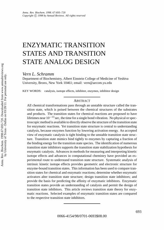
Enzymes catalyze chemical reactions at rates that are astounding relative touncatalyzed chemistry at the same conditions. Typical enzymatic rate enhance-ments are 1010 to 1015, accomplishing in 1 sec that which would require 300to 30,000,000 years in the absence of enzymes (1). Each catalytic event re-quires a minimum of three or often more steps, all of which occur within thefew milliseconds that characterize typical enzymatic reactions. According totransition state theory, the smallest fraction of the catalytic cycle is spent inthe most important step, that of the transition state. However, this step cannotoccur without participation of the enzymatic forces that occur as the Michaeliscomplex is transformed into the transition state by precise alignment of catalyticgroups by enzymatic and substrate conformational changes (Figure 1).
The original proposals of absolute reaction rate theory for chemical reac-tions defined the transition state as a distinct species in the reaction coordinatethat determined the absolute reaction rate (2). Soon thereafter, Linus Paulingproposed that the powerful catalytic action of enzymes could be explained byspecific tight binding to the transition state species (3). Because reaction rateis proportional to the fraction of the reactant in the transition state complex,the enzyme was proposed to increase the concentration of the reactive species.This proposal was formalized by Wolfenden and coworkers, who hypothesizedthat the rate increase imposed by enzymes is proportional to the affinity of
Ann
u. R
ev. B
ioch
em. 1
998.
67:6
93-7
20. D
ownl
oade
d fr
om w
ww
.ann
ualr
evie
ws.
org
by U
nive
rsity
of
Tex
as -
Dal
las
on 0
3/28
/11.
For
per
sona
l use
onl
y.
P1: DPI/ary P2: RPK/ARK/plb QC: ARK
May 12, 1998 14:26 Annual Reviews AR057-22
ENZYMATIC TRANSITION STATES 695
Figure 1 Reaction coordinate diagram for conversion of substrate (A) to enzyme-bound products(EP). Symbols are R= H-bond acceptor, H= H-bond donor,+ and− are ionic charges, and> represents hydrophobic sites. Thesolid line is an example of fully rate-limiting transition stateformation, providing intrinsic kinetic isotope effects. The energetic barriers (dashed lines) labeled“forward commitment” and “reverse commitment” make substrate binding and/or product releaserate limiting and suppress kinetic isotope effects. The transition state has unique properties of chargeand optimal H-bond alignment not found in any of the reactant species. In this example, chargerepulsion serves to clear the catalytic site and restore the enzyme to the open form (E) afterk5.
the enzyme for the transition state structure relative to the Michaelis complex(4; Figure 2). Because enzymes typically increase the noncatalyzed reactionrate by factors of 1010–1015, and Michaelis complexes often have dissociationconstants in the range of 10−3–10−6 M, it is proposed that transition state com-plexes are bound with dissociation constants in the range of 10−14–10−23 M.Analogs that resemble the transition state structures should therefore providethe most powerful noncovalent inhibitors known, even if only a small fractionof the transition state energy is captured.
Reviews from 1976, 1988, and 1995 listed enzymes known to interact withtransition state inhibitors, defined as tight binding inhibitors that resemble thehypothetical transition states or intermediates for various enzymes (5–7). Be-tween 1988 and 1995, the list grew from 33 to 132 enzymes. In 1976 and 1988,most of the inhibitors were natural products. The 1995 list of enzymes andtransition state inhibitors is dominated by intentionally synthesized inhibitors
Ann
u. R
ev. B
ioch
em. 1
998.
67:6
93-7
20. D
ownl
oade
d fr
om w
ww
.ann
ualr
evie
ws.
org
by U
nive
rsity
of
Tex
as -
Dal
las
on 0
3/28
/11.
For
per
sona
l use
onl
y.

P1: DPI/ary P2: RPK/ARK/plb QC: ARK
May 12, 1998 14:26 Annual Reviews AR057-22
696 SCHRAMM
Figure 2 The thermodynamic box that predicts transition state binding affinity (upper left) com-pares the rates of uncatalyzed (kuncat) and enzymatic (kenz) reactions and assumes that the transmis-sion coefficients from [A]‡ and [EA]‡ are equal. The dissociation constants for EA and [EA]‡ aregiven byKd andK‡d. Transition state inhibitors (lower left) invoke the unique ionization and con-formational structure found exclusively in the transition state (see Figure 1). Catalysis is preventedby a stable bond at the reaction center (represented by thesolid dot). The energetics of catalysisand binding an energetically perfect transition state analog are compared in the reaction or bindingcoordinate diagram. Energy of enzymatic transition state stabilization (11G‡) is converted intobinding energy for the transition state inhibitor (11G-I‡) to form the stable EI‡ complex, whichcannot escape by the product release pathway. The slow-onset inhibition common with transitionstate inhibitors occurs after formation of a readily reversible E•I‡ complex. The rate of onset fortight-binding inhibitors isk‡2 and the rate of escape isk‡−2. Note the unfavorable energetic barrierfor escape from the stable EI‡ complex.
in response to inhibitor development for the AIDS protease,β-lactamases,metalloproteinases, cyclooxygenases, and a growing list of enzymes that aretargets for pharmaceutical intervention. Because many inhibitor developmentsare proprietary, the list for 1995 is likely to be an incomplete representation ofthe known list of transition state inhibitors.
In 1947, an experimental approach to chemical transition states was dis-covered by Bigeleisen and coworkers, who, along with others, established therelationships among isotopic substitution, altered reaction rates, and alteredbonding between reactants and transition states (8–10). A few applications
Ann
u. R
ev. B
ioch
em. 1
998.
67:6
93-7
20. D
ownl
oade
d fr
om w
ww
.ann
ualr
evie
ws.
org
by U
nive
rsity
of
Tex
as -
Dal
las
on 0
3/28
/11.
For
per
sona
l use
onl
y.
P1: DPI/ary P2: RPK/ARK/plb QC: ARK
May 12, 1998 14:26 Annual Reviews AR057-22
ENZYMATIC TRANSITION STATES 697
of individual kinetic isotope effects were made to enzymatic reaction mecha-nisms before 1970. Qualitative results from these studies indicated whether thebond changes to the isotopically labeled atom occurred in the rate-limitingstep (e.g. Figure 1). The Steenbock Symposium of 1976, “Isotope Effectson Enzyme-Catalyzed Reactions,” provided the impetus for additional studieson enzymes (11). The 1978 bookTransition States of Biochemical Processesincluded the provocative and fundamentalist position of RL Schowen that “theentire and sole source of catalytic power is the stabilization of the transitionstate; that reactant-state interactions are by nature inhibitory and only wastecatalytic power” (12, p. 78). These works bridged the gap between chemicaland biological transition states, recognizing that even complex biological trans-formations involve formation of one or more defined transition states and aretherefore susceptible to transition state analysis based on the measurement ofkinetic isotope effects. Development of steady state and pre–steady state kineticmethods to reveal intrinsic isotope effects permitted interpretation of results interms of transition state structure (13–17).
Although the focus of this review is enzymatic transition states and relatedinhibitors, transition state similarity is not necessary for tight-binding inhibi-tion of enzymes. Any combination of multiple favorable hydrogen, ionic, orhydrophobic bonds between enzyme and substrate can provide the summationof binding interactions leading to tight-binding inhibition and is the basis forinhibitory screening from chemical and combinatorial libraries. Examples ofsuch inhibitors are found in nature as antibiotics. Streptomycin, erythromycin,and rifampicin are examples of complex natural products that have no knownsimilarity to the transition states involved in protein and RNA synthesis. In-hibitors designed to match contacts in the catalytic site or those that are sta-ble analogs of the substrate can bind tightly but do not qualify as transitionstate inhibitors. One example is the tight binding of methotrexate to dihydro-folate reductase, in which the analog is bound upside down with respect to sub-strate in the catalytic site (18). Another example is 9-deaza-9-phenyl-guanine,a powerful inhibitor of purine nucleoside phosphorylase. It was designed fromthe X-ray crystal structure of substrate and product complexes and does notresemble the transition state (19, 20) (Figure 3).
TRANSITION STATE THEORY FORENZYME-CATALYZED REACTIONS
Nature of the Transition StateAs substrate progresses from the Michaelis complex to product, chemistryoccurs by enzyme-induced changes in electron distribution in the substrate. En-zymes alter the electronic structure by protonation, proton abstraction, electron
Ann
u. R
ev. B
ioch
em. 1
998.
67:6
93-7
20. D
ownl
oade
d fr
om w
ww
.ann
ualr
evie
ws.
org
by U
nive
rsity
of
Tex
as -
Dal
las
on 0
3/28
/11.
For
per
sona
l use
onl
y.

P1: DPI/ary P2: RPK/ARK/plb QC: ARK
May 12, 1998 14:26 Annual Reviews AR057-22
698 SCHRAMM
Figure 3 Tight-binding inhibitors of purine nucleoside phosphorylase and dihydrofolate reductasethat are not transition state analogs despite high-affinity binding. TheKm values for substrates andequilibriumKi values for the inhibitors are shown.
transfer, geometric distortion, hydrophobic partitioning, and interaction withLewis acids and bases. These are accomplished by sequential protein and sub-strate conformational changes (Figure 1). When a constellation of individuallyweak forces are brought to bear on the substrate, the summation of the individ-ual energies results in large forces capable of relocating bonding electrons tocause bond-breaking and bond-making. Substrates of modest molecular size(for example, glucose), bound in the active sites of proteins, typically inter-act with two H-bonds at each donor/acceptor site. These interactions alone, intypical H-bond energies of 3 kcal/mol/H-bond, can provide over 30 kcal/mol ofenergy toward redistribution of electrons and therefore toward ionizations andcovalent bond changes. The restricted geometry and hydrophobic environmentallow short H-bonds to form that would be unfavorable in solution (21, 22).
The lifetime for chemical transition states is short, approximately 10−13 sec,the time for conversion of a bond vibrational mode to a translational mode(23, 24). This theory requires reexamination for enzymes, because the proteindomain motion resulting in transition state formation may stabilize the alteredbond lengths of the bound transition state for a lifetime sufficient for 101–106
Ann
u. R
ev. B
ioch
em. 1
998.
67:6
93-7
20. D
ownl
oade
d fr
om w
ww
.ann
ualr
evie
ws.
org
by U
nive
rsity
of
Tex
as -
Dal
las
on 0
3/28
/11.
For
per
sona
l use
onl
y.

P1: DPI/ary P2: RPK/ARK/plb QC: ARK
May 12, 1998 14:26 Annual Reviews AR057-22
ENZYMATIC TRANSITION STATES 699
vibrations. This hypothesis is indicated by the dimpled transition state featurelabeled with a question mark in Figure 1; it remains unexplored except by com-putational theory. Enzymes that form transient covalent intermediates requiretwo distinct transition states. These transition states are surrounded by lowerenergy complexes and should be distinguished from the altered vibrational stateimplied by the question mark in Figure 1. The classic interpretation of the tran-sition state supposes a short lifetime during which an infinitesimal force towardproduct or substrate leads to EP′ and EA′ respectively.
Induced Protein Conformational ChangeThe energetic problem that must be solved by enzymes is to bind tightly onlyto the unstable transition state structure while avoiding tight binding to the sub-strate and products. Enzymes often bind to the substrate at diffusion-controlledrates, and subsequent conformational or electronic changes are mandatory forcatalysis. Placing the enzyme-bound substrate in the solvent-restricted envi-ronment of the closed catalytic site permits the subsequent events to occur inthe altered solvent of the catalytic site.
Presenting the enzyme with a transition state mimic results in a mismatch ofthe substrate-recognizing features. Many transition state inhibitors are slow-onset, typified by a rapid weak binding followed by a slow tight-binding inter-action. The energetics of this interaction (Figure 2) show rapid formation ofthe encounter complex E•I‡ (similar to the EA complex of Figure 1) followedby a difficult (high-energy, slow) entry to the stable EI‡ complex. The boundinhibitor does not induce the conformational change with the efficiency of sub-strate and requires time to permit the transition state conformational change tooccur on the protein. This time corresponds to the slow onset of tight-bindinginhibition commonly observed with transition state mimics (6). These dataargue that the substrate actively induces the catalytic conformational change,because the rate of catalysis is substantially greater than the rate at which mosttight-binding inhibitors induce enzymes into the transition state configuration.This view of transition state inhibitor interaction predicts that near-perfect in-hibitors would still exhibit slow-onset inhibition because the enzyme is designedto recognize the ground state of the substrate. However, some tightly boundinhibitors are reported to achieve inhibition on the time scale of catalysis. Forexample, the inhibition of adenosine deaminases by purine riboside and of cy-tidine deaminase by pyrimidin-2-one riboside is fast (25–26). These inhibitorsare estimated to bind with dissociation constants of 10−13 and 10−12 M respec-tively, approaching the hypothetical 10−16M dissociation constant for the actualtransition states. Rapid onset occurs because these are half-reaction substratesfor the enzymes. The deaminases contain a tightly bound zinc that acts as thecatalytic site base to ionize a water molecule to the hydroxide and position it near
Ann
u. R
ev. B
ioch
em. 1
998.
67:6
93-7
20. D
ownl
oade
d fr
om w
ww
.ann
ualr
evie
ws.
org
by U
nive
rsity
of
Tex
as -
Dal
las
on 0
3/28
/11.
For
per
sona
l use
onl
y.

P1: DPI/ary P2: RPK/ARK/plb QC: ARK
May 12, 1998 14:26 Annual Reviews AR057-22
700 SCHRAMM
Figure 4 Hydration of purine riboside by a catalytic site hydroxyl generated at the tightly boundZn2+. The hydrated purine is bound tightly. An analogous reaction occurs with cytidine deaminase.
the reactive carbon of the aromatic rings (27; Figure 4). Enzymatic protonationof the adjacent aromatic ring nitrogen assists in the required rehybridization ofthe rings. These inhibitors take advantage of the substrate-induced transitionstate configuration to cause rapid hydration and tight binding of the hydratedspecies. The sp3 hybridization at the reactive carbon is a transition state feature,and without the amino leaving group, a stable complex is formed.
Tight Binding of the Transition State StructureBinding energies of enzymatic transition states are generated by the realign-ment of substrate (Michaelis) contacts as the enzyme and substrate mutuallychange their structures toward the transition state (Figure 1). The strong de-pendence of hydrogen and ionic bond energy on bond distance, angle, solventenvironment, and relative pKa values can be invoked to explain the increases inbinding forces of the transition state complex relative to the Michaelis complex(21, 28). Structural rearrangements tighten the protein around the catalytic siteto exclude solvent and to make stronger electrostatic contacts. These are shownas well-aligned H-bonds at the transition state and as ionic attraction and repul-sion as catalytic forces (Figure 1). Enzymatic reactions usually demonstratedistinct pKa values for substrate binding andkcat, testifying to the ionic changesbetween the enzyme substrate and enzyme transition state complexes (29, 30).A common mechanism for enzymatic catalysis is to generate differential chargebetween substrate and transition state, permitting electrostatic interactions tospecifically stabilize the transition state (28). The imperfect match between theenzyme and the transition state inhibitor is inevitable because it is impossible torecreate perfectly the nonequilibrium bond lengths of the transition state withstable compounds. The11G-I‡bindingenergy of Figure 2 is shown for a perfecttransition state inhibitor, with fullK ‡d of the transition state. For the necessarily
Ann
u. R
ev. B
ioch
em. 1
998.
67:6
93-7
20. D
ownl
oade
d fr
om w
ww
.ann
ualr
evie
ws.
org
by U
nive
rsity
of
Tex
as -
Dal
las
on 0
3/28
/11.
For
per
sona
l use
onl
y.
P1: DPI/ary P2: RPK/ARK/plb QC: ARK
May 12, 1998 14:26 Annual Reviews AR057-22
ENZYMATIC TRANSITION STATES 701
imperfect transition state inhibitors, thek‡2 barrier is the time to fit the imperfectinhibitor into the lowest-energy structure of the analog complex.
Relaxation of the Transition State ComplexCatalysis requires rapid relaxation of the transition state energy to allow theproducts to dissociate. The relative enzymatic affinity for the transition stateand products decreases by about 12 orders of magnitude within milliseconds.For catalytically efficient enzymes, the Michaelis complex is a diffusional eventwith a rate constant near 109 M−1sec−1. Transition state formation, productformation, and release are as rapid as this, so the diffusional event defines themaximal catalytic rate (31). The change in electron distribution as bonds arebroken creates a repulsive interaction that opens the catalytic site and expelsproducts (see Figure 1). This step is the opposite of what occurs when theMichaelis complex is converted into the transition state complex.
Computer modeling of well-understood enzymatic reactions has allowed ex-amination of the factors important in catalysis. The resulting conclusion is thatelectrostatic stabilization of the transition state is the most significant factor(28). Understanding the electrostatic nature of the transition state thereforeprovides a template for the synthesis of transition state inhibitors. The ex-perimental approach that provides the most direct information for enzymatictransition state structure is analysis of kinetic isotope effects (32–34).
EXPERIMENTAL APPROACHES TO ENZYMATICTRANSITION STATE STRUCTURE
Chemical PrecedentChemical reactivity series with leaving groups of different pKa values or elec-tron withdrawing capability can indicate the position of the transition state inthe reaction coordinate. Brønstead and Hammett plots have had wide applica-tion in chemical mechanisms, but less in enzymology because the specificityof enzymes often does not permit the use of a variety of substrates (35, 36).Behavior of model chemistry in solution for the reaction of interest providesa transition state benchmark for comparison with the transition state structureimposed by the enzyme.
Transition State InhibitorsInhibitors that bind tightly, are slow-onset, and resemble the expected tran-sition states have been used to predict state features (4–6). For example, theinhibition of deaminases and proteases by analogs with sp3 reaction centerswas useful in establishing the nature of these transition states and intermediates(37, 38). Inhibitor specificity is subject to the vagaries of enzymatic binding
Ann
u. R
ev. B
ioch
em. 1
998.
67:6
93-7
20. D
ownl
oade
d fr
om w
ww
.ann
ualr
evie
ws.
org
by U
nive
rsity
of
Tex
as -
Dal
las
on 0
3/28
/11.
For
per
sona
l use
onl
y.

P1: DPI/ary P2: RPK/ARK/plb QC: ARK
May 12, 1998 14:26 Annual Reviews AR057-22
702 SCHRAMM
Figure 5 Simplified bond vibrational basis for the relationship between a kinetic isotope effectand transition state structure. The parabolae represent the bonding environment (restoring force)for the vibrational mode of a bonded atom (1H) and its isotope (3H) in the reactant and transitionstates. Thearrowsare the activation energies for1H- and3H-labeled reactants. Inpanel A, 1H-substrate reacts more rapidly (has lower activation barrier). Inpanel B, 3H-substrate reacts morerapidly (has lower activation barrier). In practice, all significant modes are considered. InpanelC are the anticipated values of kinetic isotope effects for C1′-N9 N-ribosidic bond scission withSN1 or SN2 character. The ranges arise from variability in ring geometry and the degree of theassociated O′ nucleophile at transition states with predominant SN1 or SN2 character. The percentkinetic isotope effect (%KIE)= [(unlabeled rate/labeled rate)− 1.00] × 100%.
sites. Transition state properties cannot always be predicted, as demonstratedby methotrexate in the catalytic site of dihydrofolate reductase (18; Figure 3).Direct information on transition state structure is available from kinetic isotopeeffect studies.
Kinetic Isotope EffectsThe only method available for the direct determination of enzymatic transitionstate structure is the measurement of kinetic isotope effects. Excellent accountsof the theory and its development can be found in the literature (39–43) andare beyond the scope of this review. Kinetic isotope effects compare the enzy-matic reaction rates of isotopically labeled and unlabeled substrates (Figure 5).Isotopically labeled molecules have molecular energy different from that ofunlabeled molecules and thus require a different amount of energy to reachthe transition state, where the molecular energies may also be perturbed by theisotope. If the bonding environment for the labeled atom is less restricted in thetransition state than in the reactant, the isotope effect will be normal (the heavyisotope substrate reacting more slowly than the unlabeled substrate). Likewise,if the bonding environment at the transition state is more restricted than for thereactant, the substrate with the higher mass isotope will react more rapidly (aninverse kinetic isotope effect). Isotope effects also provide quantitative infor-mation because the magnitude of the isotope effect indicates the extent of bondchange. For some isotope effects, bond geometry can be determined because of
Ann
u. R
ev. B
ioch
em. 1
998.
67:6
93-7
20. D
ownl
oade
d fr
om w
ww
.ann
ualr
evie
ws.
org
by U
nive
rsity
of
Tex
as -
Dal
las
on 0
3/28
/11.
For
per
sona
l use
onl
y.

P1: DPI/ary P2: RPK/ARK/plb QC: ARK
May 12, 1998 14:26 Annual Reviews AR057-22
ENZYMATIC TRANSITION STATES 703
the dihedral angular dependence for conjugative isotope effectsβ to the bondbeing broken (44, 45). By measuring kinetic isotope effects at every positionin a substrate molecule that might be expected to be perturbed at the transitionstate, a unique description of the transition state can be deduced if intrinsicisotope effects are being measured (13, 46). The steps involved in transitionstate analysis by kinetic isotope effects are to
1. synthesize substrates with appropriate isotopic labels
2. measure kinetic isotope effects to an accuracy of better than 0.5%
3. compute a truncated transition state with bond lengths and angles matchingthe isotope effects (limited to 25 atoms), using normal-mode bond-energybond-order vibrational analysis (47, 48)
4. restore the complete molecular structure for the transition state by fixing thebonds established from kinetic isotope effects and optimizing the remainingstructure using semiempirical methods (49)
5. determine the electron wave function for the molecule to determine electrondistribution at the van der Waals surface (50).
EXAMPLES OF ENZYMATIC TRANSITION STATES
AMP NucleosidaseAMP nucleosidase catalyzes the hydrolysis of AMP to adenine and ribose5-phosphate under the allosteric control of MgATP and inorganic phosphate,which serve as allosteric activator and inhibitor, respectively (51, 52):
AMP+ H2O→ adenine+ ribose 5-phosphate.
The enzyme is found only in prokaryotes and is thought to play a role in theregulation of energy metabolism. However, genetic ablation of the enzymefrom Escherichia colihad only minor effects on growth rates (53). The en-zyme fromAzotobacter vinelandiirequires MgATP as akcat activator. In theabsence of activator, the enzyme binds substrate, butkcat is 10−3 of that withthe allosteric activator. This provides the opportunity to determine whetherallosteric activation is capable of changing the chemical nature of the transitionstate or only serves to change energies of activation (altered rate constants) forsteps through the reaction cycle. Systematic determination of kinetic isotopeeffects is made possible by the combined chemical and enzymatic synthesis ofAMP (Figure 6) with the isotopic labels indicated in Figure 7 (54–56).
Kinetic isotope effects for the enzyme were intrinsic based on the catalyticmechanism and by comparison with the isotope effects for acid-catalyzed
Ann
u. R
ev. B
ioch
em. 1
998.
67:6
93-7
20. D
ownl
oade
d fr
om w
ww
.ann
ualr
evie
ws.
org
by U
nive
rsity
of
Tex
as -
Dal
las
on 0
3/28
/11.
For
per
sona
l use
onl
y.

P1: DPI/ary P2: RPK/ARK/plb QC: ARK
May 12, 1998 14:26 Annual Reviews AR057-22
704 SCHRAMM
Figure 6 Combined enzymatic and chemical synthesis of specifically labeled nucleosides andnucleotides (synthesis of [9-15N, 5′-14C]ATP, AMP, and inosine). All steps from glucose to ATPare coupled in a single reaction mixture. Separate reaction mixtures are used to convert ATP to AMPand to inosine. The enzymes are HK= hexokinase, PK= pyruvate kinase, G6PDH= glucose6-phosphate dehydrogenase, 6PGDH= 6-phosphogluconate dehydrogenase, GDH= glutamatedehydrogenase, PRI= phosphoriboisomerase, PPS= 5-phosphoribosyl-1-pyrophosphate syn-thetase, AK= adenylate kinase, APRT= adenine phosphoribosyltransferase, AMD= AMPdeaminase, AP= alkaline phosphatase. Using specific labels in glucose, ribose and adenine canyield the desired labels at any position of the nucleosides and nucleotides.
solvolysis of AMP (56). The transition state for the acid-catalyzed reactionwas compared to those determined for the enzyme in the presence and absenceof allosteric activator (Figure 7). Because kinetic isotope effects report directlyon the bond environment at the transition state, comparison of the experimentalkinetic isotope effects established that the transition states differ for the enzyme-catalyzed and acid-catalyzed hydrolysis. The allosteric activator, which causesa 103-fold increase in catalytic rate, also changes the nature of the transitionstate. Quantitiation of the isotope effects in terms of bond orders for reactantsand transition states indicated that the enzymatic catalyst permitted the reactionto reach the transition state earlier, when there was more bond order remaining
Ann
u. R
ev. B
ioch
em. 1
998.
67:6
93-7
20. D
ownl
oade
d fr
om w
ww
.ann
ualr
evie
ws.
org
by U
nive
rsity
of
Tex
as -
Dal
las
on 0
3/28
/11.
For
per
sona
l use
onl
y.
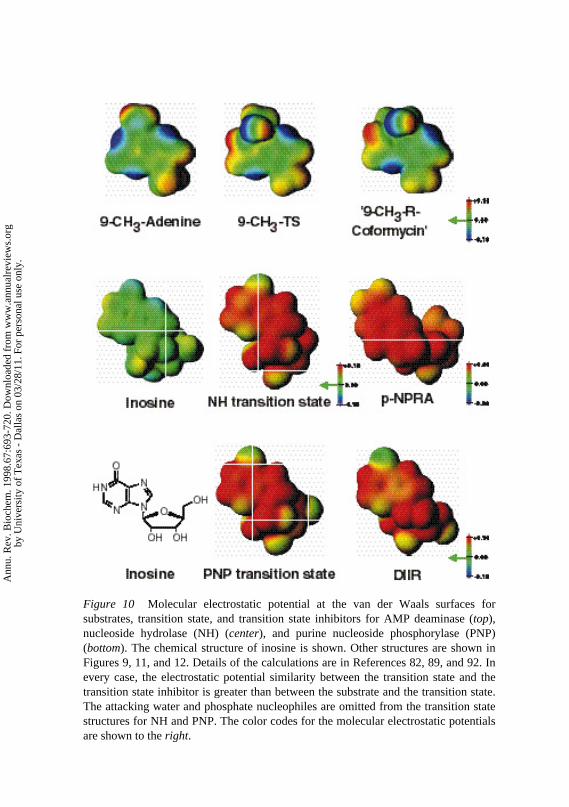
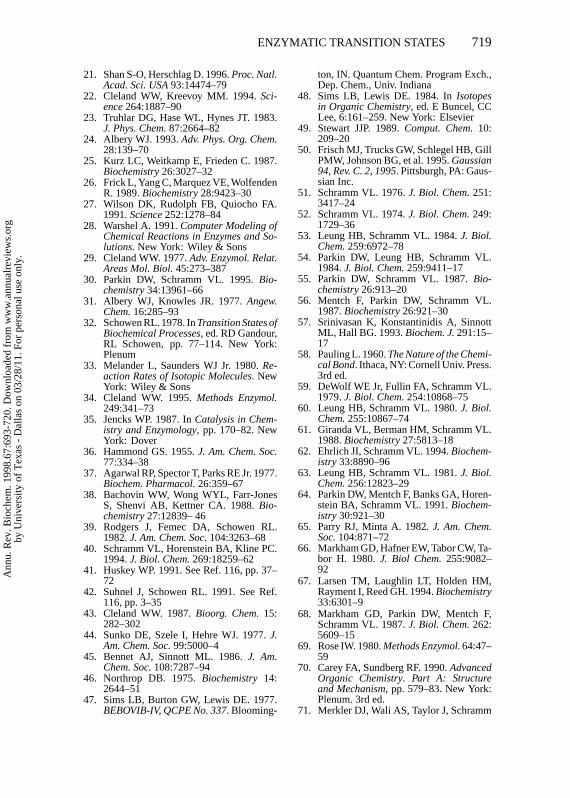
Figure 10 Molecular electrostatic potential at the van der Waals surfaces for substrates, transition state, and transition state inhibitors for AMP deaminase (top), nucleoside hydrolase (NH) (center), and purine nucleoside phosphorylase (PNP) (bottom). The chemical structure of inosine is shown. Other structures are shown in Figures 9, 11, and 12. Details of the calculations are in References 82, 89, and 92. In every case, the electrostatic potential similarity between the transition state and the transition state inhibitor is greater than between the substrate and the transition state. The attacking water and phosphate nucleophiles are omitted from the transition state structures for NH and PNP. The color codes for the molecular electrostatic potentials are shown to the right.
Ann
u. R
ev. B
ioch
em. 1
998.
67:6
93-7
20. D
ownl
oade
d fr
om w
ww
.ann
ualr
evie
ws.
org
by U
nive
rsity
of
Tex
as -
Dal
las
on 0
3/28
/11.
For
per
sona
l use
onl
y.
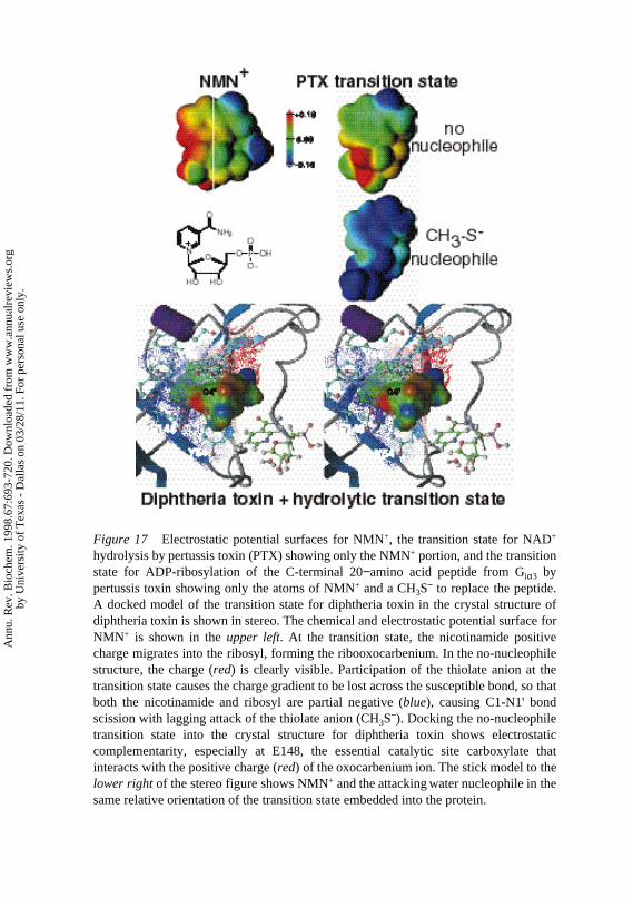
Figure 17 Electrostatic potential surfaces for NMN+, the transition state for NAD+ hydrolysis by pertussis toxin (PTX) showing only the NMN+ portion, and the transition state for ADP-ribosylation of the C-terminal 20−amino acid peptide from Giα3 by pertussis toxin showing only the atoms of NMN+ and a CH3S− to replace the peptide. A docked model of the transition state for diphtheria toxin in the crystal structure of diphtheria toxin is shown in stereo. The chemical and electrostatic potential surface for NMN+ is shown in the upper left. At the transition state, the nicotinamide positive charge migrates into the ribosyl, forming the ribooxocarbenium. In the no-nucleophile structure, the charge (red) is clearly visible. Participation of the thiolate anion at the transition state causes the charge gradient to be lost across the susceptible bond, so that both the nicotinamide and ribosyl are partial negative (blue), causing C1-N1' bond scission with lagging attack of the thiolate anion (CH3S−). Docking the no-nucleophile transition state into the crystal structure for diphtheria toxin shows electrostatic complementarity, especially at E148, the essential catalytic site carboxylate that interacts with the positive charge (red) of the oxocarbenium ion. The stick model to the lower right of the stereo figure shows NMN+ and the attacking water nucleophile in the same relative orientation of the transition state embedded into the protein.
Ann
u. R
ev. B
ioch
em. 1
998.
67:6
93-7
20. D
ownl
oade
d fr
om w
ww
.ann
ualr
evie
ws.
org
by U
nive
rsity
of
Tex
as -
Dal
las
on 0
3/28
/11.
For
per
sona
l use
onl
y.

P1: DPI/ary P2: RPK/ARK/plb QC: ARK
May 12, 1998 14:26 Annual Reviews AR057-22
ENZYMATIC TRANSITION STATES 705
Figure 7 Kinetic isotope effects (KIEs) and bond orders at the transition states for the C1′-N9bond hydrolysis of AMP by acid and AMP nucleosidases. All isotope effects were measured witha mixture of two AMP molecules, one labeled near and one distant from the C′1-N9 bond. Bothnative and mutant enzymes were activated by a factor of∼103 by the addition of MgATP. Kineticisotope effects were measured with average standard deviations of 0.4%. Bond orders for theC1′-N9 and C1′-O′ bonds were determined from bond vibrational analysis (48, 56). When bondssurrounding C1′ are weakest at the transition state, the3H1′ kinetic isotope effect is greatest. Bondorder (n) is based on the Pauling rule: rn = r1− 0.3 ln n, where r1 is the single bond length (58).
to the leaving group (Figure 7). The protein structural change induced by theallosteric activator caused the transition state to occur even earlier in the reac-tion coordinate. Changes in the transition state structure also resulted from theforced evolution of aβ-galactosidase inE. coli, causing the enzyme to becomecatalytically more efficient (57).
Substrate and inhibitor specificity studies revealed that formycin A 5′-phos-phate binds>103 times tighter to AMP nucleosidases than does substrate(59, 60). The X-ray crystal structure of this inhibitor helped to establish theproperties of the transition state (61). The inhibitor has asyn-ribosyl torsionangle, preferred in all nucleotide inhibitors of the enzyme (59). The molecularelectrostatic potential at the van der Waals surface revealed similar electronicstructures of inhibitor and transition state because of the common protonationat N7, which is not present in the substrate (62).
Random mutagenesis of the organism expressing AMP nucleosidase pro-vided an enzyme with akcat 2% that of the native enzyme (Figure 7) (63). Thepurpose of the mutagenesis was to determine if catalytic site mutation changestransition state structure. The results established substantially different kineticisotope effects for the mutant enzyme, demonstrating differences in bond struc-ture at the transition state. The mutant enzyme was less effective in stabilizing
Ann
u. R
ev. B
ioch
em. 1
998.
67:6
93-7
20. D
ownl
oade
d fr
om w
ww
.ann
ualr
evie
ws.
org
by U
nive
rsity
of
Tex
as -
Dal
las
on 0
3/28
/11.
For
per
sona
l use
onl
y.

P1: DPI/ary P2: RPK/ARK/plb QC: ARK
May 12, 1998 14:26 Annual Reviews AR057-22
706 SCHRAMM
Figure 8 Kinetic isotope effects for the SAM-synthetase reaction (left), where the35S-moleculerepresents the attacking methionine sulfur nucleophile. The isotope effects were measured in in-dividually labeled substrates but are illustrated here as a single molecule. Only symmetrical SN2displacements give primary14C isotope effects of 12.8% (see Figure 5). The transition state struc-ture consistent with all isotope effects is shown on theright. The bond orders were 0.61 and 0.35,which correspond to bond lengths of 1.96 and 1.72A for S-C5′ and C5′-O bonds, respectively. Themass difference between sulfur and oxygen accounts for the differences, which are equivalent inbond vibrational energy. C5′ is the center of reaction coordinate motion, giving the large14Cisotope effect.
the ribooxocarbenium ion of the transition state and in protonating the adenineleaving group (64).
S-Adenosylmethionine SynthetaseThe chemical reaction of S-adenosylmethionine (SAM) synthetase involves dis-placement of the tripolyphosphate of ATP by reaction of the sulfur of methioninewith C5′ of ATP (Figure 8). The reaction results in the further hydrolysis ofthe phosphate chain to phosphate and pyrophosphate, to release the productsSAM, phosphate, and pyrophosphate from the catalytic site (65–66). This isone of the few reactions of ATP in which all phosphates are displaced in a singlereaction. The reaction is irreversible under physiological conditions as a con-sequence of the tripolyphosphorolysis. Potassium ion is required for efficientcatalysis, activating the enzyme approximately 100-fold. Kinetic isotope effectstudies of the enzyme were designed to establish the nature of the transitionstate under optimal catalytic conditions and to establish the effect of K+ ion ac-tivation. Monovalent cations are common in kinase activation and are thoughtto provide a positive charge to neutralize the transition state complex at highlycharged reaction centers typified by phosphoryl transfers (67). Activation canbe achieved either by changing a rate constant or by changing the transitionstate structure.
Measurement of kinetic isotope effects with limiting K+ gave intrinsic iso-tope effects (68). The14C5′ kinetic isotope effect was 12.8%, near the theoretical
Ann
u. R
ev. B
ioch
em. 1
998.
67:6
93-7
20. D
ownl
oade
d fr
om w
ww
.ann
ualr
evie
ws.
org
by U
nive
rsity
of
Tex
as -
Dal
las
on 0
3/28
/11.
For
per
sona
l use
onl
y.
P1: DPI/ary P2: RPK/ARK/plb QC: ARK
May 12, 1998 14:26 Annual Reviews AR057-22
ENZYMATIC TRANSITION STATES 707
limit for a primary 14C isotope effect (33). This value is realized only whencarbon dominates reaction coordinate motion in a symmetric nucleophilic dis-placement reaction. The result provided a ready solution for the reaction mech-anism. In nucleophilic displacements, neighboringα- andβ-secondary3Hisotope effects are expected to be insignificant, and this was confirmed for both[5′-3H]ATP and methyl-[3H3]methionine. These kinetic isotope effects provideproof that the chemical mechanism of SAM synthetase at limiting K+ is a sym-metric SN2 reaction. Addition of K+ to activate the rate by 100-fold decreasedthe [5′-14C]ATP isotope effect, consistent with either a change in transition statestructure or increased forward commitment (Figure 1).
Substrate trapping experiments, pioneered by I Rose (69), permitted the di-rect measurement of forward commitment for SAM synthetase (the probabilitythat a bound substrate molecule will be transformed to product relative to releasefrom the catalytic site). Only substrate molecules in equilibrium with free sub-strate contribute to the observed isotope effect (13). The results established thatK+ improved catalytic efficiency by increasing the forward commitment. Theintrinsic isotope effect (corrected for commitment) was unchanged, thus thestructure of the transition state is unchanged by K+. This example of enzymaticactivation demonstrates lowering the transition state energetic barrier withouta measurable change in the bond environment of the transition state (Figure 1).
AMP Deaminase and Adenosine DeaminaseThe deaminations of AMP and adenosine are aromatic nucleophilic substitu-tions at carbon, a reaction type well known in chemistry and characterized byrate-limiting formation of an unstable tetrahedral intermediate that decomposesrapidly to yield products (70). AMP deaminase is found only in eukaryotes,and it is proposed to be involved in the regulation of the adenine nucleotidepool through the purine nucleotide cycle (71). Activation of AMP deaminasein times of energy deficiency leads to IMP formation and a decrease in thetotal adenylate pool size (72). Humans deficient in specific muscle isozymesof AMP deaminase suffer moderate difficulty in muscular work, but deficien-cies in the erythrocyte/heart isoform have no known phenotype (73). It hasbeen proposed that inhibition of the heart isozyme during recovery from heartattacks might speed recovery by preserving the adenylates and preventing oxy-gen radical damage associated with the conversion of hypoxanthine to uricacid (74). Adenosine deaminase deficiency leads to B- and T-cell immunode-ficiency, presumably as a result of the accumulation of dATP in the progenitorcells preventing clonal expansion in response to an immune challenge (75).
The natural products coformycin, deoxycoformycin, and their 5′-phosphatesare tight-binding transition state inhibitors for adenosine and AMP deami-nases, binding approximately 107-fold more tightly than substrates (76–78). The
Ann
u. R
ev. B
ioch
em. 1
998.
67:6
93-7
20. D
ownl
oade
d fr
om w
ww
.ann
ualr
evie
ws.
org
by U
nive
rsity
of
Tex
as -
Dal
las
on 0
3/28
/11.
For
per
sona
l use
onl
y.
P1: DPI/ary P2: RPK/ARK/plb QC: ARK
May 12, 1998 14:26 Annual Reviews AR057-22
708 SCHRAMM
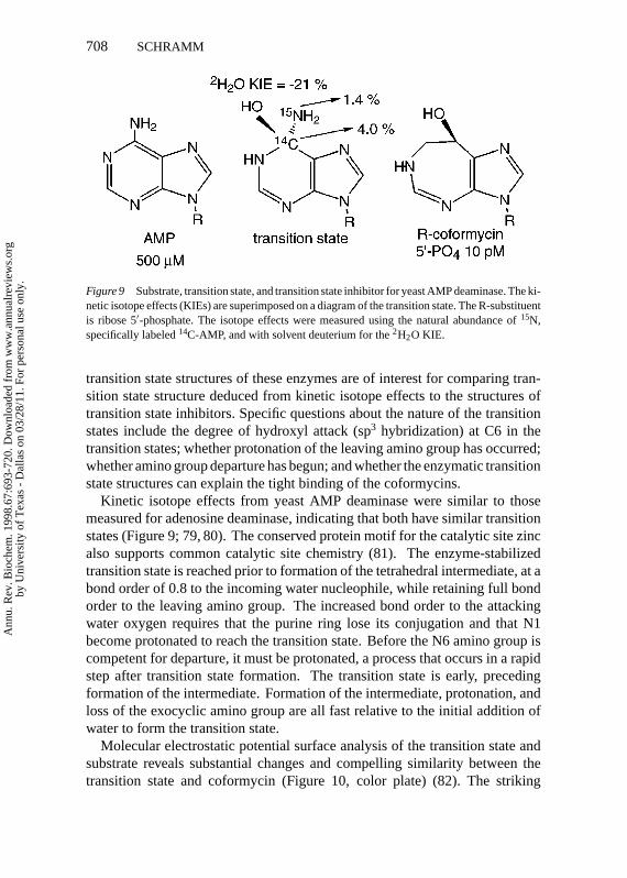
Figure 9 Substrate, transition state, and transition state inhibitor for yeast AMP deaminase. The ki-netic isotope effects (KIEs) are superimposed on a diagram of the transition state. The R-substituentis ribose 5′-phosphate. The isotope effects were measured using the natural abundance of15N,specifically labeled14C-AMP, and with solvent deuterium for the2H2O KIE.
transition state structures of these enzymes are of interest for comparing tran-sition state structure deduced from kinetic isotope effects to the structures oftransition state inhibitors. Specific questions about the nature of the transitionstates include the degree of hydroxyl attack (sp3 hybridization) at C6 in thetransition states; whether protonation of the leaving amino group has occurred;whether amino group departure has begun; and whether the enzymatic transitionstate structures can explain the tight binding of the coformycins.
Kinetic isotope effects from yeast AMP deaminase were similar to thosemeasured for adenosine deaminase, indicating that both have similar transitionstates (Figure 9; 79, 80). The conserved protein motif for the catalytic site zincalso supports common catalytic site chemistry (81). The enzyme-stabilizedtransition state is reached prior to formation of the tetrahedral intermediate, at abond order of 0.8 to the incoming water nucleophile, while retaining full bondorder to the leaving amino group. The increased bond order to the attackingwater oxygen requires that the purine ring lose its conjugation and that N1become protonated to reach the transition state. Before the N6 amino group iscompetent for departure, it must be protonated, a process that occurs in a rapidstep after transition state formation. The transition state is early, precedingformation of the intermediate. Formation of the intermediate, protonation, andloss of the exocyclic amino group are all fast relative to the initial addition ofwater to form the transition state.
Molecular electrostatic potential surface analysis of the transition state andsubstrate reveals substantial changes and compelling similarity between thetransition state and coformycin (Figure 10, color plate) (82). The striking
Ann
u. R
ev. B
ioch
em. 1
998.
67:6
93-7
20. D
ownl
oade
d fr
om w
ww
.ann
ualr
evie
ws.
org
by U
nive
rsity
of
Tex
as -
Dal
las
on 0
3/28
/11.
For
per
sona
l use
onl
y.
P1: DPI/ary P2: RPK/ARK/plb QC: ARK
May 12, 1998 14:26 Annual Reviews AR057-22
ENZYMATIC TRANSITION STATES 709
correspondence indicates that the molecular electrostatic potential surface ofthis transition state is closely related to that for the transition state inhibitor.
Nucleoside HydrolaseProtozoan parasites are purine auxotrophs, using salvage enzymes to recoverpurines from their environment (83). The nucleoside hydrolases cleave theN-ribosidic bonds of nucleosides to liberate the purine and pyrimidine basesand to generate ribose (84–88):
nucleosides+ H2O→ base+ ribose.
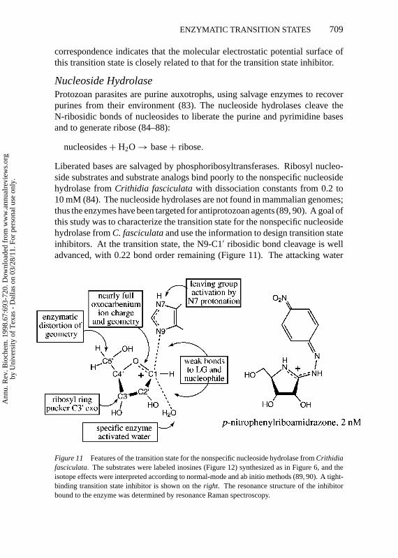
Liberated bases are salvaged by phosphoribosyltransferases. Ribosyl nucleo-side substrates and substrate analogs bind poorly to the nonspecific nucleosidehydrolase fromCrithidia fasciculatawith dissociation constants from 0.2 to10 mM (84). The nucleoside hydrolases are not found in mammalian genomes;thus the enzymes have been targeted for antiprotozoan agents (89, 90). A goal ofthis study was to characterize the transition state for the nonspecific nucleosidehydrolase fromC. fasciculataand use the information to design transition stateinhibitors. At the transition state, the N9-C1′ ribosidic bond cleavage is welladvanced, with 0.22 bond order remaining (Figure 11). The attacking water
Figure 11 Features of the transition state for the nonspecific nucleoside hydrolase fromCrithidiafasciculata. The substrates were labeled inosines (Figure 12) synthesized as in Figure 6, and theisotope effects were interpreted according to normal-mode and ab initio methods (89, 90). A tight-binding transition state inhibitor is shown on theright. The resonance structure of the inhibitorbound to the enzyme was determined by resonance Raman spectroscopy.
Ann
u. R
ev. B
ioch
em. 1
998.
67:6
93-7
20. D
ownl
oade
d fr
om w
ww
.ann
ualr
evie
ws.
org
by U
nive
rsity
of
Tex
as -
Dal
las
on 0
3/28
/11.
For
per
sona
l use
onl
y.

P1: DPI/ary P2: RPK/ARK/plb QC: ARK
May 12, 1998 14:26 Annual Reviews AR057-22
710 SCHRAMM
nucleophile lags well behind N-ribosidic bond breaking with approximately0.03 bond order from the attacking oxygen to C1′. The loss of most of theN-ribosidic bond before water attacks leaves the ribose as an oxocarbeniumcation at the transition state with C1′ rehybridized nearly completely to sp2.This flattens the ribose ring around C1′, causing the atoms of the ribosyl ringto be coplanar with the exception of C3′. The kinetic isotope effect from3H2′
is dependent on the dihedral angle to the C1′-N9 bond. Its value required thatC3′ lies below the plane of the ring, in the C3′-exo ribosyl configuration, toprovide near eclipse in the H2′-C2′-C1′-N9 dihedral angle. Solvent D2O and15N9 isotope effects established that the hypoxanthine leaving group is proto-nated, most likely at N7, prior to reaching the transition state. This createsa neutral, planar, and hydrophobic leaving group. A suprising remote 5′-3Hisotope effect of 5.1% was observed in the enzyme but not in acid-catalyzedhydrolysis of inosine, establishing enzyme-induced distortion (89, 90). The in-terpretation that the 5′-oxygen is rotated above the oxocarbenium ion is shownin Figure 11 and was recently confirmed by X-ray crystallogaphic studies witha bound transition state inhibitor (90a).
Based on the transition state structure, iminoribitol and riboamidrazone in-hibitors withKm/Ki to 200,000 have been synthesized and characterized (90–94). The best inhibitor (p-nitrophenylriboamidrazone; Figure 11) incorporatesmost of the electrostatic traits of the transition state but is structurally distinctfrom both the substrate and transition states. The similarity to the transitionstate is apparent in the comparison of the molecular electrostatic potentials ofsubstrate, transition state, and transition state inhibitor in Figure 10 (color plate)(95). Thus, the molecular electrostatic potential surface matching of inhibitorand transition states leads to powerful binding energy.
Purine Nucleoside PhosphorylaseScission of the N-ribosidic bonds of the purine nucleosides and deoxynucle-osides in mammals is accomplished almost exclusively by the phosphorolysisreaction of purine nucleoside phosphorylase (PNP). Inosine, guanosine, and2′-deoxyguanosine are the major substrates. The genetic deficiency of humanPNP causes a T-cell deficiency as the major physiological defect (96). Apopto-sis in T cells that are not immunologically stimulated to divide results in recy-cling of DNA in these thymic cells. When PNP is absent, 2′-deoxyguanosine isconverted to dGTP instead of being degraded. Imbalance of deoxynucleotidesprevents clonal expansion of normal T-cell populations. Several human dis-orders may involve T-cell action, including T-cell lymphoma, lupus, psoriasis,rheumatoid arthritis, and transplant tissue rejection. Specific inhibitors for PNPmay provide useful agents for these disorders. The transition state structure of
Ann
u. R
ev. B
ioch
em. 1
998.
67:6
93-7
20. D
ownl
oade
d fr
om w
ww
.ann
ualr
evie
ws.
org
by U
nive
rsity
of
Tex
as -
Dal
las
on 0
3/28
/11.
For
per
sona
l use
onl
y.
P1: DPI/ary P2: RPK/ARK/plb QC: ARK
May 12, 1998 14:26 Annual Reviews AR057-22
ENZYMATIC TRANSITION STATES 711
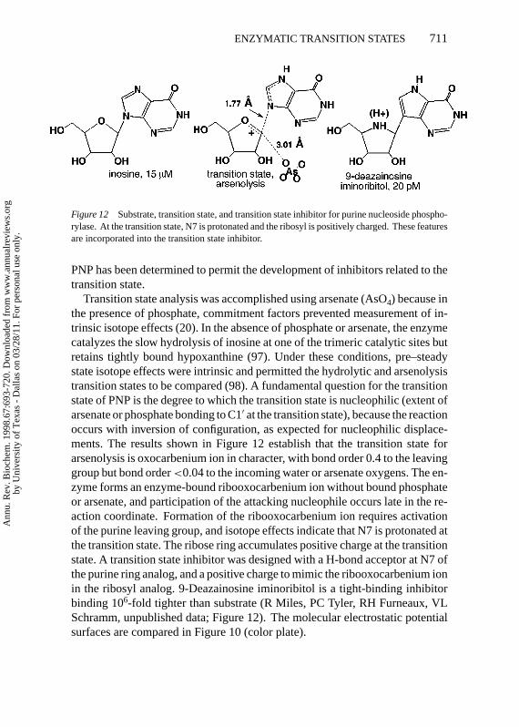
Figure 12 Substrate, transition state, and transition state inhibitor for purine nucleoside phospho-rylase. At the transition state, N7 is protonated and the ribosyl is positively charged. These featuresare incorporated into the transition state inhibitor.
PNP has been determined to permit the development of inhibitors related to thetransition state.
Transition state analysis was accomplished using arsenate (AsO4) because inthe presence of phosphate, commitment factors prevented measurement of in-trinsic isotope effects (20). In the absence of phosphate or arsenate, the enzymecatalyzes the slow hydrolysis of inosine at one of the trimeric catalytic sites butretains tightly bound hypoxanthine (97). Under these conditions, pre–steadystate isotope effects were intrinsic and permitted the hydrolytic and arsenolysistransition states to be compared (98). A fundamental question for the transitionstate of PNP is the degree to which the transition state is nucleophilic (extent ofarsenate or phosphate bonding to C1′ at the transition state), because the reactionoccurs with inversion of configuration, as expected for nucleophilic displace-ments. The results shown in Figure 12 establish that the transition state forarsenolysis is oxocarbenium ion in character, with bond order 0.4 to the leavinggroup but bond order<0.04 to the incoming water or arsenate oxygens. The en-zyme forms an enzyme-bound ribooxocarbenium ion without bound phosphateor arsenate, and participation of the attacking nucleophile occurs late in the re-action coordinate. Formation of the ribooxocarbenium ion requires activationof the purine leaving group, and isotope effects indicate that N7 is protonated atthe transition state. The ribose ring accumulates positive charge at the transitionstate. A transition state inhibitor was designed with a H-bond acceptor at N7 ofthe purine ring analog, and a positive charge to mimic the ribooxocarbenium ionin the ribosyl analog. 9-Deazainosine iminoribitol is a tight-binding inhibitorbinding 106-fold tighter than substrate (R Miles, PC Tyler, RH Furneaux, VLSchramm, unpublished data; Figure 12). The molecular electrostatic potentialsurfaces are compared in Figure 10 (color plate).
Ann
u. R
ev. B
ioch
em. 1
998.
67:6
93-7
20. D
ownl
oade
d fr
om w
ww
.ann
ualr
evie
ws.
org
by U
nive
rsity
of
Tex
as -
Dal
las
on 0
3/28
/11.
For
per
sona
l use
onl
y.
P1: DPI/ary P2: RPK/ARK/plb QC: ARK
May 12, 1998 14:26 Annual Reviews AR057-22
712 SCHRAMM
Orotate Phosphoribosyl TransferasePhosphoribosyl transferases were investigated in the pioneering kinetic isotopeeffects measured by Goiten et al in 1978 (99). The measurement of kinetic iso-tope effects from both [1-3H]- and [1-14C]5-phosphoribosyl-1-pyrophosphatesuggested a pattern consistent with a dissociative mechanism, with pyrophos-phate departure being well developed prior to the attack of the nitrogen from thepurine. However, many of the phosphoribosyl transferases demonstrated sub-stantial commitment factors, causing the kinetic isotope effects to be obscured.
Orotate phosphoribosyl transferase (OPRT), similar to other enzymes in thisgroup, does not give useful kinetic isotope effects under normal assay condi-tions. However, the use of phosphonoacetic acid as a pyrophosphate analogmade it possible to study the conversion of orotidine 5′-phosphate to orotate and5-phosphoribosyl-1-phosphonoacetic acid under conditions that gave intrinsickinetic isotope effects (100). In reactions with a concerted chemical step, thetransition state structure can be determined from kinetic isotope effect measure-ments in either the forward or reverse directions because the same transitionstate defines both reactions. Differing chemical reactivity with slow substratesmay result in a transition state that differs from the normal reaction. However,these differences are expected to be small because the template of the catalyticsite is expected to favor a specific transition state geometry for the reaction.
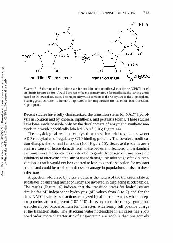
At the transition state for OPRT, the N1-C1′ bond has a residual bond orderof 0.28 and the attacking oxygen from the pyrophosphate analog is just begin-ning to participate with a bond order of<0.02 (Figure 13). The kinetic isotopeeffect from [2′-3H]orotidine 5′-phosphate is relatively large at 14%, establish-ing that the dihedral angle to the leaving group is nearly eclipsed. The remote5′-3H isotope effect is significant at 2.8%, confirming that ribosyl-transferasescommonly use geometric distortion from the ribosyl 5′-position to form thetransition state geometry. Features of this transition state require a ribooxocar-benium ion structure. The X-ray crystal structure of the enzyme has been solvedwith orotidine 5′-monophosphate or 5-phosphoribosyl-1-pyrophosphate at thecatalytic site (101, 102). Although the structure is not in a transition state con-formation, Arg156 is shown to interact with the 4-carbonyl group of orotic acidand can be proposed to activate the orotic acid leaving group.
NAD+ and the ADP-Ribosylating Toxins: Cholera,Diphtheria, and PertussisEarly investigations of the hydrolysis of nicotinamide from NAD+ and NMN+
in solution and by NAD+hydrolases used [1′-2H]- in NAD+or NMN+as the iso-topic probes (103–104). Based on the cases where isotope effects were observed,the transition states were characterized as dissociative and electropositive.
Ann
u. R
ev. B
ioch
em. 1
998.
67:6
93-7
20. D
ownl
oade
d fr
om w
ww
.ann
ualr
evie
ws.
org
by U
nive
rsity
of
Tex
as -
Dal
las
on 0
3/28
/11.
For
per
sona
l use
onl
y.
P1: DPI/ary P2: RPK/ARK/plb QC: ARK
May 12, 1998 14:26 Annual Reviews AR057-22
ENZYMATIC TRANSITION STATES 713
Figure 13 Substrate and transition state for orotidine phosphoribosyl transferase (OPRT) basedon kinetic isotope effects. Arg156 appears to be the primary group for stabilizing the leaving groupbased on the crystal structure. The major enzymatic contacts to the ribosyl are to the 5′-phosphate.Leaving group activation is therefore implicated in forming the transition state from bound orotidine5′-phosphate.
Recent studies have fully characterized the transition states for NAD+ hydrol-ysis in solution and by cholera, diphtheria, and pertussis toxins. These studieshave been made possible only by the development of enzymatic synthetic me-thods to provide specifically labeled NAD+ (105; Figure 14).
The physiological reaction catalyzed by these bacterial toxins is covalentADP-ribosylation of regulatory GTP-binding proteins. The covalent modifica-tion disrupts the normal functions (106; Figure 15). Because the toxins are aprimary cause of tissue damage from these bacterial infections, understandingthe transition state structures is intended to guide the design of transition stateinhibitors to intervene at the site of tissue damage. An advantage of toxin inter-vention is that it would not be expected to lead to genetic selection for resistantstrains and could be used to limit tissue damage in populations at risk in theseinfections.
A question addressed by these studies is the nature of the transition state assubstrates of differing nucleophilicity are involved in displacing nicotinamide.The results (Figure 16) indicate that the transition states for hydrolysis aresimilar for pH-independent hydrolysis (pH values from 3 to 7) and for theslow NAD+ hydrolysis reactions catalyzed by all three enzymes when accep-tor proteins are not present (107–110). In every case the ribosyl group haswell-developed oxocarbenium ion character, with nearly full positive chargeat the transition state. The attacking water nucleophile in all cases has a lowbond order, more characteristic of a “spectator” nucleophile than one actively
Ann
u. R
ev. B
ioch
em. 1
998.
67:6
93-7
20. D
ownl
oade
d fr
om w
ww
.ann
ualr
evie
ws.
org
by U
nive
rsity
of
Tex
as -
Dal
las
on 0
3/28
/11.
For
per
sona
l use
onl
y.

P1: DPI/ary P2: RPK/ARK/plb QC: ARK
May 12, 1998 14:26 Annual Reviews AR057-22
714 SCHRAMM
Figure 14 Enzymatic synthesis of specifically labeled NAD+. Enzymes not identified in Figure 6include 6= nicotinate phosphoribosyl transferase, 7= NAD+ pyrophosphorylase, 8= NAD+synthetase, and 11= ribokinase.
participating in bond formation. However, the magnitude of the kinetic iso-tope effects requires a small degree of nucleophilic participation, less than0.01 bond order at the transition state. The results establish that the waternucleophile does not play a role in forming the transition state, as occurs innucleophilic displacement reactions. Rather, the enzymes catalyze formationof the ribooxocarbenium ion, and the neighboring water molecule then reactswith the electron-deficient center.
Transition state structures have been solved for pertussis toxin catalyzing theADP-ribosylation of the C-terminal 20–amino acid peptide from Giα3 and theG-protein Giα1 (111, 112; Figure 16). In both cases, the ADP-ribosyl acceptoris a specific Cys sulfhydryl. The reaction for peptide ADP-ribosylation gave in-trinsic kinetic isotope effects, and the transition state structure shows increasednucleophilic participation by the thiolate anion (Figure 16). Electrostatic po-tential surfaces of the transition state for ADP-ribosylation of a thiolate anionare shown in Figure 17 (color plate). In NAD+ the positive charge is distributednearly equally in the ribosyl and nicotinamide, giving rise to an elongated, weakN-ribosidic bond. At the transition state the leaving group is neutral and the fullpositive charge has migrated into the ribose. Attack by the thiolate anion neutral-izes the oxocarbenium ion charge to form the ion-paired transition state (111).
Ann
u. R
ev. B
ioch
em. 1
998.
67:6
93-7
20. D
ownl
oade
d fr
om w
ww
.ann
ualr
evie
ws.
org
by U
nive
rsity
of
Tex
as -
Dal
las
on 0
3/28
/11.
For
per
sona
l use
onl
y.
P1: DPI/ary P2: RPK/ARK/plb QC: ARK
May 12, 1998 14:26 Annual Reviews AR057-22
ENZYMATIC TRANSITION STATES 715
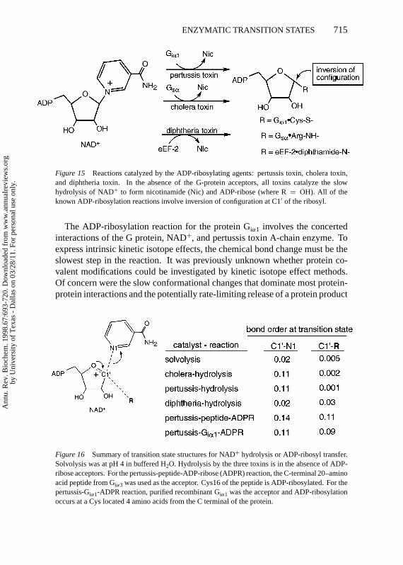
Figure 15 Reactions catalyzed by the ADP-ribosylating agents: pertussis toxin, cholera toxin,and diphtheria toxin. In the absence of the G-protein acceptors, all toxins catalyze the slowhydrolysis of NAD+ to form nicotinamide (Nic) and ADP-ribose (where R= OH). All of theknown ADP-ribosylation reactions involve inversion of configuration at C1′ of the ribosyl.
The ADP-ribosylation reaction for the protein Giα1 involves the concertedinteractions of the G protein, NAD+, and pertussis toxin A-chain enzyme. Toexpress intrinsic kinetic isotope effects, the chemical bond change must be theslowest step in the reaction. It was previously unknown whether protein co-valent modifications could be investigated by kinetic isotope effect methods.Of concern were the slow conformational changes that dominate most protein-protein interactions and the potentially rate-limiting release of a protein product
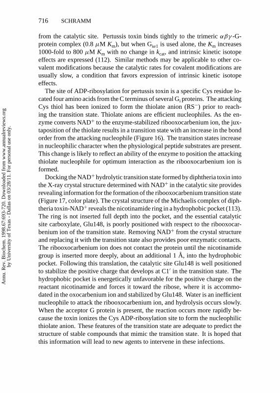
Figure 16 Summary of transition state structures for NAD+ hydrolysis or ADP-ribosyl transfer.Solvolysis was at pH 4 in buffered H2O. Hydrolysis by the three toxins is in the absence of ADP-ribose acceptors. For the pertussis-peptide-ADP-ribose (ADPR) reaction, the C-terminal 20–aminoacid peptide from Giα3 was used as the acceptor. Cys16 of the peptide is ADP-ribosylated. For thepertussis-Giα1-ADPR reaction, purified recombinant Giα1 was the acceptor and ADP-ribosylationoccurs at a Cys located 4 amino acids from the C terminal of the protein.
Ann
u. R
ev. B
ioch
em. 1
998.
67:6
93-7
20. D
ownl
oade
d fr
om w
ww
.ann
ualr
evie
ws.
org
by U
nive
rsity
of
Tex
as -
Dal
las
on 0
3/28
/11.
For
per
sona
l use
onl
y.
P1: DPI/ary P2: RPK/ARK/plb QC: ARK
May 12, 1998 14:26 Annual Reviews AR057-22
716 SCHRAMM
from the catalytic site. Pertussis toxin binds tightly to the trimericαβγ -G-protein complex (0.8µM Km), but when Giα1 is used alone, theKm increases1000-fold to 800µM Km with no change inkcat, and intrinsic kinetic isotopeeffects are expressed (112). Similar methods may be applicable to other co-valent modifications because the catalytic rates for covalent modifications areusually slow, a condition that favors expression of intrinsic kinetic isotopeeffects.
The site of ADP-ribosylation for pertussis toxin is a specific Cys residue lo-cated four amino acids from the C terminus of several Gi proteins. The attackingCys thiol has been ionized to form the thiolate anion (RS−) prior to reach-ing the transition state. Thiolate anions are efficient nucleophiles. As the en-zyme converts NAD+ to the enzyme-stabilized ribooxocarbenium ion, the jux-taposition of the thiolate results in a transition state with an increase in the bondorder from the attacking nucleophile (Figure 16). The transition states increasein nucleophilic character when the physiological peptide substrates are present.This change is likely to reflect an ability of the enzyme to position the attackingthiolate nucleophile for optimum interaction as the ribooxocarbenium ion isformed.
Docking the NAD+hydrolytic transition state formed by diphtheria toxin intothe X-ray crystal structure determined with NAD+ in the catalytic site providesrevealing information for the formation of the ribooxocarbenium transition state(Figure 17, color plate). The crystal structure of the Michaelis complex of diph-theria toxin-NAD+ reveals the nicotinamide ring in a hydrophobic pocket (113).The ring is not inserted full depth into the pocket, and the essential catalyticsite carboxylate, Glu148, is poorly positioned with respect to the ribooxocar-benium ion of the transition state. Removing NAD+ from the crystal structureand replacing it with the transition state also provides poor enzymatic contacts.The ribooxocarbenium ion does not contact the protein until the nicotinamidegroup is inserted more deeply, about an additional 1A, into the hydrophobicpocket. Following this translation, the catalytic site Glu148 is well positionedto stabilize the positive charge that develops at C1′ in the transition state. Thehydrophobic pocket is energetically unfavorable for the positive charge on thereactant nicotinamide and forces it toward the ribose, where it is accommo-dated in the oxocarbenium ion and stabilized by Glu148. Water is an inefficientnucleophile to attack the ribooxocarbenium ion, and hydrolysis occurs slowly.When the acceptor G protein is present, the reaction occurs more rapidly be-cause the toxin ionizes the Cys ADP-ribosylation site to form the nucleophilicthiolate anion. These features of the transition state are adequate to predict thestructure of stable compounds that mimic the transition state. It is hoped thatthis information will lead to new agents to intervene in these infections.
Ann
u. R
ev. B
ioch
em. 1
998.
67:6
93-7
20. D
ownl
oade
d fr
om w
ww
.ann
ualr
evie
ws.
org
by U
nive
rsity
of
Tex
as -
Dal
las
on 0
3/28
/11.
For
per
sona
l use
onl
y.
P1: DPI/ary P2: RPK/ARK/plb QC: ARK
May 12, 1998 14:26 Annual Reviews AR057-22
ENZYMATIC TRANSITION STATES 717
INHIBITOR PREDICTION FROM TRANSITIONSTATE INHIBITORS
Similarity MeasuresA goal of transition state investigation has been to use the knowledge to de-sign transition state inhibitors and to predict inhibitory strength. Transitionstates stabilized by enzymes bind strongly by virtue of their geometric andelectrostatic fit to the enzyme conformation that stabilizes the unstable elec-tronic structure. Geometric similarity of a transition state inhibitor is necessaryto provide volumetric access to the catalytic site and to establish the correctdistance to the enzymatic contacts. Electronic similarity is essential to makethe correct H-bond, ionic, and hydrophobic contacts found in the transitionstate interaction. If every detail of the transition state structure could be repro-duced in a stably bonded transition state analog, it would approach the transitionstate binding energy of 1010–1015 times tighter than the substrate. Despite theproblem of exact matching, striking similarity is apparent in molecular elec-trostatic potential surfaces of transition state structures and those of transitionstate inhibitors (Figure 10, color plate). It is possible to establish a predictiverelationship between the binding affinity of an inhibitor and its similarity to theenzymatic transition state.
Prediction of Inhibitory StrengthBagdassarian et al (114, 115) examined this relationship using three enzymes forwhich the transition state structures had been characterized by kinetic isotopeeffects. AMP deaminase, adenosine deaminase, and AMP nucleosidase wereconsidered by comparing the geometric and molecular electrostatic potentialsurfaces of the experimentally determined transition states with the substratesand a series of inhibitors exhibiting classic competitive inhibition and slow-onset, tight-binding inhibition (e.g. 78). An algorithm was used that considersthe electrostatic potential and its spatial distribution at the van der Waals surfaceof test inhibitors. These parameters were compared to those for the transitionstate structure, and an electronic similarity index (Se) was assigned, with avalue of 1.0 indicating a perfect match for molecular electrostatic potential (amolecule compared to itself gives Se = 1.0). Comparison of the transition stateSewith those from substrate and inhibitors that bind better than substrate gave astrong correlation in electrostatic similarity and binding energy (1G/RT) overa large range of binding energies encompassing the substrate and the transitionstate. Approaches that match the features of experimentally determined tran-sition states to those of proposed inhibitors have substantial potential for theprediction of new inhibitors prior to synthetic efforts.
Ann
u. R
ev. B
ioch
em. 1
998.
67:6
93-7
20. D
ownl
oade
d fr
om w
ww
.ann
ualr
evie
ws.
org
by U
nive
rsity
of
Tex
as -
Dal
las
on 0
3/28
/11.
For
per
sona
l use
onl
y.
P1: DPI/ary P2: RPK/ARK/plb QC: ARK
May 12, 1998 14:26 Annual Reviews AR057-22
718 SCHRAMM
CONCLUSIONS
Experimental access to enzymatic transition state structures has provided novelinformation about the nature of catalysis. It is reasonable to anticipate that thecontinued application of this information will permit the design and synthesisof powerful transition state inhibitors that incorporate desired pharmacologicproperties. The ability to solve transition state structures has been made pos-sible only by the development of kinetic methods to establish the rate-limitingsteps in reactions, synthetic methods for labeled compounds, and computationalchemistry and theory to quantitate kinetic isotope effects and to predict electrondistribution in partially bonded molecules. The next advance in this area willsee these technologies applied to the development of new inhibitors.
ACKNOWLEDGMENTS
Preparation of this work and research in this laboratory have been made possibleby the support of research and training grants from the National Institutes ofHealth, the United States Army, The G. Harold and Leila Y. Mathers CharitableFoundation, the American Cancer Society, and the National Sciences andEngineering Research Council (Canada). I thank Dr. Paul Berti for assistancein figure preparation.
Visit the Annual Reviews home pageathttp://www.AnnualReviews.org.
Literature Cited
1. Radzicka A, Wolfenden R. 1995.Science267:90–93
2. Glasstone S, Laidler KJ, Eyring HK.1941.The Theory of Rate Processes.NewYork: McGraw-Hill
3. Pauling L. 1948.Am. Sci.36:50–584. Wolfenden R. 1972.Acc. Chem. Res.
5:10–185. Wolfenden R. 1976.Annu. Rev. Biophys.
Bioeng.5:271–3066. Morrison JF, Walsh CT. 1988.Adv. Enzy-
mol. Relat. Areas Mol. Biol.61:201–3017. Radzicka A, Wolfenden R. 1995.Methods
Enzymol.249:284–3128. Bigeleisen J, Mayer MG. 1947.J. Chem.
Phys.15:261–679. Bigeleisen J, Wolfsberg M. 1958.Adv.
Chem. Phys.1:15–7610. Streitwiser A Jr, Jagow RH, Fahey RC,
Suzuki S. 1958.J. Am. Chem. Soc.80:2326–32
11. Cleland WW, O’Leary MH, NorthropDB, eds. 1977.Isotope Effects on Enzyme-
Catalyzed Reactions. Baltimore, MD:Univ. Park Press
12. Gandour RD, Schowen RL, eds. 1978.Transition States of Biochemical Pro-cesses. New York: Plenum
13. Northrop DB. 1981.Annu. Rev. Biochem.50:103–31
14. Cook PF, Cleland WW. 1981.Biochem-istry 20:1790–96
15. Cook PF, Oppenheimer NJ, Cleland WW.1981.Biochemistry20:1817–25
16. Cleland WW. 1982.Methods Enzymol.87:625–41
17. Scharschmidt M, Fisher MA, ClelandWW. 1984.Biochemistry23:5471–78
18. Bolin JT, Filman DJ, Matthews DA, Ham-lin RC, Kraut J. 1982.J. Biol. Chem.257:13650–62
19. Kimble E, Hadala J, Ludewig R, Peters P,Greenberg G, et al. 1995.Inflamm. Res.44:S181–82
20. Kline PC, Schramm VL. 1993.Biochem-istry 32:13212–19
Ann
u. R
ev. B
ioch
em. 1
998.
67:6
93-7
20. D
ownl
oade
d fr
om w
ww
.ann
ualr
evie
ws.
org
by U
nive
rsity
of
Tex
as -
Dal
las
on 0
3/28
/11.
For
per
sona
l use
onl
y.
P1: DPI/ary P2: RPK/ARK/plb QC: ARK
May 12, 1998 14:26 Annual Reviews AR057-22
ENZYMATIC TRANSITION STATES 719
21. Shan S-O, Herschlag D. 1996.Proc. Natl.Acad. Sci. USA93:14474–79
22. Cleland WW, Kreevoy MM. 1994.Sci-ence264:1887–90
23. Truhlar DG, Hase WL, Hynes JT. 1983.J. Phys. Chem.87:2664–82
24. Albery WJ. 1993.Adv. Phys. Org. Chem.28:139–70
25. Kurz LC, Weitkamp E, Frieden C. 1987.Biochemistry26:3027–32
26. Frick L, Yang C, Marquez VE, WolfendenR. 1989.Biochemistry28:9423–30
27. Wilson DK, Rudolph FB, Quiocho FA.1991.Science252:1278–84
28. Warshel A. 1991.Computer Modeling ofChemical Reactions in Enzymes and So-lutions. New York: Wiley & Sons
29. Cleland WW. 1977.Adv. Enzymol. Relat.Areas Mol. Biol.45:273–387
30. Parkin DW, Schramm VL. 1995.Bio-chemistry34:13961–66
31. Albery WJ, Knowles JR. 1977.Angew.Chem.16:285–93
32. Schowen RL. 1978. InTransition States ofBiochemical Processes, ed. RD Gandour,RL Schowen, pp. 77–114. New York:Plenum
33. Melander L, Saunders WJ Jr. 1980.Re-action Rates of Isotopic Molecules. NewYork: Wiley & Sons
34. Cleland WW. 1995.Methods Enzymol.249:341–73
35. Jencks WP. 1987. InCatalysis in Chem-istry and Enzymology, pp. 170–82. NewYork: Dover
36. Hammond GS. 1955.J. Am. Chem. Soc.77:334–38
37. Agarwal RP, Spector T, Parks RE Jr. 1977.Biochem. Pharmacol.26:359–67
38. Bachovin WW, Wong WYL, Farr-JonesS, Shenvi AB, Kettner CA. 1988.Bio-chemistry27:12839– 46
39. Rodgers J, Femec DA, Schowen RL.1982.J. Am. Chem. Soc.104:3263–68
40. Schramm VL, Horenstein BA, Kline PC.1994.J. Biol. Chem.269:18259–62
41. Huskey WP. 1991. See Ref. 116, pp. 37–72
42. Suhnel J, Schowen RL. 1991. See Ref.116, pp. 3–35
43. Cleland WW. 1987.Bioorg. Chem.15:282–302
44. Sunko DE, Szele I, Hehre WJ. 1977.J.Am. Chem. Soc.99:5000–4
45. Bennet AJ, Sinnott ML. 1986.J. Am.Chem. Soc.108:7287–94
46. Northrop DB. 1975.Biochemistry14:2644–51
47. Sims LB, Burton GW, Lewis DE. 1977.BEBOVIB-IV, QCPE No. 337. Blooming-
ton, IN. Quantum Chem. Program Exch.,Dep. Chem., Univ. Indiana
48. Sims LB, Lewis DE. 1984. InIsotopesin Organic Chemistry, ed. E Buncel, CCLee, 6:161–259. New York: Elsevier
49. Stewart JJP. 1989.Comput. Chem.10:209–20
50. Frisch MJ, Trucks GW, Schlegel HB, GillPMW, Johnson BG, et al. 1995.Gaussian94, Rev. C. 2, 1995. Pittsburgh, PA: Gaus-sian Inc.
51. Schramm VL. 1976.J. Biol. Chem.251:3417–24
52. Schramm VL. 1974.J. Biol. Chem.249:1729–36
53. Leung HB, Schramm VL. 1984.J. Biol.Chem.259:6972–78
54. Parkin DW, Leung HB, Schramm VL.1984.J. Biol. Chem.259:9411–17
55. Parkin DW, Schramm VL. 1987.Bio-chemistry26:913–20
56. Mentch F, Parkin DW, Schramm VL.1987.Biochemistry26:921–30
57. Srinivasan K, Konstantinidis A, SinnottML, Hall BG. 1993.Biochem. J.291:15–17
58. Pauling L. 1960.The Nature of the Chemi-cal Bond. Ithaca, NY: Cornell Univ. Press.3rd ed.
59. DeWolf WE Jr, Fullin FA, Schramm VL.1979.J. Biol. Chem.254:10868–75
60. Leung HB, Schramm VL. 1980.J. Biol.Chem.255:10867–74
61. Giranda VL, Berman HM, Schramm VL.1988.Biochemistry27:5813–18
62. Ehrlich JI, Schramm VL. 1994.Biochem-istry 33:8890–96
63. Leung HB, Schramm VL. 1981.J. Biol.Chem.256:12823–29
64. Parkin DW, Mentch F, Banks GA, Horen-stein BA, Schramm VL. 1991.Biochem-istry 30:921–30
65. Parry RJ, Minta A. 1982.J. Am. Chem.Soc.104:871–72
66. Markham GD, Hafner EW, Tabor CW, Ta-bor H. 1980.J. Biol Chem.255:9082–92
67. Larsen TM, Laughlin LT, Holden HM,Rayment I, Reed GH. 1994.Biochemistry33:6301–9
68. Markham GD, Parkin DW, Mentch F,Schramm VL. 1987.J. Biol. Chem.262:5609–15
69. Rose IW. 1980.Methods Enzymol.64:47–59
70. Carey FA, Sundberg RF. 1990.AdvancedOrganic Chemistry. Part A: Structureand Mechanism, pp. 579–83. New York:Plenum. 3rd ed.
71. Merkler DJ, Wali AS, Taylor J, Schramm
Ann
u. R
ev. B
ioch
em. 1
998.
67:6
93-7
20. D
ownl
oade
d fr
om w
ww
.ann
ualr
evie
ws.
org
by U
nive
rsity
of
Tex
as -
Dal
las
on 0
3/28
/11.
For
per
sona
l use
onl
y.
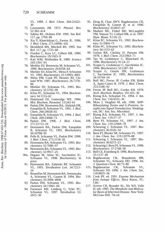
P1: DPI/ary P2: RPK/ARK/plb QC: ARK
May 12, 1998 14:26 Annual Reviews AR057-22
720 SCHRAMM
VL. 1989. J. Biol. Chem.264:21422–30
72. Lowenstein JM. 1972.Physiol. Rev.52:382–414
73. Sabina RL, Holmes EW. 1995. See Ref.117, pp. 1769–80
74. Xia Y, Khatchikian G, Zweier JL. 1996.J. Biol. Chem.271:10096–102
75. Hershfield MS, Mitchell BS. 1995. SeeRef. 117, pp. 1725–68
76. Frieden C, Kurz LC, Gilbert HR. 1980.Biochemistry19:5303–9
77. Kati WM, Wolfenden R. 1989.Science243:1591–93
78. Merkler DJ, Brenowitz M, Schramm VL.1990.Biochemistry29:8358–64
79. Merkler DJ, Kline PC, Weiss P, SchrammVL. 1993.Biochemistry32:12993–3001
80. Weiss PM, Cook PF, Hermes JD, Cle-land WW. 1987.Biochemistry26:7378–84
81. Merkler DJ, Schramm VL. 1993.Bio-chemistry32:5792–99
82. Kline PC, Schramm VL. 1994.Biochem-istry 34:1153–62
83. Hammond DJ, Gutteridge WE. 1984.Mol. Biochem. Parasitol.13:243–61
84. Parkin DW, Horenstein BA, Abdulah DR,Estupinan B, Schramm VL. 1991.J. Biol.Chem.266:20658–65
85. Estupinan B, Schramm VL. 1994.J. Biol.Chem.269:23068–73
86. Parkin DW. 1996. J. Biol. Chem.271:21713–19
87. Horenstein BA, Parkin DW, Estupi˜nanB, Schramm VL. 1991.Biochemistry30:10788–95
88. Pelle R, Schramm VL, Parkin DW. 1998.J. Biol. Chem.273:2118–26
89. Horenstein BA, Schramm VL. 1993.Bio-chemistry32:7089–97
90. Horenstein BA, Schramm VL. 1993.Bio-chemistry32:9917–25
90a. Degano M, Almo SC, Sacchettini JC,Schramm VL. 1998.Biochemistry. Inpress
91. Horenstein BA, Zabinski RF, SchrammVL. 1993. Tetrahedron Lett.34:7213–16
92. Boutellier M, Horenstein BA, SemenyakaA, Schramm VL, Ganem B. 1994.Bio-chemistry33:3994–4000
93. Parkin DW, Schramm VL. 1995.Bio-chemistry34:13961–66
94. Furneaux RH, Limberg G, Tyler PC,Schramm VL. 1997.Tetrahedron 53:2915–30
95. Deng H, Chan AWY, Bagdassarian CK,Estupinan B, Ganem B, et al. 1996.Biochemistry35:6037–47
96. Markert ML, Finkel BD, McLaughlinTM, Watson TJ, Collard HR, et al. 1997.Hum. Mutat.9:118–21
97. Kline PC, Schramm VL. 1992.Biochem-istry 31:5964–73
98. Kline PC, Schramm VL. 1995.Biochem-istry 34:1153–62
99. Goiten RK, Chelsky D, Parsons SM.1978.J. Biol. Chem.253:2963–71
100. Tao W, Grubmeyer C, Blanchard JS.1996.Biochemistry35:14–21
101. Scapin G, Grubmeyer C, Sacchettini JC.1994.Biochemistry33:1287–94
102. Scapin G, Ozturk DH, GrubmeyerC, Sacchettini JC. 1995.Biochemistry34:10744–54
103. Bull HG, Ferraz JP, Cordes EH, RibbiA, Apitz-Castro R. 1978.J. Biol. Chem.253:5186–92
104. Ferraz JP, Bull HG, Cordes EH. 1978.Arch. Biochem. Biophys.191:431–36
105. Rising KA, Schramm VL. 1994.J. Am.Chem. Soc.116:6531–36
106. Moss J, Vaughan M, eds. 1990.ADP-Ribosylating Toxins and G-Proteins. In-sights into Signal Transduction. Washing-ton, DC: Am. Soc. Microbiol.
107. Rising KA, Schramm VL. 1997.J. Am.Chem. Soc.119:27–37
108. Berti PJ, Schramm VL. 1997.J. Am.Chem. Soc.119:12069–78
109. Scheuring J, Schramm VL. 1997.Bio-chemistry36:4526–34
110. Berti PJ, Blanke SR, Schramm VL. 1997.J. Am. Chem. Soc.119:12079–88
111. Scheuring J, Schramm VL. 1997.Bio-chemistry36:8215–23
112. Scheuring J, Berti PJ, Schramm VL. 1998.Biochemistry.37:2748–58
113. Bell CE, Eisenberg D. 1996.Biochemistry35:1137–49
114. Bagdassarian CK, Braunheim BB,Schramm VL, Schwartz DD. 1996.Int.J. Quantum Chem.60:73–80
115. Bagdassarian CK, Schramm VL,Schwartz SD. 1996.J. Am. Chem. Soc.118:8825–36
116. Cook PF, ed. 1991.Enzyme Mechanismfrom Isotope Effects. Boca Raton, FL:CRC
117. Scriver CR, Beaudet AL, Sly WS, ValleD, eds. 1995.The Metabolic and Molecu-lar Basis of Inherited Disease. New York:McGraw-Hill. 7th ed.
Ann
u. R
ev. B
ioch
em. 1
998.
67:6
93-7
20. D
ownl
oade
d fr
om w
ww
.ann
ualr
evie
ws.
org
by U
nive
rsity
of
Tex
as -
Dal
las
on 0
3/28
/11.
For
per
sona
l use
onl
y.

Annual Review of Biochemistry Volume 67, 1998
CONTENTSAn Accidental Biochemist, Edwin G. Krebs 0HIV-1: Fifteen Proteins and an RNA, Alan D. Frankel and John A. T. Young 1
Sphingolipid Functions in Saccharomyces Cerevisiae : Comparison to Mammals, Robert C. Dickson 27
Transporters of Nucleotide Sugars, ATP, and Nucleotide Sulfate in the Endoplasmic Reticulum and Golgi Apparatus, Carlos B. Hirschberg, Phillips W. Robbins, and Claudia Abeijon
49
Ribonucleotide Reductases, A. Jordan and P. Reichard 71
Modified Oligonucleotides: Synthesis and Strategy for Users, Sandeep Verma and Fritz Eckstein 99
The Molecular Control of Circadian Behavioral Rhythms and Their Entrainment in Drosophila, Michael W. Young 135
Ribonuclease P: Unity and Diversity in a tRNA Processing Ribozyme, Daniel N. Frank and Norman R. Pace 153
Base Flipping, Richard J. Roberts and Xiaodong Cheng 181
The Caveolae Membrane System, Richard G. W. Anderson 199
How Cells Respond to Interferons, George R. Stark, Ian M. Kerr, Bryan R. G. Williams, Robert H. Silverman, and Robert D. Schreiber 227
Nucleocytoplasmic Transport: The Soluble Phase, Iain W. Mattaj and Ludwig Englmeier 265
Role of Small G Proteins in Yeast Cell Polarization and Wall Biosynthesis, Enrico Cabib, Jana Drgonová, and Tomás Drgon 307
RNA Localization in Development, Arash Bashirullah, Ramona L. Cooperstock, and Howard D. Lipshitz 335
Biochemistry and Genetics of von Willebrand Factor, J. Evan Sadler 395
The Ubiquitin System, Avram Hershko and Aaron Ciechanover 425Phosphoinositide Kinases, David A. Fruman, Rachel E. Meyers, and Lewis C. Cantley 481
The Green Fluorescent Protein, Roger Y. Tsien 509Alteration of Nucleosome Structure as a Mechanism of Transcriptional Regulation, J. L. Workman, and R. E. Kingston 545
Structure and Function in GroEL-Mediated Protein Folding, Paul B. Sigler, Zhaohui Xu, Hays S. Rye, Steven G. Burston, Wayne A. Fenton, and Arthur L. Horwich
581
Matrix Proteoglycans: From Molecular Design to Cellular Function, Renato V. Iozzo 609
G Protein Coupled Receptor Kinases, Julie A. Pitcher, Neil J. Freedman, and Robert J. Lefkowitz 653
Enzymatic Transition States and Transition State Analog Design, Vern L. Schramm 693
Ann
u. R
ev. B
ioch
em. 1
998.
67:6
93-7
20. D
ownl
oade
d fr
om w
ww
.ann
ualr
evie
ws.
org
by U
nive
rsity
of
Tex
as -
Dal
las
on 0
3/28
/11.
For
per
sona
l use
onl
y.

The DNA Replication Fork in Eukaryotic Cells, Shou Waga and Bruce Stillman 721
TGF-beta Signal Transduction, J. Massagué 753
Pathologic Conformations of Prion Proteins, Fred E. Cohen and Stanley B. Prusiner 793
The AMP-Activated/SNF1 Protein Kinase Subfamily: Metabolic Sensors of the Eukaryotic Cell?, D. Grahame Hardie, David Carling, and Marian Carlson
821
Ann
u. R
ev. B
ioch
em. 1
998.
67:6
93-7
20. D
ownl
oade
d fr
om w
ww
.ann
ualr
evie
ws.
org
by U
nive
rsity
of
Tex
as -
Dal
las
on 0
3/28
/11.
For
per
sona
l use
onl
y.