enzymes : mechanism and catalysis. enzymes do not change the equilibrium constant of a reaction...
TRANSCRIPT
Enzymes :Mechanism and Catalysis
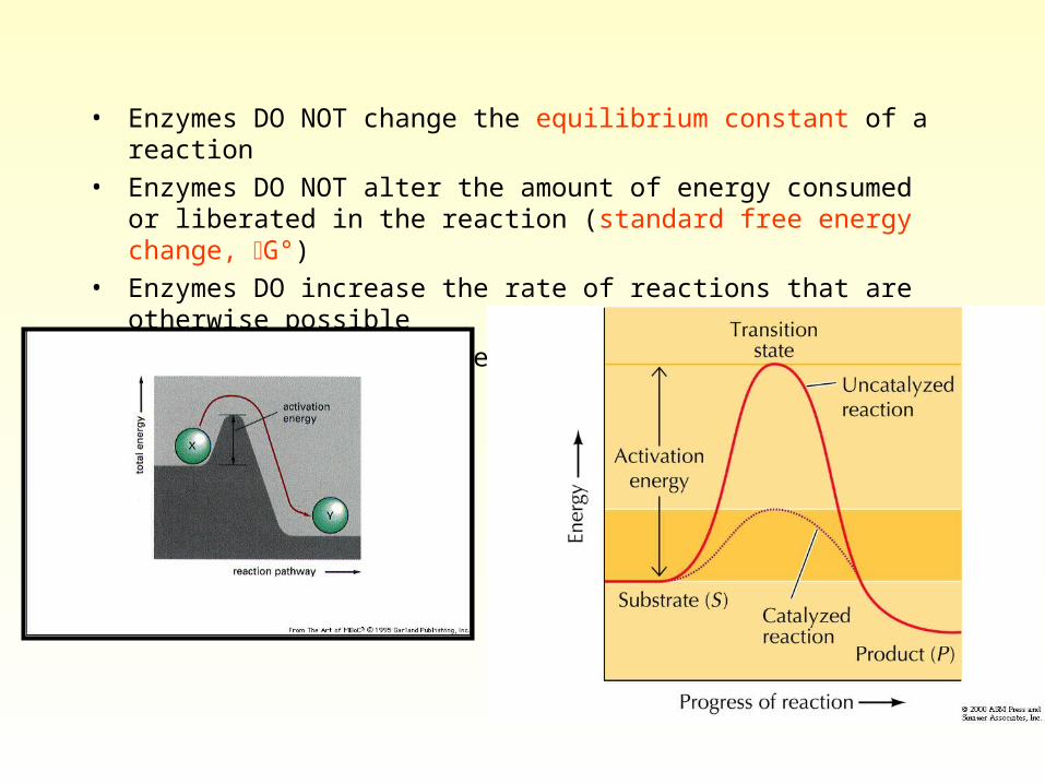
• Enzymes DO NOT change the equilibrium constant of a reaction
• Enzymes DO NOT alter the amount of energy consumed or liberated in the reaction (standard free energy change, G°)
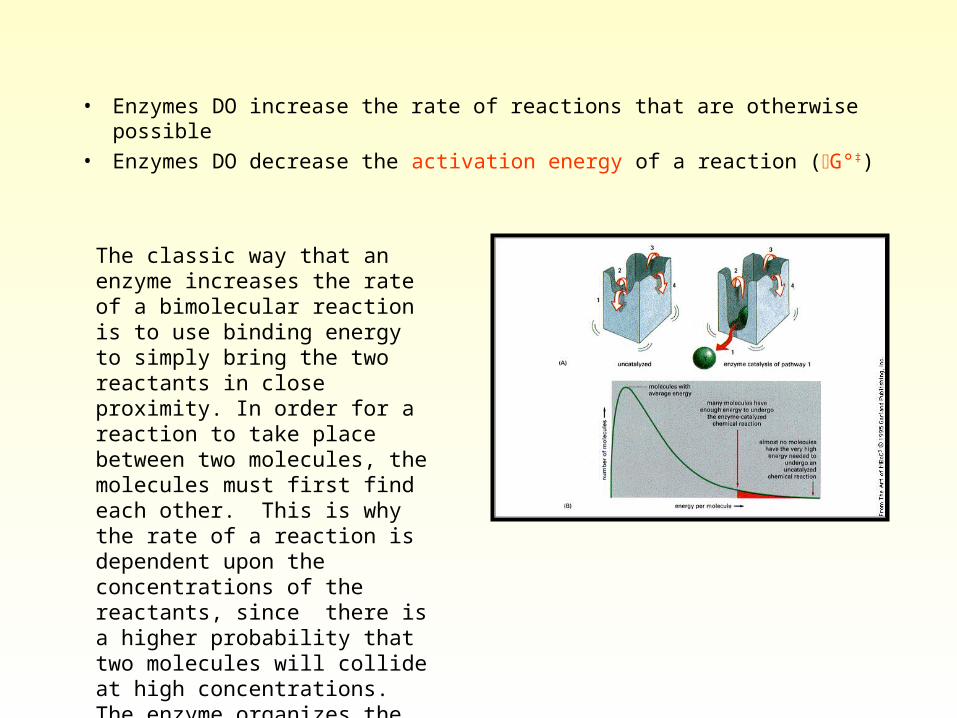
• Enzymes DO increase the rate of reactions that are otherwise possible
• Enzymes DO decrease the activation energy of a reaction (G°‡)
• Enzymes DO increase the rate of reactions that are otherwise possible
• Enzymes DO decrease the activation energy of a reaction (G°‡)
The classic way that an enzyme increases the rate of a bimolecular reaction is to use binding energy to simply bring the two reactants in close proximity. In order for a reaction to take place between two molecules, the molecules must first find each other. This is why the rate of a reaction is dependent upon the concentrations of the reactants, since there is a higher probability that two molecules will collide at high concentrations. The enzyme organizes the reaction at the active site, thereby reducing the cost in terms of ENTROPY.
• How do enzymes catalyze biochemical reactions?
– involves basic principles of organic chemistry
• What functional groups can be involved in catalysis?
– almost all alpha amino and carboxyl groups are tied up in peptide bonds
– R groups are involved in catalysis
• asp, glu
• his, lys
• ser, cys, tyr
• catalysis occurs when substrate is immobilized near these residues at the active site
General Acid-Base Catalysis
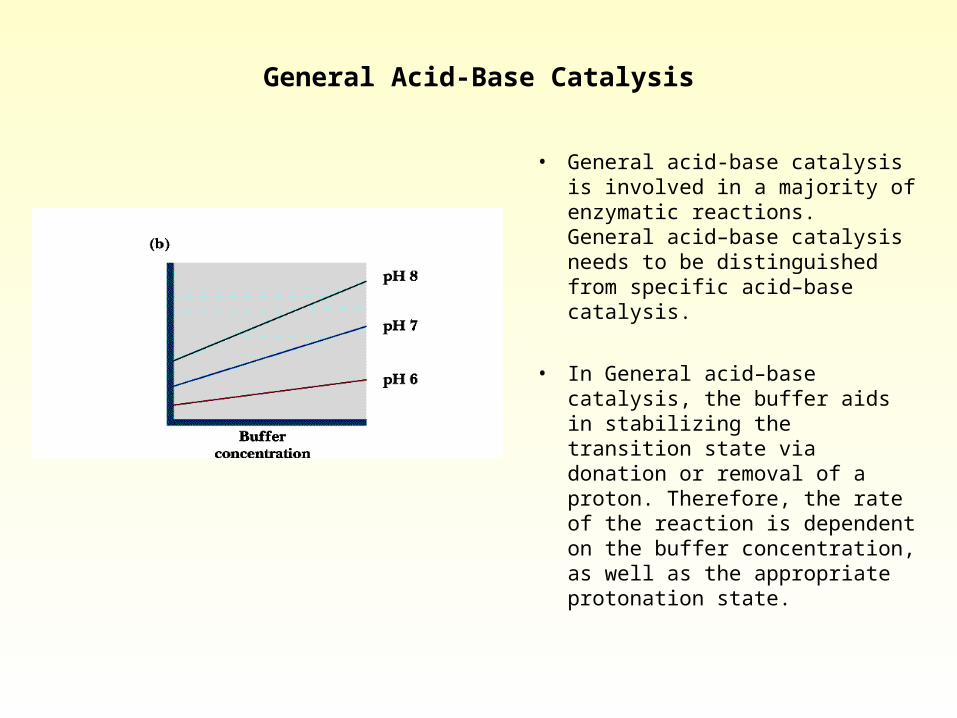
• General acid-base catalysis is involved in a majority of enzymatic reactions. General acid–base catalysis needs to be distinguished from specific acid–base catalysis.
• In General acid–base catalysis, the buffer aids in stabilizing the transition state via donation or removal of a proton. Therefore, the rate of the reaction is dependent on the buffer concentration, as well as the appropriate protonation state.
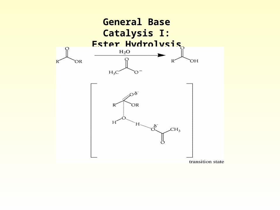
General Base Catalysis I:Ester Hydrolysis
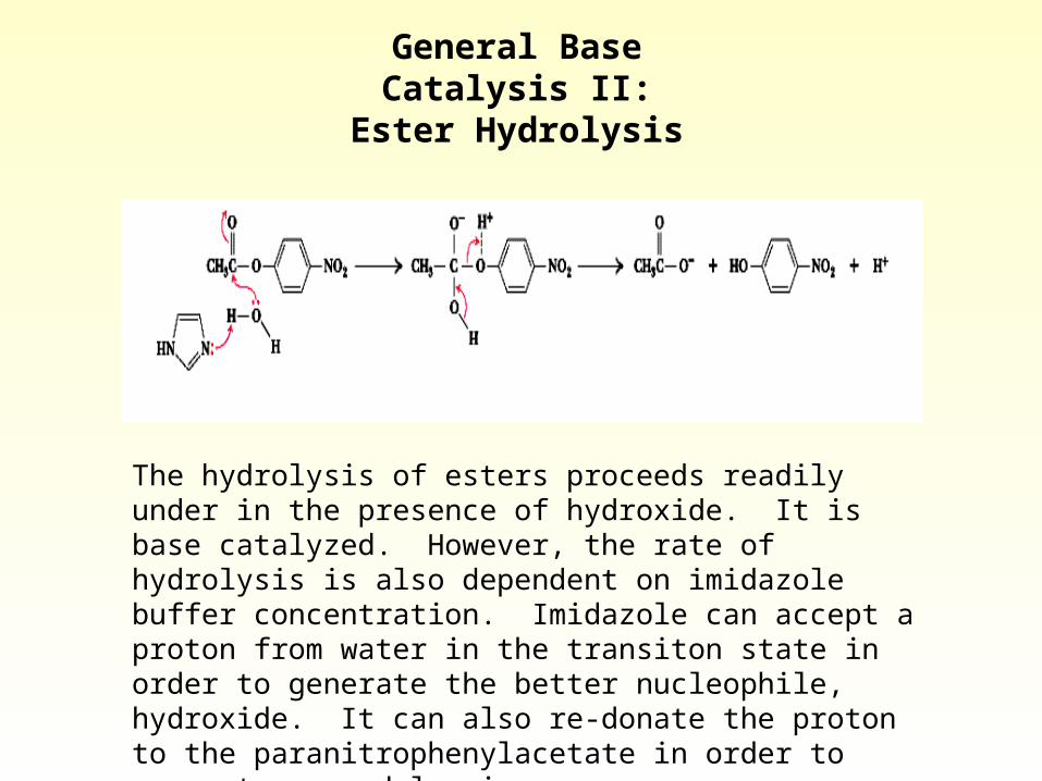
The hydrolysis of esters proceeds readily under in the presence of hydroxide. It is base catalyzed. However, the rate of hydrolysis is also dependent on imidazole buffer concentration. Imidazole can accept a proton from water in the transiton state in order to generate the better nucleophile, hydroxide. It can also re-donate the proton to the paranitrophenylacetate in order to generate a good leaving group.
General Base Catalysis II:Ester Hydrolysis
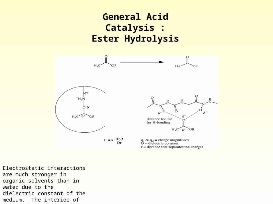
Electrostatic interactions are much stronger in organic solvents than in water due to the dielectric constant of the medium. The interior of enzymes have dielectric constants that are similar to hexane or chloroform
General Acid Catalysis :Ester Hydrolysis
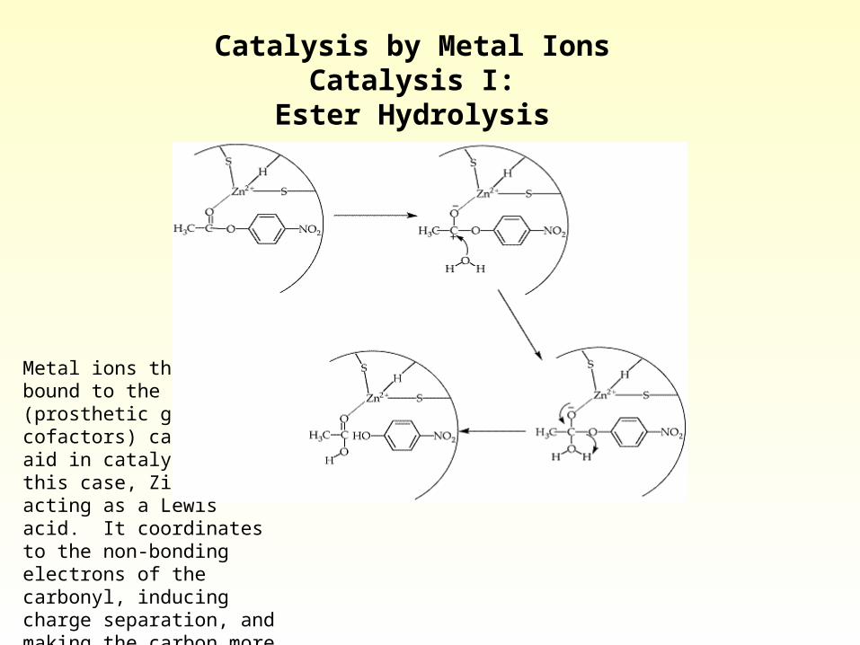
Metal ions that are bound to the protein (prosthetic groups or cofactors) can also aid in catalysis. In this case, Zinc is acting as a Lewis acid. It coordinates to the non-bonding electrons of the carbonyl, inducing charge separation, and making the carbon more electrophilic, or more susceptible to nucleophilic attack.
Catalysis by Metal Ions Catalysis I:Ester Hydrolysis
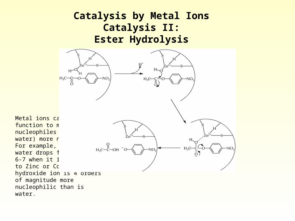
Metal ions can also function to make potential nucleophiles (such as water) more nucleophilic. For example, the pka of water drops from 15.7 to 6-7 when it is coordinated to Zinc or Cobalt. The hydroxide ion is 4 orders of magnitude more nucleophilic than is water.
Catalysis by Metal Ions Catalysis II:
Ester Hydrolysis
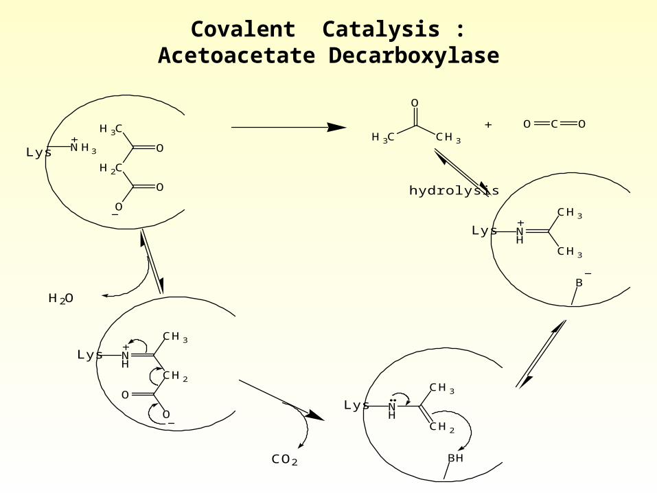
Covalent Catalysis :Acetoacetate Decarboxylase
O
H3C
H2C
O
O
O
H3C CH3
+ O C O
NH3Lys
Lys NH
CH3
CH2
O
O
H2O
Lys NH
CH3
CH2
BH
Lys NH
CH3
CH3
B
CO2
hydrolysis
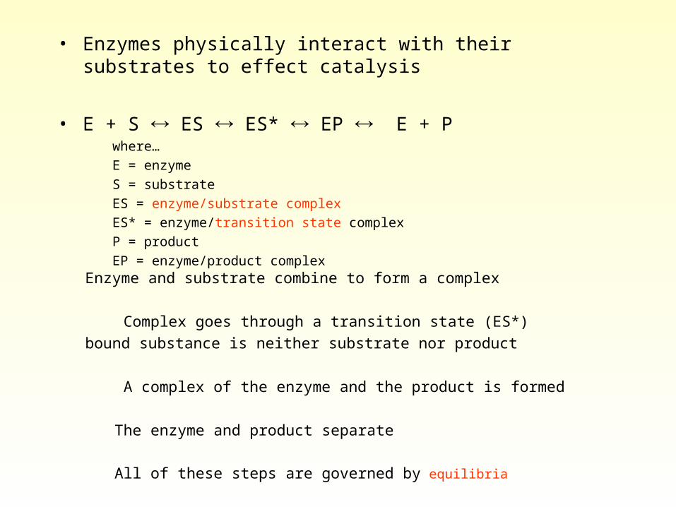
• Enzymes physically interact with their substrates to effect catalysis
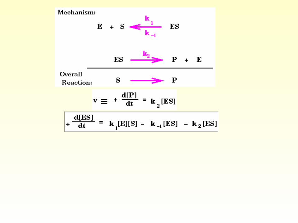
• E + S ES ES* EP E + P where…
E = enzyme
S = substrate
ES = enzyme/substrate complex
ES* = enzyme/transition state complex
P = product
EP = enzyme/product complex
Enzyme and substrate combine to form a complex
Complex goes through a transition state (ES*)
bound substance is neither substrate nor product
A complex of the enzyme and the product is formed
The enzyme and product separate
All of these steps are governed by equilibria
• Substrates bind to the enzyme’s active site– pocket in the enzyme
• Substrates bind in active site by– hydrogen bonding– hydrophobic interactions– ionic interactions
QuickTime™ and aVideo decompressor
are needed to see this picture.
QuickTime™ and aVideo decompressor
are needed to see this picture.
QuickTime™ and aVideo decompressor
are needed to see this picture.
QuickTime™ and aAnimation decompressor
are needed to see this picture.
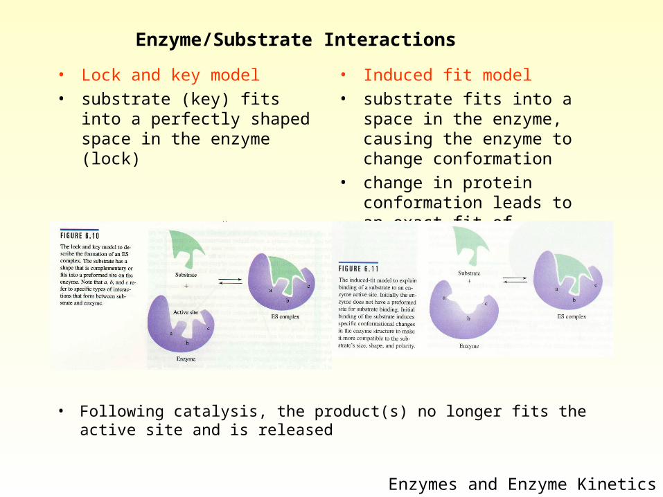
Enzyme/Substrate Interactions
• Lock and key model
• substrate (key) fits into a perfectly shaped space in the enzyme (lock)
Enzymes and Enzyme Kinetics
• Induced fit model
• substrate fits into a space in the enzyme, causing the enzyme to change conformation
• change in protein conformation leads to an exact fit of substrate with enzyme
• Following catalysis, the product(s) no longer fits the active site and is released
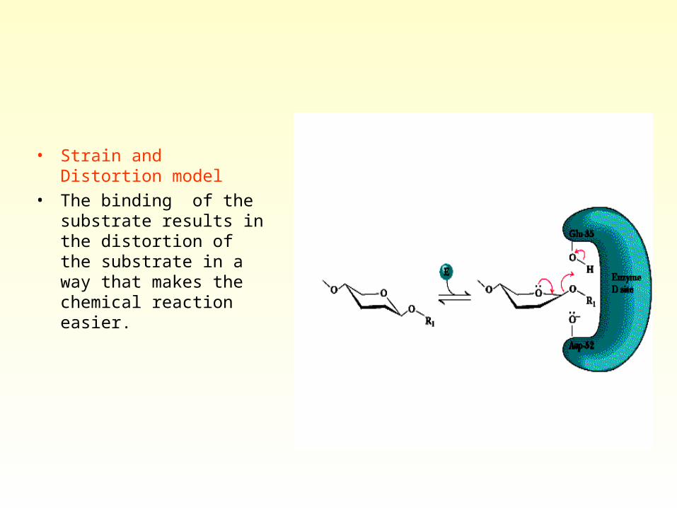
• Strain and Distortion model
• The binding of the substrate results in the distortion of the substrate in a way that makes the chemical reaction easier.
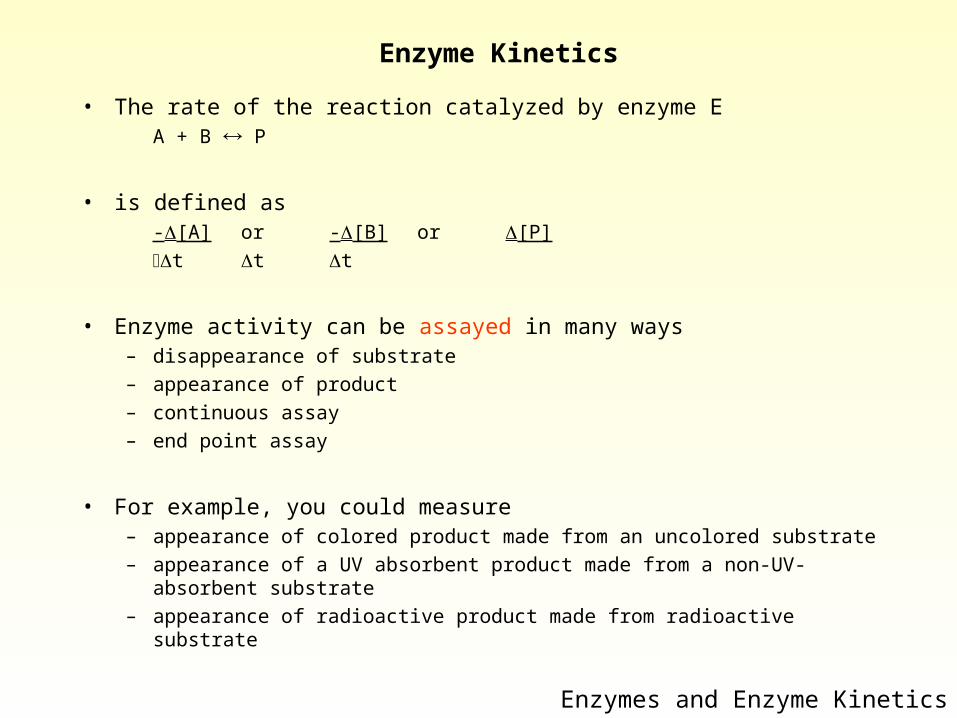
Enzyme Kinetics
• The rate of the reaction catalyzed by enzyme E A + B P
• is defined as -[A] or -[B] or [P] t t t
• Enzyme activity can be assayed in many ways– disappearance of substrate– appearance of product– continuous assay– end point assay
• For example, you could measure– appearance of colored product made from an uncolored substrate– appearance of a UV absorbent product made from a non-UV-absorbent
substrate– appearance of radioactive product made from radioactive substrate
Enzymes and Enzyme Kinetics
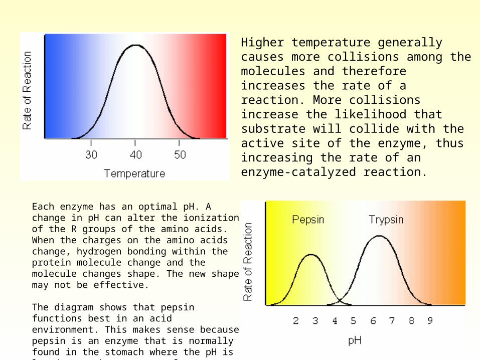
Higher temperature generally causes more collisions among the molecules and therefore increases the rate of a reaction. More collisions increase the likelihood that substrate will collide with the active site of the enzyme, thus increasing the rate of an enzyme-catalyzed reaction.
Each enzyme has an optimal pH. A change in pH can alter the ionization of the R groups of the amino acids. When the charges on the amino acids change, hydrogen bonding within the protein molecule change and the molecule changes shape. The new shape may not be effective.
The diagram shows that pepsin functions best in an acid environment. This makes sense because pepsin is an enzyme that is normally found in the stomach where the pH is low due to the presence of hydrochloric acid. Trypsin is found in the duodenum, and therefore, its optimum pH is in the neutral range to match the pH of the duodenum.
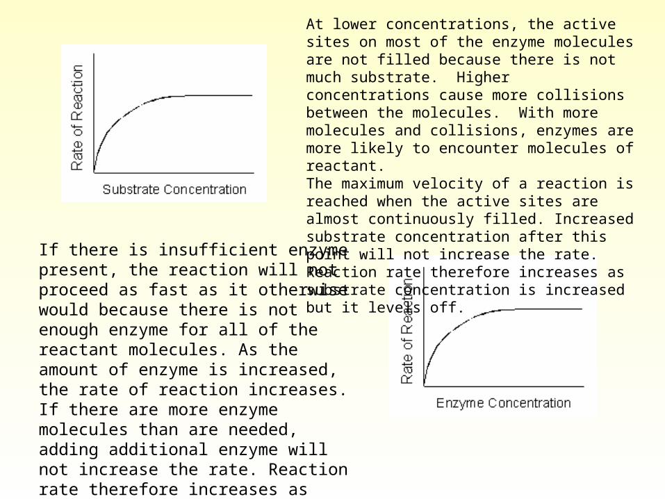
At lower concentrations, the active sites on most of the enzyme molecules are not filled because there is not much substrate. Higher concentrations cause more collisions between the molecules. With more molecules and collisions, enzymes are more likely to encounter molecules of reactant.The maximum velocity of a reaction is reached when the active sites are almost continuously filled. Increased substrate concentration after this point will not increase the rate. Reaction rate therefore increases as substrate concentration is increased but it levels off.
If there is insufficient enzyme present, the reaction will not proceed as fast as it otherwise would because there is not enough enzyme for all of the reactant molecules. As the amount of enzyme is increased, the rate of reaction increases. If there are more enzyme molecules than are needed, adding additional enzyme will not increase the rate. Reaction rate therefore increases as enzyme concentration increases but then it levels off.
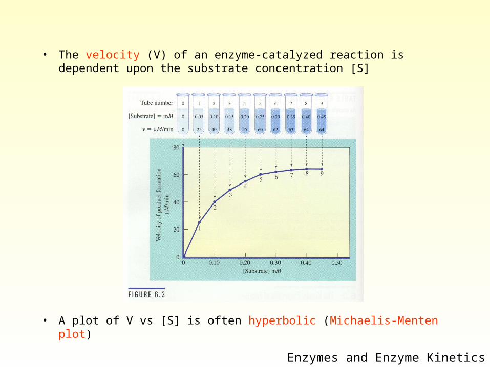
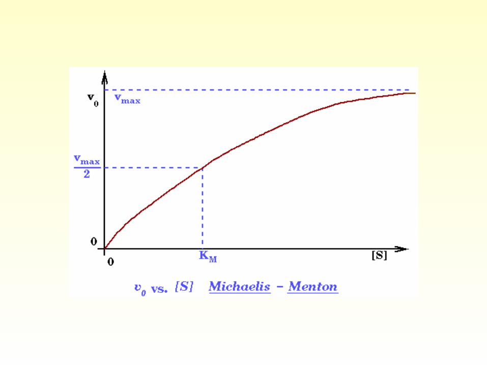
• The velocity (V) of an enzyme-catalyzed reaction is dependent upon the substrate concentration [S]
• A plot of V vs [S] is often hyperbolic (Michaelis-Menten plot)
Enzymes and Enzyme Kinetics
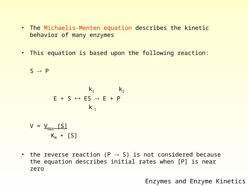
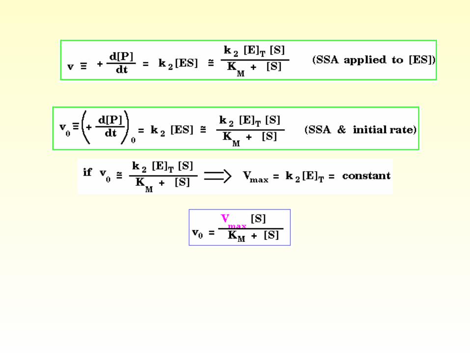
• The Michaelis-Menten equation describes the kinetic behavior of many enzymes
• This equation is based upon the following reaction:
S P
k1 k2
E + S ES E + P
k-1
V = Vmax [S]
KM + [S]
• the reverse reaction (P S) is not considered because the equation describes initial rates when [P] is near zero
Enzymes and Enzyme Kinetics
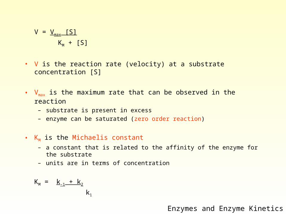
V = Vmax [S]
KM + [S]
• V is the reaction rate (velocity) at a substrate concentration [S]
• Vmax is the maximum rate that can be observed in the reaction
– substrate is present in excess– enzyme can be saturated (zero order reaction)
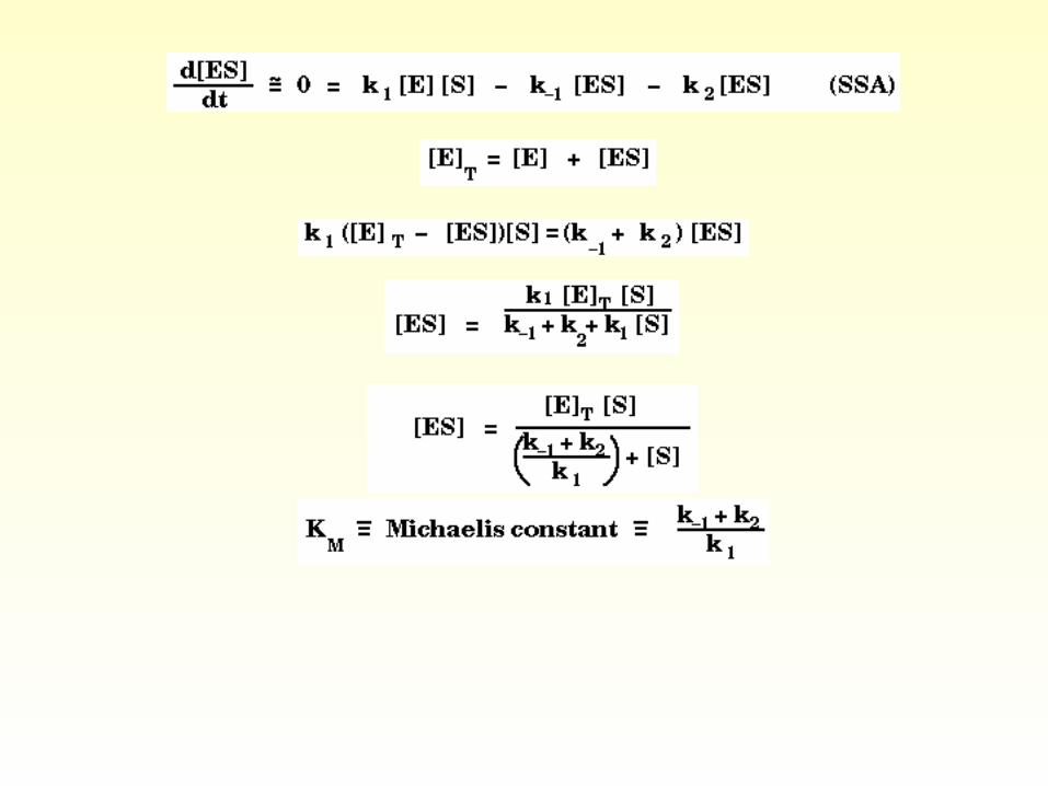
• KM is the Michaelis constant
– a constant that is related to the affinity of the enzyme for the substrate– units are in terms of concentration
KM = k-1 + k2
k1
Enzymes and Enzyme Kinetics
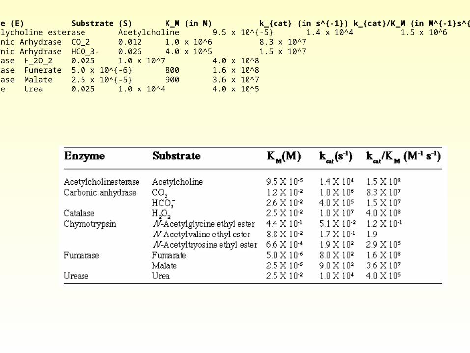
Enzyme (E) Substrate (S) K_M (in M) k_{cat} (in s^{-1}) k_{cat}/K_M (in M^{-1}s^{-1})Acetylycholine esterase Acetylcholine 9.5 x 10^{-5} 1.4 x 10^4 1.5 x 10^6Carbonic Anhydrase CO_2 0.012 1.0 x 10^6 8.3 x 10^7Carbonic Anhydrase HCO_3- 0.026 4.0 x 10^5 1.5 x 10^7Catalase H_2O_2 0.025 1.0 x 10^7 4.0 x 10^8Fumerase Fumerate 5.0 x 10^{-6} 800 1.6 x 10^8Fumerase Malate 2.5 x 10^{-5} 900 3.6 x 10^7Urease Urea 0.025 1.0 x 10^4 4.0 x 10^5
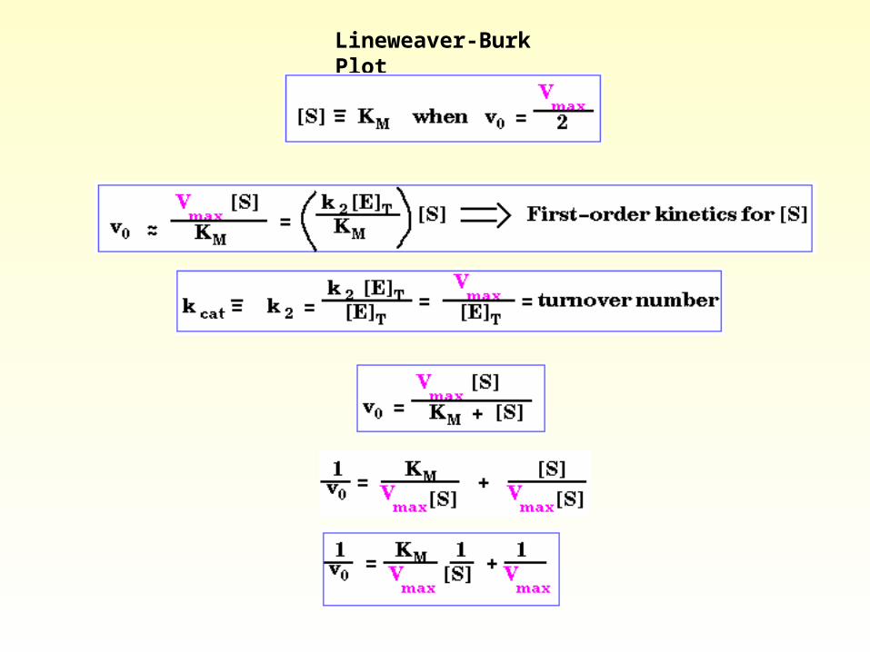
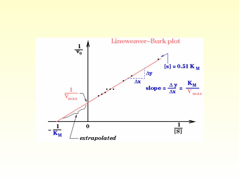
Lineweaver-Burk Plot
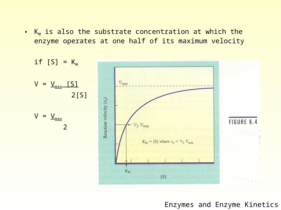
• KM is also the substrate concentration at which the enzyme operates at one half of its maximum velocity
if [S] = KM
V = Vmax [S]
2[S]
V = Vmax
2
Enzymes and Enzyme Kinetics
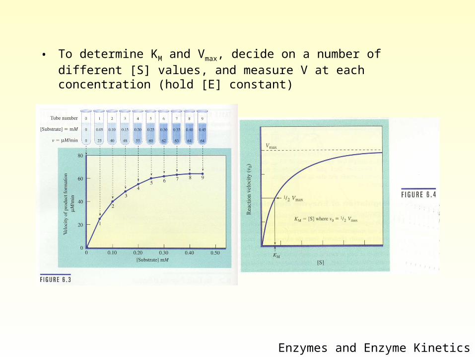
• To determine KM and Vmax, decide on a number of different [S] values, and measure V at each concentration (hold [E] constant)
Enzymes and Enzyme Kinetics
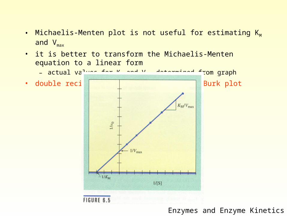
• Michaelis-Menten plot is not useful for estimating KM and Vmax
• it is better to transform the Michaelis-Menten equation to a linear form– actual values for KM and Vmax determined from graph
• double reciprocal plot or a Lineweaver-Burk plot
Enzymes and Enzyme Kinetics
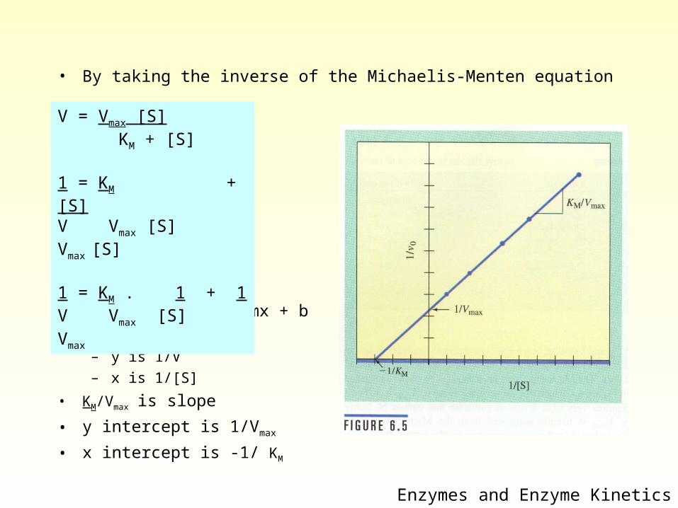
• By taking the inverse of the Michaelis-Menten equation
• same form as y = mx + b
• plot is y vs x– y is 1/V – x is 1/[S]
• KM/Vmax is slope
• y intercept is 1/Vmax
• x intercept is -1/ KM
Enzymes and Enzyme Kinetics
V = Vmax [S] KM + [S]
1 = KM + [S]V Vmax [S] Vmax [S]
1 = KM . 1 + 1V Vmax [S] Vmax
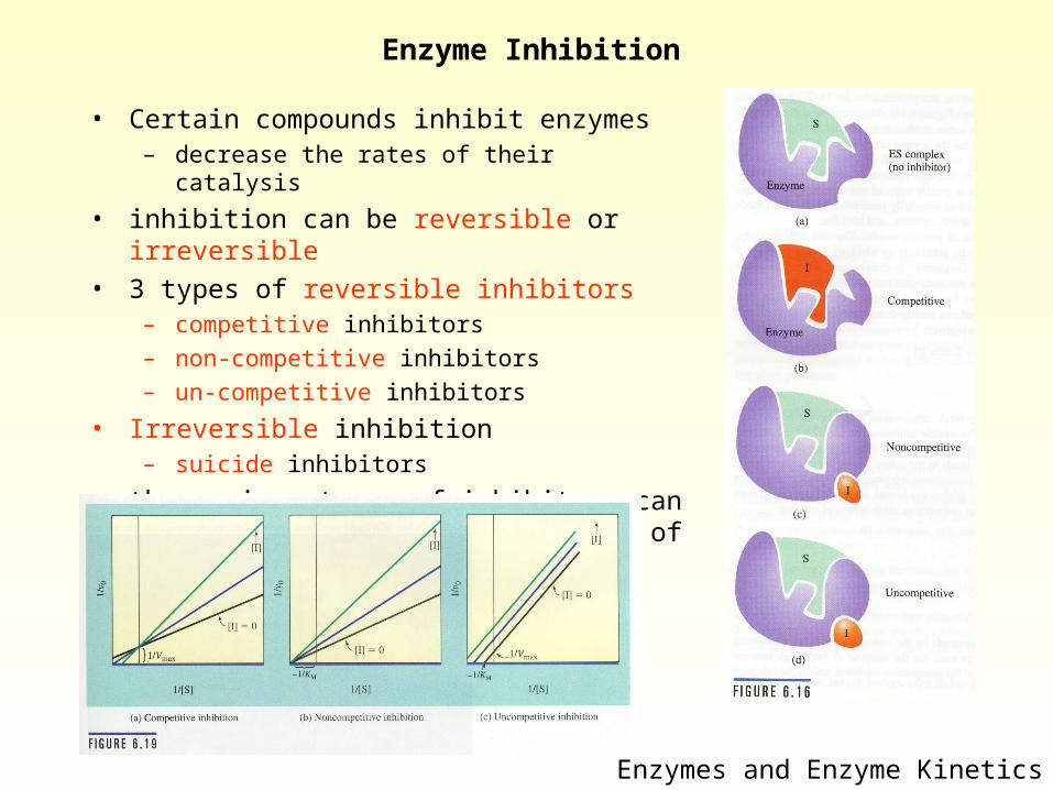
Enzyme Inhibition
• Certain compounds inhibit enzymes– decrease the rates of their catalysis
• inhibition can be reversible or irreversible
• 3 types of reversible inhibitors– competitive inhibitors– non-competitive inhibitors– un-competitive inhibitors
• Irreversible inhibition– suicide inhibitors
• the various types of inhibitors can be distinguished by the kinetics of their inhibition
Enzymes and Enzyme Kinetics
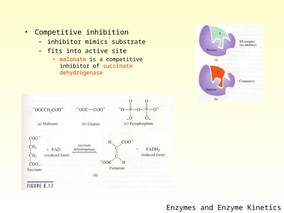
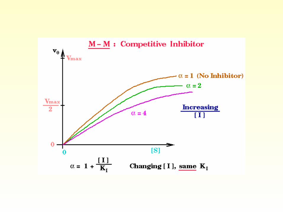
• Competitive inhibition– inhibitor mimics substrate– fits into active site
• malonate is a competitive inhibitor of succinate dehydrogenase
Enzymes and Enzyme Kinetics
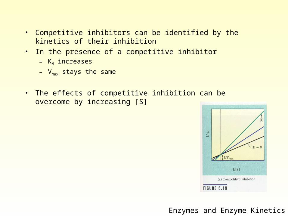
• Competitive inhibitors can be identified by the kinetics of their inhibition
• In the presence of a competitive inhibitor– KM increases
– Vmax stays the same
• The effects of competitive inhibition can be overcome by increasing [S]
Enzymes and Enzyme Kinetics
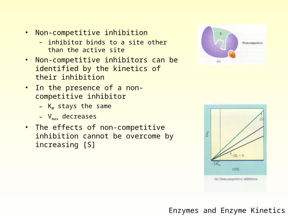
• Non-competitive inhibition– inhibitor binds to a site other than the active site
• Non-competitive inhibitors can be identified by the kinetics of their inhibition
• In the presence of a non-competitive inhibitor– KM stays the same
– Vmax decreases
• The effects of non-competitive inhibition cannot be overcome by increasing [S]
Enzymes and Enzyme Kinetics
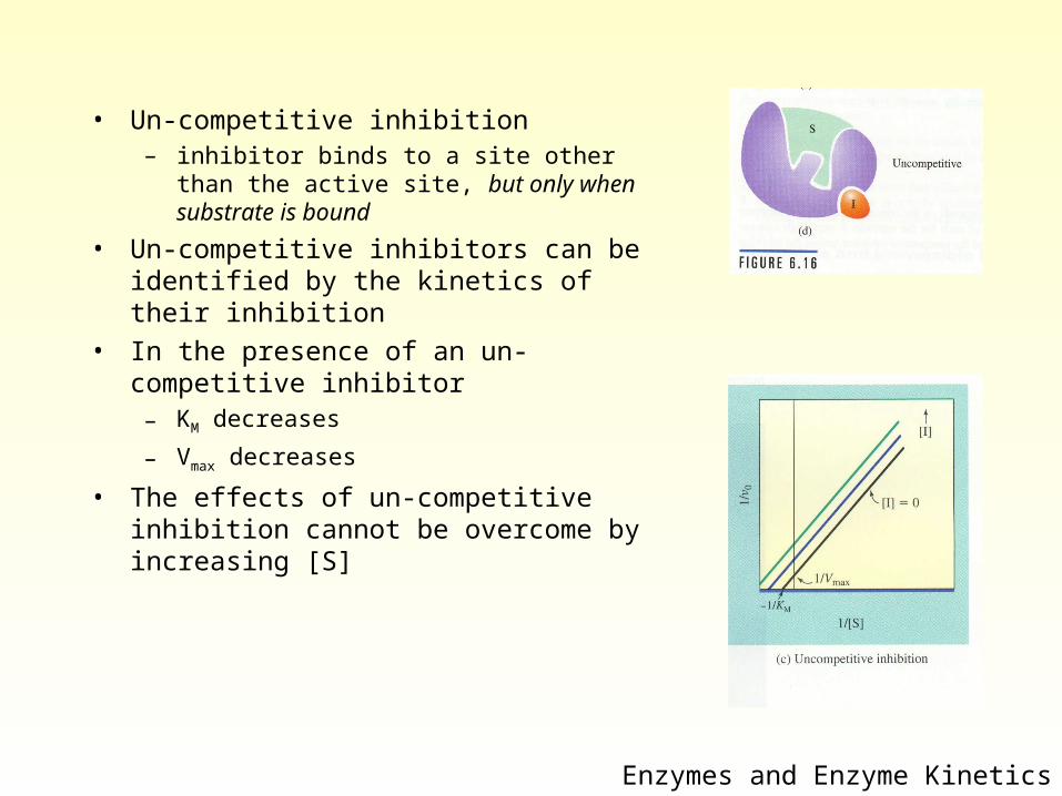
• Un-competitive inhibition– inhibitor binds to a site other than the active
site, but only when substrate is bound
• Un-competitive inhibitors can be identified by the kinetics of their inhibition
• In the presence of an un-competitive inhibitor– KM decreases
– Vmax decreases
• The effects of un-competitive inhibition cannot be overcome by increasing [S]
Enzymes and Enzyme Kinetics
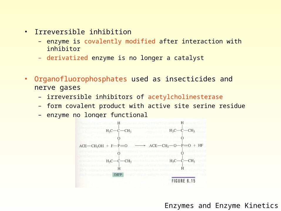
• Irreversible inhibition– enzyme is covalently modified after interaction with inhibitor– derivatized enzyme is no longer a catalyst
• Organofluorophosphates used as insecticides and nerve gases– irreversible inhibitors of acetylcholinesterase– form covalent product with active site serine residue– enzyme no longer functional
Enzymes and Enzyme Kinetics
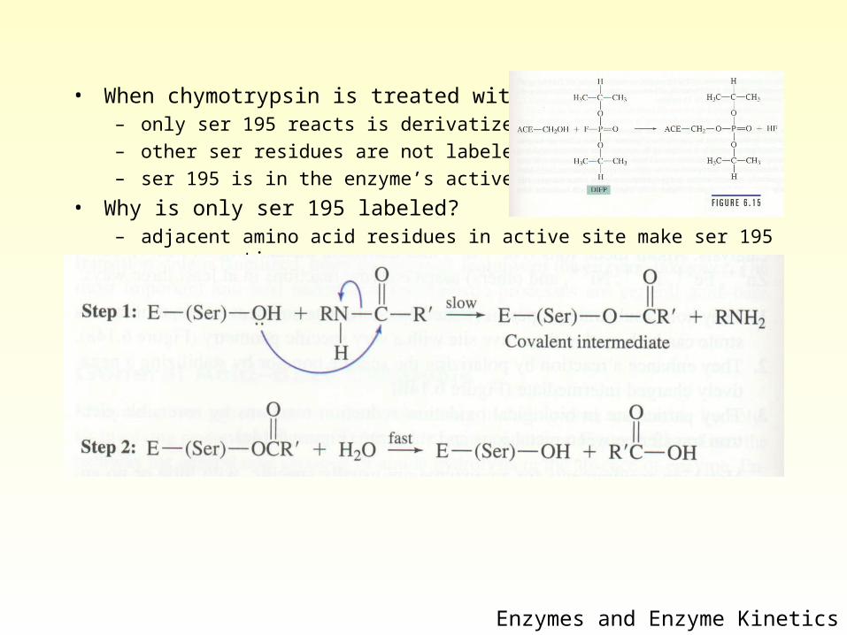
• When chymotrypsin is treated with DIPF– only ser 195 reacts is derivatized– other ser residues are not labeled– ser 195 is in the enzyme’s active site
• Why is only ser 195 labeled?– adjacent amino acid residues in active site make ser 195 more reactive
Enzymes and Enzyme Kinetics
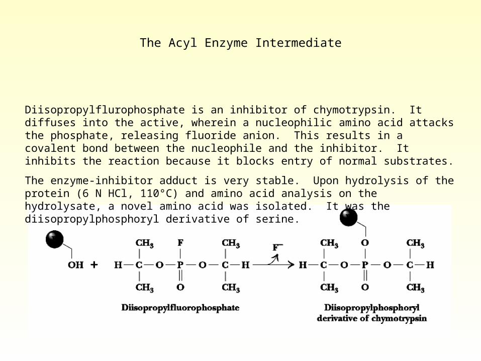
The Acyl Enzyme Intermediate
Diisopropylflurophosphate is an inhibitor of chymotrypsin. It diffuses into the active, wherein a nucleophilic amino acid attacks the phosphate, releasing fluoride anion. This results in a covalent bond between the nucleophile and the inhibitor. It inhibits the reaction because it blocks entry of normal substrates.
The enzyme-inhibitor adduct is very stable. Upon hydrolysis of the protein (6 N HCl, 110°C) and amino acid analysis on the hydrolysate, a novel amino acid was isolated. It was the diisopropylphosphoryl derivative of serine.
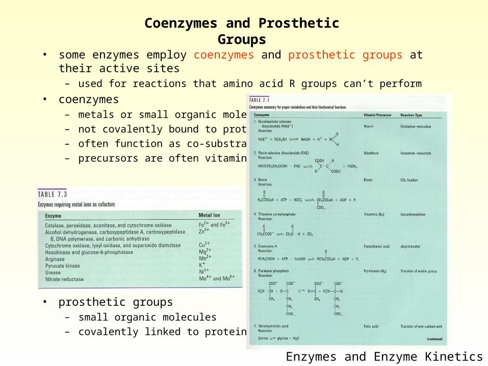
Coenzymes and Prosthetic Groups
• some enzymes employ coenzymes and prosthetic groups at their active sites
– used for reactions that amino acid R groups can’t perform
• coenzymes– metals or small organic molecules– not covalently bound to protein– often function as co-substrates– precursors are often vitamins
• prosthetic groups– small organic molecules – covalently linked to protein
Enzymes and Enzyme Kinetics
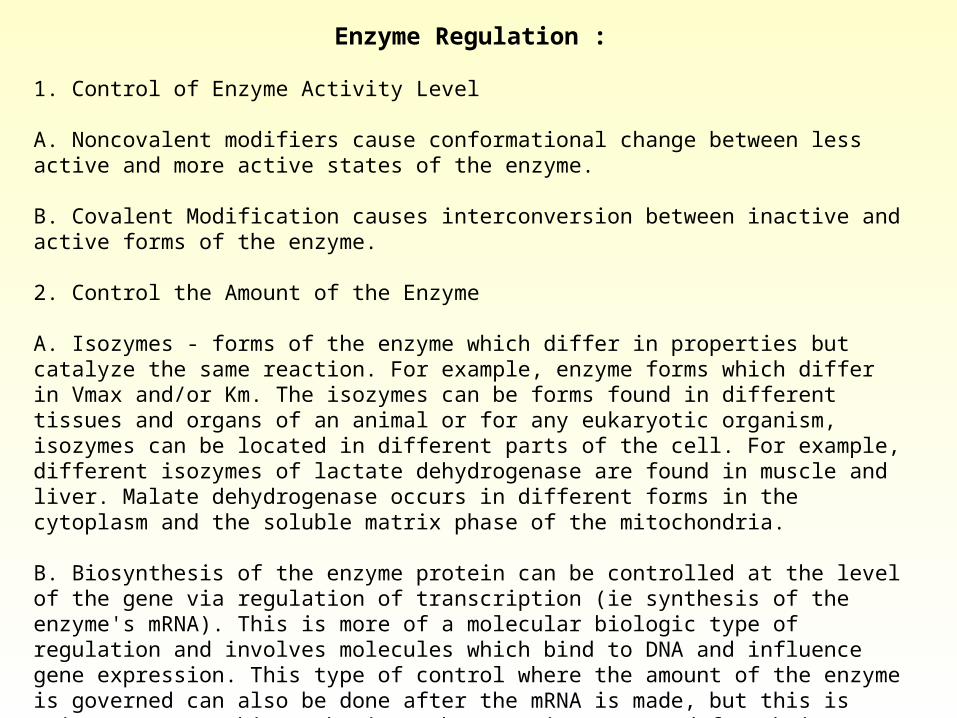
Enzyme Regulation :
1. Control of Enzyme Activity Level
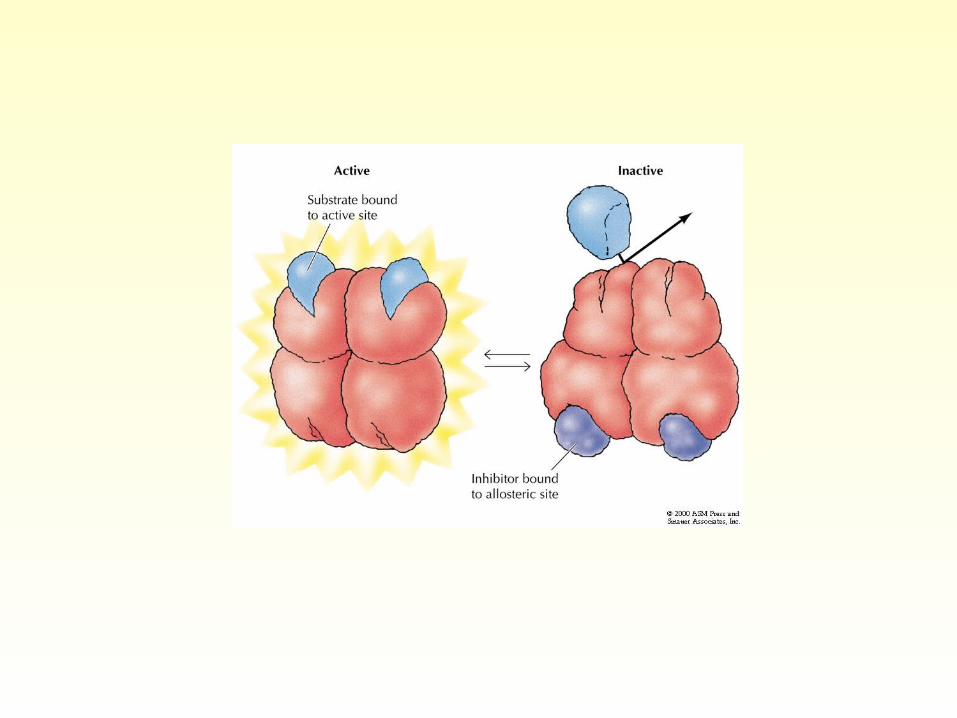
A. Noncovalent modifiers cause conformational change between less active and more active states of the enzyme.
B. Covalent Modification causes interconversion between inactive and active forms of the enzyme.
2. Control the Amount of the Enzyme
A. Isozymes - forms of the enzyme which differ in properties but catalyze the same reaction. For example, enzyme forms which differ in Vmax and/or Km. The isozymes can be forms found in different tissues and organs of an animal or for any eukaryotic organism, isozymes can be located in different parts of the cell. For example, different isozymes of lactate dehydrogenase are found in muscle and liver. Malate dehydrogenase occurs in different forms in the cytoplasm and the soluble matrix phase of the mitochondria.
B. Biosynthesis of the enzyme protein can be controlled at the level of the gene via regulation of transcription (ie synthesis of the enzyme's mRNA). This is more of a molecular biologic type of regulation and involves molecules which bind to DNA and influence gene expression. This type of control where the amount of the enzyme is governed can also be done after the mRNA is made, but this is quite rare. In this mechanism, the mRNA is prevented from being translated and since mRNA is
rather unstable, it is degraded before it is effectively used by the ribosomes to make the protein.
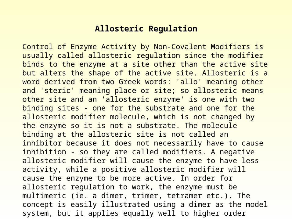
Allosteric Regulation
Control of Enzyme Activity by Non-Covalent Modifiers is usually called allosteric regulation since the modifier binds to the enzyme at a site other than the active site but alters the shape of the active site. Allosteric is a word derived from two Greek words: 'allo' meaning other and 'steric' meaning place or site; so allosteric means other site and an 'allosteric enzyme' is one with two binding sites - one for the substrate and one for the allosteric modifier molecule, which is not changed by the enzyme so it is not a substrate. The molecule binding at the allosteric site is not called an inhibitor because it does not necessarily have to cause inhibition - so they are called modifiers. A negative allosteric modifier will cause the enzyme to have less activity, while a positive allosteric modifier will cause the enzyme to be more active. In order for allosteric regulation to work, the enzyme must be multimeric (ie. a dimer, trimer, tetramer etc.). The concept is easily illustrated using a dimer as the model system, but it applies equally well to higher order multimers such as trimers and tetramers, etc.
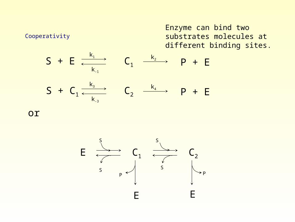
Cooperativity
S + E k1
k-1
C1k2 P + E
S + C1 k3
k-3
C2k4 P + E
Enzyme can bind two substrates molecules at different binding sites.
or
E C1 C2
E E
S S
S S
P P
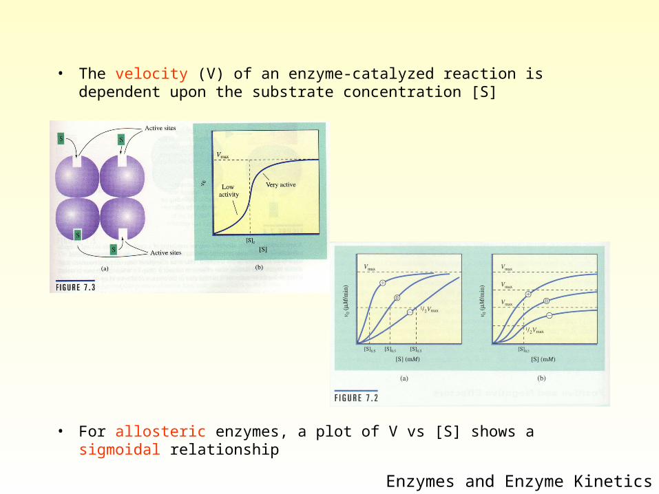
• The velocity (V) of an enzyme-catalyzed reaction is dependent upon the substrate concentration [S]
• For allosteric enzymes, a plot of V vs [S] shows a sigmoidal relationship
Enzymes and Enzyme Kinetics
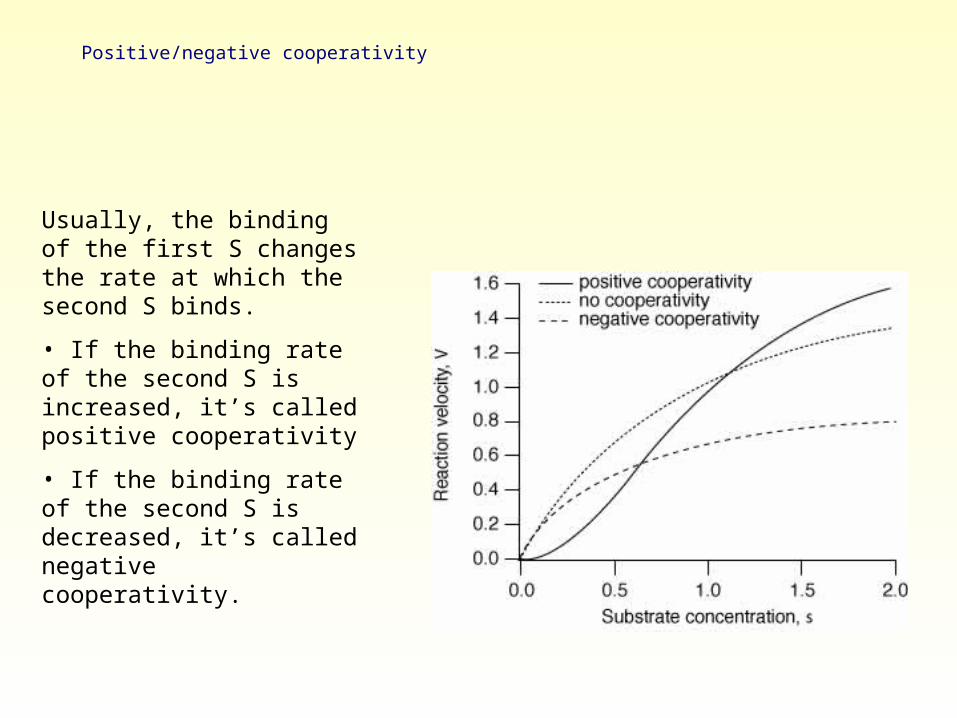
Positive/negative cooperativity
Usually, the binding of the first S changes the rate at which the second S binds.
• If the binding rate of the second S is increased, it’s called positive cooperativity
• If the binding rate of the second S is decreased, it’s called negative cooperativity.
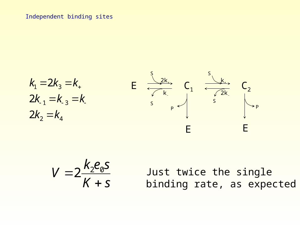
Independent binding sites
k1 2k3 k
2k 1 k 3 k2k2 k4
E C1 C2
E E
S S
S S
P P
2k+ k+
2k-k-
V 2k2e0s
K sJust twice the single binding rate, as expected
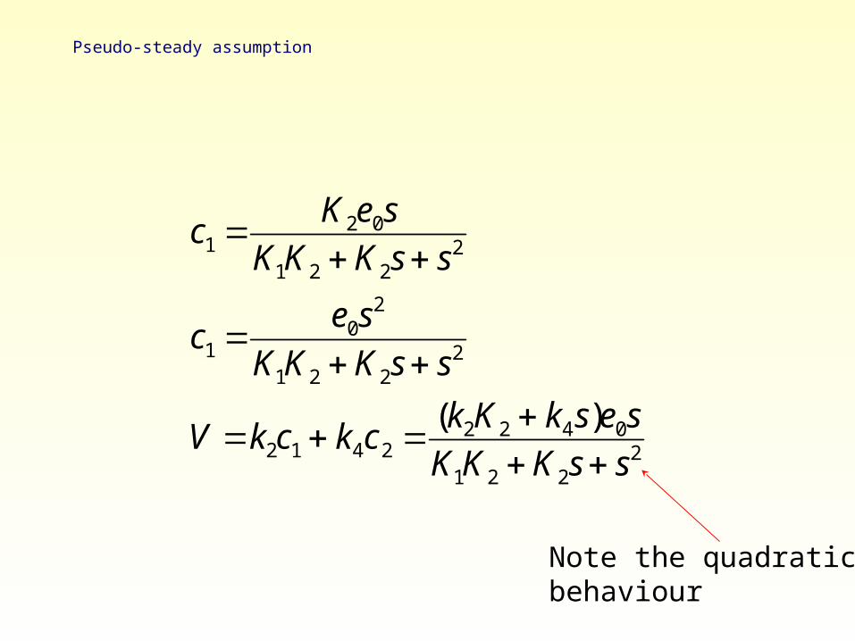
Pseudo-steady assumption
c1 K2e0sK1K2 K2s s
2
c1 e0s
2
K1K2 K2s s2
V k2c1 k4c2 (k2K2 k4s)e0s
K1K2 K2s s2
Note the quadraticbehaviour
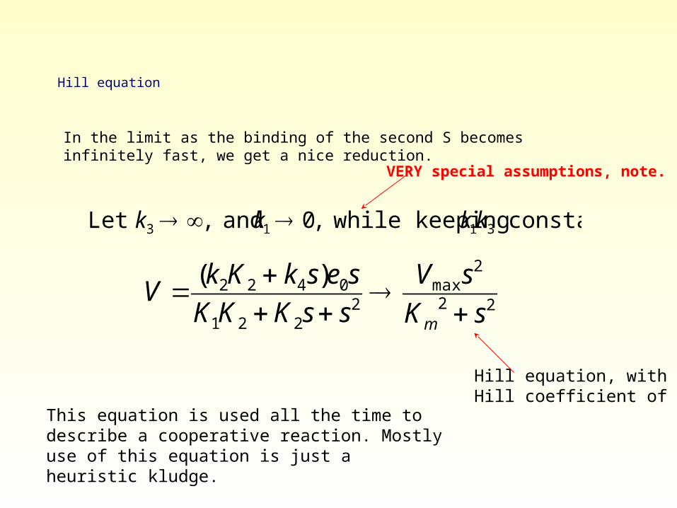
Hill equation
In the limit as the binding of the second S becomes infinitely fast, we get a nice reduction.
Let k3 , and k1 0, while keeping k1k3 constant.
V (k2K2 k4s)e0s
K1K2 K2s s2
Vmaxs2
Km2 s2
Hill equation, withHill coefficient of 2.
This equation is used all the time to describe a cooperative reaction. Mostly use of this equation is just a heuristic kludge.
VERY special assumptions, note.
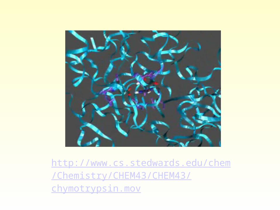
http://www.cs.stedwards.edu/chem/Chemistry/CHEM43/CHEM43/chymotrypsin.mov
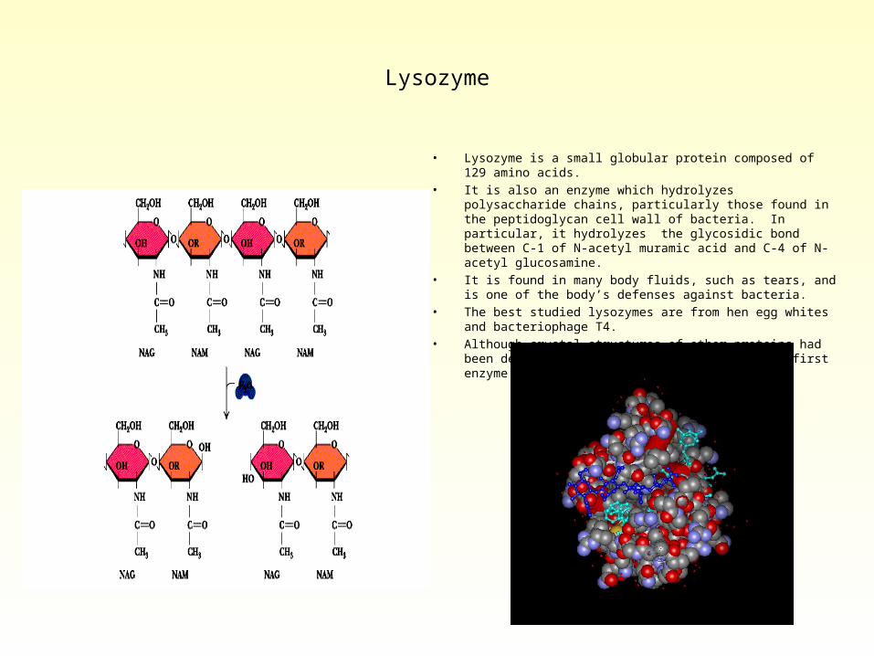
Lysozyme
• Lysozyme is a small globular protein composed of 129 amino acids.• It is also an enzyme which hydrolyzes polysaccharide chains,
particularly those found in the peptidoglycan cell wall of bacteria. In particular, it hydrolyzes the glycosidic bond between C-1 of N-acetyl muramic acid and C-4 of N-acetyl glucosamine.
• It is found in many body fluids, such as tears, and is one of the body’s defenses against bacteria.
• The best studied lysozymes are from hen egg whites and bacteriophage T4.
• Although crystal structures of other proteins had been determined previously, lysozyme was the first enzyme to have its structure determined.
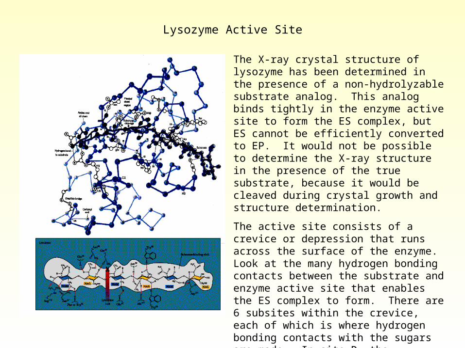
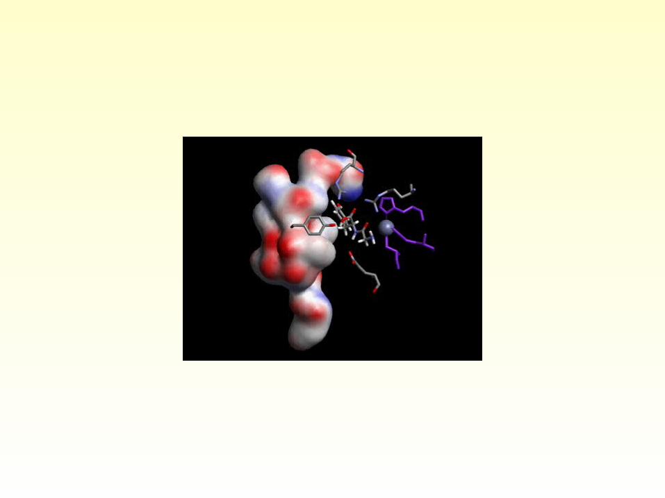
Lysozyme Active Site
The X-ray crystal structure of lysozyme has been determined in the presence of a non-hydrolyzable substrate analog. This analog binds tightly in the enzyme active site to form the ES complex, but ES cannot be efficiently converted to EP. It would not be possible to determine the X-ray structure in the presence of the true substrate, because it would be cleaved during crystal growth and structure determination.
The active site consists of a crevice or depression that runs across the surface of the enzyme. Look at the many hydrogen bonding contacts between the substrate and enzyme active site that enables the ES complex to form. There are 6 subsites within the crevice, each of which is where hydrogen bonding contacts with the sugars are made. In site D, the conformation of the sugar is distorted in order to make the necessary hydrogen bonding contacts. This distortion raises the energy of the ground state, bringing the substrate closer to the transition state for hydrolysis.
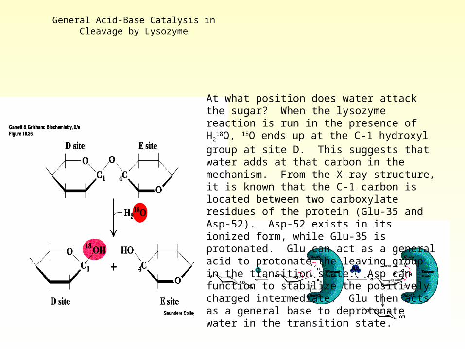
General Acid-Base Catalysis in Cleavage by Lysozyme
At what position does water attack the sugar? When the lysozyme reaction is run in the presence of H2
18O, 18O ends up at the C-1 hydroxyl group at site D. This suggests that water adds at that carbon in the mechanism. From the X-ray structure, it is known that the C-1 carbon is located between two carboxylate residues of the protein (Glu-35 and Asp-52). Asp-52 exists in its ionized form, while Glu-35 is protonated. Glu can act as a general acid to protonate the leaving group in the transition state. Asp can function to stabilize the positively charged intermediate. Glu then acts as a general base to deprotonate water in the transition state.
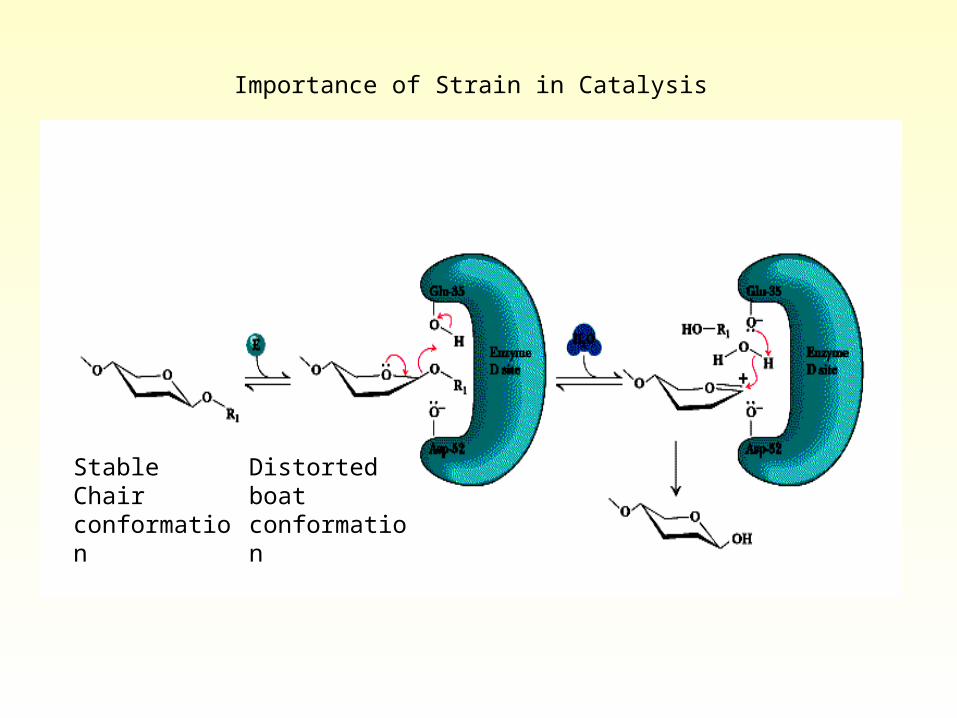
Stable Chair conformation
Distorted boat conformation
Importance of Strain in Catalysis
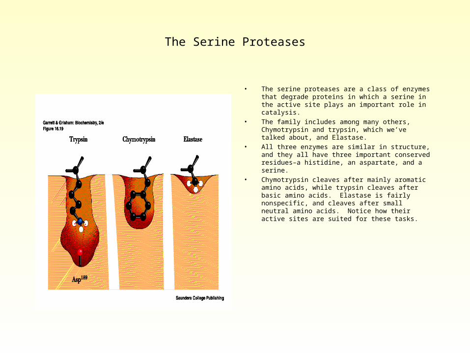
The Serine Proteases
• The serine proteases are a class of enzymes that degrade proteins in which a serine in the active site plays an important role in catalysis.
• The family includes among many others, Chymotrypsin and trypsin, which we’ve talked about, and Elastase.
• All three enzymes are similar in structure, and they all have three important conserved residues–a histidine, an aspartate, and a serine.
• Chymotrypsin cleaves after mainly aromatic amino acids, while trypsin cleaves after basic amino acids. Elastase is fairly nonspecific, and cleaves after small neutral amino acids. Notice how their active sites are suited for these tasks.
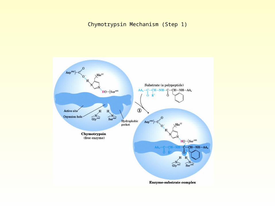
Chymotrypsin Mechanism (Step 1)
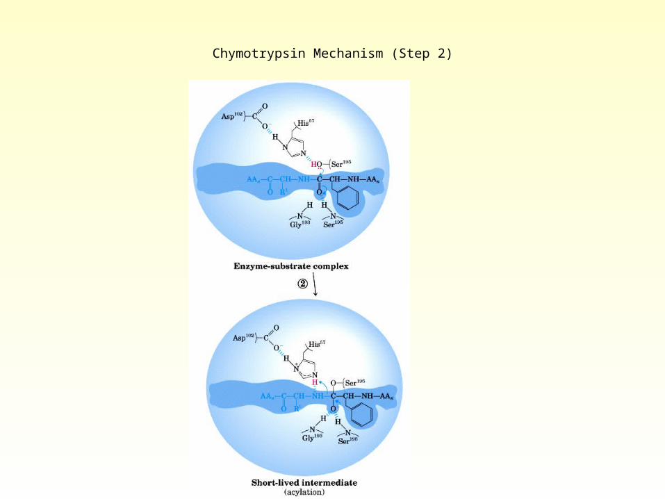
Chymotrypsin Mechanism (Step 2)
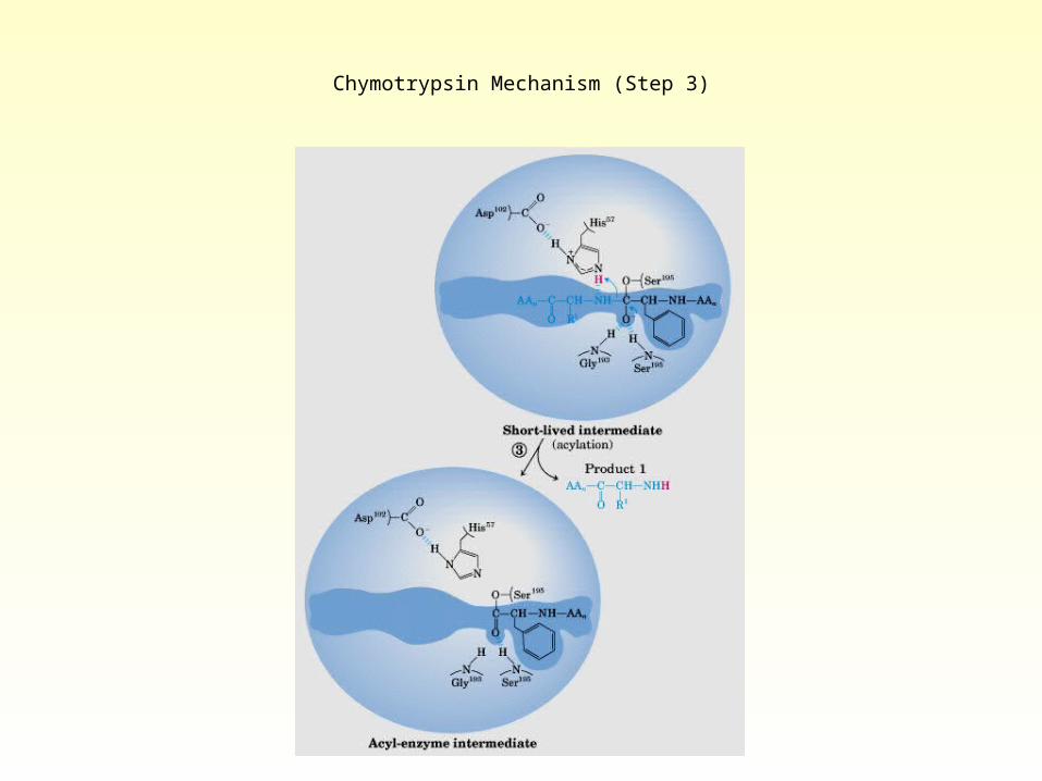
Chymotrypsin Mechanism (Step 3)
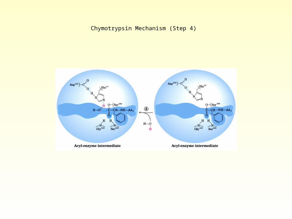
Chymotrypsin Mechanism (Step 4)
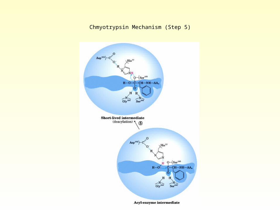
Chmyotrypsin Mechanism (Step 5)

Chymotrypsin Mechanism (Step 6)
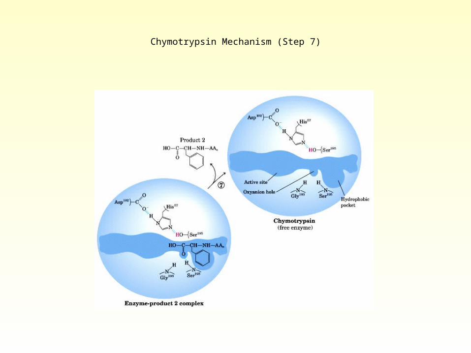
Chymotrypsin Mechanism (Step 7)