enzymes part iv: cofactors -...
TRANSCRIPT
EnzymesPart IV: CofactorsProf. Mamoun AhramDr. Diala Abu HassanSummer 2019
Catalytic strategies of enzymes
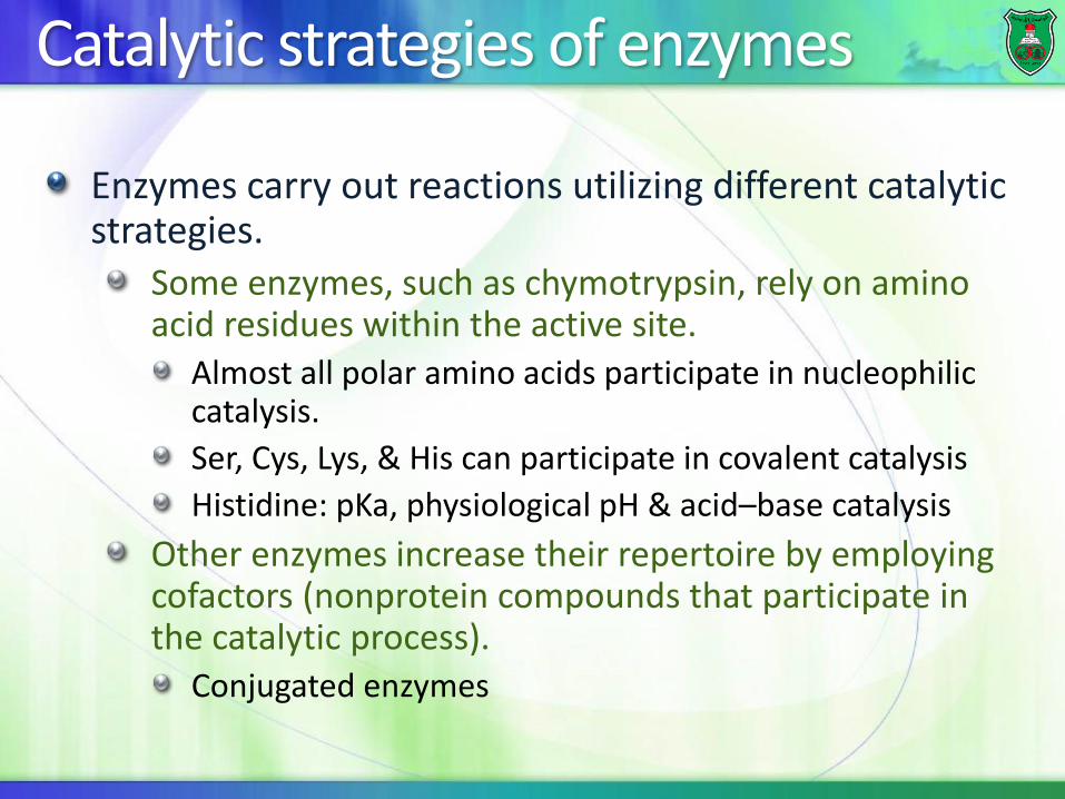
Enzymes carry out reactions utilizing different catalytic strategies.
Some enzymes, such as chymotrypsin, rely on amino acid residues within the active site.
Almost all polar amino acids participate in nucleophilic catalysis.
Ser, Cys, Lys, & His can participate in covalent catalysis
Histidine: pKa, physiological pH & acid–base catalysis
Other enzymes increase their repertoire by employing cofactors (nonprotein compounds that participate in the catalytic process).
Conjugated enzymes
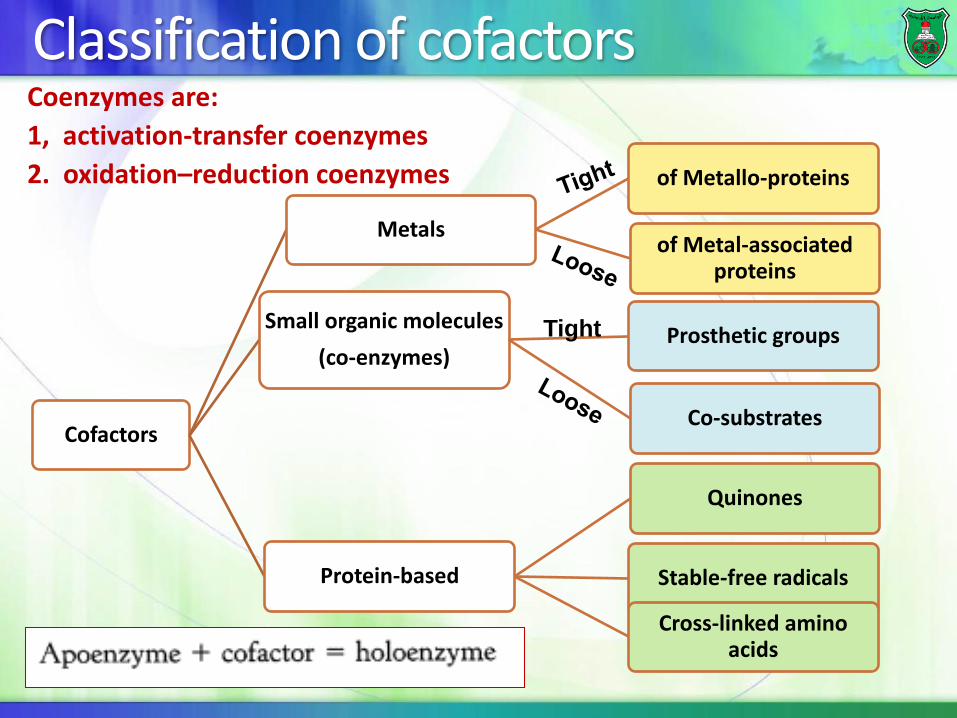
Classification of cofactors
Cofactors
Metals
of Metallo-proteins
of Metal-associated proteins
Small organic molecules
(co-enzymes)Prosthetic groups
Co-substrates
Protein-based
Quinones
Stable-free radicals
Cross-linked amino acids
Tight
Coenzymes are:
1, activation-transfer coenzymes
2. oxidation–reduction coenzymes
ACTIVATION-TRANSFER COENZYMES
They usually participate directly in catalysis by forming a covalent bond.
Characteristics:Two groups in the coenzyme:
A functional group that forms a covalent bond with substrate.
A binding group that binds tightly to the enzyme.
Dependence on the enzyme for additional specificity of substrate & additional catalytic power
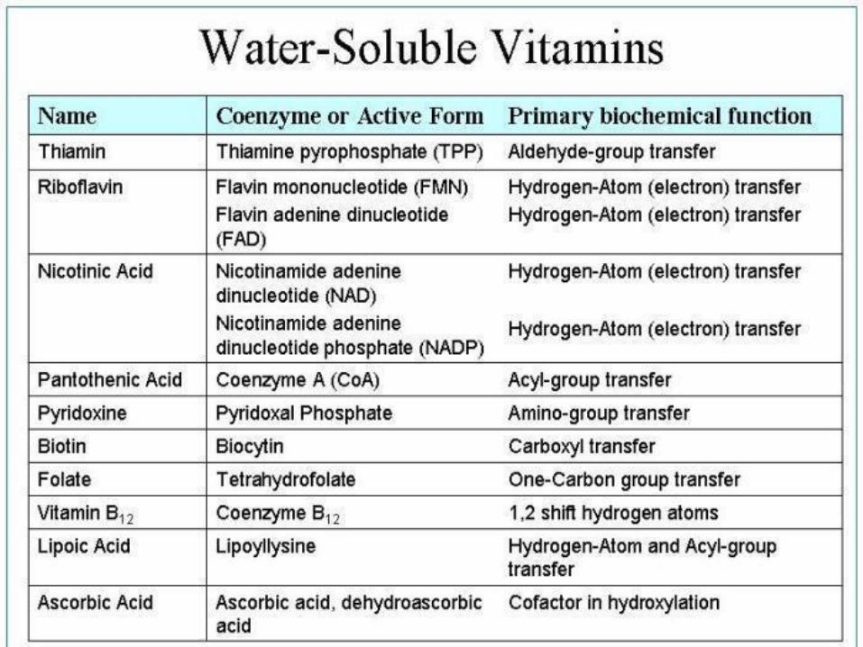
Thiamin pyrophosphate, TPP
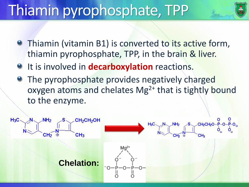
Thiamin (vitamin B1) is converted to its active form, thiamin pyrophosphate, TPP, in the brain & liver.
It is involved in decarboxylation reactions.
The pyrophosphate provides negatively charged oxygen atoms and chelates Mg2+ that is tightly bound to the enzyme.
Chelation:
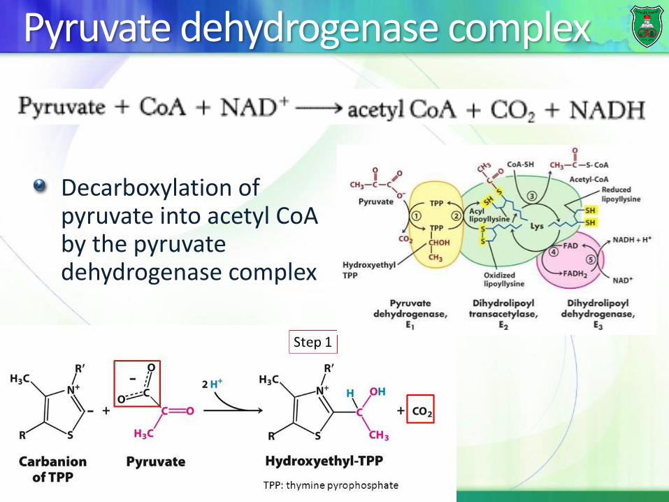
Pyruvate dehydrogenase complex
Decarboxylation of pyruvate into acetyl CoA by the pyruvate dehydrogenase complex
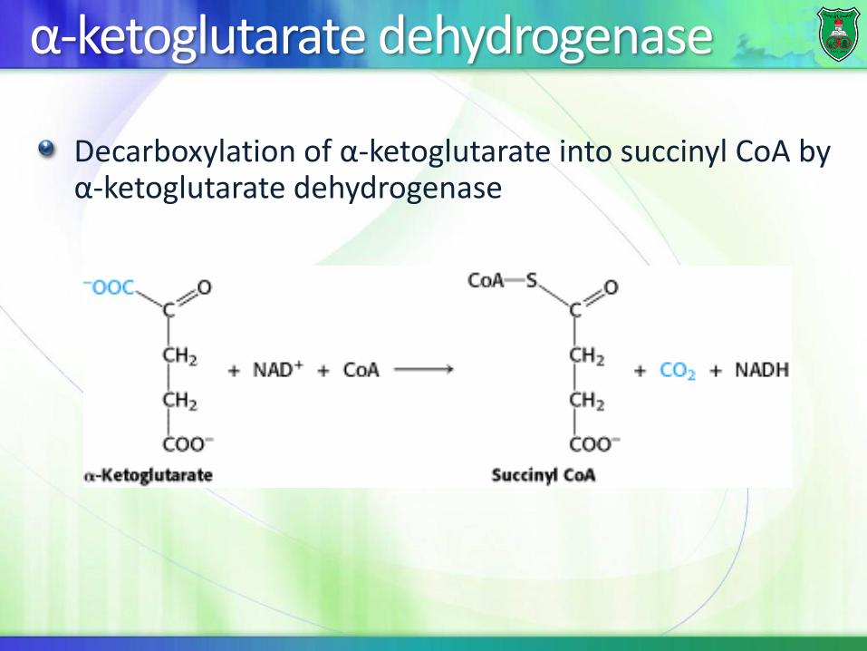
α-ketoglutarate dehydrogenase
Decarboxylation of α-ketoglutarate into succinyl CoA by α-ketoglutarate dehydrogenase
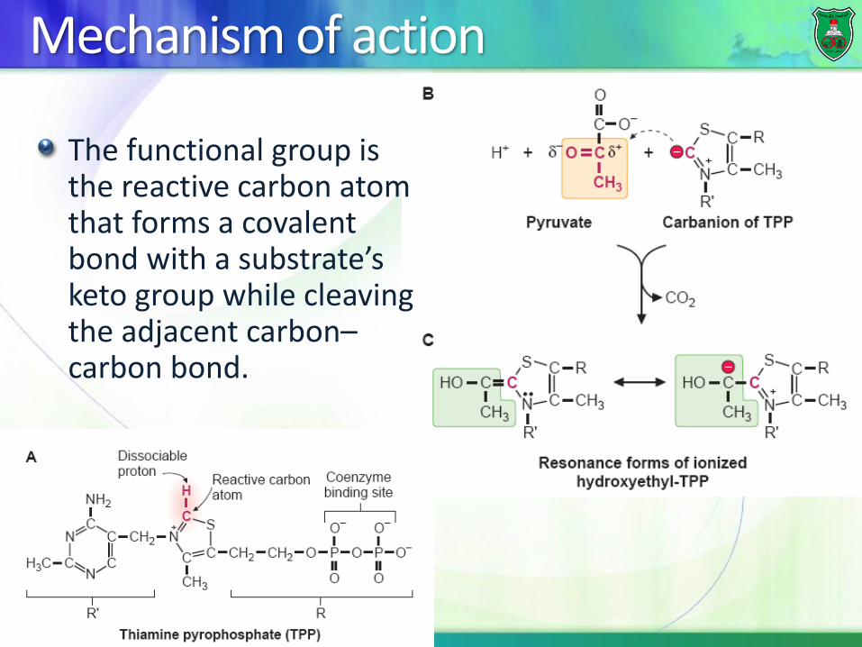
Mechanism of action
The functional group is the reactive carbon atom that forms a covalent bond with a substrate’s keto group while cleaving the adjacent carbon–carbon bond.
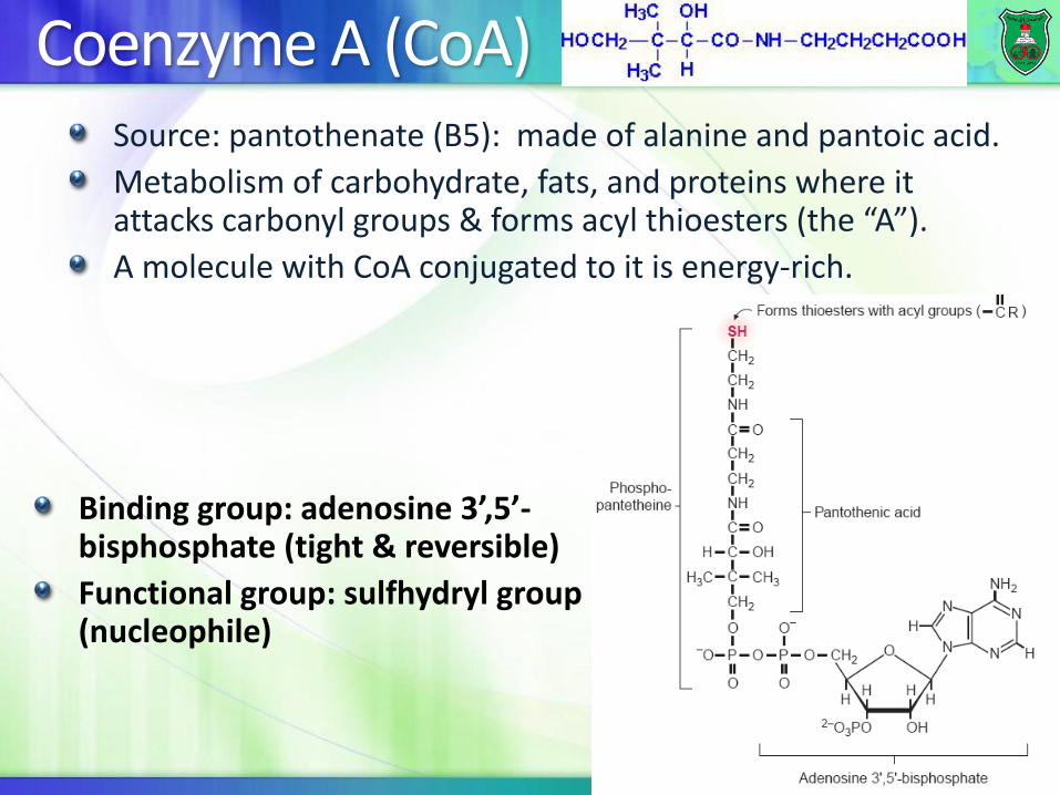
Coenzyme A (CoA) Source: pantothenate (B5): made of alanine and pantoic acid.
Metabolism of carbohydrate, fats, and proteins where it attacks carbonyl groups & forms acyl thioesters (the “A”).
A molecule with CoA conjugated to it is energy-rich.
Binding group: adenosine 3’,5’-bisphosphate (tight & reversible)
Functional group: sulfhydryl group (nucleophile)
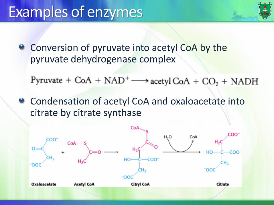
Examples of enzymes
Conversion of pyruvate into acetyl CoA by the pyruvate dehydrogenase complex
Condensation of acetyl CoA and oxaloacetate into citrate by citrate synthase
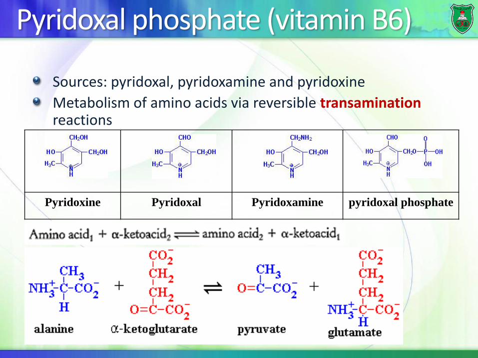
Pyridoxal phosphate (vitamin B6)
Sources: pyridoxal, pyridoxamine and pyridoxine
Metabolism of amino acids via reversible transaminationreactions
pyridoxal phosphatePyridoxaminePyridoxalPyridoxine
Examples
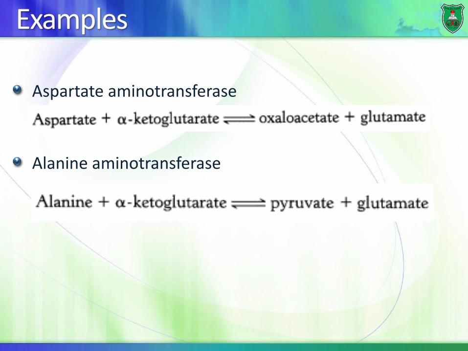
Aspartate aminotransferase
Alanine aminotransferase
Mechanism of action
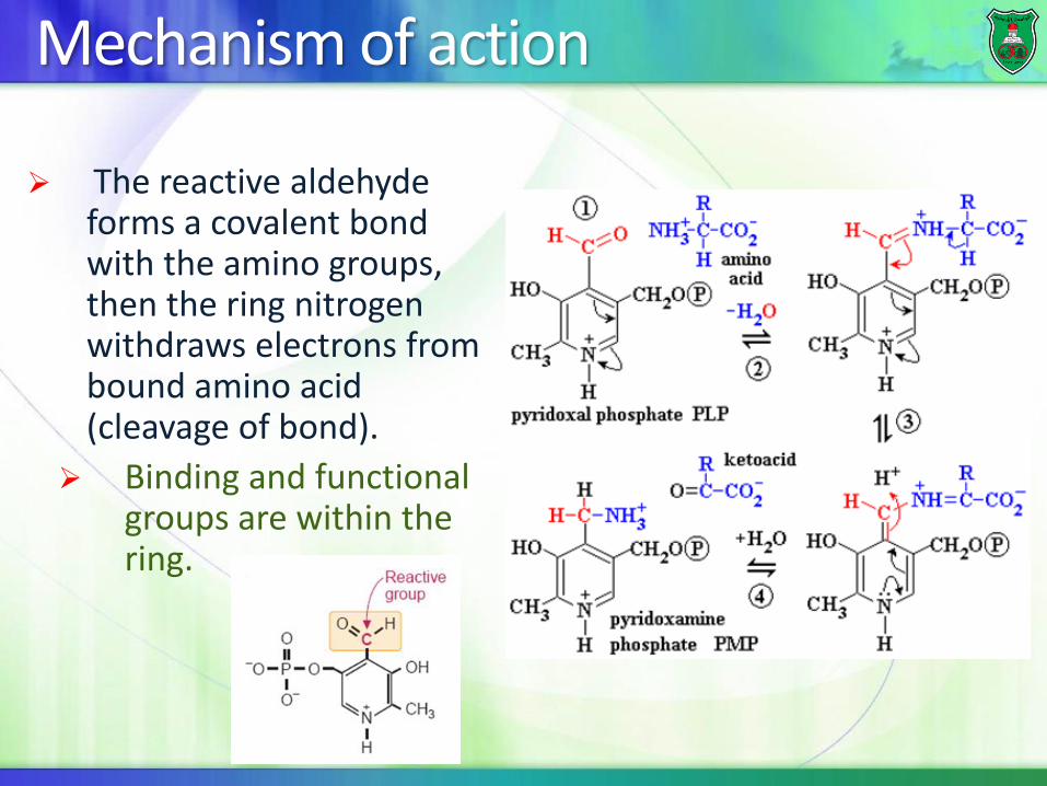
➢ The reactive aldehyde forms a covalent bond with the amino groups, then the ring nitrogen withdraws electrons from bound amino acid (cleavage of bond).
➢ Binding and functional groups are within the ring.
Biotin (vitamin B7)
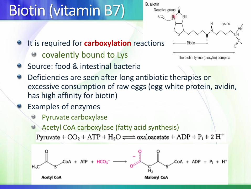
It is required for carboxylation reactions
covalently bound to LysSource: food & intestinal bacteria
Deficiencies are seen after long antibiotic therapies or excessive consumption of raw eggs (egg white protein, avidin, has high affinity for biotin)
Examples of enzymesPyruvate carboxylase
Acetyl CoA carboxylase (fatty acid synthesis)

OXIDATION–REDUCTION COENZYMES
A number of coenzymes work within oxidoreductases.
Each coenzyme has a unique functional group that accepts and donates electrons and is specific for the form of electrons it transfers (e.g., hydride ions, hydrogen atoms, oxygen).
These do not form covalent bonds with the substrate, a portion of the coenzyme binds the enzyme.
Most common: NAD+ (niacin, B3) & FAD (riboflavin, B2)
Others: work with metals to transfer single electrons to O2(Vitamins E & C)
Again: Dependence on the enzyme for additional specificity of substrate & additional catalytic power
FAD and FMN
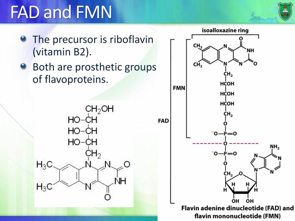
The precursor is riboflavin (vitamin B2).
Both are prosthetic groups of flavoproteins.
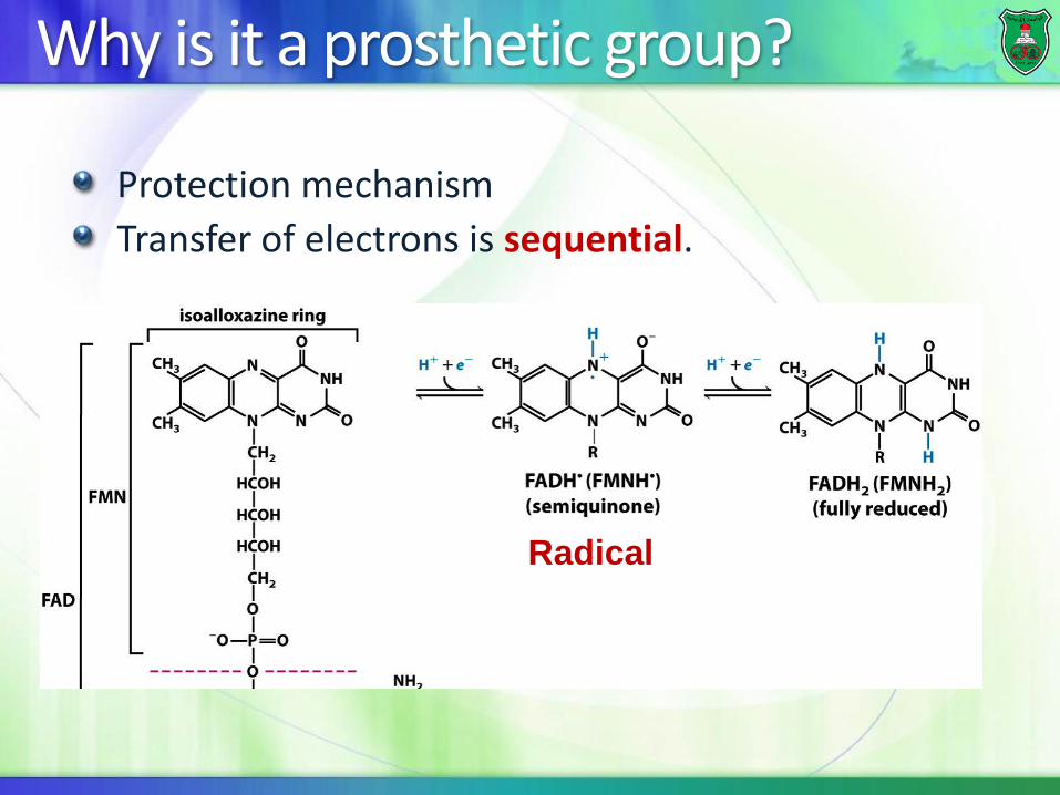
Why is it a prosthetic group?
Protection mechanism
Transfer of electrons is sequential.
Radical
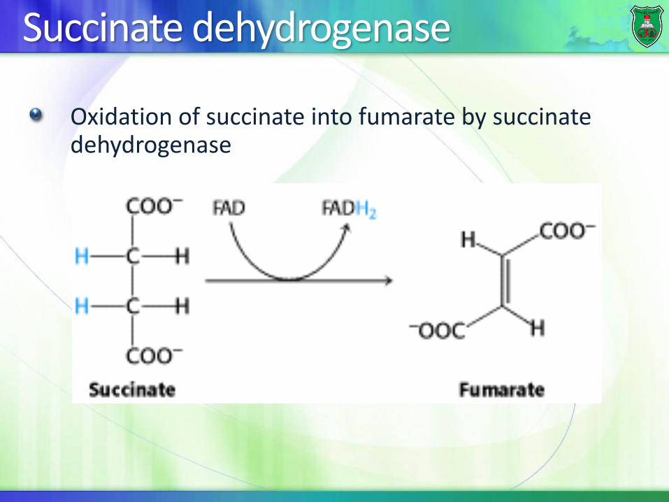
Succinate dehydrogenase
Oxidation of succinate into fumarate by succinate dehydrogenase
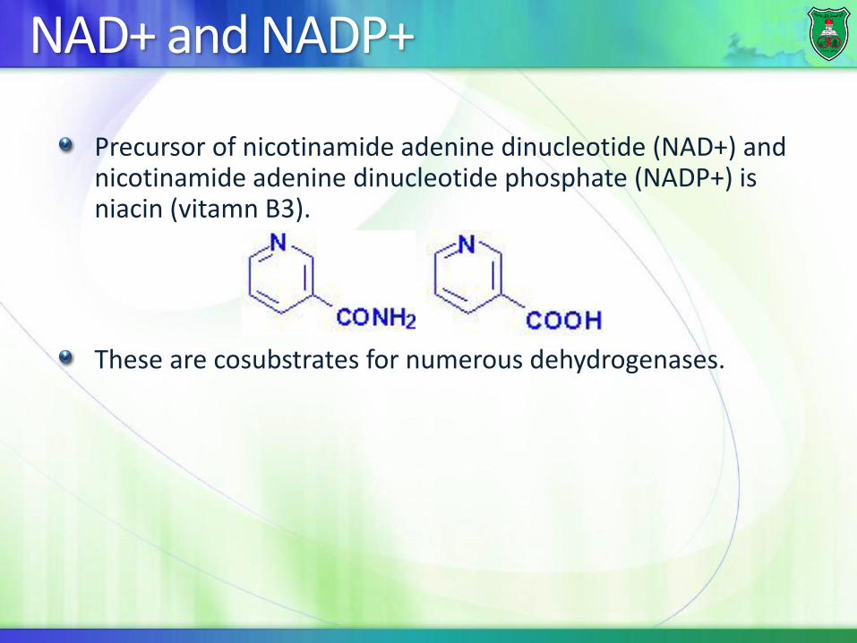
NAD+ and NADP+
Precursor of nicotinamide adenine dinucleotide (NAD+) and nicotinamide adenine dinucleotide phosphate (NADP+) is niacin (vitamn B3).
These are cosubstrates for numerous dehydrogenases.
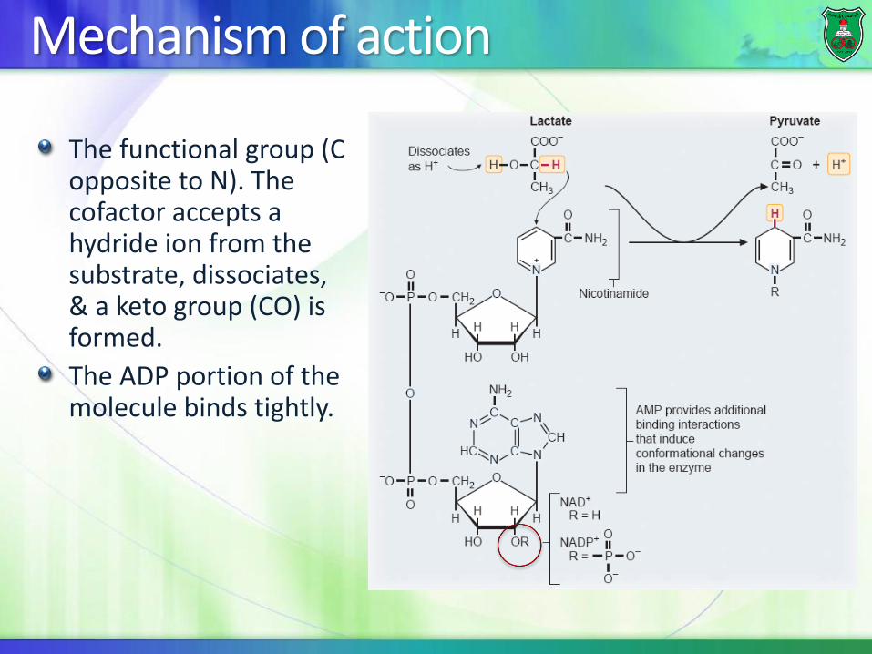
Mechanism of action
The functional group (C opposite to N). The cofactor accepts a hydride ion from the substrate, dissociates, & a keto group (CO) is formed.
The ADP portion of the molecule binds tightly.
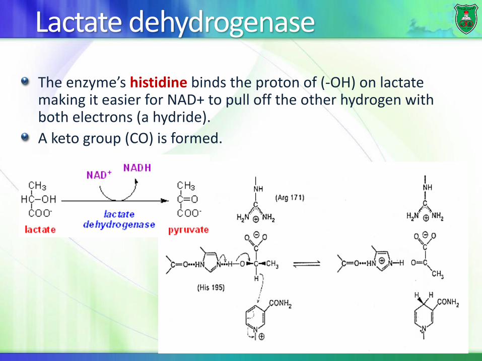
Lactate dehydrogenase
The enzyme’s histidine binds the proton of (-OH) on lactate making it easier for NAD+ to pull off the other hydrogen with both electrons (a hydride).
A keto group (CO) is formed.
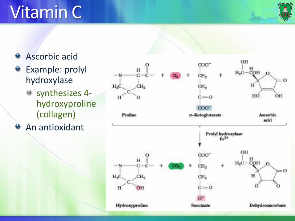
Vitamin C
Ascorbic acid
Example: prolyl hydroxylase
synthesizes 4-hydroxyproline (collagen)
An antioxidant
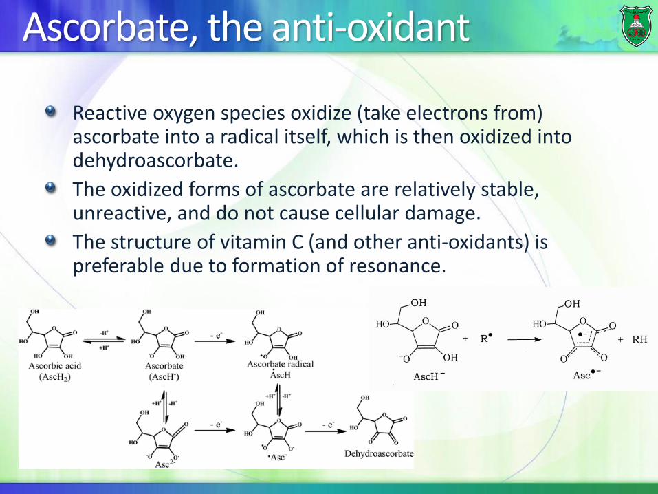
Ascorbate, the anti-oxidant
Reactive oxygen species oxidize (take electrons from) ascorbate into a radical itself, which is then oxidized into dehydroascorbate.
The oxidized forms of ascorbate are relatively stable, unreactive, and do not cause cellular damage.
The structure of vitamin C (and other anti-oxidants) is preferable due to formation of resonance.
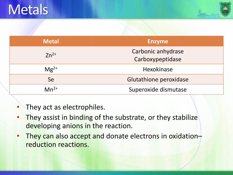
Metals
EnzymeMetal
Carbonic anhydraseCarboxypeptidase
Zn2+
HexokinaseMg2+
Glutathione peroxidaseSe
Superoxide dismutaseMn2+
• They act as electrophiles.
• They assist in binding of the substrate, or they stabilize developing anions in the reaction.
• They can also accept and donate electrons in oxidation–reduction reactions.
Advantages
• They carry positive charges and, hence, can form relatively strong yet kinetically labile (likely to be changed) bonds.
• They are stable in more than one oxidation state.
• They can bind multiple ligands in their coordination sphere enabling them to participate in binding substrates or coenzymes to enzymes. • Mg+2 connects the negatively charged phosphate groups of
thiamine pyrophosphate to basic amino acids in the enzyme.
• The phosphate groups of ATP are usually bound to enzymes through Mg+2 chelation.
Carbonic anhydrases
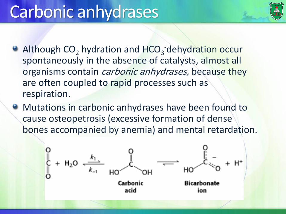
Although CO2 hydration and HCO3-dehydration occur
spontaneously in the absence of catalysts, almost all organisms contain carbonic anhydrases, because they are often coupled to rapid processes such as respiration.
Mutations in carbonic anhydrases have been found to cause osteopetrosis (excessive formation of dense bones accompanied by anemia) and mental retardation.
Zn binding to the enzyme
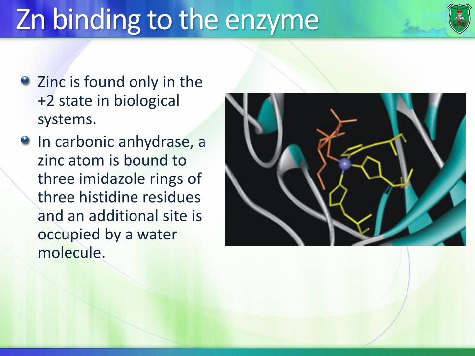
Zinc is found only in the +2 state in biological systems.
In carbonic anhydrase, a zinc atom is bound to three imidazole rings of three histidine residues and an additional site is occupied by a water molecule.
Mechanism of action
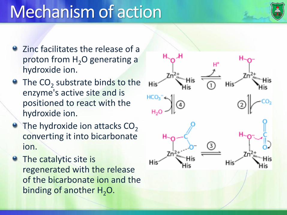
Zinc facilitates the release of a proton from H2O generating a hydroxide ion.
The CO2 substrate binds to the enzyme's active site and is positioned to react with the hydroxide ion.
The hydroxide ion attacks CO2converting it into bicarbonate ion.
The catalytic site is regenerated with the release of the bicarbonate ion and the binding of another H2O.
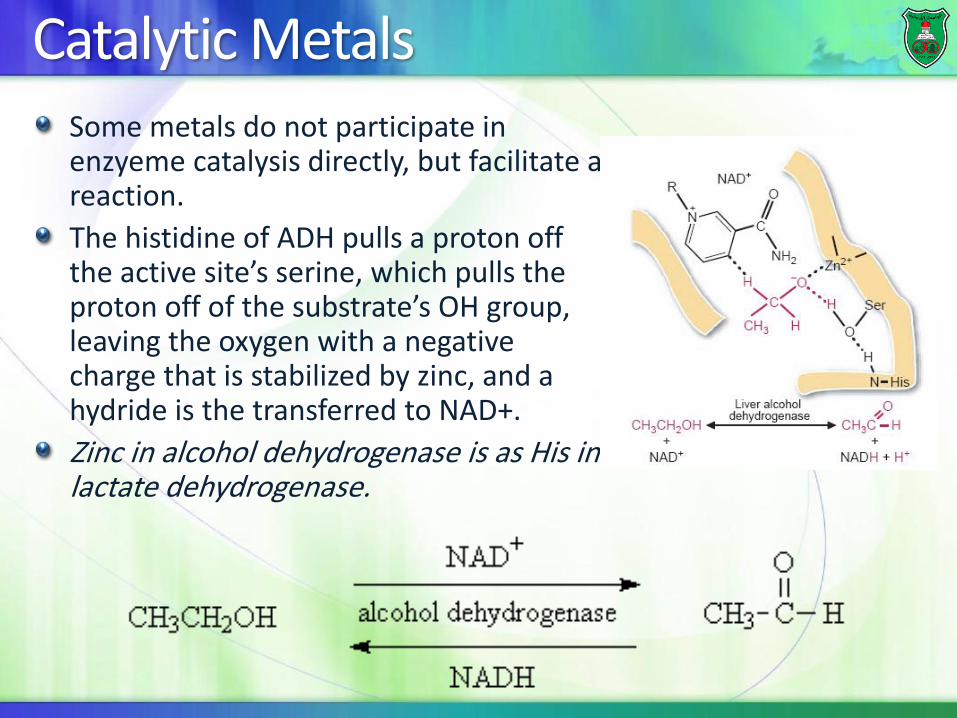
Catalytic MetalsSome metals do not participate in enzyeme catalysis directly, but facilitate a reaction.
The histidine of ADH pulls a proton off the active site’s serine, which pulls the proton off of the substrate’s OH group, leaving the oxygen with a negative charge that is stabilized by zinc, and a hydride is the transferred to NAD+.
Zinc in alcohol dehydrogenase is as His in lactate dehydrogenase.