epidemiological and histopathological analysis of soft ...elcerrahi.com/yayinlar/epidemiological and...
TRANSCRIPT

Original ArticleHand Microsurg 2018;X:X-X
doi:10.5455/handmicrosurg.16148
ABSTRACT Objectives: The spectrum of soft tissue tumors of the hand is wide. The type of tumor can vary based on demographic factors, such as a patient’s gender and age. Furthermore, certain tumors arise more commonly in particular locations on the hand. This study aimed to determine the incidence and recurrence rates of soft tissue tumors of the hand and to discuss the most common types of tumors based on tumoral parameters, demographic data, and histopathological findings with a brief review of the literature.Methods: We analyzed the clinical data and the pathology reports of all patients who underwent surgery in our hand and microsurgery department between January 1, 2007, and January 1, 2016. We evaluated the demographic data, surgical information, tumor type, size and location, histopathological diagnoses and immunohistochemical findings. We also deter-mined the recurrence rates of the patients after a 2-year follow up. Results: In our center, 302 surgical resections were performed between January 1, 2007, and January 1, 2016. Ganglion, which is a pseudotumoral lesion, was the most common space occupying lesion (n=102, 34%). Of the 302 resections, 66% were tumoral cases (n=200). Giant cell tenosynovial tumor (n=55, 27.5%) was the most common benign tumor, followed by glomus tumor (n=34, 17%), pyogenic granuloma (n=15, 7.5%), and cystic hygroma (n=14, 7%).Conclusion: In accordance with the literature, in the present study, most of the patients (99%) that underwent surgery had benign diagnoses. Synovial sarcoma was the only malignant tumor observed in the study population.
Key words: Hand, tumor, soft tissue, microsurgery
Epidemiological and histopathological analysis of soft tissue tumors of the hand - 9 years of experience from a single center
Uguray Payam Hacisalihoglu1, Ismail Bulent Ozcelik2
IntroductionThere are more than 200 types of soft tissue tum-
ors, and many subtypes [1]. These tumors are located in the superficial and deep soft tissue of the skin, and they display mesenchymal differentiation. They are catego-
rized into three main types: benign, intermediate, and malignant [2]. It has been reported that hand tumors comprise 15% of all soft tissue tumors [3,4]. Most soft tissue tumors of the hand are benign. The malignant soft tissue tumors that have more than 50 histopatho-
1Department of Pathology, 2Department of Hand and Microsurgery, Istanbul Yeni Yuzyil University Medical Faculty, Gaziosmanpasa Hospital, Istanbul, Turkey Uguray Payam Hacisalihoglu, MD, Department of Pathology, Istanbul Yeni Yuzyil University Medical Faculty, Gaziosmanpasa Hospital, Istanbul, Turkey. e-mail: [email protected] 07, 2018 / November 30, 2018
Author affiliations :
Correspondence :
Received / Accepted :
© 2018 Turkish Society for Surgery of the Hand and Upper Exremity www.handmicrosurgeryjournal.com
eJM eJManager OPEN ACCESS

Hacisalihoglu UP and Ozcelik IB
Year 2018 | Volume X | Issue X | X-XXX | Hand and Microsurgery
logical subtypes constitute less than 1% of all human malignancies and approximately 1% of the soft tissue tumors of the hand [5-7]. In hand tumors, careful his-topathological analysis, staging, and treatment are es-sential to optimize the clinical outcome. The present study aimed to determine the incidence of soft tissue tumors of the hand and to discuss the most common tumors regarding tumoral parameters, demographic data, histopathological findings, and recurrence rates with a brief review of the literature.
Patients and MethodsWe retrospectively analyzed the clinical data and
pathology reports of all patients who underwent hand surgery in our Hand and Microsurgery Department be-tween January 1, 2007, and January 1, 2016. We evalu-ated the demographic data, surgical information, tumor type, size, and location, histopathological diagnoses, and immunohistochemical findings of all the patients who had tumor diagnoses. We also determined the re-currence rates of the patients after a 2-year follow up.
ResultsTumoral parameters and demographic dataOf the 302 patients who underwent tumoral resec-
tion, 192 (63.5%) were female, and 111 (36.5%) were male. Ganglion was the most common space occupy-ing lesion (n=102; 34%). Of the patients with a gangli-on lesion, 55% were female, and 36% were male. The patients ranged in age from 5 to 70. Mean patient age was 34.4. The right wrist was the most common loca-tion for the ganglion lesion (n= 36, 37%), followed by the left wrist (n= 29, 28%) and the third finger of the right hand (n=11,1%). The dimensions of the lesions ranged between 0.3 cm and 5 cm; the mean dimension was 1.5 cm.
Since a ganglion is categorized as a pseudocystic lesion, the space-occupying lesions other than a gangli-on were interpreted as soft tissue tumors (n=200), and they were included in our study (Table 1). Eighty-sev-en of the 200 patients with soft tissue tumors (43.5%) were male, and 113 were female (56.5%). The ages of
the patients ranged between 2 and 76. Mean patient age was 37.1. The tumor was located on the right hand in 66% of the patients (n=132) and on the left hand in 34% of the patients (n=68). The dimensions of the tu-mors changed between 0.1 cm and 5 cm. Mean tumor dimension was 1.2 cm. The most common symptom upon admittance was painless swelling (96% of the cas-es). The data on the five most common types of hand soft tissue tumors are summarized in Table 2.
The most common type of tumor was the giant cell tenosynovial tumor (GCTST), (n=55, 27.5%), followed by a glomus tumor (n=34, 15.5%), pyogenic granuloma (n=15, 7.5%), cystic higroma (n=14, 7%), cavernous hemangioma (n=13, 6.5%), fibrolipoma (n=9, 4.5%), schwannoma (n=9, 4.5%), fibroma (n=8, 4%), traumatic neuroma (n=8, 4%), pilomatricoma
Table 1. Incidence rates of the hand tumors.
Tumor type Number of cases (n)
Incidence (%)
GCTST 55 27.5
Glomus tumor 34 17
Pyogenic granuloma 15 7.5
Cystic hygroma 14 7
Cavernous hemangioma 13 6.5
Fibrolipoma 9 4.5
Schwannoma 9 4.5
Fibroma 8 4
Traumatic neuroma 8 4
Pilomatricoma 7 3.5
Dermatofibroma 7 3.5
Tendon sheath fibroma 5 2.5
Angiolipoma 5 2.5
Neurofibroma 3 1.5
BPNST 3 1.5
Retiform hemangioendothelioma 1 0.5
Cutaneous myxoma 1 0.5
Chondromyxoid fibroma 1 0.5
Angiomyxoma 1 0.5
Synovial sarcoma 1 0.5
Total 200 100
Soft tissue tumors of the hand
Hand and Microsurgery | XXwww.handmicrosurgeryjournal.com
Table 2. Characteristics of the most common hand tumors.
Tumor type GCTST Glomus tumor Pyogenic granuloma Cystic hygroma
Number of cases (n) 55 34 15 14
Incidence rate (%) 27.5 15.5 7.5 7
Female gender (n) 39 21 10 11
Male gender (n) 16 13 5 3
Mean age (years) 40 42 38 37
The most common localization 3rd finger (24%) 3rd finger (28%) 1st and 5th fingers (43%) Wrist (60%)
Laterality (right) 32 (58%) 23 (74%) 8 (53%) 9 (64%)
Laterality (left) 23 (42%) 8 (26%) 7 (47%) 5 (36%)
Total excision yes yes yes yes
Mean greatest dimension (cm) 1.8 0.7 0.9 1.9
Immunohistochemistry no In 3 cases no no
Additional therapy no no no no
Cases with multiple tumors 4 3 0 0
(n=7, 3.5%), dermatofibroma (n=7, 3.5%), tendon sheath fibroma (n=5, 2.5%), angiolipoma (n=5, 2.5%), neurofibroma (n=3, 1.5%), and a benign peripheral nerve sheath tumor (BPNST) (n=3, 1.5%).
The least common hand tumors were retiform he-mangioendothelioma, cutaneous myxoma, chondro-myxoid fibroma, angiomyxoma, and synovial sarcoma (0.5%).
The benign tumors comprised 99% of all cases (n=199). Synovial sarcoma was the only malignant tu-mor observed in the study population. Of the 303 pa-
A B C
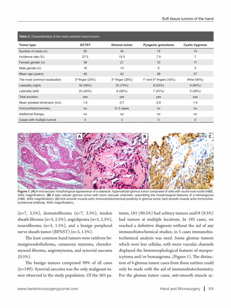
Figure 1. (A) A microscopic morphological appearance of a classical, hypercellular glomus tumor composed of cells with round-oval nuclei (H&E, 200x magnification). (B) A less cellular glomus tumor with more vascular channels; resembling the morphological features of a hemangioma (H&E, 400x magnification). (C) Anti-smooth muscle actin immunohistochemical positivity in glomus tumor (anti smooth muscle actin immunohis-tochemical antibody, 400x magnification).
tients, 181 (90.5%) had solitary tumors and19 (9.5%) had tumors at multiple locations. In 195 cases, we reached a definitive diagnosis without the aid of any immunohistochemical studies; in 5 cases immunohis-tochemical analysis was used. Some glomus tumors which were less cellular, with more vascular channels displayed the histomorphological features of myoper-icytoma and/or hemangioma. (Figure-1). The distinc-tion of 4 glomus tumor cases from these entities could only be made with the aid of immunohistochemistry. For the glomus tumor cases, anti-smooth muscle ac-

Hacisalihoglu UP and Ozcelik IB
Year 2018 | Volume X | Issue X | X-XXX | Hand and Microsurgery
Figure 2. The histomorphological and immunohistochemical characteristics of synovial sarcoma. (A) Photomicrograph is demonstrating syno-vial sarcoma: tumor cells with spindle-shaped, hyperchromatic nuclei and scant cytoplasm, arranged in fascicles. (H&E, 400X). (B) Photomi-crograph demonstrating the biphasic part of synovial sarcoma: spindle-shaped tumor cells (arrowheads) and epithelial cells forming glandular structures (arrows). (H&E, 400X). (C) Photomicrograph is demonstrating epithelial cells forming glandular structures and cords (anti-cytokeratin (AE1/AE3) immunohistochemical antibody, 400X). (D) Photomicrograph is demonstrating membranous staining with the anti-BCL-2 antibody (anti-Bcl-2 immunohistochemical antibody, 100X).
A B
DC
tin (Dako flex, clone 1A4, USA), anti-calponin (Dako Flex, clone CALP, USA), and anti-CD34 (Dako Flex, clone QBEnd 10, USA) immunohistochemical anti-bodies were used. The diagnosis of synovial sarcoma could only be reached by applying immunohistochem-istry since this tumor should always be distinguished from other mesenchymal tumors such as malignant peripheral nerve sheath tumor or solitary fibrous tu-mor. For the synovial sarcoma case, anti-cytokeratin (Dako Flex, clone AE1/AE3, USA), anti-S100 (Dako flex, polyclonal rabbit, USA), anti-beta catenin (Dako
flex, clone beta-catenin-1, USA), anti-CD34 (Dako flex, clone QBEnd 10, USA), anti-BCL-2 (Dako flex, clone124, USA), anti-smooth muscle actin (Dako flex, clone 1A4, USA), and anti CD117 (Dako flex, clone 104D2, USA) immunohistochemical antibodies were used. All of the glomus tumor cases displayed positive immunoreaction with smooth muscle actin, calponin, and CD34. In the synovial sarcoma case, we obtained positive immunoreaction with pan-cytokeratin, BCL-2, and beta-catenin and negative immunoreaction with CD34 (Figure 2).
Soft tissue tumors of the hand
Hand and Microsurgery | XXwww.handmicrosurgeryjournal.com
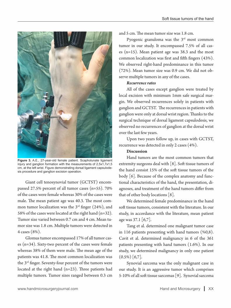
Figure 3. A.E., 27-year-old female patient. Scapholunate ligament injury and ganglion formation with the measurements of 2,5x1,7x1,5 cm, at the left wrist. Figure demonstrating dorsal ligament capsulode-sis procedure and ganglion excision operation.
Giant cell tenosynovial tumor (GCTST) encom-passed 27.5% percent of all tumor cases (n=55). 70% of the cases were female whereas 30% of the cases were male. The mean patient age was 40.3. The most com-mon tumor localization was the 3rd finger (24%), and 58% of the cases were located at the right hand (n=32). Tumor size varied between 0.7 cm and 4 cm. Mean tu-mor size was 1.8 cm. Multiple tumors were detected in 4 cases (8%).
Glomus tumor encompassed 17% of all tumor cas-es (n=34). Sixty-two percent of the cases were female whereas 38% of them were male. The mean age of the patients was 41.8. The most common localization was the 3rd finger. Seventy-four percent of the tumors were located at the right hand (n=23). Three patients had multiple tumors. Tumor sizes ranged between 0.3 cm
and 5 cm. The mean tumor size was 1.8 cm.Pyogenic granuloma was the 3rd most common
tumor in our study. It encompassed 7.5% of all cas-es (n=15). Mean patient age was 38.3 and the most common localization was first and fifth fingers (43%). We observed right-hand predominance in this tumor (72%). Mean tumor size was 0.9 cm. We did not ob-serve multiple tumors in any of the cases.
Recurrence ratesAll of the cases except ganglion were treated by
local excision with minimum 1mm safe surgical mar-gin. We observed recurrences solely in patients with ganglion and GCTST. The recurrences in patients with ganglion were only at dorsal wrist region. Thanks to the surgical technique of dorsal ligament capsulodesis, we observed no recurrences of ganglion at the dorsal wrist over the last few years.
Upon two years follow up, in cases with GCTST, recurrence was detected in only 2 cases (4%).
DiscussionHand tumors are the most common tumors that
extremity surgeons deal with [8]. Soft tissue tumors of the hand consist 15% of the soft tissue tumors of the body [6]. Because of the complex anatomy and func-tional characteristics of the hand, the presentation, di-agnoses, and treatment of the hand tumors differ from that of other body locations [8].
We determined female predominance in the hand soft tissue tumors, consistent with the literature. In our study, in accordance with the literature, mean patient age was 37.1 [6,7].
Tang et al. determined one malignant tumor case in 116 patients presenting with hand tumors (%0,8). Cavit et al. determined malignancy in 6 of the 361 patients presenting with hand tumors (1.6%). In our study, we determined malignancy in only one patient (0.5%) [6,7].
Synovial sarcoma was the only malignant case in our study. It is an aggressive tumor which comprises 5-10% of all soft tissue sarcomas [9]. Synovial sarcoma

Hacisalihoglu UP and Ozcelik IB
Year 2018 | Volume X | Issue X | X-XXX | Hand and Microsurgery
at the upper extremities comprises 10-15% of the whole body synovial sarcomas whereas synovial sarcoma of the hand comprises 4% of all extremity synovial sar-comas [10]. Synovial sarcoma is seen more commonly in the carpal region of the hand [10]. In our study, the most common localization was the carpal region of the wrist, in accordance with the literature.
The ganglion is an entity which is classified in be-nign soft tissue tumors and pseudotumors [11]. It pre-sents as white colored, thin-walled pseudocystic lesion (Figure 3). In our study, we determined that ganglion was the most common space occupying lesion; and the most common localization of the ganglion was the wrist (n=69, %68). Our results were in accordance with the literature [6,7].
Tumors such as myxoid plexiform fibrous histiocy-toma can mimic ganglion clinico-radiology. Therefore histopathological interpretation is necessary in order to reach a definitive diagnosis in cases with the pre-liminary diagnosis of ganglion [12,13]. Recurrences are common in cases with incomplete resection [13]. Tang et al., determined recurrence in 3 of 66 ganglion cases (4.5%) [7]. In our study, there was no recurrence in any of the ganglion cases during 2 years follow up.
GCTST, which was formerly called pigmented vil-lonodular synovitis, is a benign, locally aggressive pro-liferative disorder of the synovium which is composed of osteoclasts like giant cells and mononuclear inflam-matory cells. It can be localized at joint, bursa and ten-don sheath [14]. Localized form is most commonly seen at hands and feet [15]. It is the most common lesion localized at hand following ganglion [1,15,16]. In our study, we determined GCTST to be the second most common space occupying lesion as seen in the literature.
GCTST may be seen in all age groups, but it is seen most commonly between the ages 30 and 50. The fe-male/male ratio is 2 [15]. In our study, the mean age of patients with GCTST is 40, and all of the patients are female, as seen in the literature. In a study of Irmak
F, et al. it is stated that index finger was the most com-mon localization of GCTST [17]. Ushijima et al. deter-mined that the most common localization of GCTST was the fingers; whereas in a study of Di Grazia et al., hand GCTST’s were most commonly localized at the 3rd finger [18,19]. In a study of Briet et al., which has been conducted with 126 patients, the most common localization of this tumor was determined to be at the second finger [20]. Similarly, in a study of Cavit et al., it is reported that the most common localization of GCTST was the second finger (33.75%) [6]. In our study, we determined the most common localization of GCTST to be the 3rd finger (n=13, 28%).
In some patients with GCTST, due to the circum-ferential invasion of the lesion, total excision of the le-sion is impossible, thus leading recurrences [19]. In a study of Di Grazia et al., postoperative recurrence was detected in 4.7% of GCTST patients [19]. In a case se-ries composed of 80 GCTST, reported by Cavit et al., recurrence was reported in only one case [6]. In our study, upon two years follow up, we also observed re-currence in only one case (4%). The recurrent tumor was located at the 4th finger, at proximal interphalan-geal joint level and invaded the extensor mechanism. An excisional biopsy was performed by excoriating the tumor from the extensor mechanism. At the 1.5 years follow up, there was a progression in the swelling. Re-currence was detected by magnetic resonance imaging.
Glomus tumor is a mesenchymal neoplasm com-posed of cells that closely resemble the modified smooth muscle cells of the normal glomus body [21]. Most of the glomus tumors are benign but occasion-ally they present with malignant histopathological fea-tures with aggressive clinical behavior [22,23]. Its syn-onyms are glomangioma and glomangiomyoma [21]. Ten percent of the glomus tumors were determined to arise in multiple localizations. In the present study, this rate was 12%. Most of the glomus tumors are seen in young adults. In the present study, the mean age of the patients with glomus tumor was 41, in accordance with
Soft tissue tumors of the hand
Hand and Microsurgery | XXwww.handmicrosurgeryjournal.com
the literature. In a case series of Tomak et al., it is stated that, in 75% of the cases, the localization of the glomus tumor was subungual [23]. In our study, the most com-mon localization of the glomus tumor was the nail bed of the 3rd finger (n=7, %28).
Glomus tumor typically occurs in young adults. No sex predilection is seen, except in subungual le-sions, which are far more common in women [21]. In our study, 5 of 7 cases with glomus tumor at subungual localization were female (71%).
The gold standard treatment of glomus tumor is transungual or periungual surgical excision [24].
The excision of the lesions should be made me-ticulously because of its proximity to the neurovas-cular structures located at the fingers. The recurrence of the tumor is highly uncomfortable because of the never-ending pain [21]. In our study, we observed no recurrences.
Pyogenic granuloma is a benign superficial vas-cular tumor. In a study of Irmak, et al. conducted with 426 patients, pyogenic granuloma was stated to be the most common hand tumor with a 25% incidence. In the present study, it is the third most common tumor with an incidence of 7.5%. In pyogenic granuloma, most of the patients under 18 years are men [25]. Most of the patients between 19 and 40 years are women. The most common localizations are gingiva and fin-gers. The tumor typically enlarges during the first years of life, and later regresses [25]. Histopathologically, pyogenic granuloma is characterized by the relatively well-demarcated proliferation of the small capillaries in a multilobular pattern. 15 % of the hemangiomas are seen at hand and hand is the 3rd most common locali-zation of the hemangiomas seen in the whole body [6]. Patients with pyogenic granuloma who do not respond to conservative treatments such as cauterization, laser therapy or steroid injections attend to the hand and mi-crosurgery departments. This may be one of the main reasons for the low patient population in our study.
The surgical therapy of the pyogenic granuloma is
the complete excision of the lesion. In the literature, the recurrence rate was determined to be 19% during the five years follow up [6]. In our study, we determined no recurrence during the two years follow up.
ConclusionIn the present study, we determined that the most
common space-occupying lesion in hand was ganglion. GCTST was the most common soft tissue tumor fol-lowed by vascular tumors such as pyogenic granuloma, cystic, and cavernous hemangioma. In this study, soft tissue tumors of the hand were mostly (99%) benign, and the only malignant tumor was synovial sarcoma.
Conflict of interest statementThe authors have no conflicts of interest to declare.References
1. Fischer C, Montgomery EA, Thway K. Preface. In: Fischer C, Montgomery EA, Thway K (eds.) Bi-opsy interpretation of soft tissue tumors. Wolters Kluver Lippincott Williams&Wilkins, New York, 2011.
2. Fletcher CDM. WHO classification of soft tissue tumors. In: Fletcher CDM, Bridge JA, Hogen-doorn PCW, Mertens F (eds.) The WHO Classifi-cation of Tumours of Soft Tissue and Bone. IARC Press, Lyon, 2013:9.
3. Athanasian EA. Malignant Bone and Soft Tis-sue Sarcomas of the Hand. J Am Soc Surg Hand 2004;4:60–72.
4. Hsu CS, Hentz VR, Yao J. Tumors of the Hand. Lancet Oncol 2007;8:157–66.
5. Fletcher CDM, Gronchi A. Tumors of soft tis-sue-Introduction. In: Fletcher CDM, Bridge JA, Hogendoorn PCW, Mertens F (eds.) The WHO Classification of Tumours of Soft Tissue and Bone. IARC Press, Lyon, 2013:14.
6. Cavit A, Özcanli H, Sançmiş M, Ocak GA, Gürer Eİ. Tumorous Conditions of the Hand: A Retro-spective Review of 402 Cases. Turk Patoloji Derg 2018;34:66-72.
7. Tang ZH, Rajaratnam V, Desai V. Incidence and an-

Hacisalihoglu UP and Ozcelik IB
Year 2018 | Volume X | Issue X | X-XXX | Hand and Microsurgery
atomical distribution of hand tumors: a Singapore study. Singapore Med J 2017;58:714-6.
8. Zyluk A, Mazur A. Statistical and histological anal-ysis of tumors of the upper extremity. Obere Ex-trem 2015;10:252-7.
9. Weiss SW, Goldblum JR. Synovial sarcoma. In: Weiss SW, Goldblum JR (eds.) Enzinger and Weiss’s soft tissue tumours. Mosby, London, 2001:309-46.
10. Outani H, Hamada K, Oshima K, Joyama S, Naka N, Araki N, et al. Clinical outcomes for patients with synovial sarcoma of the hand. Springerplus 2014;3:649.
11. Weiss SW, Goldblum JR. Ganglion. In: Weiss SW, Goldblum JR (eds.) Enzinger and Weiss’s soft tis-sue tumours, Mosby, London, 2001:58-9.
12. Chih YL, Lan J, Huang HY. Myxoid Plexiform Fi-brohistiocytic Tumor Masquerading as Ganglion Cyst: A Case Report and Literature Review. Case Rep Pathol 2017;2017:ID5370894.
13. Bicer OS. The Epidemiology of Tumors and Tu-mor-like Lesions of the Hand and the Wrist. Clin Exp Med 2013;1:343-52.
14. Noailles T, Brulefert K, Briand S, Longis PM, Andrieu K, Chalopin A, et al. Giant cell tumor of tendon sheath: Open surgery or arthroscopic syn-ovectomy? A systematic review of the literature. Orthop Traumatol Surg Res 2017;103:809-14.
15. Weiss SW, Goldblum JR. Giant cell of tumor of the tendon sheath. In: Weiss SW, Goldblum JR (eds.) Enzinger and Weiss’s soft tissue tumours. Mosby, London, 2001:23.
16. Durmus M, Yapici M, Avşar S, Yiğit N, Bayram Y. Giant cell tumor of the tendon sheath restricting joint movement in the thumb: A case study and
review of literature. Hand Microsurg 2015;4:16-9.17. Irmak F, Basdelioglu K, Sirvan SS, Sevim KZ, Ye-
silada AK. Benign soft tissue tumors of the hand: A retrospective review of 17-year experience. Hand Microsurg 2018;7:88-92.
18. Ushijima M, Hashimoto H, Tsuneyoshi M. Giant cell tumor of the tendon sheath (nodular tenosyn-ovitis). A study of 207 cases to compare the large joint group with the common digit group. Cancer 1986;57:875-84.
19. Di Grazia S, Succi G, Fragetta F, Perrotta RE. Giant cell tumor of tendon sheath: study of 64 cases and review of literature. G Chir 2013;5-6:149-52.
20. Briët JP, Becker SJ, Oosterhoff TC, Ring D. Giant cell tumor of tendon sheath. Arch Bone Jt Surg 2015;3:19-21.
21. Folpe AL, Brems H, Legius E. Glomus tumor. In: Fletcher CDM, Bridge JA, Hogendoorn PCW (eds.) WHO Classification of Tumors of Soft Tis-sue and Bone. IARC Press, Lyon, 2013:116-7.
22. Shugart RR, Soule EH, Johnson EW. Glomus tu-mor. Surg Gynecol Obstet 1963;117:334–40.
23. Tomak Y, Akcay I, Dabak N, Eroglu L. Subungual glomus tumours of the hand: diagnosis and treat-ment of 14 cases. Scand J Plast Reconstr Surg Hand Surg 2003;37:121-4.
24. Shukla A, Verma V, Shukla R, Chaudhary R, Shar-ma S, Sajid M. Love sign’s love for glomus tumor. Hand Microsurg 2018;7:105-8.
25. Fischer C, Montgomery EA, Thway K. Pyogen-ic granuloma/lobular capillary hemangioma. In: Fischer C, Montgomery EA, Thway K (eds.) Bi-opsy interpretation of soft tissue tumors. Wolters Kluver Lippincott Williams&Wilkins, New York, 2011:399-400.
© 2018 Turkish Society for Surgery of the Hand and Upper Exremity. This is an open access article licensed under the terms of the Creative Commons Attribution NonCommercial ShareAlike 4.0 (https://creativecommons.org/licenses/by-nc-sa/4.0/) which permits unrestricted, noncommercial use, distribution and reproduction
in any medium, provided the work is properly cited.