epidemiology of human rhinoviruses -...
TRANSCRIPT

Department of Microbiology National Public Health Institute
Helsinki, Finland and
Department of Biological and Environmental Sciences Faculty of Biosciences
University of Helsinki, Finland
Soile Blomqvist
EPIDEMIOLOGY OF HUMAN RHINOVIRUSES
Academic dissertation
To be publicly discussed, with the permission of the Faculty of Biosciences of the University of Helsinki,
in the Auditorium 1041, Biocenter 2, Viikinkaari 5, Helsinki, on November 12th, 2004, at 12 noon.
Helsinki 2004

Supervised by Professor Tapani Hovi Department of Microbiology National Public Health Institute Helsinki, Finland Reviewed by Professor Timo Hyypiä Department of Virology University of Turku Turku, Finland Docent Anne Pitkäranta Department of Otorhinolaryngology University of Helsinki, Helsinki, Finland Opponent Docent Mika Mäkelä Skin and Allergy Hospital University of Helsinki Helsinki, Finland Publications of the National Public Health Institute KTL A17/2004 ISBN 951-740-469-7 print ISSN 0359-3584 print ISBN 951-740-470-0 pdf ISSN 1458-6290 pdf Publisher National Public Health Institute Mannerheimintie 166 FIN-00300 HELSINKI Copyright National Public Health Institute Yliopistopaino Helsinki 2004

To Juuso and Minja

EPIDEMIOLOGY OF HUMAN RHINOVIRUSES 4
CONTENTS ABSTRACT ...................................................................................................................6
LIST OF ORIGINAL PAPERS ...................................................................................7
ABBREVIATIONS........................................................................................................8
1 INTRODUCTION .............................................................................................9
2 REVIEW OF THE LITERATURE...............................................................10
2.1 Picornaviruses....................................................................................................10 2.2 Human Rhinoviruses .........................................................................................11
2.2.1 Early History Of Rhinovirus Research ............................................... 11 2.2.2 Assignment Of Rhinovirus Prototype Strains..................................... 11 2.2.3 General Characteristics Of Rhinoviruses............................................ 13 2.2.4 Structural Aspects............................................................................... 14
2.2.4.1 Structure Of Rhinoviruses ...........................................................14
2.2.4.2 Antigenic Structure And Neutralization Mechanisms.................16
2.2.4.3 Organization And Structure Of The Genome..............................17
2.2.5 Life Cycle Of Rhinoviruses ................................................................ 18 2.2.6 Subgroups Of Rhinoviruses................................................................ 19
2.2.6.1 Growth Properties In Cell Cultures .............................................19
2.2.6.2 Receptor Usage............................................................................19
2.2.6.3 Antiviral Sensitivity.....................................................................20
2.2.6.4 Antigenic Relationships...............................................................20
2.2.6.5 Phylogenetic Relationships..........................................................21
2.2.7 Rhinoviruses As Infectious Agents In Humans.................................. 21 2.2.7.1 Natural Course Of Rhinovirus Infection......................................21
2.2.7.2 Complications Of Rhinovirus Infection.......................................23
2.2.8 Diagnosis Of Rhinovirus Infections ................................................... 24 2.2.8.1 Virus Isolation .............................................................................24
2.2.8.2 Identification Of Rhinovirus Serotypes .......................................25
2.2.8.3 Antigen Detection........................................................................25
2.2.8.4 Detection Of Viral RNA..............................................................25
2.2.8.5 Detection Of Rhinovirus-Specific Antibodies.............................26

EPIDEMIOLOGY OF HUMAN RHINOVIRUSES 5
2.2.9 Epidemiology Of Human Rhinoviruses.............................................. 26 2.2.9.1 General Epidemiology .................................................................26
2.2.9.2 Transmission................................................................................27
2.2.9.3 Age-Dependent Variation ............................................................29
2.2.9.4 Seasonal Variation .......................................................................29
2.2.9.5 Prevalence Of Specific Rhinovirus Serotypes .............................30
2.2.9.6 Molecular Epidemiology .............................................................33
3 AIMS OF THE STUDY ..................................................................................35
4 MATERIALS AND METHODS....................................................................36
4.1 Clinical Specimens ............................................................................................36 4.2 Definitions .........................................................................................................36 4.3 Virus Strains ......................................................................................................36 4.4 Cell Lines...........................................................................................................38 4.5 Rhinovirus Isolation In Cell Culture..................................................................38 4.6 Assay For Acid Sensitivity ................................................................................38 4.7 Serological Assays.............................................................................................38 4.8 RNA Isolation....................................................................................................38 4.9 RT-PCR .............................................................................................................38 4.10 Detection Of RT-PCR Amplicons..............................................................40 4.11 Sequencing..................................................................................................40 4.12 Sequence Analysis ......................................................................................40
5 RESULTS AND DISCUSSION......................................................................41
5.1 Development Of Diagnostic Assays For Human Rhinoviruses (I, II) ...............41 5.1.1 Remarks On Virus Isolation ............................................................... 42 5.1.2 Microwell RT-PCR Hybridization ..................................................... 42
5.2 Occurrence Of Rhinovirus Infections During The First Two Years Of Life (II)46 5.3 Genetic Analysis Of Rhinovirus Strains (III) ....................................................48 5.4 Special Features Of Rhinovirus 87 (III, IV) ......................................................50
6 CONCLUSIONS..............................................................................................53
7 ACKNOWLEDGEMENTS ............................................................................54
8 REFERENCES ................................................................................................56

EPIDEMIOLOGY OF HUMAN RHINOVIRUSES 6
ABSTRACT BACKGROUND Human rhinoviruses (HRV) are the most frequent causative agents of the common cold and are also associated with such complications as acute otitis media (AOM) in children and acute community-acquired sinusitis (ACAS) in adults. Understanding of the clinical consequences of HRV infections is mainly dependent on the development of new detection assays, as the insensitivity of the conventional virus isolation method is now well known. The large number of HRV serotypes (at least 100) has hampered studies on individual HRV strains because serotyping of HRV field strains is virtually impossible in routine clinical diagnosis. MATERIALS AND METHODS A cohort of 329 children was followed from the age of two months to two years (FinOM Cohort Study). Nasopharyngeal aspirates (NPA) were collected every time the child had an upper respiratory infection or AOM. In the case of AOM, middle ear fluid (MEF) and paired sera were also obtained. In addition, four scheduled sera were collected from each child. The presence of HRV or HRV RNA in the NPA and MEF specimens was studied by virus isolation and RT-PCR hybridization. The antibodies to a mixture of HRV serotypes were determined by a complement fixation assay. A genomic region encoding the capsid protein VP4 and the N-terminus of VP2 was sequenced from all assigned HRV prototype strains, and the obtained sequences were subjected to phylogenetic analysis. RESULTS A rapid and sensitive microwell RT-PCR hybridization assay for detection of HRV RNA was developed and shown to be especially useful in the analysis of large numbers of clinical specimens. HRV infections were demonstrated to be very common in a cohort of young children. By the age of two years, 80% of children had experienced a virologically documented HRV infection, and more than 90% had HRV-specific antibodies. HRV was detected in 41% of AOM episodes. According to the phylogenetic analysis of HRV prototype and field strains, rhinoviruses clustered into two distinct genetic groups, HRV-A and HRV-B. One of the HRV serotypes, HRV87, was shown to previously be incorrectly assigned as a rhinovirus. It represents the same serotype as EV68 and belongs to enterovirus species HEV-D together with EV70. In contrast to other enteroviruses, HRV87 and EV68 are sensitive to low pH. CONCLUSIONS Rhinoviruses were shown to be very common in young children, and the close association between HRV infections and AOM was confirmed. Molecular-based methods (RT-PCR and sequencing) can be recommended as the primary tools for both diagnosis of HRV infections and characterization of HRV field strains. The acid sensitivity of HRV87 and EV68 indicates that the traditional classification of rhinoviruses and enteroviruses according to biological properties alone may be misleading.

EPIDEMIOLOGY OF HUMAN RHINOVIRUSES 7
LIST OF ORIGINAL PAPERS This thesis is based on the following original publications referred to in the text by their Roman numerals: I Blomqvist S., Skyttä A., Roivainen M. and Hovi T. 1999. Rapid detection of
human rhinoviruses in nasopharyngeal aspirates by a microwell reverse transcription-PCR-hybridization assay. Journal of Clinical Microbiology 37:2813-2816.
II Blomqvist S., Roivainen M., Puhakka T., Kleemola M. and Hovi T. 2002.
Virological and serological analysis of rhinovirus infections during the first two years of life in a cohort of children. Journal of Medical Virology 66:263-268.
III Savolainen C., Blomqvist S., Mulders M.N. and Hovi T. 2002. Genetic clustering
of all 102 human rhinovirus prototype strains: serotype 87 is close to human enterovirus 70. Journal of General Virology 83:333-340.
IV Blomqvist S., Savolainen C., Råman L., Roivainen M. and Hovi T. 2002. Human
rhinovirus 87 and enterovirus 68 represent a unique serotype with rhinovirus and enterovirus features. Journal of Clinical Microbiology 40:4218-4223.

EPIDEMIOLOGY OF HUMAN RHINOVIRUSES 8
ABBREVIATIONS ACAS acute community-acquired sinusitis AHC acute hemorrhagic conjunctivitis AOM acute otitis media ATCC American Type Culture Collection cDNA complementary DNA CF complement fixation COPD chronic obstructive pulmonary disease CPE cytopathic effect DAF decay-accelerating factor DNA deoxyribonucleic acid FinOM Studies Finnish Otitis Media Studies GMK green monkey kidney cell line HEV human enterovirus HRV human rhinovirus ICAM-1 intercellular adhesion molecule 1 Ig immunoglobulin LDL low-density lipoprotein MEF middle ear fluid mRNA messenger RNA NCR non-coding region NIAID National Institute of Allergy and Infectious Diseases NIm neutralizing immunogenic site NPA nasopharyngeal aspirate OME otitis media with effusion ORF open reading frame PCR polymerase chain reaction PV poliovirus RD rhabdomyosarcoma cell line RIVM National Institute for Public Health and the Environment RNA ribonucleic acid RT reverse transcriptase TCID tissue culture infectious dose VLDL very low-density lipoprotein VP virus protein WHO World Health Organization

INTRODUCTION
EPIDEMIOLOGY OF HUMAN RHINOVIRUSES 9
1 INTRODUCTION Acute upper respiratory infections, generally known as common colds, are the most frequent acute illnesses world-wide. The predominant role of human rhinoviruses (HRV) in the aetiology of the common cold was already established in the studies conducted in the 1960s, when only virus isolation techniques were in use. However, the lack of means for treating of rhinovirus infections and laborious diagnostic procedures have hindered progress in the studies on rhinovirus infections for some decades. Recently, rapid and sensitive molecular-based RT-PCR methods have replaced conventional virus isolation in rhinovirus diagnostics. Rhinovirus infections are estimated to account for approximately 50% of common colds in adults annually (Mäkelä et al., 1998) and 90% or more during the highest prevalence in autumn (Arruda et al.,1997; Mäkelä et al., 1998). More importantly, the exploitation of PCR is continuously increasing evidence of the association of rhinoviruses with more severe diseases such as acute otitis media (AOM), acute community-acquired sinusitis (ACAS), wheezing and exacerbations of asthma and chronic obstructive pulmonary disease. Improved understanding of the role of rhinoviruses in pathogenesis of these diseases, especially in AOM and ACAS, is anticipated to reduce unnecessary antibiotic use. With the ongoing development of anti-rhinoviral drugs, attention should now be directed towards evaluating both the laboratory methods for rapid and accurate rhinovirus diagnoses, and the clinical consequences of rhinovirus infections. While our knowledge of the frequency and clinical importance of rhinovirus infections is growing rapidly, we continue to know little about individual circulating rhinovirus strains, which may have differences in, for example, pathogenicity. Elegant molecular methods have been applied in epidemiological studies of human enterovirus strains for many years, but molecular epidemiological studies on circulating rhinoviruses are still in the initial stages.

REVIEW OF THE LITERATURE
EPIDEMIOLOGY OF HUMAN RHINOVIRUSES 10
2 REVIEW OF THE LITERATURE 2.1 PICORNAVIRUSES
The family Picornaviridae consists of small, non-enveloped RNA viruses and includes many common human and animal pathogens, e.g. polio, hepatitis A and food-and-mouth disease viruses. According to the most recent virus taxonomic proposal, the family contains nine genera (Table 1), with rhinoviruses and enteroviruses being the largest (Stanway et al., 2004). Each genus contains one or more species, and each species one or more antigenically distinct serotypes. Altogether, the family Picornaviridae contains over 200 different serotypes. The taxonomy of Picornaviridae is changing. New tentative species have been proposed in three genera (enterovirus, rhinovirus and hepatovirus), and over 20 viruses are waiting for assignment in the family. Table 1. Taxonomic structure of the family Picornaviridae. Genus Species Number of serotypes Enterovirus Bovine enterovirus 2
Human enterovirus A 13 Human enterovirus B 41 Human enterovirus C 9 Human enterovirus D 2 Poliovirus 3 Porcine enterovirus A 1 Porcine enterovirus B 2 Simian enterovirus A 2 Rhinovirus Human rhinovirus A 75 Human rhinovirus B 25 Cardiovirus Encephalomyocarditis virus 4 Theilovirus 3 Aphthovirus Equine rhinitis A virus 1
Foot-and-mouth disease virus 7 Hepatovirus Hepatitis A virus 2 Parechovirus Human parechovirus 3 Ljungan virus 1 Erbovirus Equine rhinitis B virus 2 Kobuvirus Aichi virus 1 Bovine kobuvirus 1 Teschovirus Porcine teschovirus 11

REVIEW OF THE LITERATURE
EPIDEMIOLOGY OF HUMAN RHINOVIRUSES 11
2.2 HUMAN RHINOVIRUSES
2.2.1 EARLY HISTORY OF RHINOVIRUS RESEARCH
The first successful in vitro isolation of rhinovirus was carried out by serially passaging the virus in human lung explants. This DC strain, later HRV9, was isolated in 1953 from a nasal washing of a cell biologist who worked at the Common Cold Research Unit, England, and had symptoms of upper respiratory infection (Andrewes et al., 1953; Tyrrell & Fielder, 2002). Some years later, cytopathogenic agents were isolated from nasopharyngeal washings in rhesus monkey kidney tissue cultures in two laboratories in the United States (Price, 1956; Pelon et al., 1957). These two strains, JH and 2060, were shown to be serologically identical and were named Echo 28 (later HRV1A) because of the properties they shared with enteroviruses. The breakthrough in rhinovirus isolations occurred in the early 1960s. Tyrrell and Parsons (1960) isolated strain HGP (later HRV2) in cultures of human embryonic kidney cells by imitating the conditions of the nose. They lowered the incubation temperature to 33�C and the pH of the tissue culture medium to around 7.0, slowly rotating the cultures during the incubation. At the same time, new, sensitive, semicontinuous strains of diploid human embryonic lung fibroblasts (e.g. WI-26) were developed (Hayflick & Moorehead, 1961). The “conditions of the nose” and new cell lines led to an explosive increase in isolation of new strains, many of which were found to be antigenically distinct (Taylor-Robinson & Tyrrell, 1962). Laboratories assigned the new common cold-related viruses in various ways; the names Salisbury strain, murivirus, respirovirus, rhinovirus, coryzavirus and ERC (ECHO-rhino-coryzavirus) were in use (Hilleman, 1967). However, the biological relatedness of the strains was soon noticed, and the basic properties of the virus group, especially lability in an acidic environment, were established. The name “rhinovirus”, suggested by Andrewes in 1961 because of the special adaptation of the viruses for growth in nasal epithelium, was chosen as the name for the entire virus group (Tyrrell & Chanock, 1963). 2.2.2 ASSIGNMENT OF RHINOVIRUS PROTOTYPE STRAINS
The rapid identification of new rhinovirus serotypes in the early 1960s made comparison of epidemiological data from different laboratories difficult. The National Institute of Allergy and Infectious Diseases (NIAID) and the World Health Organization (WHO) initiated a Rhinovirus Collaborative Programme, the aim of which was to compare antigenic relationships of rhinovirus strains to achieve an acceptable rhinovirus designation scheme. The Children’s Hospital in Columbus, Ohio, functioned as a reference laboratory that performed reciprocal neutralization tests with submitted candidate rhinoviruses and sera. In addition, each laboratory submitting new virus strains was ordered to test their own candidate viruses against all available sera.

REVIEW OF THE LITERATURE
EPIDEMIOLOGY OF HUMAN RHINOVIRUSES 12
Consequently, each candidate virus was tested against each specific antiserum independently in at least two laboratories. The three phases of the Rhinovirus Collaborative Programme are shown in Table 2. In Phase I, 68 candidate viruses and corresponding sera were compared by standard neutralization assays in HeLa cells (Conant & Hamparian,1968). Six pairs of viruses and three groups of three viruses were found to be serologically identical. Of these, the virus strain that had been submitted first to the Programme was assigned a prototype strain of a given serotype. In addition, two strains, Echo 28 and B632, were shown to be significantly related, but not identical, and B632 was assigned as a subtype of Echo 28. These strains were given serotype names HRV1A and HRV1B (Kapikian et al., 1967). The second phase with 73 rhinovirus candidate strains was carried out in the late 1960s. Thirty-four distinct prototype strains were numbered from HRV56 to HRV89 and the results were published in 1971 (Kapikian et al., 1971). In 1973, cross-neutralization tests were completed for 25 new candidate viruses (Phase III). A lack of financial support prevented completion of the third phase, and the results were only published in 1987 (Hamparian et al., 1987). In Phase III, 11 new prototype strains were accepted, and the numbering was extended to 100. During this stage all available epidemiological data were interpreted so that rhinoviruses identified earlier were still circulating and new serotypes were not constantly emerging. During the three-phase NIAID and WHO co-ordinated Rhinovirus Collaborative Programme, which continued for eight years, 166 submitted rhinovirus candidate strains were tested and 100 rhinovirus serotypes (and one subtype) were assigned a number and a prototype strain. Since the third phase of the Collaborative Programme, only one strain, Hanks, has been proposed as a new serotype (Gwaltney et al.,1978). In recent years, attempts to identify the serotypes of isolated rhinovirus strains have been infrequent, and many unrecognized rhinovirus serotypes may still exist. Table 2. Rhinovirus Collaborative Programme for assigning rhinovirus prototype strains. Phase No. of candidate/ Assigned serotypes Reference
accepted strains I 68 / 55 HRV1* - HRV55 Kapikian et al. (1967) II 73 / 34 HRV56 - HRV89 Kapikian et al. (1971) III 25 / 11 HRV90 - HRV100 Hamparian et al. (1987) *HRV1 has two subtypes, HRV1A and HRV1B.

REVIEW OF THE LITERATURE
EPIDEMIOLOGY OF HUMAN RHINOVIRUSES 13
2.2.3 GENERAL CHARACTERISTICS OF RHINOVIRUSES
Rhinoviruses share the basic properties of viruses in the family Picornaviridae. All picornaviruses are small (about 30 nm) and spherical in shape, and are composed of an icosahedral protein capsid enclosing a single strand of positive-sense RNA. From a phylogenetic view, the closest relatives of rhinoviruses are human enteroviruses (HEV), but these two viruses differ, for example, with regard to behaviour in an acidic environment. Rhinoviruses withstand pH values ranging from 6.0 to 8.0 but are readily inactivated below a pH of 6.0. Infectivity of human rhinovirus 14 (HRV14) is reduced at pH 5.0 in 20 minutes, and is totally lost at pH 3.0 in 10 seconds. The inactivation of HRV14 at pH 5.0 is dependent on temperature; a nearly 99% loss in infectivity occurs almost immediately at 24°C, but not in 5 minutes at 0°C (Hughes et al., 1973). In contrast to rhinoviruses, enteroviruses are stabile at low pH, but the molecular basis of the difference in acid sensitivity between rhino- and enteroviruses is not known. Acidification of HRV14 is shown to induce irreversible conformational changes both at the surface of the virus and on the capsid interior (Giranda et al., 1992). However, the acid sensitivity of rhinoviruses may be altered since acid-resistant mutants of HRV14 have been obtained by serial exposure of the viruses to pH 4.5 followed by passaging in HeLa cells (Skern et al., 1991). Due to the lack of a lipid envelope, rhinoviruses are resistant to ether, chloroform, fluorocarbon and detergents. Proteolytic enzymes, like trypsin, destroy the infectivity of some, but not all, of the serotypes (Stott & Killington, 1972). The infectivity of rhinoviruses is maintained at 24-37°C, and thus, they can survive on environmental surfaces, such as door handles and coffee cups, for hours to days (Hendley et al., 1973). Most rhinoviruses are also stabile at higher temperatures (at 50°C), but marked variation exists between the different serotypes. At frosty temperatures, rhinoviruses can survive for years (Couch, 2001).

REVIEW OF THE LITERATURE
EPIDEMIOLOGY OF HUMAN RHINOVIRUSES 14
2.2.4 STRUCTURAL ASPECTS
2.2.4.1 STRUCTURE OF RHINOVIRUSES
Detailed atomic structures of five rhinoviruses, HRV1A (Kim et al., 1989), HRV2 (Verdaguer et al., 2000), HRV3 (Zhao et al., 1996), HRV14 (Rossmann et al., 1985; Arnold & Rossmann, 1990) and HRV16 (Oliveira et al., 1993; Hadfield et al., 1997), have been determined by X-ray crystallography. The protein capsid of rhinoviruses is composed of 60 copies of protein subunits, protomers, each of which comprise a single molecule of four polypeptides, VP1 to VP4, VP1 being the most exposed. Proteins VP1 to VP3 have the same overall structural conformation, an eight-stranded antiparallel ß-barrel (Figure 1a), without having any remarkable sequence homology. Carboxyl (C) termini of proteins VP1 to VP3 are located on the surface of the virion, while amino (N) termini are in the interior. The smallest capsid protein VP4 lies on the inner surface of the capsid and is in intimate contact with RNA. (Racaniello, 2001). The capsid of rhinoviruses has icosahedral symmetry (Figure 1b). There is a star-shaped plateau at the fivefold axis of symmetry, which is surrounded by a deep cleft or canyon, and another protrusion at the threefold axis (Rossmann et al., 1985). The canyon separates the major part of five VP1 subunits, clustered about a pentamer axis, from the surrounding VP2 and VP3 subunits. Within the core of VP1, just beneath the canyon floor, is a hydrophobic tunnel, a “pocket”. (Racaniello, 2001) The overall folding pattern is well preserved in proteins VP1, VP2 and VP3 in all studied rhinoviruses to date. The structural differences between rhinovirus serotypes are located mainly on the external surface loops connecting the ß-strands and on the internal capsid surfaces, particularly in the conformation of the N-termini of proteins VP4 and VP1 (Verdaguer et al., 2000). The N-termini of VP4 and VP1 together contribute to the ß-barrel structure in HRV2, which resembles the structure found in enteroviruses (Verdaguer et al., 2000). The N-terminal ends of VP4 and VP1 are disordered in HRV3 (Zhao et al., 1996) and HRV14 (Rossmann et al., 1985), as is the VP4 N-terminus in HRV1A (Kim et al., 1989). The N-terminal amino acids of VP1 proteins form an amphipathic helix in HRV1A (Kim et al., 1989) and HRV16 (Hadfield et al., 1997).

REVIEW OF THE LITERATURE
EPIDEMIOLOGY OF HUMAN RHINOVIRUSES 15
´ Figure 1. Schematic views of the conformation of the picornavirus capsid protein (a) (Rossmann et al., 1985; Racaniello, 2001) and the icosahedral structure of the rhinovirus (b) (Rossmann et al., 1985; Racaniello, 2001). The figure is reprinted with the kind permission of the publisher.
a.
b.

REVIEW OF THE LITERATURE
EPIDEMIOLOGY OF HUMAN RHINOVIRUSES 16
2.2.4.2 ANTIGENIC STRUCTURE AND NEUTRALIZATION MECHANISMS
Neutralizing antigenic epitopes have been located on the surface of two rhinovirus serotypes, HRV2 and HRV14, by analysing neutralization-resistant mutants (Table 3). All antigenic epitopes were found in hypervariable regions of the capsid proteins at the highest points of the virus surface. Four different epitopes were shown to be involved in antibody-mediated neutralization of HRV14 (Sherry & Rueckert, 1985; Sherry et al., 1986). Three of the four epitopes, NImIB, NImII and NImIII, are composed of non-contiguous sequences located either on the same or on different capsid proteins. NImIA and NImIB lie very close to each other (Rossmann et al., 1985). The neutralization of HRV2 was shown to involve three antigenic sites, A, B and C. Site A is confined to VP1, between the fivefold symmetry axis and the canyon, and closely resembles the NImIA site of HRV14. Site B includes residues from all three external capsid proteins. The VP2 region of site B is partly analogous to the NImII site of HRV14 (Skern et al., 1987; Appleyard et al., 1990; Verdaguer et al., 2000). Several different mechanisms for antibody-driven neutralization of rhinovirus infectivity have been suggested, but not universally accepted, as reviewed by (Smith, 2001). These mechanisms include aggregation of viruses, stabilization of the viruses leading to prevention of uncoating, induction of conformational changes in the capsid, and steric blocking of cellular attachment. The major in vitro neutralization mechanism of antibodies targeted to all four antigenic sites on HRV14 is proposed to be steric blocking of attachment of the virus to cellular receptors (Colonno et al., 1989; Che et al., 1998; Smith, 2001). Antibodies to the NImIA site probably also stabilize the virus by binding bivalently across the twofold axes in the canyon region, preventing the conformational changes needed in the uncoating process (Smith et al., 1993a; Smith et al., 1993b; Che et al., 1998). Table 3. Amino acid residues involved in neutralizing antigenic epitopes of HRV2 and HRV14 determined by analysing neutralization-resistant mutants*.
VP1 VP2 VP3 HRV2 Site A 85,86,92 Site B 260,262,264,265,272,274 159,161,163,164 59,64 Site C 214,236,238 HRV14 NImIA 91,95 NImIB 83,85,138,139 NImII 210 156,158,159,161,162 NImIII 287 72,75,78,203 *Table modified from Verdaguer et al. (2000).

REVIEW OF THE LITERATURE
EPIDEMIOLOGY OF HUMAN RHINOVIRUSES 17
2.2.4.3 ORGANIZATION AND STRUCTURE OF THE GENOME
The genome of human rhinoviruses is a single-stranded, messenger-sense RNA molecule (Figure 2). The complete genomic sequences of five rhinovirus serotypes are available in GenBank (HRV1B (Hughes et al., 1988), HRV2 (Skern et al., 1985), HRV14 (Stanway et al., 1984), HRV16 (Lee et al., 1995) and HRV89 (Duechler et al., 1987). In addition, two complete sequences (HRV9 and HRV85) are published on the Picornavirus Home Page (Knowles, 1996). The length of the rhinovirus genome is 7102-7152 bases in HRV1B, HRV2, HRV9, HRV16, HRV85 and HRV89, and 7212 bases in HRV14. There are non-coding regions (NCR) at both the 5’ and 3’ ends of the genome. The 5’ NCR of the sequenced rhinoviruses is 610-625 bases long, and thus, some 100 bases shorter than that in enteroviruses. The 100-base-long insertion in enterovirus 5’ NCR, generally known as the hypervariable region, is located just before the protein-coding region. The 5’ NCR has conserved sequence and secondary structure stretches, which are involved in the initiation of replication and translation (reviewed in (Xiang et al., 1997)), and are shared by rhino- and enteroviruses (Rivera et al., 1988). A small protein, VPg, is covalently attached to the 5’ end of the genome (Lee et al., 1977) with its conserved third amino acid, tyrosine. VPg is encoded by a single viral gene (3B), and its length varies in rhinoviruses from 21 (HRV1B, HRV2, HRV9, HRV16, HRV85 and HRV89) to 23 (HRV14) amino acids. The 3’ NCR is 40-47 bases long, which is much shorter than the 70-120 bases found in enteroviruses. The genome ends with a stretch of poly(A). Residing between the 5’ and 3’ non-coding regions is a single open reading frame (ORF) that encodes a polyprotein 2100-2200 amino acids long. The polyprotein is translated as a precursor, which is further processed into individual structural and non-structural proteins. The P1 region encodes the four capsid proteins VP4, VP2, VP3 and VP1. The P2 and P3 regions encode seven proteins involved in protein processing and genome replication. (Racaniello, 2001). Figure 2. Schematic view of the organization of the rhinovirus genome.
P1 P2 P3
CAPSID PROTEINS NON-STRUCTURAL PROTEINS
POLYPROTEIN
5’ 3’
NCR NCR A n
VPg VP2 3D 2C VP3 VP4 VP1 2A 3A 3B 3C 2B

REVIEW OF THE LITERATURE
EPIDEMIOLOGY OF HUMAN RHINOVIRUSES 18
2.2.5 LIFE CYCLE OF RHINOVIRUSES
Replication of human rhinoviruses takes place in the cytoplasm of the host cell. The first step in rhinovirus life cycle is attachment to a cell surface receptor, which is either intercellular adhesion molecule 1 (ICAM-1) (major receptor group) (Greve et al., 1989; Staunton et al., 1989; Tomassini et al., 1989) or a member of the low-density lipoprotein (LDL) receptor family (minor receptor group) (Hofer et al., 1994; Gruenberger et al., 1995; Marlovits et al., 1998). ICAM-1 is a cell surface member of the immunoglobulin supergene family. The binding site of ICAM-1 has been located at the base of the canyon by cryoelectron microscopy and image reconstruction analysis of receptor-bound structures of HRV3 (Xing et al., 2003), HRV14 (Kolatkar et al., 1999) and HRV16 (Olson et al., 1993). The LDL receptor family consists of cell surface receptors, such as the LDL receptor, the very low-density lipoprotein (VLDL) receptor and the LDL receptor-related protein, which mediate the transport of lipoproteins into cells by receptor-mediated endocytosis. They are proposed to bind their ligands by electrostatic interactions with negatively charged ligand-binding domains. The VLDL receptor footprint in HRV2 is on the star-shaped dome of the icosahedral fivefold axis formed by the BC and HI loops of VP1 (Hewat et al., 2000). The attachment of the virus to the cellular receptor triggers conformational changes in the virus capsid, eventually leading to a release of viral RNA into the cytoplasm. This uncoating process is proposed to occur by different mechanisms in major and minor receptor group viruses. Binding of ICAM-1 to HRV14 initiates a rapid uncoating without a need for any cellular machinery (Greve et al., 1991; Casasnovas & Springer, 1994), whereas the release of HRV2 RNA into the cytoplasm is mediated by clathrin-dependent internalization of the virus into acidic endosomal compartments (Neubauer et al., 1987; Prchla et al., 1994; Schober et al., 1998; Snyers et al., 2003). In the cell cytoplasm, the viral genome is translated to provide viral proteins essential for genome replication and the production of new virus particles. The long polyprotein precursor first cleaves itself into the intermediates P1, P2 and P3 via two proteinases, 2A and 3C/3CD. P1 is then further cleaved to yield VP0, VP1 and VP3. VP0 is cleaved to VP4 and VP2 during viral assembly. Among other proteins synthesized are the viral RNA-dependent RNA polymerase and accessory proteins required for genome replication and mRNA synthesis. The positive-stranded RNA is copied to a negative-stranded intermediate, an RNA molecule with a complementary nucleotide sequence. Synthesis of the complementary (-) strain is initiated at the 3’ terminus of the RNA and primed by the protein VPg. This complementary (-) strain is subsequently used as a template in synthesizing large numbers of copies of the viral genome, which are used as mRNA for translation of more viral proteins or, later in the infection cycle, packaged into new virus particles. The new infectious viruses are assembled in the cytoplasm and finally released from the cell. (Racaniello, 2001). The time course of the replication cycle of rhinoviruses is similar in cell cultures and experimental infections in humans. In one-cycle in vitro growth experiments, a newly produced virus could frequently be detected in 5-7 hours, and the cycle of viral
REVIEW OF THE LITERATURE
EPIDEMIOLOGY OF HUMAN RHINOVIRUSES 19
replication was complete in 10-12 hours. However, depending on the cell line and serotype of the virus, the first appearance may be as late as 9 hours, with completion in 15-17 hours (Stott & Killington, 1972). In HRV39-challenged adult volunteers, the new virus was recovered in nasal washings 11.3 (range 8 -18) hours after inoculation. The virus titres remained almost stabile until 18 hours after challenge, then increasing again as a consequence of a second cycle of viral replication. The highest titres were reached 48 hours after virus challenge; titres began to decline on day 3 and remained low on days 4 and 5 (Harris & Gwaltney, 1996; Hendley & Gwaltney, 2004). 2.2.6 SUBGROUPS OF RHINOVIRUSES
The genus Rhinovirus contains a considerably large number of viruses, over 100 antigenically distinguishable types. Although the rhinoviruses share many common properties, such as overall structure and acid sensitivity, they do not comprise a fully homogeneous virus group, differing from each other in many important aspects, including growth properties in cell cultures, receptor usage and antiviral sensitivities. These differences, together with antigenic and phylogenetic relationships, have been exploited in classifying the rhinoviruses into subgroups. 2.2.6.1 GROWTH PROPERTIES IN CELL CULTURES
The first rhinovirus strains isolated from clinical specimens were classified as H or M strains according to their growth properties in different cell lines (Taylor-Robinson & Tyrrell, 1962; Hilleman, 1967). H strains grew only in human embryonic kidney cells, while M strains grew in both human and monkey kidney cells. Allocation of rhinoviruses into M or H strains has subsequently been shown to be of limited importance. Some of the H strains were adapted to grow in monkey cells (Douglas et al., 1966), and both M and H strains were isolated from rhinoviruses HRV15, HRV33 (Phillips et al., 1965) and HRV39 (Mufson et al., 1965). Most of the rhinovirus serotypes (at least HRV1A, HRV1B, HRV2, HRV29, HRV30, HRV31, HRV47 and HRV62) originally classified as M strains (Hilleman, 1967) belong to the minor receptor group of rhinoviruses. 2.2.6.2 RECEPTOR USAGE
Classification of rhinoviruses into either the major or minor receptor group was initiated before the receptors were known by pair-wise competition binding assays in HeLa R-19 cells (Abraham & Colonno, 1984), and after ICAM-1 was identified as a major receptor, the classification was completed with HeLa cell protection assays with monoclonal antibody to ICAM-1 (Uncapher et al., 1991). According to these results, the major receptor group consists of 91 serotypes (including the unnumbered strain Hanks) and the

REVIEW OF THE LITERATURE
EPIDEMIOLOGY OF HUMAN RHINOVIRUSES 20
minor receptor group of 10 serotypes, HRV1A, HRV1B, HRV2, HRV29, HRV30, HRV31, HRV44, HRV47, HRV49 and HRV62 (Uncapher et al., 1991). HRV87 was the only serotype that could not be classified into either of these groups. Unlike other rhinoviruses, its attachment to HeLa cells was inhibited by pre-treatment with neuraminidase, and thus, its receptor was suggested to be a sialoprotein (Uncapher et al., 991). The existence of two receptor groups was further confirmed by analysing the inhibitory activities of soluble ICAM-1 molecules. Soluble ICAM-1 inhibited the replication of all assigned major receptor group rhinoviruses, except HRV23 and HRV25, but did not affect the replication of minor receptor group rhinoviruses or HRV87 (Crump et al., 1993). 2.2.6.3 ANTIVIRAL SENSITIVITY
A variety of structurally unrelated antiviral compounds are capable of inhibiting the replication of rhinoviruses. All of these compounds share the same binding site, a hydrophobic pocket in capsid protein VP1, but have a different effect on distinct viruses. A systematic, multivariate analysis of 100 rhinovirus serotypes against a panel of 15 antiviral compounds divided rhinoviruses into two antiviral groups, A and B. Antiviral group A was shown to consist of 33 serotypes with a more than average susceptibility to elongated antiviral compounds such as WIN51711. Antiviral group B contains 67 serotypes that are susceptible to structurally shorter antiviral agents, including chalcone, dichloroflavan and R61837 (Andries et al., 1990; Andries et al., 1991). 2.2.6.4 ANTIGENIC RELATIONSHIPS
By the original definition of a rhinovirus serotype, a new serotype is approved only after it has been shown not to exhibit significant antigenic cross-reactivity with known serotypes (Kapikian et al., 1967). However, studies with hyperimmune rabbit and guinea pig sera have indicated many cross-reactions between the existing serotypes. According to a systematic analysis of antigenic relationships among 90 rhinovirus serotypes (HRV1-HRV89), 50 of these serotypes were divided into 16 antigenic groups, each of which contains 2-10 antigenically related serotypes (Cooney et al., 1982). Reciprocal neutralization was demonstrated for virus pairs HRV1A-HRV1B, HRV2-HRV49, HRV3-HRV14, HRV9-HRV32, HRV12-HRV78, HRV13-HRV41, HRV15-HRV74, HRV29-HRV44 and HRV36-HRV58 (Cooney et al., 1973, 1982). Many more serotypes have been shown to be related with one-way cross-reactions (HRV5-HRV42, HRV6-HRV14, HRV9-HRV67, HRV11-HRV40, HRV11-HRV74, HRV17-HRV42, HRV17-HRV70, HRV22-HRV61, HRV32-HRV67, HRV36-HRV50, HRV36-HRV89, HRV39-HRV54, HRV40-HRV56, HRV60-HRV38, HRV66-HRV77 and HRV76-HRV11) (Cooney & Kenny, 1970; Calhoun et al., 1974; Cooney et al., 1982). The observed heterotypic responses have usually been of a low order of magnitude, but sequential injections of related rhinovirus antigens have been shown to amplify the responses; the antigenic groups that include HRV13 and HRV41, and HRV9, HRV32 and HRV67, were determined this way (Cooney et al., 1975).
REVIEW OF THE LITERATURE
EPIDEMIOLOGY OF HUMAN RHINOVIRUSES 21
Heterotypic antibody responses have been demonstrated also in man after natural infection. Following natural infection with one of the related serotypes of HRV1A and HRV1B, HRV2 and HRV49, HRV23 and HRV30, or HRV29, HRV44 and HRV62, the antibody response to the second virus was almost equal to the homotype and was suggested to provide cross-protection for infection (Mogabgab et al., 1975). Besides multiple antigenic cross-reactions among the different rhinovirus serotypes, marked variation is another major feature in the antigenicity of rhinoviruses. Rapid antigenic variation has been demonstrated for HRV17 under in vitro immunological pressure (Patterson & Hamparian, 1997), and antigenic variants have also been described for HRV51 (Stott & Walker, 1969), HRV54 (Hamparian et al., 1987) and HRV91 (Hamparian et al., 1987). 2.2.6.5 PHYLOGENETIC RELATIONSHIPS
Genetically, human rhinoviruses cluster into two distinct groups, HRV-A and HRV-B. The existence of different genetic groups was suggested already when the first three complete genomic sequences, HRV2, HRV14 and HRV89, were determined, and HRV2 and HRV89 were found to be much more closely related to each other than to HRV14 (Duechler et al., 1987). The two groups were specified by Horsnell and colleagues in 1995 after sequencing a genomic region including the immunogenic site NImII from 12 more rhinovirus prototypes. In phylogenetic analysis, the viruses were shown to cluster into two genetic groups, group 1 containing more serotypes than HRV14-related group 2 (Horsnell et al., 1995). The same two groups were again observed after analysis of 5’ NCR sequences from 39 rhinovirus strains (both prototype and clinical strains) (Andeweg et al., 1999). 2.2.7 RHINOVIRUSES AS INFECTIOUS AGENTS IN HUMANS
2.2.7.1 NATURAL COURSE OF RHINOVIRUS INFECTION
A rhinovirus infection is initiated by delivery of the virus into the front of the nose or into the eye, where it passes down the lacrimal duct. A very small amount of the virus (as little as one tissue culture infectious dose50) can produce infection when deposited in the nose. Introduction of the rhinovirus directly into the mouth or throat does not initiate infection efficiently, as reviewed by Gwaltney and Hendley (1978). Rhinoviruses target the cells of the nasal epithelium. In some experimental infections, only a small subset of epithelial cells in upper airway tissues have been shown to become infected (Winther et al., 1986; Bardin et al., 1994; Arruda et al., 1995), but in natural infection, rhinovirus RNA has been detected in a high proportion of nasal epithelium cells by a sensitive in situ hybridization assay (Pitkäranta et al., 2003). Evidence that lower epithelial cells have a similar susceptibility to rhinovirus infection as the upper respiratory epithelium is increasing (e.g. (Gern et al., 1997; Papadopoulos et al., 1999; Papadopoulos et al., 2000; Mosser et al., 2002; Hayden, 2004). The proposed optimal growth temperature of

REVIEW OF THE LITERATURE
EPIDEMIOLOGY OF HUMAN RHINOVIRUSES 22
most rhinovirus serotypes (33°C) corresponds to the temperature of the nasal mucosa; however, thermal mapping of the airways has indicated that during quiet breathing the temperature, at least in the large airways (33-35°C) approaches the optimal temperature for rhinoviruses (reviewed by Gern, 2002). In addition, some rhinovirus strains have been shown to replicate as well or even better at lower airway temperatures (37°C) (Papadopoulos et al., 1999). However, the primary site of replication seems to be the nasal mucosa, which is indirectly supported by findings that in natural infections in man rhinovirus concentrations are higher in nasal secretions than in pharyngeal secretions, saliva or secretions obtained by simulated coughs and sneezes (Hendley et al., 1973). Rhinoviruses are believed not to infect the intestine, and attempts to infect the intestinal tract of adult volunteers have been unsuccessful (Cate et al., 1967). Attempts to isolate rhinoviruses from feces of children hospitalized with diarrheal symptoms have also failed, although rhinovirus infection could be proven by isolation of the virus from simultaneously collected nose swabs (Stott et al., 1969). While a very rare consequence of rhinovirus infection, rhinoviremia has been reported after isolating the virus from post-mortem sera of two small children (Urquhart & Stott, 1970). The predominant illness caused by rhinoviruses is acute upper respiratory infection, otherwise known as the common cold. The first symptom is often a sore throat; other typical symptoms include sneezing, nasal obstruction and nasal discharge. Hoarseness, cough, headache, fever and malaise may also occur (Couch, 2001). The incubation period of rhinovirus infection is very short, 8-12 hours in experimental infections (Naclerio et al., 1988; Harris & Gwaltney, 1996). The first symptoms occur soon after virus entry into the nose and peak on days two to three of infection. The median duration of illness is seven days in young adults, but may be up to two weeks in one-fourth of cases, or even longer in children and in the elderly. Of experimentally challenged susceptible volunteers, 95% have become infected, with 75% of infected persons developing a cold with typical symptoms. While the amount of virus in nasal secretions is small after the first three days of infection, viral shedding in nasal secretions may continue for up to three weeks (reviewed by Gwaltney, 2002). After onset of a natural rhinovirus infection in children, rhinovirus RNA was detectable in nasal secretions for two weeks in half of the cases and could still be detected after five weeks in one case (Jartti et al., 2004). Rhinovirus infection elicits serotype-specific immunity. In experimental HRV15 infections, neutralizing antibodies could be detected in serum 14-17 days after inoculation, and peak titres were reached at 4-5 weeks. Titres are proposed to persist for at least 1-3 years (Douglas, 1970). In natural rhinovirus illnesses, neutralizing antibody titres in serum rise in 75-80% of persons (Gwaltney et al., 1967; Hendley et al., 1969). IgA antibodies are found in nasal secretions in close association with serum IgG and IgA antibodies, but the clearance of nasal antibodies seems to be faster than that of serum antibodies (Cate et al., 1966). The amount of serum neutralizing antibody has been shown to be inversely correlated with the subsequent infection rate with the same serotype (Hendley et al., 1969). However, the relative importance of serum and nasal antibodies in protection against the infection remains unclear. Neutralizing antibodies (IgA) have also been detected in tears and parotid saliva.

REVIEW OF THE LITERATURE
EPIDEMIOLOGY OF HUMAN RHINOVIRUSES 23
2.2.7.2 COMPLICATIONS OF RHINOVIRUS INFECTION
While rhinovirus infections are typically mild and self-limiting illnesses, complications are not uncommon. Knowledge of these complications has greatly increased since the implementation of sensitive molecular-based techniques (RT-PCR) in the diagnosis of rhinoviruses. These complications have been the subject of a number of recent reviews (e.g. Pitkäranta & Hayden, 1998; Monto et al., 2001; Heikkinen & Jarvinen, 2003). Acute otitis media (AOM) is a frequent complication of a preceding or concomitant upper respiratory infection in children, as reviewed by Heikkinen and Chonmaitree (2003), and rhinoviruses have been identified as the most common respiratory viral pathogen associated with AOM in many studies (Arola et al., 1988, 1990a; Pitkäranta et al., 1998b; Vesa et al., 2001). The role of rhinoviruses in pathogenesis of otitis media with effusion (OME) is still uncertain. Rhinoviruses have been detected by both isolation and RT-PCR techniques in middle ear effusions of children with OME (Arola et al., 1990b; Pitkäranta et al., 1998a), but not in middle ear biopsies by in situ hybridization (Pitkäranta et al., 2002). Strong evidence suggests that rhinovirus infection is a major predisposing factor for acute community-acquired sinusitis (ACAS). Of adults with natural common colds, 42% had radiologically diagnosed ACAS on day seven of cold symptoms (Puhakka et al., 1998). Furthermore, rhinovirus RNA was detected in maxillary aspirates of 8 out of 20 patients with ACAS, suggesting that rhinoviruses in the sinus cavity are common in ACAS (Pitkäranta et al., 1997). This has later been supported by the detection of rhinovirus RNA inside epithelial cells of the maxillary sinus in 50% of patients with ACAS by in situ hybridization (Pitkäranta et al., 2001). Rhinovirus infections are associated with exacerbations of chronic respiratory diseases such as asthma (Gern, 2002), and chronic obstructive pulmonary disease (COPD) (Greenberg, 2002). The available data suggest that patients with asthma or COPD are no more susceptible to rhinovirus infection than the general population, but infections are more likely to predispose to more severe and longer-lasting lower respiratory symptoms (Hayden, 2004). In addition, rhinoviruses are the second, after respiratory syncytial virus, most common viral cause of small children being hospitalized due to bronchiolitis or pneumonia (Hayden, 2004; Papadopoulos, 2004). Rhinovirus infections are also associated with lower respiratory tract involvement and severe disease in immunocompromised patients. Seven of 22 myelosuppressed adult blood and bone marrow transplant recipients with rhinovirus infection developed fatal pneumonia (Ghosh et al., 1999). Rhinoviruses have also been detected by both culture (3/43 patients) and RT-PCR (5/43 patients) in bronchoalveolar lavage samples of patients with hematological cancer (van Elden et al., 2002) and by RT-PCR in 8% of hematopoietic stem cell transplant recipients (Ison et al., 2003).

REVIEW OF THE LITERATURE
EPIDEMIOLOGY OF HUMAN RHINOVIRUSES 24
Rhinovirus infection may be a serious risk for elderly people (Falsey et al., 1997; Monto et al., 2001; Greenberg, 2002; Graat et al., 2003). In the community-dwelling elderly, lower respiratory tract symptoms were shown to occur in 63% of those with rhinovirus infection. The median duration of the illness was 16 days (Nicholson et al., 1996,1997), which exceeds the 9.5-11 days reported for younger adults (Arruda et al., 1997). Rhinovirus infection is also the reason for hospitalization of many of the elderly with underlying heart and lung problems (Falsey et al., 2002). Rhinovirus outbreaks in long-term care facilities have been demonstrated to be a health risk for residents (Wald et al., 1995). Rhinoviruses were isolated from 33 patients during a three-week autumn outbreak in a 685-bed long-term nursing home. Of the patients with a documented rhinovirus infection, 71% had systemic symptoms, 66% had lower respiratory symptoms and 52% had new abnormalities on lung auscultation. One patient died of respiratory failure (Wald et al., 1995). 2.2.8 DIAGNOSIS OF RHINOVIRUS INFECTIONS
2.2.8.1 VIRUS ISOLATION
The traditional laboratory detection method for human rhinoviruses is isolation in cell culture (Couch, 1992). Rhinoviruses grow efficiently only in cell lines derived from human or other primate tissues, not in embryonated eggs or suckling mice. The susceptibility of different cell lines to rhinovirus serotypes varies considerably, the maximal isolation being obtained with combinations of human embryonic lung cells (WI-38 or MRC-5) and HeLa cells, which are selected for over-expression of ICAM-1 (Arruda et al., 1996). Moreover, isolation of some rhinovirus strains has succeeded only after passaging in organ cultures or in volunteers (Larson et al., 1980). The optimal conditions proposed for rhinovirus isolation in cell cultures include a growth medium pH of 7.0-7.2, an incubation temperature of 33°C and slow rotation of cultures (Couch, 1992). These recommendations are mainly based on practical experience obtained during first isolation attempts in human embryo kidney cells and have not been evaluated systematically. Some rhinovirus prototypes and wild-type strains have subsequently been shown to replicate as efficiently at 37°C as at 33°C, with the replication of certain wild-type strains being even better at 37°C (Papadopoulos et al., 1999). Supplementing the growth media with Mg2+ ions is known to increase the recovery of some rhinovirus serotypes (Cooney & Kenny, 1977). Growth of the virus in cell monolayers is detected by the manifestation of cytopathic effects (CPEs). In spontaneously degenerating HeLa cells, a blind passage on day seven is usually needed for optimal virus recovery (Arruda et al., 1996). The CPE produced by rhinoviruses and enteroviruses is so similar that it can not be used for reliable differentiation of the two genera; instead, differentiation is accomplished by assaying the acid sensitivity of the viral isolates (Couch, 1992).

REVIEW OF THE LITERATURE
EPIDEMIOLOGY OF HUMAN RHINOVIRUSES 25
2.2.8.2 IDENTIFICATION OF RHINOVIRUS SEROTYPES
Identification of the serotype of isolated rhinovirus strains can be performed with hyperimmune antisera produced in several animal species, including rabbits, guinea pigs, calves, goats and baboons. Because of the large number of rhinovirus serotypes, identification is usually done using a microneutralization assay with intersecting antiserum pools. The accepted standard for serological identity of an unknown rhinovirus is neutralization of virus concentrations ranging from 10 to 300 TCID50 by 20 units of antibody. Neutralization is carried out at 33°C for two hours, and completion of the serotype identification assay takes 4-6 days (Gwaltney, 1966; Couch, 1992). 2.2.8.3 ANTIGEN DETECTION
Antigen detection methods are frequently used to identify many other respiratory viruses, but the large number of different rhinovirus serotypes hampers their use in rhinovirus detection. However, rhinovirus field strains from nasal samples and 11 rhinovirus prototype strains were detected by immunofluorescent test using polyclonal antiserum to HRV2 after a 48-hour propagation of the viruses in cell culture (al-Mulla et al., 1994). The positive results were suggested to be due to a rhinovirus “common” antigen expressed some 48 hours after infection of HeLa Ohio cells with rhinoviruses, but the nature of this antigen remains unresolved. 2.2.8.4 DETECTION OF VIRAL RNA
The first rhinovirus RT-PCR assays were introduced in the late 1980s (Gama et al., 1988, 1989; Hyypiä et al., 1989; Torgersen et al., 1989), when only a few picornavirus genomes were completely sequenced. The binding sites for oligonucleotide primers in most of the first, and also the present, rhinovirus RT-PCR assays are short, highly conserved stretches in the 5’ non-coding region (NCR), most of which are conserved also in enteroviruses (Rivera et al., 1988). These 5’ NCR RT-PCR assays have been shown to be highly sensitive, but the differentiation of rhinoviruses from enteroviruses requires such additional steps as restriction fragment length polymorphism (Torgersen et al., 1989), hybridization with rhinovirus-specific probes (Hyypiä et al., 1989; Johnston et al., 1993; Halonen et al., 1995; Lönnrot et al., 1999; Andreoletti et al., 000; Jenison et al., 2001), sequencing of PCR amplicons (Mori & Clewley, 1994) or semi-nested (Ireland et al., 1993) or nested PCR with rhinovirus-specific primers (Andeweg et al., 1999; Steininger et al., 2001). Rhinoviruses and enteroviruses can be differentiated by the size of the RT-PCR amplicon when the RT-PCR is performed from the 5’ NCR to VP2 (or VP4) (Olive et al., 1990). While the sensitivity of this application is hindered by mismatches in the VP2 primer binding site (Santti et al., 1997; Hyypiä et al., 1998), it replaces the acid sensitivity test for clinical picornavirus isolates admirably (Atmar & Georghiou, 1993). Rhinovirus RNA can also be detected in situ either by hybridization with specific probes (Bardin et al., 1994; Pitkäranta et al., 2001) or by in situ RT-PCR (Bates et al., 1997). When tested in HRV16-infected HeLa cells, the sensitivity of in situ

REVIEW OF THE LITERATURE
EPIDEMIOLOGY OF HUMAN RHINOVIRUSES 26
RT-PCR was shown to be comparable with standard RT-PCR and greater than in situ hybridization for the detection of rhinovirus RNA (Bates et al., 1997). Nucleic acid sequence-based amplification (NASBA), an assay that directly amplifies the RNA, has also been demonstrated to be sensitive in detecting rhinovirus RNA (Samuelson et al., 1998; Loens et al., 2003). Recently, a novel technique for quick RT-PCR, real-time PCR, was shown to sensitively detect rhinovirus RNA in three hours (Dagher et al., 2004; Kares et al., 2004). In addition to real-time PCR being rapid, it enables the design of a quantitative application, but the assay demands special equipment not yet available in all laboratories performing rhinovirus diagnostics. 2.2.8.5 DETECTION OF RHINOVIRUS-SPECIFIC ANTIBODIES
The standard method for detecting rhinovirus-specific antibodies utilizes the ability of antibodies to neutralize homologous rhinovirus serotypes in cell cultures. The neutralization assay can be performed in either macro- (Douglas et al., 1968b) or microformat (Monto & Bryan, 1974). Rhinovirus antibodies can also be determined by complement fixation (Chapple et al., 1967) and haemagglutination inhibition (Reed & Hall, 1973) assays. An enzyme-linked immunoassay has been used to measure HRV2-specific IgA and IgG antibodies in sera and nasal secretions (Barclay & Al-Nakib, 1987; Barclay et al., 1988). 2.2.9 EPIDEMIOLOGY OF HUMAN RHINOVIRUSES
2.2.9.1 GENERAL EPIDEMIOLOGY
Most information on the occurrence of acute respiratory infections and the epidemiology of different causative agents comes from highly intensive longitudinal family and community studies conducted in the 1960s to 1980s (reviewed by Monto, 1994, 2002a). Selected studies in which rhinoviruses were specifically identified are presented in Table 4. In these, the detection of rhinoviruses was performed exclusively by virus isolation and assaying the acid sensitivity of isolated virus strains. Specimens were mostly collected only from persons experiencing acute respiratory symptoms (Hope-Simpson & Higgins, 1969; Monto & Cavallaro, 1972; Monto et al., 1987), but in the New York and Seattle Virus Watches, samples were also routinely collected from healthy individuals (Ketler et al., 1969; Fox et al., 1975, 1985). These earlier studies that used virus isolation for rhinovirus detection probably greatly underestimated the prevalence of rhinoviruses. Comparisons between virus isolation and RT-PCR in rhinovirus detection have clearly demonstrated the superiority of the molecular methods (Arruda et al., 1997; Hyypiä et al., 1998). Monto (2002b) has suggested that the earlier isolation frequencies should be multiplied by a factor of 1.5-3 to obtain the actual rates of rhinovirus identification.

REVIEW OF THE LITERATURE
EPIDEMIOLOGY OF HUMAN RHINOVIRUSES 27
Rhinoviruses have been shown to be by far the most frequently isolated viruses from persons with symptoms of acute respiratory illness. Rhinovirus illnesses are common in all age groups, they occur throughout the year and they are present world-wide. In longitudinal family studies, which overcome age-dependent and seasonal variations, the overall rhinovirus isolation rate in persons with acute respiratory illnesses has varied from 6.1% (Monto et al., 1987) to 23.3% (Gwaltney et al., 1966). The rates are dependent on the type of specimens collected (Hendley et al., 1969) and the isolation method used (Cooney et al., 1972). The prevalence of rhinovirus antibodies in human sera is similar in different parts of the world, indicating the ubiquitous nature of these viruses (Taylor-Robinson, 1965). Antibodies to rhinoviruses have also been detected in people living in remote areas such as Micronesian islanders, North American Eskimos and South-West African aboriginals (Brown & Taylor-Robinson, 1966). In addition, neutralizing antibodies for seven out of nine tested rhinovirus serotypes were present in sera of an even more isolated primitive Indian tribe in the Southern Amazon Basin (Thwing et al., 1993). Table 4. Selected epidemiological studies in which rhinovirus isolation was performed. Study name or site Period Study population References
Chicago 1960 - 1964 100-200 young adults Hamre et al. (1966) New York Virus Watch 1961 - 1965 average 40 families Ketler et al. (1969) Cirencester Study 1961 - 1966 ca. 3500 persons Hope-Simpson et al. (1969) Charlottesville - Virginia 1963 - 1966 320-570 adults Gwaltney et al. (1966) Charlottesville - Virginia 1965 / 1966 50 / 69 families Hendley et al. (1969) Seattle Virus Watch I 1965 - 1969 110 families with children Fox et al. (1975) Tecumseh - Michigan 1966 - 1971 families; ca. 1000 persons Monto & Ullman (1974) Seattle Virus Watch II 1975 - 1979 228 families with children Fox et al. (1985) Tecumseh - Michigan 1976 - 1981 families; ca. 1000 persons Monto et al. (1987)
2.2.9.2 TRANSMISSION
Successful transmission of rhinovirus infection is dependent on the efficient entry of the virus into a susceptible recipient. Three routes, direct contact, indirect contact and aerosol, have been shown to be efficient in rhinovirus transmission in volunteer experiments, but the relative importance of these different routes in natural infections remains unknown (Jennings & Dick, 1987). Rhinovirus particles can be recovered from the hands of infected persons even if no respiratory symptoms are present (Gwaltney et al., 1978). In the direct contact route, a brief 10-second hand-to-hand contact is
REVIEW OF THE LITERATURE
EPIDEMIOLOGY OF HUMAN RHINOVIRUSES 28
sufficient to transfer the particles onto the hands of the next person. Susceptible recipients become infected after placing contaminated fingers on their nasal or conjunctival mucosa (Hendley et al., 1973; Gwaltney et al., 1978). In indirect contact, the transmission involves contact with contaminated environmental objects. Rhinoviruses can survive on different objects, including drinking glasses, coffee cup handles and door knobs, for hours to days, and the contamination of environmental surfaces and accidental self-inoculation provide the means for infection (Hendley et al., 1973; Gwaltney & Hendley, 1982). This kind of indirect transmission can be interrupted by treating surfaces of contaminated objects with disinfectants such as phenol/alcohol sprays (Gwaltney & Hendley, 1982). The third mode of rhinovirus transmission is by infectious aerosols composed of either large or small particles, but the results from the studies of aerosol transmission are quite discrepant. These airborne particles are produced by coughing, sneezing, talking or other similar activities of the infected person, and they come primarily from the salivary pool in the mouth, where the rhinovirus concentration is usually low (the virus is found in the saliva of only 50% of infected persons), and not from the nasal secretions with high virus titres (Gwaltney & Hendley, 1978). The experimental transmission of rhinovirus strain Hanks by large-particle aerosol was very inefficient, and no transmission was accomplished by small-particle aerosol (Gwaltney et al., 1978). However, the transmission of rhinovirus type 16 was clearly more efficient by aerosol than by direct or indirect contact (Dick et al., 1987). Rhinoviruses spread most efficiently within families but frequently also in school groups, among university students and on military bases. The design of the Virus Watch family studies has provided a model to study the spread of rhinoviruses within families (Fox et al., 1975, 1985). By definition, the person whose excretion of rhinovirus or onset of the illness gives the first evidence of rhinovirus infection in the family is the introducer. The introduction rate has been shown to vary inversely with age (Hendley et al., 1969; Ketler et al., 1969; Fox et al., 1985). In the Seattle families (1965-1969) up to 50% of family episodes were initiated by children of less than two years of age. The next most common introducers were pre-school children and mothers, while fathers had the lowest introducer rate (Fox et al., 1975). Small children have been suggested to acquire their first infection from such sources as baby-sitters, guests or playmates of their older siblings (Fox et al., 1975). The frequency of secondary rhinovirus episodes varies directly with the family size (Fox et al., 1985). In rhinovirus-associated family episodes of illness, the onset of most of the secondary cases is within the first six days (Ketler et al., 1969). Small children effectively introduce the virus to siblings under the age of ten years and to their mothers (Ketler et al., 1969; Fox et al., 1985). Subclinical infections are much less effective sources of virus spread in families than symptomatic infections (Ketler et al., 1969).

REVIEW OF THE LITERATURE
EPIDEMIOLOGY OF HUMAN RHINOVIRUSES 29
2.2.9.3 AGE-DEPENDENT VARIATION
Rhinovirus infections are especially common in children, as shown in many studies (Hope-Simpson & Higgins, 1969; Monto & Ullman, 1974; Fox et al., 1975, 1985; Monto et al., 1987). In the Seattle families, the mean number of rhinovirus isolations per person-year was highest in children aged 0-1 years (0.84) (Fox et al., 1975) or under 5 years (0.85) (Fox et al., 1985). By two years of age, 86% of children had experienced at least one rhinovirus infection (Fox et al., 1975). Typically, the rate of rhinovirus infections is inversely related to age, with the exception of the age group 20-29 years (Monto et al., 1987). The increase in this age group is thought to be related to exposure of parents to young children (Monto et al., 1987). Recently, a high prevalence of rhinovirus infections has also been demonstrated among the elderly living in the community. Rhinoviruses were detected by RT-PCR in 32% of acute upper respiratory infection cases in persons 60 years of age or older (Corne et al., 2002). 2.2.9.4 SEASONAL VARIATION
Rhinovirus infections occur throughout the year, but pronounced seasonal patterns are seen depending on the type of climate. In the temperate climates of the Northern Hemisphere, the incidence of rhinovirus infections typically peaks during autumn and spring (Hamre et al., 1966), the isolation rate varying from 0% to 70% in different seasons (Gwaltney et al., 1966). The relative prominence of the autumn and spring peaks has varied depending on the study. The highest incidence of rhinovirus infections has been found in September, followed by October, (Gwaltney et al., 1966; Monto et al., 1987) or, alternatively, in May (Fox et al., 1975, 1985). During the autumn rhinoviruses comprise 80-90% of all common colds (Arruda et al., 1997; Mäkelä et al., 1998). The overall rates of respiratory illness are low in summer, but rhinoviruses are also isolated during the summer months and are responsible for most of the illnesses in this period (reviewed by Monto, 2002b). Few studies have been conducted in other climate types. However, in tropical climates, respiratory infections seem to peak simultaneously with the most intense rainfall, i.e. at the beginning and the end of the rainy season (Monto & Johnson, 1967, 1968). The reasons for the seasonal behaviour of rhinoviruses are not well understood. Attempts to demonstrate a relationship between exposure to a cold environment and contracting a common cold have failed (Douglas et al., 1968a). Rhinoviruses, as well as other picornaviruses, survive better in an environment in which the relative humidity is greater than 50% (reviewed by Hendley & Gwaltney, 1988), and an indoor relative humidity effect on virus survival has been proposed to be one important variable in determining the seasonality. The autumn peak coincides with the beginning of school, which certainly enhances the means for efficient transmission. However, if the high prevalence of rhinoviruses in autumn is a consequence of increased indoor crowding, then the spring peak would have a different, still unresolved, explanation.

REVIEW OF THE LITERATURE
EPIDEMIOLOGY OF HUMAN RHINOVIRUSES 30
2.2.9.5 PREVALENCE OF SPECIFIC RHINOVIRUS SEROTYPES
Studies in which the serotypes of isolated rhinoviruses have been identified are rather sparse (Table 5). Most of the rhinovirus isolates with a confirmed serotype are from epidemiological studies conducted in the United States before 1980s (Table 4), with the strains being isolated mainly from community-dwelling children or adults with upper respiratory symptoms. The rhinovirus strains isolated in Boston (Krilov et al., 1986) and Vienna (Kellner et al., 1991), by contrast, are exclusively from children hospitalized due to severe lower respiratory symptoms. The isolates from Moscow and Prague are from children or adults with upper or lower respiratory symptoms. The serotypes of all of the isolates shown in Table 5 were confirmed with antisera to serotypes HRV1A to HRV89, but in addition to the successfully typed strains, over 100 strains have remained untypeable in these studies. Some of the untypeable strains were included in Phase III of the Rhinovirus Collaborative Programme (Hamparian et al., 1987) and assigned serotype numbers from HRV90 to HRV100. Many strains are still unnumbered. The prevalence of distinct rhinovirus serotypes among a collection of 1582 strains (Table 5) is shown in Figure 3. In Seattle Virus Watch II, conducted during 1975-1979, nearly 600 rhinovirus strains were typed (Fox et al., 1985), but the serotype distribution is not available in the literature. The occurrence of different serotypes seems to be diverse, the number of isolations of a given type ranging from 0 (HRV17) to 58 (HRV56). The prototype strain of HRV17, which is the only serotype with no confirmed subsequent isolations, has been shown to exhibit such rapid antigenic variation under immunological pressure in vitro that after a few serial passages the progeny viruses could not be classified as HRV17 (Patterson & Hamparian, 1997). If the virus has behaved similarly in nature, type HRV17 may no longer exist. Table 5. Representative studies in which the serotypes of isolated rhinovirus strains were identified. Only studies in which serotype identification results could be readily found in the literature are included. Study Study site Study period Number Reference of isolates I Glasgow, UK 1962-1966 71 Stott (1969) II New York, USA 1963-1965 165 Fox et al. (1975) III Charlottesville, USA 1963-1966 214 Gwaltney et al.(1968) IV Prague, Czech Republic 1965-1976 63 Dreizin et al. (1979) V Tecumseh, USA 1966-1971 250 Monto et al. (1987) VI Seattle, USA 1966-1970 456 Fox et al. (1975) VII Charlottesville, USA 1969-1970 58 Calhoun et al. (1974) VIII Moscow, Soviet Union 1971-1974 61 Dreizin et al. (1979) IX Tecumseh, USA 1976-1981 194 Monto et al. (1987) X Boston, USA 1982 13 Krilov et al. (1986) XI Wien, Austria 1986-1990 37 Kellner et al. (1991)

REVIEW OF THE LITERATURE
EPIDEMIOLOGY OF HUMAN RHINOVIRUSES 31
Figure 3. Prevalence of rhinovirus serotypes. The epidemiological studies involved (I–XI) are shown in Table 5.
0
10
20
30
40
50
60
1A 2 4 6 8 10 12 14 16 18 20 22 24 26 28 30 32 34 36 38 40 42 44
Serotype
Num
ber o
f iso
late
s
0
10
20
30
40
50
60
46 48 50 52 54 56 58 60 62 64 66 68 70 72 74 76 78 80 82 84 86 88
Serotype
Num
ber o
f iso
late
s
Study I Study II Study III Study IV Study V Study VI
Study VII Study VIII Study IX Study X Study XI

REVIEW OF THE LITERATURE
EPIDEMIOLOGY OF HUMAN RHINOVIRUSES 32
The most “common” rhinovirus serotypes were sought in the 1960s and 1970s to enable the selection of these common strains for vaccine development (Fox et al., 1975). However, two studies performed in Tecumseh proposed that the prevalent serotypes change from year to year. Of the 14 common serotypes in 1966-1971, only four remained common in 1976-1981. Four of the common serotypes in 1976-1981 not appeared in 1966-1971 (Monto et al., 1987). However, some serotypes have been isolated more frequently than others. In the strain collection depicted in Figure 3, the 15 most common types (isolated over 30 times) comprise 40% of the confirmed serotypes. All serotypes do not grow equally well in cell cultures, and the common serotypes may be those that grow better. The minor receptor group rhinoviruses have wider cell tropism than the major receptor group viruses, and may thus be more easily isolated. However, the prevalence of the minor group viruses seems only rarely to exceed that of the major group viruses (Dreizin et al., 1979). In Figure 3, the minor receptor group viruses (10/90 serotypes) account for 16% of the isolates. After the first large studies conducted in New York, Charlottesville, Tecumseh and Seattle, it was proposed that a shift occurs with time to higher numbered serotypes and possibly new, untypeable viruses. This was thought to be a reflection of a progressive antigenic shift (Monto & Cavallaro, 1972; Calhoun et al., 1974; Fox et al., 1975; Fox, 1976). However, 92.8% of the isolates (194/209) from Tecumseh (1976-1981) could be typed with antisera representing types HRV1A to HRV89 (Monto et al., 1987). Similarily, the follow-up study in Seattle during 1975-1979 predicted that new serotypes will not continue to emerge in the same rate as before (Fox et al., 1985). A typical feature of the occurrence of rhinoviruses is that multiple distinct serotypes circulate simultaneously in a given population. Even in the same family, more than one serotype can be isolated (Hendley et al., 1969). Multiplicity of serotypes encountered is characteristic of the periods of increased rhinovirus incidence; for example, 10-15 serotypes were circulating simultaneously in Seattle in April, May and September 1967 and in May, September and October 1968 (Fox et al., 1975). The circulation of multiple serotypes can be readily seen in the follow-up studies. Rhinovirus infections in young adults were related to 48 different serotypes over a three-year period (1963-1966) in Charlottesville, Virginia (Gwaltney et al., 1968). Fifty-eight different serotypes were isolated in the Seattle families followed from 1965 to 1969 (Fox et al., 1975). In Tecumseh, 53 different serotypes were identified from the 181 rhinoviruses isolated during 1966-1969 (Monto & Cavallaro, 1972). Rhinoviruses can also appear as extensive outbreaks (e.g. HRV14 in Charlottesville in September 1965 (Gwaltney et al., 1968) and HRV48 in Moscow in 1971 (Dreizin et al., 1979)), but during the outbreaks serotypes other than the most prominent ones are also frequently isolated. Rhinovirus serotypes can cause simultaneously numerous outbreaks over wide geographically distinct areas. Types HRV7, HRV23, HRV29, HRV38 and HRV39 were isolated in Glasgow during the same period as these types appeared most frequently in the United States (Stott, 1969).

REVIEW OF THE LITERATURE
EPIDEMIOLOGY OF HUMAN RHINOVIRUSES 33
Some of the rhinovirus serotypes have been identified in a given population in only a single seasonal epidemic; others have appeared for two or three consecutive years, some in alternate years (Gwaltney et al., 1968). Certain serotypes persist for longer periods. In Seattle in 1965-1969, over 50% of all serotypes were isolated for ten or more months - some of them up to 40 months (Fox et al., 1975). In Tecumseh, the time span of HRV15 isolations reached 38 months (Monto & Cavallaro, 1972). In Prague, HRV31 was isolated for five successive years (Dreizin et al., 1979). Differences in infectivity of distinct rhinovirus serotypes were suggested several decades ago (Monto & Johnson, 1968; Stott, 1969), but the small number of typed rhinovirus isolates has made clinical comparisons difficult. Today, no clear indication of any relationship between the type of the rhinovirus isolated and the form of the disease has been obtained. In Moscow, 97 rhinovirus strains isolated from adults or children suffering from different upper and lower respiratory tract symptoms were serotyped, but no correlation was found between the serotype and the illness (Dreizin et al., 1979). In Vienna, 49 rhinoviruses were isolated from nasopharyngeal secretions of children hospitalized due to severe lower respiratory tract infection (Kellner et al., 1991). HRV30 was more frequently isolated in children with lower respiratory tract infection without a spastic component, while serotypes HRV40 and HRV50 were prevalent in those with severe obstructive airway disease. However, the correlations between a specific serotype and a specific diagnosis were not statistically significant. Interestingly, the only other isolates of HRV40 and HRV50 (Figure 3) were also isolated from children hospitalized for severe acute respiratory illness (Stott et al., 1969). The serotype-specific occurrence of rhinoviruses has also been studied by determining the prevalence of neutralizing antibodies to serotypes HRV1A through HRV55. In sera from 148 adults, antibodies were present to all serotypes tested, the prevalence ranging from 10% to 80%. No sharp division was present between types associated with high or low prevalence, but of the eight serotypes with the highest antibody prevalence, seven belonged to the minor receptor group (HRV1A, HRV1B, HRV2, HRV30, HRV31, HRV47, HRV49), the only major group serotype being HRV23 (Gwaltney, 1997).
2.2.9.6 MOLECULAR EPIDEMIOLOGY
Rhinoviruses, like other RNA viruses, are predisposed to extensive genetic variation mostly due to the lack of proofreading activity of the RNA-dependent RNA polymerase catalysing the synthesis of viral RNA. The rapid replication cycle and high mutation frequency of picornaviruses result in the existence of virus mixtures or quasi-species, in which the variants differ from each other by one or two bases (Domingo et al., 2001). Variation in picornavirus genomes is also enhanced by recombination, i.e. exchange of genomic material between two RNA genomes replicating in the same cell (Agol, 1997). Both intra- and interserotypic recombination is common in polioviruses (Cammack et al., 1988; Kew et al., 1995) and enteroviruses in HEV-B species (Santti et al., 1999; Lindberg et al., 2003). The recombination sites are usually located in the genomic region encoding non-structural proteins, but have also occasionally been found in the

REVIEW OF THE LITERATURE
EPIDEMIOLOGY OF HUMAN RHINOVIRUSES 34
VP1 capsid protein-coding region (Martin et al., 2002; Blomqvist et al., 2003). The high mutation frequency and recombination may be advantageous to viruses, enabling their rapid adaptation to changing environmental conditions. Studies of genetic variation in rhinoviruses are rare, although molecular methods, such as partial genome sequencing and sequence comparisons, are widely used in molecular epidemiological studies of other picornaviruses (Rodrigo & Dopazo, 1995; Oberste et al., 2003; Hughes, 2004). In rhinovirus research, molecular methods have thus far mainly been used in detection of rhinovirus RNA (RT-PCR), in attempts to identify distinct rhinovirus serotypes by restriction fragment length polymorphism (Torgersen et al., 1989) and in evaluation of the genetic relationships between rhinovirus prototype strains. The first attempt to analyse the molecular relationships of 54 different rhinovirus serotypes was performed in 1986 (Al-Nakib et al., 1986), when the complete genomic sequence was available for only HRV14 (Stanway et al., 1984). The cDNA probe constructed from the 5’ NCR of HRV14 was shown to give a positive hybridization signal with 54 of the 56 tested prototype strains, and the strength of the signal varied independently of the virus titre. HRV3, HRV4, HRV17 and HRV72 were suggested to be the closest relatives of HRV14. The clustering of rhinoviruses into different genetic groups was first proposed in 1987, when three prototype strains (HRV2, HRV14, HRV89) were completely sequenced (Duechler et al., 1987). Clustering into two genetically distinct groups was obvious also in comparisons of partial 5’ NCR sequences of 19 additional HRV prototype strains (Mori & Clewley, 1994; Andeweg et al., 1999) and 30 clinical HRV strains (Andeweg et al., 1999). HRV87 was the only rhinovirus prototype that formed a cluster of its own in the 5’ NCR analysis (Andeweg et al., 1999). The greatest genetic variation in picornaviruses is seen in the capsid protein-coding region (Palmenberg, 1989), and the sequence of VP1 capsid protein has been demonstrated to correlate well with serotype in enteroviruses (Oberste et al., 1999). Molecular relationships of 21 HRV serotypes were analysed by partially sequencing the genomic region encoding the VP2 capsid protein (Horsnell et al., 1995). The sequenced region included one of the major immunogenic sites of rhinoviruses, NimII, also known as Site B (Sherry et al., 1986, 1987). The two genetic groups were again observed. In addition, some pairs or triplets of the serotypes (HRV1A and HRV1B; HRV2 and HRV49; HRV36, HRV58 and HRV89) were shown to exhibit striking sequence similarity, correlating with the previously described antigenic relationships (Cooney et al., 1982).

AIMS OF THE STUDY
EPIDEMIOLOGY OF HUMAN RHINOVIRUSES 35
3 AIMS OF THE STUDY The objective of the Finnish Otitis Media Cohort Study (FinOM Cohort), which was carried out in Tampere, Finland, in 1994-1997, was to investigate risk factors, including respiratory virus infections, for acute otitis media (AOM) in children less than two years of age. While laboratory methods for detection of most of the common respiratory viruses were readily available, those for detection of rhinoviruses were not established and required developmental work. This thesis is based on methodological and epidemiological studies of rhinoviruses in this context. The prospective study design of the FinOM Cohort enabled us to analyse the age-dependent occurrence of rhinovirus infections during subjects’ first two years of life, with a special focus on AOM cases where specific clinical specimens, i.e. middle ear fluids and paired sera, were collected. Subsequent to the analysis of the overall prevalence of rhinovirus infections in the child cohort, we wanted to clarify which rhinoviruses were involved, and to determine this, we developed molecular tools for characterization of rhinovirus field strains. In the process of doing this, we discovered, that one of the rhinovirus prototype strains, HRV87, was genetically exceptional, and thus, it was given a special attention. SPECIFIC AIMS OF THE STUDY WERE AS FOLLOWS 1. To develop a specific and sensitive assay for detection of rhinoviruses in a large
collection of clinical specimens from the FinOM Cohort Study. 2. To analyse the occurrence of rhinovirus infections in a cohort of young children. 3. To characterize the rhinovirus prototype and field strains by molecular methods. 4. To further characterize the exceptional rhinovirus serotype HRV87.

MATERIALS AND METHODS
EPIDEMIOLOGY OF HUMAN RHINOVIRUSES 36
4 MATERIALS AND METHODS Detailed descriptions of the materials and methods used are given in the original publications (Studies I-IV) and references therein. Only a brief summary and some clarifying comments are provided here.
4.1 CLINICAL SPECIMENS
The clinical specimens, 2005 nasopharyngeal aspirates (NPA), 1140 middle ear fluids (MEF) and 1998 sera, were derived from the Finnish Otitis Media Cohort Study (FinOM Cohort) carried out from 1994 to 1997 in Tampere, Finland, by the National Public Health Institute in collaboration with the Health Centre of the City of Tampere. The study design and the sample collection procedures are described in detail elsewhere (Syrjanen et al., 2001; Vesa et al., 2001). Written informed consent was obtained from the parents of all participants. The study protocol and the consent form were approved by the Ethics Committees of the National Public Health Institute, Tampere Health Centre and Tampere University Hospital. 4.2 DEFINITIONS
Rhinovirus episode was defined as follows: The first rhinovirus-positive specimen of the child, either NPA or MEF, started the first rhinovirus episode, which continued for 30 days. Any subsequent rhinovirus-positive specimen taken during this period were included in the same episode. Likewise, an AOM episode was designated to be a 30-day period commencing with a diagnosis of AOM. 4.3 VIRUS STRAINS
The rhinovirus prototype strains used in Studies I-IV are shown in Table 6. The prototype strains were either purchased from the American Type Culture Collection (ATCC; Manassas, VA, USA) or received as a gift from the Haartman Institute, University of Helsinki, Finland, Janssen Pharmaceuticals, Beerse, Belgium, or the National Institute for Public Health and the Environment, The Netherlands. HRV31 was also received from D. Blaas, University of Vienna, Austria. The unnumbered rhinovirus strain Hanks was kindly provided by F. Hayden, Charlottesville, VA, USA. Isolation of rhinovirus field strains is described in Study I. The rhinoviruses were passaged once or twice in HeLa Ohio cells before use in the experiments. The enterovirus prototype strains (I and IV) were originally obtained from the WHO Enterovirus Reference Centre, Copenhagen, Denmark, and made available by M. Stenvik (National Public Health Institute, Helsinki, Finland).

MATERIALS AND METHODS
EPIDEMIOLOGY OF HUMAN RHINOVIRUSES 37
Table 6. Human rhinovirus prototype strains used in Studies I-IV and their origin. Serotype Prototype Origin* (Study) Serotype Prototype Origin* (Study) strain strain 1A Echo-28 RIVM (III) 51 F01-4081 RIVM (III) 1B B632 HI (I); RIVM (III,IV) 52 F01-3772 RIVM (III) 2 HGP HI (I) 53 F01-3928 RIVM (III) 3 FEB RIVM (III) 54 F01-3774 RIVM (III) 4 16/60 RIVM (III) 55 WIS 315E RIVM (III) 5 Norman RIVM (III) 56 CH82 RIVM (III) 6 Thompson RIVM (III) 57 CH47 Janssen (III) 7 68-CV11 RIVM (III) 58 21-CV20 RIVM (III) 8 MRH-CV12 ATCC (III) 59 611-CV35 RIVM (III) 9 211-CV13 HI (I); RIVM (III) 60 2268-CV37 RIVM (III) 10 204-CV14 RIVM (III) 61 6669-CV39 RIVM (III) 11 1-CV15 HI (I); RIVM (III) 62 1963M-CV40 RIVM (III) 12 181-CV6 HI (I); RIVM (III) 63 6360-CV41 RIVM (III) 13 353 HI (I); RIVM (III) 64 6258-CV44 RIVM (III) 14 1059 HI (I); RIVM (III,IV) 65 425-CV47 RIVM (III) 15 1734 RIVM (III) 66 1983-CV48 RIVM (III) 16 11757 ATCC (III) 67 1857-CV51 RIVM (III) 17 33342 RIVM (III) 68 F02-2317-Wood RIVM (III) 18 5986-CV17 RIVM (III) 69 F02-2513-Mitchinson RIVM (III) 19 6072-CV18 RIVM (III) 70 F02-2547-Treganza RIVM (III) 20 15-CV19 ATCC (III) 71 SF365 RIVM (III) 21 47-CV21 RIVM (III) 72 K2207 RIVM (III) 22 127-CV22 RIVM (III) 73 107E RIVM (III) 23 5124-CV24 RIVM (III) 74 328A RIVM (III) 24 5146-CV25 RIVM (III) 75 328F RIVM (III) 25 5426-CV12 RIVM (III) 76 H00062 RIVM (III) 26 5660-CV27 RIVM (III) 77 130-63 RIVM (III) 27 5870-CV28 RIVM (III) 78 2030-65 RIVM (III) 28 6101-CV29 RIVM (III) 79 101-1 RIVM (III) 29 5582-CV30 HI (I); RIVM (III) 80 277G Janssen (III) 30 106F RIVM (III) 81 483F2 Janssen (III) 31 140F RIVM (III); Vienna** 82 03647 Janssen (III) 32 363 RIVM (III) 83 Baylor 7 Janssen (III) 33 1200 RIVM (III) 84 432D Janssen (III) 34 137-3 RIVM (III) 85 50-525-CV54 Janssen (III) 35 164A ATCC (III) 86 121564-Johnson Janssen (III) 36 342H RIVM (III) 87 F02-3607-Corn Janssen (III);ATCC (IV) 37 151-1 RIVM (III) 88 CVD-01-0165-Dambrauskas Janssen (III) 38 CH79 HI (I) 89 41467-Gallo Janssen (III) 39 209 HI (I); RIVM (III) 90 K2305 Janssen (III) 40 1794 RIVM (III) 91 JM1 Janssen (III) 41 56110 RIVM (III) 92 SF-1662 Janssen (III) 42 56822 RIVM (III) 93 SF-1492 Janssen (III) 43 58750 ATCC (III) 94 SF-1803 Janssen (III) 44 71560 Janssen (III) 95 SF-998 ATCC (III) 45 Baylor 1 RIVM (III) 96 SF-1426 Janssen (III) 46 Baylor 2 RIVM (III) 97 SF-1372 Janssen (III) 47 Baylor 3 RIVM (III) 98 SF-4006 Janssen (III) 48 1505 HI (I); RIVM (III) 99 604 Janssen (III) 49 8213 RIVM (III) 100 K6579 Janssen (III) 50 A2 No.58 RIVM (III) unnumbered Hanks Charlottesville** *RIVM, National Institute for Public Health and the Environment, The Netherlands; HI, Haartman Institute, University of Helsinki, Finland; ATCC, American Type Culture Collection, Manassas, VA, USA; Janssen, Janssen Pharmaceuticals, Beerse, Belgium. **for origin, see text.

MATERIALS AND METHODS
EPIDEMIOLOGY OF HUMAN RHINOVIRUSES 38
4.4 CELL LINES
A rhinovirus-sensitive Ohio strain of the HeLa cell line was kindly provided by E. Arruda (University of Virginia, Charlottesville, VA, USA). The HeLa Ohio cells were maintained in Eagle’s Basal Medium (Life Technologies A/S, Roskilde, Denmark) as described in Study I. This cell line was used in passaging of rhinovirus prototype strains and also in isolation of rhinoviruses from clinical specimens (I-III). Enterovirus prototype strains (I, IV) were propagated in a human rhabdomyoma cell line, which was provided by M. A. Pallansch (Centers for Disease Control and Prevention, Atlanta, GA, USA). 4.5 RHINOVIRUS ISOLATION IN CELL CULTURE
The conventional rhinovirus isolation procedure for rolling tubes of HeLa Ohio cells (Couch, 1992) was used in method comparison (I) and in propagation of prototype and field strains of rhinoviruses (III). A microwell application of rhinovirus isolation was developed for the large number of specimens (II) and is described in detail in Study I. 4.6 ASSAY FOR ACID SENSITIVITY
Acid sensitivity of the isolated virus strains was assayed by a standard method (I, II) (Couch, 1992). In addition to the standard assay, a slightly different test procedure was employed in Study IV, which is described in the original publication (Schieble et al., 1967). 4.7 SEROLOGICAL ASSAYS
Antibodies to a mixture of rhinovirus serotypes were measured using a complement-fixation method (Hawkes, 1979), as described in Study II. The prevalence of neutralizing antibodies to HRV87 and EV68 in a collection of sera of adults (Study IV) was assayed by a microneutralization test (Couch, 1992). 4.8 RNA ISOLATION
Extraction of RNA from 100 �l of clinical specimens or suspensions of infected cell cultures was performed by using a commercial RNA isolation kit (RNEasy, Qiagen GmbH, Hilden, Germany). After elution of RNA with ribonuclease-free water, 40 units of ribonuclease inhibitor (RNasin, Promega, Madison, WI, USA) were added. The RNA extracts were immediately frozen and stored at –80�C until use. 4.9 RT-PCR
Procedures for cDNA synthesis (RT) and amplification (PCR) are described in detail in the original publications (I-IV). The RT and PCR reactions were performed as separate steps in microwell RT-PCR application (I, II) and in the RT-PCR preceding sequencing in Study III. In Study IV, all RT-PCR reactions were carried out in a single-tube procedure, as described in Oberste et al. (2000). The oligonucleotide primers and hybridization probes used in these studies are shown in Table 7.

Tab
le 7
. O
ligon
ucle
otid
e pr
imer
s and
pro
bes u
sed
in S
tudi
es I-
IV.
Stud
y C
ode
Seq
uenc
e 5’
– 3
’
Pola
rity
Loca
tion
Ref
eren
ce
I,
II, IV
H
RV
prim
er 1
G
AA A
CA
CG
G A
CA
CC
C A
AA G
TA
an
tisen
se
563-
543*
m
odifi
ed fr
om H
alon
en e
t al.
(199
5)
I, II,
IV
HR
V pr
imer
2
B
iotin
-T
CC
TC
C G
GC
CC
C T
GA
ATG
se
nse
44
4-46
1*
Hyy
piä
et a
l. (1
989)
I, II
Hyb
ridiz
atio
n pr
obe
D
NP
-AG
G G
TT A
AG G
TT A
GC
C
an
tisen
se
478-
463*
m
odifi
ed fr
om H
alon
en e
t al.
(199
5)
I, II
Bloc
king
pro
be
ATG
TG
G C
TA A
CC
TTA
AC
C C
TG C
AG
sens
e
459-
482*
S
tudy
I
III, I
V 98
95
AA
C T
AC T
TT G
GG
TG
T C
CG
TG
T
sens
e
540-
560*
S
avol
aine
n et
al.
(200
2)
III, I
V 95
65
GC
A TC
I GG
Y AR
Y TT
C C
AC C
AC C
AN C
C
antis
ense
10
83-1
058*
Sa
vola
inen
et a
l. (2
002)
IV
ncr1
CG
G T
AA Y
TT T
GT
ACG
CC
A G
TT
se
nse
63
-83*
Ande
weg
et a
l. (1
999)
IV
187
AC
I GC
I GYI
GAR
AC
I GG
N C
A
se
nse
24
35-2
454*
O
bers
te e
t al.
(200
0)
IV
188
ACI G
CI G
TI G
AR A
CI G
GN
G
se
nse
24
35-2
453*
O
bers
te e
t al.
(200
0)
IV
189
C
AR G
CI G
CI G
AR A
CI G
G N
GC
sens
e
2435
-245
4*
Obe
rste
et a
l. (2
000)
IV
222
CIC
CIG
GIG
GIA
YR
W A
CA
T
an
tisen
se
2786
-276
8*
Obe
rste
et a
l. (2
000)
IV
3D+
TT
T G
AY T
AC W
CW
GG
N T
AT G
AT G
C
sens
e
6619
-664
1**
Pulli
et a
l. (1
995)
IV
3D-
W
GS
RTT
CTT
KG
T C
CA
TC
antis
ense
71
46-7
130*
* Pu
lli et
al.
(199
5)
*
Loca
tion
acco
rdin
g to
the
com
plet
e ge
nom
ic se
quen
ce o
f HR
V1B
(Hug
hes e
t al.,
198
8).
**
Loca
tion
acco
rdin
g to
the
com
plet
e ge
nom
ic se
quen
ce o
f EV
70 (R
yan
et a
l., 1
990)
.

MATERIALS AND METHODS
EPIDEMIOLOGY OF HUMAN RHINOVIRUSES 40
4.10 DETECTION OF RT-PCR AMPLICONS
The PCR amplicons were typically visualized after electrophoresis in an ethidium bromide-stained 2% agarose gel (I, III, IV). The rhinovirus-specific PCR amplicons from the 5’ non-coding region were recognized with a specific oligonucleotide probe (Table 5) using a microwell hybridization procedure as described in Study I. 4.11 SEQUENCING
The PCR amplicons were purified before use in sequencing by commercial kits (QIAquick, Qiagen), and the purified products were stored at –20�C if not used directly in cycle sequencing (ABI Prism BigDye Terminator Cycle Sequencing Ready Reaction Kit; Applied Biosystems, Espoo, Finland). An automated DNA sequencer model 377 was used in Study III and the model 310 in Study IV. 4.12 SEQUENCE ANALYSIS
Primary sequence data were analysed with Sequencing Analysis (version 3.1, Applied Biosystems) and pair-wise comparisons were performed with Sequence Navigator (version 1, Applied Biosystems). Multiple sequence alignments were made with ClustalX version 1.64b (Thompson et al., 1997). The programs DNAdist and PROTdist from PHYLIP (Phylogeny Inference Package, version 3.572c, (Felsenstein, 1993)) were used for calculating distance matrices for DNA and proteins, respectively. The neighbour-joining option and maximum-likelihood model of nucleotide substitution in the Neighbor program (PHYLIP) were used for generation of the dendrograms. The phylogenetic trees were visualized with Njplot (Perriere & Gouy, 1996). Bootstrap analysis (Study III) was performed using Seqboot (PHYLIP) with 100 or 1000 replicates.

RESULTS AND DISCUSSION
EPIDEMIOLOGY OF HUMAN RHINOVIRUSES 41
5 RESULTS AND DISCUSSION 5.1 DEVELOPMENT OF DIAGNOSTIC ASSAYS FOR HUMAN RHINOVIRUSES (I, II)
The “gold standard” for rhinovirus detection has been virus isolation in susceptible rolling tube cell cultures (Couch, 1992). This method is extremely laborious, especially when large numbers of samples are to be analysed. To develop an easy-to-perform but sensitive rhinovirus detection method for large numbers of clinical specimens - more than 3000 in the FinOM Cohort Study - we employed two different approaches. First, to overcome the tediousness of handling the tube cultures, we developed a rhinovirus isolation assay in HeLa Ohio microwell monolayers. Second, we applied a microwell RT-PCR assay followed by HRV-specific oligonucleotide probe hybridization for detection of rhinovirus RNA directly from the clinical specimens or cell culture harvests. A comparison of the different methods for detecting rhinoviruses or rhinovirus RNA in a subset of 203 NPA specimens selected from the FinOM Cohort Study is shown in Table 8. Table 8. Comparison of different methods for detecting rhinoviruses or rhinoviral RNA in 203 nasopharyngeal (NPA) specimens. Number / percentage of rhinovirus-positive specimens
Virus isolation Virus isolation RT-PCR on RT-PCR on by tube culture by microwell culture NPA specimens cell culture harvests
52 / 25.6 17 / 8.4 98 / 48.3 67 / 33.0

RESULTS AND DISCUSSION
EPIDEMIOLOGY OF HUMAN RHINOVIRUSES 42
5.1.1 REMARKS ON VIRUS ISOLATION
In microwell isolation, HeLa Ohio cell monolayers in 96-well microtitre plates were inoculated with the specimens, after which the plate was centrifuged to facilitate rhinovirus uptake. Before Study I and analysis of the large collection of clinical specimens from the FinOM Cohort Study, the sensitivity of the microwell culture assay was evaluated with 12 rhinovirus prototype strains and found to be equal to that obtained in rolling tube cultures (data not shown). In Study I, which was mainly aimed at evaluating the adequacy of the microwell RT-PCR assay, the two isolation procedures were compared using 203 NPA specimens selected from the FinOM Cohort Study. Of these specimens, 52 (25.6%) were rhinovirus-positive in the rolling tube isolation and only 17 (8.4%) in the microwell isolation. This comparison was performed only after analysing all clinical specimens from the FinOM Cohort Study. Altogether, of the 3145 FinOM Cohort specimens (2005 NPA and 1140 MEF) analysed by microwell isolation assay, 59 were found to be rhinovirus-positive (II). This positivity rate (ca. 2 %) was unexpectedly low and showed that the sensitivity of the microwell isolation method in practice is far from optimal. It seems that the successful isolation of rhinoviruses from clinical specimens requires the rolling tube conditions, although the prototype strains with long passaging history appear to grow as well in stationary as in rolling cell cultures. Another factor contributing to the overall low positivity rate may be the use of only one cell line. The relative insensitivity of a single cell line has been noted before. In comparising 3-5 different cell lines, single cell lines could detect 38-94% of HRV-positive specimens. The best sensitivities (86-94%) were obtained with an ICAM-1-overexpressing HeLa cell line (Arruda et al., 1996). 5.1.2 MICROWELL RT-PCR HYBRIDISATION
In the microwell RT-PCR hybridization assay, all steps after the initial RNA isolation can be performed in 96-well plates using multi-channel micropipettes, which enables the analysis of a large number of specimens simultaneously. Compared with virus isolation, a considerably higher rhinovirus positivity rate (48.3%), was obtained with the direct RT-PCR hybridization assay described in Study I. When the first-passage cell culture harvests were analysed, the positivity dropped to 33% but was nevertheless much higher than in either of the cell culture procedures. This improvement in sensitivity is in accordance with other studies. Compared with rhinovirus isolation in cell culture, all published rhinovirus RT-PCR assays have increased the sensitivity of rhinovirus detection (e.g. Ireland et al., 1993; Johnston et al., 1993). Of the 69 NPA specimens studied by Lönnrot et al. (1999), six were shown to be positive by isolation and an additional ten by RT-PCR. Of 400 NPA specimens collected from 200 adults on the first and seventh days after onset of a common cold, 127 NPA samples were positive in virus isolation and 186 in RT-PCR (Hyypiä et al., 1998). Nested rhinovirus RT-PCR detected rhinovirus RNA in 23.5-23.9% of a set of clinical samples, while only 3.5-6.3% were positive in isolation (Andeweg et al., 1999). The sensitivity of RT-PCR has also been

RESULTS AND DISCUSSION
EPIDEMIOLOGY OF HUMAN RHINOVIRUSES 43
demonstrated in routine diagnostics. Six out of 229 respiratory specimens analysed during a five-year period were positive for rhinovirus by virus culture compared with 24 by RT-PCR (Vuorinen et al., 2003). The specificity of our RT-PCR hybridization assay was first evaluated with 11 rhinovirus and 17 enterovirus prototype strains (Study I). All except one of the tested prototype strains were successfully amplified, as evidenced by clearly visible RT-PCR amplicons in a 2% agarose gel. The exception was echovirus 22, which has been re-assigned to parechovirus 1 (Hyypiä et al., 1992; Stanway & Hyypiä, 1999) and is no longer classified as a member of human enteroviruses (King, 2000). In the hybridization assay, the dinitrophenyl (DNP)-labelled HRV-specific oligonucleotide probe recognized all of the rhinovirus strains and none of the enterovirus strains. Since Study I we have tested all officially recognized rhinovirus and enterovirus prototype strains with the same primer-probe combination (Nokso-Koivisto et al., 2002). Of the 102 rhinovirus stains, 96 were recognized with the probe, but again, none of the enterovirus or parechovirus prototype strains gave a positive signal. Rhinovirus prototype strains HRV12, HRV45, HRV51, HRV65, HRV71 and HRV87 were negative with the HRV probe, although they were successfully amplified in the RT-PCR. HRV87 has subsequently been classified as a member of human enteroviruses (III and IV). The reason for the negative result for the other strains is probably a sequence mismatch in the probing region of the viral genome. Although the RT-PCR hybridization assay used detected rhinoviruses with much greater sensitivity than either of the cell culture isolation methods, some of the rhinovirus strains went undetected. Eight of the 52 culture-positive specimens in the 203 NPA collection were negative in RT-PCR hybridization (I), and of the complete set of 3145 FinOM Cohort specimens, 16 were positive in virus isolation only (II). These PCR-negative strains might resemble the serotypes that were negative with the HRV probe in the prototype evaluation (see above). Alternatively, they might represent acid-sensitive enterovirus strains like HRV87 and EV68 (III and IV). Until recently, very little has been known about the molecular evolution of circulating rhinovirus strains. Today, partial or complete 5’ NCR sequences for 42 rhinovirus prototype and 95 clinical strains are available in GenBank (Andeweg et al., 1999; Loens et al., 2003; Deffernez et al., 2004). Our HRV primer and probe sequences are covered at least partially by 27 prototype and 45 clinical sequences available and are compared in Figure 4. The sequences of the prototype strains HRV13, HRV27, HRV43 and HRV45 submitted to GenBank under accession numbers AF542449-AF542452 (Loens et al., 2003) are not included in the comparison, because these sequences are 100% identical to each other and 99% identical to poliovirus 1 (PV1) strain Brunhilde, suggesting contamination of all of these rhinovirus stocks with PV1 (Davies et al., 2003; Savolainen & Hovi, 2003). As can be seen in the alignment, our primer sequences match almost perfectly with the sequences retrieved from GenBank. There are one to three base substitutions in five and three prototype strain sequences in the forward and reverse primer regions, respectively. Despite this, these strains were amplified in our RT-PCR. If the sequence mismatches are real, the sensitivity of the assay in detecting RNA of these rhinovirus strains may be decreased (not tested). The part of the 5’ NCR selected for the HRV probe attachment is more complicated. HRV51 and two clinical

RESULTS AND DISCUSSION
EPIDEMIOLOGY OF HUMAN RHINOVIRUSES 44
isolates have a one-base deletion in the middle of the probing region, which probably explains why prototype strain HRV51 was not recognized by the probe. Sequences for the other prototype strains not recognized by the probe are not yet available. The probe is identical to the corresponding sequences in 9 of the 27 prototype and 8 of the 45 clinical rhinovirus strains. Other prototype sequences have one or two base substitutions, which seems to be acceptable in the assay conditions that we used. HRV87, the only prototype strain with three substitutions, was not recognized with the probe. The sequences of the clinical strains mostly have only one mismatch, but some strains have three or four substitutions. Although we have not ascertained this, most probably the strains with the greatest variation (three or four mismatches) would have gone undetected in our assay conditions. Figure 4. ClustalX alignment showing the differences between the HRV primer and probe sequences used in Studies I and II and those of other published 5’ NCR sequences. (a) Sequences encompassing the HRV 1 antisense primer sequence are shown in the length found in GenBank. The GenBank accession numbers and references for the sequences are as follows: (b) AF542419 – AF542448 (Loens et al., 2003), (c) AF108149 – AF108187 (Andeweg et al., 1999), (d) AY062273 (Study IV), (e) NC_001752 (Lee et al., 1995), (f) (Knowles, 1996), (g) X02316 (Skern et al., 1985), (h) A10937 (Duechler et al., 1987), (i) NC_001435 (Hughes et al., 1988), (j) NC_001490 (Stanway et al., 984). * stands for a deletion.

RESULTS AND DISCUSSION
EPIDEMIOLOGY OF HUMAN RHINOVIRUSES 45
Studies I, II TCCTCCGGCCCCTGAATG MI97068210(b) ------------------ HRV51(b) -A---------T------ 95-04967(c) ------------------ HRV87(d) ------------------ 95-01841(c) ------------------ 93-24070(c) ------------------ 94-08089(c) ------------------ 94-08854(c) ------------------ 94-08121(c) ------------------ HRV62(c) ------------------ 94-09504(c) ------------------ HRV85(f) ------------------ HRV21(c) ------------------ 95-01675(c) ------------------ HRV29(c) ------------------ 93-04433(c) ------------------ MI97058122(b) ---------T-------- HRV59(b) ---------T-------- MI97058522(b) ---------T-------- M5047007(b) ---------T-------- M4067077(b) ---------T-------- M4057351(b) ------------------ HRV16(e) ------------------ M6097243(b) ------------------ M4047129(b) ------------------ 95-00753(c) ------------------ 94-01605(c) ------------------ HRV39(b) ------------------ M0107078(b) ------------------ 95-02512(c) ------------------ 95-04750 (c) ------------------ M6117258(b) ----T------------- HRV9(f) ------------------ HRV2(g) ------------------ 95-03504(c) ------------------ 95-03031(c) ------------------ 93-23024(c) ------------------ 95-01468(c) ------------------ 95-01010(c) ------------------ 93-00423(c) ------------------ 93-06388(c) ------------------ 95-01821(c) ------------------ 95-01692(c) ------------------ M5037478(b) ------------------ M7037147(b) ------------------ HRV89(h) ------------------ M6037300(b) ------------------ HRV7(c) ------------------ 95-01470(c) ------------------ HRV58(c) ------------------ 95-05109(c) ------------------ 93-22586(c) ------------------ HRV1A(c) ------------------ HRV1B(i) ------------------ 94-09389(c) ------------------ 95-01469(c) ------------------ M7037562(b) ------------------ 95-01943(c) ------------------ MI98018351(b) ------------------ 95-01936(c) ------------------ HRV84(b) ------------------ HRV6(b) ------------------ HRV37(c) ------------------ HRV86(b) ------------------ HRV17(b) ------------------ HRV91(b) ------------------ HRV70(b) ------------C----- HRV69(b) A----------------- HRV52(b) ------------------ 92-19168(c) ------------------ HRV14(j) ------------------ HRV72(c) ---G--------------
TACTTTGGGTGTCCGTGTTTC ------------- ------------ --------------------- --------------------- --------------------- --------------------- --------------------- --------------------- --------------------- --------------------- --------------------- --------------------- --------------------- --------------------- --------------------- --------------------- ------------ -------------G ------------- ------------ ------------- -------------- ------------------CCG ------------- ------------- --------------------- --------------------- ------------G- ------------ --------------------- --------------------- ------------- --------------------- --------------------- --------------------- --------------------- --------------------- --------------------- --------------------- --------------------- --------------------- --------------------- --------------------- -------------- ------------- --------------------- ------------- --------------------- --------------------- --------------------- --------------------- --------------------- --------------------- --------------------- --------------------- --------------------- --------------- --------------------- ------------ --------------------- ------------- --------------- --------------------- --------------- --------------- --------------- ----------- ------------ -------- --------------------- --------------------- ---------------------
GGCTAACCTTAACCCT ------T-C*-----C --G---T-C*-----C ------T-C*-----C ------T-C-----A- --------C------- --------C------- --------C------- --------C------- --------C------- ----------------- ---------------- ---------------- ---------------- ---------------- ---------------- ---------------C ------------A--- ------------A--- ------------A--- ---------------C ---------------C ------------A--- ------------A--- ------------A--- ------------A--- ------------A--- ------------A--- ------N---—-A--- ---------------- ------------A--C ---------------- ---------------- --------------TG ---------------- ---------------- ---------------- --------C------- --------C------- --------C------- ------T-C-C----C ------T-C------C ------------A--C ------T-C------C ------T-C------C ------T-C------C ---------------- ---------------- ---------------- ---------------C ---------------C ---------------C ------------A--- ------------A--- ------------A--- ------------A--C ------------A--- ------------A--- ------------A--- ------------A--C ------------A--C ------------A--C --------C------- ---------------C ---------------C ---------A------ ---------A------ ---------A-----C ---------A------ ---------------- ---------A------ ---------------- --------C-------
HRV strain HRV 2 sense primer HRV probe HRV 1 antisense primer (a)

RESULTS AND DISCUSSION
EPIDEMIOLOGY OF HUMAN RHINOVIRUSES 46
5.2 OCCURRENCE OF RHINOVIRUS INFECTIONS DURING THE FIRST TWO YEARS
OF LIFE (II)
The occurrence of rhinovirus infections was studied in a cohort of 329 children (FinOM Cohort Study) during the first two years of life. Rhinovirus detection comprised a combination of virus isolation in microwell HeLa Ohio cell monolayers and an RT-PCR hybridization assay on the inoculated cell cultures (described in Study I). The detection attempts were made on nasopharyngeal samples collected in every case of upper respiratory infection, whether the child had AOM or not. In the cases of AOM, middle ear fluid (MEF) specimens were also collected and analysed. Serological analysis of rhinovirus infections was performed by measuring complement-fixing antibodies to a mixture of 11 rhinovirus serotypes. Paired sera were collected from AOM cases and four scheduled serum specimens were drawn from all children. In the cohort of 329 children, altogether 544 rhinovirus episodes were detected by virological methods. Two hundred and twenty-three children (68%) had at least one rhinovirus episode during the study period, the highest number of episodes per child being eight. Rhinovirus infections were shown to start already during the first months of life; the youngest child with documented rhinovirus-related illness was seven weeks (51 days) old. The median age of the children at the onset of the first rhinovirus episode was 246 days. The highest incidence of rhinovirus episodes (>8.5 episodes/100 child-months) was observed in children aged 7-15 months, thereafter the incidence slowly decreased. More than 20% of the children had experienced their first rhinovirus infection by the age of six months, as detected by virus isolation or PCR, and 23.8% had rhinovirus-specific antibodies by that time. At the age of two years, 91.3% were seropositive, and at least one rhinovirus infection was detected by virological methods from 79.0% . These latter figures are in accord with the observation in the Seattle Virus Watch, that 86% of children had experienced a rhinovirus infection by two years of age, as detected by virus isolation (Fox et al., 1975). Rhinovirus infections were studied by both virological and serological methods in 458 episodes of AOM. Forty-one per cent of the AOM episodes were positive for rhinovirus when assayed by isolation and/or RT-PCR, while a significant antibody titre increase in paired sera was detected in only 7% of AOM episodes. The proportion of virologically confirmed rhinovirus-positive AOM episodes was higher than in previous reports. Arola et al. (1988) detected rhinovirus by virus isolation in 8% of MEF samples, with 17% of AOM cases being positive for rhinovirus. Of 363 children followed prospectively for one year, 24% had a rhinovirus-positive AOM episode, as determined by virus isolation from NPA specimens (Arola et al., 1990a). By RT-PCR, rhinovirus was detected in 35% of the 92 children with AOM (Pitkäranta et al., 1998b).

RESULTS AND DISCUSSION
EPIDEMIOLOGY OF HUMAN RHINOVIRUSES 47
Serological tests are rarely used in the diagnosis of rhinovirus infections because of their poor sensitivity. We tried to improve the conventional complement fixation (CF) test by heating and mixing 11 different rhinovirus serotypes into the antigen preparation, and most children did show a seroconversion to the antigen during the first two years of life. However, even using this antigen preparation in the diagnosis of acute rhinovirus infections, the virological methods proved to be much more sensitive. The virological findings in the FinOM Cohort Study have been reported in detail elsewhere (Vesa et al., 2001; Nokso-Koivisto et al., 2004). In the FinOM Cohort Study, rhinoviruses were clearly the most frequently found viruses in the cases of acute upper respiratory infections and/or AOM. The occurrence of rhinovirus infections in children was shown to follow the seasonal patterns previously demonstrated in temperate climates. Rhinovirus infections were prevalent throughout the year, but the most pronounced peaks were seen in spring and autumn (Figure 5). Of the two years with complete seasonal data, the spring peak (March) was most prominent in 1995, and the autumn peak (August) in 1996. The seasonal variation in the occurrence of rhinoviruses coincided with the variation in AOM episodes. This continued in the FinOM Vaccine Trial, conducted after the FinOM Cohort Study, and described by Nokso-Koivisto et al. (2004). The typical seasonal patterns of rhinovirus prevalence in temperate climates also reflect another feature frequently associatiated with rhinovirus infections; the occurrence of asthma exacerbations with hospital admission was shown to be highest in the autumn and spring, and the relationship was stronger for pediatric admissions (Johnston et al., 1996).
Figure 5. Seasonal variation of rhinovirus-specific ( ) and AOM ( ) episodes in the FinOM Cohort Study, expressed as monthly rates per 100 children. The figure is adapted from Vesa et al. (2001) with a kind permission of the publisher.

RESULTS AND DISCUSSION
EPIDEMIOLOGY OF HUMAN RHINOVIRUSES 48
5.3 GENETIC ANALYSIS OF RHINOVIRUS STRAINS (III)
The virological (virus isolation and RT-PCR) and serological methods described in Studies I and II recognize the rhinoviruses as a group and do not separate individual rhinovirus strains with differences in, for example, pathogenicity. Recently, molecular methods have successfully been used in rapid genetic identification of circulating human enterovirus strains (Oberste et al., 2000), and this prompted us to explore the possibility of genetic typing of HRV field strains. To prepare tools for molecular characterization of rhinovirus field strains, we sequenced the complete VP4 and partial VP2 capsid protein-coding regions from all previously non-sequenced 97 rhinovirus prototype strains. For phylogenetic analysis, the corresponding sequences of HRV1B, HRV2, HRV14, HRV16 and HRV89 were obtained from GenBank. Prototype strain HRV31, used in Study III, was later found to be contaminated with strain HRV32 (Vlasak et al., 2003). The revised sequence of HRV31 was submitted to GenBank (AF343583) in June 2003. The phylogenetic analysis of the rhinovirus prototype strains (with the revised HRV31 sequence) is shown in Figure 6. The sequences of all but one of the prototype strains clustered into the two previously described phylogenetic clades (Duechler et al., 1987; Horsnell et al., 1995), with 76 serotypes in the HRV1B-related group (HRV-A) and 25 in the HRV14-related group (HRV-B). HRV87, the only exception, appeared to belong to the enterovirus species HEV-D. All ten rhinovirus serotypes known to belong to the minor receptor group (Uncapher et al., 1991) clustered into the genetic group HRV-A. They did not form a distinct subcluster of their own, instead being located in three different branches among the major receptor group rhinoviruses. Also found in the same “minor receptor branches” were the serotypes HR23 and HRV25, whose classification into the major receptor group has previously been questioned (Crump et al., 1993). The same tripartite clustering of minor receptor group viruses was later also seen in the VP1 capsid protein region (Vlasak et al., 2003; Ledford et al., 2004; Laine et al., submitted). The known antigenic sites of rhinoviruses do not involve the amino acids from the VP4/VP2 region analysed in this study (Sherry & Rueckert, 1985; Sherry et al., 1986; Skern et al., 1987; Verdaguer et al., 2000). However, most of the pairs of rhinovirus serotypes previously shown to have reciprocal antigenic relationships (Cooney et al., 1973, 1982) cluster very close to each other in the phylogenic tree. Antigenic relatives HRV12 and HRV78 seem to be the only exceptions, with the strains clustering in distinct branches in the HRV-A group. Rhinovirus strains with one-way antigenic relationships (Cooney & Kenny, 1970; Calhoun et al., 1974; Cooney et al., 1982) are genetically not so closely related, but they do always cluster within the same genetic group, HRV-A or HRV-B.
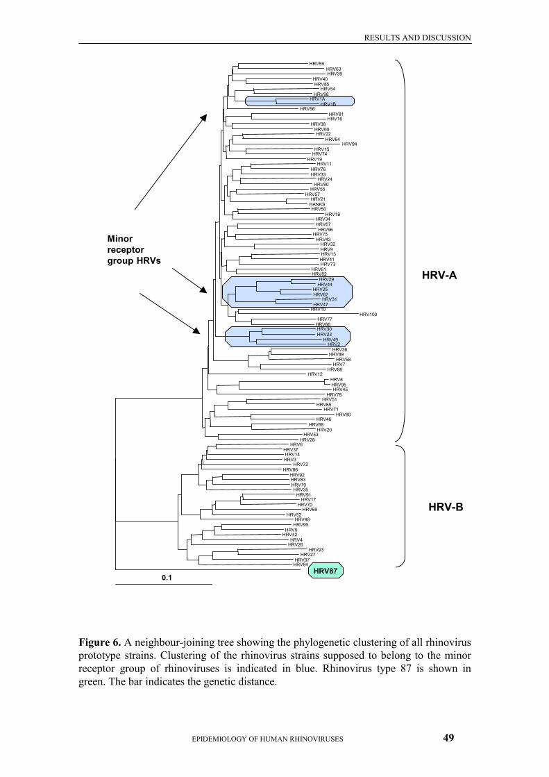
RESULTS AND DISCUSSION
EPIDEMIOLOGY OF HUMAN RHINOVIRUSES 49
Figure 6. A neighbour-joining tree showing the phylogenetic clustering of all rhinovirus prototype strains. Clustering of the rhinovirus strains supposed to belong to the minor receptor group of rhinoviruses is indicated in blue. Rhinovirus type 87 is shown in green. The bar indicates the genetic distance.
0.1
HRV59 HRV63 HRV39
HRV40 HRV85
HRV54 HRV98
HRV1A HRV1B
HRV56 HRV81
HRV16 HRV38
HRV60 HRV22
HRV64 HRV94
HRV15 HRV74
HRV19 HRV11
HRV76 HRV33
HRV24 HRV90
HRV55 HRV57
HRV21 HANKS HRV50
HRV18 HRV34 HRV67 HRV96
HRV75 HRV43
HRV32 HRV9 HRV13
HRV41 HRV73
HRV61 HRV82
HRV29 HRV44
HRV25 HRV62
HRV31HRV47
HRV10HRV100
HRV77 HRV66 HRV30HRV23
HRV49 HRV2
HRV36 HRV89
HRV58 HRV7
HRV88 HRV12
HRV8 HRV95 HRV45
HRV78 HRV51
HRV65 HRV71
HRV80 HRV46
HRV68 HRV20
HRV53 HRV28
HRV6 HRV37 HRV14 HRV3
HRV72 HRV86
HRV92 HRV83 HRV79 HRV35
HRV91 HRV17
HRV70 HRV69
HRV52 HRV48
HRV99 HRV5
HRV42 HRV4
HRV26 HRV93
HRV27 HRV97
HRV84 HRV87
HRV-A
HRV-B
Minor receptor group HRVs
RESULTS AND DISCUSSION
EPIDEMIOLOGY OF HUMAN RHINOVIRUSES 50
Recently, the genetic relationships of both rhinovirus prototype and field strains were studied also in the genomic region encoding non-structural proteins (Savolainen et al., 2004). The same clustering into two genetic groups, HRV-A and HRV-B, was observed in the 3D polymerase coding gene, in addition to a third distinctly different cluster. Based on the phylogenetic analysis, the authors proposed that within-species recombination events occurred during the evolution of rhinoviruses. The phylogenetic analysis was broadened to also include the 61 previously sequenced rhinovirus field isolates from the FinOM Cohort Study. Of these 61 field strains, 24 clustered into HRV-A and 37 into HRV-B, as previously described by Savolainen et al., (2002). This distribution was strikingly different from that seen within the prototype strains (A:B ratio 3:1) or within the field strains serotyped some decades ago (Figure 3, A:B ratio 1301:159 or about 8:1). However, because only a minor subset of rhinovirus-positive specimens (61/833) from the FinOM Cohort were subjected to sequencing, the distribution of the analysed field strains probably does not indicate the actual prevalence of circulating HRV-A and HRV-B strains. Most of the field strains had more than 90% identical nucleotides with the nearest prototype strain, and they could be easily linked to a single prototype. In this respect at least, the VP4/VP2 coding region can be used to genotype field strains. However, for enteroviruses, sequence comparisons in the VP1 region give more reliable serotype identification than those in the VP4/VP2 region (Oberste et al., 1999), and whether this also applies to rhinoviruses remains to be analysed. Considering the laborious serotype identification with the conventional neutralization assays and the known antigenic variation in rhinoviruses, genetic typing in the VP4/VP2 region offers promise in the characterization of rhinovirus strains.
5.4 SPECIAL FEATURES OF RHINOVIRUS 87 (III, IV)
Human rhinovirus serotype 87 differs from the other rhinoviruses in several ways. While the receptors of the other rhinoviruses were characterized some time ago, the receptor for HRV87 remains unknown (Uncapher et al., 1991). Analysis of genetic relationships of rhinoviruses at the 5’ NCR has revealed the deviant clustering of HRV87 (Andeweg et al., 1999). Moreover, in Study III, we showed that in the VP4/VP2 region HRV87 clustered closer to EV70 than to rhinoviruses. The characterization of HRV87 was further continued with more extensive sequence analysis to assess antigenic characteristics, acid sensitivity and receptor usage. After the initial comparison of the VP4/VP2 sequence of HRV87 with the complete rhinovirus and enterovirus genomic sequences in GenBank, we noted that the similarity of HRV87 was even greater with the partial VP2 sequence of EV68 (Pulli et al., 1995) than with EV70. We subsequently sequenced four distinct genomic regions of the HRV87 prototype strain and two sublines of the EV68 prototype strain Fermon and compared them with the complete genomic sequence of EV70 obtained from GenBank (Ryan et al., 1990). Phylogenetic analysis showed that HRV87 clustered into the HEV-D group, together with EV68 and EV70, at the two capsid coding regions and the 3D polymerase coding region. At the 5’ NCR, HRV87 and EV68 clustered into genetic

RESULTS AND DISCUSSION
EPIDEMIOLOGY OF HUMAN RHINOVIRUSES 51
group 2 (Pöyry et al., 1996) with EV70 and HEV-C enteroviruses, including polioviruses. The 5’ NCR of HRV87 contained the additional hypervariable region typical of enteroviruses, but not present in rhinovirus genomes. The nucleotide sequence encoding the VP1 capsid protein has been shown to correlate well with enterovirus serotype (Oberste et al., 1999), and based on this, HRV87 and EV68 are clearly members of the same serotype. EV70 is a different serotype but belongs to the same genetic cluster, HEV-D. One-way antigenic cross-reactivity was demonstrated between HRV87 and EV68. Antisera raised against EV68 effectively neutralized both HRV87 and EV68, while antisera to HRV87 had only a slight effect on the infectivity of EV68. Accordingly, EV68 is the prime strain of HRV87, since by definition, a prime strain exhibits a broader antigenicity than a “prototype” virus; HRV87 should thus be considered a prototype strain of this virus pair. Prime strains have previously been described for both enterovirus (Pallansch & Roos, 2001) and rhinovirus prototype strains; for example, the strain Chase is a prime strain of HRV22 (Schieble et al., 1970). In addition, several antigenic variants of HRV17 that fulfil the criteria for being a prime strain, have been isolated (Patterson & Hamparian, 1997). The HEV-D cluster, the smallest of the four genetic clusters of enteroviruses (HEV-A to HEV-D), has previously been reported to have only two representatives, EV68 and EV70. The prototype strain Fermon of EV68 was isolated from a throat swab of a 10-month-old female with pneumonia and bronchiolitis (Schieble et al., 1967), but no other isolations of the virus are found in the literature. The same holds true for HRV87, now shown to belong to the same serotype as EV68. Antibodies to this virus pair appear, however, to be rather common among Finnish people, as represented by the plasma pool preparation and the few individual sera studied by us. However, nothing is known about any clinical disease caused by these viruses. The acid sensitivity makes infection of the alimentary tract suspect, and the infection may well occur in the respiratory tract, as is typical of rhinoviruses. In contrast to EV68, the clinical syndromes caused by EV70 are well known. EV70 is an atypical enterovirus in that its primary replication site is not the alimentary tract, but rather the eye. The first acute haemorrhagic conjunctivitis (AHC) epidemic reported to be EV70-associated occurred in 1969 in Ghana, and since then, two pandemics and smaller outbreaks have been described to comprise approximately 100 million cases of AHC (reviewed in Alexander & Dimock, 2002). Besides being a causative agent of AHC, EV70 has a propensity to infect the central nervous system, occasionally causing paralysis or meningoencephalitis (Pallansch & Roos, 2001). We assessed the capability of a monoclonal antibody to the decay-accelerating factor (DAF) to inhibit the infection caused by HRV87, EV68 and EV70, observing that the CPE induced by these viruses in HeLa Ohio cells was clearly inhibited by pre-treatment of the cells with the antibody. The HRV87 binding has previously been known to be dependent on the presence of sialic acids on the cell surface (Uncapher et al., 1991). The HeLa cell receptor for EV70 has been demonstrated to be the DAF (Karnauchow et al., 1996), but the binding of EV70 to the cell surface is also dependent on the presence of sialic acids (Utagawa et al., 1982; Alexander & Dimock, 2002), which are not found in DAF (Alexander & Dimock, 2002). To date, EV70 is the only human enterovirus observed to require cell surface sialic acids for attachment. The DAF, by contrast, has

RESULTS AND DISCUSSION
EPIDEMIOLOGY OF HUMAN RHINOVIRUSES 52
been shown to serve as a cellular attachment protein for a number of enteroviruses, including CBV1 (Shafren et al., 1995), CBV3 (Bergelson et al., 1995; Shafren et al., 1995; Martino et al., 1998), CBV5 (Shafren et al., 1995) and CAV21 (Shafren et al., 1997; Newcombe et al., 2003), as well as some echoviruses (Bergelson et al., 1994). These viruses seem to require accessory molecules in addition to DAF for successful infection; for example, both ICAM-1 and DAF are used by CAV21 (Shafren et al., 1997). Although we showed, that the infection of HeLa Ohio cells by HRV87 and EV68 is dependent on DAF, as is also the case for EV70, it remains unresolved whether the sialic acid moiety involved is shared by these viruses. While the DAF is expressed almost ubiquitously in mammalian (Lublin & Atkinson, 1989), a putatively distinct co-receptor may give rise to different tissue tropism of these viruses. Our study clearly showed that HRV87 and EV68 belong to the same serotype in the HEV-D cluster of enteroviruses - a finding later supported by others (Ishiko et al., 2002; Ledford et al., 2004). Initial misidentification of viruses in the family Picornaviridae has been noted regularly. Parechoviruses 1 and 2, for example, were originally assigned as enterovirus serotypes echovirus 22 and echovirus 23, respectively, but have subsequently been shown to represent a genus of their own (Hyypiä et al., 1992). In addition, the virus pairs echovirus 1 and 8, coxsackievirus A11 and A15, and coxsackievirus A13 and A18, have been shown to exhibit such strong serological and genetic relationships that they are now classified as single serotypes (Oberste et al., 1999; Stanway et al., 2004). The most striking feature of virus pair HRV87/EV68 is that the viruses are acid-sensitive, a feature typical of rhinoviruses. This result conflicts with the traditional classification criteria, which state that enteroviruses are acid-resistant. This discrepancy has in part led to a proposal by the Picornavirus Study Group of the International Committee for the Taxonomy of Viruses (T. Hovi, pers. comm.) that the rhinovirus and enterovirus genera be combined to form a single genus with the suggested name Enterhinovirus.

CONCLUSIONS
EPIDEMIOLOGY OF HUMAN RHINOVIRUSES 53
6 CONCLUSIONS 1. We developed a rapid and sensitive microwell RT-PCR hybridization assay for the
detection of rhinoviral RNA in clinical specimens. The specificity of the assay was assessed with a complete collection of rhinovirus and enterovirus prototype strains, a step not performed by others. Despite the observation that a few rhinovirus prototype and field strains go unrecognized in the hybridization step, the assay clearly exceeds the sensitivity of virus isolation in a single cell line. The assay is especially useful for analysis of large numbers of clinical specimens.
2. Rhinovirus infections were shown to be very common in young children, with
infections occurring already during the very first months of life. By the age of two years, 79% of children had experienced a virologically documented rhinovirus infection, and 91.3% had rhinovirus-specific antibodies. A close association between rhinovirus infection and acute otitis media (AOM) was confirmed by rhinoviruses being detected by isolation or RT-PCR in 41% of all AOM episodes studied.
3. Genetic relationships between all 102 rhinovirus prototype strains were studied for
the first time. In the VP4/VP2 genomic region, the strains were shown to cluster into the two established species, Human rhinovirus A (HRV-A) and Human rhinovirus B (HRV-B), with only one exception, HRV87. HRV-A contained 76 and HRV-B 25 prototype strains. The clustering of previously sequenced field strains suggests that genetic typing of rhinoviruses is possible and may be a useful tool in characterizing rhinovirus field strains.
4. Rhinovirus serotype 87 was shown both antigenically and genetically to represent
the same serotype as enterovirus 68, and thus, to belong to the enterovirus species HEV-D together with EV70. Based on the prevalence of neutralizing antibodies, the virus pair HRV87/EV68 is relatively common among Finnish people. HRV87/EV68 appears to share the HeLa cell DAF receptor with EV70. In contrast to other enteroviruses, HRV87 and EV68 are sensitive to an acidic environment.

ACKNOWLEDGEMENTS
EPIDEMIOLOGY OF HUMAN RHINOVIRUSES 54
7 ACKNOWLEDGEMENTS This study was carried out at the Enterovirus Laboratory of the Department of Microbiology, National Public Health Institute, Helsinki, Finland. I thank Professors Jussi Huttunen and Pekka Puska, the former and present Head of the Institute, respectively, for providing excellent working facilities in the Institute. I wish to thank The Steering Group of FinOM Studies for giving me the opportunity to participate in these unique studies right from the beginning. My warmest gratitude is due to my supervisor, Professor Tapani Hovi. He introduced me to the intriguing world of picornaviruses and was always available when I needed guidance in scientific questions. Tapani was an invaluable source of optimism and encouragement during the days when the work did not fare as planned. Professor Carl G. Gahmberg is acknowledged for giving me the opportunity to study biochemistry in the Department of Biochemistry, University of Helsinki. I will always remember the inspiring scientific atmosphere of the Department. Professor Timo Hyypiä and Docent Anne Pitkäranta are acknowledged for their expert review of this thesis. I am grateful for pleasant discussions and their invariably positive and encouraging attitudes, not only during the review process but also throughout the rhinovirus studies. Carol Ann Pelli is kindly thanked for reviewing the language. I thank Docent Merja Roivainen, Head of the Enterovirus Laboratory, for valuable help in technical issues. I am particularly grateful to her for allowing me to concentrate on my writing regardless of the workload and tight time schedule in the Lab. Marjaana Kleemola, Riitta Räty and all other members of the Laboratory of Respiratory Viruses are thanked for delightful collaboration over the years of the FinOM Studies. Mirja Stenvik deserves my warmest thanks for introducing me to the challenging world of isolation of viruses. She is also especially thanked for her warmth and empathy – she always had time for a smile however crowded the days were. I feel lucky to having been able to share the Lab and scientific concerns with Carita Savolainen, who introduced me to the four important colours of life (electropherograms). Carita’s wonderful sense of humour and enthusiasm for her work has invariably brightened my days. Without Kristiina Aitkoski and Lotta Krogius we would know nothing of the rhinoviruses in the FinOM Cohort. Their technical expertise is highly appreciated - as is their positive attitude, which remained unchanged even some thousands of virus cultures and RT-PCRs later. Sinikka Sopanen and Raija Tyni are acknowledged for providing us with the excellent cell cultures. Their contribution has been invaluable to these studies.

ACKNOWLEDGEMENTS
EPIDEMIOLOGY OF HUMAN RHINOVIRUSES 55
I thank all former and present members of the Enterovirus Laboratory. Many of them have contributed importantly to these studies and, all have helped to create an enjoyable working atmosphere. Arja Skyttä is thanked for continuing with the virus isolations during my first maternity leave. Mick Mulders is thanked for his inspiring ideas, and for initiating the sequencing of the rhinoviruses. Antero Airaksinen is thanked for excellent lessons on how to manage with the awkward computers. Laura Råman is acknowledged for taking good care of the rhinovirus strain collection. Johanna Nokso-Koivisto is thanked for being great company in the Lab and for all the medical advice she gave to my family and myself. Pia Laine is thanked for stimulating discussions on scientific and other concerns, and for her expert assistance with the computer software. I wish her best of luck for the future. My warmest thanks go to Eija Penttilä, Elisa Lamminsalo, Päivi Hirttiö and Mervi Eskelinen. I sincerely appreciate their expertise and commitment to the work they do. I feel honoured to be able to share the Lab with them. My mother, Raija, and grandmother, Aino, are thanked for unconditional love and support throughout my life. Raija is also thanked for taking care of Juuso and Minja whenever needed. Finally, my heartfelt gratitude goes to Kai - the man in my life - for his continuous love and encouragement in all the years that we have known one another. Juuso and Minja, our beloved children, are acknowledged for reminding me of what is really important in life. I love you all so much. This work was partly supported by Wyeth-Lederle Vaccines and Pediatrics, Merck & Co., Inc., Pasteur Mérieux Sérums et Vaccins, Orion Reseach Foundation, the Academy of Finland and the Päivikki and Sakari Sohlberg Foundation. Helsinki, 13 October, 2004 Soile Blomqvist

REFERENCES
EPIDEMIOLOGY OF HUMAN RHINOVIRUSES 56
8 REFERENCES Abraham, G. & Colonno, R. J. (1984). Many rhinovirus serotypes share the same cellular
receptor. J Virol 51, 340-5. Agol, V. I. (1997). Recombination and other genomic rearrangements in picornaviruses.
Seminars in Virology 8, 77-84. Alexander, D. A. & Dimock, K. (2002). Sialic acid functions in enterovirus 70 binding and
infection. J Virol 76, 11265-72. al-Mulla, W., el Mekki, A. & al-Nakib, W. (1994). Rapid culture-amplified immunofluorescent
test for the detection of human rhinoviruses in clinical samples: evidence of a common epitope in culture. J Med Virol 42, 182-7.
Al-Nakib, W., Stanway, G., Forsyth, M., Hughes, P. J., Almond, J. W. & Tyrrell, D. A. (1986). Detection of human rhinoviruses and their molecular relationship using cDNA probes. J Med Virol 20, 289-96.
Andeweg, A. C., Bestebroer, T. M., Huybreghs, M., Kimman, T. G. & de Jong, J. C. (1999). Improved detection of rhinoviruses in clinical samples by using a newly developed nested reverse transcription-PCR assay. J Clin Microbiol 37, 524-30.
Andreoletti, L., Lesay, M., Deschildre, A., Lambert, V., Dewilde, A. & Wattre, P. (2000). Differential detection of rhinoviruses and enteroviruses RNA sequences associated with classical immunofluorescence assay detection of respiratory virus antigens in nasopharyngeal swabs from infants with bronchiolitis. J Med Virol 61, 341-6.
Andrewes, T. O., Chaproniere, D. M., Gompels, A. E. H., Pereira, H. G. & Roden, A.T. (1953). Propagation of common-cold virus in tissue cultures. Lancet 2, 546.
Andries, K., Dewindt, B., Snoeks, J., Willebrords, R., Stokbroekx, R. & Lewi, P. J. (1991). A comparative test of fifteen compounds against all known human rhinovirus serotypes as a basis for a more rational screening program. Antiviral Res 16, 213-25.
Andries, K., Dewindt, B., Snoeks, J., Wouters, L., Moereels, H., Lewi, P. J. & Janssen, P. A. (1990). Two groups of rhinoviruses revealed by a panel of antiviral compounds present sequence divergence and differential pathogenicity. J Virol 64, 1117-23.
Appleyard, G., Russell, S. M., Clarke, B. E., Speller, S. A., Trowbridge, M. & Vadolas, J. (1990). Neutralization epitopes of human rhinovirus type 2. J Gen Virol 71, 1275-82.
Arnold, E. & Rossmann, M. G. (1990). Analysis of the structure of a common cold virus, human rhinovirus 14, refined at a resolution of 3.0 Å. J Mol Biol 211, 763-801.
Arola, M., Ruuskanen, O., Ziegler, T., Mertsola, J., Nanto-Salonen, K., Putto-Laurila, A., Viljanen, M. K. & Halonen, P. (1990a). Clinical role of respiratory virus infection in acute otitis media. Pediatrics 86, 848-55.
Arola, M., Ziegler, T., Puhakka, H., Lehtonen, O. P. & Ruuskanen, O. (1990b). Rhinovirus in otitis media with effusion. Ann Otol Rhinol Laryngol 99, 451-3.
Arola, M., Ziegler, T., Ruuskanen, O., Mertsola, J., Nanto-Salonen, K. & Halonen, P. (1988). Rhinovirus in acute otitis media. J Pediatr 113, 693-5.
Arruda, E., Boyle, T. R., Winther, B., Pevear, D. C., Gwaltney, J. M., Jr. & Hayden, F. G. (1995). Localization of human rhinovirus replication in the upper respiratory tract by in situ hybridization. J Infect Dis 171, 1329-33.
Arruda, E., Crump, C. E., Rollins, B. S., Ohlin, A. & Hayden, F. G. (1996). Comparative susceptibilities of human embryonic fibroblasts and HeLa cells for isolation of human rhinoviruses. J Clin Microbiol 34, 1277-9.
Arruda, E., Pitkäranta, A., Witek, T. J., Jr., Doyle, C. A. & Hayden, F. G. (1997). Frequency and natural history of rhinovirus infections in adults during autumn. J Clin Microbiol 35, 2864-8.

REFERENCES
EPIDEMIOLOGY OF HUMAN RHINOVIRUSES 57
Atmar, R. L. & Georghiou, P. R. (1993). Classification of respiratory tract picornavirus isolates as enteroviruses or rhinoviruses by using reverse transcription-polymerase chain reaction. J Clin Microbiol 31, 2544-6.
Barclay, W. S. & Al-Nakib, W. (1987). An ELISA for the detection of rhinovirus specific antibody in serum and nasal secretion. J Virol Methods 15, 53-64.
Barclay, W. S., Callow, K. A., Sergeant, M. & al-Nakib, W. (1988). Evaluation of an enzyme-linked immunosorbent assay that measures rhinovirus-specific antibodies in human sera and nasal secretions. J Med Virol 25, 475-82.
Bardin, P. G., Johnston, S. L., Sanderson, G., Robinson, B. S., Pickett, M. A., Fraenkel, D. J. & Holgate, S. T. (1994). Detection of rhinovirus infection of the nasal mucosa by oligonucleotide in situ hybridization. Am J Respir Cell Mol Biol 10, 207-13.
Bates, P. J., Sanderson, G., Holgate, S. T. & Johnston, S. L. (1997). A comparison of RT-PCR, in-situ hybridisation and in-situ RT-PCR for the detection of rhinovirus infection in paraffin sections. J Virol Methods 67, 153-60.
Bergelson, J. M., Chan, M., Solomon, K. R., St John, N. F., Lin, H. & Finberg, R. W. (1994). Decay-accelerating factor (CD55), a glycosylphosphatidylinositol-anchored complement regulatory protein, is a receptor for several echoviruses. Proc Natl Acad Sci U S A 91, 6245-8.
Bergelson, J. M., Mohanty, J. G., Crowell, R. L., St John, N. F., Lublin, D. M. & Finberg, R. W. (1995). Coxsackievirus B3 adapted to growth in RD cells binds to decay-accelerating factor (CD55). J Virol 69, 1903-6.
Blomqvist, S., Bruu, A. L., Stenvik, M. & Hovi, T. (2003). Characterization of a recombinant type 3/type 2 poliovirus isolated from a healthy vaccinee and containing a chimeric capsid protein VP1. J Gen Virol 84, 573-80.
Brown, P. K. & Taylor-Robinson, D. (1966). Respiratory virus antibodies in sera of persons living in isolated communities. Bull World Health Organ 34, 895-900.
Calhoun, A. M., Jordan, W. S., Jr. & Gwaltney, J. M., Jr. (1974). Rhinovirus infections in an industrial population. V. Change in distribution of serotypes. Am J Epidemiol 99, 58-64.
Casasnovas, J. M. & Springer, T. A. (1994). Pathway of rhinovirus disruption by soluble intercellular adhesion molecule 1 (ICAM-1): an intermediate in which ICAM-1 is bound and RNA is released. J Virol 68, 5882-9.
Cate, T. R., Douglas, R. G., Jr., Johnson, K. M., Couch, R. B. & Knight, V. (1967). Studies on the inability of rhinovirus to survive and replicate in the intestinal tract of volunteers. Proc Soc Exp Biol Med 124, 1290-5.
Cate, T. R., Rossen, R. D., Douglas, R. G., Jr., Butler, W. T. & Couch, R. B. (1966). The role of nasal secretion and serum antibody in the rhinovirus common cold. Am J Epidemiol 84, 352-63.
Chapple, P. J., Head, B. & Tyrrell, D. A. (1967). A complement fixing antigen from an M rhinovirus (brief report). Arch Gesamte Virusforsch 21, 123-6.
Che, Z., Olson, N. H., Leippe, D., Lee, W. M., Mosser, A. G., Rueckert, R. R., Baker, T. S. & Smith, T. J. (1998). Antibody-mediated neutralization of human rhinovirus 14 explored by means of cryoelectron microscopy and X-ray crystallography of virus-Fab complexes. J Virol 72, 4610-22.
Colonno, R. J., Callahan, P. L., Leippe, D. M., Rueckert, R. R. & Tomassini, J. E. (1989). Inhibition of rhinovirus attachment by neutralizing monoclonal antibodies and their Fab fragments. J Virol 63, 36-42.
Conant, R. M. & Hamparian, V. V. (1968). Rhinoviruses: Basis for a numbering system. II. Serologic characterization of prototype strains. J of Immunol 100, 114-119.
Cooney, M. K., Fox, J. P. & Kenny, G. E. (1982). Antigenic groupings of 90 rhinovirus serotypes. Infect Immun 37, 642-7.

REFERENCES
EPIDEMIOLOGY OF HUMAN RHINOVIRUSES 58
Cooney, M. K., Hall, C. E. & Fox, J. P. (1972). The Seattle virus watch. 3. Evaluation of isolation methods and summary of infections detected by virus isolations. Am J Epidemiol 96, 286-305.
Cooney, M. K. & Kenny, G. E. (1970). Reciprocal neutralizing cross-reaction between rhinovirus types 9 and 32. J Immunol 105, 531-3.
Cooney, M. K. & Kenny, G. E. (1977). Demonstration of dual rhinovirus infection in humans by isolation of different serotypes in human heteroploid (HeLa) and human diploid fibroblast cell cultures. J Clin Microbiol 5, 202-7.
Cooney, M. K., Kenny, G. E., Tam, R. & Fox, J. P. (1973). Cross relationships among 37 rhinoviruses demonstrated by virus neutralization with potent monotypic rabbit antisera. Infect Immun 7, 335-340.
Cooney, M. K., Wise, J. A., Kenny, G. E. & Fox, J. P. (1975). Broad antigenic relationships among rhinovirus serotypes revealed by cross-immunization of rabbits with different serotypes. J Immunol 114, 635-9.
Corne, J. M., Marshall, C., Smith, S., Schreiber, J., Sanderson, G., Holgate, S. T. & Johnston, S. L. (2002). Frequency, severity, and duration of rhinovirus infections in asthmatic and non-asthmatic individuals: a longitudinal cohort study. Lancet 359, 831-4.
Couch, R. (1992). Rhinoviruses. In Laboratory Diagnosis of Viral Infections, pp. 709-729. Edited by E. H. Lennette. New York: Marcel Dekker, Inc.
Couch, R. B. (2001). Rhinoviruses. In Fields VIROLOGY, 4. edn, pp. 777-797. Edited by D. M. Knipe & P. M. Howley. Philadelphia: Lippincott Williams & Wilkins.
Crump, C. E., Arruda, E. & Hayden, F. G. (1993). In vitro inhibitory activity of soluble ICAM-1 for the numbered serotypes of human rhinovirus. Antiviral Chem Chemoth 4, 323-327.
Dagher, H., Donninger, H., Hutchinson, P., Ghildyal, R. & Bardin, P. (2004). Rhinovirus detection: comparison of real-time and conventional PCR. J Virol Methods 117, 113-21.
Davies, M., Bruce, C., Bewley, K., Outlaw, M., Mioulet, V., Lloyd, G. & Clegg, C. (2003). Poliovirus type 1 in working stocks of typed human rhinoviruses. Lancet 361, 1187-8.
Deffernez, C., Wunderli, W., Thomas, Y., Yerly, S., Perrin, L. & Kaiser, L. (2004). Amplicon sequencing and improved detection of human rhinovirus in respiratory samples. J Clin Microbiol 42, 3212-8.
Dick, E. C., Jennings, L. C., Mink, K. A., Wartgow, C. D. & Inhorn, S. L. (1987). Aerosol transmission of rhinovirus colds. J Infect Dis 156, 442-8.
Domingo, E., Biebricher, C. K., Eigen, M. & Holland, J. J. (2001). Quasispecies and RNA Virus Evolution: Principles and Consequences. Austin, Texas: Eurekah.com/Landes Bioscience.
Douglas, G., Jr., Lindgren, K. M. & Couch, R. (1968a). Exposure to cold environment and rhinovirus common cold. Failure to demonstrate effect. N Engl J M 279, 742-747.
Douglas, R. G., Jr. (1970). Pathogenesis of rhinovirus common colds in human voluteers. Ann Otol Rhinol Laryngol 79, 563-71.
Douglas, R. G., Jr., Cate, T. R. & Couch, R. B. (1966). Growth and cytopathic effect of H type rhinoviruses in monkey kidney tissue culture. Proc Soc Exp Biol Med 123, 238-41.
Douglas, R. G., Jr., Fleet, W. F., Cate, T. R. & Couch, R. B. (1968b). Antibody to rhinovirus in human sera. I. Standardization of a neutralization test. Proc Soc Exp Biol Med 127, 497-502.
Dreizin, R. S., Strizova, V., Ponomareva, T. I., Syrucek, L., Vikhnovich, E. M., Borovkova, N. M., Geine, G. V., Klimanskaya, E. V., Bruckova, M., Kuncova, L. & Berkovicova, V. (1979). The spread of rhinoviruses in the Soviet Union (USSR) and in Czechoslovakia (CSSR) and the role of their pathology. J Hyg Epidemiol Microbiol Immunol 23, 174-85.

REFERENCES
EPIDEMIOLOGY OF HUMAN RHINOVIRUSES 59
Duechler, M., Skern, T., Sommergruber, W., Neubauer, C., Gruendler, P., Fogy, I., Blaas, D. & Kuechler, E. (1987). Evolutionary relationships within the human rhinovirus genus: comparison of serotypes 89, 2, and 14. Proc Natl Acad Sci U S A 84, 2605-9.
Falsey, A. R., McCann, R. M., Hall, W. J., Criddle, M. M., Formica, M. A., Wycoff, D. & Kolassa, J. E. (1997). The "common cold" in frail older persons: impact of rhinovirus and coronavirus in a senior daycare center. J Am Geriatr Soc 45, 706-11.
Falsey, A. R., Walsh, E. E. & Hayden, F. G. (2002). Rhinovirus and coronavirus infection-associated hospitalizations among older adults. J Infect Dis 185, 1338-41.
Felsenstein, J. (1993). PHYLIP (Phylogeny Inference Package) version 3.5c. Distributed by the author, Department of Genetics, University of Washington, Seattle, USA.
Fox, J. P. (1976). Is a rhinovirus vaccine possible? Am J Epidemiol 103, 345-54. Fox, J. P., Cooney, M. K. & Hall, C. E. (1975). The Seattle virus watch. V. Epidemiologic
observations of rhinovirus infections, 1965-1969, in families with young children. Am J Epidemiol 101, 122-43.
Fox, J. P., Cooney, M. K., Hall, C. E. & Foy, H. M. (1985). Rhinoviruses in Seattle families, 1975-1979. Am J Epidemiol 122, 830-46.
Gama, R. E., Horsnell, P. R., Hughes, P. J., North, C., Bruce, C. B., al-Nakib, W. & Stanway, G. (1989). Amplification of rhinovirus specific nucleic acids from clinical samples using the polymerase chain reaction. J Med Virol 28, 73-7.
Gama, R. E., Hughes, P. J., Bruce, C. B. & Stanway, G. (1988). Polymerase chain reaction amplification of rhinovirus nucleic acids from clinical material. Nucleic Acids Res 16, 9346.
Gern, J. E. (2002). Rhinovirus respiratory infections and asthma. Am J Med 112, 19S-27S. Gern, J. E., Galagan, D. M., Jarjour, N. N., Dick, E. C. & Busse, W. W. (1997). Detection of
rhinovirus RNA in lower airway cells during experimentally induced infection. Am J Respir Crit Care Med 155, 1159-61.
Ghosh, S., Champlin, R., Couch, R., Englund, J., Raad, I., Malik, S., Luna, M. & Whimbey, E. (1999). Rhinovirus infections in myelosuppressed adult blood and marrow transplant recipients. Clin Infect Dis 29, 528-32.
Giranda, V. L., Heinz, B. A., Oliveira, M. A., Minor, I., Kim, K. H., Kolatkar, P. R., Rossmann, M. G. & Rueckert, R. R. (1992). Acid-induced structural changes in human rhinovirus 14: possible role in uncoating. Proc Natl Acad Sci U S A 89, 10213-7.
Graat, J. M., Schouten, E. G., Heijnen, M. L., Kok, F. J., Pallast, E. G., de Greeff, S. C. & Dorigo-Zetsma, J. W. (2003). A prospective, community-based study on virologic assessment among elderly people with and without symptoms of acute respiratory infection. J Clin Epidemiol 56, 1218-23.
Greenberg, S. B. (2002). Viral respiratory infections in elderly patients and patients with chronic obstructive pulmonary disease. Am J Med 112, 28S-32S.
Greve, J. M., Davis, G., Meyer, A. M., Forte, C. P., Yost, S. C., Marlor, C. W., Kamarck, M. E. & McClelland, A. (1989). The major human rhinovirus receptor is ICAM-1. Cell 56, 839-47.
Greve, J. M., Forte, C. P., Marlor, C. W., Meyer, A. M., Hoover-Litty, H., Wunderlich, D. & McClelland, A. (1991). Mechanisms of receptor-mediated rhinovirus neutralization defined by two soluble forms of ICAM-1. J Virol 65, 6015-23.
Gruenberger, M., Wandl, R., Nimpf, J., Hiesberger, T., Schneider, W. J., Kuechler, E. & Blaas, D. (1995). Avian homologs of the mammalian low-density lipoprotein receptor family bind minor receptor group human rhinovirus. J Virol 69, 7244-7.
Gwaltney, J. M. (1997). Rhinoviruses. In Viral Infections of humans - Epidemiology and control, 4 edn. Edited by A. S. Evans.
Gwaltney, J. M. (2002). Clinical significance and pathogenesis of viral respiratory infections. Am J Med 112, 13S-18S.

REFERENCES
EPIDEMIOLOGY OF HUMAN RHINOVIRUSES 60
Gwaltney, J. M., Jr. (1966). Micro-neutralization test for identification of rhinovirus serotypes. Proc Soc Exp Biol Med 122, 1137-41.
Gwaltney, J. M., Jr. & Hendley, J. O. (1978). Rhinovirus transmission: one if by air, two if by hand. Am J Epidemiol 107, 357-61.
Gwaltney, J. M., Jr. & Hendley, J. O. (1982). Transmission of experimental rhinovirus infection by contaminated surfaces. Am J Epidemiol 116, 828-33.
Gwaltney, J. M., Jr., Hendley, J. O., Simon, G. & Jordan, W. S., Jr. (1966). Rhinovirus infections in an industrial population. I. The occurrence of illness. N Engl J Med 275, 1261-8.
Gwaltney, J. M., Jr., Hendley, J. O., Simon, G. & Jordan, W. S., Jr. (1967). Rhinovirus infections in an industrial population. II. Characteristics of illness and antibody response. Jama 202, 494-500.
Gwaltney, J. M., Jr., Hendley, J. O., Simon, G. & Jordan, W. S., Jr. (1968). Rhinovirus infections in an industrial population. 3. Number and prevalence of serotypes. Am J Epidemiol 87, 158-66.
Gwaltney, J. M., Jr., Moskalski, P. B. & Hendley, J. O. (1978). Hand-to-hand transmission of rhinovirus colds. Ann Intern Med 88, 463-7.
Hadfield, A. T., Lee, W., Zhao, R., Oliveira, M. A., Minor, I., Rueckert, R. R. & Rossmann, M. G. (1997). The refined structure of human rhinovirus 16 at 2.15 Å resolution: implications for the viral life cycle. Structure 5, 427-41.
Halonen, P., Rocha, E., Hierholzer, J., Holloway, B., Hyypiä, T., Hurskainen, P. & Pallansch, M. (1995). Detection of enteroviruses and rhinoviruses in clinical specimens by PCR and liquid-phase hybridization. J Clin Microbiol 33, 648-53.
Hamparian, V. V., Colonno, R. J., Cooney, M. K., Dick, E. C., Gwaltney, J. M., Jr., Hughes, J. H., Jordan, W. S., Jr., Kapikian, A. Z., Mogabgab, W. J., Monto, A. & et al. (1987). A collaborative report: rhinoviruses-extension of the numbering system from 89 to 100. Virology 159, 191-2.
Hamre, D., Connelly, A. P., Jr. & Procknow, J. J. (1966). Virologic studies of acute respiratory disease in young adults. IV. Virus isolations during four years of surveillance. Am J Epidemiol 83, 238-49.
Harris, J. M., 2nd & Gwaltney, J. M., Jr. (1996). Incubation periods of experimental rhinovirus infection and illness. Clin Infect Dis 23, 1287-90.
Hawkes, R. A. (1979). General principles underlying laboratory diagnosis of viral infections. In Diagnostic procedures for viral, rickettsial and chlamydial infections., 5 edn, pp. 35-42. Edited by E. H. Lennette & N. J. Schmidt. Washington, DC: American Public Health Association.
Hayden, F. G. (2004). Rhinovirus and the lower respiratory tract. Rev Med Virol 14, 17-31. Hayflick, L. & Moorehead, P. S. (1961). The serial cultivation of human diploid cell strains.
Exp Cell Res 25, 585-621. Heikkinen, T. & Chonmaitree, T. (2003). Importance of respiratory viruses in acute otitis
media. Clin Microbiol Rev 16, 230-41. Heikkinen, T. & Järvinen, A. (2003). The common cold. Lancet 361, 51-9. Hendley, J. O. & Gwaltney, J. M., Jr. (1988). Mechanisms of transmission of rhinovirus
infections. Epidemiol Rev 10, 243-58. Hendley, J. O. & Gwaltney, J. M., Jr. (2004). Viral titers in nasal lining fluid compared to viral
titers in nasal washes during experimental rhinovirus infection. J Clin Virol 30, 326-8. Hendley, J. O., Gwaltney, J. M., Jr. & Jordan, W. S., Jr. (1969). Rhinovirus infections in an
industrial population. IV. Infections within families of employees during two fall peaks of respiratory illness. Am J Epidemiol 89, 184-96.
Hendley, J. O., Wenzel, R. P. & Gwaltney, J. M., Jr. (1973). Transmission of rhinovirus colds by self-inoculation. N Engl J Med 288, 1361-4.

REFERENCES
EPIDEMIOLOGY OF HUMAN RHINOVIRUSES 61
Hewat, E. A., Neumann, E., Conway, J. F., Moser, R., Ronacher, B., Marlovits, T. C. & Blaas, D. (2000). The cellular receptor to human rhinovirus 2 binds around the 5-fold axis and not in the canyon: a structural view. Embo J 19, 6317-25.
Hilleman, M. R. (1967). Present knowledge of the rhinovirus group of viruses. Curr Top Microbiol Immunol 41, 1-22.
Hofer, F., Gruenberger, M., Kowalski, H., Machat, H., Huettinger, M., Kuechler, E. & Blass, D. (1994). Members of the low density lipoprotein receptor family mediate cell entry of a minor-group common cold virus. Proc Natl Acad Sci U S A 91, 1839-42.
Hope-Simpson, R. E. & Higgins, P. G. (1969). A respiratory virus study in Great Britain: review and evaluation. Prog Med Virol 11, 354-407.
Horsnell, C., Gama, R. E., Hughes, P. J. & Stanway, G. (1995). Molecular relationships between 21 human rhinovirus serotypes. J Gen Virol 76, 2549-55.
Hughes, A. L. (2004). Phylogeny of the Picornaviridae and differential evolutionary divergence of picornavirus proteins. Infect Genet Evol 4, 143-52.
Hughes, J. H., Thomas, D. C., Hamparian, V. V. & Cramblett, H. G. (1973). Acid liability of rhinovirus type 14: effect of pH, time, and temperature. Proc Soc Exp Biol Med 144, 555-60.
Hughes, P. J., North, C., Jellis, C. H., Minor, P. D. & Stanway, G. (1988). The nucleotide sequence of human rhinovirus 1B: molecular relationships within the rhinovirus genus. J Gen Virol 69, 49-58.
Hyypiä, T., Auvinen, P. & Maaronen, M. (1989). Polymerase chain reaction for human picornaviruses. J Gen Virol 70, 3261-8.
Hyypiä, T., Horsnell, C., Maaronen, M., Khan, M., Kalkkinen, N., Auvinen, P., Kinnunen, L. & Stanway, G. (1992). A distinct picornavirus group identified by sequence analysis. Proc Natl Acad Sci U S A 89, 8847-51.
Hyypiä, T., Puhakka, T., Ruuskanen, O., Mäkelä, M., Arola, A. & Arstila, P. (1998). Molecular diagnosis of human rhinovirus infections: comparison with virus isolation. J Clin Microbiol 36, 2081-3.
Ireland, D. C., Kent, J. & Nicholson, K. G. (1993). Improved detection of rhinoviruses in nasal and throat swabs by seminested RT-PCR. J Med Virol 40, 96-101.
Ishiko, H., Miura, R., Shimada, Y., Hayashi, A., Nakajima, H., Yamazaki, S. & Takeda, N. (2002). Human rhinovirus 87 identified as human enterovirus 68 by VP4-based molecular diagnosis. Intervirol 45, 136-41.
Ison, M. G., Hayden, F. G., Kaiser, L., Corey, L. & Boeckh, M. (2003). Rhinovirus infections in hematopoietic stem cell transplant recipients with pneumonia. Clin Infect Dis 36, 1139-43.
Jartti, T., Lehtinen, P., Vuorinen, T., Koskenvuo, M. & Ruuskanen, O. (2004). Persistence of rhinovirus and enterovirus RNA after acute respiratory illness in children. J Med Virol 72, 695-9.
Jenison, R., Rihanek, M. & Polisky, B. (2001). Use of a thin film biosensor for rapid visual detection of PCR products in a multiplex format. Biosens Bioelectron 16, 757-63.
Jennings, L. C. & Dick, E. C. (1987). Transmission and control of rhinovirus colds. Eur J Epidemiol 3, 327-35.
Johnston, S. L., Pattemore, P. K., Sanderson, G., Smith, S., Campbell, M. J., Josephs, L. K., Cunningham, A., Robinson, B. S., Myint, S. H., Ward, M. E., Tyrrell, D. A. & Holgate, S. T. (1996). The relationship between upper respiratory infections and hospital admissions for asthma: a time-trend analysis. Am J Respir Crit Care Med 154, 654-60.
Johnston, S. L., Sanderson, G., Pattemore, P. K., Smith, S., Bardin, P. G., Bruce, C. B., Lambden, P. R., Tyrrell, D. A. & Holgate, S. T. (1993). Use of polymerase chain reaction for diagnosis of picornavirus infection in subjects with and without respiratory symptoms. J Clin Microbiol 31, 111-7.

REFERENCES
EPIDEMIOLOGY OF HUMAN RHINOVIRUSES 62
Kapikian, A. Z., (chairman), Conant, R. M., Hamparian, V. V., Chanock, R. M., Chapple, P. J., Dick, E. C., Fenters, J. D., Gwaltney, J. M., Jr., Hamre, D., Holper, J. C., Jordan, W. S., Jr., Lennette, E. H., Melnick, J. L., Mogabgab, W. J., Mufson, M. A., Phillips, C. A., Schieble, J. H. & Tyrrell, D. A. J. (1967). Rhinoviruses: a numbering system. Nature (London) 213, 761-3.
Kapikian, A. Z., (Chairman), Conant, R. M., Hamparian, V. V., Chanock, R. M., Dick, E. C., Gwaltney, J. M., Jr., Hamre, D., Jordan, W. S., Jr., Kenny, G. E., Lennette, E. H., Melnick, J. L., Mogabgab, W. J., Phillips, C. A., Schieble, J. H., Stott, E. J. & Tyrrell, D. A. J. (1971). A collaborative report: Rhinoviruses - extension of the numbering system. Virology 43, 524-526.
Kares, S., Lönnrot, M., Vuorinen, P., Oikarinen, S., Taurianen, S. & Hyöty, H. (2004). Real-time PCR for rapid diagnosis of entero- and rhinovirus infections using LightCycler. J Clin Virol 29, 99-104.
Karnauchow, T. M., Tolon, D. L., Harrison, B. A., Altman, E., Lublin, D. M. & Dimock, K. (1996). The HeLa cell receptor for enterovirus 70 is decay-accelerating factor (CD55). J Virol 70, 5143-52.
Kellner, G., Popow-Kraupp, T., Binder, C., Goedl, I., Kundi, M. & Kunz, C. (1991). Respiratory tract infections due to different rhinovirus serotypes and the influence of maternal antibodies on the clinical expression of the disease in infants. J Med Virol 35, 267-72.
Ketler, A., Hall, C. E., Fox, J. P. et al. (1969). The Virus Watch program: A continuing surveillance of viral infections in metropolitan New York families. 8. Am J Epidemiol 90, 244-254.
Kim, S. S., Smith, T. J., Chapman, M. S., Rossmann, M. C., Pevear, D. C., Dutko, F. J., Felock, P. J., Diana, G. D. & McKinlay, M. A. (1989). Crystal structure of human rhinovirus serotype 1A (HRV1A). J Mol Biol 210, 91-111.
King, A. M. Q., Brown, F., Christian, P., Hovi, T., Hyypiä, T., Knowles, N.J., Lemon, S.M., Minor, P.D., Palmenberg, A.C., Skern, T. and Stanway, G. (2000). Picornaviridae. In Virus Taxonomy. Seventh Report of the International Committee for the Taxonomy of Viruses., pp. 657-673. Edited by M. H. V. Van Regenmortel, Fauquet, C.M., Bishop, D.H.L., Calisher, C.H., Carsten, E.B., Estes, M.K., Lemon, S.M., Maniloff, J., Mayo, M.A., McGeoch, D.J., Pringle, C.R. and Wickner, R.B. New York, San Diego: Academic Press.
Knowles, N. J. (1996). http://www.iah.bbsrc.ac.uk/virus/Picornaviridae/index.html Kolatkar, P. R., Bella, J., Olson, N. H., Bator, C. M., Baker, T. S. & Rossmann, M. G. (1999).
Structural studies of two rhinovirus serotypes complexed with fragments of their cellular receptor. Embo J 18, 6249-59.
Krilov, L., Pierik, L., Keller, E., Mahan, K., Watson, D., Hirsch, M., Hamparian, V. & McIntosh, K. (1986). The association of rhinoviruses with lower respiratory tract disease in hospitalized patients. J Med Virol 19, 345-52.
Laine, P., Savolainen, C., Blomqvist, S. & Hovi, T. Phylogenetic analysis of human rhinovirus capsid protein VP1 and 2A protease coding sequences confirms shared genus-like relationships with human enteroviruses. Submitted.
Larson, H. E., Reed, S. E. & Tyrrell, D. A. (1980). Isolation of rhinoviruses and coronaviruses from 38 colds in adults. J Med Virol 5, 221-9.
Ledford, R. M., Patel, N. R., Demenczuk, T. M., Watanyar, A., Herbertz, T., Collett, M. S. & Pevear, D. C. (2004). VP1 sequencing of all human rhinovirus serotypes: insights into genus phylogeny and susceptibility to antiviral capsid-binding compounds. J Virol 78, 3663-74.
Lee, W. M., Wang, W. & Rueckert, R. R. (1995). Complete sequence of the RNA genome of human rhinovirus 16, a clinically useful common cold virus belonging to the ICAM-1 receptor group. Virus Genes 9, 177-81.

REFERENCES
EPIDEMIOLOGY OF HUMAN RHINOVIRUSES 63
Lee, Y. F., Nomoto, A., Detjen, B. M. & Wimmer, E. (1977). A protein covalently linked to poliovirus genome RNA. Proc Natl Acad Sci U S A 74, 59-63.
Lindberg, A. M., Andersson, P., Savolainen, C., Mulders, M. N. & Hovi, T. (2003). Evolution of the genome of Human enterovirus B: incongruence between phylogenies of the VP1 and 3CD regions indicates frequent recombination within the species. J Gen Virol 84, 1223-35.
Loens, K., Ieven, M., Ursi, D., De Laat, C., Sillekens, P., Oudshoorn, P. & Goossens, H. (2003). Improved detection of rhinoviruses by nucleic acid sequence-based amplification after nucleotide sequence determination of the 5' noncoding regions of additional rhinovirus strains. J Clin Microbiol 41, 1971-6.
Lönnrot, M., Sjöroos, M., Salminen, K., Maaronen, M., Hyypiä, T. & Hyöty, H. (1999). Diagnosis of enterovirus and rhinovirus infections by RT-PCR and time-resolved fluorometry with lanthanide chelate labeled probes. J Med Virol 59, 378-84.
Lublin, D. M. & Atkinson, J. P. (1989). Decay-accelerating factor: biochemistry, molecular biology, and function. Annu Rev Immunol 7, 35-58.
Mäkelä, M. J., Puhakka, T., Ruuskanen, O., Leinonen, M., Saikku, P., Kimpimäki, M., Blomqvist, S., Hyypiä, T. & Arstila, P. (1998). Viruses and bacteria in the etiology of the common cold. J Clin Microbiol 36, 539-42.
Marlovits, T. C., Abrahamsberg, C. & Blaas, D. (1998). Very-low-density lipoprotein receptor fragment shed from HeLa cells inhibits human rhinovirus infection. J Virol 72, 10246-50.
Martino, T. A., Petric, M., Brown, M., Aitken, K., Gauntt, C. J., Richardson, C. D., Chow, L. H. & Liu, P. P. (1998). Cardiovirulent coxsackieviruses and the decay-accelerating factor (CD55) receptor. Virology 244, 302-14.
Mogabgab, W. J., Holmes, B. J. & Pollock, B. (1975). Antigenic relationships of common rhinovirus types from disabling upper respiratory illnesses. Dev Biol Stand 28, 400-11.
Monto, A. S. (1994). Studies of the community and family: acute respiratory illness and infection. Epidemiol Rev 16, 351-73.
Monto, A. S. (2002a). Epidemiology of viral respiratory infections. Am J Med 112, 4S-12S. Monto, A. S. (2002b). The seasonality of rhinovirus infections and its implications for clinical
recognition. Clin Ther 24, 1987-97. Monto, A. S. & Bryan, E. R. (1974). Microneutralization test for detection of rhinovirus
antibodies. Proc Soc Exp Biol Med 145, 690-4. Monto, A. S., Bryan, E. R. & Ohmit, S. (1987). Rhinovirus infections in Tecumseh, Michigan:
frequency of illness and number of serotypes. J Infect Dis 156, 43-9. Monto, A. S. & Cavallaro, J. J. (1972). The Tecumseh study of respiratory illness. IV.
Prevalence of rhinovirus serotypes, 1966-1969. Am J Epidemiol 96, 352-60. Monto, A. S., Fendrick, A. M. & Sarnes, M. W. (2001). Respiratory illness caused by
picornavirus infection: a review of clinical outcomes. Clin Ther 23, 1615-27. Monto, A. S. & Johnson, K. M. (1967). A community study of respiratory infections in the
tropics. I. Description of the community and observations on the activity of certain respiratory agents. Am J Epidemiol 86, 78-92.
Monto, A. S. & Johnson, K. M. (1968). A community study of respiratory infections in the tropics. II. The spread of six rhinovirus isolates within the community. Am J Epidemiol 88, 55-68.
Monto, A. S. & Ullman, B. M. (1974). Acute respiratory illness in an American community. The Tecumseh Study. JAMA 227, 164-9.
Mori, J. & Clewley, J. P. (1994). Polymerase chain reaction and sequencing for typing rhinovirus RNA. J Med Virol 44, 323-9.

REFERENCES
EPIDEMIOLOGY OF HUMAN RHINOVIRUSES 64
Mosser, A. G., Brockman-Schneider, R., Amineva, S., Burchell, L., Sedgwick, J. B., Busse, W. W. & Gern, J. E. (2002). Similar frequency of rhinovirus-infectible cells in upper and lower airway epithelium. J Infect Dis 185, 734-43.
Mufson, M. A., Kawana, R., James, H. D., Jr., Gauld, L. W., Bloom, H. H. & Chanock, R. M. (1965). A description of six new rhinoviruses of human origin. Am J Epidemiol 81, 32-43.
Naclerio, R. M., Proud, D., Lichtenstein, L. M., Kagey-Sobotka, A., Hendley, J. O., Sorrentino, J. & Gwaltney, J. M. (1988). Kinins are generated during experimental rhinovirus colds. J Infect Dis 157, 133-42.
Neubauer, C., Frasel, L., Kuechler, E. & Blaas, D. (1987). Mechanism of entry of human rhinovirus 2 into HeLa cells. Virology 158, 255-8.
Newcombe, N. G., Andersson, P., Johansson, E. S., Au, G. G., Lindberg, A. M., Barry, R. D. & Shafren, D. R. (2003). Cellular receptor interactions of C-cluster human group A coxsackieviruses. J Gen Virol 84, 3041-50.
Nicholson, K. G., Kent, J., Hammersley, V. & Cancio, E. (1996). Risk factors for lower respiratory complications of rhinovirus infections in elderly people living in the community: prospective cohort study. BMJ 313, 1119-23.
Nicholson, K. G., Kent, J., Hammersley, V. & Cancio, E. (1997). Acute viral infections of upper respiratory tract in elderly people living in the community: comparative, prospective, population based study of disease burden. BMJ 315, 1060-4.
Nokso-Koivisto, J., Pitkäranta, A., Blomqvist, S., Jokinen, J., Kleemola, M., Takala, A., Kilpi, T. & Hovi, T. (2002). Viral etiology of frequently recurring respiratory tract infections in children. Clin Infect Dis 35, 540-6.
Nokso-Koivisto, J., Räty, R., Blomqvist, S., Kleemola, M., Syrjänen, R., Pitkäranta, A., Kilpi, T. & Hovi, T. (2004). Presence of specific viruses in the middle ear fluids and respiratory secretions of young children with acute otitis media. J Med Virol 72, 241-8.
Oberste, M. S., Maher, K., Flemister, M. R., Marchetti, G., Kilpatrick, D. R. & Pallansch, M. A. (2000). Comparison of classic and molecular approaches for the identification of untypeable enteroviruses. J Clin Microbiol 38, 1170-4.
Oberste, M. S., Maher, K., Kilpatrick, D. R. & Pallansch, M. A. (1999). Molecular evolution of the human enteroviruses: correlation of serotype with VP1 sequence and application to picornavirus classification. J Virol 73, 1941-8.
Oberste, M. S., Nix, W. A., Maher, K. & Pallansch, M. A. (2003). Improved molecular identification of enteroviruses by RT-PCR and amplicon sequencing. J Clin Virol 26, 375-7.
Olive, D. M., Al-Mufti, S., Al-Mulla, W., Khan, M. A., Pasca, A., Stanway, G. & Al-Nakib, W. (1990). Detection and differentiation of picornaviruses in clinical samples following genomic amplification. J Gen Virol 71, 2141-7.
Oliveira, M. A., Zhao, R., Lee, W. M., Kremer, M. J., Minor, I., Rueckert, R. R., Diana, G. D., Pevear, D. C., Dutko, F. J., McKinlay, M. A. & et al. (1993). The structure of human rhinovirus 16. Structure 1, 51-68.
Olson, N. H., Kolatkar, P. R., Oliveira, M. A., Cheng, R. H., Greve, J. M., McClelland, A., Baker, T. S. & Rossmann, M. G. (1993). Structure of a human rhinovirus complexed with its receptor molecule. Proc Natl Acad Sci U S A 90, 507-11.
Pallansch, M. A. & Roos, R. P. (2001). Enteroviruses: Polioviruses, Coxsackieviruses, Echoviruses, and Newer Enteroviruses. In Fields VIROLOGY, 4. edn, pp. 723-775. Edited by D. M. Knipe & P. M. Howley. Philadelphia: Lippincott Williams & Wilkins.
Palmenberg, A. C. (1989). Sequence alignments of picornaviral capsid proteins. In Molecular aspects of picornavirus infection and detection., pp. 211-241. Edited by B. L. Semler & E. Ehrenfeld. Washington, DC.: American Society of Microbiology.

REFERENCES
EPIDEMIOLOGY OF HUMAN RHINOVIRUSES 65
Papadopoulos, N. G. (2004). Do rhinoviruses cause pneumonia in children? Paediatr Respir Rev 5, S191-5.
Papadopoulos, N. G., Bates, P. J., Bardin, P. G., Papi, A., Leir, S. H., Fraenkel, D. J., Meyer, J., Lackie, P. M., Sanderson, G., Holgate, S. T. & Johnston, S. L. (2000). Rhinoviruses infect the lower airways. J Infect Dis 181, 1875-84.
Papadopoulos, N. G., Sanderson, G., Hunter, J. & Johnston, S. L. (1999). Rhinoviruses replicate effectively at lower airway temperatures. J Med Virol 58, 100-4.
Patterson, L. J. & Hamparian, V. V. (1997). Hyper-antigenic variation occurs with human rhinovirus type 17. J Virol 71, 1370-4.
Pelon, W., Mogabgab, W. J., Phillips, I. A. & Pierce, W. E. (1957). A cytopathogenic agent isolated from naval recruits with mild respiratory illness. Proc Soc Exp Biol Med 94, 262-267.
Perriere, G. & Gouy, M. (1996). WWW-query: an on-line retrieval system for biological sequence banks. Biochimie 78, 364-9.
Phillips, C. A., Riggs, S., Melnick, J. L. & Grim, C. A. (1965). Rhinoviruses Associated with Common Colds in a Student Population. Jama 192, 277-80.
Pitkäranta, A., Arruda, E., Malmberg, H. & Hayden, F. G. (1997). Detection of rhinovirus in sinus brushings of patients with acute community-acquired sinusitis by reverse transcription-PCR. J Clin Microbiol 35, 1791-3.
Pitkäranta, A. & Hayden, F. G. (1998). Rhinoviruses: important respiratory pathogens. Ann Med 30, 529-37.
Pitkäranta, A., Jero, J., Arruda, E., Virolainen, A. & Hayden, F. G. (1998a). Polymerase chain reaction-based detection of rhinovirus, respiratory syncytial virus, and coronavirus in otitis media with effusion. J Pediatr 133, 390-4.
Pitkäranta, A., Puhakka, T., Mäkelä, M. J., Ruuskanen, O., Carpen, O. & Vaheri, A. (2003). Detection of rhinovirus RNA in middle turbinate of patients with common colds by in situ hybridization. J Med Virol 70, 319-23.
Pitkäranta, A., Rihkanen, H., Carpen, O. & Vaheri, A. (2002). Rhinovirus RNA in children with longstanding otitis media with effusion. Int J Pediatr Otorhinolaryngol 66, 247-50.
Pitkäranta, A., Starck, M., Savolainen, S., Pöyry, T., Suomalainen, I., Hyypiä, T., Carpen, O. & Vaheri, A. (2001). Rhinovirus RNA in the maxillary sinus epithelium of adult patients with acute sinusitis. Clin Infect Dis 33, 909-11.
Pitkäranta, A., Virolainen, A., Jero, J., Arruda, E. & Hayden, F. G. (1998b). Detection of rhinovirus, respiratory syncytial virus, and coronavirus infections in acute otitis media by reverse transcriptase polymerase chain reaction. Pediatrics 102, 291-5.
Pöyry, T., Kinnunen, L., Hyypiä, T., Brown, B., Horsnell, C., Hovi, T. & Stanway, G. (1996). Genetic and phylogenetic clustering of enteroviruses. J Gen Virol 77, 1699-717.
Prchla, E., Kuechler, E., Blaas, D. & Fuchs, R. (1994). Uncoating of human rhinovirus serotype 2 from late endosomes. J Virol 68, 3713-23.
Price, W. H. (1956). The isolation of a new virus associated with respiratory clinical disease in humans. Proc Natl Acad Sci USA 42, 892-6.
Puhakka, T., Mäkelä, M. J., Alanen, A., Kallio, T., Korsoff, L., Arstila, P., Leinonen, M., Pulkkinen, M., Suonpää, J., Mertsola, J. & Ruuskanen, O. (1998). Sinusitis in the common cold. J Allergy Clin Immunol 102, 403-8.
Pulli, T., Koskimies, P. & Hyypiä, T. (1995). Molecular comparison of coxsackie A virus serotypes. Virology 212, 30-8.
Racaniello, V. R. (2001). Picornaviridae: The Viruses and Their Replication. In Fields VIROLOGY, 4. edn, pp. 685-722. Edited by D. M. Knipe & P. M. Howley. Philadelphia: Lippincott Williams & Wilkins.
Reed, S. E. & Hall, T. S. (1973). Hemagglutination-inhibition test in rhinovirus infections of volunteers. Infect Immun 8, 1-3.
REFERENCES
EPIDEMIOLOGY OF HUMAN RHINOVIRUSES 66
Rivera, V. M., Welsh, J. D. & Maizel, J. V., Jr. (1988). Comparative sequence analysis of the 5' noncoding region of the enteroviruses and rhinoviruses. Virology 165, 42-50.
Rodrigo, M. J. & Dopazo, J. (1995). Evolutionary analysis of the picornavirus family. J Mol Evol 40, 362-71.
Rossmann, M. G., Arnold, E., Erickson, J. W., Frankenberger, E. A., Griffith, J. P., Hecht, H. J., Johnson, J. E., Kamer, G., Luo, M., Mosser, A. G. & et al. (1985). Structure of a human common cold virus and functional relationship to other picornaviruses. Nature 317, 145-53.
Ryan, M. D., Jenkins, O., Hughes, P. J., Brown, A., Knowles, N. J., Booth, D., Minor, P. D. & Almond, J. W. (1990). The complete nucleotide sequence of enterovirus type 70: relationships with other members of the picornaviridae. J Gen Virol 71, 2291-9.
Samuelson, A., Westmoreland, D., Eccles, R. & Fox, J. D. (1998). Development and application of a new method for amplification and detection of human rhinovirus RNA. J Virol Methods 71, 197-209.
Santti, J., Hyypiä, T. & Halonen, P. (1997). Comparison of PCR primer pairs in the detection of human rhinoviruses in nasopharyngeal aspirates. J Virol Methods 66, 139-47.
Santti, J., Hyypiä, T., Kinnunen, L. & Salminen, M. (1999). Evidence of recombination among enteroviruses. J Virol 73, 8741-9.
Savolainen, C. & Hovi, T. (2003). Caveat: poliovirus may be hiding under other labels. Lancet 361, 1145-6.
Savolainen, C., Mulders, M. N. & Hovi, T. (2002). Phylogenetic analysis of rhinovirus isolates collected during successive epidemic seasons. Virus Res 85, 41-6.
Schieble, J. H., Fox, V. L. & Lennette, E. H. (1967). A probable new human picornavirus associated with respiratory diseases. Am J Epidemiol 85, 297-310.
Schieble, J. H., Lennette, E. H. & Fox, V. L. (1970). Antigenic variation of rhinovirus type 22. Proc Soc Exp Biol Med 133, 329-33.
Schober, D., Kronenberger, P., Prchla, E., Blaas, D. & Fuchs, R. (1998). Major and minor receptor group human rhinoviruses penetrate from endosomes by different mechanisms. J Virol 72, 1354-64.
Shafren, D. R., Bates, R. C., Agrez, M. V., Herd, R. L., Burns, G. F. & Barry, R. D. (1995). Coxsackieviruses B1, B3, and B5 use decay accelerating factor as a receptor for cell attachment. J Virol 69, 3873-7.
Shafren, D. R., Dorahy, D. J., Ingham, R. A., Burns, G. F. & Barry, R. D. (1997). Coxsackievirus A21 binds to decay-accelerating factor but requires intercellular adhesion molecule 1 for cell entry. J Virol 71, 4736-43.
Sherry, B., Mosser, A. G., Colonno, R. J. & Rueckert, R. R. (1986). Use of monoclonal antibodies to identify four neutralization immunogens on a common cold picornavirus, human rhinovirus 14. J Virol 57, 246-57.
Sherry, B. & Rueckert, R. (1985). Evidence for at least two dominant neutralization antigens on human rhinovirus 14. J Virol 53, 137-43.
Skern, T., Neubauer, C., Frasel, L., Grundler, P., Sommergruber, W., Zorn, M., Kuechler, E. & Blaas, D. (1987). A neutralizing epitope on human rhinovirus type 2 includes amino acid residues between 153 and 164 of virus capsid protein VP2. J Gen Virol 68, 315-23.
Skern, T., Sommergruber, W., Blaas, D., Gruendler, P., Fraundorfer, F., Pieler, C., Fogy, I. & Kuechler, E. (1985). Human rhinovirus 2: complete nucleotide sequence and proteolytic processing signals in the capsid protein region. Nucleic Acids Res 13, 2111-26.
Skern, T., Torgersen, H., Auer, H., Kuechler, E. & Blaas, D. (1991). Human rhinovirus mutants resistant to low pH. Virology 183, 757-63.

REFERENCES
EPIDEMIOLOGY OF HUMAN RHINOVIRUSES 67
Smith, T. J. (2001). Antibody interactions with rhinovirus: lessons for mechanisms of neutralization and the role of immunity in viral evolution. Curr Top Microbiol Immunol 260, 1-28.
Smith, T. J., Olson, N. H., Cheng, R. H., Chase, E. S. & Baker, T. S. (1993a). Structure of a human rhinovirus-bivalently bound antibody complex: implications for viral neutralization and antibody flexibility. Proc Natl Acad Sci U S A 90, 7015-8.
Smith, T. J., Olson, N. H., Cheng, R. H., Liu, H., Chase, E. S., Lee, W. M., Leippe, D. M., Mosser, A. G., Rueckert, R. R. & Baker, T. S. (1993b). Structure of human rhinovirus complexed with Fab fragments from a neutralizing antibody. J Virol 67, 1148-58.
Snyers, L., Zwickl, H. & Blaas, D. (2003). Human rhinovirus type 2 is internalized by clathrin-mediated endocytosis. J Virol 77, 5360-9.
Stanway, G., Brown, F., Christian, P., Hovi, T., Hyypiä, T., King, A. M. Q., Knowles, N. J., Lemon, S. M., Minor, P. D., Pallansch, M. A., Palmenberg, A. C. & Skern, T. (2004). Picornaviridae. In Virus Taxonomy, VIIIth Report of the ICTV, pp. 757-778. Edited by C. M. Fauquet, M. A. Mayo, J. Maniloff, U. Desselberger & L. A. Ball. London: Elsevier/Academic Press.
Stanway, G., Hughes, P. J., Mountford, R. C., Minor, P. D. & Almond, J. W. (1984). The complete nucleotide sequence of a common cold virus: human rhinovirus 14. Nucleic Acids Res 12, 7859-75.
Stanway, G. & Hyypiä, T. (1999). Parechoviruses. J Virol 73, 5249-54. Staunton, D. E., Merluzzi, V. J., Rothlein, R., Barton, R., Marlin, S. D. & Springer, T. A.
(1989). A cell adhesion molecule, ICAM-1, is the major surface receptor for rhinoviruses. Cell 56, 849-53.
Steininger, C., Aberle, S. W. & Popow-Kraupp, T. (2001). Early detection of acute rhinovirus infections by a rapid reverse transcription-PCR assay. J Clin Microbiol 39, 129-33.
Stott, E. J. (1969). Rhinovirus infections in Glasgow in 1962-66. Bull World Health Organ 41, 947-52.
Stott, E. J., Eadie, M. B. & Grist, N. R. (1969). Rhinovirus infections of children in hospital; isolation of three possibly new rhinovirus serotypes. Am J Epidemiol 90, 45-52.
Stott, E. J. & Killington, R. A. (1972). Rhinoviruses. Annu Rev Microbiol 26, 503-24. Stott, E. J. & Walker, M. (1969). Antigenic variation among strains of rhinovirus type 51.
Nature 224, 1311-2. Syrjänen, R. K., Kilpi, T. M., Kaijalainen, T. H., Herva, E. E. & Takala, A. K. (2001).
Nasopharyngeal carriage of Streptococcus pneumoniae in Finnish children younger than 2 years old. J Infect Dis 184, 451-9.
Taylor-Robinson, D. (1965). Respiratory virus antibodies in human sera from different regions of the world. Bull World Health Organ 32, 833-47.
Taylor-Robinson, D. & Tyrrell, D. A. J. (1962). Serotypes of viruses (rhinoviruses) isolated from common colds. Lancet i, 452-4.
Thompson, J. D., Gibson, T. J., Plewniak, F., Jeanmougin, F. & Higgins, D. G. (1997). The CLUSTAL_X windows interface: flexible strategies for multiple sequence alignment aided by quality analysis tools. Nucleic Acids Res 25, 4876-82.
Thwing, C. J., Arruda, E., Vieira Filho, J. P., Castelo Filho, A. & Gwaltney, J. M., Jr. (1993). Rhinovirus antibodies in an isolated Amazon Indian tribe. Am J Trop Med Hyg 48, 771-5.
Tomassini, J. E., Graham, D., DeWitt, C. M., Lineberger, D. W., Rodkey, J. A. & Colonno, R. J. (1989). cDNA cloning reveals that the major group rhinovirus receptor on HeLa cells is intercellular adhesion molecule 1. Proc Natl Acad Sci U S A 86, 4907-11.
Torgersen, H., Skern, T. & Blaas, D. (1989). Typing of human rhinoviruses based on sequence variations in the 5' non-coding region. J Gen Virol 70, 3111-6.
Tyrrell, D. & Fielder, M. (2002). Cold Wars. The fight against the common cold. New York: Oxford University Press Inc.

REFERENCES
EPIDEMIOLOGY OF HUMAN RHINOVIRUSES 68
Tyrrell, D. A. J. & Chanock, R. M. (1963). A description of rhinoviruses. Science 141, 152-3. Uncapher, C. R., DeWitt, C. M. & Colonno, R. J. (1991). The major and minor group receptor
families contain all but one human rhinovirus serotype. Virology 180, 814-7. Urquhart, G. E. & Stott, E. J. (1970). Rhinoviraemia. Br Med J 4, 28-30. Utagawa, E. T., Miyamura, K., Mukoyama, A. & Kono, R. (1982). Neuraminidase-sensitive
erythrocyte receptor for enterovirus type 70. J Gen Virol 63, 141-8. Wald, T. G., Shult, P., Krause, P., Miller, B. A., Drinka, P. & Gravenstein, S. (1995). A
rhinovirus outbreak among residents of a long-term care facility. Ann Intern Med 123, 588-93.
van Elden, L. J., van Kraaij, M. G., Nijhuis, M., Hendriksen, K. A., Dekker, A. W., Rozenberg-Arska, M. & van Loon, A. M. (2002). Polymerase chain reaction is more sensitive than viral culture and antigen testing for the detection of respiratory viruses in adults with hematological cancer and pneumonia. Clin Infect Dis 34, 177-83.
Verdaguer, N., Blaas, D. & Fita, I. (2000). Structure of human rhinovirus serotype 2 (HRV2). J Mol Biol 300, 1179-94.
Vesa, S., Kleemola, M., Blomqvist, S., Takala, A., Kilpi, T. & Hovi, T. (2001). Epidemiology of documented viral respiratory infections and acute otitis media in a cohort of children followed from two to twenty-four months of age. Pediatr Infect Dis J 20, 574-81.
Winther, B., Gwaltney, J. M., Jr., Mygind, N., Turner, R. B. & Hendley, J. O. (1986). Sites of rhinovirus recovery after point inoculation of the upper airway. Jama 256, 1763-7.
Vlasak, M., Blomqvist, S., Hovi, T., Hewat, E. & Blaas, D. (2003). Sequence and structure of human rhinoviruses reveal the basis of receptor discrimination. J Virol 77, 6923-30.
Vuorinen, T., Vainionpää, R. & Hyypiä, T. (2003). Five years' experience of reverse-transcriptase polymerase chain reaction in daily diagnosis of enterovirus and rhinovirus infections. Clin Infect Dis 37, 452-5.
Xiang, W., Paul, A. & Wimmer, E. (1997). RNA signals in entero- and rhinovirus genome replication. Seminars in Virology 8, 256-73.
Xing, L., Casasnovas, J. M. & Cheng, R. H. (2003). Structural analysis of human rhinovirus complexed with ICAM-1 reveals the dynamics of receptor-mediated virus uncoating. J Virol 77, 6101-7.
Zhao, R., Pevear, D. C., Kremer, M. J., Giranda, V. L., Kofron, J. A., Kuhn, R. J. & Rossmann, M. G. (1996). Human rhinovirus 3 at 3.0 Å resolution. Structure 4, 1205-20.