epigenetic regulation of nfatc1 transcription and
TRANSCRIPT

Children's Mercy Kansas City Children's Mercy Kansas City
SHARE @ Children's Mercy SHARE @ Children's Mercy
Manuscripts, Articles, Book Chapters and Other Papers
2-6-2019
Epigenetic regulation of NfatC1 transcription and Epigenetic regulation of NfatC1 transcription and
osteoclastogenesis by nicotinamide phosphoribosyl transferase osteoclastogenesis by nicotinamide phosphoribosyl transferase
in the pathogenesis of arthritis. in the pathogenesis of arthritis.
Xuanan Li Children's Mercy Hospital
Shamima Islam Children's Mercy Hospital
Min Xiong Children's Mercy Hospital
Ndona N. Nsumu Children's Mercy Hospital
Mark W. Lee
See next page for additional authors
Follow this and additional works at: https://scholarlyexchange.childrensmercy.org/papers
Recommended Citation Recommended Citation Li, X., Islam, S., Xiong, M., Nsumu, N. N., Lee, M. W., Zhang, L., Ueki, Y., Heruth, D. P., Lei, G., Ye, S. Epigenetic regulation of NfatC1 transcription and osteoclastogenesis by nicotinamide phosphoribosyl transferase in the pathogenesis of arthritis. Cell Death Discov 5, 62-62 (2019).
This Article is brought to you for free and open access by SHARE @ Children's Mercy. It has been accepted for inclusion in Manuscripts, Articles, Book Chapters and Other Papers by an authorized administrator of SHARE @ Children's Mercy. For more information, please contact [email protected].

Creator(s) Creator(s) Xuanan Li, Shamima Islam, Min Xiong, Ndona N. Nsumu, Mark W. Lee, Li Qin Zhang, Yasuyoshi Ueki, Daniel P. Heruth, Guanghua Lei, and Shui Qing Ye
This article is available at SHARE @ Children's Mercy: https://scholarlyexchange.childrensmercy.org/papers/2795
Li et al. Cell Death Discovery (2019) 5:62
https://doi.org/10.1038/s41420-018-0134-6 Cell Death Discovery
ART ICLE Open Ac ce s s
Epigenetic regulation of NfatC1transcription and osteoclastogenesis bynicotinamide phosphoribosyl transferase inthe pathogenesis of arthritisXuanan Li1,2,3, Shamima Islam1, Min Xiong1, Ndona N. Nsumu1, Mark W. Lee Jr.4, Li Qin Zhang1,2, Yasuyoshi Ueki5,Daniel P. Heruth 1, Guanghua Lei3 and Shui Qing Ye1,2
AbstractNicotinamide phosphoribosyltransferase (NAMPT) functions in NAD synthesis, apoptosis, and inflammation.Dysregulation of NAMPT has been associated with several inflammatory diseases, including rheumatoid arthritis (RA).The purpose of this study was to investigate NAMPT’s role in arthritis using mouse and cellular models. Collagen-induced arthritis (CIA) in DBA/1J Nampt+/− mice was evaluated by ELISA, micro-CT, and RNA-sequencing (RNA-seq). Invitro Nampt loss-of-function and gain-of-function studies on osteoclastogenesis were examined by TRAP staining,nascent RNA capture, luciferase reporter assays, and ChIP-PCR. Nampt-deficient mice presented with suppressedinflammatory bone destruction and disease progression in a CIA mouse model. Nampt expression was required for theepigenetic regulation of the Nfatc1 promoter and osteoclastogenesis. Finally, RNA-seq identified 690 differentiallyexpressed genes in whole ankle joints which associated (P < 0.05) with Nampt expression and CIA. Selected target wasvalidated by RT-PCR or functional characterization. We have provided evidence that NAMPT functions as a genetic riskfactor and a potential therapeutic target to RA.
IntroductionRheumatoid arthritis (RA) is characterized by synovial
inflammation and bone erosion1,2. Unfortunately, currenttherapies for arthritis are inadequate and there remains aneed for additional therapeutic targets.Nicotinamide phosphoribosyltransferase (NAMPT) is
an essential gene3 which functions in NAD synthesis,apoptosis, and inflammation4. NAMPT is expressed innearly all organs, tissues, and cells examined4. Because ofits pleiotropic functions, dysregulated NAMPT expression
has been implicated in the pathogenesis of several dis-eases, including arthritis, though the role of NAMPT inthese disorders remains to be elucidated4. Using acollagen-induced arthritis (CIA) mouse model, Bussoet al. demonstrated that FK866, a NAMPT inhibitor,effectively reduced the severity and progression ofarthritis5. The progression of CIA was also slowed byselective siRNA knockdown of NAMPT in Ly6Chigh
monocytes6. However, no study has been conducted tosystematically evaluate the molecular mechanisms ofNampt in arthritis in well-established Nampt knockdown(Nampt+/−) and Nampt overexpression (NamptOE) miceto substantiate that Nampt is a genetic risk factor andpotential therapeutic target in RA.This study investigated the molecular mechanisms of
Nampt in arthritis through integrative approaches of CIAmouse models, in vitro cellular experimentation, and
© The Author(s) 2019OpenAccessThis article is licensedunder aCreativeCommonsAttribution 4.0 International License,whichpermits use, sharing, adaptation, distribution and reproductionin any medium or format, as long as you give appropriate credit to the original author(s) and the source, provide a link to the Creative Commons license, and indicate if
changesweremade. The images or other third partymaterial in this article are included in the article’s Creative Commons license, unless indicated otherwise in a credit line to thematerial. Ifmaterial is not included in the article’s Creative Commons license and your intended use is not permitted by statutory regulation or exceeds the permitted use, you will need to obtainpermission directly from the copyright holder. To view a copy of this license, visit http://creativecommons.org/licenses/by/4.0/.
Correspondence: Daniel P. Heruth ([email protected]) orGuanghua Lei ([email protected]) or Shui Qing Ye ([email protected])1Division of Experimental and Translational Genetics, Children’s Mercy, KansasCity, MO 64108, USA2Department of Biomedical and Health Informatics, University of MissouriKansas City School of Medicine, Kansas City, MO 64108, USAFull list of author information is available at the end of the article.Edited by I. Harris
Official journal of the Cell Death Differentiation Association
1234
5678
90():,;
1234
5678
90():,;
1234567890():,;
1234
5678
90():,;
transcriptional profiling. We validated Nampt’s involve-ment in arthritis using a CIA mouse model7,8 in wild-typeand Nampt heterozygous knockdown DBA/1J mice, andwe investigated further the pathways underlying Nampt’smechanism in arthritis through loss-of-function and gain-of-function studies experiments in mouse bone marrow-derived macrophages (BMM) and RNA sequencing(RNA-seq) of CIA mouse tissue. Selected targets fromRNA-seq discovery were experimentally validated. Ourresults support the hypothesis that NAMPT is a geneticrisk factor and a potential therapeutic target in RA.
ResultsDecreased inflammation and suppressed bone erosion incollagen-induced arthritis in DBA/1J Nampt+/− miceTo explore the molecular mechanisms of altered Nampt
expression in arthritis, we characterized CIA in Nampt+/−
and Nampt+/+ mice. The progression of observableinflammation was less severe in Nampt+/− mice com-pared to Nampt+/+ with a significant difference in themedian arthritic index from day 28 to 70 post-immunization (Fig. 1a). The incidence of arthritis (indexscore > 1) was decreased slightly in Nampt+/− mice (85%,11/13) compared to Nampt+/+ (100%, 11/11). At day 70post-immunization, the mice were euthanized for tissueisolation and sample analysis.To elucidate potential mechanisms by which the
induction of arthritis is suppressed in Nampt+/− mice, wemeasured the serum levels of the arthritogenic anti-mouseCII auto-antibody, which plays a crucial role in theinitiation of CIA7. The serum levels of the CII antibodywere decreased significantly in Nampt+/− mice comparedwith Nampt+/+ mice (Fig. 1b). The decreased immuneresponse corresponded with lower levels of circulatingNampt in the heterozygous mice compared to wild-typemice (Fig. 1b).To evaluate the effect of CIA on focal bone loss, we
analyzed the hind paw and talus by micro-computed-tomographic (micro-CT) imaging. Micro-CT revealedvisible differences between the hind paw and talus ofNampt+/+ and Nampt+/− mice with CIA (Fig. 1c). Toquantify bone loss, we contoured the talus to determinebone volume (BV). The average total BV of the talus weresignificantly lower in both CIA Nampt+/+ and Nampt+/−
mice compared with their non-immunized controls(Fig. 1d). Correction of the BV by the total volume of thetalus (BV/TV) revealed bone loss was significantly milderin CIA Nampt+/− compared with CIA Nampt+/+ mice(Fig. 1d).
Attenuated osteoclastogenesis in Nampt-deficient primaryBMM and RAW 264.7 cellsOur finding that Nampt+/− mice were protected against
bone erosion in CIA led us to hypothesize that Nampt
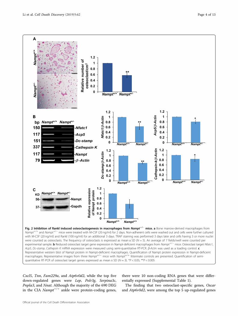
plays a critical role in osteoclast differentiation and thatdecreased Nampt expression attenuates osteoclast for-mation. To test this potential mechanism, we examinedRankl-dependent osteoclast differentiation in BMM iso-lated from Nampt+/+ and Nampt+/− mice (Fig. 2). TRAPactivity, a histochemical marker of osteoclastogenesis9,was detected by cell staining (Fig. 2a). M-CSF-dependentNampt+/+ BMM were able to produce TRAP+ cells fol-lowing a 3-day stimulation with M-CSF and Rankl, whilestimulated Nampt+/− BMM produced significantly fewerTRAP+ cells. The relative number of differentiatedosteoclasts from Nampt+/− BMM was significantly lowercompared with Nampt+/+ BMM (Fig. 2a). Decreaseddifferentiation of Nampt-deficient macrophages intoTRAP+ cells correlated with lower expression of Acp5, thegene encoding TRAP protein. mRNA levels for keyosteoclast markers, including Nfatc1, Dc-stamp, andCathepsin K, were also lower in osteoclasts derived fromNampt+/− BMM compared with Nampt+/+ controls, thusvalidating the attenuation of osteoclastogenesis (Fig. 2b).Western blot analyses verified that Nampt expressionwas decreased in Nampt+/− BMM relative to Nampt+/+
BMM (Fig. 2c).To investigate the mechanism by which Nampt-
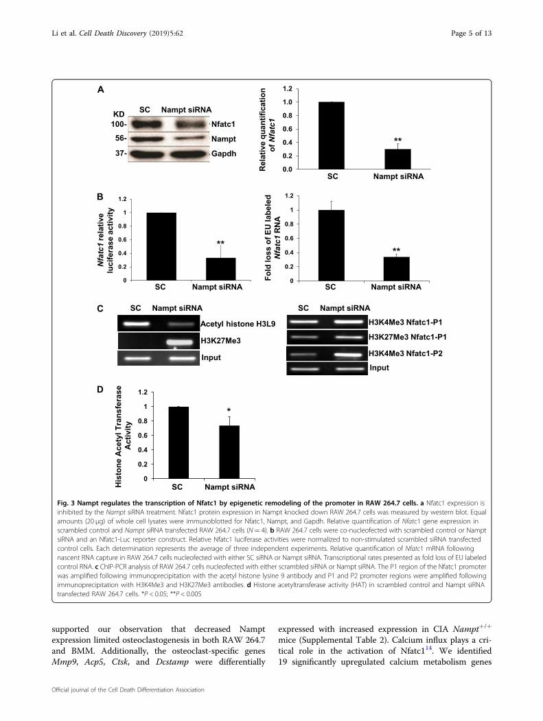
deficiency attenuated macrophage differentiation intoTRAP+ cells, we examined the expression of Nfactc1, anessential transcriptional regulator of osteoclast differ-entiation10. Silencing of Nampt expression by siRNA inRAW 264.7 cells inhibited expression of Nfatc1 proteinand mRNA (Fig. 3a). To determine the regulatorymechanism of Nfatc1 expression, we tested mRNA sta-bility in response to decreased Nampt levels. Althoughthere was a decrease in mRNA stability, it was not suffi-cient to account entirely for the loss in protein levels (datanot shown). Therefore, we investigated Nfatc1 transcrip-tion using luciferase reporter and nascent RNA captureassays. The relative luciferase activity decreased sig-nificantly in RAW 264.7 cells co-transfected with theNfatc1 promoter luciferase reporter and Nampt siRNArelative to the Nfatc1 promoter luciferase reporter andscrambled siRNA control cells (Fig. 3b). Nascent RNAcapture in RAW 264.7 cells validated further that synth-esis of Nfatc1 mRNA required Nampt expression(Fig. 3b).Epigenetic re-modeling of the Nfatc1 promoter plays a
critical role in Nfatc1 expression11. Therefore, we per-formed ChIP-PCR analyses in Nampt-deficient RAW264.7 cells to characterize the transcriptional regulation ofthe Nfatc1 promoter. The interaction of acetylated his-tones, which represents open chromatin, with the Nfatc1P1 promoter decreased following Nampt knockdown(Fig. 3c). Conversely, the presence of methylated histones,which represents closed chromatin, increased at theNfatc1 P1 and P2 promoter regions following Nampt
Li et al. Cell Death Discovery (2019) 5:62 Page 2 of 13
Official journal of the Cell Death Differentiation Association

siRNA knockdown compared with scrambled siRNAtransfected controls (Fig. 3c). These observations corre-sponded with decreased histone acetyltransferase (HAT)activity in RAW 264.7 cells subjected to Nampt knock-down (Fig. 3d). The epigenetic remodeling was consistentwith the decreased transcriptional activity observed byluciferase reporter and nascent RNA capture assays(Fig. 3b).To determine if Nampt enzymatic activity was
required for the Nampt–NfatC1–osteoclastogenesispathway, we treated RAW 264.7 and Nampt+/+ BMMwith the enzymatic inhibitors, FK86612 and MC413
during Rankl-induced differentiation. Formation ofTRAP+ cells was decreased significantly in RAW 264.7cells treated with FK866 and MC4 relative to theDMSO-vehicle control cells (Fig. 4a, b). Differentiationof Nampt+/+ BMM also decreased significantly inresponse to FK866 and MC4 relative to the control cells(Fig. 4c, d). In both RAW 264.7 cells and Nampt+/+
BMM, MC4 was as effective as MTX in blocking theformation of TRAP+ cells. The combination of MC4 andMTX in RAW 264.7 and Nampt+/+ BMM significantly
blocked osteoclast differentiation compared with MC4or MTX treatment alone (Fig. 4b, d). These findingsstrongly support the requirement of Nampt enzymaticactivity to promote osteoclastogenesis.
Transcriptomic profiling of whole ankle joints in CIA miceidentifies genes and pathways associated withosteoclastogenesisTo investigate the molecular mechanisms of Nampt in
the pathogenesis of arthritis, we sequenced RNA isolatedfrom whole ankle joints of Nampt+/+ and Nampt+/− micewith and without CIA (Fig. 5). We initially determined theDEG in two comparison groups. There were 1613 DEG inCIA Nampt+/+ mice compared with non-CIA controlNampt+/+ mice and 1778 DEG in CIA Nampt+/− micecompared with CIA Nampt+/+ mice. Comparison of thesetwo lists identified 721 genes that were present in bothgroups (Fig. 5a), with 690 genes inversely regulated(Fig. 5b). We hypothesized these 690 genes were asso-ciated with Nampt’s role in the pathogenesis of CIA(Supplemental Table 1). The top five up-regulated pro-tein-encoding genes in CIA Nampt+/+ ankle were Oscar,
Fig. 1 Decreased NAMPT expression attenuates inflammation and bone erosion in collagen-induced arthritis. CIA was induced in 8 weeksold, male DBA/1J Nampt+/+ and Nampt+/− mice. Observable inflammation was observed for 10 weeks. Serum and hind limbs were collected forELISA and micro-CT analysis, respectively. a Changes in joint inflammation scores in Nampt+/+ (n= 11) and Nampt+/− (n= 13). Arthritic index scoreis reported as mean ±median absolute deviation. b Serum concentrations of anti-collagen antibody (scale ×100,000) and NAMPT. The results arepresented as mean ± SD; Nampt+/+ CIA (n= 3), Nampt+/− CIA (n= 5). c Representative micro-CT images of left hind paws and talus bones. Arrowrepresents the location of the talus in ankle joint. d Bone volume (BV) and bone volume per total volume (BV/TV) in talus are presented as mean ± SD;Nampt+/+ (n= 5), Nampt+/− (n= 4), Nampt+/+ CIA (n= 6), 7 Nampt+/− CIA (n= 6). *P < 0.05, **P < 0.005
Li et al. Cell Death Discovery (2019) 5:62 Page 3 of 13
Official journal of the Cell Death Differentiation Association
Cxcl5, Tnn, Fam229a, and Atp6v0d2, while the top fivedown-regulated genes were Lep, Polr3g, Serpina3c,Pnpla3, and Nnat. Although the majority of the 690 DEGin the CIA Nampt+/+ ankle were protein-coding genes,
there were 10 non-coding RNA genes that were differ-entially expressed (Supplemental Table 1).The finding that two osteoclast-specific genes, Oscar
and Atp6v0d2, were among the top 5 up-regulated genes
Fig. 2 Inhibition of Rankl induced osteoclastogenesis in macrophages from Nampt+/− mice. a Bone marrow-derived macrophages fromNampt+/+ and Nampt+/− mice were treated with M-CSF (20 ng/ml) for 2 days. Non-adherent cells were washed out and cells were further culturedwith M-CSF (20 ng/ml) and Rankl (100 ng/ml) for an additional 3 days. TRAP staining was performed 3 days later and cells having 3 or more nucleiwere counted as osteoclasts. The frequency of osteoclasts is expressed as mean ± SD (N= 5). An average of 7 fields/well were counted perexperimental sample. b Reduced osteoclast target gene expression in Nampt-deficient macrophages from Nampt+/− mice. Osteoclast target Nfatc1,Acp5, Dc-stamp, Cathepsin K mRNA expression were measured using semi-quantitative RT-PCR. β-Actin was used as a loading control. cRepresentative western blot of Nampt protein in Nampt-deficient macrophages. Quantification of Nampt protein expression in Nampt-deficientmacrophages. Representative images from three Nampt+/− mice with Nampt+/+ littermate controls are presented. Quantification of semi-quantitative RT-PCR of osteoclast target genes expressed as mean ± SD (N= 3). *P < 0.05; **P < 0.005
Li et al. Cell Death Discovery (2019) 5:62 Page 4 of 13
Official journal of the Cell Death Differentiation Association
supported our observation that decreased Namptexpression limited osteoclastogenesis in both RAW 264.7and BMM. Additionally, the osteoclast-specific genesMmp9, Acp5, Ctsk, and Dcstamp were differentially
expressed with increased expression in CIA Nampt+/+
mice (Supplemental Table 2). Calcium influx plays a cri-tical role in the activation of Nfatc114. We identified19 significantly upregulated calcium metabolism genes
SC Nampt siRNA
Nfatc1
Nampt
Gapdh
KD100-
56-
37-
0.0
0.2
0.4
0.6
0.8
1.0
1.2
SC Nampt siRNA
Rel
ativ
e qu
antif
icat
ion
of N
fatc
1
**
A
0
0.2
0.4
0.6
0.8
1
1.2
SC Nampt siRNA
Fold
loss
of E
U la
bele
d N
fatc
1 R
NA
**
0
0.2
0.4
0.6
0.8
1
1.2
SC Nampt siRNA
Nfa
tc1
rela
tive
luci
fera
se a
ctiv
ity
**
B
SC Nampt siRNA
Acetyl histone H3L9
H3K27Me3
Input
SC Nampt siRNA
H3K4Me3 Nfatc1-P1
H3K27Me3 Nfatc1-P1
H3K4Me3 Nfatc1-P2
Input
C
0
0.2
0.4
0.6
0.8
1
1.2
His
tone
Ace
tyl T
rans
fera
se
Act
ivity
*
SC Nampt siRNA
D
Fig. 3 Nampt regulates the transcription of Nfatc1 by epigenetic remodeling of the promoter in RAW 264.7 cells. a Nfatc1 expression isinhibited by the Nampt siRNA treatment. Nfatc1 protein expression in Nampt knocked down RAW 264.7 cells was measured by western blot. Equalamounts (20 µg) of whole cell lysates were immunoblotted for Nfatc1, Nampt, and Gapdh. Relative quantification of Nfatc1 gene expression inscrambled control and Nampt siRNA transfected RAW 264.7 cells (N= 4). b RAW 264.7 cells were co-nucleofected with scrambled control or NamptsiRNA and an Nfatc1-Luc reporter construct. Relative Nfatc1 luciferase activities were normalized to non-stimulated scrambled siRNA transfectedcontrol cells. Each determination represents the average of three independent experiments. Relative quantification of Nfatc1 mRNA followingnascent RNA capture in RAW 264.7 cells nucleofected with either SC siRNA or Nampt siRNA. Transcriptional rates presented as fold loss of EU labeledcontrol RNA. c ChIP-PCR analysis of RAW 264.7 cells nucleofected with either scrambled siRNA or Nampt siRNA. The P1 region of the Nfatc1 promoterwas amplified following immunoprecipitation with the acetyl histone lysine 9 antibody and P1 and P2 promoter regions were amplified followingimmunoprecipitation with H3K4Me3 and H3K27Me3 antibodies. d Histone acetyltransferase activity (HAT) in scrambled control and Nampt siRNAtransfected RAW 264.7 cells. *P < 0.05; **P < 0.005
Li et al. Cell Death Discovery (2019) 5:62 Page 5 of 13
Official journal of the Cell Death Differentiation Association

within the 690 DEG (Supplemental Table 3). Our pre-vious finding that knockdown of NAMPT expressionsignificantly attenuated calcium influx into human pul-monary artery endothelial cells15, supports our hypothesisthat decreased Nampt expression may inhibit osteoclastdifferentiation by inhibiting calcium influx via mediatingexpression of calcium metabolism genes.To gain further insight into the biological functions
associated with Nampt mediated pathogenesis of CIA, wesubmitted the 690 DEG for pathway analysis. Theseinclude several pathways associated with osteoclastogen-esis, including the Inhibition of Matrix Metalloproteasesand the Role of Osteoblasts, Osteoclasts and Chondrocytesin Rheumatoid Arthritis (Fig. 5c, Supplemental Table 4).IPA analysis predicted target molecules in the dataset of
690 DEG that are either activated or inhibited by well-characterized upstream regulators. TNFα (5.575 activa-tion z-score; 7.73E−46 P-value of overlap), TGFB1 (5.436;
9.4E−36), and LPS (5.985; 4.87E−32) are the top 3 acti-vators, while the top 3 inhibitory regulators are the drugsdexamethasone (−3.074; 2.12E−34) and rosiglitazone(−4.791; 7.56E−24), and the kinase inhibitor PD98059(−3.847; 1.33E−23) (Supplemental Table 5). Upstreamregulator analysis also identified factors, such as cytokines(Tnfsf11, CSF1), transcriptional regulators (Fos, Nfatc1),and signaling proteins (Nfκb, Erk1/2, P38 Mapk, Src,P13k, Akt) that are known to promote osteoclast differ-entiation and bone resorption (Supplemental Table 6).We next performed functional enrichment analyses for
GO terms and KEGG to predict potential biologicalprocesses and pathways involved in the Nampt associatedpathogenesis of CIA. The top biological processes linkedwith the 690 DEG include GO:0007155—cell adhesion,GO:0022610—biological adhesion, GO:0001501—skeletalsystem development, GO:0001503—ossification andGO:0060348—bone development (Fig. 5d, Supplemental
Fig. 4 Inhibition of Rankl induced osteoclasts differentiation by Nampt enzyme inhibitors MC4, FK866 with or without MTX in RAW 264.7and BMM. a RAW cells were pretreated with 2 nM MC4 or FK866 or 50 nM MTX for 3 h and then stimulated with Rankl (100 ng/ml) for 5 days. TRAPstaining was performed using a TRACP and ALP double staining kit. b The frequency of TRAP-positive cells expressed as mean of triplicatedeterminations ± SD. c BMM were isolated from wild-type mice pretreated with 2 nM MC4 or FK866 or 50 nM MTX for 3 h and then stimulated withRankl (100 ng/ml) for 5 days. TRAP staining was performed using a TRACP and ALP double staining kit. d The frequency of TRAP-positive cellsexpressed as mean of triplicate determinations ± SD. *P < 0.05; **P < 0.005; ***P < 0.001
Li et al. Cell Death Discovery (2019) 5:62 Page 6 of 13
Official journal of the Cell Death Differentiation Association
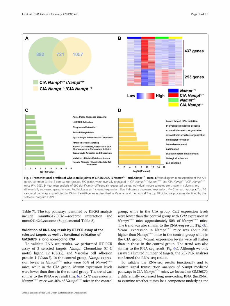
Table 7). The top pathways identified by KEGG analysisinclude mmu04512:ECM—receptor interaction andmmu04142:Lysosome (Supplemental Table 8).
Validation of RNA-seq result by RT-PCR assay of theselected targets as well as functional validation ofGM26870, a long non-coding RNATo validate RNA-seq results, we performed RT-PCR
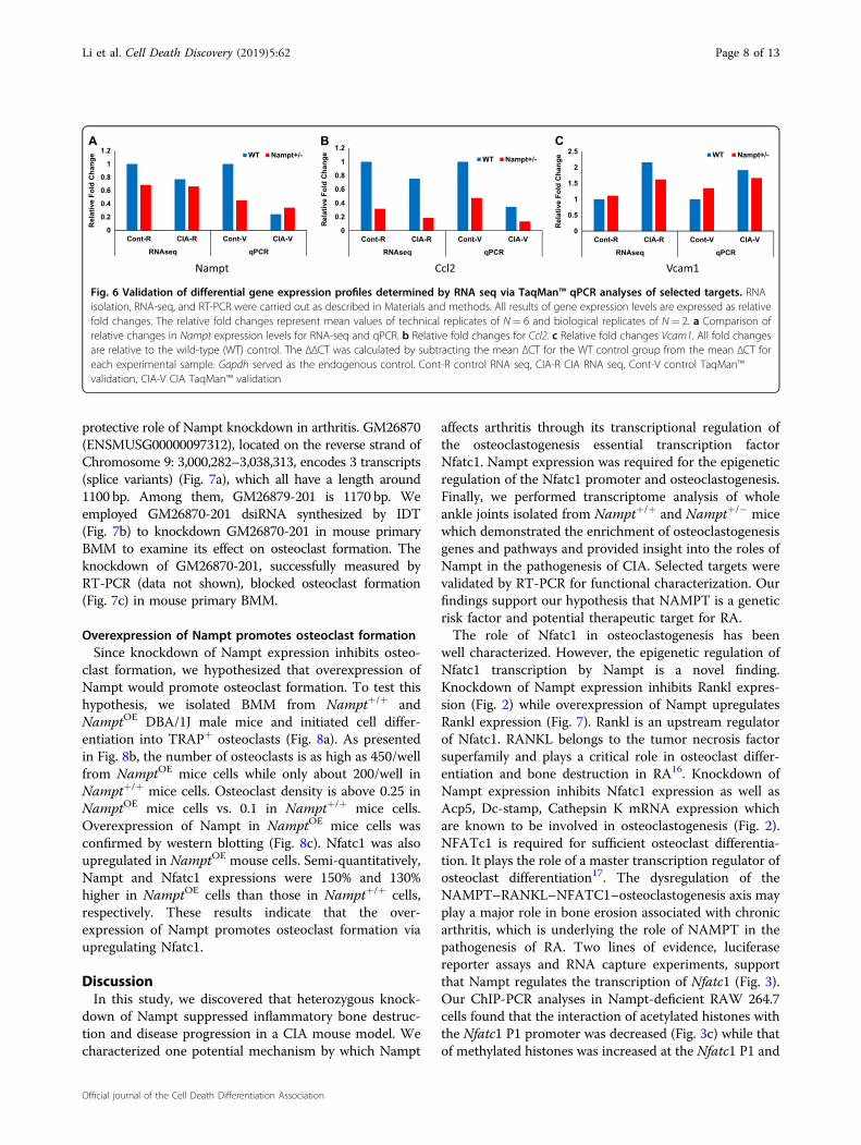
assay of 3 selected targets: Nampt, Chemokine (C–Cmotif) ligand 12 (Ccl12), and Vascular cell adhesionprotein 1 (Vcam1). In the control group, Nampt expres-sion levels in Nampt+/− mice were 40% of Nampt+/+
mice, while in the CIA group, Nampt expression levelswere lower than those in the control group. The trend wassimilar to the RNA-seq result (Fig. 6a). Ccl2 expression inNampt+/− mice was 40% of Nampt+/+ mice in the control
group, while in the CIA group, Ccl2 expression levelswere lower than the control group with Ccl2 expression inNampt+/− mice approximately 50% of Nampt+/+ mice.The trend was also similar to the RNA-seq result (Fig. 6b).Vcam1 expression in Nampt+/− mice was about 20%higher than Nampt+/+ mice in the control group while inthe CIA group, Vcam1 expression levels were all higherthan in those in the control group. The trend was alsosimilar to the RNA-seq result (Fig. 6c). Although we onlyassayed a limited number of targets, the RT-PCR analysesconfirmed the RNA-seq results.To validate the RNA-seq results functionally and to
initiate signal transduction analyses of Nampt mediatedpathways in CIA Nampt+/− mice, we focused on GM26870,a differentially expressed long non-coding RNA (lncRNA),to examine whether it may be a component underlying the
Fig. 5 Transcriptional profiles of whole ankle joints of CIA in DBA/1J Nampt+/+ and Nampt+/− mice. a Venn diagram representation of the 721genes common to the 2 comparison groups. 690 genes were inversely regulated in CIA Nampt+/+/Nampt+/+ and CIA Nampt+/−/CIA Nampt+/+
mice (P < 0.05). b Heat map analysis of 690 significantly differentially expressed genes. Individual mouse samples are shown in columns anddifferentially expressed genes in rows. Red indicates an increased expression. Blue indicates a decreased expression. N= 2 for each group. c Top 10canonical pathways as predicted by IPA for the 690 genes as described in Materials and methods. d The top 10 biological processes identified by thesoftware program DAVID
Li et al. Cell Death Discovery (2019) 5:62 Page 7 of 13
Official journal of the Cell Death Differentiation Association
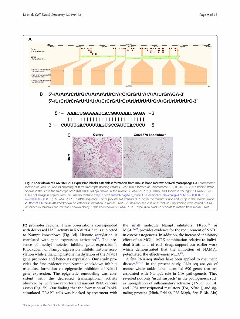
protective role of Nampt knockdown in arthritis. GM26870(ENSMUSG00000097312), located on the reverse strand ofChromosome 9: 3,000,282–3,038,313, encodes 3 transcripts(splice variants) (Fig. 7a), which all have a length around1100 bp. Among them, GM26879-201 is 1170 bp. Weemployed GM26870-201 dsiRNA synthesized by IDT(Fig. 7b) to knockdown GM26870-201 in mouse primaryBMM to examine its effect on osteoclast formation. Theknockdown of GM26870-201, successfully measured byRT-PCR (data not shown), blocked osteoclast formation(Fig. 7c) in mouse primary BMM.
Overexpression of Nampt promotes osteoclast formationSince knockdown of Nampt expression inhibits osteo-
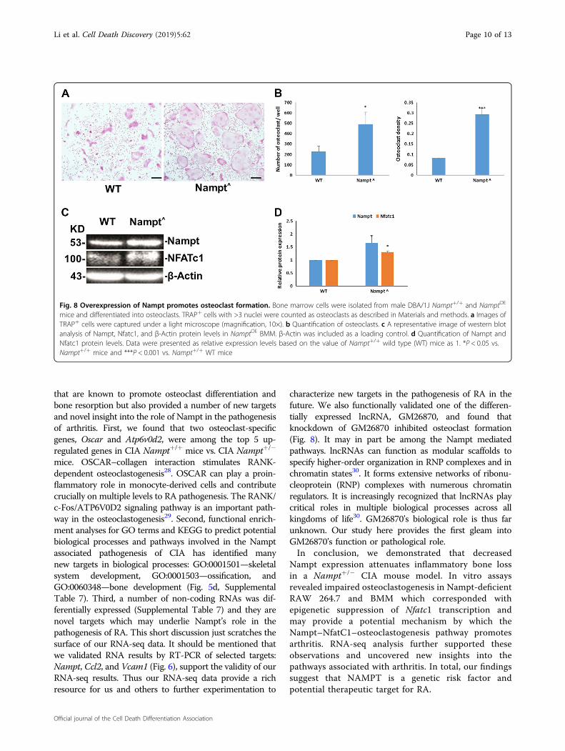
clast formation, we hypothesized that overexpression ofNampt would promote osteoclast formation. To test thishypothesis, we isolated BMM from Nampt+/+ andNamptOE DBA/1J male mice and initiated cell differ-entiation into TRAP+ osteoclasts (Fig. 8a). As presentedin Fig. 8b, the number of osteoclasts is as high as 450/wellfrom NamptOE mice cells while only about 200/well inNampt+/+ mice cells. Osteoclast density is above 0.25 inNamptOE mice cells vs. 0.1 in Nampt+/+ mice cells.Overexpression of Nampt in NamptOE mice cells wasconfirmed by western blotting (Fig. 8c). Nfatc1 was alsoupregulated in NamptOE mouse cells. Semi-quantitatively,Nampt and Nfatc1 expressions were 150% and 130%higher in NamptOE cells than those in Nampt+/+ cells,respectively. These results indicate that the over-expression of Nampt promotes osteoclast formation viaupregulating Nfatc1.
DiscussionIn this study, we discovered that heterozygous knock-
down of Nampt suppressed inflammatory bone destruc-tion and disease progression in a CIA mouse model. Wecharacterized one potential mechanism by which Nampt
affects arthritis through its transcriptional regulation ofthe osteoclastogenesis essential transcription factorNfatc1. Nampt expression was required for the epigeneticregulation of the Nfatc1 promoter and osteoclastogenesis.Finally, we performed transcriptome analysis of wholeankle joints isolated from Nampt+/+ and Nampt+/− micewhich demonstrated the enrichment of osteoclastogenesisgenes and pathways and provided insight into the roles ofNampt in the pathogenesis of CIA. Selected targets werevalidated by RT-PCR for functional characterization. Ourfindings support our hypothesis that NAMPT is a geneticrisk factor and potential therapeutic target for RA.The role of Nfatc1 in osteoclastogenesis has been
well characterized. However, the epigenetic regulation ofNfatc1 transcription by Nampt is a novel finding.Knockdown of Nampt expression inhibits Rankl expres-sion (Fig. 2) while overexpression of Nampt upregulatesRankl expression (Fig. 7). Rankl is an upstream regulatorof Nfatc1. RANKL belongs to the tumor necrosis factorsuperfamily and plays a critical role in osteoclast differ-entiation and bone destruction in RA16. Knockdown ofNampt expression inhibits Nfatc1 expression as well asAcp5, Dc-stamp, Cathepsin K mRNA expression whichare known to be involved in osteoclastogenesis (Fig. 2).NFATc1 is required for sufficient osteoclast differentia-tion. It plays the role of a master transcription regulator ofosteoclast differentiation17. The dysregulation of theNAMPT–RANKL–NFATC1–osteoclastogenesis axis mayplay a major role in bone erosion associated with chronicarthritis, which is underlying the role of NAMPT in thepathogenesis of RA. Two lines of evidence, luciferasereporter assays and RNA capture experiments, supportthat Nampt regulates the transcription of Nfatc1 (Fig. 3).Our ChIP-PCR analyses in Nampt-deficient RAW 264.7cells found that the interaction of acetylated histones withthe Nfatc1 P1 promoter was decreased (Fig. 3c) while thatof methylated histones was increased at the Nfatc1 P1 and
0
0.5
1
1.5
2
2.5
Cont-R CIA-R Cont-V CIA-V
RNAseq qPCR
WT Nampt+/-C
Rel
ativ
e Fo
ld C
hang
e
0
0.2
0.4
0.6
0.8
1
1.2
Cont-R CIA-R Cont-V CIA-V
RNAseq qPCR
WT Nampt+/-
B
Rel
ativ
e Fo
ld C
hang
e
0
0.2
0.4
0.6
0.8
1
1.2
Cont-R CIA-R Cont-V CIA-V
RNAseq qPCR
WT Nampt+/-A
Rel
ativ
e Fo
ld C
hang
e
1macV2lcCtpmaN
Fig. 6 Validation of differential gene expression profiles determined by RNA seq via TaqMan™ qPCR analyses of selected targets. RNAisolation, RNA-seq, and RT-PCR were carried out as described in Materials and methods. All results of gene expression levels are expressed as relativefold changes. The relative fold changes represent mean values of technical replicates of N= 6 and biological replicates of N= 2. a Comparison ofrelative changes in Nampt expression levels for RNA-seq and qPCR. b Relative fold changes for Ccl2. c Relative fold changes Vcam1. All fold changesare relative to the wild-type (WT) control. The ΔΔCT was calculated by subtracting the mean ΔCT for the WT control group from the mean ΔCT foreach experimental sample. Gapdh served as the endogenous control. Cont-R control RNA seq, CIA-R CIA RNA seq, Cont-V control TaqMan™validation, CIA-V CIA TaqMan™ validation
Li et al. Cell Death Discovery (2019) 5:62 Page 8 of 13
Official journal of the Cell Death Differentiation Association
P2 promoter regions. These observations correspondedwith decreased HAT activity in RAW 264.7 cells subjectedto Nampt knockdown (Fig. 3d). Histone acetylation iscorrelated with gene expression activation18. The pre-sence of methyl moieties inhibits gene expression19.Knockdown of Nampt expression inhibits histone acet-ylation while enhancing histone methylation of the Nfatc1gene promoter and hence its expression. Our study pro-vides the first evidence that Nampt knockdown inhibitsosteoclast formation via epigenetic inhibition of Nfatc1gene expression. The epigenetic remodeling was con-sistent with the decreased transcriptional activityobserved by luciferase reporter and nascent RNA captureassays (Fig. 3b). Our finding that the formation of Rankl-stimulated TRAP+ cells was blocked by treatment with
the small molecule Nampt inhibitors, FK86612 orMC413,20, provides evidence for the requirement of NAD+
in osteoclastogenesis. In addition, the increased inhibitoryeffect of an MC4+MTX combination relative to indivi-dual treatments of each drug, support our earlier workwhich demonstrated that the inhibition of NAMPTpotentiated the effectiveness MTX21.A few RNA-seq studies have been applied to rheumatic
diseases22–27. In the present study, RNA-seq analysis ofmouse whole ankle joints identified 690 genes that areassociated with Nampt’s role in CIA pathogenesis. Theyrevealed not only “usual suspects” in the pathogenesis suchas upregulation of inflammatory activator (TNFα, TGFB1,and LPS), transcriptional regulators (Fos, Nfatc1), and sig-naling proteins (Nfκb, Erk1/2, P38 Mapk, Src, P13k, Akt)
Fig. 7 Knockdown of GM26870-201 expression blocks osteoblast formation from mouse bone marrow-derived macrophages. a Chromosomelocation of GM26870 and its encoding of three transcripts (splicing variants). GM26870 is located at Chromosome 9: 3,000,282–3,038,313 reverse strand.Shown in the left is the transcript GM26870-201 (1170 bp), shown in the middle is GM26870-202 (1170 bp), and shown in the right is GM26870-203(1143 bp). Image is copied from the Ensembl website (http://useast.ensembl.org/Mus_musculus/Gene/Splice?db=core;g=ENSMUSG00000097312;r=9:3000282-3038313). b GM26870-201 dsiRNA sequence. The duplex dsiRNA consists of 25 bp in the forward strand and 27 bp in the reverse strand.c Effect of GM26870-201 knockdown on osteoclast formation in mouse BMM. Cell isolation and culture as well as Trap staining were carried out asdescribed in Materials and methods. Shown clearly is that knockdown of GM26870-201 expression blocks osteoclast formation from mouse BMM
Li et al. Cell Death Discovery (2019) 5:62 Page 9 of 13
Official journal of the Cell Death Differentiation Association
that are known to promote osteoclast differentiation andbone resorption but also provided a number of new targetsand novel insight into the role of Nampt in the pathogenesisof arthritis. First, we found that two osteoclast-specificgenes, Oscar and Atp6v0d2, were among the top 5 up-regulated genes in CIA Nampt+/+ mice vs. CIA Nampt+/−
mice. OSCAR–collagen interaction stimulates RANK-dependent osteoclastogenesis28. OSCAR can play a proin-flammatory role in monocyte-derived cells and contributecrucially on multiple levels to RA pathogenesis. The RANK/c‑Fos/ATP6V0D2 signaling pathway is an important path-way in the osteoclastogenesis29. Second, functional enrich-ment analyses for GO terms and KEGG to predict potentialbiological processes and pathways involved in the Namptassociated pathogenesis of CIA has identified manynew targets in biological processes: GO:0001501—skeletalsystem development, GO:0001503—ossification, andGO:0060348—bone development (Fig. 5d, SupplementalTable 7). Third, a number of non-coding RNAs was dif-ferentially expressed (Supplemental Table 7) and they arenovel targets which may underlie Nampt’s role in thepathogenesis of RA. This short discussion just scratches thesurface of our RNA-seq data. It should be mentioned thatwe validated RNA results by RT-PCR of selected targets:Nampt, Ccl2, and Vcam1 (Fig. 6), support the validity of ourRNA-seq results. Thus our RNA-seq data provide a richresource for us and others to further experimentation to
characterize new targets in the pathogenesis of RA in thefuture. We also functionally validated one of the differen-tially expressed lncRNA, GM26870, and found thatknockdown of GM26870 inhibited osteoclast formation(Fig. 8). It may in part be among the Nampt mediatedpathways. lncRNAs can function as modular scaffolds tospecify higher-order organization in RNP complexes and inchromatin states30. It forms extensive networks of ribonu-cleoprotein (RNP) complexes with numerous chromatinregulators. It is increasingly recognized that lncRNAs playcritical roles in multiple biological processes across allkingdoms of life30. GM26870’s biological role is thus farunknown. Our study here provides the first gleam intoGM26870’s function or pathological role.In conclusion, we demonstrated that decreased
Nampt expression attenuates inflammatory bone lossin a Nampt+/− CIA mouse model. In vitro assaysrevealed impaired osteoclastogenesis in Nampt-deficientRAW 264.7 and BMM which corresponded withepigenetic suppression of Nfatc1 transcription andmay provide a potential mechanism by which theNampt–NfatC1–osteoclastogenesis pathway promotesarthritis. RNA-seq analysis further supported theseobservations and uncovered new insights into thepathways associated with arthritis. In total, our findingssuggest that NAMPT is a genetic risk factor andpotential therapeutic target for RA.
Fig. 8 Overexpression of Nampt promotes osteoclast formation. Bone marrow cells were isolated from male DBA/1J Nampt+/+ and NamptOE
mice and differentiated into osteoclasts. TRAP+ cells with >3 nuclei were counted as osteoclasts as described in Materials and methods. a Images ofTRAP+ cells were captured under a light microscope (magnification, 10×). b Quantification of osteoclasts. c A representative image of western blotanalysis of Nampt, Nfatc1, and β-Actin protein levels in NamptOE BMM. β-Actin was included as a loading control. d Quantification of Nampt andNfatc1 protein levels. Data were presented as relative expression levels based on the value of Nampt+/+ wild type (WT) mice as 1. *P < 0.05 vs.Nampt+/+ mice and ***P < 0.001 vs. Nampt+/+ WT mice
Li et al. Cell Death Discovery (2019) 5:62 Page 10 of 13
Official journal of the Cell Death Differentiation Association
Materials and methodsAntibodies and chemicalsRPMI 1640 and DMEM were purchased from Life
Technologies. Lipopolysaccharide-Escherichia coli 055:B5 was obtained from Sigma Aldrich (#L6529; St. Louis,MO). TRACP and ALP double staining kit (#MK300) waspurchased from Clontech (Mountain View, CA). Anti-TRAP1 antibody (#ab151239) was from Abcam (Cam-bridge, MA). Phospho-MAPK family antibody samplerkit (#9910), pNF-κB p105 (#4806), pNF-κBp65 (#3033),Acetyl-Histone H3 (Lys9) (#9649), Tri-Methyl-HistoneH3 (Lys27) (#9733) antibodies, Simple ChIP EnzymaticChromatin IP kit (#9003), and cell lysis buffer (#9803)were purchased from Cell Signaling Technology (Beverly,MA). Recombinant Mouse M-CSF (#576406) and pur-ified anti-NFATc1 antibody (#649601) were purchasedfrom Biolegend (San Diego, CA). GAPDH (#sc25778),anti-Mouse and anti-Rabbit secondary antibodies werefrom Santa Cruz Biotechnology (Santa Cruz, CA).Nampt siRNA (Stealth_116; 5′-CCACCCAACACAAGCAAAGUUUAUU-3′) and Scrambled control siRNA(stealth_con 116, 5′-CCACAACAACAAACGUUGAUCCAUU-3′), Click-iT Nascent RNA capture kit(#C10365), Superscript III first strand synthesis supermixfor qRT-PCR (#11752-050), Superscript VILO cDNAsynthesis Supermix (#11754–050), and mouse Ranklrecombinant protein (#RP-8601) were from (Thermo-Fisher Scientific, Waltham, MA). TaqMan® geneexpression assays for Nfatc1 (Mm00479445_m1), Acp5(mCG22832), and TaqMan® gene expression master mix(#4369016) were purchased from Applied Biosystems(Foster City, CA). SF cell line 4D Nucleofector X kit wasfrom Lonza (Alpharetta, GA). Anti-Nampt antibody(#A300–372A) was purchased from Bethyl Laboratories(Montgomery, TX).
Cell cultureThe murine macrophage cell line RAW 264.7 (TIB- 71,
ATCC®) was maintained in RPMI 1640 media containing10% FBS and antibiotics. All cells were grown at 37 °C, 5%CO2.
Animal studiesMice were maintained in an Association for Assessment
and Accreditation of Laboratory Animal Care accreditedinstitution in a temperature-controlled, pathogen-freefacility with a 12 h light/dark cycle. The mice were ganghoused with free access to food (Irradiated Teklad Global18% Rodent Diet, Envigo, cat# 2918.15) and water.DBA/1J mice (The Jackson Laboratory) were crossed withC57BL/6J Nampt+/− mice31. Offspring were backcrossedto DBA/1J for >10 generations to develop congenicDBA/1J Nampt+/−. To induce arthritis, 10-week-old,male mice were immunized with bovine collagen type II
(CII, 100 µg, Chondrex) in Complete Freund’s Adjuvant(CFA, 100 µg Mycobacterium tuberculosis, Chondrex) byintradermal injection at the base of the tail. A collagenbooster (100 µg) in Incomplete Freund’s Adjuvant (IFA,Chondrex) was administered at day 218. Mice were eval-uated for the onset of inflammation and scored (scale 0–4per paw) as described by Brand et al.8. Mice were scoredtwice per week for 10 weeks. At the end of the 10-weekperiod, the animals were euthanized and tissue was iso-lated for analysis. Circulating serum levels of anti-mouseCII antibody (Chondrex) and Nampt (AdipoGen®) weredetermined by ELISA. The left hind limb was scannedwith a vivaCT40 (Scanco) in the Skeletal Imaging and Invitro-In vivo Mechanical Core in the University of Mis-souri Kansas City, School of Dentistry, as described pre-viously32. The right hind limb was collected for RNAisolation and gene expression analyses.Overexpression (NamptOE) mice were generated in our
lab as described previously33.
Isolation of bone marrow-derived macrophageIsolation of BMM was performed as previously
described34.
In vitro osteoclastogenesisOsteoclasts were generated from bone marrow as
described previously35.
siRNA mediated Nampt knockdownRAW 264.7 cells were 4D-nucleofected with Nampt and
scrambled siRNA (50 nM/2 × 106 cells) in SF solution(Amaxa). Cells were seeded at 5 × 10⁴ nucleofected cells/well/24-well plate for TRAP staining and 1 × 10⁵ cells/well/6-well plate for protein isolation.
Protein extraction and western blot analysisProtein extraction and western blot analysis were car-
ried according to our previous procedure33.
ChIP assaySimpleChIP® Enzymatic Chromatin IP assays (Cell
Signaling Technology) with Acetyl-Histone H3 (Lys9)and Tri-Methyl-Histone H3 (Lys27) antibodies wereutilized according to the manufacturer’s instruction.Immunoprecipitated DNA was reverse cross-linked,purified and analyzed by PCR (primers: NFATc1-617-F5′-GGAAGCCTGCGATTTTACAT-3′, NFATc1-426-R5′-ACGAAACGGGAAGGAAAG-3′).
Histone acetyltransferase (HAT) assayHAT enzyme activity was quantified in Nampt-
deficient and scrambled control cells using an EpiQuikHAT assay (EpiGentek) according to the manufacturer’sinstruction.
Li et al. Cell Death Discovery (2019) 5:62 Page 11 of 13
Official journal of the Cell Death Differentiation Association
Luciferase reporter assaysLuciferase reporter assays were performed as described
previously34.
RNA isolation, quantitative RT-PCR, and nascent RNAcaptureRNA isolation, quantitative RT-PCR, and nascent RNA
capture were performed as we previously described36.
RNA-seqRNA was isolated from flash frozen whole ankle joints
collected after the 10-week CIA observation period.cDNA sequencing libraries were prepared with an Illu-mina TruSeq Stranded Total RNA Sample Prep Kit andsubjected to 2 × 101 paired-end sequencing as describedpreviously3. Mapping of RNA-seq reads and transcriptassembly and abundance estimation were conductedusing Tuxedo Suite pipeline (TopHat v1.3.0/Bowtiev0.12.7/Cufflinks v1.0.3) and reported in Fragments PerKilobase of exon per Million fragments mapped (FPKM).To identify genes which were differentially expressed, foldchanges for each gene were calculated by dividing theaverage FPKM for the case by the average FPKM for thecontrol. We determined fold changes for two comparisongroups: (1) CIA Nampt+/+ mice compared with non-CIAcontrol Nampt+/+ mice, and (2) CIA Nampt+/− micecompared with CIA Nampt+/+ mice.
Functional pathway analysisKyoto Encyclopedia of Genes and Genomes (KEGG)
analyses to identify cellular pathways and biological pro-cesses associated with differentially expressed genes(DEG) were performed with DAVID37. Ingenuity PathwayAnalysis (IPA) (Ingenuity Systems) predicted functionaland canonical pathways.
StatisticsStatistical analyses were performed with Sigma Stat (v4.0,
Systat Software, Inc.). Results were expressed as mean ± SD.P < 0.05 was considered statistically significant.
Data availabilityThe RNA-seq data have been deposited to Gene Expression Omnibus (http://www.ncbi.nlm.nih.gov/geo; accession number GSE121793).
AcknowledgementsThe authors are grateful to Drs. Inna Sokolovsky and Tomoyuki Mukai forexcellent technical assistance. This work was supported in part by NationalInstitutes of Health Grant HL 080042 (S.Q.Y.), by the Katherine Berry RichardsonFoundation (D.P.H.), Children’s Mercy and UMKC (S.Q.Y.), and the William R.Brown/Missouri endowment (S.Q.Y.) as well as a postdoctoral fellowship fromMinistry of Education of the People’s Republic of China to X.L.
Author details1Division of Experimental and Translational Genetics, Children’s Mercy, KansasCity, MO 64108, USA. 2Department of Biomedical and Health Informatics,University of Missouri Kansas City School of Medicine, Kansas City, MO 64108,USA. 3Department of Orthopedics, Xiangya Hospital, Central South University,
Changsha 410005, China. 4Department of Chemistry, University of Missouri,Columbia, MO 65211, USA. 5Department of Oral and Craniofacial Sciences,School of Dentistry, University of Missouri-Kansas City, Kansas City, MO 64108,USA
Author contributionsD.P.H., L.Q.Z., G.L. and S.Q.Y. conceptualized and designed the research. X.L., S.I.,D.P.H. and S.Q.Y. wrote the manuscript. X.L., S.I., M.X., N.N.N. and D.P.H. acquireddata. X.L., S.I., D.P.H., G.L. and S.Q.Y. analyzed data. M.W.L. and Y.U. provided samples,reagents, and other project assistance. All authors critically revised the manuscript.D.P.H., G.L. and S.Q.Y. provided overall project guidance and supervision.
Conflict of interestThe authors declare that they have no conflict of interest.
Study approvalAnimal studies were approved by the University of Missouri Kansas City’sInstitutional Animal Care and Use Committee.
Publisher’s noteSpringer Nature remains neutral with regard to jurisdictional claims inpublished maps and institutional affiliations.
The online version of this article (https://doi.org/10.1038/s41420-018-0134-6)contains supplementary material, which is available to authorized users.
Received: 5 October 2018 Revised: 15 November 2018 Accepted: 29November 2018
References1. Scott, D. L., Wolfe, F. & Huizinga, T. W. Rheumatoid arthritis. Lancet 376,
1094–1108 (2010).2. Glant, T. T., Mikecz, K. & Rauch, T. A. Epigenetics in the pathogenesis of
rheumatoid arthritis. BMC Med. 12, 35 (2014).3. Zhang, L. Q. et al. Metabolic and molecular insights into an essential role of
nicotinamide phosphoribosyltransferase. Cell Death Dis. 8, e2705 (2017).4. Zhang, L. Q., Heruth, D. P. & Ye, S. Q. Nicotinamide phosphoribosyltransferase
in human diseases. J. Bioanal. Biomed. 3, 13–25 (2011).5. Busso, N. et al. Pharmacological inhibition of nicotinamide phosphoribosyl-
transferase/visfatin enzymatic activity identifies a new inflammatory pathwaylinked to NAD. PLoS One 3, e2267 (2008).
6. Présumey, J. et al. Nicotinamide phosphoribosyltransferase/visfatin expressionby inflammatory monocytes mediates arthritis pathogenesis. Ann. Rheum. Dis.72, 1717–1724 (2013).
7. Seki, N. et al. Type II collagen-induced murine arthritis. I. Induction and per-petuation of arthritis require synergy between humoral and cell-mediatedimmunity. J. Immunol. 140, 1477–1484 (1988).
8. Brand, D. D., Latham, K. A. & Rosloniec, E. F. Collagen-induced arthritis. Nat.Protoc. 2, 1269–1275 (2007).
9. Vu, T. H. et al. MMP-9/gelatinase B is a key regulator of growth plate angio-genesis and apoptosis of hypertrophic chondrocytes. Cell 93, 411–422 (1998).
10. Winslow, M. M. et al. Calcineurin/NFAT signaling in osteoblasts regulates bonemass. Dev. Cell 10, 771–782 (2006).
11. Pham, L. V., Tamayo, A. T., Li, C., Bueso-Ramos, C. & Ford, R. J. An epigeneticchromatin remodeling role for NFATc1 in transcriptional regulation of growthand survival genes in diffuse large B-cell lymphomas. Blood 116, 3899–3906(2010).
12. Hasmann, M. & Schemainda, I. FK866, a highly specific noncompetitive inhi-bitor of nicotinamide phosphoribosyltransferase, represents a novelmechanism for induction of tumor cell apoptosis. Cancer Res. 63, 7436–7442(2003).
13. Lee, M. W. Jr., Sevryugina, Y. V., Khan, A. & Ye, S. Q. Carboranes increase thepotency of small molecule inhibitors of nicotinamide phosphoribosyltranfer-ase. J. Med. Chem. 55, 7290–7294 (2012).
14. Kajiya, H. Calcium signaling in osteoclast differentiation and bone resorption.Adv. Exp. Med. Biol. 740, 917–932 (2012).
Li et al. Cell Death Discovery (2019) 5:62 Page 12 of 13
Official journal of the Cell Death Differentiation Association
15. Ye, S. Q. et al. Pre-B-cell-colony-enhancing factor is critically involved inthrombin-induced lung endothelial cell barrier dysregulation. Microvasc. Res.70, 142–151 (2005).
16. Tanaka, S., Tanaka, Y., Ishiguro, N., Yamanaka, H. & Takeuchi, T. RANKL: atherapeutic target for bone destruction in rheumatoid arthritis. Mod. Rheu-matol. 28, 9–16 (2018).
17. Kim, J. H. & Kim, N. Regulation of NFATc1 in osteoclast differentiation. J. BoneMetab. 21, 233–241 (2014).
18. Verdone, L., Agricola, E., Caserta, M. & Di Mauro, E. Histone acetylation in generegulation. Brief. Funct. Genom. Proteom. 5, 209–221 (2006).
19. Razin, A. & Cedar, H. DNA methylation and gene expression. Microbiol. Rev. 55,451–458 (1991).
20. Huang, P. et al. MC-PPEA as a new and more potent inhibitor of CLP-inducedsepsis and pulmonary inflammation than FK866. Drug Des. Devel. Ther. 11,629–641 (2017).
21. Funk, R. S. et al. Nicotinamide phosphoribosyltransferase attenuates metho-trexate response in juvenile idiopathic arthritis and in vitro. Clin. Transl. Sci. 9,149–157 (2016).
22. Heruth, D. P., Gibson, M., Grigoryev, D. N., Zhang, L. Q. & Ye, S. Q. RNA-seqanalysis of synovial fibroblasts brings new insights into rheumatoid arthritis.Cell Biosci. 2, 43 (2012).
23. Shi, L. et al. The SLE transcriptome exhibits evidence of chronic endotoxinexposure and has widespread dysregulation of non-coding and coding RNAs.PLoS ONE 9, e93846 (2014).
24. Tandon, M., Gallo, A., Jang, S.-I., Illei, G. G. & Alevizos, I. Deep sequencing ofshort RNAs reveals novel microRNAs in minor salivary glands of patients withSjögren’s syndrome. Oral Dis. 18, 127–131 (2012).
25. Stone, R. C. et al. RNA-Seq for enrichment and analysis of IRF5 transcriptexpression in SLE. PLoS ONE 8, e54487 (2013).
26. Moncrieffe, H. et al. Transcriptional profiles of JIA patient blood withsubsequent poor response to methotrexate. Rheumatology. https://doi.org/10.1093/rheumatology/kex206 (2017).
27. Choi, S. et al. Transcription factor NFAT5 promotes macrophage survival inrheumatoid arthritis. J. Clin. Invest. 127, 954–969 (2017).
28. Schultz, H. S. et al. OSCAR-collagen signaling in monocytes plays a proin-flammatory role and may contribute to the pathogenesis of rheumatoidarthritis. Eur. J. Immunol. 46, 952–963 (2016).
29. Zhu, X., Zeng, Z., Qiu, D. & Chen, J. Vgamma9Vdelta2 T cells inhibitimmature dendritic cell transdifferentiation into osteoclasts throughdownregulation of RANK, cFos and ATP6V0D2. Int. J. Mol. Med. 42,2071–2079 (2018).
30. Rinn, J. L. & Chang, H. Y. Genome regulation by long noncoding RNAs. Annu.Rev. Biochem. 81, 145–166 (2012).
31. Hong, S. B. et al. Essential role of pre-B-cell colony enhancing factor inventilator-induced lung injury. Am. J. Respir. Crit. Care Med. 178, 605–617(2008).
32. Mukai, T. et al. Loss of SH3 domain-binding protein 2 function suppressesbone destruction in tumor necrosis factor-driven and collagen-inducedarthritis in mice. Arthritis Rheumatol. 67, 656–667 (2015).
33. Zhang, L. Q. et al. Novel protective role of nicotinamide phosphoribosyl-transferase in acetaminophen-induced acute liver injury in mice. Am. J. Pathol.188, 1640–1652 (2018).
34. Ling, M. et al. Epigenetic regulation of Runx2 transcription and osteoblastdifferentiation by nicotinamide phosphoribosyltransferase. Cell Biosci. 7, 27(2017).
35. Takayanagi, H. et al. Induction and activation of the transcription factorNFATc1 (NFAT2) integrate RANKL signaling in terminal differentiation ofosteoclasts. Dev. Cell 3, 889–901 (2002).
36. Bi, G. et al. Up-regulation of SFTPB expression and attenuation of acute lunginjury by pulmonary epithelial cell-specific NAMPT knockdown. FASEB J. 32,3583–3596 (2018).
37. da Huang, W., Sherman, B. T. & Lempicki, R. A. Systematic and integrativeanalysis of large gene lists using DAVID bioinformatics resources. Nat. Protoc. 4,44–57 (2009).
Li et al. Cell Death Discovery (2019) 5:62 Page 13 of 13
Official journal of the Cell Death Differentiation Association