epigenetic regulation of satellite cell fate during
TRANSCRIPT
REVIEW Open Access
Epigenetic regulation of satellite cell fateduring skeletal muscle regenerationJimmy Massenet1,2, Edward Gardner1,3, Bénédicte Chazaud2 and F. Jeffrey Dilworth1,3,4*
Abstract
In response to muscle injury, muscle stem cells integrate environmental cues in the damaged tissue to mediateregeneration. These environmental cues are tightly regulated to ensure expansion of muscle stem cell populationto repair the damaged myofibers while allowing repopulation of the stem cell niche. These changes in muscle stemcell fate result from changes in gene expression that occur in response to cell signaling from the muscleenvironment.Integration of signals from the muscle environment leads to changes in gene expression through epigeneticmechanisms. Such mechanisms, including post-translational modification of chromatin and nucleosomerepositioning, act to make specific gene loci more, or less, accessible to the transcriptional machinery. In youth, themuscle environment is ideally structured to allow for coordinated signaling that mediates efficient regeneration.Both age and disease alter the muscle environment such that the signaling pathways that shape the healthymuscle stem cell epigenome are altered. Altered epigenome reduces the efficiency of cell fate transitions requiredfor muscle repair and contributes to muscle pathology. However, the reversible nature of epigenetic changes holdsout potential for restoring cell fate potential to improve muscle repair in myopathies.In this review, we will describe the current knowledge of the mechanisms allowing muscle stem cell fate transitionsduring regeneration and how it is altered in muscle disease. In addition, we provide some examples of howepigenetics could be harnessed therapeutically to improve regeneration in various muscle pathologies.
Keywords: Muscle stem cells, Regeneration, Epigenetics, Cell fate, Duchenne muscular dystrophy
BackgroundThe development and regeneration of skeletal muscleare mediated by muscle stem cells (MuSCs), also termedsatellite cells, that act coordinately to ensure the efficientformation of myofibers while repopulating the niche toallow for repair after future injuries. To mediate myofi-ber formation, MuSCs must transit through multiple cellfates before achieving their fully differentiated state.Each of these intermediate cell fates share an identical
genome that is used as a blueprint for determining theiridentity. However, the characteristics of each cell fateare determined through alternate interpretation of thegenomic blueprint where epigenetic mechanisms areused to determine the subset of genes that will beexpressed. These epigenetic mechanisms achieve differ-ential gene expression by controlling the accessibility ofthe transcriptional machinery to specific loci. Indeed,not all genes within the nucleus can be accessible forgene expression as six billion base pairs of genetic infor-mation encoded by human diploid genome must behighly compacted to reside within the confines of thenuclear membrane. DNA compaction occurs throughthe formation of nucleosomes linking 147 bp and is re-peated across the genome at 200 bp intervals with the
© The Author(s). 2021 Open Access This article is licensed under a Creative Commons Attribution 4.0 International License,which permits use, sharing, adaptation, distribution and reproduction in any medium or format, as long as you giveappropriate credit to the original author(s) and the source, provide a link to the Creative Commons licence, and indicate ifchanges were made. The images or other third party material in this article are included in the article's Creative Commonslicence, unless indicated otherwise in a credit line to the material. If material is not included in the article's Creative Commonslicence and your intended use is not permitted by statutory regulation or exceeds the permitted use, you will need to obtainpermission directly from the copyright holder. To view a copy of this licence, visit http://creativecommons.org/licenses/by/4.0/.The Creative Commons Public Domain Dedication waiver (http://creativecommons.org/publicdomain/zero/1.0/) applies to thedata made available in this article, unless otherwise stated in a credit line to the data.
* Correspondence: [email protected] Center for Stem Cell Research, Regenerative Medicine Program,Ottawa Hospital Research Institute, 501 Smyth Rd, Mailbox 511, Ottawa, ONK1H 8L6, Canada3Department of Cellular and Molecular Medicine, University of Ottawa,Ottawa, ON K1H 8L6, CanadaFull list of author information is available at the end of the article
Massenet et al. Skeletal Muscle (2021) 11:4 https://doi.org/10.1186/s13395-020-00259-w
linker histone H1 protein allowing the protection ofinter-nucleosomal DNA. These 10-nm fibers then con-dense through a disordered self-aggregation to formchromatids [1]. While the DNA organization within eachcell has been suggested to be unique [2], general rulesemerge. For instance, genes that are not expressed tend tobe associated with the nuclear periphery while transcribedgenes tend to aggregate in the nuclear lumen. This distinctnuclear organization between cells is the basis for estab-lishing and maintaining cell-specific gene expression pro-grams. The chromatin state is fluid to allow expression ofthe needed genes in a spacio-temporal manner. Onemechanism allowing fluidity of the chromatin structure isthe dynamic nature of nucleosome association with DNAwhere nucleosome displacement allows incorporation ofunmarked histones within newly formed nucleosomes.This histone exchange represents more than a simpleturnover mechanism, as canonical histones in nucleo-somes can be replaced by different histone variants. His-tone variants alter the chemical properties of nucleosomesand change their stability.Ability to alter chromatin organization is critical for
mediating decisions of cell fate and differentiation. In re-sponse to environmental cues, chromatin organization ismodified and alters the accessibility of the transcriptionmachinery to the gene sequences, which is modulated byseveral levels of regulation. The first level lies within theDNA itself where the presence of specific sequence ele-ments surrounding the gene can act as promoters or en-hancers that serve as binding sites for transcriptionfactors (TFs), which can recruit the transcriptional ma-chinery to locus. The ability to recruit the transcriptionalmachinery may be increased by promoter-enhancer in-teractions that can occur through chromatin looping tomodulate gene expression. A second level of regulationis established through differential accessibility of TFs orthe transcription machinery to DNA elements. This canbe performed by modulation of chromatin conformation,caused by reversible modifications of either DNA or his-tones through epigenetic processes.DNA methylation is a widely used epigenetic mechan-
ism to regulate chromatin accessibility. The predomin-ant DNA modification in mammals is CpG methylation,where the addition of a methyl group at carbon 5 of thedeoxyribonucleotide cytosine alters the affinity of the TFfor its DNA element. CpG methylation stabilizes gene si-lencing through two potential modes: (I) blocking accessof DNA binding proteins required for transcription byimpairing their ability to bind DNA elements or (II) at-traction of TFs containing methyl-CpG-binding domainsthat are able to repress transcription. It has been pro-posed that DNA methylation does not trigger gene re-pression but instead stabilizes repression at alreadysilenced genes [3].
In contrast to the limited number of characterizedDNA modifications, histones undergo a wide-variety ofpost-translational modifications (PTMs) includingacetylation, methylation, phosphorylation, ubiquitina-tion, ADP-ribosylation, and citrullination. Among them,some histone PTMs are known to allow chromatin com-paction while others direct chromatin decompaction.For instance, trimethylation of lysine 9 and lysine 27 ofhistone 3 (H3K9me3 and H3K27me3) or trimethylationof lysine 20 of histone 4 (H4K20me3) are associated withlocal chromatin compaction. This compaction modu-lates TF or RNA polymerase II access to target se-quences and leads to the repression of gene expression.In contrast, acetylation of lysine 9 of histone 3(H3K9Ac) and lysine 20 of histone 4 (H4K20Ac) and thetrimethylation of the lysine 4 of histone 3 (H3K4me3)lead to relaxation of the chromatin state, improving ac-cessibility of the transcription machinery and increasinglocal gene expression [4–6]. Presence of relaxed chroma-tin at gene enhancer caused by monomethylation of ly-sine 20 of histone 3 (H3K20me1) and acetylation oflysine 27 of histone 3 (H3K27Ac) also modulates geneexpression [7, 8]. Thus, the relationship between histonemodifications and transcriptional output reveal how theepigenetic code regulates gene expression.In the present review, we will discuss the role for epi-
genetic regulators in mediating the regenerative functionof MuSCs in adults. The role for diseased and agedmuscle environment in modifying the epigenetic land-scape of MuSCs will also be examined.
MuSCs mediate regeneration of skeletal muscleResiding at the periphery of the muscle fiber betweenthe sarcolemma and the basal lamina [9], MuSCs laydormant in a quiescent state, ready to respond to muscleinjury. The fate of MuSCs is regulated by a series of TFsthat include Pax7 and a family of myogenic basic-Helix-loop-helix proteins, termed the myogenic regulatory fac-tors (MRFs), that include MYOD, MYF5, MRF4, andmyogenin (MYOG) proteins [10–12]. These muscle TFswork with other, more ubiquitous TFs, to establish theepigenetic states to regulate muscle regeneration.MuSC quiescent state is characterized by the expres-
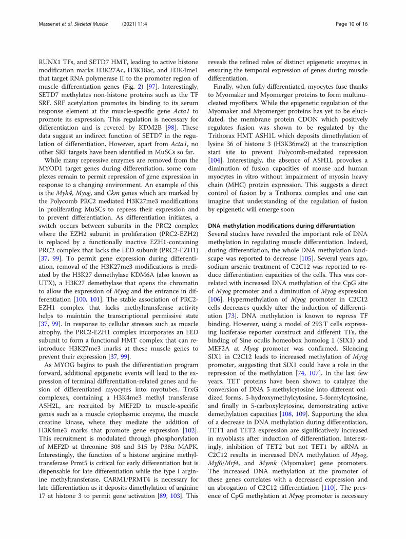
sion of PAX7 and FOXO transcription factors [13]. Myf5and Myod1 are also transcribed in quiescent MuSCs, butpost-transcriptional regulation prevents their translation(PAX7+FOXO+MYF5-MYOD1-) (Fig. 1) [13, 14]. Morespecifically, Myf5 mRNA is sequestered in messenger ri-bonucleoprotein granules (mRNPs) to avoid its transla-tion [15] while Myod1 mRNA retains an intron thatprevents its translocation out of the nucleus [16]. Aftermuscle injury, MuSC activation leads to symmetric and/or asymmetric division. Asymmetric divisions produce aPAX7+MYF5+ cell, destined to the myogenic program
Massenet et al. Skeletal Muscle (2021) 11:4 Page 2 of 16
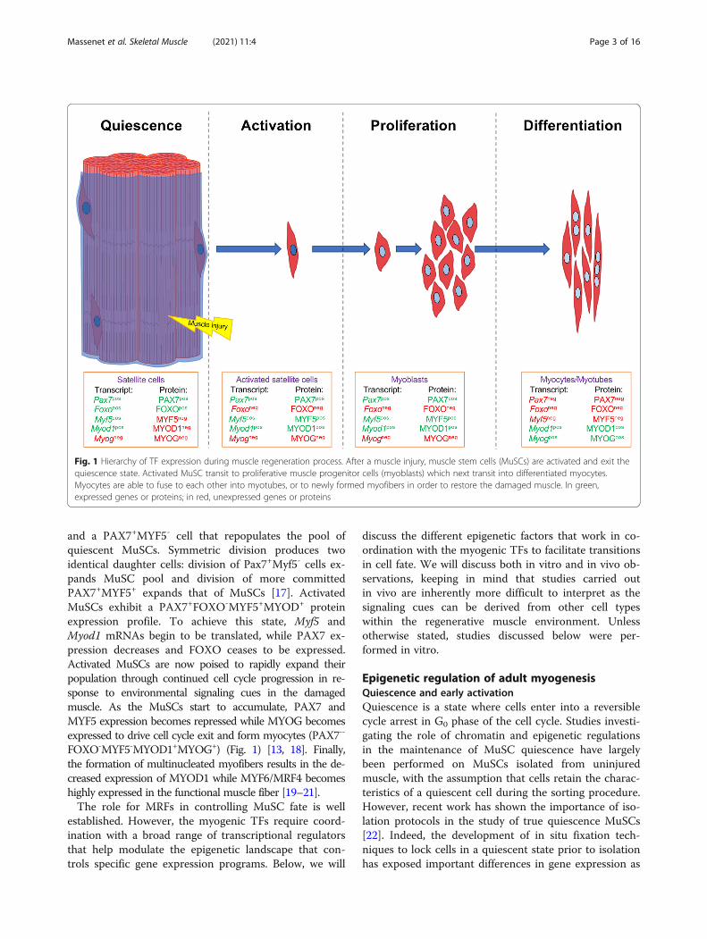
and a PAX7+MYF5- cell that repopulates the pool ofquiescent MuSCs. Symmetric division produces twoidentical daughter cells: division of Pax7+Myf5- cells ex-pands MuSC pool and division of more committedPAX7+MYF5+ expands that of MuSCs [17]. ActivatedMuSCs exhibit a PAX7+FOXO-MYF5+MYOD+ proteinexpression profile. To achieve this state, Myf5 andMyod1 mRNAs begin to be translated, while PAX7 ex-pression decreases and FOXO ceases to be expressed.Activated MuSCs are now poised to rapidly expand theirpopulation through continued cell cycle progression in re-sponse to environmental signaling cues in the damagedmuscle. As the MuSCs start to accumulate, PAX7 andMYF5 expression becomes repressed while MYOG becomesexpressed to drive cell cycle exit and form myocytes (PAX7--
FOXO-MYF5-MYOD1+MYOG+) (Fig. 1) [13, 18]. Finally,the formation of multinucleated myofibers results in the de-creased expression of MYOD1 while MYF6/MRF4 becomeshighly expressed in the functional muscle fiber [19–21].The role for MRFs in controlling MuSC fate is well
established. However, the myogenic TFs require coord-ination with a broad range of transcriptional regulatorsthat help modulate the epigenetic landscape that con-trols specific gene expression programs. Below, we will
discuss the different epigenetic factors that work in co-ordination with the myogenic TFs to facilitate transitionsin cell fate. We will discuss both in vitro and in vivo ob-servations, keeping in mind that studies carried outin vivo are inherently more difficult to interpret as thesignaling cues can be derived from other cell typeswithin the regenerative muscle environment. Unlessotherwise stated, studies discussed below were per-formed in vitro.
Epigenetic regulation of adult myogenesisQuiescence and early activationQuiescence is a state where cells enter into a reversiblecycle arrest in G0 phase of the cell cycle. Studies investi-gating the role of chromatin and epigenetic regulationsin the maintenance of MuSC quiescence have largelybeen performed on MuSCs isolated from uninjuredmuscle, with the assumption that cells retain the charac-teristics of a quiescent cell during the sorting procedure.However, recent work has shown the importance of iso-lation protocols in the study of true quiescence MuSCs[22]. Indeed, the development of in situ fixation tech-niques to lock cells in a quiescent state prior to isolationhas exposed important differences in gene expression as
Fig. 1 Hierarchy of TF expression during muscle regeneration process. After a muscle injury, muscle stem cells (MuSCs) are activated and exit thequiescence state. Activated MuSC transit to proliferative muscle progenitor cells (myoblasts) which next transit into differentiated myocytes.Myocytes are able to fuse to each other into myotubes, or to newly formed myofibers in order to restore the damaged muscle. In green,expressed genes or proteins; in red, unexpressed genes or proteins
Massenet et al. Skeletal Muscle (2021) 11:4 Page 3 of 16
well as histone post-transcriptional modifications. Exten-sive changes in epigenetic marks on histone H3 are ob-served during the 3-h period needed to isolate MuSCs,though no differences in DNA methylation were observedwithin this time frame [16, 22, 23]. Based on these find-ings, one can assume that most of the epigenetic informa-tion collected using isolated MuSC analysis do notrepresent the quiescent state but a transition between qui-escence and activation known as early activation [24, 25].Maintenance of the quiescent state requires repression
of the genes coding for both cell cycle proteins and per-manent cell cycle exit. p53 was shown to maintain a re-versible cell cycle arrest in quiescent MuSCs whereasthe activation of tumor suppressor ARF (p16INK4a) leadsto a definitive cell cycle arrest and senescence [24, 26].To maintain this balance, different pathways contributeto the quiescent MuSC transcriptional network. In par-ticular, the expression of forkhead box (FOXO) tran-scription factors, FOXO1, FOXO3A, and FOXO4, werereported to be required for the maintenance of theMuSC quiescence [13]. FOXO3 maintains the expressionof Notch pathway components [27]. Active Notch path-way leads to a decreased expression of MDM2 which al-lows accumulation of p53 to maintain cell cycle arrestand quiescence until injury [26–28]. On the other hand,p16INK4A needs to be kept in a repressed state to preventMuSCs from entering into a definitive senescent cellcycle arrest [26].In the quiescent state, transcription levels are relatively
low due to the condensed state of the chromatin [29].Nevertheless, many genes are expressed including MRFswhich show accumulation of mRNA but not of theirencoded proteins. This shows that MuSCs use additionalmechanisms beyond transcriptional regulation to modulatetheir fate as it is the case for Myf5 and Myod1 mRNA nu-clear retention. That being said, Myod1 expression levelsmust be minimized. This reduction must be controlledthrough epigenetic mechanisms. As a matter of fact, ex-pression of a H4K20me2 methyltransferase, Suv4-20 h1, isrequired for condensation of chromatin and repression ofMyod1 expression in quiescent MuSCs [29, 30].
Histone post-translational regulation of MuSC quiescenceand activationThough the expression of PAX7 and FOXO transcrip-tion factors are key features of quiescent MuSCs, it isunclear how their expression is controlled duringquiescence.In activated MuSCs, Pax7 expression is regulated by
the antagonism of Polycomb (PcG) and Trithorax(TrxG) group proteins to silence or activate its expres-sion, respectively. Indeed, the lysine methyltransferaseMLL1 (a Trithorax group sub-unit) KO mice see a lossof Pax7 expression in activated and proliferating MuSCs
(by the addition of H3K4me3 marks at its promoter)while having no effect on Pax7 expression in quiescentMuSCs [31].To maintain quiescence, MuSCs prevent cell cycle
entry through the expression of specific cell cycle inhibi-tors. As mentioned above, the choice of cell cycle inhibi-tors is indispensable, as the expression of p16INK4a cellcycle inhibitor leads to senescence and permanent cellcycle exit. The repression of p16INK4a in MuSCs is as-sured by the Polycomb PRC1 complex where the Ring1BE3 ubiquitin ligase mediates H2A monoubiquitination atlysine 119 (H2AUb) of the INK4a locus [26, 32, 33]. Inaddition, the Polycomb PRC2 complex containing theEZH2 subunit was shown to bind at the INK4a pro-moter to control its transcription in in vitro culture ofmouse embryonic fibroblasts through the depositing ofthe repressive H3K27me3 mark [34]. Regulation of theINK4a locus by PRC2 is also likely to occur in MuSCs asa MuSC-specific KO of EZH2 prevented the expansionof the stem cell population [35].MYOD1 has an important function in MuSC commit-
ment, and its expression is needed for MuSC activationand proliferation. While the gene is expressed at lowlevels, a repressive chromatin environment is maintainedat the Myod1 locus to prevent high-level expression untilactivation. Histone methyl transferase (HMT) Suv4-20h1 adds H4K20me2 marks at Myod1 promoter and at itsdistal regulatory region (DRR), 5 kb upstream of MyoD1transcription starting site. Addition of H4K20me2 atthese sites induces heterochromatin formation and de-creased Myod1 expression in early activated MuSCs[29]. Since this mechanism is necessary to repressMyod1 expression during quiescence too [29, 30], onecan assume that these histone PTMs are already presentat the quiescent state and maintained during the earlyactivation. The DRR and promoter of Myod1 are alsomarked by the repressive H3K9me2 modification. Whilethe enzyme responsible for H3K9me2 marking is notknown, some mechanisms maintaining this mark havebeen uncovered. The E3 ubiquitin ligase Deltex2 is es-sential to maintain H3K9me2 mark through the inhib-ition of lysine demethylase jumonji domain containing1C (JMJD1c) enzyme [36]. Inhibition of JMJD1c func-tions prevents demethylation to allow maintenance ofH3K9me2 marks at the Myod1 promoter and DRR. It isnoted that the removal of H3K9me2 at these regulatoryregions is necessary for the increased expression ofMyod1 that drives MuSC activation [36].Finally, the expression of muscle-specific genes is also
repressed in quiescent and activated MuSCs. In this case,the PRC2 complex mediates the addition of H3K27me3marks to myosin heavy chain 2b (Myh4) and Myogenin(Myog) promoters and to muscle creatine kinase gene(MCK) enhancer, leading to their repression of
Massenet et al. Skeletal Muscle (2021) 11:4 Page 4 of 16

expression [37]. While this level of regulation is inferredfrom early activated MuSCs, it will be important to con-firm whether PRC2 is also present at these muscle-specific genes in true quiescent MuSCs.
The DNA methylation landscape during MuSC quiescenceand early activationTechnical limitations have hindered our understandingof functions of DNA methylation in the maintenance ofMuSC quiescent state. One could infer that the DNA ismethylated at the MyoD locus to prevent transcriptionbased on the original lineage conversion studies in fibro-blast cell lines [38]. However, studies in primary fibro-blasts have shown that the MyoD locus is notmethylated in normal conditions and only becomesmethylated as part of a genome-wide increase in CpG is-land methylation in response to crisis [39]. Thus, therole for DNA methylation at MyoD and other genes inregulating the transition between satellite cell quiescenceand activation is an area that still needs to be exploredand will be facilitated as new technologies that allowanalysis of DNA methylation on a small number of cellsand improvement of techniques to isolate and study qui-escent MuSCs become available.
Epigenetic histone modifications contributing to theproliferative state of MuSCsMYF5 is a key transcription factor contributing to theproliferation of MuSCs. The Myf5 gene is alreadyexpressed in quiescent MuSCs but, upon activation, tri-methylation of H3K4 (H3K4me3) at its promoter leadsto an increase of its expression. Marking of the Myf5promoter by H3K4me3 is mediated by the HMT com-plex, WDR5/ASH2L/MLL1 [40]. This HMT complex isrecruited by PAX7 through an interaction that requiresmethylation of PAX7 by the CARM1 protein [41, 42].HMT recruitment at Myf5 underlies the importance ofPAX7 expression for activation and proliferation ofMuSCs. In the same context, MLL1 KO also displaysdiminution of Myf5 gene and protein expression in pro-liferating primary myoblasts and C2C12 cells [31, 43].These effects observed in proliferating myoblasts areconsistent with the fact that Pax7 expression needs to bemaintained during the transition from quiescence toproliferation and during the proliferation. Maintainingthe open state of chromatin at the Pax7 gene is attrib-uted to a direct effect of the switch/sucrose nonfermen-table chromatin remodeling complex (SWI/SNF)chromatin remodeling complex. Indeed, the Brg1 SWI/SNF subunit is phosphorylated by casein kinase 2 andcontributes to the formation of SWI/SNF complex atPax7 gene, promoting its expression and leading toMuSC proliferation [44, 45].
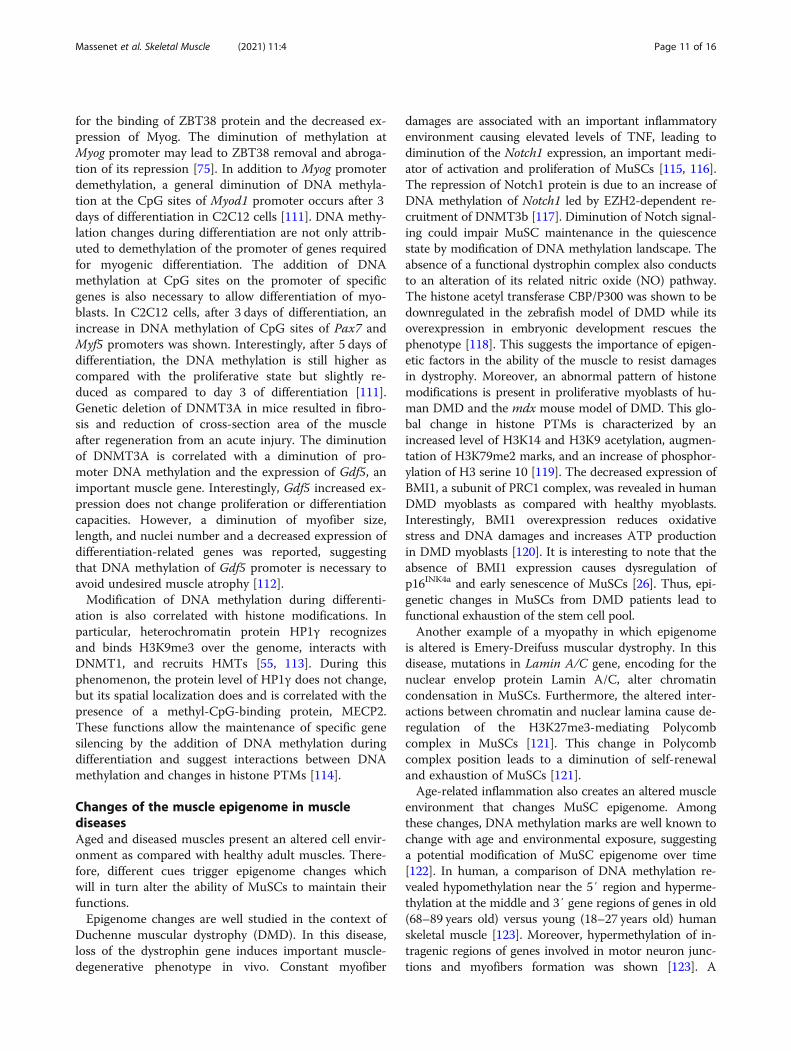
In proliferating MuSCs, PAX7 is recruited to areas ofopen chromatin and its presence correlates with activehistone marks H3K4me1 and H3K27Ac. In particular,PAX7 facilitates chromatin accessibility at gene loci en-coding MRFs through activation of transcriptional en-hancers [46]. Among these MRFs, MYOD1 plays a keyrole in regulating both proliferation and differentiation.In proliferating cells, the Msh homeobox 1 (MSX1) TFwas shown to bind a mouse specific isoform of histoneH1, H1b, at the core enhancer region (CER) of Myod1 inorder to induce chromatin compaction and to reduceMyod1 expression [47]. Additionally, histone deacety-lases (HDACs) are known to be necessary for the main-tenance of proliferation, and class IIA HDACs, HDAC4and HDAC5, are recruited by the H3K9me3 methyl-transferase SUV39H1 to specifically target Myod1 pro-moter. In this way, they modulate Myod1 expression,which underlines the importance of chromatin shape atMyod1 gene for maintenance of proliferation or entrancein differentiation (Fig. 2) [48]. The accumulation of therepressive epigenetic factors at the Myod1 gene in prolif-erating MuSCs is modulated in response to Notch1 sig-naling to prevent differentiation where expression ofMyod1 oscillates due to transient expression of theNotch-regulated transcriptional inhibitor HES1 [49, 50].Lahmann et al. showed that decreased HES1 expressionleads to maintenance of MYOD expression and differen-tiation [50]. These mechanisms likely work in tandem toorchestrate temporal control of the transition from pro-liferation to differentiation.Though expressed at lower levels in proliferating cells,
MYOD1 contributes to both proliferation and differenti-ation. MYOD1 ensures the repression of muscle differ-entiation genes during proliferation and then theactivation of the same genes in response to differenti-ation cues [12]. A role for MYOD1 in both proliferationand differentiation may seem contradictory but is readilyunderstood when one considers that this TF can be botha repressor and an activator at specific genes dependingon the context. In this model, MYOD1 and MEF2Dinteract with the scaffold protein KAP1 [51]. In prolifer-ation conditions, MYOD1, MEF2D, and KAP1 act to-gether to stabilize the association of both co-repressors(G9A, HDAC1) [48, 52] and coactivators (P300 andLSD1) [53, 54] at the muscle differentiation genes. Theassembly of this enhanceosome-type complex establishesa poised chromatin state at the promoters where the re-pressive enzymes dominate to limit gene expression.During differentiation, the increased expression ofmitogen-activated protein kinase (MAPK) P38α (P38α)leads to activation of MSK1 kinase which phosphorylatesKAP1 at serine 473 [51]. The phosphorylated KAP1 pro-tein no longer interacts with the co-repressors, but con-tinues to maintain an interaction with the coactivators,
Massenet et al. Skeletal Muscle (2021) 11:4 Page 5 of 16
leading to the establishment of an open chromatin stateand the expression of muscle target genes (Fig. 2) [51].In other words, when both co-repressors and
coactivators associate with MYOD1, the co-repressorsdominate. Signals from the environment induce phos-phorylation by MSK1 that in turn displaces the co-
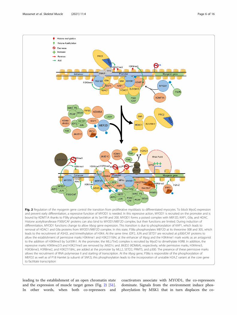
Fig. 2 Regulation of the myogenin gene control the transition from proliferative myoblasts to differentiated myocytes. To block MyoG expressionand prevent early differentiation, a repressive function of MYOD1 is needed. In this repressive action, MYOD1 is recruited on the promoter and isbound by KDMT1A thanks to P38γ phosphorylation at its Ser199 and 200. MYOD1 forms a poised complex with MEF2D, KAP1, G9a, and HDAC.Histone acetyltransferase P300/CAF proteins can also bind to MYOD1/MEF2D complex, but their functions are limited. During induction ofdifferentiation, MYOD1 functions change to allow Myog gene expression. This transition is due to phosphorylation of KAP1, which leads toremoval of HDAC1 and G9a proteins from MYOD1/MEF2D complex. In this state, P38α phosphorylates MEF2D at its threonine 308 and 305, whichleads to the recruitment of ASH2L and trimethylation of H3K4. At the same time JDP2, JUN and SETD7 are recruited at p300/CAF proteins toallow the establishment of permissive marks H3K4me1 and H3K27/18Ac at the enhancer of Myog and the H3K4me1 mark works as an antagonistto the addition of H3K9me3 by SuV39h1. At the promoter, the MLL/TrxG complex is recruited by MyoD to dimethylate H3R8. In addition, therepressive marks H3K9me2/3 and H3K27me3 are removed by JMJD1c and JMJD2 (KDM6A), respectively, while permissive marks, H3K4me3,H3K36me3, H3R8me2, and H3K27/18Ac, are added at the promoter by MLL2, SETD2, PRMT5, and p300. The presence of these permissive marksallows the recruitment of RNA polymerase II and starting of transcription. At the Myog gene, P38α is responsible of the phosphorylation ofMEFD2 as well as of P18 Hamlet (a subunit of SNF2); this phosphorylation leads to the incorporation of unstable H2A.Z variant at the core geneto facilitate transcription
Massenet et al. Skeletal Muscle (2021) 11:4 Page 6 of 16
repressors from the locus [51]. MYOD1 contribution toMuSC proliferation directly suggests the possibility ofregulation of Myod1 expression level duringproliferation.Additional co-repressors are associated with the
MYOD1-MEF2D-KAP1 complex in proliferating cells.Heterochromatin proteins HP1α and β interact withMYOD1-MEF2D-KAP1 to repress its activity at targetgene promoters to maintain proliferation [55]. Similarly,the formation of a HDAC1/MYOD1-KAP1 complex al-lows deacetylation of MYOD1 target genes. One of theirtargets, Myog, which is necessary for differentiation,shows reduced acetylation and a recruitment ofSUV39h1 thanks to the presence of HDAC1/MYOD1complex. In C2C12 cell line, the formation of anothercomplex containing MYOD1/P300/CBP-associated fac-tor (PCAF) and the HDAC SIRT2 is necessary to main-tenance of proliferation and repression of differentiation[56]. Such as other SIRT enzymes, SIRT2 is dependentof the level of NAD+, revealing the implication of themetabolism for the maintenance of proliferation. The re-moval of histone acetylation and the recruitment ofSuv39h1 leads to an enrichment of H3K9me3 marks andmaintenance of the chromatin under a closed state, lead-ing to repression of Myog expression and impeding thestart of differentiation (Fig. 2) [57, 58]. Another study re-vealed the function of P38γ MAPK into the phosphoryl-ation of the Ser199 and 200 of MYOD1 to allow therecruitment SUV39h1/KMT1a to the Myog promoter,reducing its expression during proliferation [59].
Moreover, SNAIl family transcriptional repressor 1,SNAI1, associated with HDAC1/2 can bind E-box atdifferentiation-related genes and prevents the binding ofMYOD1 and target gene expression [60]. This mechan-ism suggests the importance of the SNAI1/HDAC1/2complex in promoting proliferation by blocking MYOD1from initiating differentiation.To maintain myoblast proliferation, repression of dif-
ferentiation is not enough. Cells must also maintain theexpression of genes involved in cell cycle progression.Like in most cell types, E2F protein family plays a keyrole in regulating cell cycle through the recruitment ofhistone acetyltransferase P300/CBP and PCAF/GCN5histone acetylases to the cyclin genes [61, 62]. In primarymouse myoblasts, the E2F1/PCAF complex mediatesacetylation of histones at E2F1 target genes to allow pas-sage through the G1/S cell cycle checkpoint [63]. Inaddition to the recruitment of acetyltransferase, studiesin many different cell types have shown that E2F pro-teins mediate recruitment of H3K4 histone methyltrans-ferases from the KMT2 family [64]. In the musclesystem, proliferating C2C12 cells utilize MLL5 to depositH3K4me3 marks at the cyclin A2 gene, a factor neces-sary for progression through G1/S cell cycle checkpoint[65]. Finally, H3K36me methyltransferase SET2 KO inC2C12 cells compromise G1/S and G2/M phase transi-tion by decreasing levels of cyclin D1, CDK4, CDK6, andcyclin E2. This result indicates an essential function ofthe H3K36 methyltransferase SET2 for the maintenanceof myoblast proliferation [66]. Thus, it is clear that
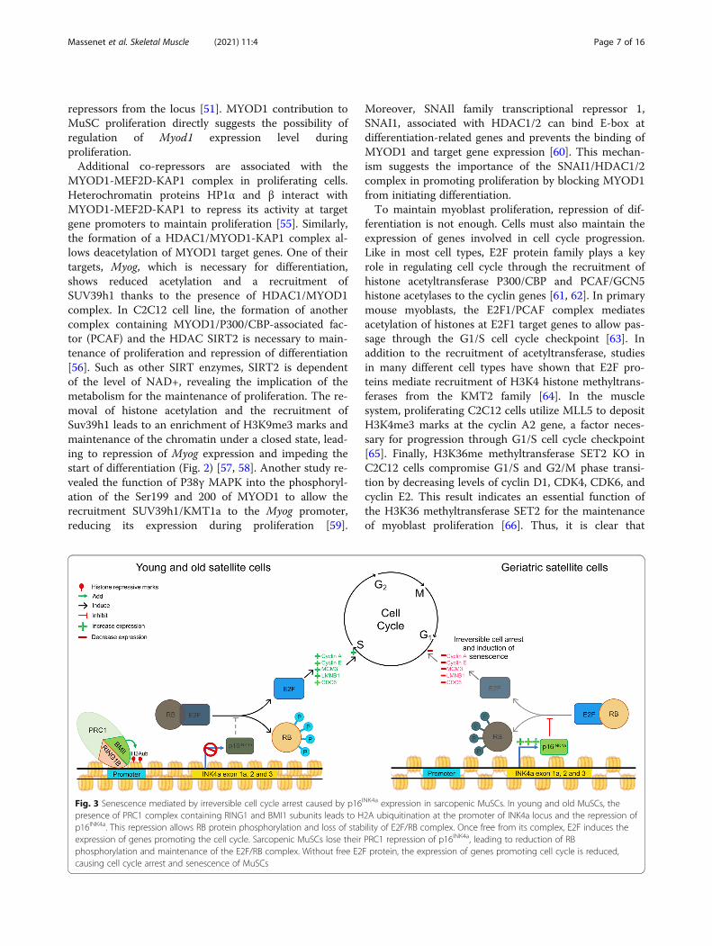
Fig. 3 Senescence mediated by irreversible cell cycle arrest caused by p16INK4a expression in sarcopenic MuSCs. In young and old MuSCs, thepresence of PRC1 complex containing RING1 and BMI1 subunits leads to H2A ubiquitination at the promoter of INK4a locus and the repression ofp16INK4a. This repression allows RB protein phosphorylation and loss of stability of E2F/RB complex. Once free from its complex, E2F induces theexpression of genes promoting the cell cycle. Sarcopenic MuSCs lose their PRC1 repression of p16INK4a, leading to reduction of RBphosphorylation and maintenance of the E2F/RB complex. Without free E2F protein, the expression of genes promoting cell cycle is reduced,causing cell cycle arrest and senescence of MuSCs
Massenet et al. Skeletal Muscle (2021) 11:4 Page 7 of 16
epigenetic modifications of histones contributes to main-tain the cell cycle progression though we still lack an indepth understanding of all the players involved in myo-blast expansion.The degree to which genes are marked by histone
post-translational modifications depends not only on thepresence of epigenetic enzymes, but also on the avail-ability of key co-factors that contribute to their enzym-atic activity. Indeed, the energy source available to thecell will affect the availability of these co-factors. Theglycolytic environment of proliferating MuSCs ensuresan abundance of several intermediates of the tricarb-oxylic acid cycle (TCA) that are needed for establish-ment of epigenetic modifications. In particular, oxidativedecarboxylation of pyruvate supplies the acetyl-CoAdonor of acetyl utilized by the HAT proteins [67]. Simi-larly, the metabolite α-ketoglutarate is a necessary co-factor for the demethylation of either DNA or histones,by ten-eleven translocation (TET) and JMJD proteins,respectively. The functions of the histone demethylaseLSD1 and the HDACs also depends on the availability ofFAD+ and NAD+ in the cell [68]. Independently of theTCA, the intracellular methionine is transformed in S-adenosylmethionine which is necessary for the action ofDNA methyltransferase (DNMT) proteins [68]. Under-standing these points highlight the importance of themetabolism in the epigenetic regulation of the MuSC.Indeed, after isolation of mouse MuSCs, during the tran-sition from quiescence to proliferation, a shift betweenfatty acid oxidation and glycolysis occurs [69]. This shiftinduces a decrease of NAD+ levels which reduces theactivity of SIRT1 family of NAD+-dependent HDAC en-zymes. The activity of SIRT1 is associated with a regula-tion of H4K16Ac marks associated to muscle geneexpression [69]. While characterization of metabolicpathways contributing to the epigenetic regulation ofMuSC proliferation remains in its infancy, new tech-nologies in the area of metabolomics make this an excit-ing area of current research.
The DNA methylation of proliferating MuSCsDNA methylation plays a key role in preventing the ex-pression of cell cycle inhibitors during MuSC prolifera-tion. Among them, repression of cyclin-dependentkinase inhibitor 1C (CDN1C or P57kip2) is required toprevent differentiation. Interestingly, deleting DNAmethyltransferase DNMT3A in MuSCs leads to a dimin-ution of proliferation that correlates with increased ex-pression of the Cdkn1c gene and decreased DNAmethylation at its promoter sequence. This loss of prolif-eration in the DNMT3A KO myoblasts can be rescuedwith Cdkn1c KD, indicating an important function of denovo methylation in the maintenance of MuSC prolifera-tion [70]. The control of Cdkn1c DNA methylation can
occur indirectly through MYOD1 functions. Indeed,MYOD1 has been shown to control the expression atranscriptional repressor Zinc Finger Protein 238(ZFP238) in C2C12 cells [71]. This is significant as theZFP238 protein is able to recruit DNMT3A and HDAC1at the promoter of myogenic genes to repress their ex-pression [72]. It is possible that in MuSCs, ZFP238 isalso present at Cdkn1c promoter and recruits DNMT3A.At the moment, there is no work to support these hy-pothesis and analysis of the presence of ZFP238 andDNMT3A at the Cdkn1c promoter has to beinvestigated.The continued proliferation of myoblasts is also
dependent on repression of Myog gene expression toprevent differentiation. This repression was shown to bepossible thanks to the methylation at the Myog promoter[73, 74]. The Myog promoter has only 1.4% of CpG di-nucleotides. However, even if the region is not rich inCpG island, bisulphite and methylation-sensitive restric-tion endonuclease analysis revealed a hypermethylationstate during proliferation of C2C12 cells, necessary forMyog repression [73]. Additionally, methylation of thescattered CpG sites is necessary for the binding of amethyl-CpG-binding protein, ZBT38 (CIBZ). The pres-ence of ZBT38 at CpG islands within the Myog pro-moter acts to inhibit its expression and maintenance ofproliferation as the knockout of ZBT38 leads to differen-tiation of C2C12 cells [75].Taken together, these findings show that as MuSCs
start to proliferate, TFs target the epigenetic machineryto genes necessary to ensure cell cycle progression andthose preventing differentiation.
Epigenetic regulation of the differentiation processAfter the expansion phase, MuSCs undergo cell cycle ar-rest and transit towards differentiation [76]. Cell cyclearrest is initiated through the expression of the cell cycleinhibitors CDKN1C or CDKN1A (p21) [77]. Moreover,a progressive increase of MYOD1 protein expressionleads to the increased expression of early differentiationmarkers such as Myog and coincides with the abrogationof MYF5 and PAX7 expression [78]. Coincident with theonset of differentiation, MuSCs are marked by hyperace-tylation of histones H3 and H4 while decreased methyla-tion of H3K9 and K27 is observed [79, 80]. Theepigenetic reprogramming of MuSCs allows the openingof chromatin at specific muscle loci needed fordifferentiation.
The control of MuSC differentiation by histone post-transcriptionalThe differentiation of human myoblasts has been shownto result in a large change in the histone PTM landscapewith a global diminution of H3K9me3 and H4K20me3
Massenet et al. Skeletal Muscle (2021) 11:4 Page 8 of 16
repressive marks. In particular, H3K9me3 is erased fromMyod1 and Myog loci [79]. These results point to theimportance of histone modification modulation for myo-blast differentiation.One of the first steps during the transition to differen-
tiation is the stop of MuSC proliferation by cell cycle ar-rest. In this context, the downregulation of Pax7expression must occur. Indeed, PRC2 interacts with YinYang 1 TF (YY1) via the phosphorylation of the threo-nine 372 of EZH2, a subunit of PRC2 complex, by p38α.This interaction allows replacement of H3K4me3 marksby H3K27me3 and leads to the formation of a repressivechromatin state at Pax7 promoter and to the repressionof its expression [81]. Among TFs involved in differenti-ation, E2F is a family of eight proteins, parts of a com-plex including retinoblastoma-associated protein (RB),retinoblastoma-like protein 1 (RBL1) and 2 (RBL2)pocket proteins. Their function is to control the gene ex-pression of proteins regulating the cell cycle and pro-moting differentiation in various tissues [82, 83]. RB,which was shown to bind HDAC1 for the maintenanceof proliferation, can also regulate cell cycle exit throughits interaction with E2F4. RB recruits HMT to promoteH3K9me3 and H3K27me3 marks at the gene promoterof proteins promoting the cell cycle to decrease their ex-pression, stop the cell cycle, and start differentiation.This repression is PRC1 and PRC2 dependent where theaddition of H2AK119Ub1 and H3K27me3 marks estab-lish a silent state [84]. This silent state could be main-tained by the recruitment of dimerization partner, RB-like, E2F, and MuvB (dREAM) chromatin compactioncomplex to target genes through interactions between itsL3MBTL1 subunit, E2F4 and HP1γ heterochromatinprotein [85].Induction of differentiation coincides with an in-
creased expression of MYOD1. The increased Myod1 ex-pression is established through JMJD1c-drivendemethylation of H3K9me3 marks in its promoter [36].Once MYOD1 is expressed at high levels, a switch be-tween the SNAI1/HDAC1/2 complex and MYOD1 oc-curs at E-box of muscle target genes to promotedifferentiation [60].Myog expression is also essential for MuSCs to com-
mit to differentiation (Fig. 2) and is regulated by a superenhancer upstream of the transcription start site [86].The ability of TFs such as MYOD1, MEF2D, SIX4, andFOXO3 to create a transcriptional competent state atthe myogenin promoter depends upon the combined ac-tivity of multiple epigenetic enzymes. One of the initialevents in activating the Myog gene is the removal of therepressive H3K9me2 and H3K9me3 marks from the pro-moter by the action of the lysine demethylase JMJD2/KDM4A [87]. Moreover, focal adhesion kinase (FAK)helps achieve the open chromatin state by facilitating
the departure of HDAC enzymes from the promoter. Inthis case, FAK binds the methyl-CpG-binding proteinMBD2, where it induced phosphorylation of HDAC1that breaks up the HDAC1/MBD2 interaction and disso-ciates it from the promoter [88]. Once the repressivemarks are cleared, the promoter can then be modified toaccumulate transcriptionally permissive marks. One ofthe first marks to appear is the dimethylation at arginine8 in histone 3 (H3R8me2) within the promoter. This isachieved through the PRMT5 protein, a type II argininemethyltransferase. Once the H3R8me2 mark is in place,the epigenetic mark acts to allow a stable association ofthe chromatin remodeling complex SWI/SNF throughrecognition of modified histone tail by the Brg1 subunitof the complex. The association of SWI/SNF with thepromoter then allows chromatin decompaction for RNApolymerase II to access the gene [89]. In addition, thehistone methyltransferase SETD7 is targeted to the Myogpromoter by MYOD1 to introduce the H3K4me1 mark.SETD7 is required for differentiation as silencing ofSETD7 leads to a reduced number of myotubes and lossof expression of Myog [90–92]. The addition ofH3K4me1 by SETD7 prevents the reintroduction of re-pressive H3K9me3 repressive marks by blockingSuv39h1 function [90–92]. Without the presence ofHDAC1 at Myog promoter, the histone acetyltransferaseP300/CAF leads to its enrichment of H3K9 and H3K14acetylation and the expression of Myog [58]. The associ-ation of the histone acetyltransferase P300 with theMyog promoter is facilitated by chromatin-binding pro-tein, NUPR1 (P8), which also recruits the RNA helicaseDDX5 to the locus to promoter high levels of gene ex-pression [93]. The introduction of H3K36me3 marks isalso essential to high level expression of Myog as silencingof SETD2 blocked its expression and prevented myotubeformation during differentiation [66]. Finally, histone ex-changes within the nucleosome can alter the expression ofthe Myog gene. During the differentiation process, thesubunit ZNHI1 (p18Hamlet) of SNF2 complex is phos-phorylated by P38α which allows its recruitment to Myogpromoter and allows the replacement of H2A histones byits less-stable variant H2A.Z (Fig. 2) [94].For differentiation to proceed, cells must also begin to
express functional genes that define the muscle lineage.Activation of these MYOD1 target genes requires the re-cruitment of SWI/SNF complex which is facilitated bythe presence of histone 4 hyperacetylation that is recog-nized by the bromodomain of the transcription activatorBRG1 (SMCA4), leading to chromatin decompactionand gene expression [95]. SWI/SNF recruitment is facili-tated by the incorporation of the MYOD1-associatedSMRD3 (BAF60C) subunit into the chromatin remodel-ing complex [96]. In addition, MyoD recruits the histoneacetyl transferase P300, the JDP2, the AP1 (JUN) and
Massenet et al. Skeletal Muscle (2021) 11:4 Page 9 of 16
RUNX1 TFs, and SETD7 HMT, leading to active histonemodification marks H3K27Ac, H3K18ac, and H3K4me1that target RNA polymerase II to the promoter region ofmuscle differentiation genes (Fig. 2) [97]. Interestingly,SETD7 methylates non-histone proteins such as the TFSRF. SRF acetylation promotes its binding to its serumresponse element at the muscle-specific gene Acta1 topromote its expression. This regulation is necessary fordifferentiation and is revered by KDM2B [98]. Thesedata suggest an indirect function of SETD7 in the regu-lation of differentiation. However, apart from Acta1, noother SRF targets have been identified in MuSCs so far.While many repressive enzymes are removed from the
MYOD1 target genes during differentiation, some com-plexes remain to permit repression of gene expression inresponse to a changing environment. An example of thisis the Myh4, Myog, and Ckm genes which are marked bythe Polycomb PRC2 mediated H3K27me3 modificationsin proliferating MuSCs to repress their expression andto prevent differentiation. As differentiation initiates, aswitch occurs between subunits in the PRC2 complexwhere the EZH2 subunit in proliferation (PRC2-EZH2)is replaced by a functionally inactive EZH1-containingPRC2 complex that lacks the EED subunit (PRC2-EZH1)[37, 99]. To permit gene expression during differenti-ation, removal of the H3K27me3 modifications is medi-ated by the H3K27 demethylase KDM6A (also known asUTX), a H3K27 demethylase that opens the chromatinto allow the expression of Myog and the entrance in dif-ferentiation [100, 101]. The stable association of PRC2-EZH1 complex that lacks methyltransferase activityhelps to maintain the transcriptional permissive state[37, 99]. In response to cellular stresses such as muscleatrophy, the PRC2-EZH1 complex incorporates an EEDsubunit to form a functional HMT complex that can re-introduce H3K27me3 marks at these muscle genes toprevent their expression [37, 99].As MYOG begins to push the differentiation program
forward, additional epigenetic events will lead to the ex-pression of terminal differentiation-related genes and fu-sion of differentiated myocytes into myotubes. TrxGcomplexes, containing a H3K4me3 methyl transferaseASH2L, are recruited by MEF2D to muscle-specificgenes such as a muscle cytoplasmic enzyme, the musclecreatine kinase, where they mediate the addition ofH3K4me3 marks that promote gene expression [102].This recruitment is modulated through phosphorylationof MEF2D at threonine 308 and 315 by P38α MAPK.Interestingly, the function of a histone arginine methyl-transferase Prmt5 is critical for early differentiation but isdispensable for late differentiation while the type I argin-ine methyltransferase, CARM1/PRMT4 is necessary forlate differentiation as it deposits dimethylation of arginine17 at histone 3 to permit gene activation [89, 103]. This
reveals the refined roles of distinct epigenetic enzymes inensuring the temporal expression of genes during muscledifferentiation.Finally, when fully differentiated, myocytes fuse thanks
to Myomaker and Myomerger proteins to form multinu-cleated myofibers. While the epigenetic regulation of theMyomaker and Myomerger proteins has yet to be eluci-dated, the membrane protein CDON which positivelyregulates fusion was shown to be regulated by theTrithorax HMT ASH1L which deposits dimethylation oflysine 36 of histone 3 (H3K36me2) at the transcriptionstart site to prevent Polycomb-mediated repression[104]. Interestingly, the absence of ASH1L provokes adiminution of fusion capacities of mouse and humanmyocytes in vitro without impairment of myosin heavychain (MHC) protein expression. This suggests a directcontrol of fusion by a Trithorax complex and one canimagine that understanding of the regulation of fusionby epigenetic will emerge soon.
DNA methylation modifications during differentiationSeveral studies have revealed the important role of DNAmethylation in regulating muscle differentiation. Indeed,during differentiation, the whole DNA methylation land-scape was reported to decrease [105]. Several years ago,sodium arsenic treatment of C2C12 was reported to re-duce differentiation capacities of the cells. This was cor-related with increased DNA methylation of the CpG siteof Myog promoter and a diminution of Myog expression[106]. Hypermethylation of Myog promoter in C2C12cells decreases quickly after the induction of differenti-ation [73]. DNA methylation is known to repress TFbinding. However, using a model of 293 T cells express-ing luciferase reporter construct and different TFs, thebinding of Sine oculis homeobox homolog 1 (SIX1) andMEF2A at Myog promoter was confirmed. SilencingSIX1 in C2C12 leads to increased methylation of Myogpromoter, suggesting that SIX1 could have a role in therepression of the methylation [74, 107]. In the last fewyears, TET proteins have been shown to catalyze theconversion of DNA 5-methylcytosine into different oxi-dized forms, 5-hydroxymethylcytosine, 5-formylcytosine,and finally in 5-carboxylcytosine, demonstrating activedemethylation capacities [108, 109]. Supporting the ideaof a decrease in DNA methylation during differentiation,TET1 and TET2 expression are significatively increasedin myoblasts after induction of differentiation. Interest-ingly, inhibition of TET2 but not TET1 by siRNA inC2C12 results in increased DNA methylation of Myog,Myf6/Mrf4, and Mymk (Myomaker) gene promoters.The increased DNA methylation at the promoter ofthese genes correlates with a decreased expression andan abrogation of C2C12 differentiation [110]. The pres-ence of CpG methylation at Myog promoter is necessary
Massenet et al. Skeletal Muscle (2021) 11:4 Page 10 of 16
for the binding of ZBT38 protein and the decreased ex-pression of Myog. The diminution of methylation atMyog promoter may lead to ZBT38 removal and abroga-tion of its repression [75]. In addition to Myog promoterdemethylation, a general diminution of DNA methyla-tion at the CpG sites of Myod1 promoter occurs after 3days of differentiation in C2C12 cells [111]. DNA methy-lation changes during differentiation are not only attrib-uted to demethylation of the promoter of genes requiredfor myogenic differentiation. The addition of DNAmethylation at CpG sites on the promoter of specificgenes is also necessary to allow differentiation of myo-blasts. In C2C12 cells, after 3 days of differentiation, anincrease in DNA methylation of CpG sites of Pax7 andMyf5 promoters was shown. Interestingly, after 5 days ofdifferentiation, the DNA methylation is still higher ascompared with the proliferative state but slightly re-duced as compared to day 3 of differentiation [111].Genetic deletion of DNMT3A in mice resulted in fibro-sis and reduction of cross-section area of the muscleafter regeneration from an acute injury. The diminutionof DNMT3A is correlated with a diminution of pro-moter DNA methylation and the expression of Gdf5, animportant muscle gene. Interestingly, Gdf5 increased ex-pression does not change proliferation or differentiationcapacities. However, a diminution of myofiber size,length, and nuclei number and a decreased expression ofdifferentiation-related genes was reported, suggestingthat DNA methylation of Gdf5 promoter is necessary toavoid undesired muscle atrophy [112].Modification of DNA methylation during differenti-
ation is also correlated with histone modifications. Inparticular, heterochromatin protein HP1γ recognizesand binds H3K9me3 over the genome, interacts withDNMT1, and recruits HMTs [55, 113]. During thisphenomenon, the protein level of HP1γ does not change,but its spatial localization does and is correlated with thepresence of a methyl-CpG-binding protein, MECP2.These functions allow the maintenance of specific genesilencing by the addition of DNA methylation duringdifferentiation and suggest interactions between DNAmethylation and changes in histone PTMs [114].
Changes of the muscle epigenome in musclediseasesAged and diseased muscles present an altered cell envir-onment as compared with healthy adult muscles. There-fore, different cues trigger epigenome changes whichwill in turn alter the ability of MuSCs to maintain theirfunctions.Epigenome changes are well studied in the context of
Duchenne muscular dystrophy (DMD). In this disease,loss of the dystrophin gene induces important muscle-degenerative phenotype in vivo. Constant myofiber
damages are associated with an important inflammatoryenvironment causing elevated levels of TNF, leading todiminution of the Notch1 expression, an important medi-ator of activation and proliferation of MuSCs [115, 116].The repression of Notch1 protein is due to an increase ofDNA methylation of Notch1 led by EZH2-dependent re-cruitment of DNMT3b [117]. Diminution of Notch signal-ing could impair MuSC maintenance in the quiescencestate by modification of DNA methylation landscape. Theabsence of a functional dystrophin complex also conductsto an alteration of its related nitric oxide (NO) pathway.The histone acetyl transferase CBP/P300 was shown to bedownregulated in the zebrafish model of DMD while itsoverexpression in embryonic development rescues thephenotype [118]. This suggests the importance of epigen-etic factors in the ability of the muscle to resist damagesin dystrophy. Moreover, an abnormal pattern of histonemodifications is present in proliferative myoblasts of hu-man DMD and the mdx mouse model of DMD. This glo-bal change in histone PTMs is characterized by anincreased level of H3K14 and H3K9 acetylation, augmen-tation of H3K79me2 marks, and an increase of phosphor-ylation of H3 serine 10 [119]. The decreased expression ofBMI1, a subunit of PRC1 complex, was revealed in humanDMD myoblasts as compared with healthy myoblasts.Interestingly, BMI1 overexpression reduces oxidativestress and DNA damages and increases ATP productionin DMD myoblasts [120]. It is interesting to note that theabsence of BMI1 expression causes dysregulation ofp16INK4a and early senescence of MuSCs [26]. Thus, epi-genetic changes in MuSCs from DMD patients lead tofunctional exhaustion of the stem cell pool.Another example of a myopathy in which epigenome
is altered is Emery-Dreifuss muscular dystrophy. In thisdisease, mutations in Lamin A/C gene, encoding for thenuclear envelop protein Lamin A/C, alter chromatincondensation in MuSCs. Furthermore, the altered inter-actions between chromatin and nuclear lamina cause de-regulation of the H3K27me3-mediating Polycombcomplex in MuSCs [121]. This change in Polycombcomplex position leads to a diminution of self-renewaland exhaustion of MuSCs [121].Age-related inflammation also creates an altered muscle
environment that changes MuSC epigenome. Amongthese changes, DNA methylation marks are well known tochange with age and environmental exposure, suggestinga potential modification of MuSC epigenome over time[122]. In human, a comparison of DNA methylation re-vealed hypomethylation near the 5′ region and hyperme-thylation at the middle and 3′ gene regions of genes in old(68–89 years old) versus young (18–27 years old) humanskeletal muscle [123]. Moreover, hypermethylation of in-tragenic regions of genes involved in motor neuron junc-tions and myofibers formation was shown [123]. A
Massenet et al. Skeletal Muscle (2021) 11:4 Page 11 of 16
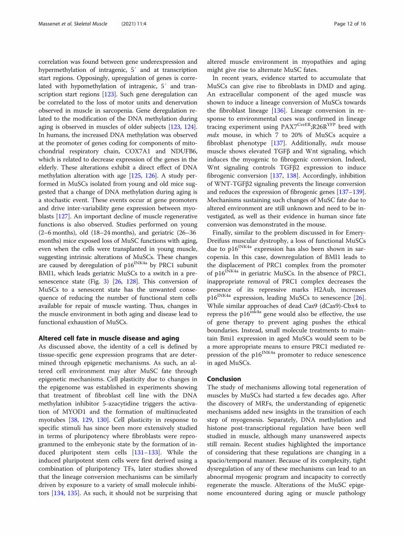
correlation was found between gene underexpression andhypermethylation of intragenic, 5′ and at transcriptionstart regions. Opposingly, upregulation of genes is corre-lated with hypomethylation of intragenic, 5′ and tran-scription start regions [123]. Such gene deregulation canbe correlated to the loss of motor units and denervationobserved in muscle in sarcopenia. Gene deregulation re-lated to the modification of the DNA methylation duringaging is observed in muscles of older subjects [123, 124].In humans, the increased DNA methylation was observedat the promoter of genes coding for components of mito-chondrial respiratory chain, COX7A1 and NDUFB6,which is related to decrease expression of the genes in theelderly. These alterations exhibit a direct effect of DNAmethylation alteration with age [125, 126]. A study per-formed in MuSCs isolated from young and old mice sug-gested that a change of DNA methylation during aging isa stochastic event. These events occur at gene promotersand drive inter-variability gene expression between myo-blasts [127]. An important decline of muscle regenerativefunctions is also observed. Studies performed on young(2–6months), old (18–24months), and geriatric (26–36months) mice exposed loss of MuSC functions with aging,even when the cells were transplanted in young muscle,suggesting intrinsic alterations of MuSCs. These changesare caused by deregulation of p16INK4a by PRC1 subunitBMI1, which leads geriatric MuSCs to a switch in a pre-senescence state (Fig. 3) [26, 128]. This conversion ofMuSCs to a senescent state has the unwanted conse-quence of reducing the number of functional stem cellsavailable for repair of muscle wasting. Thus, changes inthe muscle environment in both aging and disease lead tofunctional exhaustion of MuSCs.
Altered cell fate in muscle disease and agingAs discussed above, the identity of a cell is defined bytissue-specific gene expression programs that are deter-mined through epigenetic mechanisms. As such, an al-tered cell environment may alter MuSC fate throughepigenetic mechanisms. Cell plasticity due to changes inthe epigenome was established in experiments showingthat treatment of fibroblast cell line with the DNAmethylation inhibitor 5-azacytidine triggers the activa-tion of MYOD1 and the formation of multinucleatedmyotubes [38, 129, 130]. Cell plasticity in response tospecific stimuli has since been more extensively studiedin terms of pluripotency where fibroblasts were repro-grammed to the embryonic state by the formation of in-duced pluripotent stem cells [131–133]. While theinduced pluripotent stem cells were first derived using acombination of pluripotency TFs, later studies showedthat the lineage conversion mechanisms can be similarlydriven by exposure to a variety of small molecule inhibi-tors [134, 135]. As such, it should not be surprising that
altered muscle environment in myopathies and agingmight give rise to alternate MuSC fates.In recent years, evidence started to accumulate that
MuSCs can give rise to fibroblasts in DMD and aging.An extracellular component of the aged muscle wasshown to induce a lineage conversion of MuSCs towardsthe fibroblast lineage [136]. Lineage conversion in re-sponse to environmental cues was confirmed in lineagetracing experiment using PAX7CreER;R26RYFP bred withmdx mouse, in which 7 to 20% of MuSCs acquire afibroblast phenotype [137]. Additionally, mdx mousemuscle shows elevated TGFβ and Wnt signaling, whichinduces the myogenic to fibrogenic conversion. Indeed,Wnt signaling controls TGFβ2 expression to inducefibrogenic conversion [137, 138]. Accordingly, inhibitionof WNT-TGFβ2 signaling prevents the lineage conversionand reduces the expression of fibrogenic genes [137–139].Mechanisms sustaining such changes of MuSC fate due toaltered environment are still unknown and need to be in-vestigated, as well as their evidence in human since fateconversion was demonstrated in the mouse.Finally, similar to the problem discussed in for Emery-
Dreifuss muscular dystrophy, a loss of functional MuSCsdue to p16INK4a expression has also been shown in sar-copenia. In this case, downregulation of BMI1 leads tothe displacement of PRC1 complex from the promoterof p16INK4a in geriatric MuSCs. In the absence of PRC1,inappropriate removal of PRC1 complex decreases thepresence of its repressive marks H2Aub, increasesp16INK4a expression, leading MuSCs to senescence [26].While similar approaches of dead Cas9 (dCas9)-Cbx4 torepress the p16ink4a gene would also be effective, the useof gene therapy to prevent aging pushes the ethicalboundaries. Instead, small molecule treatments to main-tain Bmi1 expression in aged MuSCs would seem to bea more appropriate means to ensure PRC1 mediated re-pression of the p16INK4a promoter to reduce senescencein aged MuSCs.
ConclusionThe study of mechanisms allowing total regeneration ofmuscles by MuSCs had started a few decades ago. Afterthe discovery of MRFs, the understanding of epigeneticmechanisms added new insights in the transition of eachstep of myogenesis. Separately, DNA methylation andhistone post-transcriptional regulation have been wellstudied in muscle, although many unanswered aspectsstill remain. Recent studies highlighted the importanceof considering that these regulations are changing in aspacio/temporal manner. Because of its complexity, tightdysregulation of any of these mechanisms can lead to anabnormal myogenic program and incapacity to correctlyregenerate the muscle. Alterations of the MuSC epige-nome encountered during aging or muscle pathology
Massenet et al. Skeletal Muscle (2021) 11:4 Page 12 of 16
conduct gene dysregulations and to the diminution ofthe capacity of MuSCs to regenerate. Recently, modifica-tions of these regulations were reported to alter themaintenance of a healthy MuSC fate in mouse models[137, 139]. These modifications are not well understoodyet. Possibly, the important advances in technologies toanalyze epigenomes of a small number of cells will allowthe discovering of the mechanisms leading to abnormalcell fate decisions of MuSCs.
AbbreviationsAAV: Adeno-associated virus; CER: Core enhancer region; dCas9: Dead Cas9;DMD: Duchenne muscular dystrophy; DNMT: DNA methyltransferase;dREAM: Dimerization partner, RB-like, E2F, and MuvB; DRR: Distal regulatoryregion; FAK: Focal adhesion kinase; H2AUb: Monoubiquitination of lysine 119of histone 2A; H3K20me1: Monomethylation of lysine 20 of histone 3;H3K27Ac: Acetylation of lysine 27 of histone 3; H3K27me3: Trimethylation oflysine 9 of histone 3; H3K36me2: Dimethylation of lysine 36 of histone 3;H3K4me3: Trimethylation of the lysine 4 of histone 3; H3K9Ac: Acetylation oflysine 9 of histone 3; H3K9me3: Trimethylation of lysine 9 of histone 3;H3R8me2: Dimethylation of arginine 8 of histone 3; H4K20Ac: Acetylation oflysine 20 of histone 4; H4K20me3: Trimethylation of lysine 20 of histone 4;HDAC: Histone deacetylase; HMT: Histone methyltransferase; MAPK: Mitogen-activated protein kinase; MCK: Muscle creatine kinase; MeCP2: Methyl-CpG-binding protein 2; MHC: Myosine heavy chain; MRF: Myogenic regulatoryfactor; MuSC: Muscle stem cell; MYOG: Myogenin; NO: Nitric oxide;PCAF: P300/CBP-associated factor; PcG: Polycomb group; PRC: Polycombrepressive complex; PTM: Post-translational modification; SWI/SNF: Switch/sucrose nonfermentable chromatin remodeling complex; TET: Ten-eleventranslocation; TF: Transcription factor; TrxG: Trithorax group
AcknowledgementsJM was the recipient of a MITACS Global link scholarship.
Authors’ contributionsJM, EG, BC, and FJD conceived, discussed, and wrote the manuscript. Theauthors read and approved the final manuscript.
FundingWork in the Dilworth lab was supported by the Canadian Institutes of HealthResearch.
Availability of data and materialsNot applicable.
Ethics approval and consent to participateNot applicable.
Consent for publicationAll authors have consent for publication of this article.
Competing interestsThe authors declare that they have no competing interests.
Author details1Sprott Center for Stem Cell Research, Regenerative Medicine Program,Ottawa Hospital Research Institute, 501 Smyth Rd, Mailbox 511, Ottawa, ONK1H 8L6, Canada. 2Institut NeuroMyoGène, Université Claude Bernard Lyon 1,CNRS 5310, INSERM U1217, 8 Rockefeller Ave, 69008 Lyon, France.3Department of Cellular and Molecular Medicine, University of Ottawa,Ottawa, ON K1H 8L6, Canada. 4LIFE Research Institute, University of Ottawa,Ottawa, ON K1H 8L6, Canada.
Received: 16 October 2020 Accepted: 20 December 2020
References1. Ou HD, Phan S, Deerinck TJ, Thor A, Ellisman MH, O’Shea CC. ChromEMT:
Visualizing 3D chromatin structure and compaction in interphase andmitotic cells. Science. 2017;357:eaag0025.
2. Bintu B, Mateo LJ, Su J-H, Sinnott-Armstrong NA, Parker M, Kinrot S, et al.Super-resolution chromatin tracing reveals domains and cooperativeinteractions in single cells. Science. 2018;362:eaau1783.
3. Mohandas T, Sparkes R, Shapiro L. Reactivation of an inactive human Xchromosome: evidence for X inactivation by DNA methylation. Science.1981;211:393–6.
4. Rice JC, Allis CD. Histone methylation versus histone acetylation: newinsights into epigenetic regulation. Curr Opin Cell Biol. 2001;13:263–73.
5. Schuettengruber B, Martinez A-M, Iovino N, Cavalli G. Trithorax groupproteins: switching genes on and keeping them active. Nat Rev Mol CellBiol. 2011;12:799–814.
6. Zhang CL, McKinsey TA, Olson EN. Association of class II histonedeacetylases with heterochromatin protein 1: potential role for histonemethylation in control of muscle differentiation. Mol Cell Biol. 2002;22:7302–12.
7. Heintzman ND, Stuart RK, Hon G, Fu Y, Ching CW, Hawkins RD, et al. Distinctand predictive chromatin signatures of transcriptional promoters andenhancers in the human genome. Nat Genet. 2007;39:311–8.
8. Herz H-M, Mohan M, Garruss AS, Liang K, Takahashi Y –h, Mickey K, et al.Enhancer-associated H3K4 monomethylation by Trithorax-related, theDrosophila homolog of mammalian Mll3/Mll4. Genes Dev. 2012;26:2604–20.
9. Mauro A. Satellite cell of skeletal muscle fibers. J Biophys Biochem Cytol.1961;9:493–5.
10. Aziz A, Liu Q-C, Dilworth FJ. Regulating a master regulator: establishingtissue-specific gene expression in skeletal muscle. Epigenetics. 2010;5:691–5.
11. Segalés J, Perdiguero E, Muñoz-Cánoves P. Epigenetic control of adultskeletal muscle stem cell functions. FEBS J. 2015;282:1571–88.
12. Singh K, Dilworth FJ. Differential modulation of cell cycle progressiondistinguishes members of the myogenic regulatory factor family oftranscription factors. FEBS J. 2013;280:3991–4003.
13. García-Prat L, Perdiguero E, Alonso-Martín S, Dell’Orso S, Ravichandran S,Brooks SR, et al. FoxO maintains a genuine muscle stem-cell quiescent stateuntil geriatric age. Nat Cell Biol. 2020. Available from: http://www.nature.com/articles/s41556-020-00593-7.
14. Beauchamp JR, Heslop L, Yu DS, Tajbakhsh S, Kelly RG, Wernig A, et al.Expression of CD34 and Myf5 defines the majority of quiescent adultskeletal muscle satellite cells. J Cell Biol. 2000;151:1221–34.
15. Crist CG, Montarras D, Buckingham M. Muscle satellite cells are primed formyogenesis but maintain quiescence with sequestration of Myf5 mRNAtargeted by microRNA-31 in mRNP granules. Cell Stem Cell. 2012;11:118–26.
16. Yue L, Wan R, Luan S, Zeng W, Cheung TH. Dek modulates global intronretention during muscle stem cells quiescence exit. Dev Cell. 2020;53:661–76.
17. Kuang S, Kuroda K, Le Grand F, Rudnicki MA. Asymmetric self-renewal andcommitment of satellite stem cells in muscle. Cell. 2007;129:999–1010.
18. Yablonka-Reuveni Z, Rudnicki MA, Rivera AJ, Primig M, Anderson JE,Natanson P. The transition from proliferation to differentiation is delayed insatellite cells from mice lacking MyoD. Dev Biol. 1999;210:440–55.
19. Hinterberger TJ, Sassoon DA, Rhodes SJ, Konieczny SF. Expression of themuscle regulatory factor MRF4 during somite and skeletal myofiberdevelopment. Dev Biol. 1991;147:144–56.
20. Lazure F, Blackburn DM, Corchado AH, Sahinyan K, Karam N, Sharanek A,et al. Myf6/MRF4 is a myogenic niche regulator required for themaintenance of the muscle stem cell pool. EMBO Rep. 2020. Available from:https://onlinelibrary.wiley.com/doi/10.15252/embr.201949499.
21. Zhu Z, Boone MJ. MRF4 can substitute for myogenin during early stages ofmyogenesis. Dev Dyn. 1997;209:233–41.
22. Machado L, Esteves de Lima J, Fabre O, Proux C, Legendre R, Szegedi A,et al. In situ fixation redefines quiescence and early activation of skeletalmuscle stem cells. Cell Rep. 2017;21:1982–93.
23. van Velthoven CTJ, de Morree A, Egner IM, Brett JO, Rando TA.Transcriptional profiling of quiescent muscle stem cells in vivo. Cell Rep.2017;21:1994–2004.
Massenet et al. Skeletal Muscle (2021) 11:4 Page 13 of 16
24. Liu L, Cheung TH, Charville GW, Hurgo BMC, Leavitt T, Shih J, et al.Chromatin modifications as determinants of muscle stem cell quiescenceand chronological aging. Cell Rep. 2013;4:189–204.
25. Liu L, Cheung TH, Charville GW, Rando TA. Isolation of skeletal muscle stemcells by fluorescence-activated cell sorting. Nat Protoc. 2015;10:1612–24.
26. Sousa-Victor P, Gutarra S, García-Prat L, Rodriguez-Ubreva J, Ortet L, Ruiz-Bonilla V, et al. Geriatric muscle stem cells switch reversible quiescence intosenescence. Nature. 2014;506:316–21.
27. Gopinath SD, Webb AE, Brunet A, Rando TA. FOXO3 Promotes quiescencein adult muscle stem cells during the process of self-renewal. Stem Cell Rep.2014;2:414–26.
28. Bjornson CRR, Cheung TH, Liu L, Tripathi PV, Steeper KM, Rando TA. Notchsignaling is necessary to maintain quiescence in adult muscle stem cells.STEM CELLS. 2012;30:232–42.
29. Boonsanay V, Zhang T, Georgieva A, Kostin S, Qi H, Yuan X, et al. Regulationof skeletal muscle stem cell quiescence by Suv4-20 h1-dependentfacultative heterochromatin formation. Cell Stem Cell. 2016;18:229–42.
30. Li Y, Dilworth FJ. compacting chromatin to ensure muscle satellite cellquiescence. Cell Stem Cell. 2016;18:162–4.
31. Addicks GC, Brun CE, Sincennes M-C, Saber J, Porter CJ, Francis Stewart A,et al. MLL1 is required for PAX7 expression and satellite cell self-renewal inmice. Nat Commun. 2019;10:4256.
32. Cao R, Tsukada Y, Zhang Y. Role of Bmi-1 and Ring1A in H2A Ubiquitylationand Hox gene silencing. Mol Cell. 2005;20:845–54.
33. Wang H, Wang L, Erdjument-Bromage H, Vidal M, Tempst P, Jones RS, et al.Role of histone H2A ubiquitination in Polycomb silencing. Nature. 2004;431:873–8.
34. Agherbi H, Gaussmann-Wenger A, Verthuy C, Chasson L, Serrano M, DjabaliM. Polycomb mediated epigenetic silencing and replication timing at theINK4a/ARF locus during senescence. Blagosklonny MV, editor. PLoS ONE.2009;4:e5622.
35. Juan AH, Derfoul A, Feng X, Ryall JG, Dell’Orso S, Pasut A, et al. PolycombEZH2 controls self-renewal and safeguards the transcriptional identity ofskeletal muscle stem cells. Genes Dev. 2011;25:789–94.
36. Luo D, de Morree A, Boutet S, Quach N, Natu V, Rustagi A, et al. Deltex2represses MyoD expression and inhibits myogenic differentiation by actingas a negative regulator of Jmjd1c. Proc Natl Acad Sci U S A. 2017;114:E3071–80.
37. Caretti G. The Polycomb Ezh2 methyltransferase regulates muscle geneexpression and skeletal muscle differentiation. Genes Dev. 2004;18:2627–38.
38. Jones PA, Wolkowicz MJ, Rideout WM, Gonzales FA, Marziasz CM, CoetzeeGA, et al. De novo methylation of the MyoD1 CpG island during theestablishment of immortal cell lines. Proc Natl Acad Sci. 1990;87:6117–21.
39. Diede SJ, Yao Z, Keyes CC, Tyler AE, Dey J, Hackett CS, et al. Fundamentaldifferences in promoter CpG island DNA hypermethylation between humancancer and genetically engineered mouse models of cancer. Epigenetics.2013;8:1254–60.
40. McKinnell IW, Ishibashi J, Le Grand F, Punch VGJ, Addicks GC, Greenblatt JF,et al. Pax7 activates myogenic genes by recruitment of a histonemethyltransferase complex. Nat Cell Biol. 2008;10:77–84.
41. Diao Y, Guo X, Li Y, Sun K, Lu L, Jiang L, et al. Pax3/7BP Is a Pax7- and Pax3-binding protein that regulates the proliferation of muscle precursor cells byan epigenetic mechanism. Cell Stem Cell. 2012;11:231–41.
42. Kawabe Y, Wang YX, McKinnell IW, Bedford MT, Rudnicki MA. Carm1Regulates Pax7 transcriptional activity through MLL1/2 recruitment duringasymmetric satellite stem cell divisions. Cell Stem Cell. 2012;11:333–45.
43. Cai S, Zhu Q, Guo C, Yuan R, Zhang X, Nie Y, et al. MLL1 promotes myogenesisby epigenetically regulating Myf5. Cell Prolif. 2020;53 Available from: https://onlinelibrary.wiley.com/doi/abs/10.1111/cpr.12744. [cited 2020 Mar 10].
44. Padilla-Benavides T, Nasipak BT, Imbalzano AN. Brg1 Controls the expressionof Pax7 to promote viability and proliferation of mouse primary myoblasts:primary myoblasts require Brg1. J Cell Physiol. 2015;230:2990–7.
45. Padilla-Benavides T, Nasipak BT, Paskavitz AL, Haokip DT, Schnabl JM,Nickerson JA, et al. Casein kinase 2-mediated phosphorylation of Brahma-related gene 1 controls myoblast proliferation and contributes to SWI/SNFcomplex composition. J Biol Chem. 2017;292:18592–607.
46. Lilja KC, Zhang N, Magli A, Gunduz V, Bowman CJ, Arpke RW, et al. Pax7remodels the chromatin landscape in skeletal muscle stem cells. PloS One.2017;12:e0176190.
47. Lee H. Msx1 Cooperates with histone H1b for inhibition of transcription andmyogenesis. Science. 2004;304:1675–8.
48. Puri PL, Iezzi S, Stiegler P, Chen TT, Schiltz RL, Muscat GE, et al. Class Ihistone deacetylases sequentially interact with MyoD and pRb duringskeletal myogenesis. Mol Cell. 2001;8:885–97.
49. Bröhl D, Vasyutina E, Czajkowski MT, Griger J, Rassek C, Rahn H-P, et al.Colonization of the satellite cell niche by skeletal muscle progenitor cellsdepends on Notch signals. Dev Cell. 2012;23:469–81.
50. Lahmann I, Bröhl D, Zyrianova T, Isomura A, Czajkowski MT, Kapoor V, et al.Oscillations of MyoD and Hes1 proteins regulate the maintenance ofactivated muscle stem cells. Genes Dev. 2019;33:524–35.
51. Singh K, Cassano M, Planet E, Sebastian S, Jang SM, Sohi G, et al. A KAP1phosphorylation switch controls MyoD function during skeletal muscledifferentiation. Genes Dev. 2015;29:513–25.
52. Ling BMT, Bharathy N, Chung T-K, Kok WK, Li S, Tan YH, et al. Lysinemethyltransferase G9a methylates the transcription factor MyoD andregulates skeletal muscle differentiation. Proc Natl Acad Sci. 2012;109:841–6.
53. Dilworth FJ, Seaver KJ, Fishburn AL, Htet SL, Tapscott SJ. In vitrotranscription system delineates the distinct roles of the coactivators pCAFand p300 during MyoD/E47-dependent transactivation. Proc Natl Acad Sci.2004;101:11593–8.
54. Choi J, Jang H, Kim H, Kim S-T, Cho E-J, Youn H-D. Histone demethylaseLSD1 is required to induce skeletal muscle differentiation by regulatingmyogenic factors. Biochem Biophys Res Commun. 2010;401:327–32.
55. Ait-Si-Ali S, Guasconi V, Fritsch L, Yahi H, Sekhri R, Naguibneva I, et al. ASuv39h-dependent mechanism for silencing S-phase genes indifferentiating but not in cycling cells. EMBO J. 2004;23:605–15.
56. Fulco M, Schiltz RL, Iezzi S, King MT, Zhao P, Kashiwaya Y, et al. Sir2regulates skeletal muscle differentiation as a potential sensor of the redoxstate. Mol Cell. 2003;12:51–62.
57. Mal AK. Histone methyltransferase Suv39h1 represses MyoD-stimulatedmyogenic differentiation. EMBO J. 2006;25:3323–34.
58. Mal A, Harter ML. MyoD is functionally linked to the silencing of a muscle-specific regulatory gene prior to skeletal myogenesis. Proc Natl Acad Sci.2003;100:1735–9.
59. Gillespie MA, Le Grand F, Scimè A, Kuang S, von Maltzahn J, Seale V, et al.p38-γ–dependent gene silencing restricts entry into the myogenicdifferentiation program. J Cell Biol. 2009;187:991–1005.
60. Soleimani VD, Yin H, Jahani-Asl A, Ming H, Kockx CEM, van Ijcken WFJ, et al. Snailregulates MyoD binding-site occupancy to direct enhancer switching anddifferentiation-specific transcription in myogenesis. Mol Cell. 2012;47:457–68.
61. Takahashi Y, Rayman JB, Dynlacht BD. Analysis of promoter binding by theE2F and pRB families in vivo: distinct E2F proteins mediate activation andrepression. Genes Dev. 2000;14:804–16.
62. Taubert S, Gorrini C, Frank SR, Parisi T, Fuchs M, Chan H-M, et al. E2F-dependent histone acetylation and recruitment of the Tip60acetyltransferase complex to chromatin in late G1. Mol Cell Biol. 2004;24:4546–56.
63. Rao VK, Ow JR, Shankar SR, Bharathy N, Manikandan J, Wang Y, et al. G9apromotes proliferation and inhibits cell cycle exit during myogenicdifferentiation. Nucleic Acids Res. 2016;44:8129–43.
64. Nightingale KP, Gendreizig S, White DA, Bradbury C, Hollfelder F, Turner BM.Cross-talk between histone modifications in response to histonedeacetylase inhibitors: MLL4 links histone H3 acetylation and histone h3k4methylation. J Biol Chem. 2007;282:4408–16.
65. Sebastian S, Sreenivas P, Sambasivan R, Cheedipudi S, Kandalla P, PavlathGK, et al. MLL5, a trithorax homolog, indirectly regulates H3K4 methylation,represses cyclin A2 expression, and promotes myogenic differentiation. ProcNatl Acad Sci. 2009;106:4719–24.
66. Yi X, Tao Y, Lin X, Dai Y, Yang T, Yue X, et al. Histone methyltransferaseSetd2 is critical for the proliferation and differentiation of myoblasts.Biochim Biophys Acta Mol Cell Res. 1864;2017:697–707.
67. Pietrocola F, Galluzzi L, Bravo-San Pedro JM, Madeo F, Kroemer G. Acetyl CoenzymeA: a central metabolite and second messenger. Cell Metab. 2015;21:805–21.
68. Yucel N, Wang YX, Mai T, Porpiglia E, Lund PJ, Markov G, et al. Glucosemetabolism drives histone acetylation landscape transitions that dictatemuscle stem cell function. Cell Rep. 2019;27:3939–3955.e6.
69. Ryall JG, Dell’Orso S, Derfoul A, Juan A, Zare H, Feng X, et al. The NAD(+)-dependent SIRT1 deacetylase translates a metabolic switch into regulatoryepigenetics in skeletal muscle stem cells. Cell Stem Cell. 2015;16:171–83.
70. Naito M, Mori M, Inagawa M, Miyata K, Hashimoto N, Tanaka S, et al.Dnmt3a regulates proliferation of muscle satellite cells via p57Kip2. PLoSGenet. 2016;12:e1006167.
Massenet et al. Skeletal Muscle (2021) 11:4 Page 14 of 16
71. Yokoyama S, Ito Y, Ueno-Kudoh H, Shimizu H, Uchibe K, Albini S, et al. Asystems approach reveals that the myogenesis genome network isregulated by the transcriptional repressor RP58. Dev Cell. 2009;17:836–48.
72. Fuks F. Dnmt3a binds deacetylases and is recruited by a sequence-specificrepressor to silence transcription. EMBO J. 2001;20:2536–44.
73. Fuso A, Ferraguti G, Grandoni F, Ruggeri R, Scarpa S, Strom R, et al. Earlydemethylation of non-CpG, CpC-rich, elements in the myogenin 5’-flankingregion: a priming effect on the spreading of active demethylation? CellCycle. 2010;9:3965–76.
74. Palacios D, Summerbell D, Rigby PWJ, Boyes J. Interplay between DNAmethylation and transcription factor availability: implications fordevelopmental activation of the mouse myogenin gene. Mol Cell Biol. 2010;30:3805–15.
75. Oikawa Y, Omori R, Nishii T, Ishida Y, Kawaichi M, Matsuda E. The methyl-CpG-binding protein CIBZ suppresses myogenic differentiation by directlyinhibiting myogenin expression. Cell Res. 2011;21:1578–90.
76. Skapek SX, Rhee J, Kim PS, Novitch BG, Lassar AB. Cyclin-mediated inhibitionof muscle gene expression via a mechanism that is independent of pRBhyperphosphorylation. Mol Cell Biol. 1996;16:7043–53.
77. Zhang P, Wong C, Liu D, Finegold M, Harper JW, Elledge SJ. p21(CIP1) andp57(KIP2) control muscle differentiation at the myogenin step. Genes Dev.1999;13:213–24.
78. Berkes CA, Tapscott SJ. MyoD and the transcriptional control of myogenesis.Semin Cell Dev Biol. 2005;16:585–95.
79. Bhanu NV, Sidoli S, Yuan Z-F, Molden RC, Garcia BA. Regulation of proline-directed kinases and the trans-histone code H3K9me3/H4K20me3 duringhuman myogenesis. J Biol Chem. 2019;294:8296–308.
80. Asp P, Blum R, Vethantham V, Parisi F, Micsinai M, Cheng J, et al. Genome-wide remodeling of the epigenetic landscape during myogenicdifferentiation. Proc Natl Acad Sci. 2011;108:E149–58.
81. Palacios D, Mozzetta C, Consalvi S, Caretti G, Saccone V, Proserpio V, et al.TNF/p38α/polycomb signaling to Pax7 locus in satellite cells linksinflammation to the epigenetic control of muscle regeneration. Cell StemCell. 2010;7:455–69.
82. Balciunaite E, Spektor A, Lents NH, Cam H, te Riele H, Scime A, et al. Pocketprotein complexes are recruited to distinct targets in quiescent andproliferating cells. Mol Cell Biol. 2005;25:8166–78.
83. Dimova DK. Cell cycle-dependent and cell cycle-independent control oftranscription by the Drosophila E2F/RB pathway. Genes Dev. 2003;17:2308–20.
84. Schwartz YB, Pirrotta V. Polycomb silencing mechanisms and themanagement of genomic programmes. Nat Rev Genet. 2007;8:9–22.
85. Trojer P, Li G, Sims RJ, Vaquero A, Kalakonda N, Boccuni P, et al. L3MBTL1, ahistone-methylation-dependent chromatin lock. Cell. 2007;129:915–28.
86. Peng XL, So KK, He L, Zhao Y, Zhou J, Li Y, et al. MyoD- and FoxO3-mediated hotspot interaction orchestrates super-enhancer activity duringmyogenic differentiation. Nucleic Acids Res. 2017;45:8785–805.
87. Verrier L, Escaffit F, Chailleux C, Trouche D, Vandromme M. A new isoformof the histone demethylase JMJD2A/KDM4A is required for skeletal muscledifferentiation. Cox GA, editor. PLoS Genet. 2011;7:e1001390.
88. Luo S-W, Zhang C, Zhang B, Kim C-H, Qiu Y-Z, Du Q-S, et al. Regulation ofheterochromatin remodelling and myogenin expression during muscledifferentiation by FAK interaction with MBD2. EMBO J. 2009;28:2568–82.
89. Dacwag CS, Ohkawa Y, Pal S, Sif S, Imbalzano AN. The protein argininemethyltransferase Prmt5 is required for myogenesis because it facilitatesATP-dependent chromatin remodeling. Mol Cell Biol. 2007;27:384–94.
90. Nishioka K. Set9, a novel histone H3 methyltransferase that facilitatestranscription by precluding histone tail modifications required forheterochromatin formation. Genes Dev. 2002;16:479–89.
91. Wang H, Cao R, Xia L, Erdjument-Bromage H, Borchers C, Tempst P, et al.Purification and functional characterization of a histone H3-lysine 4-specificmethyltransferase. Mol Cell. 2001;8:1207–17.
92. Tao Y, Neppl RL, Huang Z-P, Chen J, Tang R-H, Cao R, et al. The histonemethyltransferase Set7/9 promotes myoblast differentiation and myofibrilassembly. J Cell Biol. 2011;194:551–65.
93. Sambasivan R, Cheedipudi S, Pasupuleti N, Saleh A, Pavlath GK, Dhawan J.The small chromatin-binding protein p8 coordinates the association of anti-proliferative and pro-myogenic proteins at the myogenin promoter. J CellSci. 2009;122:3481–91.
94. Cuadrado A, Corrado N, Perdiguero E, Lafarga V, Muñoz-Canoves P, NebredaAR. Essential role of p18Hamlet/SRCAP-mediated histone H2A.Z chromatinincorporation in muscle differentiation. EMBO J. 2010;29:2014–25.
95. de la Serna IL, Ohkawa Y, Berkes CA, Bergstrom DA, Dacwag CS, Tapscott SJ,et al. MyoD targets chromatin remodeling complexes to the myogeninlocus prior to forming a stable DNA-bound complex. Mol Cell Biol. 2005;25:3997–4009.
96. Forcales SV, Albini S, Giordani L, Malecova B, Cignolo L, Chernov A, et al.Signal-dependent incorporation of MyoD-BAF60c into Brg1-based SWI/SNFchromatin-remodelling complex: BAF60c-MyoD marks chromatin for SWI/SNF recruitment. EMBO J. 2012;31:301–16.
97. Blum R, Dynlacht BD. The role of MyoD1 and histone modifications in theactivation of muscle enhancers. Epigenetics. 2013;8:778–84.
98. Joung H, Kang J-Y, Kim J-Y, Kwon D-H, Jeong A, Min H-K, et al. SRF is a non-histone methylation target of KDM2B and SET7 in the regulation ofmyogenesis. bioRxiv. 2020;2020(04):17.046342 Cold Spring HarborLaboratory.
99. Stojic L, Jasencakova Z, Prezioso C, Stützer A, Bodega B, Pasini D, et al.Chromatin regulated interchange between polycomb repressive complex 2(PRC2)-Ezh2 and PRC2-Ezh1 complexes controls myogenin activation inskeletal muscle cells. Epigenetics Chromatin. 2011;4:16.
100. Faralli H, Wang C, Nakka K, Benyoucef A, Sebastian S, Zhuang L, et al. UTXdemethylase activity is required for satellite cell–mediated muscleregeneration. J Clin Invest. 2016;126:1555–65.
101. Seenundun S, Rampalli S, Liu Q-C, Aziz A, Palii C, Hong S, et al. UTXmediates demethylation of H3K27me3 at muscle-specific genes duringmyogenesis. EMBO J. 2010;29:1401–11.
102. Rampalli S, Li L, Mak E, Ge K, Brand M, Tapscott SJ, et al. p38 MAPK signalingregulates recruitment of Ash2L-containing methyltransferase complexes tospecific genes during differentiation. Nat Struct Mol Biol. 2007;14:1150–6.
103. Dacwag CS, Bedford MT, Sif S, Imbalzano AN. Distinct protein argininemethyltransferases promote ATP-dependent chromatin remodeling functionat different stages of skeletal muscle differentiation. Mol Cell Biol. 2009;29:1909–21.
104. Castiglioni I, Caccia R, Garcia-Manteiga JM, Ferri G, Caretti G, Molineris I,et al. The Trithorax protein Ash1L promotes myoblast fusion by activatingCdon expression. Nat Commun. 2018;9:5026.
105. Tsumagari K, Baribault C, Terragni J, Varley KE, Gertz J, Pradhan S, et al. Earlyde novo DNA methylation and prolonged demethylation in the musclelineage. Epigenetics. 2013;8:317–32.
106. Steffens AA, Hong G-M, Bain LJ. Sodium arsenite delays the differentiationof C2C12 mouse myoblast cells and alters methylation patterns on thetranscription factor myogenin. Toxicol Appl Pharmacol. 2011;250:154–61.
107. Liu Y, Chu A, Chakroun I, Islam U, Blais A. Cooperation between myogenicregulatory factors and SIX family transcription factors is important formyoblast differentiation. Nucleic Acids Res. 2010;38:6857–71.
108. Pfaffeneder T, Hackner B, Truß M, Münzel M, Müller M, Deiml CA, et al. Thediscovery of 5-formylcytosine in embryonic stem cell DNA. Angew ChemInt Ed. 2011;50:7008–12.
109. Tahiliani M, Koh KP, Shen Y, Pastor WA, Bandukwala H, Brudno Y, et al.Conversion of 5-methylcytosine to 5-hydroxymethylcytosine in mammalianDNA by MLL Partner TET1. Science. 2009;324:930–5.
110. Zhong X, Wang Q-Q, Li J-W, Zhang Y-M, An X-R, Hou J. Ten-eleventranslocation-2 (Tet2) is involved in myogenic differentiation of skeletalmyoblast cells in vitro. Sci Rep. 2017;7:43539.
111. Chao Z, Zheng X-L, Sun R-P, Liu H-L, Huang L-L, Cao Z-X, et al.Characterization of the methylation status of Pax7 and myogenic regulatorfactors in cell myogenic differentiation. Asian-Australas J Anim Sci. 2016;29:1037–43.
112. Hatazawa Y, Ono Y, Hirose Y, Kanai S, Fujii NL, Machida S, et al. ReducedDnmt3a increases Gdf5 expression with suppressed satellite celldifferentiation and impaired skeletal muscle regeneration. FASEB J Off PublFed Am Soc Exp Biol. 2018;32:1452–67.
113. Smallwood A, Esteve P-O, Pradhan S, Carey M. Functional cooperationbetween HP1 and DNMT1 mediates gene silencing. Genes Dev. 2007;21:1169–78.
114. Agarwal N, Hardt T, Brero A, Nowak D, Rothbauer U, Becker A, et al. MeCP2interacts with HP1 and modulates its heterochromatin association duringmyogenic differentiation. Nucleic Acids Res. 2007;35:5402–8.
115. Tidball JG. Inflammatory processes in muscle injury and repair. Am J Physiol-Regul Integr Comp Physiol. 2005;288:R345–53.
116. Porter JD. A chronic inflammatory response dominates the skeletal musclemolecular signature in dystrophin-deficient mdx mice. Hum Mol Genet.2002;11:263–72.
Massenet et al. Skeletal Muscle (2021) 11:4 Page 15 of 16
117. Acharyya S, Sharma SM, Cheng AS, Ladner KJ, He W, Kline W, et al. TNFinhibits Notch-1 in skeletal muscle cells by Ezh2 and DNA methylationmediated repression: implications in Duchenne muscular dystrophy. Bryk M,editor. PLoS ONE. 2010;5:e12479.
118. Bajanca F, Vandel L. Epigenetic regulators modulate muscle damage inDuchenne muscular dystrophy model. PLoS Curr. 2017. Available from:http://currents.plos.org/md/article/epigenetic-regulators-modulate-muscle-damage-in-duchenne-muscular-dystrophy-model/.
119. Colussi C, Gurtner A, Rosati J, Illi B, Ragone G, Piaggio G, et al. Nitric oxidedeficiency determines global chromatin changes in Duchenne musculardystrophy. FASEB J. 2009;23:2131–41.
120. Dibenedetto S, Niklison-Chirou M, Cabrera CP, Ellis M, Robson LG, Knopp P,et al. Enhanced energetic state and protection from oxidative stress inhuman myoblasts overexpressing BMI1. Stem Cell Rep. 2017;9:528–42.
121. Bianchi A, Mozzetta C, Pegoli G, Lucini F, Valsoni S, Rosti V, et al.Dysfunctional polycomb transcriptional repression contributes to lamin A/C–dependent muscular dystrophy. J Clin Invest. 2020;130:2408–21.
122. Hannum G, Guinney J, Zhao L, Zhang L, Hughes G, Sadda S, et al. Genome-wide methylation profiles reveal quantitative views of human aging rates.Mol Cell. 2013;49:359–67.
123. Zykovich A, Hubbard A, Flynn JM, Tarnopolsky M, Fraga MF, Kerksick C, et al.Genome-wide DNA methylation changes with age in disease-free humanskeletal muscle. Aging Cell. 2014;13:360–6.
124. Parker MH. The altered fate of aging satellite cells is determined bysignaling and epigenetic changes. Front Genet. 2015;6. Available from:http://journal.frontiersin.org/Article/10.3389/fgene.2015.00059/abstract.
125. Ling C, Poulsen P, Simonsson S, Rönn T, Holmkvist J, Almgren P, et al. Geneticand epigenetic factors are associated with expression of respiratory chaincomponent NDUFB6 in human skeletal muscle. J Clin Invest. 2007;117:3427–35.
126. Rönn T, Poulsen P, Hansson O, Holmkvist J, Almgren P, Nilsson P, et al. Ageinfluences DNA methylation and gene expression of COX7A1 in humanskeletal muscle. Diabetologia. 2008;51:1159–68.
127. Hernando-Herraez I, Evano B, Stubbs T, Commere P-H, Jan Bonder M, ClarkS, et al. Ageing affects DNA methylation drift and transcriptional cell-to-cellvariability in mouse muscle stem cells. Nat Commun. 2019;10:4361.
128. Conboy IM, Conboy MJ, Wagers AJ, Girma ER, Weissman IL, Rando TA.Rejuvenation of aged progenitor cells by exposure to a young systemicenvironment. Nature. 2005;433:760–4.
129. Davis RL, Weintraub H, Lassar AB. Expression of a single transfected cDNAconverts fibroblasts to myoblasts. Cell. 1987;51:987–1000.
130. Taylor SM, Jones PA. Multiple new phenotypes induced in 10 T1/2 and 3 T3cells treated with 5-azacytidine. Cell. 1979;17:771–9.
131. Takahashi K, Yamanaka S. Induction of pluripotent stem cells from mouseembryonic and adult fibroblast cultures by defined factors. Cell. 2006;126:663–76.
132. Takahashi K, Tanabe K, Ohnuki M, Narita M, Ichisaka T, Tomoda K, et al.Induction of pluripotent stem cells from adult human fibroblasts by definedfactors. Cell. 2007;131:861–72.
133. Yu J, Vodyanik MA, Smuga-Otto K, Antosiewicz-Bourget J, Frane JL, Tian S,et al. Induced pluripotent stem cell lines derived from human somatic cells.Science. 2007;318:1917–20.
134. Hirano K, Nagata S, Yamaguchi S, Nakagawa M, Okita K, Kotera H, et al. Humanand mouse induced pluripotent stem cells are differentially reprogrammed inresponse to kinase inhibitors. Stem Cells Dev. 2012;21:1287–98.
135. Ying Q-L, Wray J, Nichols J, Batlle-Morera L, Doble B, Woodgett J, et al. Theground state of embryonic stem cell self-renewal. Nature. 2008;453:519–23.
136. Stearns-Reider KM, D’Amore A, Beezhold K, Rothrauff B, Cavalli L, WagnerWR, et al. Aging of the skeletal muscle extracellular matrix drives a stem cellfibrogenic conversion. Aging Cell. 2017;16:518–28.
137. Biressi S, Miyabara EH, Gopinath SD, Carlig PM, Rando TA. A Wnt-TGF 2 axisinduces a fibrogenic program in muscle stem cells from dystrophic mice.Sci Transl Med. 2014;6:267ra176.
138. Pessina P, Kharraz Y, Jardí M, Fukada S, Serrano AL, Perdiguero E, et al.Fibrogenic cell plasticity blunts tissue regeneration and aggravates musculardystrophy. Stem Cell Rep. 2015;4:1046–60.
139. Brack AS, Conboy MJ, Roy S, Lee M, Kuo CJ, Keller C, et al. Increased Wntsignaling during aging alters muscle stem cell fate and increases fibrosis.Science. 2007;317:807–10.
Publisher’s NoteSpringer Nature remains neutral with regard to jurisdictional claims inpublished maps and institutional affiliations.
Massenet et al. Skeletal Muscle (2021) 11:4 Page 16 of 16