epigenetics in diabetic nephropathy, immunity and metabolism · epigenetics in diabetic...
TRANSCRIPT
REVIEW
Epigenetics in diabetic nephropathy, immunity and metabolism
Samuel T. Keating1 & Janna A. van Diepen1& Niels P. Riksen1
& Assam El-Osta2,3,4
Received: 28 February 2017 /Accepted: 22 June 2017 /Published online: 11 November 2017# The Author(s) 2017. This article is an open access publication
Abstract When it comes to the epigenome, there is a fine linebetween clarity and confusion—walk that line and you willdiscover another fascinating level of transcription control.With the genetic code representing the cornerstone of rulesfor information that is encoded to proteins somewhere abovethe genome level there is a set of rules by which chemicalinformation is also read. These epigenetic modifications showa different side of the genetic code that is diverse and regulat-ed, hence modifying genetic transcription transiently, rangingfrom short- to long-term alterations. While this complexitybrings exquisite control it also poses a formidable challengeto efforts to decode mechanisms underlying complex disease.Recent technological and computational advances have im-proved unbiased acquisition of epigenomic patterns to im-prove our understanding of the complex chromatin landscape.Key to resolving distinct chromatin signatures of diabetic
complications is the identification of the true physiologicaltargets of regulatory proteins, such as reader proteins that rec-ognise, writer proteins that deposit and eraser proteins thatremove specific chemical moieties. But how might a diversegroup of proteins regulate the diabetic landscape from anepigenomic perspective? Drawing from an ever-expandingcompendium of experimental and clinical studies, this reviewdetails the current state-of-play and provides a perspective ofchromatin-dependent mechanisms implicated in diabetic com-plications, with a special focus on diabetic nephropathy. Wehypothesise a codified signature of the diabetic epigenomeand provide examples of prime candidates for chemical mod-ification. As for the pharmacological control of epigeneticmarks, we explore future strategies to expedite and refine thesearch for clinically relevant discoveries. We also consider thechallenges associated with therapeutic strategies targeting epi-genetic pathways.
Keywords Chromatin . Diabetes . Diabetic complications .
Diabetic nephropathy . Epigenetics . EWAS .Histone . Innateimmunememory . Vascular
Abbreviations5hmC 5-Hydroxymethylcytosine5mC 5-MethylcytosineBCG Bacillus Calmette–Guérin vaccineCpG Cytosine–guanine dinucleotideCTCF CCCTC-binding factorDCCT Diabetes Control and Complications TrialDNMT DNA methyltransferaseEDIC Epidemiology of Diabetes Interventions and
ComplicationsESRD End-stage renal diseaseEZH2 Enhancer of zeste 2 repressive complex 2 subunit
Electronic supplementary material The online version of this article(https://doi.org/10.1007/s00125-017-4490-1) contains a slideset of thefigures for download, which is available to authorised users.
* Samuel T. [email protected]
* Assam [email protected]
1 Department of Internal Medicine, Department of Internal Medicine(463), Radboud University Medical Center, Nijmegen, PO Box9101, 6500 HB Nijmegen, the Netherlands
2 Central Clinical School, Monash University, 99 Commercial Road,Melbourne, VIC 3004, Australia
3 Department of Pathology, The University of Melbourne,Parkville, VIC, Australia
4 Hong Kong Institute of Diabetes and Obesity, Prince of WalesHospital, The Chinese University of Hong Kong, Hong Kong, SAR,China
Diabetologia (2018) 61:6–20https://doi.org/10.1007/s00125-017-4490-1

HAT Histone acetyltransferaseHDAC Histone deacetylaseKLF4 Kruppel-like factor 4mTOR Mammalian target of rapamycinoxLDL Oxidised low-density lipoproteinLp(a) Lipoprotein(a)NPHS1 NephrinPTEC Proximal tubular epithelial cellTET Ten-eleven translocationTXNIP Thioredoxin-interacting protein
Introduction
Vascular disease affecting nearly all types of blood vessels iscommon to both type 1 and type 2 diabetes mellitus.Accelerated rates of clinically defined macrovascular compli-cations, such as myocardial infarction and stroke, which resultfrom large vessel atherosclerosis remain the leading causes ofmorbidity and premature mortality in the diabetic population.Diabetes is also associated with the occurrence of adversemicrovascular complications, manifesting clinically as reti-nopathy, neuropathy and nephropathy.
Extensive debate surrounds the extent to which diabeticmicrovascular and macrovascular complications represent acontinuous pathological spectrum. Closely related to this de-bate is the question of why not all people with diabetes com-plications experience more advanced forms of vascular dis-ease. Though yet to be completely defined mechanistically,the persis tent and harmful effects of antecedenthyperglycaemia may at least partly explain the variation invascular deterioration. Nonetheless, the fundamental reasonswhy a proportion of diabetic individuals appear to beprotected from serious complications remain poorly under-stood. Despite the promises of the genetic revolution, contem-porary knowledge of the impact of genetic variation on diabe-tes does not adequately explain the disproportionate distribu-tion and severity of diabetic vascular complications.
Realisation of novel preventative and therapeutic ap-proaches hinges on improved characterisation of the molecu-lar events and interactions that underlie the development andprogression of diabetic vasculopathology. Interestingly, how-ever, some in the field have shifted their research focus tounderstanding the post-translational and covalent chemicalchromatin modifications that contribute to transcriptional reg-ulation via structural adaptation. Insight from cultured cellsand preclinical models, as well as clinical samples, hashighlighted the importance of chromatin modifications in thepersistent inflammatory response to glycaemic variability.Thus, epigenetics may be able to provide an explanation asto why some individuals with diabetes are predisposed to de-veloping vascular disease and are more likely to progress to
advanced stages of complications and/or develop other asso-ciated vascular pathologies.
Glycaemic memories and vascular complicationsof diabetes
The problem of hyperglycaemic persistence As mentionedabove, long-term inadequate glycaemic control is a major riskfactor in the development of vascular complications. Despitethe proclivity for patients with good metabolic control to havea significantly decreased risk for developing complications,vascular disease may still develop and progress even withintensive treatment regimens [1]. This is particularly true forindividuals with a history of suboptimal glycaemic controlwho develop vascular disease despite good current metaboliccontrol, a phenomenon known as ‘glycaemic memory’ or‘legacy effect’.
The landmark Diabetes Control and Complications Trial(DCCT) was the first to demonstrate that achieving near-normal blood glucose levels ameliorates microvascular com-plications of type 1 diabetes [2]. Moreover, by switching bothgroups of study participants to the intensive insulin regimen inthe wake of the successful completion of DCCT, theEpidemiology of Diabetes Interventions and Complications(EDIC) follow-up study not only confirmed the durability ofthe effects of glucose control on more advanced stages ofcomplications, but also revealed that, despite stringent long-term glycaemic control, previous periods of suboptimal bloodglucose continued to be a risk factor for chronic microvascularcomplications [3]. While provocative, these findings were notwithout precedent , as vascular memory of priorhyperglycaemia had been suggested by earlier studies of var-ious experimental animal models [4–6]. As regards type 2diabetes, observational studies suggest the enduring conse-quences of antecedent hyperglycaemia underlying vascularrisk [7–9].
Microvascular and macrovascular complicationsAlthough hyperglycaemia is demonstrably a principal causeof microvasculopathy—the microvasculature of the retina isparticularly susceptible to excess glucose and diabetic ne-phropathy is not observed in the absence of hyperglycaemia[10]—the efficacy of glucose-lowering interventions to re-duce cardiovascular risk is still questioned [11]. Are the path-ogenic characteristics of microvascular disease also related tothe development of macrovascular disease? Indeed, diabeticindividuals with microvascular complications are especiallyprone to accelerated atherosclerosis and premature mortality[12]. Of the numerous organ systems affected by diabetes, theimpact on renal function is the most pronounced. The diabetickidney is considered a primary failing organ and its clinicalfeatures are increasingly considered to be indicative of overall
Diabetologia (2018) 61:6–20 7
vascular damage. Individuals with diabetic nephropathy en-dure an exceptionally high risk of cardiovascular disease, andboth increased urinary albumin excretion and reduced GFRare prognostic of cardiovascular morbidity and mortality [13].
Diabetes in the GWAS era Unlike the near inexorable pro-gression to retinopathy, more than half of all individuals withtype 1 diabetes do not develop renal complications [14].While insufficient metabolic and haemodynamic control, aswell as prolonged disease duration, may explain some cases,the fact that individuals with strict compliance can developclinically evident nephropathy whereas many individuals withsimilar or worse control do not, illustrates the disproportionatedistribution of the diabetic nephropathy burden [10].Similarly, not all people with microalbuminuria progress tomacroalbuminuria or end-stage renal disease (ESRD), appar-ently protected despite decades of chronic hyperglycaemiaand haemodynamic stress. Furthermore, increased risk of re-nal disease aggregates in families [15], as exemplified by thefinding that the incidence of nephropathy in diabetic childrenof individuals with diabetic nephropathy is more than threetimes that in children of individuals without renal disease [16,17].
Thus, the search for genetic factors associated with diabeticnephropathy susceptibility, using initially linkage analyses,candidate gene-based approaches, and, more recently,hypothesis-free GWAS has been extensive. Yet, even withthe advent of modern sequencing technologies, intensive ef-forts have yielded only a limited number of consistent geneticassociations, and the impact on clinical management has so farbeen negligible [18]. Large collaborations drawing from suf-ficiently powered sampling, such as those recently publishedby the Surrogate markers for Micro-and Macro- vascular hardendpoints for Innovative diabetes Tools (SUMMIT) consor-tium [19], provide additional motivation for genetic studies inpursuit of the enigmatic heritability of chronic kidney disease.
Editing and interpreting chromatin modificationsMethylation is unique in the way that it is enriched at cytosinebases of the DNA template, primarily, but not exclusively, atcytosine–guanine (CpG) dinucleotides, as well as on the tailsof chromatinised histones. When written by DNA methyl-transferase enzymes (DNMT1, DNMT3a, and DNMT3b inhumans) to the 5-carbon position of cytosine (5-methylcytosine, 5mC), the methyl modification is historicallyassociated with transcriptional silencing by recruitment ofspecific factors that actively remodel the chromatin structure,as well as by the disruption of transcription factor bindingsites. On the other hand, 5mC enrichment can preclude bind-ing of transcriptional repressors such as CCCTC-bindingfactor (CTCF), which is associated with altered chromatinstructures and thus aberrant gene activation [20]. Indeed, theprecise location of the modification relative to genetically
encoded regulatory elements is central to the epigenetic func-tion of 5mC. Recent characterisation of the ten-eleven-translocation (TET) family of proteins that hydroxylate 5mCto 5-hydroxymethylcytosine (5hmC) has inspired strong inter-est in DNA demethylation pathways [21]. As for the role ofthe demethylation and chromatin remodelling by the TETproteins, the enrichment of 5hmC at the gene body is impli-cated in transcriptional activation. Indeed, transcriptional in-duction of adipocyte differentiation is dynamically regulatedby the binding of CTCF to chromatin [22].
Methylation of lysine and arginine residues on histone tailsis similarly associated with both transcriptional activation andrepression, depending not only on the position of the substrateresidue within a specified histone tail, but also the degree ofmodification (mono-, di-, or tri-methylation, Table 1).Trimethylated lysines at position 4 of the H3 histone tail(H3K4m3) are associated with active promoters, whereas his-tones methylated at H3K9 and H3K27 are predominantlyenriched at repressed genes. Monomethylated H3K4(H3K4m1) denotes distal enhancers [23], and plays a regula-tory role at specific promoters [24, 25]. A dynamic network ofhighly specific methyl writers (methyltransferase) and erasers(demethylase) regulate these and many other sites predomi-nantly on the tails of H3 and H4 histones [26]. Such epigeneticmarks are read by multi-subunit chromatin remodelling com-plexes, though their precise function, including interactionwith traditional transcription factors, as well as mechanismsregulating gene-specific enrichment, remain to be definitivelycharacterised (Fig. 1).
Further emphasising the importance of chromatinised ly-sine residues is histone acetylation, which promotes an openchromatin structure by electrostatic charge disruption and fa-cilitates the assembly of transcriptional machinery [27]. Theacetyl-writing activities of histone acetyltransferases (HATs)are mechanistically opposed by histone deacetylases(HDACs), a dichotomy that has been exploited for the clinicaltreatment of heart disease and cancer [28, 29]. Furthermore,there is obvious interplay between histone methylation andacetylation in their competition for lysine substrates. Otherimportant though less well studied histone modifications in-clude phosphorylation, sumoylation, ubiquitination, ADP-ribosylation, and O-GlcNAcylation.
Epigenetic changes in diabetic nephropathy
We have previously described the important role of the SET7lysine methyltransferase in vascular endothelial cells in writ-ing a specific high-glucose-mediated H3K4m1 signature atthe promoter of the RELA gene [24, 26], which encodes theproinflammatory p65 subunit of NFκB. In accordance withthe concept of glycaemic memory, this modification persistedin cultured human vascular cells and rodents beyond
8 Diabetologia (2018) 61:6–20

euglycaemic restoration. Importantly, the particular SET7-dependent H3K4m1 signature was recently identified inmonocytes of diabetic individuals [30]. In addition, specificenrichment of histone acetylation was observed at severalgenes related to diabetes complications in DCCT/EDIC par-ticipants who received conventional treatment as comparedwith those who received intensive therapy [31], which mayhave implications for metabolic memory in vascular compli-cations. Could similar mechanisms be responsible for the per-sistence and progression of diabetic nephropathy? Chromatinmodifications are increasingly implicated in renal pathophys-iology, and while the persistence of the majority of associa-tions remains untested, it is evident that their influence reachesbeyond roles in metabolic memory. We recently described theimportance of cell-specific epigenetic changes in atheroscle-rosis [32] and in the section below we adopt a similar ap-proach for diabetic nephropathy, highlighting recent key
examples of chromatinised changes in podocytes and proxi-mal epithelial cells.
Transcriptional control in podocytes Podocytes are visceralepithelial cells that line the urinary space of the renal corpus-cle. These highly specialised cells derive their name from longinterdigitating foot processes that form intercellular cleftscalled slit pores, bridged by diaphragms consisting ofpodocyte-specific proteins, such as NPHS1 (also known asnephrin) and NPHS2 (also known as podocin). Structural dis-turbance of the slit diaphragm proteins results in insufficientfiltration and proteinuria, exemplified by congenital kidneyfailure arising from defects in nephrin [33]. Podocyte injuryand loss, through detachment, apoptosis, or epithelial to mes-enchymal transition (EMT) [34], are strong predictors of dia-betic nephropathy progression [35] and are closely linked toglomerulosclerosis [36].
Table 1 Sites and regulators of chromatin modifications
Substrate Target Modification Relationship to transcription Writer
DNA CpG Cytosine methylation Repressive/activating DNMT1, DNMT3a, DNMT3b
H3 histone H3R2 Arginine methylation Repressive PRMT6, CARM1
H3K4 Lysine methylation Activating KMT2A-E, SET7, SETD3, SETMAR,SETD1A, SETD1B, NSD3, SMYD1,SMYD2, SMYD3
H3R8 Arginine methylation Repressive PRMT5
H3K9 Lysine acetylation Activating ELP3, KAT2A
H3K14 Lysine methylation Repressive KAT2A, EHMT2, EZH2, SETDB1,SETDB2, SUV39H1, SUV39H2
Lysine acetylation Activating CLOCK, KAT6A, KAT2A, MGEA5,KAT2B, KAT5
H3R17 Arginine methylation Activating CARM1
H3K18 Lysine acetylation Activating CREBBP, ELP3, EP300
H3K23 Lysine acetylation Activating KAT2A, EP300
H3R26 Arginine methylation Activating CARM1
H3K27 Lysine acetylation Activating CREBBP, EP300
Lysine methylation Repressive EZH1, EZH2, SETDB1, SETDB2,SUV39H1, SUV39H2, EHMT2, NSD3
H3K36 Lysine methylation Activating SETD2, SETD3, SMYD2, SETMAR, NSD2
H3K79 Lysine methylation Activating DOT1L
H4 histone H4R3 Lysine methylation Repressive/activating PRMT1, PRMT7
H4K5 Lysine acetylation Activating CREBBP, KAT2A, KAT5, KAT7, EP300
H4K8 Lysine acetylation Activating KAT5, CREBBP, KAT2A, EP300, KAT7
H4K12 Lysine acetylation Activating CREBBP, KAT2A, KAT5, EP300, KAT7
H4K16 Lysine acetylation Activating CREBBP, KAT2A, EP300
H4K20 Lysine methylation Repressive KMT5B, KMT5C, SET8
CARM1, coactivator associated arginine methyltransferase 1; CLOCK, clock circadian regulator; CREBBP, CREB binding protein; DNMT, DNAmethyltransferase; DOT1L, DOT1 like histone lysine methyltransferase; EHMT2, euchromatin histone lysine methyltransferase 2; ELP3, elongatoracetyltransferase complex subunit 3; EP300, E1A binding protein p300; EZH1, enhancer of zeste polycomb repressive complex 1 subunit; KAT, K(lysine) acetyltransferase; KMT, lysine (K)-specific methyltransferase; MGEA5, meningioma expressed antigen 5 (hyaluronidase); NSD, nuclearreceptor binding SET domain protein; PRMT, protein arginine methyltransferase; SET7, SET domain containing lysine methyltransferase; SETD,SET domain containing; SETDB, SET domain bifurcated; SETMAR, SET domain and mariner transposase fusion protein; SMYD, SET and MYNDdomain containing; SUV39H, suppressor of variegation 3-9 homologue
Diabetologia (2018) 61:6–20 9

Expression of the pluripotency-associated Kruppel-likefactor 4 (KLF4) [37] correlates positively withNPHS1 expres-sion and inversely with proteinuria in rodent and humanpodocytes [38]. Moreover, transient restoration of KLF4 indiseased glomeruli re-establishes the normal podocyte pheno-type and attenuates proteinuria. KLF4 binds a specific motifon the NPHS1 promoter and upregulates gene expression byreduced methylation or demethylation of adjacent CpG sites.Methyl profiling of cultured human podocytes overexpressingKLF4 revealed widespread changes in 5mC, highlighted byreduced CpG methylation at promoters of other epithelialgenes, such as NPHS2 and SYNPO (which encodessynaptopodin), in striking contrast to hypermethylation at pro-moters of mesenchymal genes, such as VIM (which encodesvimentin) and CTGF (which encodes connective tissuegrowth factor), thereby indicating KLF4-dependent determi-nation of podocyte phenotype by gene-specific methylation.The potential implications of these findings are significant, notonly for slit diaphragm maintenance but also for epithelial tomesenchymal transition observed in advanced diabetic ne-phropathy. The slit diaphragm proteins NPHS1 and nephrin-like 3 (NEPH3) are encoded by genes (located on chromo-some 19q13.12 in a head-to-head orientation) that share abidirectional promoter and have been shown to be dependenton 5mC for co-regulation and expression [39].
Specifically within the diabetic setting, the chromatin-dependent regulation of glucose-mediated oxidative stress inpodocytes is emerging as a critical mediator of diabetic ne-phropathy. For example, the H3K27-methylating function ofenhancer of zeste 2 repressive complex 2 subunit (EZH2)recently emerged as an important regulator of diabetic ne-phropathy by repressing the transcription factor PAX6 andsubsequently dampening expression of the endogenous anti-oxidant inhibitor thioredoxin-interacting protein (TXNIP).Inhibition of EZH2 augments proteinuria, podocytopathy, glo-merular Txnip expression, and renal oxidative stress in a ratmodel of diabetes [40]. Disease-related genes are rarely regu-lated by a single mechanism, but, rather, can reflect the inter-play of multiple epigenetic determinants. Indeed, glucose-mediated Txnip expression is coordinated by histone acetyla-tion and methylation in kidneys from diabetic Sur1-E1506K+/+ mice [41]. Similarly, promoter CpG hypomethy-lation and concomitant H3 histone hyperacetylation by theGCN5 histone acetyltransferase were found to drive proteinC-dependent expression of the critical mediator of oxidativestress p66Shc in podocytes exposed to high glucose [42]. Thisepigenetic signature closely mirrors the specif ichyperglycaemia-induced changes that activate and maintainp66Shc expression beyond restoration of euglycaemia in cul-tured vascular endothelial cells and diabetic mice [43], there-fore representing a possible epigenetic mechanism ofglycaemic memory in the podocyte. The importance of H3acetylation for p66Shc gene regulation was further underscored
by pharmacological and molecular experiments targeting classIII HDACs, and specifically the NAD+-dependent SIRT1HDAC [44].
Transcriptional control in proximal tubule epithelial andglomerular mesangial cells Contiguous with podocytes inrenal structure, proximal tubular epithelial cells (PTECs) playa crucial role in renal function, reabsorbing much of the glu-cose and amino acids, as well as sodium, from the glomerularfiltrate. Both high glucose and abnormal protein traffickingthrough the glomerulus induce inflammation andtubulointerstitial lesions through PTEC activation, and the ex-tent of interstitial fibrosis ultimately determines the rate ofdecline in renal function [45]. Recent in vivo investigationshighlight differential 5mC patterns associated with genes suchas Sglt2 (also known as Slc5a2) andG6pc, indicating that 5mCunderlies selective glucose handling by PTECs in the kidney[46]. While PTECs isolated from db/dbmice exhibited a com-parable genome-wide methylation profile to PTECs from con-trol animals, significant differences in 5mC were observed atgenes implicated in sugar reabsorption (Slc5a2), nephropathy(Met) and hypertension (Agt) [46]. An important component ofthe renin-angiotensin system, Agt was shown to accumulateaberrant epigenetic changes early in the pathogenesis of dia-betic nephropathy. Acetylated H3K9 was enriched at the Agtpromoter as early as 5 weeks in the diabetic kidney and pre-ceded both DNA hypomethylation and H3K9m3. The impor-tance of H3 acetylation was further underscored by Agt tran-scriptional activation in response to HDAC inhibition. In con-trast, these epigenetic changes were resistant to the glucose-lowering drug pioglitazone. In human diabetic nephropathy,microdissected tubuli from individuals with diabetic kidneydisease exhibited differential methylation of genes implicatedin fibrogenesis [47]. Central to nephron function is the networkof capillaries that form the glomerulus. Activation ofmesangial cells by advanced glycation end-products and highglucose increase proinflammatory and profibrotic cytokines,including angiotensin II (ANGII). Recent studies have shownthat the ANGII type 1 receptor antagonist (AT1R) losartan,which is used to treat renal complications of diabetes, alterspost-translational modifications on histones in glomeruli fromdb/db mice [48]. In mesangial cells cultured underhyperglycaemic conditions, losartan attenuates histone acety-lation at RAGE (also known as AGER), PAI1 (also known asSERPINE1) and MCP-1 (also known as CCL2) promoters.
Chromatin modifications regulate inflammationin diabetic nephropathy
Kidney biopsies from experimental diabetes models or in-dividuals with diabetes are characterised by enhanced mac-rophage infiltration [49, 50]. Furthermore, a localised pro-inflammatory response is well characterised in the
10 Diabetologia (2018) 61:6–20

Era
sers
KK
KR
Rea
ders
Rea
ders
Rea
ders
Writ
ers
Enz
ymes
that
add
am
ark
such
as
acet
ylas
es, m
ethy
lase
san
d ph
osph
oryl
ases
Enz
ymes
that
rem
ove
a m
ark
such
as
deac
etyl
ases
,de
met
hyla
ses
and
phos
phat
ases
Pro
tein
s th
at in
terp
ret
hist
one
mar
ks in
clud
e th
e br
omod
omai
n,ch
rom
odom
ain
read
ers
Pro
tein
s th
at in
terp
ret m
odifi
edcy
tosi
ne r
esid
ues
such
as
5-m
ethy
lcyt
osin
e (5
mC
) an
d5-
hydr
oxym
ethy
lcyt
osin
e(5
hmC
) in
clud
e M
BD
and
TE
T
Pro
tein
s th
at in
terp
ret m
odifi
edad
enos
ine
resi
dues
suc
h as
N6 -
met
hyla
deno
sine
(m
6A)
incl
ude
YT
H a
nd H
NR
NP
CG
6A
His
tone
Tail
DN
A
RN
A
CH
RO
MO
SO
ME
CH
RO
MA
TIN
NU
CLE
OS
OM
ER
EF
ER
EN
CE
SD
ET
ER
MIN
AN
TE
PIG
EN
ET
IC C
OD
E
CH
RO
MA
TIN
FIB
RE
Writ
erH
isto
ne m
ark
Era
ser
His
tone
rea
der
DN
A r
eade
rR
NA
rea
der
SE
T7
H
3K4m
e1[2
4, 2
6, 3
0, 1
02, 1
03, 1
06, 1
07]
SE
T7
H
3K4m
e1[3
0]
SU
V39
H1
H3K
9me3
Mec
hani
stic
stu
dies
Pro
filin
g st
udie
s
[109
, 110
]
[31]
H3K
9ac
[40,
111
]E
ZH
2
H3K
27m
e3
[82]
H3K
9me2
[108
]S
ET
DB
1 H
3K9m
e2
[113
]
[107
]S
UV
39H
1/H
2
[42,
112
]K
AT
2A
H3
acet
yl
[94,
119
]C
TC
FM
echa
nist
ic s
tudi
es 5mC
5mC
TE
T2
5h
mC
[46,
78,
85,
121
-130
]
[54,
79,
80,
87,
88,
132-
149]
SIR
T1
5m
C[4
7]
[150
]
5mC
and
5hm
C[1
31]
KLF
4
5mC
[38]
DN
MT
3B
5mC
(no
n-C
pG)
[120
]
Mec
hani
stic
stu
dies
FT
O[1
52, 1
53]
Mec
hani
stic
stu
dies
Pro
filin
g st
udie
s
FT
O
m
6A[1
54]
FT
OF
TO
[151
]
m
6A[1
55]
Pro
filin
g st
udie
s
Mec
hani
stic
stu
dies
[26,
108
]K
DM
1A
H3K
9me2
/3
[114
]K
DM
6B
H3K
27m
e3[1
15]
PH
F2
H
3K9m
e2
[99,
116
]H
DA
C3
[118
]H
DA
C7
[117
]H
DA
C4
GLY
AT
L1
H3
acet
yl
Diabetologia (2018) 61:6–20 11

vasculature and kidneys under diabetic conditions, exem-plified by proinflammatory cytokine and chemokine secre-tion and the overproduction of reactive oxygen species [51,52]. The local presence and contribution of activated mac-rophages to the aetiology of both diabetic nephropathy andcardiovascular disease [53] may point towards a generalsystemic role in diabetes complications. Studies have iden-tified a potential role of chromatin modifications inmonocyte-derived inflammatory gene expression in thecontext of diabetes, including persistent vascular complica-tions [30, 31, 54]. Long-term memory spanning decades invascular cells such as macrophages could potentially beexplained by persistent epigenetic profiles of progenitorcells, though such an association is yet to be demonstrated.In the acute setting, chromatin modifications in monocyteshave been implicated recently in functional changes associ-ated with the exciting concept of trained innate immunity(also termed innate immune memory). This is a fascinatingnew field with tremendous scope and biological implica-tions, not only with respect to inflammatory diseases, butfor all diseases in which monocytes play a pathological role[55].
Trained immunity: chromatin-dependent memoryImmunological memory had classically been viewed as beingcharacteristic of only the adaptive immune system (T and Blymphocytes). However, it has recently emerged that innateimmune cells exhibit memory-like behaviour, characterisedby an increased proinflammatory response to secondary infec-tions [55–57]. Importantly, the trained memory of monocytesis non-specific, meaning that an encounter with a certain path-ogen can also protect against infection by unrelated pathogens[56, 57]. The heightened responsiveness of monocytes ischaracterised by enhanced secretion of proinflammatory me-diators associated with widespread changes in chromatin pat-terns [58]. Burgeoning interest is rapidly uncovering an
intricate program of chromatin modification underlying notonly training for a heightened immune response, but also tol-erance to re-stimulation, which is basically the opposite oftraining [59]. Some aspects of this macrophage memory areat least partly dependent on the induction of latent enhancers[60], constitutively unmarked distal elements that acquire sig-nature epigenetic features of enhancers (H3K4m1 andH3K27ac) upon stimulation with certain microbial products.Moreover, after the initial stimulation, H3K4m1 persists atdecommissioned regulatory elements to mediate a faster re-sponse to re-stimulation, further emphasising the role of thisspecific histone modification in transcriptional memory andinflammation [24].
The non-specific trained memory of monocytes is thoughtto have beneficial effects in numerous immunological set-tings, including vaccination programmes [61]. In contrast,trained immunity may play a maladaptive role in chronic in-flammatory (metabolic) diseases such as atherosclerosis [62,63]. Especially since microbial training of monocytes not onlyenhances responsiveness to subsequent pathogens, but alsoprimes transcription of chemokines and scavenger receptorsto promote foam cell formation [62, 64]. This suggests animportant link between trained immunity and metabolic dis-eases. Even more relevant are experiments demonstrating thatnon-pathogen-related pro-atherosclerotic metabolites such asoxidised LDL (oxLDL) and lipoprotein(a) [Lp(a)] can alsoinduce trained immunity [64, 65]. Monocytes trained byoxLDL or Lp(a) exhibit a long-term pro-atherogenic mono-cyte phenotype, which is associated with specific and persis-tent H3K4m3 enrichment at activated promoters, and is ac-cordingly attenuated by pan-methyltransferase inhibition [64,65].
Chronic hyperglycaemia associates with monocyte activa-tion, induced directly by glucose or by other endogenous com-pounds associated with hyperglycaemia, such as AGEs [66,67]. Whether glucose or AGEs promote epigenetic
�Fig. 1 Codified signature of the diabetic epigenome. Readers, writers and erasers in diabetes. Modification of the diabetic epigenome includes post-translational modifications to the tails of histones, carried out by histone-modifying enzymes (known a ‘writers’), such as SET7 [24, 26, 30, 102, 103,106, 107], SETDB1 [108], SUV39H1 [109, 110], EZH2 [40, 111], KAT2A [42, 112] and GLYATL1 [113]. Experimental studies that provide mech-anistic insights for specific determinants are grouped to include the enzyme and corresponding modified histone, whereas informative profiling studiesusing clinical cohorts are separated with examples such as SET7 [30], SUV39H1/H2 [107], H3K9 acetylation [31] and H3K9me2 [82]. The epigeneticcode is dynamic and eraser enzymes are implicated in diabetes such as KDM6B [114], PHF2 [115], KDM1A [26, 108], HDAC3 [99, 116], HDAC4[117] and HDAC7 [118]. Protein readers such as CTCF recognise post-translational histone modifications including methylation of cytosine residues inCpG dinucleotides [94, 119]. Genome readers regulate transcriptional responses and include KLF4 [38], SIRT1 [47], as well as non-CpGmethylation byDNMT3B [120]. The DNA template is subject to modification and recent experimental studies have shown an association with 5mC [46, 78, 85,121–130] and 5hmC [131]. Clinical profiling studies for DNAmodification have also shown an association with 5mC [54, 79, 80, 87, 88, 132–149] and5hmC [150]. Post-transcriptional gene regulation by RNA modifications include the writers, erasers and readers of N
6-methyladenosine (m6A). FTO
[151–153] is an m6A eraser implicated in metabolic homeostasis [154] and is associated with type 2 diabetes [155] CARM1, coactivator associatedarginine methyltransferase 1; DNMT3B, DNA methyltransferase 3B; FTO, fat mass and obesity-associated protein; GLYATL1, glycine-N-acyltrans-ferase like 1; HNRNP, heterogeneous nuclear ribonucleoprotein; KAT2A, K (lysine) acetyltransferase 2A; KDM, lysine (K)-specific demethylase; NSD,nuclear receptor binding SET domain protein; MBD, methyl-CpG binding domain protein; PHF2, PHD finger protein 2; PRMT, protein argininemethyltransferase; SETDB, SET domain bifurcated; SETMAR, SET domain and mariner transposase fusion protein; SIRT1, sirtuin 1; SUV39H,suppressor of variegation 3-9 homologue; YTH, YTH domain protein. Blank fields in the mechanistic and profiling studies refer to either enzymes ormodified determinants that were not reported in the studies listed
12 Diabetologia (2018) 61:6–20

reprogramming of monocytes, macrophages or progenitorcells remains to be determined; however, this could play animportant role in the phenomenon of hyperglycaemic memoryin individuals with diabetes [68].
Immunometabolism and diabetic vascular complicationsThe epigenetic reprogramming of monocytes clearly involvesmarked changes in cellular metabolism, which is determinedby their activation status [58, 69]. Specifically, oxidative phos-phorylation is used as a primary metabolic process by restingcells, which contrasts with a profound switch to aerobic gly-colysis (Warburg effect) upon activation [69]. The glycolyticswitch is under the control of the Akt–mammalian target ofrapamycin (mTOR)– hypoxia-inducible factor-1α (HIF1α)pathway and aimed at optimising immune cell function, in-cluding macromolecular synthesis and enhanced cytokineproduction. Recent multilevel -omics analysis revealed thatglycolysis, glutaminolysis and cholesterol synthesis are non-redundant pathways for the induction of trained immunity bythe microbial cell wall component β-glucan [70].
Interestingly, the metabolic state of the cell is linked to aparticular epigenetic program [71]. Indeed, the intracellularchanges in the metabolic milieu may in fact drive the epigeneticreprogramming of monocytes during trained immunity [72]. Forexample, accumulation of fumarate inβ-glucan-trained cells, dueto glutamine replenishment of the tricarboxylic acid cycle, inte-grates immune and metabolic circuits with epigenetic regulationby inhibiting the lysine demethylase 5 (KDM5) histonedemethylase. Moreover, fumarate induces epigeneticreprogramming similar to β-glucan-mediated trained immunity[70].
The relationship between metabolic processes and epigeneticchanges has so far been studied predominantly in the field ofcancer research [73], but could potentially have major implica-tions for cellular behaviour in disturbed metabolic environmentssuch as diabetes [74]. Assuming that chronic hyperglycaemiaincreases glucose availability as a substrate for innate immunecells, this may affect intracellular metabolism (e.g. stimulate gly-colysis), including changes in intermediate metabolites that pro-mote epigenetic changes [68]. Chromatin-dependent immuno-logical training by fumarate [70] is particularly relevant for in-flammation in diabetic nephropathy because this metabolite wasrecently shown to accumulate in the kidneys of diabetic rats [75].
Predisposition, progression and prognosis
The heritability of epigenomic signatures continues to be in-tensely debated, though several plausible mechanisms oftransgenerational transmission of acquired phenotypes havebeen described (reviewed elsewhere [76]), including metabol-ic traits [77]. Recent studies demonstrate that epigenetic reg-ulation underlying phenotypic determinants of adult metabol-ic health is influenced in utero and by the early postnatal
environment [74, 78]. The potential for shared environmentalexposures to impart similar epigenetic patterns among relatedindividuals cannot discount the influence of genetic variation.Perhaps the phenotypic consequences of some susceptiblegenes are only revealed under specific patterns of epi-regulation induced by environmental variation or diabetes-specific processes, thereby confounding their discovery bytraditional GWAS. Previously unmarked genomic regulatoryelements can be commissioned by stimulus-dependent persis-tent chromatin modifications [60], and similar undiscoveredmechanisms could be responsible for a latent susceptibility todiabetic nephropathy or exacerbate the effects of disease-associated genetic variants.
Expansion of high-throughput sequencing technologies toinclude the profiling of chromatin modifications on a genome-wide scale represents a new approach towards understandingpredisposition to diabetic nephropathy. Studies comparing di-abetic individuals with and without diabetic nephropathy re-ported differential DNA methylation at numerous genes, in-cluding several previously identified by GWAS [54, 79, 80].One example is the gene encoding unc-13 homologue B(UNC13B), which is associated with glucose-mediated apo-ptosis in glomerular cells [81], and is hypermethylated near itstranscription start site in peripheral blood cells of type 2 dia-betes patients with diabetic nephropathy [79]. Furthermoreblood derived from individuals with type 1 diabetes revealedan association between diabetic nephropathy and differentialmethylation at genes involved in mitochondrial function [80].This study compared methylation patterns in African-American and Hispanic diabetic individuals with ESRD anddiabetic people without nephropathy. Interestingly, partici-pants with ESRD being treated with haemodialysis show sig-nificantly reduced methylation.
Immune cell subtypes are distinguished by epigenetic pro-files [82], and differences between monocytes of differentindividuals are relatively stable [83], consistent with mono-cytes being an appropriate and practical source of material.Such a profiling strategy provides opportunities to gain greaterinsight into gene-regulating events specific to the pathologicalproperties of monocytes in diabetes complications, as well asother vascular cell types. Epigenomes derived from peripheralblood [79] and saliva [80] exhibit marked differences betweenindividuals with and without diabetic nephropathy, indicating,at least in principle, the utility of such a proxy for disease risk.However, the importance of cell-specific epigenomes cannotbe overlooked [32].
While certainly an attractive approach to understandingcomplex disease phenotypes, several challenges limit the in-terpretability of epigenomic profiles. Notably, the aforemen-tioned studies have used methylation array hybridisation tech-nology that predominantly assays promoters, and to date thereare no unbiased profiles of DNAmethylation in the context ofdiabetic nephropathy. Genome coverage is critical to
Diabetologia (2018) 61:6–20 13
understand differential methylation outside promoter regions.For example, the application of next-generation sequencinggenerates comprehensive maps of DNA methylation data,thereby reducing the limitations often attributed to array com-position [54]. Contrary to popular belief as it specifically per-tains to the field of oncology, not all methylation sites are bornequal [84]. While it is appreciated that genes are repressed bymethylation, repression is not strictly restricted to promotersof genes and highlights the importance of methodological de-tection [85].
Shaping the diabetes epigenome Altered DNA methylationpatterns at specific loci can distinguish phenotypic cases fromcontrols to reveal possible causal mechanisms. However, thecross-sectional approach can also be confounded by reversecausation, where the interrogated epigenomes are shaped by(rather than cause) the disease [86], as described recently instudies of BMI [87] and type 1 diabetes [88]. Exerting an evengreater influence is the DNA sequence itself, which is estimat-ed to account for up to 80% of inter-individual DNA methyl-ation [86]. To circumvent both issues of causality, epigenomeprofiling strategies should be reconsidered to include concur-rent genotyping and transcriptome profiling of the same cellsfrom a single individual [86].
Inter-individual epigenetic differences may prove to bevaluable predictive biomarkers of diabetic nephropathy sus-ceptibility and development. Progression of atherosclerosiscan indeed be associated with the degree of DNAmethylationwithin plaques [89], and prospective approaches could pro-vide similar insight into the role of chromatin modifications indiabetic nephropathy, with potential prognostic applications.Sampling of individuals prior and subsequent to disease onsetpermits the discovery of epigenetic changes that precede andpossibly even predict the overt phenotype, while reducing theeffects of genetic variation that confound cross-sectional stud-ies [86]. Further strengthening the relationship between epi-genetics and metabolic memory, recent longitudinal profilingof monocyte DNA methylation from the same individuals at7–6 year intervals identified loci-specific differential DNAmethylation established during the DCCT that persists forseveral years during the EDIC Study. A noteworthy discoverywas the persistent hypomethylation of nephropathy-associatedTXNIP, an effect that was replicated in a cell culture model ofhyperglycaemic variability [54].
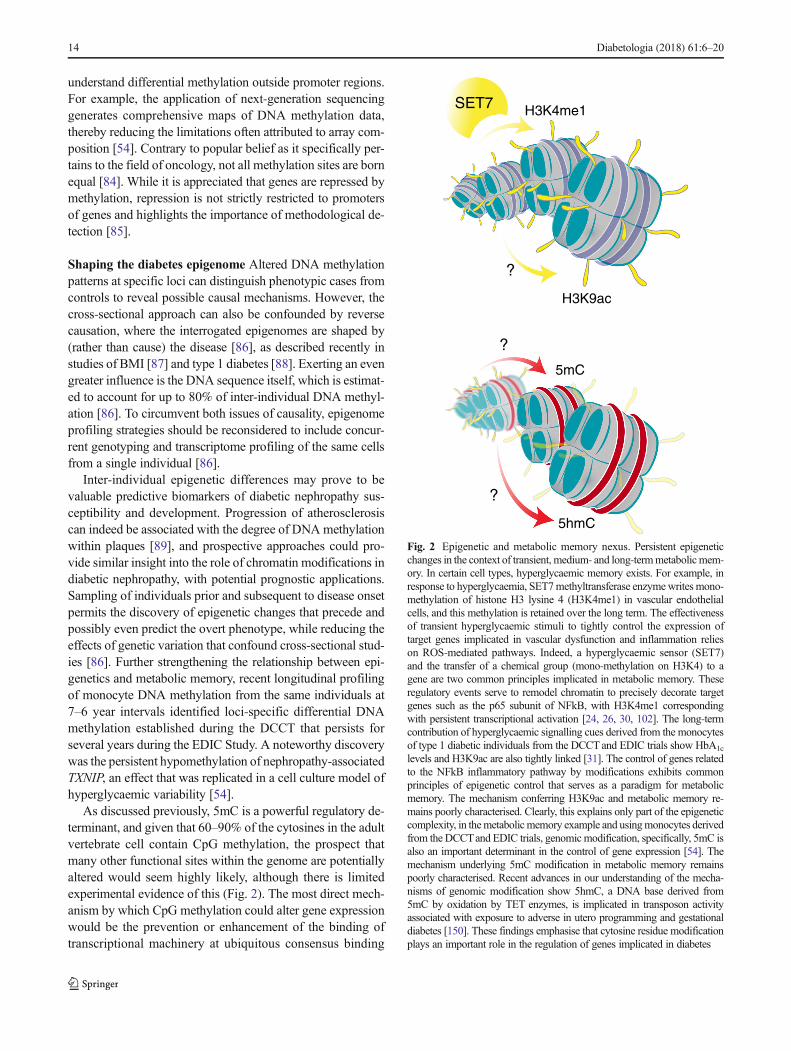
As discussed previously, 5mC is a powerful regulatory de-terminant, and given that 60–90% of the cytosines in the adultvertebrate cell contain CpG methylation, the prospect thatmany other functional sites within the genome are potentiallyaltered would seem highly likely, although there is limitedexperimental evidence of this (Fig. 2). The most direct mech-anism by which CpG methylation could alter gene expressionwould be the prevention or enhancement of the binding oftranscriptional machinery at ubiquitous consensus binding
5mC
?
5hmC
H3K9ac
?
H3K4me1
?
SET7
Fig. 2 Epigenetic and metabolic memory nexus. Persistent epigeneticchanges in the context of transient, medium- and long-termmetabolicmem-ory. In certain cell types, hyperglycaemic memory exists. For example, inresponse to hyperglycaemia, SET7methyltransferase enzymewrites mono-methylation of histone H3 lysine 4 (H3K4me1) in vascular endothelialcells, and this methylation is retained over the long term. The effectivenessof transient hyperglycaemic stimuli to tightly control the expression oftarget genes implicated in vascular dysfunction and inflammation relieson ROS-mediated pathways. Indeed, a hyperglycaemic sensor (SET7)and the transfer of a chemical group (mono-methylation on H3K4) to agene are two common principles implicated in metabolic memory. Theseregulatory events serve to remodel chromatin to precisely decorate targetgenes such as the p65 subunit of NFkB, with H3K4me1 correspondingwith persistent transcriptional activation [24, 26, 30, 102]. The long-termcontribution of hyperglycaemic signalling cues derived from the monocytesof type 1 diabetic individuals from the DCCTand EDIC trials show HbA1c
levels and H3K9ac are also tightly linked [31]. The control of genes relatedto the NFkB inflammatory pathway by modifications exhibits commonprinciples of epigenetic control that serves as a paradigm for metabolicmemory. The mechanism conferring H3K9ac and metabolic memory re-mains poorly characterised. Clearly, this explains only part of the epigeneticcomplexity, in themetabolicmemory example and usingmonocytes derivedfrom the DCCTand EDIC trials, genomicmodification, specifically, 5mC isalso an important determinant in the control of gene expression [54]. Themechanism underlying 5mC modification in metabolic memory remainspoorly characterised. Recent advances in our understanding of the mecha-nisms of genomic modification show 5hmC, a DNA base derived from5mC by oxidation by TET enzymes, is implicated in transposon activityassociated with exposure to adverse in utero programming and gestationaldiabetes [150]. These findings emphasise that cytosine residue modificationplays an important role in the regulation of genes implicated in diabetes
14 Diabetologia (2018) 61:6–20

sites. For example, CTCF is a chromatin insulator that servesto regulate access of distant enhancers to promoters [90].Indeed, CTCF binding is methylation sensitive [91], bindingimprinted control regions only at the unmethylated parentalallele to regulate specific gene expression patterns [92]. CTCFbinding also protects regions from DNA methylation [93].This diversity in methylation distribution provides a directmechanism to regulate gene expression, offering a simpleand elegant means of controlling key target genes in signallingcomplexes and core pathways implicated in type 2 diabetes[94]. The alternative possibility is that transcriptional repres-sors that assemble on chromatin recognise the methylationmoiety together with other transcriptional components [95].Indeed, methylation-specific binding proteins potentiallyserve to control gene expression of many functional sites with-in the genome in a chromatin context, thereby offering anattractive model to explain the capacity of gene function moreeffectively [96].
Targeting chromatin modifications in the clinic
With increased exploration of chromatin modifications in var-ious medical contexts, it may be possible in the future not onlyto track causal mechanisms of vascular disease, but also tocapture an individual patient’s positionwithin a complex spec-trum of pathophysiological processes, thereby supporting tai-lored approaches to the anticipation and prevention of diabeticcomplications [97]. The epigenome is responsive to internaland external stimuli as diverse as disease-specific processes,nutrition [74] and exercise [98]. Exploitation of this plasticityhas fast become a novel avenue of investigation to improvedysregulated gene function. Whereas metabolic manipulationof the chromatin landscape is a relatively recent suggestion,pharmacological compounds that modulate epigenetic regula-tors have a longer history in the clinic. Common to both strat-egies is the imperative challenge of specificity.
The emerging picture of epigenetic regulation is one ofremarkable complexity. Defining the tissue-specificrelative contributions of epigenetic writers and erasersremains an unresolved but critical issue for developingnovel therapeutic epigenetic modulators. WhereasHDAC3 deletion from the macrophage is vasculo-protective [99], deletion of the same enzyme from endo-thelial cells exacerbates macrovascular disease [100].Many enzymes that modify histones also target specificamino acids on other proteins, including transcriptionfactors, to post-translationally regulate their stability andactivity, with major implications for the interpretation ofgene expression profiles. In addition to writing H3K4m1,SET7 methylates a variety of transcription factors indifferent cellular contexts [101], and is therefore implicatedin chromatin-dependent and chromatin-independent gene
regulation [102, 103]. Furthermore, evidence is emerging tosuggest that histone-modifying enzymes can post-translationally modify and regulate each other [74], meaningthat a single enzyme could potentially influence many distinctmodifications. Finally, the mechanisms driving loci-specificenrichment of chromatin modifications remain largelyuncharacterized, though several examples of non-codingRNA and transcription factor co-recruitment have recentlyemerged [104, 105]. The respective repertoire of gene-localising mechanisms is likely to reflect cell type specificityof distinct gene programs.
Despite these formidable challenges, the epigenome is richin opportunity. Compounds that target epigenetic pathwaysare increasingly investigated pre-clinically, and drugs thatare already used in clinical management of diabetes may im-pact the epigenetic landscape. For example, metformin pre-vents trained immunity by the Bacillus Calmette–Guérin(BCG) vaccine via mTOR inhibition and suppression of gly-colysis [69]. Whether this holds true for immunological train-ing in the context of diabetes remains to be elucidated.
Conclusion
Increasingly accessible technologies that permit unbiasedacquisition of genome-wide patterns boast the capacity totransform our understanding of the chromatin landscape inthe occurrence and progression of complex diseases. Theimmense potential for epigenetics to explain many aspectsof diabetic vascular complications is evident in recent sci-entific literature. By sensitising the genome to environmen-tal variation, these molecular signatures shape diverse phe-notypes and functional programs. Chromatin modificationsinfluence deleterious changes in gene expression that, undersome circumstances, can endure improvements in metabolicmanagement. Similarly, persistent epigenetic modificationsdrive the non-specific memory of proinflammatory macro-phages, a process that is increasingly implicated in vasculardisease and could prove to be instrumental to resolving theimportant debate as to whether diabetic microvascular andmacrovascular pathology share a common pathology.Epigenomic profiling of circulating cells may further shedlight on the phenotypic variation and disproportionate bur-den of diabetic vascular complications. Combined withclassical genetic approaches, epigenomic profiling has po-tential to identify molecular trajectories underlying diabeticvascular disease development. While the extent that patho-logical chromatin changes can be manipulated in humandiabetic complications remains to be established, the clini-cal applicability of epigenetic interventions will be greatlyadvanced by a deeper understanding of the cell type-specific functions and interactions of chromatin-modifyingmachinery in the diabetic vasculature.
Diabetologia (2018) 61:6–20 15

Funding The authors acknowledge support from the National HealthandMedical Research Council (NHMRC) (APP1113188, APP1082572).JAvD is supported by a Veni Grant from The Netherlands Organizationfor Scientific Research (91616083) and received funding from the DutchDiabetes Foundation (2013.81.1674). NPR is supported by a dr. E.Dekker grant from the Netherlands Heart Foundation (2012T051), andreceived funding from the European Union’s Horizon 2020 research andinnovation programme under grant agreement no. 667837.
Duality of interest The authors declare that there is no duality of inter-est associated with this manuscript.
Contribution statement All authors were responsible for drafting thearticle and revising it critically for important intellectual content. Allauthors approved the version to be published.
Open Access This article is distributed under the terms of the CreativeCommons At t r ibut ion 4 .0 In te rna t ional License (h t tp : / /creativecommons.org/licenses/by/4.0/), which permits unrestricted use,distribution, and reproduction in any medium, provided you give appro-priate credit to the original author(s) and the source, provide a link to theCreative Commons license, and indicate if changes were made.
References
1. Thomas MC, Groop PH, Tryggvason K (2012) Towards under-standing the inherited susceptibility for nephropathy in diabetes.Curr Opin Nephrol Hypertens 21:195–202
2. Nathan DM, Cleary PA, Backlund JY et al (2005) Intensive dia-betes treatment and cardiovascular disease in patients with type 1diabetes. N Engl J Med 353:2643–2653
3. DCCT/EDIC The Diabetes Control and Complications Trial/Epidemiology of Diabetes Interventions and ComplicationsResearch Group (2000) Retinopathy and nephropathy in patientswith type 1 diabetes four years after a trial of intensive therapy.N Engl J Med 342:381–389
4. Hammes HP, Klinzing I, Wiegand S, Bretzel RG, Cohen AM,Federlin K (1993) Islet transplantation inhibits diabetic retinopa-thy in the sucrose-fed diabetic Cohen rat. Invest Ophthalmol VisSci 34:2092–2096
5. Roy S, Sala R, Cagliero E, Lorenzi M (1990) Overexpression offibronectin induced by diabetes or high glucose: phenomenonwith a memory. Proc Natl Acad Sci U S A 87:404–408
6. Engerman RL, Kern TS (1987) Progression of incipient diabeticretinopathy during good glycemic control. Diabetes 36:808–812
7. Gaede P, Oellgaard J, Carstensen B et al (2016) Years of lifegained by multifactorial intervention in patients with type 2 dia-betes mellitus and microalbuminuria: 21 years follow-up on theSteno-2 randomised trial. Diabetologia 59:2298–2307
8. Hayward RA, Reaven PD, Wiitala WL et al (2015) Follow-up ofglycemic control and cardiovascular outcomes in type 2 diabetes.N Engl J Med 372:2197–2206
9. Holman RR, Paul SK, Bethel MA,Matthews DR, Neil HA (2008)10-year follow-up of intensive glucose control in type 2 diabetes.N Engl J Med 359:1577–1589
10. Thomas MC (2016) Epigenetic mechanisms in diabetic kidneydisease. Curr Diab Rep 16:31
11. Mannucci E, Dicembrini I, Lauria A, Pozzilli P (2013) Is glucosecontrol important for prevention of cardiovascular disease in dia-betes? Diabetes Care 36(Suppl 2):S259–S263
12. OrasanuG, Plutzky J (2009) The pathologic continuum of diabeticvascular disease. J Am Coll Cardiol 53:S35–S42
13. Sasso FC, Chiodini P, Carbonara O et al (2012) High cardiovas-cular risk in patients with type 2 diabetic nephropathy: the predic-tive role of albuminuria and glomerular filtration rate. The NID-2Prospective Cohort Study. Nephrol Dial Transplant 27:2269–2274
14. Chawla A, Chawla R, Jaggi S (2016) Microvasular andmacrovascular complications in diabetes mellitus: distinct or con-tinuum? Indian J Endocrinol Metab 20:546–551
15. The Diabetes Control and Complications Trial Research Group(1997) Clustering of long-term complications in families with di-abetes in the diabetes control and complications trial. Diabetes 46:1829–1839
16. Quinn M, Angelico MC, Warram JH, Krolewski AS (1996)Familial factors determine the development of diabetic nephropa-thy in patients with IDDM. Diabetologia 39:940–945
17. Seaquist ER, Goetz FC, Rich S, Barbosa J (1989) Familial clus-tering of diabetic kidney disease. Evidence for genetic susceptibil-ity to diabetic nephropathy. N Engl J Med 320:1161–1165
18. Ma RC, Cooper ME (2017) Genetics of diabetic kidney disease—from the worst of nightmares to the light of dawn? J Am SocNephrol 28:389–393
19. Sandholm N, Van Zuydam N, Ahlqvist E et al (2017) The geneticlandscape of renal complications in type 1 diabetes. J Am SocNephrol 28:557–574
20. FlavahanWA, Drier Y, Liau BB et al (2016) Insulator dysfunctionand oncogene activation in IDHmutant gliomas. Nature 529:110–114
21. Breiling A, Lyko F (2015) Epigenetic regulatory functions ofDNA modifications: 5-methylcytosine and beyond. EpigeneticsChromatin 8:24
22. Dubois-Chevalier J, Oger F, Dehondt H et al (2014) A dynamicCTCF chromatin binding landscape promotes DNAhydroxymethylation and transcriptional induction of adipocytedifferentiation. Nucleic Acids Res 42:10943–10959
23. Heintzman ND, Stuart RK, Hon G et al (2007) Distinct and pre-dictive chromatin signatures of transcriptional promoters and en-hancers in the human genome. Nat Genet 39:311–318
24. El-Osta A, Brasacchio D, Yao D et al (2008) Transient high glu-cose causes persistent epigenetic changes and altered gene expres-sion during subsequent normoglycemia. J Exp Med 205:2409–2417
25. Cheng J, Blum R, Bowman C et al (2014) A role for H3K4monomethylation in gene repression and partitioning of chromatinreaders. Mol Cell 53:979–992
26. Brasacchio D, Okabe J, Tikellis C et al (2009) Hyperglycemiainduces a dynamic cooperativity of histone methylase anddemethylase enzymes associated with gene-activating epigeneticmarks that coexist on the lysine tail. Diabetes 58:1229–1236
27. Fu LL, TianM, Li X et al (2015) Inhibition of BET bromodomainsas a therapeutic strategy for cancer drug discovery. Oncotarget 6:5501–5516
28. Yoon S, Eom GH (2016) HDAC and HDAC inhibitor: from can-cer to cardiovascular diseases. Chonnam Med J 52:1–11
29. McKinsey TA (2012) Therapeutic potential for HDAC inhibitorsin the heart. Annu Rev Pharmacol Toxicol 52:303–319
30. Paneni F, Costantino S, Battista R et al (2015) Adverse epigeneticsignatures by histonemethyltransferase Set7 contribute to vasculardysfunction in patients with type 2 diabetes mellitus. CircCardiovasc Genet 8:150–158
31. Miao F, Chen Z, Genuth S et al (2014) Evaluating the role ofepigenetic histone modifications in the metabolic memory of type1 diabetes. Diabetes 63:1748–1762
32. Keating ST, Plutzky J, El-Osta A (2016) Epigenetic changes indiabetes and cardiovascular risk. Circ Res 118:1706–1722
33. Yoshizawa C, Kobayashi Y, Ikeuchi Y et al (2016) Congenitalnephrotic syndrome with a novel NPHS1 mutation. Pediatr Int58:1211–1215
16 Diabetologia (2018) 61:6–20

34. Yamaguchi Y, Iwano M, Suzuki D et al (2009) Epithelial-mesenchymal transition as a potential explanation for podocytedepletion in diabetic nephropathy. Am J Kidney Dis 54:653–664
35. Pagtalunan ME, Miller PL, Jumping-Eagle S et al (1997)Podocyte loss and progressive glomerular injury in type II diabe-tes. J Clin Invest 99:342–348
36. Petermann AT, Pippin J, Krofft R et al (2004) Viable podocytesdetach in experimental diabetic nephropathy: potential mechanismunderlying glomerulosclerosis. Nephron Exp Nephrol 98:e114–e123
37. Takahashi K, Tanabe K, Ohnuki M et al (2007) Induction of plu-ripotent stem cells from adult human fibroblasts by defined fac-tors. Cell 131:861–872
38. Hayashi K, Sasamura H, Nakamura M et al (2014) KLF4-dependent epigenetic remodelingmodulates podocyte phenotypesand attenuates proteinuria. J Clin Invest 124:2523–2537
39. Ristola M, Arpiainen S, Saleem MA, Holthofer H, Lehtonen S(2012) Transcription of nephrin-Neph3 gene pair is synergisticallyactivated by WT1 and NF-kappaB and silenced by DNA methyl-ation. Nephrol Dial Transplant 27:1737–1745
40. Siddiqi FS, Majumder S, Thai K et al (2016) The histone methyl-transferase enzyme enhancer of zeste homolog 2 protects againstpodocyte oxidative stress and renal injury in diabetes. J Am SocNephrol 27:2021–2034
41. De Marinis Y, Cai M, Bompada P et al (2016) Epigenetic regula-tion of the thioredoxin-interacting protein (TXNIP) gene by hy-perglycemia in kidney. Kidney Int 89:342–353
42. Bock F, Shahzad K, Wang H et al (2013) Activated protein Cameliorates diabetic nephropathy by epigenetically inhibiting theredox enzyme p66Shc. Proc Natl Acad Sci U S A 110:648–653
43. Paneni F, Mocharla P, Akhmedov A et al (2012) Gene silencing ofthe mitochondrial adaptor p66Shc suppresses vascular hyperglyce-mic memory in diabetes. Circ Res 111:278–289
44. Zhou S, Chen HZ, Wan YZ et al (2011) Repression of P66Shcexpression by SIRT1 contributes to the prevention ofhyperglycemia-induced endothelial dysfunction. Circ Res 109:639–648
45. Tang SC, Lai KN (2012) The pathogenic role of the renal proximaltubular cell in diabetic nephropathy. Nephrol Dial Transplant 27:3049–3056
46. Marumo T, Yagi S, Kawarazaki W et al (2015) Diabetes inducesaberrant DNAmethylation in the proximal tubules of the kidney. JAm Soc Nephrol 26:2388–2397
47. Hasegawa K, Wakino S, Simic P et al (2013) Renal tubular Sirt1attenuates diabetic albuminuria by epigenetically suppressingClaudin-1 overexpression in podocytes. Nat Med 19:1496–1504
48. Reddy MA, Sumanth P, Lanting L et al (2014) Losartan reversespermissive epigenetic changes in renal glomeruli of diabetic db/dbmice. Kidney Int 85:362–373
49. Furuta T, Saito T, Ootaka T et al (1993) The role of macrophagesin diabetic glomerulosclerosis. Am J Kidney Dis 21:480–485
50. Sassy-Prigent C, Heudes D, Mandet C et al (2000) Early glomer-ular macrophage recruitment in streptozotocin-induced diabeticrats. Diabetes 49:466–475
51. Wada J, Makino H (2016) Innate immunity in diabetes and dia-betic nephropathy. Nat Rev Nephrol 12:13–26
52. Tanaka M, Masuda S, Matsuo Y et al (2016) Hyperglycemia andinflammatory property of circulating monocytes are associatedwith inflammatory property of carotid plaques in patients under-going carotid endarterectomy. J Atheroscler Thromb 23:1212–1221
53. Kon V, Linton MF, Fazio S (2011) Atherosclerosis in chronickidney disease: the role of macrophages. Nat Rev Nephrol 7:45–54
54. Chen Z, Miao F, Paterson AD et al (2016) Epigenomic profilingreveals an association between persistence of DNA methylation
andmetabolic memory in the DCCT/EDIC type 1 diabetes cohort.Proc Natl Acad Sci U S A 113:E3002–E3011
55. Netea MG, Joosten LA, Latz E et al (2016) Trained immunity: aprogram of innate immune memory in health and disease. Science352:aaf1098
56. Kleinnijenhuis J, Quintin J, Preijers F et al (2012) BacilleCalmette-Guérin induces NOD2-dependent nonspecific protec-tion from reinfection via epigenetic reprogramming ofmonocytes.Proc Natl Acad Sci U S A 109:17537–17542
57. Quintin J, Saeed S, Martens JH et al (2012) Candidaalbicans infection affords protection against reinfection viafunctional reprogramming of monocytes. Cell Host Microbe12:223–232
58. Saeed S, Quintin J, Kerstens HH et al (2014) Epigenetic program-ming of monocyte-to-macrophage differentiation and trained in-nate immunity. Science 345:1251086
59. Novakovic B, Habibi E, Wang SY et al (2016) Beta-glucan re-verses the epigenetic state of LPS-induced immunological toler-ance. Cell 167:1354–1368.e1314
60. Ostuni R, Piccolo V, Barozzi I et al (2013) Latent enhancers acti-vated by stimulation in differentiated cells. Cell 152:157–171
61. Blok BA, Arts RJ, van Crevel R, Benn CS, Netea MG (2015)Trained innate immunity as underlying mechanism for the long-term, nonspecific effects of vaccines. J Leukoc Biol 98:347–356
62. Bekkering S, Joosten LA, van der Meer JW, Netea MG, RiksenNP (2013) Trained innate immunity and atherosclerosis. CurrOpin Lipidol 24:487–492
63. Christ A, Bekkering S, Latz E, Riksen NP (2016) Long-term ac-tivation of the innate immune system in atherosclerosis. SeminImmunol 28:384–393
64. Bekkering S, Quintin J, Joosten LA, van der Meer JW, Netea MG,Riksen NP (2014) Oxidized low-density lipoprotein induces long-term proinflammatory cytokine production and foam cell forma-tion via epigenetic reprogramming of monocytes. ArteriosclerThromb Vasc Biol 34:1731–1738
65. van der Valk FM, Bekkering S, Kroon J et al (2016) Oxidizedphospholipids on lipoprotein(a) elicit arterial wall inflammationand an inflammatory monocyte response in humans. Circulation134:611–624
66. Yan SF, Ramasamy R, Schmidt AM (2008) Mechanisms of dis-ease: advanced glycation end-products and their receptor in in-flammation and diabetes complications. Nat Clin PractEndocrinol Metab 4:285–293
67. Shanmugam N, Reddy MA, Guha M, Natarajan R (2003) Highglucose-induced expression of proinflammatory cytokine and che-mokine genes in monocytic cells. Diabetes 52:1256–1264
68. van Diepen JA, Thiem K, Stienstra R, Riksen NP, Tack CJ, NeteaMG (2016) Diabetes propels the risk for cardiovascular disease:sweet monocytes becoming aggressive? Cell Mol Life Sci 73:4675–4684
69. Cheng SC, Quintin J, Cramer RA et al (2014) mTOR- and HIF-1alpha-mediated aerobic glycolysis as metabolic basis for trainedimmunity. Science 345:1250684
70. Arts RJ, Novakovic B, Ter Horst R et al (2016) Glutaminolysisand fumarate accumulation integrate immunometabolic and epi-genetic programs in trained immunity. Cell Metab 24:807–819
71. Donohoe DR, Collins LB, Wali A, Bigler R, Sun W, Bultman SJ(2012) The Warburg effect dictates the mechanism of butyrate-mediated histone acetylation and cell proliferation. Mol Cell 48:612–626
72. Cheng SC, Joosten LA, Netea MG (2014) The interplay betweencentral metabolism and innate immune responses. CytokineGrowth Factor Rev 25:707–713
73. Kaelin WG Jr, McKnight SL (2013) Influence of metabolism onepigenetics and disease. Cell 153:56–69
Diabetologia (2018) 61:6–20 17

74. Keating ST, El-Osta A (2015) Epigenetics and metabolism. CircRes 116:715–736
75. de Oliveira AA, de Oliveira TF, Bobadilla LL et al (2017)Sustained kidney biochemical derangement in treated experimen-tal diabetes: a clue to metabolic memory. Sci Rep 7:40544
76. Sharma A (2017) Transgenerational epigenetics: integrating somato germline communication with gametic inheritance. MechAgeing Dev 163:15–22
77. Cropley JE, Eaton SA, Aiken A et al (2016) Male-lineage trans-mission of an acquired metabolic phenotype induced by grand-paternal obesity. Mol Metab 5:699–708
78. Khurana I, Kaspi A, Ziemann M et al (2016) DNA methylationregulates hypothalamic gene expression linking parental diet dur-ing pregnancy to the offspring’s risk of obesity in Psammomysobesus. Int J Obes 40:1079–1088
79. Bell CG, Teschendorff AE, Rakyan VK, Maxwell AP, Beck S,Savage DA (2010) Genome-wide DNA methylation analysis fordiabetic nephropathy in type 1 diabetes mellitus. BMCMed Genet3:33
80. Sapienza C, Lee J, Powell J et al (2011) DNA methylation profil-ing identifies epigenetic differences between diabetes patients withESRD and diabetes patients without nephropathy. Epigenetics 6:20–28
81. Tregouet DA, Groop PH, McGinn S et al (2008) G/T substitutionin intron 1 of the UNC13B gene is associated with increased riskof nephropathy in patients with type 1 diabetes. Diabetes 57:2843–2850
82. Miao F, Wu X, Zhang L, Riggs AD, Natarajan R (2008) Histonemethylation patterns are cell-type specific in human monocytesand lymphocytes and well maintained at core genes. J Immunol180:2264–2269
83. Reinius LE, Acevedo N, Joerink M et al (2012) Differential DNAmethylation in purified human blood cells: implications for celllineage and studies on disease susceptibility. PLoS One 7:e41361
84. Baylin SB, Jones PA (2011) A decade of exploring the cancerepigenome–biological and translational implications. Nat RevCancer 11:726–734
85. Pirola L, Balcerczyk A, Tothill RW et al (2011) Genome-wideanalysis distinguishes hyperglycemia regulated epigenetic signa-tures of primary vascular cells. Genome Res 21:1601–1615
86. Birney E, Smith GD, Greally JM (2016) Epigenome-wide associ-ation studies and the interpretation of disease-omics. PLoS Genet12:e1006105
87. Richmond RC, Sharp GC, Ward ME et al (2016) DNA methyla-tion and BMI: investigating identified methylation sites at HIF3Ain a causal framework. Diabetes 65:1231–1244
88. RakyanVK, Beyan H, Down TA et al (2011) Identification of type1 diabetes-associated DNA methylation variable positions thatprecede disease diagnosis. PLoS Genet 7:e1002300
89. Valencia-Morales Mdel P, Zaina S, Heyn H et al (2015) The DNAmethylation drift of the atherosclerotic aorta increases with lesionprogression. BMC Med Genet 8:7
90. Lewis A, Murrell A (2004) Genomic imprinting: CTCF protectsthe boundaries. Curr Biol 14:R284–R286
91. Kanduri C, Pant V, Loukinov D et al (2000) Functional associationof CTCF with the insulator upstream of the H19 gene is parent oforigin-specific and methylation-sensitive. Curr Biol 10:853–856
92. Demars J, Shmela ME, Khan AW et al (2014) Genetic variantswithin the second intron of the KCNQ1 gene affect CTCF bindingand confer a risk of Beckwith-Wiedemann syndrome upon mater-nal transmission. J Med Genet 51:502–511
93. Fedoriw AM, Stein P, Svoboda P, Schultz RM, Bartolomei MS(2004) Transgenic RNAi reveals essential function for CTCF inH19 gene imprinting. Science 303:238–240
94. Stitzel ML, Sethupathy P, Pearson DS et al (2010) Globalepigenomic analysis of primary human pancreatic islets provides
insights into type 2 diabetes susceptibility loci. Cell Metab 12:443–455
95. El-Osta A, Kantharidis P, Zalcberg JR, Wolffe AP (2002)Precipitous release of methyl-CpG binding protein 2 and histonedeacetylase 1 from the methylated human multidrug resistancegene (MDR1) on activation. Mol Cell Biol 22:1844–1857
96. Harikrishnan KN, ChowMZ, Baker EK et al (2005) Brahma linksthe SWI/SNF chromatin-remodeling complex with MeCP2-dependent transcriptional silencing. Nat Genet 37:254–264
97. McCarthy MI (2015) Genomic medicine at the heart of diabetesmanagement. Diabetologia 58:1725–1729
98. King-Himmelreich TS, Schramm S, Wolters MC et al (2016) Theimpact of endurance exercise on global and AMPK gene-specificDNA methylation. Biochem Biophys Res Commun 474:284–290
99. Hoeksema MA, Gijbels MJ, Van den Bossche J et al (2014)Targeting macrophage histone deacetylase 3 stabilizes atheroscle-rotic lesions. EMBO Mol Med 6:1124–1132
100. Zampetaki A, Zeng L, Margariti A et al (2010) Histonedeacetylase 3 is critical in endothelial survival and atherosclerosisdevelopment in response to disturbed flow. Circulation 121:132–142
101. Keating ST, El-Osta A (2013) Transcriptional regulation by theSet7 lysine methyltransferase. Epigenetics 8:361–372
102. Okabe J, Orlowski C, Balcerczyk A et al (2012) Distinguishinghyperglycemic changes by Set7 in vascular endothelial cells. CircRes 110:1067–1076
103. Keating ST, Ziemann M, Okabe J, Khan AW, Balcerczyk A, El-Osta A (2014) Deep sequencing reveals novel Set7 networks. CellMol Life Sci 71:4471–4486
104. Lian Y,Wang J, Feng J et al (2016) Long non-coding RNA IRAINsuppresses apoptosis and promotes proliferation by binding toLSD1 and EZH2 in pancreatic cancer. Tumour Biol 37:14929–14937
105. Battistelli C, Cicchini C, Santangelo L et al (2017) The snail re-pressor recruits EZH2 to specific genomic sites through the enroll-ment of the lncRNA HOTAIR in epithelial-to-mesenchymal tran-sition. Oncogene 36:942–955
106. Yuan H, Reddy MA, Deshpande S et al (2016) Epigenetic histonemodifications involved in profibrotic gene regulation by 12/15-lipoxygenase and its oxidized lipid products in diabetic nephrop-athy. Antioxid Redox Signal 24:361–375
107. Syreeni A, El-Osta A, Forsblom C et al (2011) Genetic examina-tion of SETD7 and SUV39H1/H2 methyltransferases and the riskof diabetes complications in patients with type 1 diabetes.Diabetes 60:3073–3080
108. Musri MM, Carmona MC, Hanzu FA, Kaliman P, Gomis R,Parrizas M (2010) Histone demethylase LSD1 regulates adipo-genesis. J Biol Chem 285:30034–30041
109. Villeneuve LM, Reddy MA, Lanting LL, Wang M, Meng L,Natarajan R (2008) Epigenetic histone H3 lysine 9 methylationin metabolic memory and inflammatory phenotype of vascularsmooth muscle cells in diabetes. Proc Natl Acad Sci U S A 105:9047–9052
110. Hardikar AA, Satoor SN, Karandikar MS et al (2015)Multigenerational undernutrition increases susceptibility to obe-sity and diabetes that is not reversed after dietary recuperation.Cell Metab 22:312–319
111. Chen H, Gu X, Su IH et al (2009) Polycomb protein Ezh2 regu-lates pancreatic beta-cell Ink4a/Arf expression and regeneration indiabetes mellitus. Genes Dev 23:975–985
112. Sakai M, Tujimura-Hayakawa T, Yagi T et al (2016) The GCN5-CITED2-PKA signalling module controls hepatic glucose metab-olism through a cAMP-induced substrate switch. Nat Commun 7:13147
113. Vecellio M, Spallotta F, Nanni S et al (2014) The histone acetylaseactivator pentadecylidenemalonate 1b rescues proliferation and
18 Diabetologia (2018) 61:6–20

differentiation in the human cardiac mesenchymal cells of type 2diabetic patients. Diabetes 63:2132–2147
114. Gallagher KA, Joshi A, CarsonWF et al (2015) Epigenetic chang-es in bone marrow progenitor cells influence the inflammatoryphenotype and alter wound healing in type 2 diabetes. Diabetes64:1420–1430
115. Okuno Y, Ohtake F, Igarashi K et al (2013) Epigenetic regulationof adipogenesis by PHF2 histone demethylase. Diabetes 62:1426–1434
116. Sathishkumar C, Prabu P, Balakumar M et al (2016)Augmentation of histone deacetylase 3 (HDAC3) epigenetic sig-nature at the interface of proinflammation and insulin resistancein patients with type 2 diabetes. Clin Epigenetics 8:125
117. Wang X, Liu J, Zhen J et al (2014) Histone deacetylase 4 selec-tively contributes to podocyte injury in diabetic nephropathy.Kidney Int 86:712–725
118. Daneshpajooh M, Bacos K, Bysani M et al (2017) HDAC7 isoverexpressed in human diabetic islets and impairs insulin secre-tion in rat islets and clonal beta cells. Diabetologia 60:116–125
119. Bell AC, Felsenfeld G (2000) Methylation of a CTCF-dependentboundary controls imprinted expression of the Igf2 gene. Nature405:482–485
120. Barres R, Osler ME, Yan J et al (2009) Non-CpG methylation ofthe PGC-1alpha promoter through DNMT3B controls mitochon-drial density. Cell Metab 10:189–198
121. Pennarossa G, Maffei S, Campagnol M, Tarantini L, Gandolfi F,Brevini TA (2013) Brief demethylation step allows the conversionof adult human skin fibroblasts into insulin-secreting cells. ProcNatl Acad Sci U S A 110:8948–8953
122. VolkmarM,Dedeurwaerder S, CunhaDA et al (2012) DNAmeth-ylation profiling identifies epigenetic dysregulation in pancreaticislets from type 2 diabetic patients. EMBO J 31:1405–1426
123. Ling C, Del Guerra S, Lupi R et al (2008) Epigenetic regulation ofPPARGC1A in human type 2 diabetic islets and effect on insulinsecretion. Diabetologia 51:615–622
124. Kirchner H, Sinha I, Gao H et al (2016) Altered DNAmethylationof glycolytic and lipogenic genes in liver from obese and type 2diabetic patients. Mol Metab 5:171–183
125. Babu M, Durga Devi T, Makinen P et al (2015) Differential pro-moter methylation of macrophage genes is associated with im-paired vascular growth in ischemic muscles of hyperlipidemicand type 2 diabetic mice: genome-wide promoter methylationstudy. Circ Res 117:289–299
126. Radford EJ, Ito M, Shi H et al (2014) In utero effects. In uteroundernourishment perturbs the adult sperm methylome and inter-generational metabolism. Science 345:1255903
127. Dayeh T, Volkov P, Salo S et al (2014) Genome-wide DNAmeth-ylation analysis of human pancreatic islets from type 2 diabeticand non-diabetic donors identifies candidate genes that influenceinsulin secretion. PLoS Genet 10:e1004160
128. Yang BT, Dayeh TA, Volkov PA et al (2012) Increased DNAmethylation and decreased expression of PDX-1 in pancreaticislets from patients with type 2 diabetes. Mol Endocrinol 26:1203–1212
129. Park JH, Stoffers DA, Nicholls RD, Simmons RA (2008)Development of type 2 diabetes following intrauterine growthretardation in rats is associated with progressive epigenetic silenc-ing of Pdx1. J Clin Invest 118:2316–2324
130. Multhaup ML, Seldin MM, Jaffe AE et al (2015) Mouse-humanexperimental epigenetic analysis unmasks dietary targets and ge-netic liability for diabetic phenotypes. Cell Metab 21:138–149
131. Kalani A, Kamat PK, Tyagi N (2015) Diabetic stroke severity:epigenetic remodeling and neuronal, glial, and vascular dysfunc-tion. Diabetes 64:4260–4271
132. Volkov P, Bacos K, Ofori JK et al (2017) Whole-genome bisulfitesequencing of human pancreatic islets reveals novel differentially
methylated regions in type 2 diabetes pathogenesis. Diabetes 66:1074–1085
133. Soriano-Tarraga C, Jimenez-Conde J, Giralt-Steinhauer E et al(2016) Epigenome-wide association study identifies TXNIP geneassociated with type 2 diabetes mellitus and sustained hypergly-cemia. Hum Mol Genet 25:609–619
134. Kulkarni H, Kos MZ, Neary J et al (2015) Novel epigenetic de-terminants of type 2 diabetes inMexican-American families. HumMol Genet 24:5330–5344
135. Orozco LD, Morselli M, Rubbi L et al (2015) Epigenome-wideassociation of liver methylation patterns and complex metabolictraits in mice. Cell Metab 21:905–917
136. Gu T, Falhammar H, Gu HF, Brismar K (2014) Epigenetic analy-ses of the insulin-like growth factor binding protein 1 gene in type1 diabetes and diabetic nephropathy. Clin Epigenetics 6:10
137. Ruchat SM, Houde AA, Voisin G et al (2013) Gestational diabetesmellitus epigenetically affects genes predominantly involved inmetabolic diseases. Epigenetics 8:935–943
138. Mudry JM, Lassiter DG, Nylen C et al (2016) Insulin and glucosealter death-associated protein kinase 3 (DAPK3) DNA methyla-tion in human skeletal muscle. Diabetes 66:651–662
139. Volkov P, Olsson AH, Gillberg L et al (2016) A genome-widemQTL analysis in human adipose tissue identifies genetic variantsassociated with DNA methylation, gene expression and metabolictraits. PLoS One 11:e0157776
140. Clarke-Harris R, Wilkin TJ, Hosking J et al (2014)PGC1alpha promoter methylation in blood at 5-7 yearspredicts adiposity from 9 to 14 years (EarlyBird 50).Diabetes 63:2528–2537
141. Zeng L, Kanwar YS, Amro N et al (2003) Epigenetic and geneticanalysis of p16 in dermal fibroblasts from type 1 diabetic patientswith nephropathy. Kidney Int 63:2094–2102
142. Paul DS, Teschendorff AE, DangMA et al (2016) Increased DNAmethylation variability in type 1 diabetes across three immuneeffector cell types. Nat Commun 7:13555
143. Reichetzeder C, Dwi Putra SE, Pfab T et al (2016) Increased glob-al placental DNAmethylation levels are associated with gestation-al diabetes. Clin Epigenetics 8:82
144. Bacos K, Gillberg L, Volkov P et al (2016) Blood-based bio-markers of age-associated epigenetic changes in human islets as-sociate with insulin secretion and diabetes. Nat Commun 7:11089
145. Ronn T, Volkov P, Gillberg L et al (2015) Impact of age,BMI and HbA1c levels on the genome-wide DNA methyl-ation and mRNA expression patterns in human adipose tis-sue and identification of epigenetic biomarkers in blood.Hum Mol Genet 24:3792–3813
146. Yuan W, Xia Y, Bell CG et al (2014) An integrated epigenomicanalysis for type 2 diabetes susceptibility loci in monozygotictwins. Nat Commun 5:5719
147. Nilsson E, Jansson PA, Perfilyev A et al (2014) Altered DNAmethylation and differential expression of genes influencing me-tabolism and inflammation in adipose tissue from subjects withtype 2 diabetes. Diabetes 63:2962–2976
148. El Hajj N, Pliushch G, Schneider E et al (2013) Metabolic pro-gramming ofMEST DNAmethylation by intrauterine exposure togestational diabetes mellitus. Diabetes 62:1320–1328
149. Ling C, Poulsen P, Simonsson S et al (2007) Genetic and epige-netic factors are associated with expression of respiratory chaincomponent NDUFB6 in human skeletal muscle. J Clin Invest 117:3427–3435
150. Sun M, Song MM, Wei B et al (2016) 5-Hydroxymethylcytosine-mediated alteration of transposon activity associated with the ex-posure to adverse in utero environments in human. Hum MolGenet 25:2208–2219
Diabetologia (2018) 61:6–20 19

151. Frayling TM, Timpson NJ, Weedon MN et al (2007) A commonvariant in the FTO gene is associated with body mass index andpredisposes to childhood and adult obesity. Science 316:889–894
152. Fischer J, Koch L, Emmerling C et al (2009) Inactivation of theFto gene protects from obesity. Nature 458:894–898
153. Church C, Moir L, McMurray F et al (2010) Overexpression ofFto leads to increased food intake and results in obesity. Nat Genet42:1086–1092
154. Wu W, Feng J, Jiang D et al (2017) AMPK regulates lipid accu-mulation in skeletal muscle cells through FTO-dependent demeth-ylation of N6-methyladenosine. Sci Rep 7:41606
155. Shen F, Huang W, Huang JT et al (2015) Decreased N6-methyladenosine in peripheral blood RNA from diabetic patientsis associated with FTO expression rather than ALKBH5. J ClinEndocrinol Metab 100:E148–E154
20 Diabetologia (2018) 61:6–20