epigenetics in evolution and disease - harvard university · 272 epigenetics in evolution and...
TRANSCRIPT

part four
Epigenetics in Evolution and Disease


271
Epigenetics: Linking Genotype and Phenotype in Development and Evolution, ed. Benedikt Hallgrímsson and Brian K. Hall. Copyright by The Regents of the University of California. All rights of reproduction in any form reserved.
16
Epigenetic Integration, Complexity, and Evolvability of the Head
rethinking the functional matrix hypothesis
Daniel E. Lieberman
As I get older, I find myself increasingly hesi-tant to use the word epigenetics because I worry about employing a term that is so liable to en-gender confusion and disagreement. Many bi-ologists define epigenetics in a narrow sense solely as heritable changes in the phenotype that derive from molecular mechanisms other than sequence changes in the genotype (the classic example being methylation). However,
Waddington, who coined the word in 1942, and other early users of the term had a broader concept in mind, one that captured the variable effects of interactions between genes, embry-onic development, and the environment. Ac-cording to this original and more encompass-ing definition, epigenetics refers to the vast set of processes by which alternative, variable phenotypes––cellular, anatomical, physiologi-cal, even behavioral––derive from a given geno-type (see Haig, 2004). Not surprisingly, there is a rich literature on epigenetics in complex or-ganisms because many layers and types of epi-genetic interactions are essential to initiate the development and integration of diverse units so that they grow and function together appropri-ately (Wagner, 2001; Kirschner and Gerhart, 2005; West-Eberhard, 2003). Epigenetic inter-actions occur at each hierarchical level of devel-opment including within the genome; among cells, tissues, and organs; and between an or-ganism and its environment.
ContEnts
Epigenetic Integration During Craniofacial Development
PatterningMorphogenesisGrowth
The Functional Matrix Hypothesis RevisitedApplying the Model (Brain Size in Human
Evolution)References

272 e p i g e n e t i c s i n e v o l u t i o n a n d d i s e a s e
Epigenetic interactions play major roles in the development and evolution of every com-plex phenotype, but vertebrate heads are an ex-treme case that merits particular consideration because of their special complexity, both struc-turally and functionally. Structurally, heads are complex because they comprise an astonishing array of diverse tissues and organs in a compar-atively small, restricted space. In a typical adult human, these structures include 22 bones that derive from hundreds of ossification centers, 32 teeth, many dozens of muscles, the brain (itself comprised of many major units), eyes, olfactory bulbs, the organs of balance, the organs of hear-ing, the pharynx, various glands, as well as the many nerves, veins, arteries, and sinuses that supply, drain, and innervate these structures. Functionally, the head participates in a wide array of tasks including perception, cognition, memory, vision, taste, olfaction, hearing, bal-ance, chewing, swallowing, vocalization, respi-ration, and thermoregulation. How could such a degree of complexity and multifunctionality develop, let alone evolve, without massive levels of epigenetic interactions?
A thought experiment may help to illustrate this point. Imagine you are an engineer and have been given the task of creating a robotic head of human size that carries out the same functions as a human head and with similar lev-els of performance. To complicate matters fur-ther, the robotic head needs not only to perform all these functions effectively under many cir-cumstances (extreme heat, cold, dryness, wet-ness, when upside-down, etc.) but also to be able to grow from the size of a walnut to that of a soccer ball without any compromise in func-tion. Needless to say, you would probably quit your job or go mad because this kind of engi-neering is currently impossible, even unimagi-nable. Yet, evolution has managed this feat ef-fectively and with many different models. Over countless generations, natural selection has op-erated more like a tinker than an engineer, using a wide range of heritable and available components in novel ways to create heads that function superbly in a wide range of demand-
ing tasks, grow manyfold in size, and even look attractive in many cases.
The complexity of the head, both structurally and functionally, raises a paradox. From an en-gineering standpoint, one might imagine that very complex and functionally vital parts of the body like the head are tightly constrained. Any change, such as a larger brain or smaller ca-nines, would presumably disrupt how the head grows and functions, leading to a loss of inte-gration and, hence, a decline in performance. In a world full of competitors and predators, natural selection would eliminate any variant that could not see, smell, chew, or hear as well as its competitors. Yet, heads are actually very evolvable (capable of generating a wide range of heritable, phenotypic variation), as is evident from the extraordinary diversity of mammalian skulls in terms of size, shape, and function (es-pecially food processing, vision, olfaction, hear-ing, locomotion, cognition, thermoregulation, and vocalization). There is also much evidence for high levels of evolvability at a smaller scale of evolution. For example, a large proportion of the anatomical differences among species of hominins (species more closely related to a human than to a chimpanzee) lie above, rather than below, the neck.
Two nonexclusive hypotheses account for the paradox of complexity––that the most intri-cate, functionally vital parts of the body are as evolvable as, if not more evolvable than, other less complex regions. The first is that the greater complexity of some regions of the phenotype such as the head reflects more intense levels of selection. Given the head’s vital roles in func-tions with strong fitness effects (such as cogni-tion, vision, and smell), it is possible that natu-ral selection has acted more strongly on the head than on some other regions of the body with fewer vital functions (such as the knees). The second nonexclusive hypothesis––and the focus of this chapter––is that very complex re-gions such as the head derive their evolvability from the way they are integrated during devel-opment through many interacting layers of epi-genetic interactions (Lieberman, 2011). Accord-
i n t e g r a t i o n a n d e v o l v a b i l i t y o f t h e h e a d 273
ing to this model, the evolvable complexity of some intricate parts of the phenotype is an emergent property of the special network of many epigenetic processes that help them form. Put differently, high levels of epigenetic integration at multiple levels of development allow the head to tolerate and adjust to a wide range of variations. Because variation is an essential component of evolution, high levels of epigenetic integration lead to high levels of evolvability.
My goal in this chapter is to articulate and discuss this hypothesis rather than to test it rig-orously, but I will point out some key lines of supporting evidence. I will start by reviewing how epigenetic mechanisms operate during craniofacial ontogeny, making the case that heads are put together in a special way that pro-motes high levels of epigenetic interaction both within and between units. The model I outline is essentially a reformulation and modification of Melvin Moss’ functional matrix hypothesis (Moss, 1968, 1997a, 1997b, 1997c, 1997d). Ac-cording to the model, the head’s organs initially differentiate and then grow in a way that causes them to share overlapping portions of the same skeletal framework. Although we do not yet know many details about most of the epigenetic mechanisms in the head and how they work, we have a good idea of their general nature. In par-ticular, as the head’s many modules develop, grow, and function, they interact with each other directly and indirectly to accommodate high levels of variation without compromising function. I will conclude by considering briefly how the model applies to key transformations of the head that occurred during human evolu-tion, focusing on brain size.
EpIgEnEtIC IntEgratIon DurIng CranIofaCIal DEvElopmEnt
It would be quixotic to try to devise a compre-hensive model of epigenetic interactions during development for the head, let alone any other region of the body. Evolution by tinkering has produced too many exceptions to any one iden-
tifiable rule. In addition, complex phenotypes develop via a time-dependent network of inter-actions rather than via a linear set of processes (see Wilkins, 2002), so any comprehensive model would be too complex to be of any gen-eral use. That said, epigenetic interactions are intense in the head at every level of develop-ment, and it is reasonable to hypothesize that they help to set up an overall configuration that may be especially tolerant to a wide range of variation. Because every unit initially grows so that it partially shares its skeletal framework with other units, the head commences its devel-opment as a highly integrated structure in which there is much potential for much epigen-etic interaction, and hence much mutual ac-commodation, among units. Before discussing this hypothesis, I review how epigenetic interac-tions are involved in craniofacial development via three major steps: patterning, morphogene-sis, and growth.
pattErnIng
Integration and modularity are complementary characteristics of complex systems (for review, see Klingenberg, 2008). As in the rest of the body, epigenetic interactions play a key role in patterning the head, helping to create discrete units in the right place, at the right time, and with information about their eventual identity. Patterning processes in the head, however, cre-ate a structure that is integrated in a special way. Only the first four somites in the head de-rive from the axial skeleton, which is patterned by Hox genes (for review, see Deschamps and van Nes, 2005). The rest of the head is pat-terned via interactions between many cell lines: (1) segments of the neural tube, initially the prosencephalon, mesencephalon, and rhomb-encephalon; (2) the notochord; (3) mesenchyme of mesodermal origin; (4) surface ectoderm; (4) the cranial end of the gut tube; and (5) several streams of neural crest cells that migrate from regions around the brain toward the face. These latter cells are especially important. One set of neural crest cells migrates over the forebrain (prosencephalon) like a hood to create the fron-

274 e p i g e n e t i c s i n e v o l u t i o n a n d d i s e a s e
tonasal prominence (Figure 16.1). As they migrate, cells in the prominence interact with neural placodes (especially optic and olfactory) and other tissues to differentiate into many of the units that eventually form the upper and middle facial skeleton around the eyes, nose, and the upper part of the pharynx. The other set of neural crest cells, the branchial arches, mi-grate like collars around the side of the head to create the middle ear, the lower jaw, and the throat around the pharynx (Figure 16.1). These cells, too, are patterned by inductive interac-tions with cells they encounter as they stream into position (see below).
To reiterate, complex epigenetic interactions are involved in patterning every region of the body, but a key point about the head is that its initial units become intricately integrated with each other in an architecturally convoluted ar-rangement from the start so that skeletal units grow around and between organs and func-tional spaces. For example, the cells of the fron-tonasal prominence that migrate over the fore-brain to differentiate into the skeletal framework around the eyes and nose depend on numerous interactions with tissues they encounter along the way in order to bifurcate and diverge around these primordial organs and then re-fuse below them. To accomplish these tricky maneuvers, the frontonasal prominence relies on inductive signals from the nasal and olfactory placodes and vice versa so that the eyes and nose form surrounded by regions of mesenchyme that form a skeletal framework (see Marcucio et al., 2005). Not surprisingly, the most common type of cra-niofacial disorders is palatal clefting, which re-sults from improper coordination of these events (see Marazita and Mooney, 2004). In ad-dition, derivatives of the frontonasal promi-nence must integrate with the cranial base along with the pharynx and branchial arches to grow around the structures that form the lower face. Similarly convoluted migrations and inter-actions characterize most other craniofacial structures, setting up an embryonic head in which it quickly becomes difficult to discern straightforward boundaries between units. For example, the four bones of the cranial base de-rive from 13 pairs of condensations––some me-sodermal in origin, others neuroectodermal in origin––whose formation appears to be depen-dent on interactions with the notochord, brain, sensory placodes, nerves, and other tissues (most of these inductive interactions have yet to be worked out). As a consequence, many of the traditional divisions of the skull (e.g., basicra-nium, neurocranium, and face; prechordal and postchordal) do not map on directly to any bones. The sphenoid, for instance, is often con-sidered an endochondral, basicranial bone of
1
1
1
2
2
2
3
3
3
4
4
4
5
5
5
NeuralBasicranialFrontonasalBranchial
fIgurE 16.1 Schematic and highly simplified view of the head’s early morphogenesis (in lateral and anterior views), showing movements of the frontonasal prominences (black arrows) and branchial arches (gray arrows) over the top and around the head. Note that the prominences and branchial arches interact with and envelop various placodes and orga-nizing centers as they stream into final position. Thus, the eyes, nose, pharynx, and other organs and spaces develop surrounded by a shared structural framework.

i n t e g r a t i o n a n d e v o l v a b i l i t y o f t h e h e a d 275
mesodermal origin, but it actually contains some elements that are neural crest and others that are paraxial mesoderm (Jiang et al., 2002); in addition, some of its components are basicra-nial and grow via endochondral ossification, while others are neurocranial and grow via in-tramembranous ossification. As we shall see below, there is reason to believe that this byzan-tine arrangement, made possible by innumera-ble epigenetic interactions, underlies some of the head’s ability to be successfully complex.
morpHogEnEsIs
A second level of epigenetic interaction impor-tant to craniofacial development occurs during morphogenesis, when precursor cells in partic-ular units differentiate into the cell types that form distinct tissues and organs. In the head, as in the rest of the body, differentiation typically occurs because of inductive interactions with neighboring tissues. As an example, the basioc-cipital derives from paraxial mesodermal cells above the notochord and below the brain stem that differentiate into sclerotome, then into mesenchyme, and then into chondrocytes and osteoblasts which form the bone through endo-chondral ossification (see McBratney-Owen et al., 2008). Transplant experiments provide dra-matic evidence of the importance of local in-ductive interactions between the brain and ba-sicranium that drive the fate of these cells. Reorienting an embryonic chick brain by 180° causes a concomitant reversal in the shape of the underlying cranial base (Schowing, 1968). Teeth provide another classic example of induc-tion elsewhere in the head. Epithelial cells transplanted from the incisor to the molar re-gions of the jaw cause local mesenchymal cells to develop into incisors rather than molars (see Peters and Balling, 1999; Jernvall and Thesleff, 2000).
Although there is nothing necessarily unique about the nature of the inductive interactions involved in morphogenetic differentiation in the vertebrate head, the complex ways that the head’s units become arranged with respect to
each other do lead to some special properties. For one, the head has a greater density of or-gans (such as the eyes, vestibular system, and pituitary gland) and functional spaces (such as the pharynx, oral cavity, and sinuses) compared to other parts of the body. More importantly, be-cause the branchial arches and the derivatives of the frontonasal prominence stream around the organs of sense and the pharynx and then differentiate into skeletal condensations, these organs and spaces share a common, architec-turally complex skeletal framework. As the cor-onal section shown in Figure 16.2 illustrates, few walls of bone or cartilage are unique to any single organ or space. For example, the floor of the anterior cranial fossa is the roof of the orbit, the medial wall of the orbit is the lateral wall of the nasal cavity, the roof of the nasopharynx is the floor of the brain stem, and so on. Be-cause the head’s organs and spaces grow in and
Olfactory bulb
Frontal lobe
eyeball
tongue
oral cavity
nasal cavity
max. sinus
masseter
fIgurE 16.2 Coronal section through a head, highlighting selected key functional matrices that are either organs (e.g., the brain, olfactory bulbs, eyeballs, tongue, teeth), spaces (e.g., the pharynx and sinuses), or muscles (e.g., the masse-ter muscle). Importantly, all these functional matrices share a common skeletal framework. Almost every wall of bone is part of the cranial component of more than one functional matrix.
276 e p i g e n e t i c s i n e v o l u t i o n a n d d i s e a s e
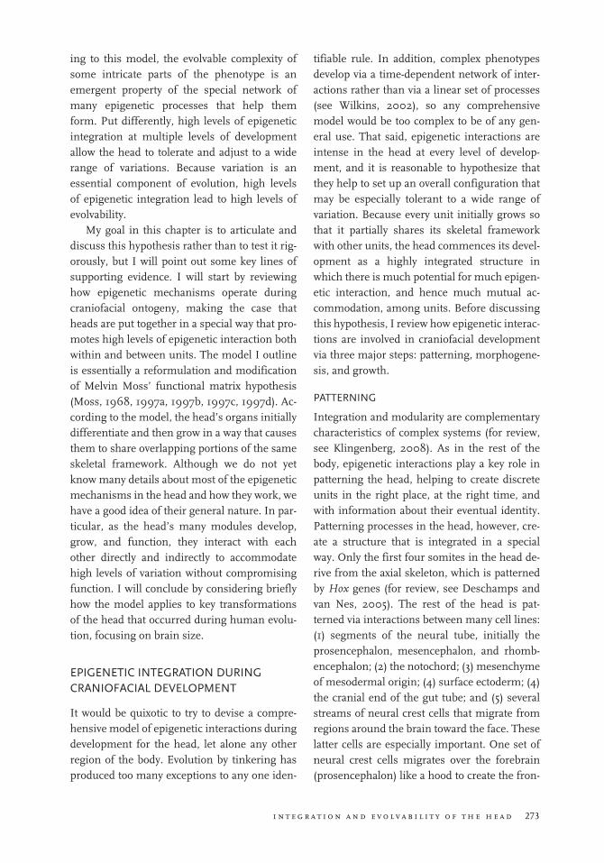
around each other with a common skeletal framework, many different factors can affect the size and shape of each morphogenetic unit. One key factor is the size and shape of a given initial condensation, which Atchley and Hall (1991) modeled as a function of five major pa-rameters: (1) the number of initial mesenchy-mal stem cells, largely a function of the number of cells that migrated there in the first place; (2) the time of initiation; (3) the rate of cell division; (4) the percentage of mitotically active cells; and (5) the rate of cell death. These initial parame-ters have important effects on the size of the resulting adult phenotype (Cottrill et al., 1987; Cohen, 2000). In addition, the special way that the head’s skeletal units are arranged around organs and spaces both requires and ensures a high degree of integration because the shape and size of each unit necessarily has effects on the shape and size of its neighbors. As shown by numerous studies, covariation among units of the skull is extremely high even in disparate regions, with especially strong levels of covaria-tion between the widths of adjoining units (e.g., Zelditch and Carmichael, 1989; Cheverud, 1995; Lieberman et al., 2000a; Marroig and Cheverud, 2001; Strait, 2001; Polanski and Franciscus, 2006; Hallgrimsson et al., 2007). We will re-turn later to these reciprocal interactions and their effects on integration and evolvability.
growtH
The final type of epigenetic integration to con-sider is how particular units interact during growth to attain their final size and shape. This aspect of ontogeny, which can generate signifi-cant levels of phenotypic variation via mecha-nisms such as heterochrony (see Gould, 1977; Atchley and Hall, 1991; McKinney and McNa-mara, 1991), involves a broad range of epige-netic interactions that contribute to substantial morphological integration and promote or maintain functional integration (Olson and Miller, 1958; Chernoff and Magwene, 1999; Young and Badyaev, 2006). Many of these in-teractions occur at the systemic level through shared responses to circulating hormones such
as growth hormone and insulin-like growth fac-tor I, permitting disparate regions to grow in a common trajectory and with appropriate scal-ing (see Shea and Gomez, 1988). For instance, most components of the face grow in a skeletal growth trajectory along with the rest of the body but not the brain and basicranium, thus main-taining constant scaling between the skeletal units that surround the pharynx, the oral cavity, the nasal cavity, and body size (for details, see Lieberman, 2011). At a more regional level, a va-riety of epigenetic interactions (e.g., cell–cell signaling and responses to mechanical stimuli) enable neighboring organs and spaces and their surrounding skeletal frameworks to accommo-date each other’s growth. For example, as the brain grows, it generates tension in the dura mater, which secretes fibroblast growth factor 2 (Fgf2) that binds to receptors in the sutures, stimulating bone growth (Opperman, 2000; Morriss-Kay and Wilkie, 2005). Thus, during normal growth, differing amounts of growth in the brain trigger appropriate rates of vault growth; disruptions of this epigenetic pathway, such as mutations to the Fgf receptors in su-tures, cause synostoses that lead to abnormal compensatory growth in other sutures (Richts-meier, 2002; Marie et al., 2005).
An important, general point (critical but not unique to the head) is that mechanisms of epi-genetic integration during growth are essential because it would be impossible to preprogram how the organism’s many units grow to accom-modate each other without compromising func-tion. The masticatory system provides an excel-lent example of how morphological integration maintains functional integration during growth in response to mechanical and other stimuli (Olson and Miller, 1958; Herring, 1993). In order to have proper occlusion, the teeth from the upper and lower jaws must fit each other precisely. An engineer might build the two jaws as mirrors of each other, but the evolutionary origins of the head require much more complex tinkering for several reasons. First, the mandi-ble and maxilla have different embryonic ori-gins (only the former derives from the first

i n t e g r a t i o n a n d e v o l v a b i l i t y o f t h e h e a d 277
branchial arch) and are patterned somewhat differently (see Mina, 2001; Cerny et al., 2004; Lee et al., 2004; Depew et al., 2002, 2005). Sec-ond, the mandible and maxilla grow through a different set of processes: The mandible grows to a large extent in the condyles as a secondary ossification center against the base of the mid-dle cranial fossa, whereas the maxilla is dis-placed downward and forward from the rest of the face (for reviews, see Enlow, 1990; Sperber, 2001). These differences set up many integra-tive challenges, including the need for the man-dible to grow in a way relative to the maxilla (and vice versa) that maintains proper occlusion of the lower and upper teeth, even though the mandible articulates with the cranial base while the maxilla mostly grows downward from the nasal cavity. If the cranial base were a stable platform, this coordination would not be so complex; but the cranial base changes its angle and length in ways that convolute this arrange-ment in several ways. Among other factors, ex-pansion of brain volume relative to cranial base length during ontogeny causes the cranial base to flex, whereas facial elongation causes the cra-nial base to extend, thereby shortening or lengthening, respectively, the distance between the temporomandibular joint and the teeth (Biegert, 1963; Ross and Ravosa, 1993; Lieber-man and McCarthy, 1999; Lieberman et al., 2008). In addition, most vectors of facial growth alter the position of the maxillary arch, and hence the upper dentition, relative to the man-dibular arch, and hence the lower dentition. Since these and other changes occur at different rates, in different ways, and with different ef-fects, a high degree of epigenetic integration must occur throughout postnatal ontogeny to coordinate growth of the upper and lower jaws to maintain effective occlusion. These integra-tive mechanisms are poorly known but include alterations in the rate of growth in the mandibu-lar condyles, repositioning the teeth within the alveolar crests from differing amounts of ten-sion and compression, variation in growth rates within the alveolar crest, and differential elon-gation and widening of both the mandibular
and maxillary arches (Moyers, 1988; Enlow, 1990; Herring, 1993).
tHE funCtIonal matrIx HypotHEsIs rEvIsItED
As described above, the basic epigenetic pro-cesses necessary for craniofacial growth and de-velopment do not differ substantially from those in other parts of the body. However, the way the head’s units become arranged and then interact with each other may be special in a certain re-spect. Namely, the processes by which the head initially forms cause almost every organ and functional space to be partially encapsulated in a skeletal framework that is shared to some ex-tent with other organs and functional spaces. As Figure 16.2 makes clear, frontal lobe growth will affect orbital growth and vice versa, eyeball growth will affect nasal cavity growth and vice versa, and so on. From a functional standpoint, this seemingly byzantine arrangement requires a high degree of epigenetic integration so that units can accommodate each other and still per-form their functions effectively in spite of varia-tions in each other’s size and shape. For exam-ple, if brain growth repositions the orbits, then the orbits need to reposition the nasal and oral cavities to function properly. If so, then it is rea-sonable to hypothesize that the same mecha-nisms that accommodate variations among units during growth also accommodate consid-erable variations among units between individ-uals, leading to the potential for evolutionary change over many generations.
The idea that the head comprises a number of mutually accommodating functional units––albeit not in an evolutionary context––is well known as the functional matrix hypothesis (FMH), a concept first proposed by Van der Klaauw (1948–1952) and then elaborated by Moss (see Moss and Young, 1960; Moss, 1968, 1997a, 1997b, 1997c, 1997d). According to the FMH, the head is comprised of a series of func-tional matrices, defined as “genetically deter-mined and functionally maintained” soft tissues and the spaces they occupy (Moss, 1968, 69).

278 e p i g e n e t i c s i n e v o l u t i o n a n d d i s e a s e
Functional matrices include organs such as the olfactory bulbs, spaces such as the inner ear and nasopharynx, and muscles such as the tempora-lis. Each functional matrix is enclosed to some extent by skeletal tissue, a functional cranial component. According to Moss’s hypothesis, each functional cranial component derives its shape principally from the shape and/or func-tions of the soft tissue and spaces it encloses. The functional cranial components of the above functional matrices are the cribriform plate, the petrous temporal, the nasal capsule, and the temporal fossa. Importantly, the FMH posits that the major determinants of head shape are not the functional cranial components but the functional matrices (organs and spaces) they contain. As functional matrices grow and func-tion, they strongly influence the shape of their capsules via epigenesis. As an example, brain growth stimulates growth in the surrounding cranial vault so that the vault fits perfectly around the brain (see Richtsmeier et al., 2006). Similarly, as the eyeball grows, it stimulates growth in the surrounding orbit (Sarnat, 1982). Therefore, according to a strict interpretation of the FMH, the overall shape of the skull is basi-cally an emergent property of the shape of its constituent functional matrices.
The FMH as formulated (perhaps overfor-mulated) by Moss has additional details not reviewed here (e.g., the difference between cap-sular and periosteal matrices). Although influ-ential, the FMH is not as widely accepted as it deserves because the original hypothesis was oversimplified. One problem is that it is untrue that skeletal units have no intrinsic genetic reg-ulation (for review, see Hall, 2005) and that organ shapes are entirely heritable (e.g., Als-birk, 1977; Rogers et al. 2007). In addition, it should be evident that functional matrices are not independent of each other, particularly be-cause many of them share parts of the same cra-nial components (e.g., the lateral wall of the nasal cavity is the medial wall of the orbit). From the perspective of integration, a better way to think about functional matrices is that the head forms an amalgamated complex of
units in which any given functional matrix ex-erts a strong morphogenetic influence on its skeletal capsule (functional cranial component) but in which there are also reciprocal epigenetic interactions, hence integration, among neigh-boring functional matrices and their cranial components. For example the size and shape of the brain have a strong influence on the shape of the cranial vault, which accounts for why in-dividuals with hydrocephaly or microcephaly grow appropriately sized and shaped vaults around their, respectively, large or small brains (Babineau and Kronman, 1969; Rönning, 1995). However, it is also clear that the shape of the cranial vault itself can strongly influence the shape of the brain, as is evident from changes in brain shape that derive from craniosynosto-ses or head binding (Antón, 1989; Cheverud et al., 1992).
A more integrated FMH (hereafter, IFMH) that considers reciprocal epigenetic interactions among skeletal and nonskeletal components and other modules of the head may also be a useful way to think about how heads grow and function because it accounts for how the head can manage to have so many different units with complex functions and yet allow these dif-ferent interdependent units to accommodate each other. As noted above, the floor of the cra-nial base is part of the skeletal capsule of the brain, but the cranial base floor is also part of the skeletal capsule of the eyeball. In addition, the medial wall of the orbit is the lateral wall of the nasal capsule, the floor of the nasal capsule is the roof of the oral cavity, the roof of the oro-pharynx is part of the posterior cranial fossa floor, and so on. To reiterate, each of these shared walls necessitates integration via epigen-etic interactions. For instance, as the brain grows, it causes growth (via flexion, drift, and/or elongation) in the floor of the anterior cranial fossa, which in turn affects the growth of the orbits and face; in turn, the growth of the eye-ball and that of the face also have effects on the growth of the roof of the orbit via the same wall of bone. These mutual, sometimes reciprocal, interactions among units ensure high levels of

i n t e g r a t i o n a n d e v o l v a b i l i t y o f t h e h e a d 279
integration, which helps to explain why analy-ses of the skull routinely display such high lev-els of correlation and covariance (see Hallgríms-son et al., 2007). For example, growth of the occipital lobe of the brain affects its skeletal cap-sule, the posterior cranial fossa, which in turn affects the middle cranial fossa, which in turn affects the midface, and so on. Also, to a lesser extent, the same pathway appears to work in re-verse so that changes in facial shape affect the shape of the middle cranial fossa and even the posterior cranial fossa. These reciprocal path-ways can lead to some surprising patterns of correlation. For instance, individuals with schizophrenia tend to have significantly wider temporal lobes than controls, which leads to significantly wider middle cranial fossae and, hence, significantly wider midfaces than nor-mal controls (McGrath et al., 2002); similarly, individuals with cleft palates develop asymme-tries and other morphological changes not only in the midface but also in the neurocranium (Young et al., 2007; Parsons et al., 2008).
In short, the way in which the head develops lends itself to a more epigenetic, integrated model of functional matrices that takes into ac-count how reciprocal interactions between and among organs, spaces, and their skeletal com-ponents creates an integrated whole. This IFMH model, which incorporates more func-tional feedback among modules, not only helps to account for how the head’s many bones, or-gans, and other tissues manage to perform doz-ens of disparate functions effectively during growth but also may explain how the head man-ages to permit so many successful variants. In other words, one can hypothesize that the inten-sity of epigenetic integration in the head ac-counts for its paradoxically high degree of evolv-ability despite its many vital functions.
applyIng tHE moDEl (BraIn sIzE In Human EvolutIon)
It is beyond the scope of this chapter to test the model described above. For one, the IFMH is a general model of craniofacial development that
will be difficult to test. One prediction, for ex-ample, might be that levels of integration are higher in the skull than the postcranium. Al-though one can quantitatively compare levels of integration among regions using methods such as those described by Wagner (1984), interpret-ing such data is complicated by multiple pro-cesses that cause integration such as pleiotropy, linkage, and shared mechanical responses to loading. In addition, mechanisms and patterns of integration may be qualitatively different in the skull versus the postcranium, where organs are not encapsulated by a skeletal framework in the same way. Muscles do attach to postcranial bones, creating periosteal matrices; but inter-preting these muscle–bone interactions as functional matrices can be difficult to compre-hend when many muscles attach to a single bone (more than 20 muscles insert on the femur) and by other functional roles, notably weight bearing during locomotion. Additional models, such as beam models, may be more useful for considering how epigenetic interac-tions influence postcranial morphology (see Carter and Beaupré, 2001; Currey, 2002).
Another problem with rigorously testing the IMFH model is that it does not lend itself easily to simple, falsifiable predictions (for reviews, see Watson, 1982; Herring, 1993; Moss 1997a, 1997b, 1997c, 1997d; Radlanski and Renz, 2006). The model essentially posits that skulls accommodate considerable variation because the head’s structure and the nature of its many epigenetic interactions allow many disparate craniofacial modules to influence each other in multiple ways. One basic prediction, that levels of integration in the skull are high but extremely variable within and between species, has been demonstrated by numerous studies (e.g., Zeld-itch and Carmichael, 1989; Cheverud, 1995; Marroig and Cheverud, 2001; Strait, 2001; Hall-grimsson et al., 2007). In a more focused test of the hypothesis among primate skulls, McCar-thy (2004) found that covariation between ma-trices decreases with distance (e.g., that anterior cranial fossa shape covaries more strongly with orbit shape than with oral cavity shape) and that

280 e p i g e n e t i c s i n e v o l u t i o n a n d d i s e a s e
these relationships tend to scale with negative allometry as they become more distant. Another prediction of the model is that the narrow sense heritabilities (h2) of most structures in the skull—both organs and bones—should be low, reflecting their many variable epigenetic influ-ences. Measuring, comparing, and interpreting heritabilities are fraught with complications; but most meta-analyses of the craniofacial com-plex yield h2 values between 0.1 and 0.4 for most measurements, with a mean of about 0.3 (see Hunter, 1990). Even tooth crown sizes, typically considered very genetic traits, have h2 estimates of about 0.6 (Harris and Johnson, 1991). These values, of course, refute Moss’s original formulation of the FMH, that the forms of functional matrices such as tooth crowns are entirely genetic and that the forms of functional cranial components such as the mandibular cor-pus have no intrinsic genetic basis. However, as noted above, the original FMH was oversimpli-fied in this respect, and the evidence suggests that both types of units have some combination of intrinsic genetic and extrinsic regulation.
There is much to gain from examining the details of the IFMH model in greater detail and testing it at different levels, but it is also worth-while to develop a model from an evolutionary perspective to ask how well the model explains observed patterns of evolvability. In this con-text, human evolution is a useful test case be-cause we know much about human craniofacial growth and development and because the fossil record of human evolution is so well docu-mented and studied that we have a very good idea of the many craniofacial transformations that occurred since the divergence of the human and chimpanzee lineages about 6–8 million years ago (see Figure 16.3). Of these transfor-mations, one of the most important occurred in the genus Homo in terms of brain size (accom-panied by reductions in tooth size and facial prognathism). Chimpanzees have endocranial volumes (ECVs) of about 400 cc, and early hominin australopiths have slightly larger ECVs (400–580 cc); but a major transition occurred about 2 million years ago with the origins of the
genus Homo, when ECVs started to increase substantially in terms of both absolute and rela-tive size (Figure 16.4). Although modest in-creases in brain size in the very earliest species of the genus Homo may have been driven largely by increases in body size, later increases also involved relatively larger brains (Ruff et al., 1997; Holloway et al., 2004; Rightmire, 2004; Lordkipanidze et al., 2007; Lieberman, 2007). Clearly, these increases had enormous effects on craniofacial growth that were potentially a challenge for mechanisms of integration.
A highly genetic view of skull growth would suggest that encephalization in human evolu-tion required substantial selection in order for the various parts of the skull to accommodate the increases in brain size. In fact, one might argue that constraints on growing and accom-modating a big brain within the skull may help to explain why so few animals have very large brains. The IFMH, however, suggests that in-creases in brain size were not highly con-strained from a developmental perspective be-cause the skull in Homo was easily able to accommodate bigger brains by taking advan-tage of epigenetic mechanisms that already op-erate in mammals to accommodate increases in brain size during ontogeny. Further, these mechanisms account for the wide range of con-figurations evident among different species of the genus Homo in terms of brain size and over-all craniofacial shape. Based on our current knowledge of craniofacial growth, three sets of epigenetic mechanisms of integration were probably the most important.
1. The first of these sets of mechanisms are the effects of intracranial pressure (ICP) on neurocranial growth that cause compo-nents of the braincase to grow superiorly, laterally, and posteriorly as the brain grows in volume (see Figure 16.5). As noted above, increases in brain mass and/or fluids within the cranial cavity (e.g., ce-rebrospinal fluid) generate tension in the dura mater, which expresses signaling factors such as Fgf2 that activate osteo-

i n t e g r a t i o n a n d e v o l v a b i l i t y o f t h e h e a d 281
blasts in sutures or their precursors, the fontanelles (see Cohen, 2000; Wilkie and Morriss-Kay, 2001). Brain expansion also causes drift in the floor of the anterior, middle, and posterior cranial fossae (Duterloo and Enlow, 1970) and some degree of mediolateral and anteroposte-rior expansion of the synchondroses of the cranial base (reviewed in Enlow, 1990; Lieberman, 2011). There is abundant evidence that these growth mechanisms and constraints accommodated increased brain size in human evolution. As hom-
inin brains get bigger, so does the neuro-cranium become predictably longer, taller, wider, and more rounded (see Lieberman et al., 2000a; Bookstein et al., 2003). Moreover, in the one hominin species that appears to have undergone a reduc-tion in brain size from dwarfism, H. flore-siensis, the braincase shrank in just the manner predicted by the scaling relation-ship between shape and size in the genus Homo (Baab and McNulty, 2009). Brains, and hence vaults, however, do not grow as perfect spheres because of several
fIgurE 16.3 Major craniofacial transformations in human evolution in the context of a hypothetical hom-inin phylogeny (dashed lines indicate less secure evolutionary relationships).

282 e p i g e n e t i c s i n e v o l u t i o n a n d d i s e a s e
constraints. First, the dural bands— reflections of the dura mater between the two hemispheres (the falx cerebri) and below the occipital lobe above the cerebel-lum (the tentorium cerebelli)—insert at several locations within the cranial base. These bands appear to act as anchors that constrain the length and width of the basi-cranium and neurocranium (Moss and Young, 1960; Friede, 1981; Jeffery, 2002). In addition, the area of the cranial base, especially the width, constrains growth of the brain and neurocranium, with rela-tively wider cranial bases accommodating wider neurocrania (Lieberman et al., 2000a).
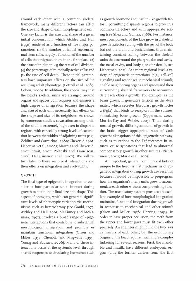
2. A second important mechanism of epi-genetic integration is angulation of the cranial base. As initially proposed by Bolk (1926), Weidenreich (1941), Biegert (1963), and Gould (1977), the basicranial
platform on which the brain sits can accommodate a larger brain relative to the length of the cranial base by being more flexed (Figure 16.6a). This spatial packing hypothesis has been tested most rigor-ously in primates (Figure 16.6b), which display a strong correlation across species between cranial base flexion and an index of brain size relative to cranial base length (Ross and Ravosa, 1993; McCarthy, 2001; Lieberman et al., 2000b). There is also some correlation among hominins be-tween ECV and cranial base flexion (Ross and Henneberg, 1995; Spoor, 1997). Cra-nial base flexion is measured most com-monly as cranial base angle 1 (CBA1), the angle in the sagittal plane between the line connecting basion and sella (the cen-ter of the hypophyseal fossa) and the line connecting sella to the foramen cecum (the most anteroinferior point on the cra-
1000900800700600500400300
1100120013001400150016001700
7 6 5 4 3 2 1 0
Time before present (millions of years)
End
ocra
nial
vol
ume
(cm
3 )
Au. afa
rens
is
Ar. ram
idus
Au. ae
thiop
icus
Au. af
rican
us
Au. bo
isei
Au. ga
rhi
Au. ro
bustu
s
H. ere
ctus
H. heid
elber
gens
is
H. nea
nder
thale
nsis
H. hab
ilis
H. rud
olphe
nsis
H. sap
iens
(Plei
stoce
ne)
H. sap
iens
(Holo
cene
)
Sahela
nthr
opus
1000900
800700
600
500
400
300
1100
1200
1300
1400
1500
1600
170018001900
End
ocra
nial
vol
ume
(cm
3 )
fIgurE 16.4 Endocranial vol-ume versus time (top) and by species (bottom). Note that major increases in volume did not really start until about 2 million years ago in Homo erectus. Data from Ruff et al. (1997) and Holloway et al. (2004).
i n t e g r a t i o n a n d e v o l v a b i l i t y o f t h e h e a d 283
nial base). In chimpanzees, CBA1 aver-ages 159°; in australopiths such as Australopithecus africanus, CBA1 is approx-imately 147°; and in Homo sapiens, it aver-ages 137° (Lieberman, 2011). However, the shift from less flexed to more flexed cra-nial bases does not correlate solely with brain volume because, as emphasized above, the cranial base is sandwiched be-tween the brain and face and, thus, has to accommodate variations in facial size. As shown by Lieberman and McCarthy (1999) for chimpanzees and by Lieber-man et al. (2008) for mice, longer and
more prognathic faces are also associated with a kind of reverse spatial packing, which causes the cranial base to extend (although possibly at different synchon-droses than where cranial base flexion oc-curs). The extent to which cranial base flexion and extension accommodate and integrate variations in both facial size and brain size aspect of the cranial base is also evident from variations in CBA1 among hominin species. Some australopiths such as Au. aethiopicus have very extended cranial bases (156°) because they have small brains (410 cc) combined with very
fIgurE 16.5 Vectors and pro-cesses of neurocranial and basi-cranial growth (modified from Lieberman et al., 2000a). Ten-sion in the dura mater gener-ated by the expansion of the brain induces one deposition with sutures and synchondro-ses. Brain expansion also in-duces drift (deposition on the outside combined with resorp-tion on the inside) in the basi-cranium and lower portion of the neurocranium. Note that the relative amount of growth in different vectors is constrained by the dural bands and by other factors that influence the shape and size of the basicranium.
284 e p i g e n e t i c s i n e v o l u t i o n a n d d i s e a s e
prognathic faces, but other australopiths such as Au. boisei have more flexed cranial bases (ca. 135°) because their brains are slightly larger (average 492 cc) and be-cause they have more orthognathic, ven-trally rotated faces designed to generate and withstand large masticatory forces (see Rak, 1983; Hylander, 1988; Kimbel et al., 2004). Similarly, there is a consid-erable variation in CBA1 within the genus Homo, with some species such as H. hei-delbergensis and H. neanderthalensis having fairly extended cranial bases (published values for CBA1 range 139°–150°) and some H. erectus crania with smaller faces having more flexed cranial bases (Baba et al., 2003; Lieberman, 2011).
3. A final aspect of epigenetic integration especially relevant to increased brain size in human evolution is skull width. Most mammals, including apes, have relatively long and narrow skulls; but increases in brain size in human evolution have been accommodated to a large extent by wider posterior, middle, and anterior cranial fos-sae (Weidenreich, 1941). Because the face grows downward/ventrally from the ante-rior cranial fossa and forward from the middle cranial fossa (see Enlow, 1990), a wider basicranium and neurocranium also leads to a wider face, particularly in the upper portions around the orbits but also in the middle and lower portions of the face (Lieberman et al., 2000a, 2004;
190
180
170
160
150
140
0.6 0.8 1.0 1.2 1.4 1.6
r2=0.65
H. sapiensCra
nial
bas
e A
ngle
1 (
°)
Index of Relative Encephalization (ECV0.33/cranial base length)
fIgurE 16.6 Top: Changes in the cranial base angle (CBA1) between a neonatal chimpanzee and a human (modified from Lieberman, 2011). Note that CBA1 extends in the chimpan-zee but flexes in the human be-cause of the combination of a relatively larger brain and a rela-tively shorter face. Bottom: The relationship between CBA1 and brain size relative to cranial base length (quantified as the index of relative encephaliza-tion) in primates (from McCarthy, 2001). Primates with relatively bigger brains have more flexed cranial bases, but humans are more flexed than predicted by the primate regres-sion. ECV, endocranial volume.

i n t e g r a t i o n a n d e v o l v a b i l i t y o f t h e h e a d 285
Bastir et al., 2008). These width increases and their effects on overall craniofacial in-tegration are probably accommodated by a number of other epigenetic mechanisms, which probably explains why width di-mensions are among the strongest sources of covariation in the mammalian skull (Hallgrimsson et al., 2007), includ-ing in humans (Polanski and Franciscus, 2006). For example, lateral expansion of the brain and posterior cranial fossa ap-pears to cause lateral (coronal) rotation of the adjoining petrous portions of the tem-poral (Dean, 1988; Spoor, 1997; but see Jeffery and Spoor, 2002). Note also that rotation of the petrous portion of the tem-poral also rotates the temporomandibular joints, helping to align the condyle and ramus of the mandible with the orienta-tion of the maxillary tooth row (bigger brains set up wider faces with wider max-illary arches that necessitate a more coro-nally oriented temporomandibular joint) (Lieberman, 2011).
Few mammals have evolved very big brains, probably because brain tissue is extremely metabolically expensive (Elia, 1992). However, highly encephalized mammals, such as homi-nins that have been able to pay for larger brains and benefit in terms of fitness, have also been able to accommodate larger brains without sub-stantial reengineering by using existing epigen-etic mechanisms that arise from how heads grow in the first place. Notably, the same mech-anisms that permit mammals to grow bigger brains during ontogeny without compromising functions such as mastication, vision, and olfac-tion have also been able to accommodate varia-tions in adult brain size among individuals and, hence, evolutionary shifts over time. Evidence for this accommodation is abundant in the per-formance of individuals with developmental problems that affect brain growth such as mi-crocephalus and hydrocephalus. Many humans with ECVs more than three standard deviations above and below the species mean can still hear,
chew, smile, and locomote effectively. Similar kinds of epigenetic mechanisms also help hu-mans of varying brain volume to function well in spite of wide variations in tooth size, eyeball size, face size, and so on. These, of course, are extreme cases, but they illustrate how learning more about the many mechanisms by which in-dividuals with these disorders do (and do not) adapt to variations, we will surely learn more about the mechanisms that permit skulls to be so variable and evolvable. In turn, unraveling the epigenetic mechanisms that make skulls so variable and evolvable has much potential for yielding generalizable insights into how epige-netic interactions are structured during devel-opment to permit mutual accommodation of organs, tissues, spaces, and skeletal structures.
In short, it has long been appreciated by de-velopmental biologists that epigenetic interac-tions are involved in many aspects of embryo-genesis, but I hope that the above examples illuminate how these sorts of general interac-tions, which involve various kinds of feedback mechanisms, are also vitally important through-out an organism’s ontogeny. In the case of the head, there is good reason to believe that the initial (apparently convoluted) way the head grows sets up the potential for many future in-teractions, thus permitting a wide range of structures that can function in many different ways. Also, as long as skeletal units grow in and around functional units, the many parts of the head can push and pull on each other to permit the head to function well in these different con-figurations, thus permitting selection to oper-ate. This general model, of course, needs much further testing, and many key details remain poorly studied and barely understood. In addi-tion, functional matrix-type models need to be applied to other complex aspects of phenotype elsewhere in the body. Yet, I am confident that this sort of research on the relationship be-tween complexity and evolvability will continue to provide excellent examples of why Wadding-ton’s original, broad definition of epigenetics is both necessary and useful. Although some bi-ologists are tempted to impose a simplifying,

286 e p i g e n e t i c s i n e v o l u t i o n a n d d i s e a s e
reductionist definition on the concept, without complex epigenetic processes neither biologists nor their heads would be here.
rEfErEnCEs
Alsbirk, P. H. 1977. Variation and heritability of ocu-lar dimensions. A population study among adult Greenland Eskimos. Acta Ophthalmol 55:443–56.
Antón, S. C. 1989. Intentional cranial vault deforma-tion and induced changes in the cranial base and face. Am J Phys Anthropol 79:253–67.
Atchley, W. R., and B. K. Hall. 1991. A model for development and evolution of complex morpho-logical structures. Biol Rev Camb Philos Soc 66: 101–57.
Baab, K. L., and K. P. McNulty. 2009. Size, shape, and asymmetry in fossil hominins: The status of the LB1 cranium based on 3D morphometric anal-yses. J Hum Evol doi:10.1016/j.jhevol.2008. 08.011.
Baba, H., F. Aziz, Y. Kaifu, G. Suwa, R. T. Kono, and T. Jacob. 2003. Homo erectus calvarium from the Pleistocene of Java. Science 299:1384–8.
Babineau, T. A., and J. H. Kronman. 1969. A cephalo-metric evaluation of the cranial base in micro-cephaly. Angle Orthod 39:57–63.
Bastir, M., A. Rosas, D. E. Lieberman, and P. O’Higgins. 2008. Middle cranial fossa anatomy and the origin of modern humans. Anat Rec 291:130–40.
Biegert, J. 1963. The evaluation of characters of the skull, hands and feet for primate taxonomy. In Classification and Human Evolution, ed. S. L. Washburn, 116–45. Chicago: Aldine de Gruyter.
Bolk, L. 1926. Das Problem der Menschwerdung. Jena, Germany: Gustav Fischer.
Bookstein, F. L., P. Gunz, P. Mitteroecker, H. Pros-singer, K. Schaefer, and H. Seidler. 2003. Cranial integration in Homo: Singular warps analysis of the midsagittal plane in ontogeny and evolution. J Hum Evol 44:167–87.
Carter, D. R., and G. S. Beaupré. 2001. Skeletal Func-tion and Form: Mechanobiology of Skeletal Develop-ment, Aging, and Regeneration. New York: Cam-bridge University Press.
Cerny, R., P. Lwigale, R. Ericsson, D. Meulemans, H. H. Epperlein, and M. Bronner-Fraser. 2004. Developmental origins and evolution of jaws: New interpretation of “maxillary” and “mandibu-lar.” Dev Biol 276:225–36.
Chernoff, B., and P. M. Magwene. 1999. Morphologi-cal integration: Forty years later. In Morphological Integration, 319–53. Chicago: University of Chi-cago Press.
Cheverud, J. M. 1995. Morphological integration in the saddle-back tamarin (Saguinus fuscicollis) cra-nium. Am Nat 145:63–89.
Cheverud, J. M., L. A. Kohn, L. W. Konigsberg, and S. R. Leigh. 1992. Effects of fronto-occipital artifi-cial cranial vault modification on the cranial base and face. Am J Phys Anthropol 88:323–45.
Cohen, M. M., Jr. 2000. Sutural biology. In Cranio-synostosis: Diagnosis, Evaluation and Management, ed. M. M. Cohen and R. E. MacLean, 11–23. New York: Oxford University Press.
Cottrill, C. P., C. W. Archer, and L. Wolpert. 1987. Cell sorting and aggregrate formation in micro-mass culture. Dev Biol 122:503–15.
Currey, J. D. 2002. Bones: Structure and Mechanics. Princeton, NJ: Princeton University Press.
Dean, M. C. 1988. Growth processes in the cranial base of hominoids and their bearing on morpho-logical similarities that exist in the cranial base of Homo and Paranthropus. In Evolutionary History of the “Robust” Australopithecines, ed. F. E. Grine, 107–12. New York: Aldine de Gruyter.
Depew, M. J., T. Lufkin, and J. R. L. Rubenstein. 2002. Specification of jaw subdivision by Dlx genes. Science 298:381–85.
Depew, M. J., C. A. Simpson, M. Morasso, and J. L. Rubenstein. 2005. Reassessing the Dlx code: The genetic regulation of branchial arch skeletal pat-tern and development. J. Anat 207:501–61.
Deschamps, J., and J. van Nes. 2005. Developmental regulation of the Hox genes during axial morpho-genesis in the mouse. Development 132:2931–42
Duterloo, H. S., and D. H. Enlow. 1970. A compara-tive study of cranial growth in Homo and Macaca. Am J Anat 127:357–68.
Elia, M. 1992. Organ and tissue contribution to meta-bolic weight. In Energy Metabolism: Tissue Deter-minants and Cellular Corollaries, ed. J. M. Kinney and H. N. Tucker, 61–79. New York: Raven Press.
Enlow, D. H. 1990. Facial Growth, 3rd ed. Philadel-phia: Saunders.
Friede, H. 1981. Normal development and growth of the human neurocranium and cranial base. Scand J Plast Reconstr Surg 115:163–9.
Gould, S. J. 1977. Ontogeny and Phylogeny. Cam-bridge, MA: Belknap Press.
Haig, D. 2004. The (dual) origin of epigenetics. Cold Spring Harb Symp Quant Biol 69:1–4.
Hall, B. K. 2005. Bones and Cartilage: Developmental and Evolutionary Skeletal Biology. Amsterdam: Elsevier.
Hallgrímsson, B., D. E. Lieberman, W. Lie, A. F. Ford-Hutchinson, and F. R. Jirik. 2007. Epigenetic in-teractions and the structure of phenotypic varia-tion in the skull. Evol Dev 9:76–91.

i n t e g r a t i o n a n d e v o l v a b i l i t y o f t h e h e a d 287
Harris, E. F., and M. G. Johnson. 1991. Heritability of craniometric and occlusal variables: A longitudi-nal sib analysis. Am J Orthod Dentofacial Orthop 99:258–68.
Herring, S. W. 1993. Epigenetic and functional influ-ences on skull growth. In The Skull, ed. J. Hanken and B. K. Hall, 237–71. Chicago: University of Chi-cago Press.
Holloway, R. L., D. C. Broadfield, M. S. Yuan, J. H. Schwartz, and I. Tattersall. 2004. The Human Fossil Record. Vol. 3 of Brain Endocasts—The Paleo-neurological Evidence. New York: Wiley.
Hunter, W. S. 1990. Heredity in the craniofacial com-plex. In Facial Growth, 3rd ed., ed. D. H. Enlow, 249–66. Philadelphia: Saunders.
Hylander, W. L. 1988. Implications of in vivo experi-ments for interpreting the functional signifi-cance of “robust” australopithecine jaws. In Evolutionary History of the “Robust” Australopithe-cines, ed. F. Grine, 55–83. New York: Aldine de Gruyter.
Jeffery, N. 2002. Differential regional brain growth and rotation of the prenatal human tentorium cerebelli. J Anat 200:135–44.
Jeffery, N., and F. Spoor. 2002. Brain size and the human cranial base: A prenatal perspective. Am J Phys Anthropol 118:324–40.
Jernvall, J., and I. Thesleff. 2000. Reiterative sig-naling and patterning during mammalian tooth morphogenesis. Mech Dev 92:19–29.
Jiang, X., S. Iseki, R. E. Maxson, H. M. Sucov, and G. M. Morriss-Kay. 2002. Tissue origins and in-teractions in the mammalian skull vault. Dev Biol 241:106–16.
Kimbel, W. H., Y. Rak, and D. C. Johanson. 2004. The Skull of Australopithecus afarensis. Oxford: Ox-ford University Press.
Kirschner, M. W., and J. C. Gerhart. 2005. The Plausi-bility of Life. New Haven, CT: Yale University Press.
Klingenberg, C. P. 2008. Morphological integration and developmental modularity. Annu Rev Ecol Evol Syst 39:115–32.
Lee, S. H., O. Bédard, M. Buchtová, K. Fu, and J. M. Richman. 2004. A new origin for the maxillary jaw. Dev Biol 276:207–24.
Lieberman, D. E. 2007. Homing in on early Homo. Nature 449:291–2.
Lieberman, D. E. 2011. The Evolution of the Human Head. Cambridge, MA: Harvard University Press.
Lieberman, D. E., B. Hallgrímsson, W. Liu, T. A. Par-sons, and H. A. Jamniczky. 2008. Spatial pack-ing, cranial base angulation, and craniofacial shape variation in the mammalian skull: Testing a new model using mice. J Anat 212:720–35.
Lieberman, D. E., G. E. Krovitz, and B. McBratney-Owen. 2004. Testing hypotheses about tinkering in the fossil record: The case of the human skull. J Exp Zoolog B Mol Dev Evol 302:302–21.
Lieberman, D. E., and R. C. McCarthy. 1999. The on-togeny of cranial base angulation in humans and chimpanzees and its implications for reconstruct-ing pharyngeal dimensions. J Hum Evol 36:487–517.
Lieberman, D. E., K. M. Mowbray, and O. M. Pearson. 2000a. Basicranial influences on overall cranial shape. J Hum Evol 38:291–315.
Lieberman, D. E., C. F. Ross, and M. J. Ravosa. 2000b. The primate cranial base: Ontogeny, function and integration. Ybk J Phys Anthropol 43:117–69.
Lordkipanidze, D., T. Jashashvili, A. Vekua, M. A. Ponce de León, C. P. Zollikofer, G. P. Rightmire, H. Pontzer, et al. 2007. Postcranial evidence from early Homo from Dmanisi, Georgia. Nature 449:305–10.
Marazita, M. L., and M. Mooney. 2004. Current con-cepts in the embryology and genetics of cleft lip and cleft palate. Clin Plast Surg 31:125–40.
Marcucio, R. S., D. R. Cordero, D. Hu, and J. A. Helms. 2005. Molecular interactions coordinat-ing the development of the forebrain and face. Dev Biol 284:48–61.
Marie, P. J., J. D. Coffin, and M. M. Hurley. 2005. FGF and FGFR signaling in chondrodys-plasias and craniosynostosis. J Cell Biochem 96:888–96.
Marroig, G., and J. Cheverud. 2001. A comparison of phenotypic variation and covariation patterns and the role of phylogeny, ecology and ontogeny dur-ing cranial evolution of New World monkeys. Evo-lution 55:2576–600.
McBratney-Owen, B., S. Iseki, S. D. Bamforth, B. R. Olsen, and G. M. Morriss-Kay. 2008. Develop-ment and tissue origins of the mammalian cra-nial base. Dev Biol 322:121–32.
McCarthy, R. C. 2001. Anthropoid cranial base archi-tecture and scaling relationships. J Hum Evol 40:41–66.
McCarthy, R. C. 2004. Constraints and primate cra-niofacial growth and form. PhD diss., George Washington University.
McGrath, J., O. El-Saadi, V. Grim, S. Cardy, B. Chaple, D. Chant, D. E. Lieberman, and B. Mowry. 2002. Minor physical anomalies and quantitative measures of the head in psychosis. Arch Gen Psy-chiatry 59:458–64.
McKinney, M. L., and K. J. McNamara. 1991. Heter-ochrony: The Evolution of Ontogeny. New York: Ple-num Press.

288 e p i g e n e t i c s i n e v o l u t i o n a n d d i s e a s e
Mina, M. 2001. Regulation of mandibular growth and morphogenesis. Crit Rev Oral Biol Med 12:276–300.
Morriss-Kay, G. M., and A. O. Wilkie. 2005. Growth of the normal skull vault and its alteration in cra-niosynostosis: Insights from human genetics and experimental studies. J Anat 207:637–53.
Moss, M. L. 1968. The primacy of functional matrices in orofacial growth. Dent Practitioner 19:63–73.
Moss, M. L. 1997a. The functional matrix hypothesis revisited. 1. The role of mechanotransduction. Am J Orthod Dentofacial Orthop 112:8–11.
Moss, M. L. 1997b. The functional matrix hyopothe-sis revisited. 2. The role of an osseous connected cellular network. Am J Orthod Dentofacial Orthop 112:221–6.
Moss, M. L. 1997c. The functional matrix hypothesis revisited. 3. The genomic thesis. Am J Orthod Dentofacial Orthop 112:338-342.
Moss, M. L. 1997d. The functional matrix hypothesis revisited. 4. The epigenetic antithesis and the re-solving synthesis. Am J Orthod Dentofacial Orthop 112:410–14.
Moss, M. L., and R. W. Young. 1960. A functional ap-proach to craniology. Am J Phys Anthropol 18:281–92.
Moyers, R. L. 1988. Handbook of Orthodontics, 4th ed. Chicago: Yearbook Medical Publishers.
Olson, E. R., and R. L. Miller. 1958. Morphological In-tegration. Chicago: University of Chicago Press.
Opperman, L. A. 2000. Cranial sutures as intramem-branous bone growth sites. Dev Dyn 219:472–85.
Parsons, T. E., E. Kristensen, L. Hornung, V. M. Diewert, S. K. Boyd, R. Z. German, and B. Hall-grímsson. 2008. Phenotypic variability and cra-niofacial dysmorphology: increased shape vari-ance in a mouse model for cleft lip. J Ana. 135–43.
Peters, H., and R. Balling. 1999. Teeth. Where and how to make them. Trends Genet 15:59–65.
Polanski, J. M., and R. G. Franciscus. 2006. Patterns of craniofacial integration in extant Homo, Pan, and Gorilla. Am J Phys Anthropol 75:195–96.
Radlanski, R. J., and H. Renz. 2006. Genes and forces and forms. Mechanical aspects during prenatal craniofacial development. Dev Dyn 235:1219–29.
Rak, Y. 1983. The Australopithecine Face. New York: Academic Press.
Richtsmeier, J. T. 2002. Cranial vault morphology and growth in craniosynostoses. In Understanding Craniofacial Anomalies: The Etiopathogenesis of Craniosynostoses and Facial Clefting, ed. M. P. Mooney and M. I. Siegel, 321–41. New York: Wiley-Liss.
Richtsmeier, J. T., K. Aldridge, V. B. DeLeon, J. Pan-chal, A. A. Kane, J. L. Marsh, P. Yan, and T. M. Cole III 2006. Phenotypic integration of neuro-cranium and brain. J Exp Zoolog B Mol Dev Evol 306:360–78.
Rightmire, G. P. 2004. Brain size and encephaliza-tion in early to mid-Pleistocene Homo. Am J Phys Anthropol 124:109–23.
Rogers, J., P. Kochunov, J. Lancaster, W. Shelledy, D. Glahn, J. Blangero, and P. Fox. 2007. Heritability of brain volume, surface area and shape: An MRI study in an extended pedigree of baboons. Hum Brain Mapp 28:576–83.
Rönning, H. J. 1995. Growth of the cranial vault: In-fluence of intracranial and extracranial pressures. Acta Odontol Scand 53:192–5.
Ross, C. F., and M. Henneberg. 1995. Basicranial flexion, relative brain size, and facial kyphosis in Homo sapiens and some fossil hominids. Am J Phys Anthropol 98:575–93.
Ross, C. F., and M. J. Ravosa. 1993. Basicranial flex-ion, relative brain size, and facial kyphosis in non-human primates. Am J Phys Anthropol 91:305–24.
Ruff, C. B., E. Trinkaus, and T. W. Holliday. 1997. Body mass and encephalization in Pleistocene Homo. Nature 387:173–6.
Sarnat, B. G. 1982. Eye and orbital size in the young and adult. Some postnatal experimental and clini-cal relationships. Ophthalmologica 185:74–8.
Schowing, J. 1968. Demonstration of the inductive role of the brain in osteogenesis of the embryonic skull of the chicken. J Embryol Exp Morphol 19:83–94.
Shea, B. T., and A. M. Gomez. 1988. Tooth scaling and evolutionary dwarfism: An investigation of al-lometry in human pygmies. Am J Phys Anthropol 77:117–32.
Sperber, G. 2001. Craniofacial Development. Hamil-ton, Canada: B. C. Decker.
Spoor, C. F. 1997. Basicranial architecture and rela-tive brain size of Sts 5 (Australopithecus africanus) and other Plio-Pleistocene hominids. S Afr J Sci 93:182–6.
Strait, D. S. 2001. Integration, phylogeny, and the hominid cranial base. Am J Phys Anthropol 114: 273–97.
van der Klaauw, C. 1948–1952. Size and position of the functional components of the skull. Arch Neerland Zool 9:1–559.
Wagner, G. P. 1984. On the eigenvalue distribution of genetic and phenotypic dispersion matrices—evi-dence for a nonrandom organization of quantita-tive character variation. J Math Biol 21:77–95.
Wagner, G. P. 2001. The Character Concept in Evolu-tionary Biology. New York: Academic Press.

i n t e g r a t i o n a n d e v o l v a b i l i t y o f t h e h e a d 289
Watson, W. G. 1982. The functional matrix revisited. Am J Orthod 81:71–3.
Weidenreich, F. 1941. The brain and is rôle in the phylogenetic transformation of the human skull. Trans Am Philos Soc 31:328–442.
West-Eberhard, M. J. 2003. Developmental Plasticity and Evolution. Oxford: Oxford University Press.
Wilkie, A. O., and G. M. Morriss-Kay. 2001. Genetics of craniofacial development and malformation. Nat Rev Genet 2:458–68.
Wilkins, A. S. 2002. The Evolution of Developmental Pathways. Sunderland, MA: Sinauer Associates.
Young, N. M., S. Wat, V. M. Diewert, L. W. Browder, and B. Hallgrímsson. 2007. Comparative mor-phometrics of embryonic facial morphogenesis: implications for cleft-lip etiology. Anat Rec 290:123–39.
Young, R. L., and A. V. Badyaev. 2006. Evolutionary persistence of phenotypic integration: Influence of developmental and functional relationships on complex trait evolution. Evolution 60:1291–9.
Zelditch, M. L., and A. C. Carmichael. 1989. Ontoge-netic variation in patterns of developmental and functional integration in skulls of Sigmodon fulvi-venter. J Mammal 70:477–84.