epilepsy & behavior - repositorio.ufrn.br filebehavioral and eeg effects of gabaergic...
TRANSCRIPT
Epilepsy & Behavior 24 (2012) 391–398
Contents lists available at SciVerse ScienceDirect
Epilepsy & Behavior
j ourna l homepage: www.e lsev ie r .com/ locate /yebeh
Behavioral and EEG effects of GABAergic manipulation of the nigro-tectal pathway inthe Wistar audiogenic rat (WAR) strain II: An EEG wavelet analysis and retrogradeneuronal tracer approach
Franco Rossetti a,b,1, Marcelo Cairrão Araújo Rodrigues b,2, Simone S. Marroni a,b, Artur Fernandes b,Maira Licia Foresti a,b, Rodrigo N. Romcy-Pereira a,3,Dráulio Barros de Araújo a,c,3, Norberto Garcia-Cairasco a,b,⁎a Neuroscience and Behavioral Sciences Department, Ribeirão Preto School of Medicine, University of São Paulo, Ribeirão Preto, São Paulo, Brazilb Physiology Department, Ribeirão Preto School of Medicine, University of São Paulo, Ribeirão Preto, São Paulo, Brazilc Physics and Mathematics Department of the School of Philosophy, Sciences and Letters, University of São Paulo, Ribeirão Preto, São Paulo, Brazil
⁎ Corresponding author at: Neurophysiology andLaboratory, University of São Paulo, USP, Ribeirão PBandeirantes, 3900 Ribeirão Preto, SP 14049‐900, Braz
E-mail address: [email protected] (N. Garcia-Cai1 Walter Reed Army Institute of Research, Silver Sprin2 Physiology and Pharmacology Department, Feder
Recife, Brazil.3 Brain Institute, Federal University of Rio Grande do
1525-5050 © 2012 Elsevier Inc.doi:10.1016/j.yebeh.2012.04.133
Open access under the Elsevier
a b s t r a c t
a r t i c l e i n f oArticle history:Received 28 March 2012Accepted 21 April 2012Available online 16 June 2012
Keywords:Audiogenic seizuresSustained EEG oscillationBicucullineMuscimolFluorogold
The role of the substantia nigra pars reticulata (SNPr) and superior colliculus (SC) network in rat strains sus-ceptible to audiogenic seizures still remain underexplored in epileptology. In a previous study from our lab-oratory, the GABAergic drugs bicuculline (BIC) and muscimol (MUS) were microinjected into the deep layersof either the anterior SC (aSC) or the posterior SC (pSC) in animals of the Wistar audiogenic rat (WAR) strainsubmitted to acoustic stimulation, in which simultaneous electroencephalographic (EEG) recording of theaSC, pSC, SNPr and striatum was performed. Only MUS microinjected into the pSC blocked audiogenic sei-zures. In the present study, we expanded upon these previous results using the retrograde tracer Fluorogold(FG) microinjected into the aSC and pSC in conjunction with quantitative EEG analysis (wavelet transform),in the search for mechanisms associated with the susceptibility of this inbred strain to acoustic stimulation.Our hypothesis was that the WAR strain would have different connectivity between specific subareas of thesuperior colliculus and the SNPr when compared with resistant Wistar animals and that these connectionswould lead to altered behavior of this network during audiogenic seizures. Wavelet analysis showed thatthe only treatment with an anticonvulsant effect was MUS microinjected into the pSC region, and this treat-ment induced a sustained oscillation in the theta band only in the SNPr and in the pSC. These data suggestthat in WAR animals, there are at least two subcortical loops and that the one involved in audiogenic seizuresusceptibility appears to be the pSC-SNPr circuit. We also found that WARs presented an increase in the num-ber of FG+ projections from the posterior SNPr to both the aSC and pSC (primarily to the pSC), with both act-ing as proconvulsant nuclei when compared with Wistar rats. We concluded that these two differentsubcortical loops within the basal ganglia are probably a consequence of the WAR genetic background.
© 2012 Elsevier Inc. Open access under the Elsevier OA license.
1. Introduction
The Wistar audiogenic rat (WAR) is an inbred rodent strain sus-ceptible to audiogenic seizures and derived from Wistar rats [1–3].The audiogenic seizures are characterized by an initial period ofwild running, jumping and atonic falling episodes followed bytonic-clonic seizures (opisthotonus plus forelimb and hindlimb tonic
Experimental Neuroethologyreto School of Medicine, Av.il. Fax: +55 16 3633 0017.rasco).g, MD, USA.al University of Pernambuco,
Norte, Natal, Brazil.
OA license.
hyperextensions and clonic convulsions) ending with apnea andpostictal depression [2–4].
Several studies of audiogenic strains appear in the literature, butthe roles of the network formed by substantia nigra pars reticulata(SNPr) and superior colliculus (SC) still remain underexplored. Thecharacterization of these networks is important for epileptology.When these studies are conducted in different strains, the resultswould show the effects of genetic variability on the circuitry affectingepilepsy. In situ hybridization studies for c-Fos mRNA showed in-creased labeling of the deep SC of genetically epilepsy-prone rats(GEPRs), suggesting the involvement of the deep SC in the propaga-tion of seizures [5]. Additionally, there is a rapid burst firing of neu-rons in the deep SC layers in freely moving GEPRs, suggesting thatthis structure plays a key role in triggering the wild running behavior[6]. Strasbourg audiogenic rats have a 40% reduction of GABAergic

Table 1SI with behavioral descriptions according to Garcia-Cairasco et al. [3], categorized intodiscreet variables for statistical purposes by Rossetti et al. (cSI; [20]).
SI Seizure behaviors cSI
0.00 No seizures 00.11 One running 10.23 One wild running (running plus jumping plus atonic falling) (WR1) 20.38 Two wild runnings (WR2) 30.61 Tonic convulsion (opisthotonus) 40.85 Tonic seizures plus generalized clonic convulsions (TS) 50.90 Head ventral flexion plus cSI 5 60.95 Forelimb hyperextension plus cSI 6a (FH) 71.00 Hindlimb hyperextension plus cSI 7a (HH) 8
a Categories that are generally followed by hindlimb clonic convulsions (HCC).
392 F. Rossetti et al. / Epilepsy & Behavior 24 (2012) 391–398
receptors in the SNPr [7], and additional studies of GEPR animalsshowed that there is a lower release of GABA (stimulated by highpotassium concentration) in the SNPr compared with the releaseobserved in non-audiogenic control rats [1]. The geneticallyepilepsy-prone hamster (GPG/Vall) presents morphological andneurochemical abnormalities in the deep SC layers [8]. In WARs,sectioning of the unilateral or bilateral subcollicular deep SC layersignificantly reduced audiogenic seizure severity. When the sec-tioning was performed bilaterally, the latency for running episodes,when present, was increased [9].
Neurophysiological measurements and behavioral responses havebeen used with success in investigating the neural networks underly-ing many seizure behaviors [10,11]. However, these results are de-pendent on the quantitative analytical methods used to extract theinformation from the experimental data [12]. Synchronous oscillatoryactivity is important in coordinating the firing of distributed neuronalpopulations during behavioral coding. Specific oscillatory patterns arealtered in several neurological disorders, revealing abnormal networkfunctioning [13]. The sustained aspects of these oscillations (theirmaintenance in time) are important, allowing us to correlate behaviorwith synchronization in characteristic frequency bands over time.
In our previous study [14], the GABAergic drugs bicuculline (BIC;GABAA antagonist) and muscimol (MUS; GABAA agonist) were micro-injected into the deep layers of either the anterior region of SC (aSC)or the posterior part of SC (pSC) in WARs submitted to acoustic stim-ulation with simultaneous EEG recording of the aSC, pSC, SNPr andstriatum. Only MUSmicroinjected in pSC blocked audiogenic seizures,following the same protocol of microinjections made in GEPRs byMerril et al. [15]. In the current study, we used quantitative analysis(wavelet transform) to study the EEG before injection (basal period),after microinjection of BIC and MUS into the deep layers of SC, duringthe adaptation period and during the audiogenic seizure period inWARs.
All WARs described in this study are those used in our previouspaper [14]. In the current study, a new group was added and micro-injected with the retrograde tracer Fluorogold (FG) to study the neu-roanatomical connections comparing the SC subareas (anterior andposterior) and the SNPr between WARs and Wistar rats. In theserats, FG was microinjected into the aSC and pSC to analyze the projec-tions from the anterior region of the SNPr (aSNPr) and posteriorportion of the SNPr (pSNPr). These specific subregions of the SNPrhave been respectively implicated in anticonvulsant (aSNPr) andproconvulsant (pSNPr) effects in several epilepsy models in normalrats [16–19].
Our hypothesis was that theWAR strain would have different con-nections between the aSC/pSC and the SNPr when compared with theresistant Wistar rat controls. If present, these differential connectionswould lead to different convulsant or anticonvulsant actions of thisnetwork during the expression of audiogenic seizures.
2. Methods
2.1. GABAergic drug groups
All animals and experimental procedures in these groups were thesame as those used in our previous work [14]. Briefly, for additionaldetails on the EEG experimental protocol used in Section 2.1.1 of the cur-rent study, see Rossetti et al. [14]. For the quantitative EEG evaluation(wavelet analyses) described in Section 2.1.2 of the current study, weused the same EEG recordings reported in Rossetti et al. [14].
2.1.1. Experimental procedures [14]Fifteen male WARs (250–300 g) received phosphate buffer (PBS),
BIC and MUS (Tocris; Westwoods Business Park, Ellisville, MO, USA)microinjections into the aSC (n=6; aSC group) or pSC (n=9; pSCgroup).
Prior to any manipulations, all WARs were tested for audiogenic sei-zures using three acoustic stimuli that were done 48 h apart (screeningtest). The experimental groups comprised animals that experienced a sei-zure with a categorized mesencephalic seizure severity index (cSI,Table 1) of at least 4 in at least one of the three screening tests. Behavioralevaluationswere performed in accordancewith the seizure severity indexdeveloped by Garcia-Cairasco et al. [3] andmodified by Rossetti et al. [20].
Three days after the screening test, the animals were stereotaxical-ly implanted with two twisted monopolar Teflon-coated electrodes(0.008″; AM System; Carlsborg, USA), which were placed in the stria-tum (anterior–posterior (AP)=3.0 mm; lateral (L)=+2.3 mm; ven-tral (V)=−5.0 mm) and SNPr (AP=−5.4 mm; L=+2.3 mm; V=−8.0 mm). One chemitrode, an electrode coupled with a cannula(for simultaneous recording and drug microinjections), was im-planted in the aSC (AP=−6.4 mm; L=+1.5 mm; V=−5.0 mm) orpSC (AP=−7.3 mm; L=+1.5 mm; V=−4.47 mm; Â=28° refersto the sagittal plane). All coordinates were obtained from the atlas ofPaxinos and Watson [21] and are relative to the bregma suture.
Seven days after the surgery, the animals were unilaterally micro-injected with 200 nl of PBS (50 mM, pH=7.4), stimulated with soundand video-EEG recorded. After 48 h, the animals were unilaterallymicroinjected with 200 nl of BIC (0.1 nmol, according to Merrill etal. [15]) and then stimulated with sound and video-EEG recorded.The animals were unilaterally microinjected with 200 nl of PBS andthen stimulated with sound and video-EEG recorded 48 h later.After an additional 48‐h period, the animals were unilaterally micro-injected with 200 nl of MUS (1.75 nmol, according to Rossetti et al.[20]) and then stimulated with sound and video-EEG recorded. Allthe EEG recording procedures were conducted according to Dutra Mo-raes et al. [22] and are detailed in Rossetti et al. [14].
At the end of the experiments, the animals were perfused with70 ml of PBS (50 mM, pH 7.4) followed by 300 ml of paraformalde-hyde/PBS (4%, pH 7.4). The brains were removed and post-fixed in4% paraformaldehyde for 1 h, cryoprotected in 20% sucrose for 24 hat 4 °C, frozen in isopentane and sliced into 40-μm-thick coronal sec-tions on a cryostat (Micron-Zeiss HM-505-E; Walldorf, Germany).The sections were mounted on gelatin-subbed glass slides and storedat−20 °C until Nissl staining was used to reveal the correct electrodeand chemitrode placement.
2.1.2. Quantitative EEG evaluationTo evaluate the effects of drugs tested in WARs, a time–frequency
spectral analysis using wavelet transform [12,20] was performed onthe EEG traces. EEG tracing is a method developed for the analysisof non-stationary signals by separating the EEG time-series into itstemporal and spectral components. This method has already beenused in several studies of the correlation of EEG oscillatory patternsand behavioral responses [23–25].
We analyzed frequencies between 3 and 20 Hz during the basalperiod (6 min prior to any manipulation), adaptation period (6 minprior to acoustic stimulation) and audiogenic seizure period.
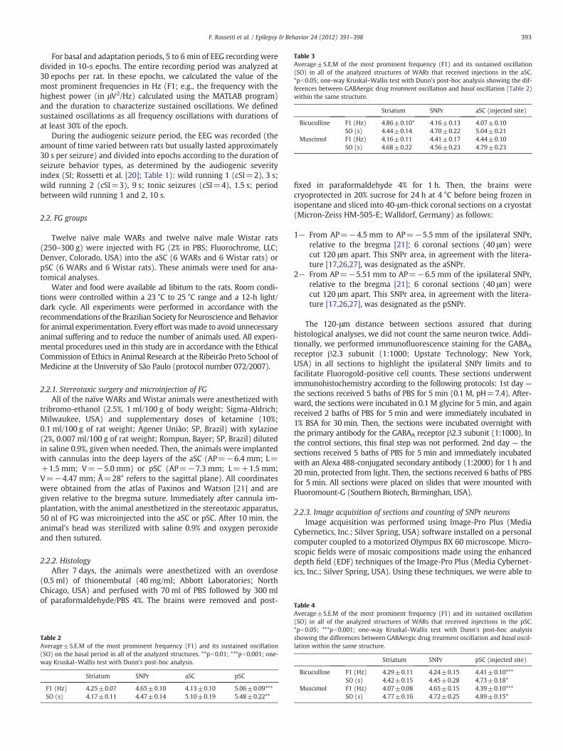
Table 3Average±S.E.M of the most prominent frequency (F1) and its sustained oscillation(SO) in all of the analyzed structures of WARs that received injections in the aSC.*pb0.05; one-way Kruskal–Wallis test with Dunn's post-hoc analysis showing the dif-ferences between GABAergic drug treatment oscillation and basal oscillation (Table 2)within the same structure.
Striatum SNPr aSC (injected site)
Bicuculline F1 (Hz) 4.86±0.10* 4.16±0.13 4.07±0.10SO (s) 4.44±0.14 4.70±0.22 5.04±0.21
Muscimol F1 (Hz) 4.16±0.11 4.41±0.17 4.44±0.10SO (s) 4.68±0.22 4.56±0.23 4.79±0.23
393F. Rossetti et al. / Epilepsy & Behavior 24 (2012) 391–398
For basal and adaptation periods, 5 to 6 min of EEG recording weredivided in 10-s epochs. The entire recording period was analyzed at30 epochs per rat. In these epochs, we calculated the value of themost prominent frequencies in Hz (F1; e.g., the frequency with thehighest power (in μV2/Hz) calculated using the MATLAB program)and the duration to characterize sustained oscillations. We definedsustained oscillations as all frequency oscillations with durations ofat least 30% of the epoch.
During the audiogenic seizure period, the EEG was recorded (theamount of time varied between rats but usually lasted approximately30 s per seizure) and divided into epochs according to the duration ofseizure behavior types, as determined by the audiogenic severityindex (SI; Rossetti et al. [20]; Table 1): wild running 1 (cSI=2), 3 s;wild running 2 (cSI=3), 9 s; tonic seizures (cSI=4), 1.5 s; periodbetween wild running 1 and 2, 10 s.
2.2. FG groups
Twelve naïve male WARs and twelve naïve male Wistar rats(250–300 g) were injected with FG (2% in PBS; Fluorochrome, LLC;Denver, Colorado, USA) into the aSC (6 WARs and 6 Wistar rats) orpSC (6 WARs and 6 Wistar rats). These animals were used for ana-tomical analyses.
Water and food were available ad libitum to the rats. Room condi-tions were controlled within a 23 °C to 25 °C range and a 12-h light/dark cycle. All experiments were performed in accordance with therecommendations of the Brazilian Society for Neuroscience and Behaviorfor animal experimentation. Every effort wasmade to avoid unnecessaryanimal suffering and to reduce the number of animals used. All experi-mental procedures used in this study are in accordance with the EthicalCommission of Ethics in Animal Research at the Ribeirão Preto School ofMedicine at the University of São Paulo (protocol number 072/2007).
2.2.1. Stereotaxic surgery and microinjection of FGAll of the naïve WARs and Wistar animals were anesthetized with
tribromo-ethanol (2.5%, 1 ml/100 g of body weight; Sigma-Aldrich;Milwaukee, USA) and supplementary doses of ketamine (10%;0.1 ml/100 g of rat weight; Agener União; SP, Brazil) with xylazine(2%, 0.007 ml/100 g of rat weight; Rompun, Bayer; SP, Brazil) dilutedin saline 0.9%, given when needed. Then, the animals were implantedwith cannulas into the deep layers of the aSC (AP=−6.4 mm; L=+1.5 mm; V=−5.0 mm) or pSC (AP=−7.3 mm; L=+1.5 mm;V=−4.47 mm; Â=28° refers to the sagittal plane). All coordinateswere obtained from the atlas of Paxinos and Watson [21] and aregiven relative to the bregma suture. Immediately after cannula im-plantation, with the animal anesthetized in the stereotaxic apparatus,50 nl of FG was microinjected into the aSC or pSC. After 10 min, theanimal's head was sterilized with saline 0.9% and oxygen peroxideand then sutured.
2.2.2. HistologyAfter 7 days, the animals were anesthetized with an overdose
(0.5 ml) of thionembutal (40 mg/ml; Abbott Laboratories; NorthChicago, USA) and perfused with 70 ml of PBS followed by 300 mlof paraformaldehyde/PBS 4%. The brains were removed and post-
Table 2Average±S.E.M of the most prominent frequency (F1) and its sustained oscillation(SO) on the basal period in all of the analyzed structures. **pb0.01; ***pb0.001; one-way Kruskal–Wallis test with Dunn's post-hoc analysis.
Striatum SNPr aSC pSC
F1 (Hz) 4.25±0.07 4.65±0.10 4.13±0.10 5.06±0.09***SO (s) 4.17±0.11 4.47±0.14 5.10±0.19 5.48±0.22**
fixed in paraformaldehyde 4% for 1 h. Then, the brains werecryoprotected in 20% sucrose for 24 h at 4 °C before being frozen inisopentane and sliced into 40-μm-thick coronal sections on a cryostat(Micron-Zeiss HM-505-E; Walldorf, Germany) as follows:
1− From AP=−4.5 mm to AP=−5.5 mm of the ipsilateral SNPr,relative to the bregma [21]; 6 coronal sections (40 μm) werecut 120 μm apart. This SNPr area, in agreement with the litera-ture [17,26,27], was designated as the aSNPr.
2− From AP=−5.51 mm to AP=−6.5 mm of the ipsilateral SNPr,relative to the bregma [21]; 6 coronal sections (40 μm) werecut 120 μm apart. This SNPr area, in agreement with the litera-ture [17,26,27], was designated as the pSNPr.
The 120-μm distance between sections assured that duringhistological analyses, we did not count the same neuron twice. Addi-tionally, we performed immunofluorescence staining for the GABAA
receptor β2.3 subunit (1:1000; Upstate Technology; New York,USA) in all sections to highlight the ipsilateral SNPr limits and tofacilitate Fluorogold-positive cell counts. These sections underwentimmunohistochemistry according to the following protocols: 1st day —
the sections received 5 baths of PBS for 5 min (0.1 M, pH=7.4). After-ward, the sections were incubated in 0.1 M glycine for 5 min, and againreceived 2 baths of PBS for 5 min and were immediately incubated in1% BSA for 30 min. Then, the sections were incubated overnight withthe primary antibody for the GABAA receptor β2.3 subunit (1:1000). Inthe control sections, this final step was not performed. 2nd day — thesections received 5 baths of PBS for 5 min and immediately incubatedwith an Alexa 488-conjugated secondary antibody (1:2000) for 1 h and20 min, protected from light. Then, the sections received 6 baths of PBSfor 5 min. All sections were placed on slides that were mounted withFluoromount-G (Southern Biotech, Birminghan, USA).
2.2.3. Image acquisition of sections and counting of SNPr neuronsImage acquisition was performed using Image-Pro Plus (Media
Cybernetics, Inc.; Silver Spring, USA) software installed on a personalcomputer coupled to a motorized Olympus BX 60 microscope. Micro-scopic fields were of mosaic compositions made using the enhanceddepth field (EDF) techniques of the Image-Pro Plus (Media Cybernet-ics, Inc.; Silver Spring, USA). Using these techniques, we were able to
Table 4Average±S.E.M of the most prominent frequency (F1) and its sustained oscillation(SO) in all of the analyzed structures of WARs that received injections in the pSC.*pb0.05; ***pb0.001; one-way Kruskal–Wallis test with Dunn's post-hoc analysisshowing the differences between GABAergic drug treatment oscillation and basal oscil-lation within the same structure.
Striatum SNPr pSC (injected site)
Bicuculline F1 (Hz) 4.29±0.11 4.24±0.15 4.41±0.10***SO (s) 4.42±0.15 4.45±0.28 4.73±0.18*
Muscimol F1 (Hz) 4.07±0.08 4.65±0.15 4.39±0.10***SO (s) 4.77±0.16 4.72±0.25 4.89±0.15*

394 F. Rossetti et al. / Epilepsy & Behavior 24 (2012) 391–398
count the neurons in detail because each piece of the mosaic in thepanoramic view could be observed with high magnitude (ex: 10×)and good resolution (see Fig. 3A). Moreover, we were able to conductthree-dimensional cell counting through the entire slice.
We used the mosaic images to count FG-positive neurons in theanterior and posterior SNPr into the nigral network. This work wasperformed with the aid of the manual tag technique (Image Pro-Plus) and Adobe Photoshop CS3 (Adobe System Incorporated, USA).
2.3. Statistical analyses
For both experimental groups (GABAergic drugs and FG), statisti-cal analyses were performed using the SigmaStat 3.1 software (SystatSoftware UK Limited; London, UK). All data were initially tested fornormality and followed by the one-way ANOVA test with Tukey'sand Dunn's post-hoc comparisons. The differences were consideredsignificant for pb0.05.
Fig. 1. Representative graphics of the wavelet transform analyses and the first most prominjections; B — interval between the first and second wild running called INTER WRs comparegraphic correspond to the pSC group. After performing mathematical transformations, theplotted in the frequency×intensity graphs and represented by the following: red line (STRthe spectrum is represented as calibration bars within each graph. ***pb0.001, **pb0.01, *pintensity values in each frequency. Abbreviations: PBS2 — second PBS microinjection; MUSpSC = posterior superior colliculus.
3. Results
3.1. Wavelet analyses of GABAergic drug groups
3.1.1. Basal periodDuring the basal period, the nuclei oscillate normally in the theta
band (4 to 8 Hz), but the pSC oscillated with the highest frequencyand duration, oscillating for 50% more than the average time (Table 2).
3.1.2. Adaptation periodThe injections of PBS into both nuclei (aSC and pSC) caused no
changes in the F1 or SO in any of the studied structures (data notshown). The microinjection of BIC into the aSC caused an increasein the F1 oscillation of the striatum (Table 3). The aSC nucleusdid not present any changes in its oscillation. Additionally, themicroinjection of MUS into the aSC caused no effects on the oscilla-tion of either the striatum or the SNPr. However, microinjections of
ent frequency (F1) graphic of A — tonic seizure (TS) comparing PBS and MUS microin-d with the second wild running (WR2). All the behaviors and periods analyzed in thiscomponent frequencies (in Hz) of the trace emerge, and their intensities (μV2/Hz) are), blue line (SNPr) and green line (pSC). In the time×frequency graphs, the power ofb0.05, Kruskal–Wallis test with Tukey's post-hoc test. Hot colors correspond to greater— muscimol microinjection; STR = striatum; SNPr = substantia nigra pars reticulata;

395F. Rossetti et al. / Epilepsy & Behavior 24 (2012) 391–398
GABAergic drugs (MUS and BIC) into the pSC reduced both F1 andSO (Table 4).
3.1.3. Audiogenic seizure periodThe decrease of F1 and SO in the pSC caused by MUS microinjec-
tion coincided with the anticonvulsant behavioral effects during thesound stimulation. 7 of the 9 WARs had partially blocked seizuresduring the tonic phase (opisthotonus; cSI=4) and with complete ab-sence of clonic seizures and tonic hyperextension behaviors. Two ofthe nineWARs that were microinjected did not present any behavior-al seizures. Early termination of seizures correlated with increased F1and SO during tonic seizures in all structures, primarily the SNPr andpSC, compared with animals that presented complete audiogenic sei-zures. In the striatum, SNPr and pSC, there was an increase of F1 in thealpha band (8 to 12 Hz) but there was only a significant difference ofSO between the SNPr and the pSC (Fig. 1A).
Additionally, following the first wild running, there was a periodthat we called INTER-WRs, which was characterized by the absenceof seizures followed by a second wild running period. During this sec-ond wild running period, F1 increased in the SNPr and pSC in a similarmanner until the early end of audiogenic seizure period. However, inthis period, F1 oscillated in the theta band (between 6 and 8 Hz;Fig. 1B). These effects did not occur after BIC microinjections (datanot shown).
3.2. FG groups
Only animals with accurate microinjections of FG in the deeplayers of aSC and pSC were considered for analyses: aSC group
Fig. 2. Microinjection locations of FG into the aSC and pSC. All coordinates were obtained frbregma suture). WAR aSC group (n=4); WAR pSC group (n=4); Wistar aSC group (n=4
(WARs: n=4; Wistar rats: n=4); pSC group (WARs: n=4; Wistarrats: n=5) (Fig. 2).
GABAA receptor immunofluorescence allowed us to delineate theSNPr because this specific nucleus is rich in GABAA receptors. OnlyFG+ neurons of the anterior SNPr and posterior SNPr in this regionwere counted (Fig. 3).
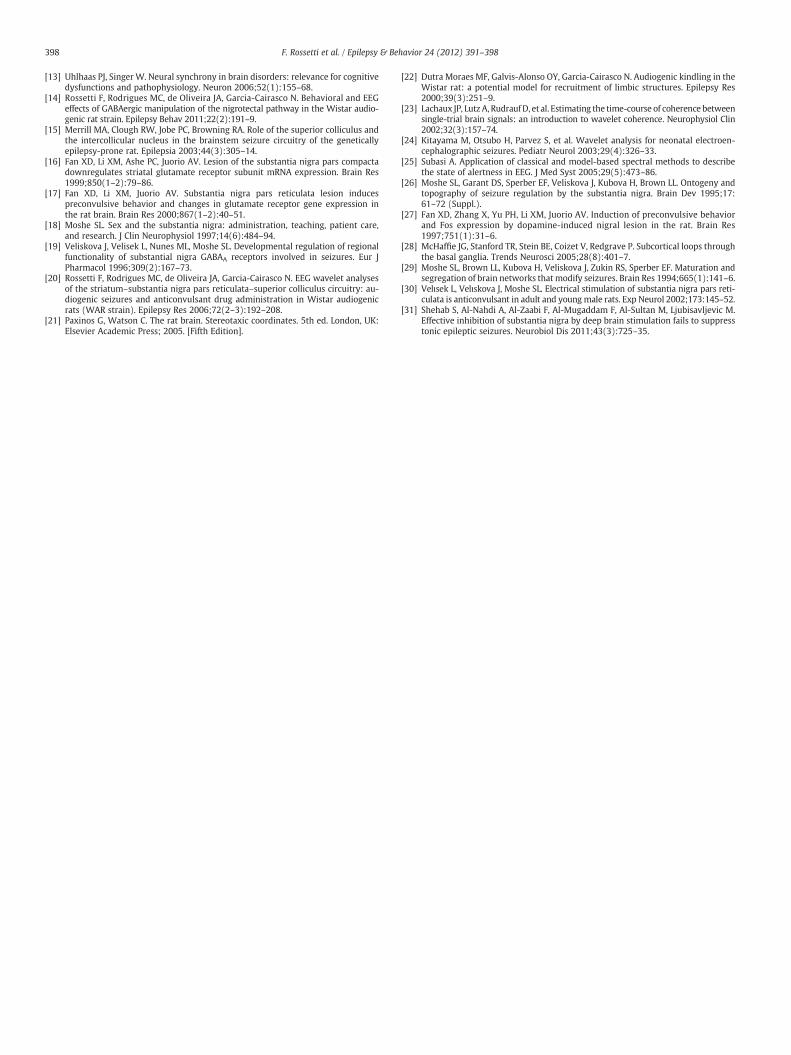
The quantitative analysis of FG+ cells showed that the number ofneurons projecting from the posterior SNPr to both the aSC and pSCregions was higher in WARs than in Wistar rats (Fig. 4) and that thenumber of projections from the posterior SNPr to both regions ofthe SC was higher than the projections from the anterior SNPr.
4. Discussion
BIC microinjection into the aSC increased F1 in the striatum, andGABAergic (MUS and BIC) microinjections into the pSC decreased F1and SO during the adaptation period. Our results clearly demonstratethat the SC in WARs has two defined regions that express distinct EEGfeatures and receptor phenotypes (or chemical features) as observedwith different responses to GABAergic drugs. This finding corrobo-rates our preliminary division of SC into two regions, which we ini-tially formulated due to the literature descriptions of three differentsubcortical loops that are formed by connections between the basalganglia nuclei, SC and thalamus [28] and characterized in our previ-ous studies [14]. The SC receives projections from the SNPr in threedifferent regions (one from the superficial layers and two from thedeep layers) that make connections with the thalamus. The lattersends projections into the striatum, which also sends projectionsback into the SNPr, closing the loop [28]. However, none of the
om the atlas of Paxinos and Watson [21] (AP — antero-posterior in mm, relative to the) and Wistar pSC group (n=5).
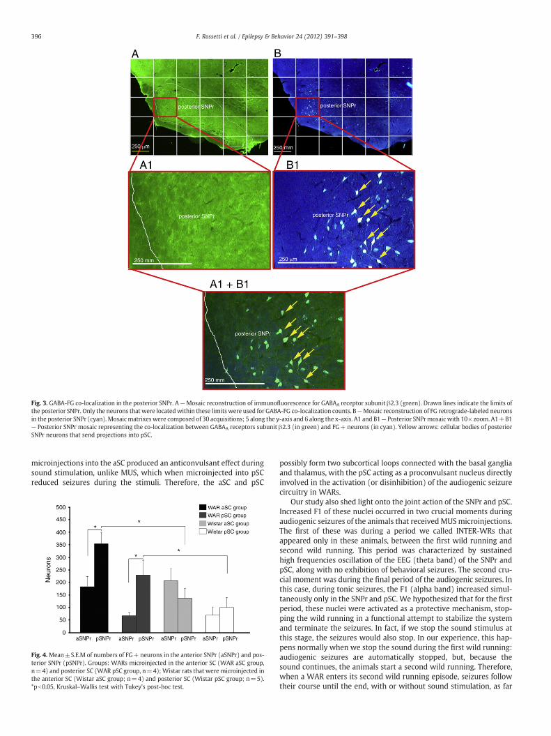
Fig. 3. GABA-FG co-localization in the posterior SNPr. A—Mosaic reconstruction of immunofluorescence for GABAA receptor subunit β2.3 (green). Drawn lines indicate the limits ofthe posterior SNPr. Only the neurons that were locatedwithin these limits were used for GABA-FG co-localization counts. B—Mosaic reconstruction of FG retrograde-labeled neuronsin the posterior SNPr (cyan). Mosaicmatrixes were composed of 30 acquisitions; 5 along the y-axis and 6 along the x-axis. A1 and B1— Posterior SNPrmosaic with 10× zoom. A1+B1— Posterior SNPr mosaic representing the co-localization between GABAA receptors subunit β2.3 (in green) and FG+ neurons (in cyan). Yellow arrows: cellular bodies of posteriorSNPr neurons that send projections into pSC.
396 F. Rossetti et al. / Epilepsy & Behavior 24 (2012) 391–398
microinjections into the aSC produced an anticonvulsant effect duringsound stimulation, unlike MUS, which when microinjected into pSCreduced seizures during the stimuli. Therefore, the aSC and pSC
Fig. 4.Mean±S.E.M of numbers of FG+ neurons in the anterior SNPr (aSNPr) and pos-terior SNPr (pSNPr). Groups: WARs microinjected in the anterior SC (WAR aSC group,n=4) and posterior SC (WAR pSC group, n=4); Wistar rats that were microinjected inthe anterior SC (Wistar aSC group; n=4) and posterior SC (Wistar pSC group; n=5).*pb0.05, Kruskal–Wallis test with Tukey's post-hoc test.
possibly form two subcortical loops connected with the basal gangliaand thalamus, with the pSC acting as a proconvulsant nucleus directlyinvolved in the activation (or disinhibition) of the audiogenic seizurecircuitry in WARs.
Our study also shed light onto the joint action of the SNPr and pSC.Increased F1 of these nuclei occurred in two crucial moments duringaudiogenic seizures of the animals that receivedMUSmicroinjections.The first of these was during a period we called INTER-WRs thatappeared only in these animals, between the first wild running andsecond wild running. This period was characterized by sustainedhigh frequencies oscillation of the EEG (theta band) of the SNPr andpSC, along with no exhibition of behavioral seizures. The second cru-cial moment was during the final period of the audiogenic seizures. Inthis case, during tonic seizures, the F1 (alpha band) increased simul-taneously only in the SNPr and pSC. We hypothesized that for the firstperiod, these nuclei were activated as a protective mechanism, stop-ping the wild running in a functional attempt to stabilize the systemand terminate the seizures. In fact, if we stop the sound stimulus atthis stage, the seizures would also stop. In our experience, this hap-pens normally when we stop the sound during the first wild running:audiogenic seizures are automatically stopped, but, because thesound continues, the animals start a second wild running. Therefore,when a WAR enters its second wild running episode, seizures followtheir course until the end, with or without sound stimulation, as far

397F. Rossetti et al. / Epilepsy & Behavior 24 (2012) 391–398
as tonic seizures occur (the presence of tonic seizure is the criterionfor stopping the sound). We concluded that in this exact moment ofthe audiogenic seizure, after the first running and before the second,both the SNPr and pSC act in conjunction to stop the seizures withhigher frequency than the one presented in the INTER-WRs period.This result is even more evident in another study from our groupthat used phenobarbital (PHE) systemic injection; the EEG oscillationof the SNPr and SC decreased both during the adaptation period andduring the sound stimulation in animals that had audiogenic seizuresblocked by PHE [20]. When the animal had seizures, there was anincrease of F1 in both nuclei, which returned to normal when theseizures ended [20]. Nevertheless, when we bilaterally injected eitherPHE or MUS into the SNPr, the audiogenic seizures of WARs were notblocked and temporally coincided with an increase of F1 in the SC.The animals that had audiogenic seizures presented simultaneousoscillations (alpha band) of the SNPr and SC during hindlimb clonicseizures, cSI=8 [20].
The participation of the SNPr in seizure activity has been discussedin the literature. Microinjections of dopamine into the anterior SNPrin Wistar rats induced preconvulsive behaviors, such as staring,immobilization, facial and mouth movements and wet dog shakesassociated with Fos oncoprotein expression in the limbic system[27] and in the RNAm expression of the glutamatergic receptorsGLUR1, GLUR2 and NMDAR1 [17]. Microinjection of dopamine intothe posterior SNPr in Wistar rats showed neither pre-convulsivebehavior nor Fos expression [27]. According to these authors, thespecific effects in the anterior SNPr can be due to the action of dopa-mine in D1 receptors, which are located in GABAergic neuronal termi-nals that cause GABA release and therefore provoke reduction of theiractivity and, consequently, cause disinhibitory effects in the thalamicnuclei. Therefore, when the anterior SNPr of Wistar rats is inhibited,preconvulsive behaviors appear. This does not happen when theposterior SNPr is inhibited [16,17,27]. The authors conclude that inthe Wistar strain, the anterior SNPr acts as an anticonvulsantnucleus, whereas the posterior SNPr acts as a proconvulsant nucleus.
In our FG studies, we observed that the neuronal projections fromthe anterior and posterior SNPr to both aSC and pSC regions formdifferent connectivity patterns in WARs compared to those fromWistar animals. In WARs, the posterior SNPr sends more projectionsto both SC regions than the anterior SNPr. The genetic backgroundof the WARs might explain the differences in the connectivity be-tween the SNPr and SC, mainly in the posterior SNPr, which is proba-bly crucial to their audiogenic seizure susceptibility. This finding isinteresting because according to Fan et al. [16,17,27], the posteriorSNPr is called the proconvulsant region of SNPr in Wistar animals. Be-cause WAR is a Wistar-derived strain, the proconvulsant feature ofthe posterior SNPr is preserved in WARs; it is tempting to speculatethat the higher number of projections from the proconvulsant regionof the SNPr to the pSC would be an important functional and neuro-anatomical component to explain the audiogenic susceptibility ofthe WARs.
In an animal seizure model induced by fluorothyl in Sprague–Dawley rats, bilateral microinjections of (Z)-3-[(aminoiminomethyl)thio]prop-2-enoic acid (ZAPA, an agonist of the low-affinity GABAA
receptor site), gamma-vinyl-GABA and MUS into the anterior SNPrcaused anticonvulsant effects. However, the same microinjectionsinto the posterior SNPr caused proconvulsant effects [19,29]. The mi-croinjections of BIC (100 ng) were proconvulsant in the anteriorSNPr but ineffective in the posterior SNPr [19]. These studies charac-terized the anterior SNPr as a proconvulsant nucleus and the posteri-or SNPr as an anticonvulsant nucleus in the Sprague–Dawley strain,which can be compared with our findings with WARs, a geneticallydeveloped strain.
One final comment can be made on the impact of the currentstudy to the proposal of SNPr as a potential target for deep brain stim-ulation. Conflicts in the literature are mostly due to the use of normal
rats made epileptic by different, usually chemical, treatments [30,31],in contrast with the endogenous epileptogenicity of WARs.
In conclusion, the SC in WARs presents two functional sub-regionsdefined with distinct GABAergic drug response and EEG characteris-tics, forming at least two subcortical loops. The genetic backgroundof the WARs certainly causes important changes in the neuronal pro-jections between the posterior SNPr and the aSC and pSC regions,compared with the WAR parent strain (Wistar). This new pool ofdata strongly supports the view that changes in the nigro-tectal path-way might be one of the causes of audiogenic seizure susceptibilitycondition. In addition, only the pSC participates in the audiogenic sei-zure circuitry inWARs as a proconvulsant nucleus, possibly due to theincreased number of projections from the posterior SNPr, a nucleusalso found to be proconvulsant in the Wistar strain when challengedwith GABAergic drugs [16,17,27].
Acknowledgments
Franco Rossetti, Marcelo C.A. Rodrigues, Simone S. Marroni, ArturFernandes and Maira Lícia Foresti were supported by FAPESP fellow-ships (03/12420-2, 03/01134-9, 05/58939-4, 05/513366-9, 06/60768-6, respectively). Rodrigo Neves Romcy-Pereira was supportedby a PRODOC-CAPES fellowship, and Dráulio Barros de Araújo wassupported by FAPESP, CNPq and CAPES. Norberto Garcia-Cairascoholds a CNPQ Research Fellowship and is supported by FAPESP(2003/00873-2;2007/50261-4), FAPESP-Cinapce (2005/56447-7),CNPq, PRONEX, CAPES-PROAP-PROEX and FAEPA grants. The presentstudy was also supported by FAPESP (03/11381-3, 03/00873-2)grants. Thanks to Flávio Del Vecchio and José Antonio Cortes deOliveira for technical assistance and to Leonardo Fidelis Filho andEduardo Gomes for the care of the animals at the vivarium of thePhysiology Department of the Ribeirão Preto School of Medicine atthe University of São Paulo, São Paulo, Brazil. We confirm that wehave read the Journal's position on issues involved in ethical publica-tion and affirm that this report is consistent with those guidelines.
References
[1] Doretto MC, Fonseca CG, Lobo RB, Terra VC, Oliveira JA, Garcia-Cairasco N. Quan-titative study of the response to genetic selection of the Wistar audiogenic ratstrain (WAR). Behav Genet 2003;33(1):33–42.
[2] Garcia-Cairasco N. A critical review on the participation of inferior colliculus inacoustic-motor and acoustic-limbic networks involved in the expression ofacute and kindled audiogenic seizures. Hear Res 2002;168(1–2):208–22.
[3] Garcia-Cairasco N, Doretto MC, Ramalho MJ, Antunes-Rodrigues J, Nonaka KO.Audiogenic and audiogenic-like seizures: locus of induction and seizure severitydetermine postictal prolactin patterns. Pharmacol Biochem Behav 1996;53(3):503–10.
[4] Carolino RO, Beleboni RO, Pizzo AB, et al. Convulsant activity and neurochemicalalterations induced by a fraction obtained from fruit Averrhoa carambola(Oxalidaceae: Geraniales). Neurochem Int 2005;46(7):523–31.
[5] Ribak CE, Manio AL, Navetta MS, Gall CM. In situ hybridization for c-fos mRNAreveals the involvement of the superior colliculus in the propagation of seizureactivity in genetically epilepsy-prone rats. Epilepsy Res 1997;26(3):397–406.
[6] Faingold CL, Randall ME. Neurons in the deep layers of superior colliculus play acritical role in the neuronal network for audiogenic seizures: mechanisms forproduction of wild running behavior. Brain Res 1999;815(2):250–8.
[7] Deransart C, Le-Pham BT, Hirsch E, Marescaux C, Depaulis A. Inhibition of thesubstantia nigra suppresses absences and clonic seizures in audiogenic rats, butnot tonic seizures: evidence for seizure specificity of the nigral control. Neurosci-ence 2001;105(1):203–11.
[8] Fuentes-Santamaria V, Alvarado JC, Herranz AS, Garcia-Atares N, Lopez DE.Morphologic and neurochemical alterations in the superior colliculus of the genet-ically epilepsy-prone hamster (GPG/Vall). Epilepsy Res 2007;75(2–3):206–19.
[9] Doretto MC, Cortes-de-Oliveira JA, Rossetti F, Garcia-Cairasco N. Role of the superiorcolliculus in the expression of acute and kindled audiogenic seizures inWistar audio-genic rats. Epilepsia 2009;50(12):2563–74.
[10] Jensen O, Gelfand J, Kounios J, Lisman JE. Oscillations in the alpha band (9–12 Hz)increase with memory load during retention in a short-term memory task. CerebCortex 2002;12(8):877–82.
[11] Makeig S, Debener S, Onton J, Delorme A. Mining event-related brain dynamics.Trends Cogn Sci 2004;8(5):204–10.
[12] Romcy-Pereira RN, de Araujo DB, Leite JP, Garcia-Cairasco N. A semi-automatedalgorithm for studying neuronal oscillatory patterns: a wavelet-based timefrequency and coherence analysis. J Neurosci Methods 2008;167(2):384–92.
398 F. Rossetti et al. / Epilepsy & Behavior 24 (2012) 391–398
[13] Uhlhaas PJ, Singer W. Neural synchrony in brain disorders: relevance for cognitivedysfunctions and pathophysiology. Neuron 2006;52(1):155–68.
[14] Rossetti F, Rodrigues MC, de Oliveira JA, Garcia-Cairasco N. Behavioral and EEGeffects of GABAergic manipulation of the nigrotectal pathway in the Wistar audio-genic rat strain. Epilepsy Behav 2011;22(2):191–9.
[15] Merrill MA, Clough RW, Jobe PC, Browning RA. Role of the superior colliculus andthe intercollicular nucleus in the brainstem seizure circuitry of the geneticallyepilepsy-prone rat. Epilepsia 2003;44(3):305–14.
[16] Fan XD, Li XM, Ashe PC, Juorio AV. Lesion of the substantia nigra pars compactadownregulates striatal glutamate receptor subunit mRNA expression. Brain Res1999;850(1–2):79–86.
[17] Fan XD, Li XM, Juorio AV. Substantia nigra pars reticulata lesion inducespreconvulsive behavior and changes in glutamate receptor gene expression inthe rat brain. Brain Res 2000;867(1–2):40–51.
[18] Moshe SL. Sex and the substantia nigra: administration, teaching, patient care,and research. J Clin Neurophysiol 1997;14(6):484–94.
[19] Veliskova J, Velisek L, Nunes ML, Moshe SL. Developmental regulation of regionalfunctionality of substantial nigra GABAA receptors involved in seizures. Eur JPharmacol 1996;309(2):167–73.
[20] Rossetti F, Rodrigues MC, de Oliveira JA, Garcia-Cairasco N. EEG wavelet analysesof the striatum–substantia nigra pars reticulata–superior colliculus circuitry: au-diogenic seizures and anticonvulsant drug administration in Wistar audiogenicrats (WAR strain). Epilepsy Res 2006;72(2–3):192–208.
[21] Paxinos G, Watson C. The rat brain. Stereotaxic coordinates. 5th ed. London, UK:Elsevier Academic Press; 2005. [Fifth Edition].
[22] Dutra Moraes MF, Galvis-Alonso OY, Garcia-Cairasco N. Audiogenic kindling in theWistar rat: a potential model for recruitment of limbic structures. Epilepsy Res2000;39(3):251–9.
[23] Lachaux JP, Lutz A, RudraufD, et al. Estimating the time-course of coherence betweensingle-trial brain signals: an introduction to wavelet coherence. Neurophysiol Clin2002;32(3):157–74.
[24] Kitayama M, Otsubo H, Parvez S, et al. Wavelet analysis for neonatal electroen-cephalographic seizures. Pediatr Neurol 2003;29(4):326–33.
[25] Subasi A. Application of classical and model-based spectral methods to describethe state of alertness in EEG. J Med Syst 2005;29(5):473–86.
[26] Moshe SL, Garant DS, Sperber EF, Veliskova J, Kubova H, Brown LL. Ontogeny andtopography of seizure regulation by the substantia nigra. Brain Dev 1995;17:61–72 (Suppl.).
[27] Fan XD, Zhang X, Yu PH, Li XM, Juorio AV. Induction of preconvulsive behaviorand Fos expression by dopamine-induced nigral lesion in the rat. Brain Res1997;751(1):31–6.
[28] McHaffie JG, Stanford TR, Stein BE, Coizet V, Redgrave P. Subcortical loops throughthe basal ganglia. Trends Neurosci 2005;28(8):401–7.
[29] Moshe SL, Brown LL, Kubova H, Veliskova J, Zukin RS, Sperber EF. Maturation andsegregation of brain networks that modify seizures. Brain Res 1994;665(1):141–6.
[30] Velısek L, Velıskova J, Moshe SL. Electrical stimulation of substantia nigra pars reti-culata is anticonvulsant in adult and young male rats. Exp Neurol 2002;173:145–52.
[31] Shehab S, Al-Nahdi A, Al-Zaabi F, Al-Mugaddam F, Al-Sultan M, Ljubisavljevic M.Effective inhibition of substantia nigra by deep brain stimulation fails to suppresstonic epileptic seizures. Neurobiol Dis 2011;43(3):725–35.