epilepsy in children with autistic spectrum disorder · background epilepsy is a common childhood...
TRANSCRIPT

Objectives :
Background
Pathophysiology of ASD in Epileptic patients
Association between Epilepsy and ASD
EEG changes in ASD
Treatment of epilepsy in ASD patients

Background
Epilepsy is a common childhood disorder
The prevalence is estimated to be 2-3 % in general population
Epilepsy in children with autistic spectrum disorders(ASD) is more common and range from 8-38%

Historical Background
Autism was tied to epilepsy in Kanner’s initial description of autism over 60 years ago
The initial studies on the relationship of autism to epilepsy and to electroencephalogram (EEG) abnormalities in the 1960s
In 1960,epilepsy was classified as a neurological versus mental disorder in the WHO classification system for epilepsy

Historical Background
Early efforts to classify autism were led by Lorna Wing who introduced the concept of the “autism triad” and highlighted common impairments in social, language and repetitive behaviors among children with cognitive deficits
Wing L, Gould J. Severe impairments of social interaction and associated abnormalities in children: epidemiology and classification. J Autism Dev Disord 1979;9:11–29
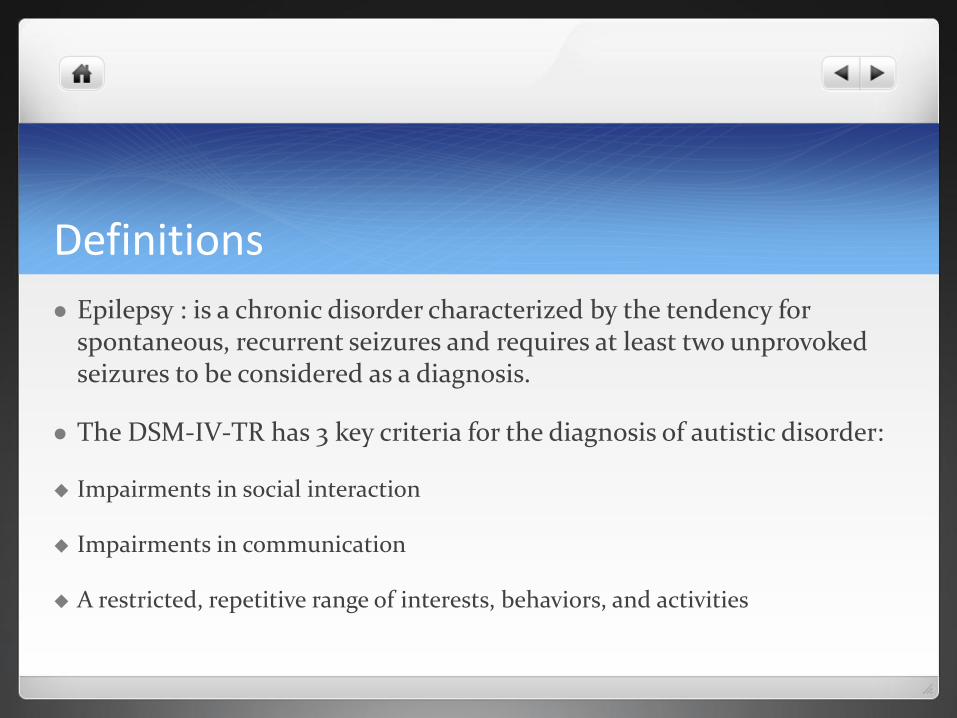
Definitions
Epilepsy : is a chronic disorder characterized by the tendency for spontaneous, recurrent seizures and requires at least two unprovoked seizures to be considered as a diagnosis.
The DSM-IV-TR has 3 key criteria for the diagnosis of autistic disorder:
Impairments in social interaction
Impairments in communication
A restricted, repetitive range of interests, behaviors, and activities
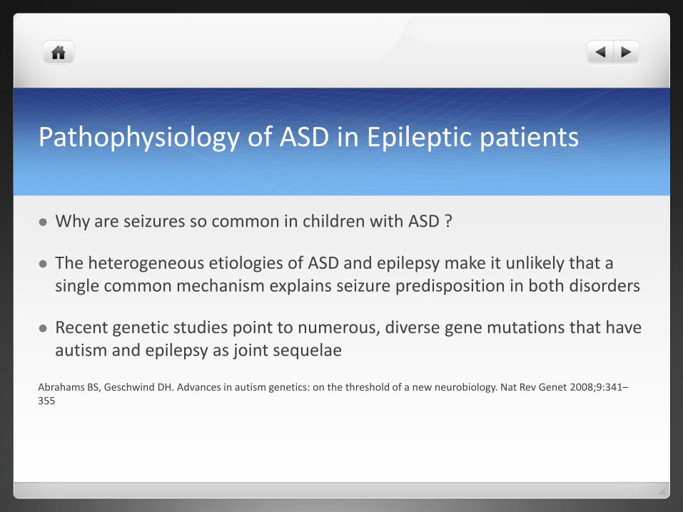
Pathophysiology of ASD in Epileptic patients
Why are seizures so common in children with ASD ?
The heterogeneous etiologies of ASD and epilepsy make it unlikely that a single common mechanism explains seizure predisposition in both disorders
Recent genetic studies point to numerous, diverse gene mutations that have autism and epilepsy as joint sequelae
Abrahams BS, Geschwind DH. Advances in autism genetics: on the threshold of a new neurobiology. Nat Rev Genet 2008;9:341–355


Pathophysiology of ASD in Epileptic patients
A common neurobiological antecedent
structural or developmental lesions
genetic susceptibilities
environmental insults
Abnormal brain circuitry underlying ASD could predispose the brain to seizures

Pathophysiology of ASD in Epileptic patients
Neuropathological Studies from Post-mortem patients of with autism showed cortical malformation, Polymicrogyria and megalenecephaly. A. Bailey et al Brain (1998),
121, 889–905
A clinicopathological study of autism 897
Table 2 Pathological findings in cerebral cortex and underlying white matter
Case Cortical dysgenesis White matter Other
1 Slightly irregular laminar pattern in Numerous patches of ectopic grey mattersuperior frontal gyrus with clusters of and increased numbers of single neuronsabnormally orientated pyramidal cells.White matter/layer 6 boundary poorlydefined.
2 Thickened cortex and increased neuronaldensity. Increased numbers of neurons inlamina 1 of frontal cortex, includinginverted pyramidal cells.
3 Increased neuronal density and mild focal Patch of nerve cells deep in the deep Small shallow focus of gliosis in frontaldisturbance of laminar pattern in frontal white matter of anterior frontal cortex. cortex. Larger area of gliosis involvingcortex Increased number of single neurons. full cortical thickness in one occipital
lobe.4 Thickened cortices and increased Subpial gliosis.
neuronal density in frontal cortex andcingulate gyri. Laminar pattern ofsuperior temporal gyrus disorganised.
5 Increased number of single neurons in Numerous corpora amylacea withinsuperior temporal gyrus. subpial zone and molecular layer of
insular cortex.
6 Increased number of white matterneurons.
Table 3 Pathological findings in the brainstem
Case Olivary dysplasia Neuronal ectopia Other
1 Dysplastic olives with apparent Bilateral groups of neurons lateral to Large medulla oblongata with smallreduplication of medial accessory olive. olives and in inferior cerebellar peduncle. pyramids. Aberrant pontine tract
bilaterally. Small midbrain.2 Bilateral groups of neurons lateral to Unusually small aqueduct.
olives. Group of neurons in one inferiorcerebellar peduncle.
3 Bilateral break in the inferior end of the Bilateral band-like groups of neurons Hypertrophy of striae medullaris andinferomedial olives. lateral to olives. enlarged arcuate nuclei.
4 Small groups of neurons in inferior Widely dispersed locus coeruleus.cerebellar peduncles.
5 Bilateral break in the anterior portions of Slight flattening of medullathe inferior olives. anteroposterior. Fissure separating
pyramids poorly defined. Prominentexternal arcuate nuclei and externalarcuate fibres.
6 Adequate section not available. Neurons of locus coeruleus looselygrouped.
patchy increase in GFAP in Bergmann glia, without an
obvious increase in cell number. In the right cerebral hemi-
sphere, there were neurons in the white matter, and the deep
white matter arteries showed scanty lymphocytic cuffing.
The basal ganglia were unremarkable. No obvious abnormal-
ity was noted in the hippocampus in a section taken from a
cryoprotected block.
The neuropathological findings (in the cerebral cortex and
underlying white matter, the brainstem and the cerebellum)
are summarized in Tables 2–4.
(During revision of this manuscript the brain and spinal
cord of a cachectic, handicapped 41-year-old woman with
autism became available for study. She met ADI criteria
for autism. The brain weighed 1233 g. There was partial
reduplication of the inferior olive at one level. Purkinje
cells were irregularly aligned with some lying in the deep
molecular layer. The Bergman glia appeared excessively
numerous and there was an excess of corpora amylacea
in the molecular layer. There were excess numbers of
multipolar neurons in the molecular layer of the right
cerebral hemisphere. The amygdaloid nucleus contained
tightly packed round or elongated clusters of mature large
neurons, some within bundles of myelinated fibres that
appeared out of place.)

Pathophysiology of ASD in Epileptic patients
Independent
Epilepsy
ASD
Common Pathway
Epilepsy
ASD
Epilepsy Causation
Epilepsy
Epileptiformdischarges
Developmental
ASD
Epilepsy
Genetic
Lesional
Multifactorial
ASD
Autistic Regresion

The Association between Epilepsy and ASD
The frequency of epilepsy in ASD range from 5-38%.
The age peak from 1-5 years and > 10 years and adolescent
All seizure types can be associated with autism like complex partial, atypical absence, myoclonic, and tonic-clonic seizures
Tuchman RF, Rapin I, Shinnar S. Autistic anddysphasic children, II: epilepsy. Pediatrics 1991; 88:1219–25.

Examples for seizure types:

Be careful ….
Not all the abnormal movements are seizures

The Association between Epilepsy and ASD
The prevalence of autism in children with epilepsy around 27 % and it increased in patients with mental retardation.
The developing brain is well known to be more susceptible to seizures than the mature brain Holmes GL. Epilepsia1997; 38: 12–30.
Epileptiform discharges on electroencephalography (EEG) without clinical seizures can cause behavioural and cognitive impairment
Binnie C, Channon S, Marston D. Behaviouralcorrelates of interictal spikes. In: Smith D, Treiman D, Trimble M, eds. Neurobehaviouralcorrelates of epilepsy. New York: Raven Press, 1991:113–26.

The Association between Epilepsy and ASD
Roberto Tuchman and Isabelle RapinLancet Neurology 2002; 1: 352–58

The Association between Epilepsy and ASD
Autism
Epilepsy
Developmental Language Disorder
LKS
ESES
BRET
Disintegrative Disorder


Landau- Kleffner Syndrome( LKS)
Acquired aphasia, seizures, and an epileptic electroencephalogram
A language regression between the age of 2- 8 years. Peak age is 4-5 years
Progressive loss of speech in a previously well child
Patients rend to have partial onset seizures , Atypical absence
Epileptiform EEG abnormalities and/ or clinical seizures

EEG changes :

EEG changes :

Psychiatric symptoms in LKS
Hyperactivity, inattentiveness, impulsivity and aggression
Abnormal social behaviors resembling autistic symptoms.
Psychiatrist described it as "the symptom of autism" secondary to a medical condition, or a depressive adjustment disorder

The differences Between ASD & LKS :
ASD LKS
Age of regression 18-24 months 3 years
Regression affect behavioral
Only language
Epileptiform activity Centro-temporal , Infrequent
Fronto-temporo-parietalFrequent

Treatment :
Anti – Epileptic Drugs :
IV Ig
Steroid
Surgery

Outcome
Epilepsy usually well controlled
Language deficits usually persist; sometimes correlated with the epileptiform EEG


Electrical Status Epilepticus in Sleep( CSWS)
An epileptic disorder characterized by specific EEG abnormalities which is associated with language, cognitive and behavioral regression in children
Global regression in cognitive and/or motor skills
Age 3 -14 years with a peak age of 5 - 7 years
Up to one third of children with CSWS have a preceding neurologic condition, such as meningitis or neonatal encephalopathy

EEG changes:

EEG criteria
Various SWI criteria are proposed by different authors for ESES
The commonly used definition in clinical studies is "bilateral secondarily generalized 1.5 to 4.5 Hz spike-waves occuring during greater than 85% of slow wave sleep
Focal, multifocal, unilateral, asymmetric/symmetric bilateral, diffuse, or more restricted.
The ESES/CSWS pattern may be continuous, fragmented, or periodic

Psychiatric symptoms in CSWS
Severe decline in IQ, short term memory deficits and poor reasoning
The language impairment includes an expressive aphasia as well as difficulties with lexical and syntactic skills
Reduced attention, hyperactivity, disinhibition and aggression
Motor deficits and apraxia, ataxia, dystonia, dyspraxia

Seizure type in CSWS :
Seizures can present in many types; partial onset or secondarily generalized, typical or atypical absence
Focal MRI abnormalities have been reported

Treatment
Anti-Epileptic Drugs : Valproic Acid, Levetiracetam and Clobazam
IV Ig
Steroid
Surgery

Benign Childhood Epilepsy with Centrotemporal Spikes (Rolandic epilepsy)
Age: 5-9 years
Remission: By 16 years of age
Nocturnal seizures with sensory motor symptoms involving the face and oropharynx
EEG:
Treatment:


EEG in ASD
Normal EEGs are frequently seen
Paroxysmal epileptiform activity (spike, spike-wave, polyspikes, and polyspikes and waves)
Rossi et al. studied EEG finding in 106 patients with ASD found:
57.5% had no seizure and no paroxysmal activity
18.9 had central,temporal and parietal spikes as seen in benign epilepsy of childhood with centro-temporal spikes( BRECT) Epilepsia, 48(Suppl. 9):33–35, 2007

EEG in ASD

EEG in ASD
Occipital spikes similar to those seen in benign occipital epilepsy of childhood were seen on video-EEG monitoring in 17% Nass et al., 1998
191
Fig. 6. Benign occipital epilepsy. Four-year-old girl with seizures. Note the independent, bi-occipital sharp and
slow wave complexes with eyes closed.
11_Riviello 4/11/07 9:07 AM Page 191
EEG in ASD
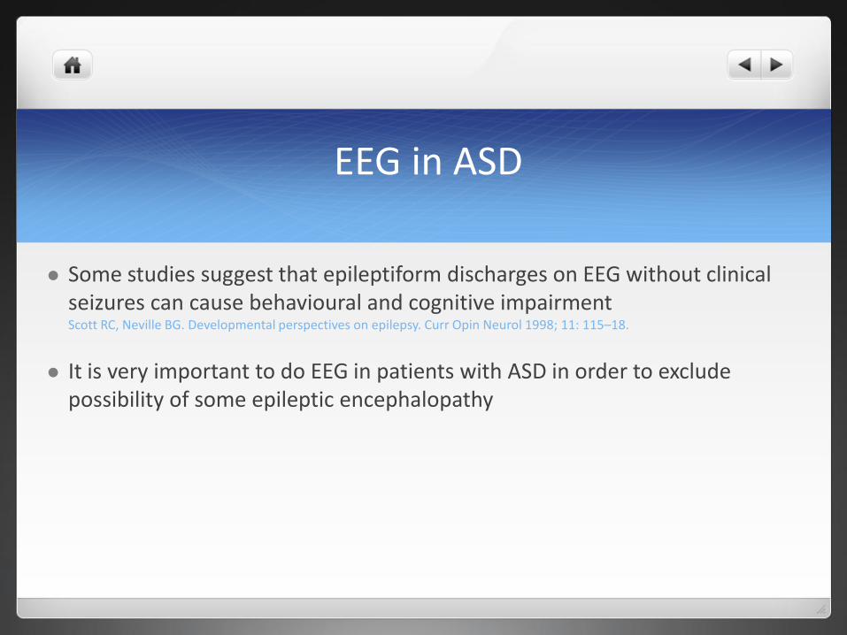
Some studies suggest that epileptiform discharges on EEG without clinical seizures can cause behavioural and cognitive impairment Scott RC, Neville BG. Developmental perspectives on epilepsy. Curr Opin Neurol 1998; 11: 115–18.
It is very important to do EEG in patients with ASD in order to exclude possibility of some epileptic encephalopathy


Treatment of epilepsy in ASD patients
We can divide the cases as following:
ASD patient who has clinical seizure and EEG abnormalities
ASD patient who has no clinical seizure but EEG abnormalities

Treatment of epilepsy in ASD patients
ASD patient who has clinical seizure and EEG abnormalities:
The treatment of epilepsy in ASD children is not different from the treatment of other children with epilepsy
The choices of Anti-Epileptic Drugs ( AED ) depend on clinical semiology of seizure as well as EEG abnormalities

Treatment :
Choices of anti-epileptic medications
Start with small dose and build up slowly
Explain to the parents possible side effects
Educate the parent about how to manage the attack of seizure

What to do during a convulsion :

Treatment of epilepsy in ASD patients

Treatment of epilepsy in ASD patients
There are several clinical reports of the use of valproic acid in children with autism with or without clinical seizures but with epileptiform abnormalities on the EEG Plioplys AV. Autism: electroencephalogram abnormalities and clinical improvement with valproic acid. Arch Pediatr AdolescMed 1994; 148: 220–22.
In a double-blind placebo controlled study with lamotrigine in children with autism without seizures, no favorable effects were observed on the outcome measures for autistic symptoms Belsito K, Law P. Kirk K, et al. (2001) Lamotrigine therapy for autistic disorder:a randomized double-blind , placebo controlled trial. J Autism Dev Disord 31:175–181

ASD patient who has no clinical seizure but EEG abnormalities
Controversial issue ??
A limited number of clinical reports on the effectiveness of valproate in reducing overactivity instability and impulsivity in children with autism and abnormal electroencephalograms
There are reports that the language of limited numbers of children with LKS and of those with autism has improved in response to anticonvulsants, especially valproic acid, ethosuximide, and benzodiazepines
Marescaux C et al Epilepsia 1990; 31: 768–77

ASD patient who has no clinical seizure but EEG abnormalities
No definite recommendations can be made

Is there any other modality of treatments?
Epilepsy surgery
Vagal Nerve stimulation
Ketogenic diet

Treatment of epilepsy in ASD patients
Surgical treatment :
Surgery may improve the seizures but not the autism
Multiple subpial transections used in patient with LKS however it is not well validated

Treatment of epilepsy in ASD patients

127 patients identified
33.9% had Treatment Resistant Epilepsy( TRE)
27.5% were seizure free
38.6% had infrequent seizures
Earlier onset seizures was in TRE patients
3 patients had surgery , 1 with no improvement
9 had VNS , 2 patients with limited improvement
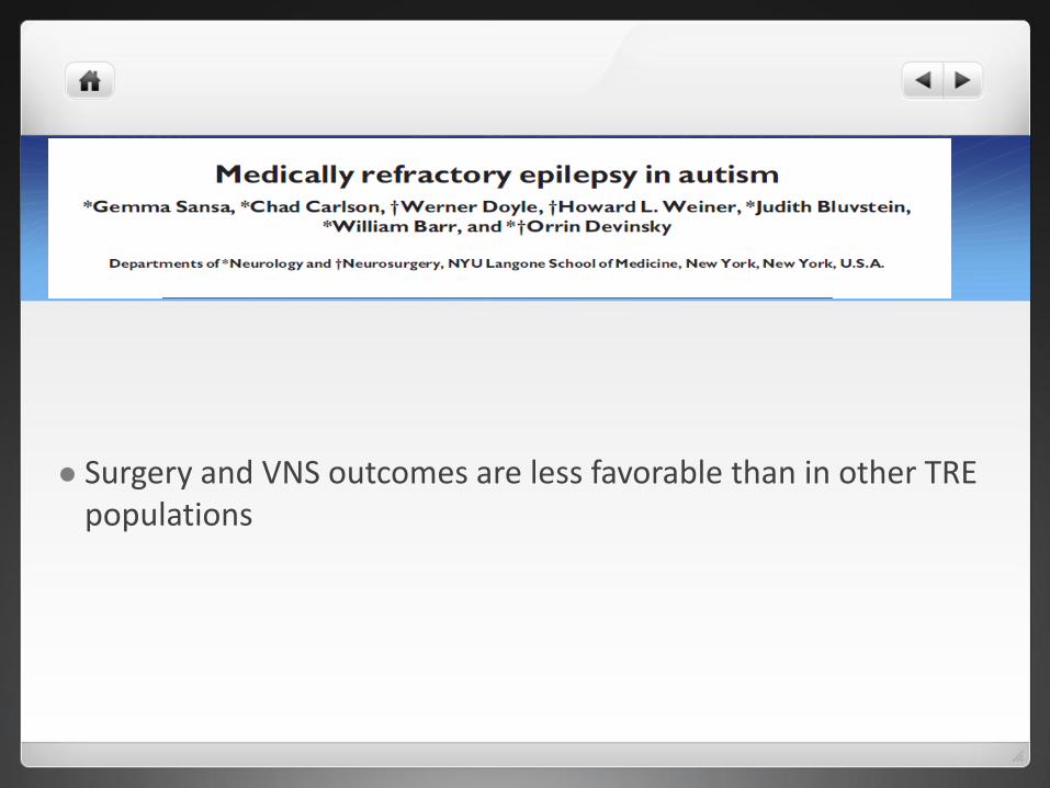
Surgery and VNS outcomes are less favorable than in other TRE populations

Outcome :
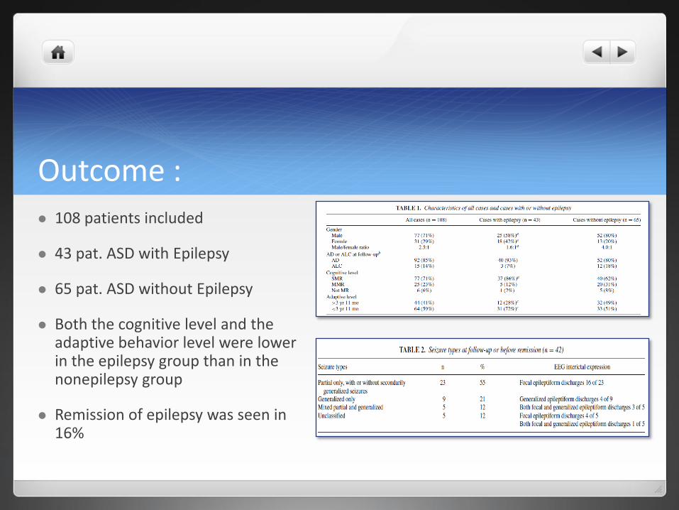
Outcome : 108 patients included
43 pat. ASD with Epilepsy
65 pat. ASD without Epilepsy
Both the cognitive level and the adaptive behavior level were lower in the epilepsy group than in the nonepilepsy group
Remission of epilepsy was seen in 16%

Conclusion : Epilepsy is common and should be routinely investigated in patients with
ASD
Epilepsy and ASD share a common pathway and genetic susceptibility and environmental factors plays a roll in pathophysiology of both conditions
Epileptic encephalopathy and epileptic syndromes like LKS,CSWS should be rolled out in patients with ASD
The treatment of epilepsy in ASD children is not different from the treatment of other children with epilepsy however the choices of antiepileptic drugs may be different

Thank [email protected]