episode 28 aortic dissection, acute limb ischemia & compartment
TRANSCRIPT

EPISODE 28 - VASCULAR CATASTROPHES PEARLS & PITFALLS - EMERGENCYMEDICINECASES.COM
Aortic dissection is an uncommon diagnosis with high mortality, and often difficult to identify.
The Classic presentation: acute, “tearing” or “ripping” chest pain reaching maximal intensity at onset,
radiating to back and/or between shoulder blades. Only 25% of patients have the triad of this pain, widened
mediastinum, and pulse deficit. The most common descriptor of the pain is “sharp”, ~5% presented painlessly (1), and 10% with syncope (IRAD(2).
Risk factors for Dissection: Hypertension, age, male gender, family history, recent deceleration injury (i.e.
MVC with airbag), prior cardiac surgery,
known preexisting aortic aneurysm,
recent cardiac cath.
In younger patients (<40): cocaine or
amphetamine use, pregnancy, connective tissue diseases (e.g. Marfan Syndrome), congenital heart disease, bicuspid aortic
valve (9x risk), and weight lifting.
3 Important Questions:
• quality of pain (most commonly “sharp” but highest LR for “tearing”)
• pain intensity at onset
• radiation of pain (back and/or belly)
90% of the ED docs suspected dissection
even before investigations were done if all 3 of these questions were asked (1).
“Chest pain Plus...” CP + focal neuro deficit or pain below diaphragm
or limb ischemia: think dissection!
EPISODE 28: VASCULAR EMERGENCIES WITH DR. ANIL
CHOPRA & DR. DAVID CARR
First: exclude other causes of chest pain such as
MI! ECG abnormalities are common (~70% in
IRAD database) (2), but abnormalities include non-
specific ST changes, LVH, infarction (i.e. inferior
territory from dissection of right
coronary artery), or other ischemic
changes. Ischemic ECG changes are
either due to chronic coronary
disease or extension of dissection
into RCA.
WHAT TO LOOK FOR ON THE ECG Pitfalls of ECGs, troponin, and D-dimer:An ECG showing ischemic changes or a slightly elevated troponin may lead to assumption that pain is due to cardiac ischemia, and a positive D-dimer may lead to assumption that the patient has a PE. Treating PE or ACS equates to “a clean kill” for a patient who has a dissection!
Although D-dimer will usually be positive in nearly all patients with dissection, it cannot be used as a rule-out test as low levels have been found in younger patients, or patients with a thrombosed "false lumen". Both D-dimer and troponin could be normal in patients presenting early with dissection.
Clues to diagnosis of Aortic Dissection:
Aortic Dissection continued:Physical examination:
Carefully check for diastolic murmur
of aortic regurgitation (retrograde
dissection), signs of Marfan syndrome
(pectus excavatum, ‘gangly’
appearance), and pulse deficit in
radials and femoral arteries.
What about BP? Many patients
have a difference in blood pressure
between arms normally, so a BP
difference does not rule in
dissection; nor does a lack thereof
rule out dissection. However, a BP
difference may heighten your
suspicion of dissection in the right
clinical context.
Chest X-Ray Findings:
Although 1/3 of patients have a normal chest x-ray, the 2 most
important abnormalities are a widened mediastinum and the
"calcium sign".
Calcium Sign: separation of the
outermost portion of the aorta from the
calcified intima by >5mm.
Other possible findings on
CXR: loss of aortic knob, trachea
displaced to right, left mainstream bronchus displaced downward,
disparity of ascending and descending aorta caliber, apical
capping, pleural effusion (usually
left), localized bulge in the aorta (3).
Remember to compare with
previous CXR if available!
What about on bedside ultrasound: Check for a
pericardial effusion, which occurs
from dissection into the pericardial space. If an effusion is seen,
management is similar to other scenarios for tamponade (drain only
if unstable). Although sensitivity is
poor for dissection, check if the abdominal aorta appears normal.
What imaging test to get?
Stable Patient: CT scan with arterial
contrast.
Unstable (or unable to receive IV contrast): transesophageal echo
(TEE) which is equally sensitive to CT with arterial contrast (4)
ED Treatment of Dissection
Aggressive BP & HR control to
decrease stress on the aortic wall!! Goal SBP110–120, HR60
1st Line: short acting B-blocker such
as esmolol or labetolol
2nd line: nitroprusside (0.25-1.0mcg/
kg/min) but only in addition to B-blocker (to avoid reflex tachycardia!)
Consult a surgeon! All type A
dissections require urgent surgery!While most type B dissections can
be treated medically, certain type B dissections require urgent surgery
(for ongoing pain, expanding
diameter, evidence of aortic rupture) => consult surgery early in all cases.
Time is key: type A (aortic arch involved) kill 1-2% of patients per hour.
Type B (arch not involved) fare better.
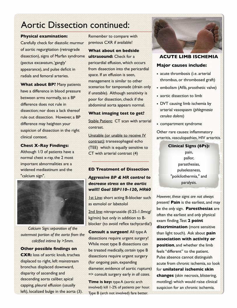
ACUTE LIMB ISCHEMIA
Major causes include:
• acute thrombosis (i.e. arterial
thrombus, or thrombosed graft)
• embolism (Afib, prosthetic valve)
• aortic dissection to limb
• DVT causing limb ischemia by
arterial vasospasm (phlegmasia
cerulea dolens)
• compartment syndrome
Other rare causes: inflammatory arteritis, vasculopathies, HIV arteritis.
Clinical Signs (6Ps):
pain, pallor,
parasthesias, pulselessness,
"poikilothermia," and
paralysis.
However, these signs are not always present! Pain is the earliest, and may
be the only sign. Paresthesias are
often the earliest and only physical exam finding. Test 2 point
discrimination (more sensitive
than light touch). Ask about pain association with activity or
position, and whether the limb
feels “different” to the patient.Pulse absence cannot distinguish
acute from chronic ischemia, so look for unilateral ischemic skin
changes (skin necrosis, blistering,
mottling) which would raise clinical suspicion for an chronic ischemia.

Acute Limb Ischemia continued:Blue Toe Syndrome: Painful
cyanotic discoloration of portions of
the foot, caused by micro-embolic showers from proximal source.
Although pulses are preserved, this is
managed as an acute occlusive condition.
Who goes to the OR?
True acute limb ischemia should get
angiography in the OR and definitive
management. Arterial dopplers may be done for stable patients with a history
of claudication.
A rough rule of thumb for surgical
indication: loss of light touch.
Treatment of limb ischemia:
Time is limb! Salvage time depends on
collaterals, and patient-specific factors. If sensory loss to light touch is
minimal, viability may be excellent, but
ischemia can occur within hours.
Acute Treatment: ASA, UFH
80U/kg bolus, then 18U/kg/h*, ample pain meds, and an urgent surgical
consult for endovascular or surgical
revascularization.
*UFH may inhibit clot propagation and
further distal thrombosis, but no
established benefit in literature.
What about thrombolysis?
While not superior to surgery, it may be the treatment of choice in patients
with occluded grafts, collaterals and
chronic insufficiency, or for occlusions of small, inaccessible arteries.
Sending patients home with non-critical ischemia? Remember to help
optimize comorbidities, and review
anti-platelet agents: ASA (plus clopidigrel in refractory patients) to
improve outcomes.
COMPARTMENT SYNDROME
Definition: Increased pressure within a limited space which compromises tissue function. Most commonly from fractures (including open fractures), but also soft tissue injury, reperfusion injury, minor trauma, major burns, and limb compression in pts “found down”. Compartment syndrome can occur in leg, arm, hand, or abdomen.
Key clinical features
Increasing Pain out of Proportion(intractable severe pain, usually “aching”)Altered Sensationcheck loss of light touch / 2ptPain on Passive StretchMuscle weakness (late finding)Tenderness and swelling in the compartment (can feel woody, tense)
**pulses are usually still present, and the limb oxygen saturation is often preserved.**
How to Examine and Stretch Compartments of Lower Leg:Anterior: pain with passive ankle plantar-flexion, +/- weak ankle dorsiflexion, and loss of sensation between first two toesLateral: pain with passive ankle inversion, +/- weak ankle eversion, and similar findings to anterior compartmentSuperficial posterior: pain with passive ankle dorsiflexionDeep posterior (difficult to palpate): pain with passive toe extension, weak toe flexion
Do serial physical exams and call a surgeon if you are suspicious! Consider measuring a compartment pressure if unsure of diagnosis (see left).
MEASURING COMPARTMENT
PRESSURESCompartment pressure measurement can help confirm Compartment Syndrome, but the Dx can often be made clinically alone. The needle/tubing/manometer technique can be done with readily available ED
equipment. **Measure pressures as close to the fracture or injury site as possible. If unsure, call the surgeon to assist in the diagnosis!
While awaiting for definitive management by fasciotomy: remove constrictive dressings, elevate limb to the level of heart, keep blood pressure adequate (fluids for hypotensive patients!), give oxygen, and relieve pain.
References:
1) Roseman HS, et al. Chest. 1998;114: 793-5.
2) Hagan, PG et al. JAMA 2000; 283:897-903.
3) Marx et al. Rosen's Emergency Medicine: Concepts and Clinical Practice, 6th ed. 2006.
4) Shiga et al. Arch Intern Med 2006;166:1350-6.
SUBSCRIBE TO EMCASES