epma and tem investigations on the interdiffusion layer of the pnn/pzt functionally gradient...
TRANSCRIPT

JOURNAL OF MATERIALS SCIENCE35 (2000 )1031– 1036
EPMA and TEM investigations on the
interdiffusion layer of the PNN/PZT
functionally gradient piezoelectric ceramics
XINHUA ZHU∗, JIANMIN ZHU, SHUNHUA ZHOU, QI LI,ZHIGUO LIU, NAIBEN MINGNational Laboratory of Solid State of Microstructures, Department of Physics,Nanjing University, Nanjing 210093, People’s Republic of China
ZHONGYAN MENGSchool of Materials Science and Engineering, Shanghai University (Jiading Campus),Shanghai 201800, People’s Republic of ChinaE-mail: [email protected]
The compositional profile, distribution of the phases and the ordering behavior in theinterdiffusion layer of the PNN/PZT functionally gradient piezoelectric ceramics have beeninvestigated by electron probe microbeam analyses (EPMA) and transmission electronmicroscopy (TEM) respectively. The results show that the thickness of the interdiffusionlayers (d) for Ni2+, Nb5+, Ti4+ and Zr4+ ions are ordered as d2+
Ni >d5+Nb >d4+
Ti >d4+Zr . It is
demonstrated by TEM observation and selected area electron diffraction (SAED) patternsthat a clear interface between the rhombohedral and pseudocubic phases exists in theinterdiffusion layer. The SAED studies also reveal the presence of F spots along the [111]direction of the perovskite cubic unit cell. The origin of this superstructure is determined.C© 2000 Kluwer Academic Publishers
1. IntroductionFunctionally gradient materials (FGM) are character-ized by the compositional and/or microstructural grada-tion over macroscopic/microscopic distances [1]. Withthe research and development of the FGM, the scopeof which has been extended into other functional mate-rials, such as the piezoelectric ceramic materials, fromthe initial research aim at obtaining the materials withmore advantages in thermal stress relaxation because ofthe gradient joints within the FGM reducing the residualstresses produced at the interfaces. A typical exampleis the development of the FGM piezoelectric ceramicactuator to overcome the performance limitations ofthe usual piezoelectric bimorphs [2]. The FGM piezo-electric ceramic actuator is composed of three layers:a piezoelectric ceramic layer, a high dielectric ceramiclayer and a sandwich, whose compositions, microstruc-tures and electrical properties vary gradually. The com-positional and microstructural variations of the sand-wich (an interdiffusion layer) have great effect on theinterfacial bonding strength and the electric-induceddisplacement characteristics of an FGM piezoelectricactuator [3]. As a consequence, it is necessary to in-vestigate the interdiffusion reaction and the interfacestructure of the sandwich within the PNN/PZT FGMpiezoelectric ceramic actuator before the proceedingconditions of the FGM piezoelectric ceramic actuators
∗ Author to whom all correspondence should be addressed.
are optimized. In this paper, the compositional pro-files, distribution of the phases and short range order-ing phenomena in the interdiffusion layer formed inthe PNN/PZT interdiffusion couple were investigatedby electron probe microbeam analyses (EPMA) andtransmission electron microscopy (TEM), respectively.
2. ExperimentalThe interdiffusion couple A-B was constructedby first pressing A powder (composition (mol%):0.8Pb(Ni1/3Nb2/3)O3–0.2Pb(Zr0.6Ti0.4)O3) in a mouldand then adding B powder (composition (mol%):0.2Pb(Ni1/3Nb2/3)O3–0.8Pb(Zr0.6Ti0.4)O3) and press-ing again, both at 30 MPa, to give pellets (8= 20 mmin diameter) of about 4 mm each in thickness, detailsin Ref. [4]. In this way, optimal contact between thereactant pellets was assured. The interdiffusion cou-ple was sintered at 1250◦C in air for two hours. Thecompositional profiles were obtained by EPMA ontransverse cuts of the annealed interdiffusion couple.The EPMA experiment was performed in the EPMA-8705QH (Daojin Corp., Japan), operated at 20 kV with10µm step width.
Specimens for electron microscopy were thinned me-chanically and Ar+-ion beam milled after they had beenmounted onto 3 mm Mo grids for TEM observation.
0022–2461 C© 2000 Kluwer Academic Publishers 1031
Samples were coated with carbon before examination inthe electron microscope. Structural investigations werecarried out with JEM-200CX TEM (JEOL, Japan) op-erated at 200 kV, using a double-tilt stage.
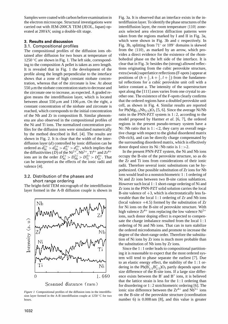
3. Results and discussion3.1. Compositional profilesThe compositional profiles of the diffusion ions ob-tained after diffusion for two hours at temperature of1250◦C are shown in Fig. 1. The left side, correspond-ing to the composition A pellet is taken as zero length.It is revealed that in Fig. 1 the development of theprofile along the length perpendicular to the interfaceshows that a zone of high constant niobate concen-tration, whereas that of the zirconate is low. At about550µm the niobate concentration starts to decrease andthe zirconate one to increase, as expected. A graded re-gion means the interdiffusion layer, which is locatedbetween about 550µm and 1106µm. On the right, aconstant concentration of the niobate and zirconate isreached, which corresponds to the initial concentrationof the Nb and Zr in composition B. Similar phenom-ena are also observed in the compositional profiles ofthe Ni and Ti ions. The normalized concentration pro-files for the diffusion ions were simulated numericallyby the method described in Ref. [4]. The results areshown in Fig. 2. It is clear that the width of the inter-diffusion layer (d) controlled by ionic diffusion can beordered asd2+
Ni > d5+Nb > d4+
Ti > d4+Zr , which implies that
the diffusitivities (D) of the Ni2+, Nb5+, Ti4+ and Zr4+ions are in the orderD2+
Ni > D5+Nb > D4+
Ti > D4+Zr . That
can be interpreted as the effects of the ionic radii andvalence [4].
3.2. Distribution of the phases andshort range ordering
The bright-field TEM micrograph of the interdiffusionlayer formed in the A-B diffusion couple is shown in
Figure 1 Compositional profiles of the diffusion ions in the interdiffu-sion layer formed in the A-B interdiffusion couple at 1250◦C for twohours.
Fig. 3a. It is observed that an interface exists in the in-terdiffusion layer. To identify the phase structures of theinterdiffusion layer, the room temperature〈110〉 zone-axis selected area electron diffraction patterns weretaken from the regions marked by I and II in Fig. 3a,which were shown in Fig. 3b and c respectively. InFig. 3b, splitting from 71◦ or 109◦ domains is skewedfrom the 〈110〉, as marked by an arrow, which pro-vides a direct evidence for the existence of the rhom-bohedral phase on the left side of the interface. It isclear that in Fig. 3c besides the (strong) allowed reflec-tions originating from the cubic perovskite structure,extra (weak) superlattice reflections (F-spots ) appear atpositions of (h+ 1
2, k+ 12, l + 1
2) from the fundamen-tal reflections for a cubic perovskite unit cell with alattice constant a. The intensity of the superstructurespot along the [111] axes varies from one crystal to an-other one. The existence of the F-spots clearly confirmsthat the ordered regions have a doubled perovskite unitcell, as shown in Fig. 4. Similar results are reportedfor Pb(Mg1/3Nb2/3)O3 [5, 6]. Since the average Ni/Nbratio in the PNN-PZT system is 1 : 2, according to themodel proposed by Harmeret al. [6, 7], the orderedregions in the present pseudo-ternary system have aNi : Nb ratio that is 1 :<2, they carry an overall nega-tive charge with respect to the global disordered matrix(Nb-rich), and can be directly charge compensated bythe surrounding disordered matrix, which is effectivelydonor doped since its Ni : Nb ratio is 1 :>2.
In the present PNN-PZT system, the Ni and Nb ionsoccupy the B-site of the perovskite structure, so as dothe Zr and Ti ions from considerations of their ionicradii. Therefore several ionic substitutions can be hy-pothesized. One possible substitution of Zr ions for Nbions would lead to a nonstoichiometric 1 : 1 ordering ofNi and Zr ions between two B-site cation sublattices.However such local 1 : 1 short-range ordering of Ni andZr ions in the PNN-PZT solid solution carries the localB-site valence of+3, which is electrostatically less fa-vorable than the local 1 : 1 ordering of Zr and Nb ions(local valence+4.5) formed by the substitution of Zrfor Ni ions on the B-site of perovskite structure. Withhigh valence Zr4+ ions replacing the low valence Ni2+ions, such donor doping effect is expected to compen-sate the charge imbalance resulted from the local 1 : 1ordering of Ni and Nb ions. That can in turn stabilizethe ordered microdomains and promote to increase thedegree of the short-range order. Therefore the substitu-tion of Ni ions by Zr ions is much more probable thanthe substitution of Nb ions by Zr ions.
Since the 1 : 1 order leads to compositional partition-ing it is reasonable to expect that the most ordered sys-tem will tend to phase separate the earliest [7]. Dueto an elastic energy effect, the stability of the 1 : 1 or-dering in the Pb(B′1/3B′′2/3)O3 partly depends upon thesize difference of the B-site ions. If a large size differ-ence exists between the B′ and B′′ ions, it is believedthat the lattice strain is less for the 1 : 1 ordering thanfor disordering or 1 : 2 stoichiometric ordering [6]. Theionic size difference between the Zr4+ and Nb5+ ionson the B-site of the perovskite structure (coordinationnumber 6) is 0.008 nm [8], and this value is greater
1032

Figure 2 Normalized concentration profiles simulated numerically for the diffusion ions, and compared to that obtained from the EPMA experiment(diffusion conditions: 1250◦C/2 hrs).
than the difference between the Ni2+ and Nb5+ ions(0.005 nm) [8]. Therefore the stability of the 1 : 1 Nb-Zrshort-range ordering is much higher than that of the 1 : 1Ni-Nb short-range ordering. Consideration of the pos-sible 1 : 1 ordering of Ni and Ti ions yields an averagevalence of+3 on B-site and is therefore electrostaticallyless favorable than the Ni:Nb order. The Nb:Ti order isalso unlikely since this type of 1 : 1 ordering involvesthe substitution of the small-sized Ti ion for the rela-tively large-sized Ni ion which requires less favorablestrain energy than the Ni:Nb ordering (R2+
Ni = 0.069 nm,R4+
Ti = 0.0605 nm) [8]. Ti ions are believed to di-lute the forces responsible for the ordering process[9]. The degree of the B-site 1 : 1 order is decreasedwith increasing the PT content, which is identified by
the PT content dependence of the domain morpholo-gies for the 0.2Pb(Ni1/3Nb2/3)O3–0.8Pb(Zr1−xTix)O3
samples. Fig. 5a–c illustrate the dependence of theroom-temperature bright field image on PT con-tent in the 0.2Pb(Ni1/3Nb2/3)O3–0.8Pb(Zr1−xTix)O3
specimens for compositionx= 0.30, 0.50 and 0.60,respectively. It is revealed that in the rhombohedral-rich side (x= 0.30), only local random contrast rep-resenting short-range order polar clusters or nan-odomains is observed, and no evidence of micro-sizeddomains is found. For compositions near the PNN-PZ-PT morphotropic phase boundary (x= 0.50), normalmicron-sized domains appear, and become stable in thetetragonal-rich field (x= 0.60), in which a well-defined90◦ ferroelectric macro-domains can readily be seen.
1033

Figure 3 (a) Bright-field TEM micrograph of the interdiffusion layer formed in the A-B diffusion couple at room temperature, (b) and (c) for theroom temperature〈110〉 zone-axis selected area electron diffraction patterns taken from the regions marked by I and II in Fig. 3a, respectively.
1034

Based on the facts discussed above, it can be con-cluded that the phase structures of the interdiffusionlayer formed in the PNN-PZT system are composed ofthe rhombohedral and pseudocubic phases. The1
2[111]superlattice reflections (F-spots) are originated from the
Figure 4 Proposed model for the atomic configuration of the12{111}-
ordered structure in the PNN-PZT crystalline solution, illustrating theformation of two distinct B-site cation sublattice.βI andβII for nickel(or zirconate) and niobium respecrively.
Figure 5 Bright-field TEM micrographs at room temperature showing the domain morphologies of the 0.2Pb(Ni1/3Nb2/3)O3–0.8Pb(Zr1−xTix)O3
samples for compositions (a)x= 0.30, (b)x= 0.50, and (c)x= 0.60, respectively.
enhanced short-range, non-stoichiometric 1 : 1 orderingof the Zr and Nb cations.
4. ConclusionsUsing the EPMA and TEM techniques the com-positional profiles, distribution of the phase andshort-range ordering of the interdiffusion layer formedin the PNN/PZT functionally gradient piezoelectricceramics have been investigated. It is found that thethickness of the interdiffusion layer (d) of the Ni2+,Nb5+, Ti4+ and Zr4+ ions decreases in the orderd2+
Ni > d5+Nb > d4+
Ti > d4+Zr . A clear interface between
the rhombohedral and pseudocubic phases exists inthe interdiffusion layer. The12[111] superlattice spots(F-spots) are originated from the enhanced short-range,non-stoichiometric 1 : 1 ordering of the Zr and Nbcations on an F-centred superlattices, whose intensitiesvary from one crystal to another one along the [111]axes.
AcknowledgementsThis work is supported by the opening project ofNational Laboratory of Solid State Microstructures(M981308), Nanjing University, and also partially sup-ported by the project of the National Natural ScienceFoundation of China (59832050), and by a grant forState Key Program for Basic Research of China.
1035

References1. N. C H E R R A D I, A . K A W A S A K I andM . G A S I K , Composites
Engineering4 (1994) 883.2. X . H. Z H U, Q. W A N G andZ. Y . M E N G, J. Mater. Sci. Lett.
14 (1995) 516.3. X . H. Z H U andZ. Y . M E N G, Sensors & Actuators A: Physical,
48 (1995) 169.4. X . H. Z H U, J. X U andZ. Y . M E N G, J. Mater. Sci.33 (1998)
1023.5. C. A . R A N D A L andA . S. B H A L L A , Jpn. J. Appl. Phys.29
(1990) 327.
6. J. C H E N, H. C H A N andM . P. H A R M E R, J. Amer. Ceram. Soc.72 (1989) 593.
7. M . P. H A R M E R, J. C H E N, P. P E N G, H. M . C H A N andD. M . S M Y T H, Ferroelectrics97 (1989) 263.
8. R. D. S H A N N O N, Acta. Cryst. A32 (1976) 751.9. A . D. H I L T O N , D. J. B A R B E R, C. A . R A N D A L L and
T. R. S H R O U T, J. Mater. Sci.25 (1990) 3461.
Received 7 July 1997and accepted 22 July 1999
1036